Note: Descriptions are shown in the official language in which they were submitted.
CA 02482969 2010-08-03
DILATION BALLOON FOR ENDOSCOPE
DESCRIPTION
Technical Field
[0002] This invention relates to medical devices, more particularly to
balloons used in
endoscopy to dilate strictures.
Background of the Invention
[0003] Through the endoscope, balloon dilation of tight esophageal strictures
is
frequently carried out with fluoroscopic monitoring. A stricture is considered
to be
"tight" if an endoscope cannot be passed through it. Fluoroscopic monitoring
of tight
stricture dilation is believed to help prevent sudden fracture or splitting of
the stricture
and thus reduce the risk of esophageal perforation during the dilation
procedure.
Currently available dilation balloons are made of transparent material.
However, the
tapered or domed butt design of the proximal end of currently available
dilation balloons
severely limits stricture wall visualization when the face of the endoscope is
approximated to the butt of the balloon. Also, the misalignment produced by
current
dilation balloon design between the dilation balloon and endoscope insertion
shaft as
described below further limits stricture wall visualization. Therefore,
fluoroscopic
monitoring must be relied upon for monitoring purposes.
[0004] Examination and accurate measurement of an esophageal stricture can
only be
accomplished visually or endosonographically if the endoscope can be passed
completely
through the stricture. Two techniques exist for accomplishing complete
stricture passage
CA 02482969 2004-06-16
WO 03/086524 PCT/US03/11036
2
with balloon dilation. The traditional method is to pass and inflate
successively larger
balloons across the stricture until a diameter of 15 to 16 mm is achieved. The
last
dilation balloon is then removed and the instrument is maneuvered through the
stricture
under direct unguided operator control. The post-dilation 15 or 16 mm diameter
stricture
lumen is 5 or 6 mm larger than the diameter of a standard video endoscope and
2 to 3 mm
larger than the diameter of an echoendoscope. However, stricture elasticity,
luminal
tortuosity, and frequent shelving (stepped areas along the stricture) can
prevent passage of
the instrument, despite an apparently adequate dilation.
[0005] An alternative method for accomplishing complete stricture passage with
balloon
dilation is the "balloon-scope train method". The stricture is dilated to a
diameter 1 or 2
mm larger than the diameter of the endoscope. The endoscope is then pushed up
against
the proximal end of the inflated dilation balloon to form a balloon-scope
"train". The
combination of balloon and endoscope is then advanced through the stricture.
Although
currently available dilation balloons are made of transparent material, their
design permits
only limited monitoring and inspection of the stricture wall as the maneuver
is carried
out.
[0006] Unfortunately, current dilation balloon design hinders not only
visualization of the
stricture wall during dilation and subsequent instrument passage, but also
actively
impedes the passage of the "balloon - scope train". Figure 1 depicts a
currently available
esophageal dilation balloon (for example, the QUANTUM TTC Balloon Dilator,
which is the subject ofU.S. Patent No. 5,681,344 to Kelly) and endoscope in a
"balloon-
scope train" configuration. Because the instrument accessory channel outlet on
the
endoscope face is off-center with respect to the endoscope insertion shaft and
the balloon
support wire is centered with respect to the balloon, the flat face of the
endoscope
protrudes over one side of the balloon. The protruding endoscope face tends to
catch
tumor shelves and resist passage through tortuous areas resulting in difficult
passage and
on occasion failure of passage. Also, because the current tapered or domed
butt balloon
designs prevent the endoscope from being cinched up tight against the rear of
the balloon,
CA 02482969 2005-08-01
3
a significant gap is created, which exacerbates the tendency of the endoscope
face to
catch on tumor shelves and in tortuous areas of a stricture.
What is needed is a dilation balloon that will permit direct visualization of
the
stricture wall through the transparent material ofthe balloon for purposes of
stricture wall
monitoring during dilation and that will align properly with the insertion
shaft of the
endoscope to facilitate passage of the endoscope through the stricture using
the balloon-
scope train method.
Summary of the Invention
An object of the present invention is to provide a dilation balloon for
endoscope.
In accordance with an aspect of the present invention, there is provided a
balloon
catheter for use with an endoscope comprising:
a) a dilation balloon comprising a proximal end and a distal end, wherein said
proximal end comprises a proximal opening, said proximal opening being offset
from a
central longitudinal axis; and
b) a shaft connected to said proximal opening, wherein said shaft comprises a
lumen for providing an infusion pathway into said dilation balloon.
In accordance with another aspect of the invention, there is provided a
balloon
catheter for use with an endoscope comprising:
a) a dilation balloon comprising a proximal end and a distal end, wherein said
proximal end is truncated comprising a proximal opening and said distal end is
tapered
comprising a distal opening, said proximal and distal openings being offset
from a central
longitudinal axis;
b) a shaft connected to said proximal opening, wherein said shaft comprises a
lumen for providing an infusion pathway into said dilation balloon;
c) a tip portion connected to said distal opening; and
d) a support element, wherein said support element is connectively disposed
between said shaft and said tip portion, such that said infusion pathway is
maintained
between said shaft and said dilation balloon,
CA 02482969 2005-08-01
3a
wherein when said dilation balloon is inflated said proximal end substantially
abuts and aligns with the endoscope forming a substantially unitary
cylindrical unit with
the endoscope.
In accordance with another aspect of the invention, there is provided a
balloon
catheter for use with an endoscope comprising:
a) a dilation balloon comprising a central axis and a luminal axis, wherein
said
luminal axis is offset from said central axis; and
b) a shaft connected to said dilation balloon along said luminal axis.
The dilation balloon of the subject invention preferably comprises a balloon
portion mounted about a shaft that, when inflated, produces a configuration
comprising a
tapered distal end and a proximal end or butt that is substantially flat
(preferably
truncated) and is adapted to generally conform with the outer contours of the
endoscope
through which it is introduced when the balloon is pulled back against the
endoscope
face. The close engagement of the subject balloon catheter and endoscope, when
forming
a balloon-scope train, enables the scope to more readily navigate strictures
and tortuous
body lumen, as well as allows the balloon to act as a lens for viewing
anatomical
structure within the body lumen, such as tumors, strictures, and the inner
luminal wall
surface itself. The term "engage" is used herein to define when the balloon
portion and
endoscope come into contact in a manner made possible by the configuration of
the
balloon portion such that the scope and balloon portion generally fit closely
against, or
couple with one another, to generally form a single functional unit.
Generally, the balloon
portion is positioned relative to the shaft such that the central axis of the
balloon portion
and the central axis of the endoscope are generally in alignment with one
another when in
engagement, regardless of the position of the instrument channel along the
endoscope
face. As used herein, the term "endoscope" includes any elongate medical
device having
a viewing lens, port, camera, etc., located about the distal end thereof that
is capable of
remote transmission of images from within the body of a patient, through
video,
CA 02482969 2004-06-16
WO 03/086524 PCT/US03/11036
4
ultrasound and other energy waves, direct observation, etc. to a screen,
viewing port, etc.
where it can be viewed by a clinician, typically in real time.
[0009] In one embodiment of the present invention, the dilation balloon
includes a shaft
made of a flexible catheter tubing, such as Pellethane; a balloon portion made
of non-
compliant material, such as transparent polyethylene terephthalate (PTE); a
support
element, such as a solid, tapered nitinol wire that extends from the distal
end of the shaft
and longitudinally traverses the balloon; and a flexible tip portion. Unlike
the standard
PTE dilation balloon, the cross-sectional center of the present balloon is
offset relative to
both the balloon shaft, which supplies infustate to fill the balloon, and the
support wire.
This offset results in the balloon having an eccentric shape following
inflation, relative to
the luminal axis, which comprises the original passageway that extends
longitudinally
through the balloon portion, intersecting the distal and proximal openings.
The degree of
offset generally corresponds to the distance between the instrument or working
channel of
the endoscope and the scope's central axis, thus allowing the balloon, when
inflated and
properly oriented, to become concentrically aligned with the scope and
generally
eliminating or reducing exposure of the otherwise-protruding edge along the
endoscope
face. This allows the balloon-scope train, which generally forms a common
cylindrical
unit, to be navigated through a complex stricture with greater ease by better
protecting the
endoscope face from butting against a shelf or other portion of a stricture
during
advancement. As used herein, a "common cylindrical unit" is defined as
endoscope and
balloon catheter combination in which the inflated balloon portion, when fully
abutted
against the endoscope face, generally extends distally therefrom as a
continuous unit and
without any significant gaps existing between the proximal end of the balloon
portion and
the distal face of the endoscope. Furthermore, the balloon portion is
generally
concentrically aligned with the body of the scope. The balloon portion can be
somewhat
larger or smaller than the scope, or increase or decrease in diameter somewhat
over its
length; however, the balloon provides a functional extension that generally
follows the
contours of the scope for at least a portion of the balloon's length, such as
up until the
distal taper. With regard to the cross-sectional profile of the balloon, the
definition of
CA 02482969 2004-06-16
WO 03/086524 PCT/US03/11036
"cylindrical" would include a tubular shape that is not generally round. For
example, the
balloon portion may comprise an elongate, but squarish or triangular shape.
Furthermore,
it should be noted that the present invention does not necessarily require
that all
embodiments of the balloon portion form a common cylindrical unit with the
endoscope.
For example, the balloon portion may be spherical or some other shape, yet
comprise a
material or configuration that allows it to effectively abut and engage the
endoscope face
to function in the manner previously described.
[0010] In another aspect of the invention, the balloon is formed such that the
proximal
end is generally truncate in shape, having a substantially flat butt, rather
than comprising
a standard tapered or domed configuration. The truncated end permits all or a
substantial
portion of the endoscope face to be drawn up against the proximal end of the
balloon,
thereby significantly reducing or eliminating any gaps that would otherwise
exist. By
advancing the endoscope face and viewing port against the transparent balloon
material,
the liquid-filled balloon acts like a lens to permit improved visualization of
the
anatomical structures adjacent to the balloon. This is especially significant
during a
dilation procedure in the esophagus. With the goal of being able to achieve
maximum
dilation of the stricture or tumor without causing a fissure to form in the
esophageal wall
due to over-inflation of the balloon, being able to clearly visualize and
monitor the tissues
during inflation provides an important clinical benefit over existing
treatment modalities,
especially fluoroscopy, during which detection of a developing (issue is
generally not
possible. In addition, when filled with a liquid, such as water or saline,
typically acts like
a magnifying lens to make structures adjacent the walls of the balloon appear
larger, thus
aiding with diagnosis and monitoring of a procedure.
[0011] In yet another aspect of the present invention, the balloon catheter
includes an
inner shaft that extends from within the main shaft and through the balloon
portion,
instead of a support wire, to accommodate optional ancillary instrumentation
that maybe
used in a procedure, such as a standard wire guide. The inner shaft terminates
about the
distal tip portion, which includes a passageway via which the wire guide may
enter and
CA 02482969 2010-08-03
6
exit the balloon catheter to aid in cannulation or perform some other
function. The
infustate for inflation of the balloon is supplied via the outer shaft through
the space
between the outer and inner shafts.
[0012] In still yet another aspect of the invention, the posterior end of the
balloon portion
is further modified to facilitate positive engagement with the face of the
endoscope and/or
aid with alignment between the endoscope and balloon when the endoscopist is
drawing
the balloon back against the scope. In one embodiment, the positive end of the
balloon
portion is concave in shape to receive the distal face of the endoscope, which
typically
has a rounded shape. In a different embodiment, the posterior end of the
balloon portion
includes a guide element, such as one or more rings, flaps, ridges, etc.
affixed around the
outer ridge of the posterior end that could guide and/or align the tip of the
endoscope
against the posterior end of the balloon portion. The guide element(s) may
also serve to
further shield any gap that exists between the scope and balloon to prevent
tissue or
materials from entering that space, possibly causing an obstruction that
hinders further
advancement or impairs visibility. A different approach to facilitating
alignment between
the balloon portion and endoscope is found in an embodiment that provides an
alignment
marking on the portion of the catheter external to the scope, such as the
proximal hub.
The marking is positioned such that when oriented in a predetermined manner,
the larger
side of the eccentric balloon is aligned with the corresponding side of the
endoscope face,
typically having the viewing port or lens, such that the scope and balloon are
generally
aligned concentrically.
Brief Description of the Drawings
[0014] Embodiments of the present invention will now be described by way of
example
with reference to the accompanying drawings, in which:
CA 02482969 2004-06-16
7
[0015] Fig. 1 depicts a partially-sectioned side view of a prior art dilation
balloon being
used with a standard endoscope;
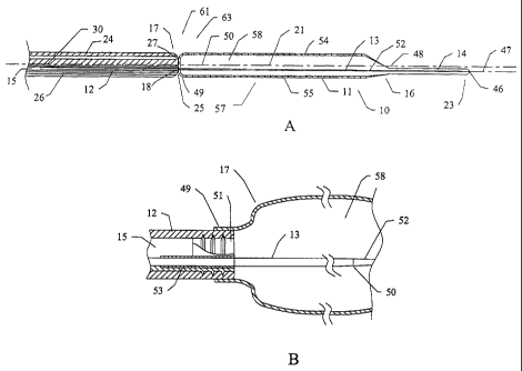
[0016] Fig. 2A depicts a partially-sectioned side view of the illustrative
embodiment of
the present invention in engagement with the endoscope of Fig. 1;
[0017] Fig. 2B depicts a partially-sectioned detail view of the embodiment of
Fig. 2A;
[0018] Fig. 3 depicts an end view of the face of a standard endoscope having
an
instrument channel offset from the central axis;
[0019] Fig. 4A depicts a cross-sectional view of an embodiment of the present
invention
configured for use with a standard wire guide;
[0020] Fig. 4B is a cross-sectional view taken along line 4a-4a of Fig. 4A;
[0021] Fig. 5 depicts an embodiment of the present invention, wherein the
posterior end
of the balloon portion is generally concave;
[0022] Fig. 6 depicts an embodiment of the present invention, wherein the
posterior end
includes a guide element to facilitate engagement with the endoscope face; and
[0023] Fig. 7 depicts an embodiment of the posterior connector of the present
invention
which includes an alignment marker.
Detailed Description of the Drawing
[0024] The present invention includes embodiments of aballoon catheter 10,
such as that
depicted in Fig. 2, configured for engagement with an endoscope to facilitate
negotiation
of the scope through a stricture or other difficult or tortuous pathway within
the body,
and/or to abut the viewing port 27 or objective lens of the endoscope face
such that
anatomical structures of interest can be viewed. The illustrative balloon
catheter 10
comprises a dilation balloon portion 11; typically made of a clear, non-
distensible
polymer material such as transparent polyethylene terephthalate (PET); a
shaft, made of a
flexible catheter material 12 attached proximally to the balloon portion and
having a
CA 02482969 2004-06-16
B
passageway 15 that communicates with the balloon portion 11 to supply
infusate, such as
water, or saline, to expand the balloon; a support element 13 or wire, that
extends beyond
the distal end 18 of the shaft, through the distal end 16 of the balloon, and
terminating
within a flexible tip portion 14, made of a suitable medical grade elastomer
tubing, such
as Pelethane 2363-80AE. The tip portion 14, which generally provides an
atraumatic
means of cannulating a stricture or generally guiding the balloon through a
passageway,
includes a rounded tip with the central bore of the tip 46 being filled with
an adhesive at
the distal end 16. In an embodiment of the subject invention, the support
element 13 is a
kink-resistant material such as nitinol, stainless steel, or other non-
superelastic materials
and alloys.
[0025] The illustrative balloon portion 11, depicted in Figs. 2A-2B, comprises
a main
portion 57 that is generally uniformly cylindrical in shape, and a tapered
portion 22
toward the distal end 16 of the balloon portion 11. The proximal end 17 of the
balloon
portion 11 is generally truncate in shape such that the proximal end 17 can be
cinched or
drawn against the distal face 25 of an endoscope 24 from which it has been
advanced,
such that there is broad area of contact between the balloon portion 11 and at
least a
substantial cross section of the endoscope face 25, which is depicted in Fig.
3. the area of
contact includes the viewing port 27 or objective lens, and preferably, but
not essentially,
the light source 28 such that the balloon portion generally serves as an
extension of the
lens 27, thereby enabling the endoscopist a relatively unobstructed and
undistorted view
through the balloon interior 58, which permits visualization of the anatomical
structures
within the body conduit. When obstructions from tissues or fluids do occur,
they still can
be dislodged from the lens or space between the balloon and endoscope using a
stream of
saline, water, etc. delivered from the flush port 29. When illustrative
balloon portion 11
is inflated and held against the endoscope 24, the resulting balloon-scope
train 61
generally forms a common cylindrical unit 63.
[0026] The main portion 57 of the balloon portion 11 includes a central axis
21 that
intersects the cross-sectional center point of the main cylindrical portion
57. The balloon
CA 02482969 2004-06-16
9
portion 11 also includes alumina! axis 47 that inteisec>s theproxima149 and
distal 48 openings
48, 49 of the balloon portion. The luminal axis 47 of the present invention
comprises the
original lumen of the tubing used to form the balloon portion 11, but unlike a
standard
dilation balloon, such as the `344 balloon, is offset relative to the central
axis 21 to allow
alignment with the endoscope. Generally, it is desired that the balloon
portion 11 and
outer contours of the endoscope 24 be concentrically aligned with one another
to
maximize the field of view and reduce ledges or surfaces that are prone to
catch upon a
shelf or stricture during advancement of the balloon-scope train 61. Although
having the
balloon diameter closely match that of the endoscope provides the ideal
clinical situation
for introduction of the balloon-scope train 61, it is not necessary to the
invention that the
balloon and scope be of the same diameter. Often, multiple sizes of balloons
are used
with a given endoscope for a single procedure, such as in esophageal dilation
procedures,
where attempting to fully dilate in a single, rather than multiple stages,
increases the risk
of rupture. The standard sizes of endoscopes used in gastrointestinal
procedures are 8.5,
9.5, and 11.5 mm, which are generally compatible with the most preferred range
of
balloon diameters for the illustrative embodiment (10-16 nun).
[00271 The balloon portion 11 and shaft 12 are attached to one another by
inserting the
distal end 18 of the shaft 12 into the proximal opening 49 and bonding thereto
using a
well-known method such as an ultraviolet-curable adhesive. The shaft 12, which
is
aligned with the luminal axis 47, is therefore, offset relative to the central
axis 21. Also
aligned with the luminal axis 47, is the support element 13, or stiffener,
which can be, but
is not to be limited to, for example, a 0.027" solid flexible nitinol wire,
that extends the
length of the catheter shaft 12, through the balloon portion 11, then
terminating within the
tip portion 14. The support element 13 includes a tapered portion 52 that
begins at a
point 50 within the interior 58 of the balloon portion and tapers down about
two-thirds
the original diameter (in this example, approximately 0.010") at the tip 23.
As shown in
Fig. 2a, the support element 13 is attached to an insert 51 that is embedded
into the sheath
lumen 15 about the distal tip. The insert, is preferably, but not essentially,
made of a
physiologically inert, radiopaque material, such as 303 stainless steel. To
avoid the
CA 02482969 2010-08-03
difficulty of soldering to the support element 13, a piece of metal cannula 53
is crimped
over the support wire 13 and soldered or otherwise affixed to the insert 51,
thereby
longitudinally securing the support wire relative to the shaft 12 and balloon
portion 11.
[0028] In the illustrative embodiment, the catheter shaft 12 includes a single
lumen 15
that houses the support element 13 and provides an infusion pathway to the
balloon
portion 11, whereby water or saline is introduced, via the hub, using a
conunonly-
available infusion device appropriate for the balloon volume. The balloon is
maintained
in a deflated state and is folded and inserted into a delivery sheath (not
shown). It is then
advanced from the delivery sheath into the instrument (accessory) channel of
the
endoscope, which typically is a minimum of 2.8 rnm for the illustrative
esophageal
dilation balloon, as well as the related pyloric, or colonic embodiments in
which the
balloon is 18 nun or smaller in diameter when inflated. Larger diameter
balloons, e.g.,
19-20mmn, may require an instrument channel of up 3.7 nun or greater.
Typically, the
balloon is lubricated to ease insertion into the endoscope instrument channel.
The shaft
12 of the illustrative embodiment and related embodiments can be provided in a
shaft
passageway 26 and has an OD of approximately 0.085" and an ID of approximately
0.058". The esophageal and colonic embodiments
typically have an overall length, including balloon, of approximately 180 cm,
although
any length that is appropriate for a particular endoscope may be used. The
colonic
dilation balloon catheter 10 is typically longer, e.g., 240 cm.
[0029] The balloon portion 10 of the illustrative embodiment of Fig. 2 is
formed by a
well-known means, such as blow molding, whereby a length of PTE tubing,
sufficient in
length to form the fu1al desired length of the balloon, is placed and clamped
within a
mold confornling to the final shape of the fully distended balloon. Hot air is
passed
through the tubing, causing the tubing to expand against the contours of the
mold. The
tubing and molding process parameters necessary to achieve the desired balloon
are
determined by the required burst strength and recommended pressure of the
balloon, the
material used, and the size of the balloon. One source of the balloon portion
10 of the
illustrative embodiment is Advanced Polymers, Inc. (Salem, NH). The typical
range of
CA 02482969 2004-06-16
WO 03/086524 PCT/US03/11036
11
diameters for an 8 cm long esophageal dilation balloon is generally about 6 to
19 mm,
with a more preferred range of 12-18 mm. Minimum specified burst pressures
typically
average 175 psi for a 12 mm balloon, down to about 122 mm for an 18 mm
diameter
balloon, with the corresponding recommended pressures being about 90 and 50
psi,
respectively. Pyloric and colonic dilation balloons are typically shorter in
length (e.g., 5.5
cm); however, the recommended pressures are generally the same as the longer
esophageal balloons for corresponding diameters. In the illustrative
invention, the
balloon portion 11, because of its eccentric shape, is divisible into a first
longitudinal
portion 54 and a second longitudinal portion 55 along the luminal axis 47,
with the first
longitudinal portion 54 comprising the larger volume of the two. Because the
original
tubing requires greater expansion within one side of the eccentric-shaped mold
than the
other to contact the outer mold surface, the thicknesses found along the wall
59 of the
first longitudinal portion 54 will generally be thinner than that found along
the wall 60 of
the second longitudinal portion 55. Generally, the thickness and strength of
the first
portion wall 59 determines the burst and recommended pressures that are
specified for a
given balloon catheter 10.
[0030] A second embodiment of the present invention is depicted in Fig. 4 that
is adapted
for use with a wire guide 34. The illustrative wire-guided dilation balloon 10
includes an
inner sheath 62 coaxially disposed within the outer sheath 12 to which the
balloon portion
11 is attached. The inner sheath 62 serves as the conduit for a wire guide 34,
in one
embodiment a standard 0.035" wire guide, that is loaded into, and is
extendable from the
inner sheath passageway 45. In the illustrative embodiment, both the inner and
outer
sheaths 12, 62 are made of poly-ether ether ketone (PEEK), with the outer
sheath 12
having and OD of 0.85" and the inner sheath 62 having an OD of 0.50". The
inner sheath
62 is sized to allow the flow of infusate through sheath passageway 15 within
the annular
space between the two sheaths 12, 62 and into the interior 5 8 of the balloon
portion 11 to
expand the balloon. The inner sheath 62 terminates within the distal tip
portion 14 about
the distal end 16 of the balloon portion or a few millimeters past. The wire
guide 34 is
typically utilized a support element 13 for adding stiffness or pushability to
the balloon
CA 02482969 2004-06-16
WO 03/086524 PCT/US03/11036
12
catheter 10, or it maybe introduced separately into the patient. The inner
sheath 62 alone
may provide sufficient stiffness and pushability to function as the support
element 13 for
some applications, which can in some embodiments make a separate support
element 13,
such as a nitinol wire, unnecessary. If desired, a wire guide 34 that is most
suitable as a
support element 13, may at some point be replaced with a different wire guide
having
characteristics more desirable for a particular procedure. In the illustrative
embodiment,
the outer and inner sheaths 12, 62 are typically fixed relative to one another
longitudinally
by a standard hub (not shown), which provides access for the wire guide, and a
port for
the infusion of balloon infusate.
[00311 In certain embodiments, the proximal end of the balloon is indented.
Such
indentations can permit the endoscopist to lock or otherwise more completely
engage the
proximal end of the balloon with the distal end of the endoscope, thereby
resisting
rotational movement and thus minimizing rotational loss of balloon/scope
alignment.
One such exemplary embodiment comprises an indentation which effectively
results in a
circumferential flange at the proximal end of the balloon that is configured
to frictionally
engage the distal end of the endoscope.
[00321 Figs. 5-6 depicts embodiments of the balloon portion 11 that include a
positive
engagement guide 36 that is intended to facilitate or improve engagement
and/or
alignment with the face 25 of the endoscope 24. Typically, engagement results
when the
proximal end 17 of balloon both tightly abuts the endoscope face 25 and is
correctly
aligned so that central axis 21 of the balloon is generally aligned with
central axis 30 of
the endoscope. Fig. 5 depicts a positive engagement guide 36 that comprises a
receiving
area 64 comprising a concave surface 37 at the proximal end 17 of the balloon
portion 11
to receive the endoscope face 25, which is typically rounded distally and
therefore,
naturally conforms to the concave surface 37. The concave shape of the
proximal end 17
can increase the available area of the endoscope face 25 contacting the
balloon portion
11, and possibly assisting with alignment as the balloon pulled back to engage
the scope.
CA 02482969 2004-06-16
WO 03/086524 PCT/US03/11036
13
[0033] Fig. 6 depicts a balloon portion 11 that includes a guide structure 38
along the
outer edge of the truncate proximal end 17 to help facilitate correct
alignment and proper
engagement between the scope 24 and balloon portion 11. As the balloon
catheter 10 is
pulled back toward the endoscope face 25, the guide structure 38 provides an
additional
means to help guide the endoscope against the balloon portion 11. The
illustrative guide
structure 38 comprises a flap-like structure that is bonded to or formed with
the balloon
portion 11 and that defines a receiving area 64. The guide structure 38 acts
to properly
seat the endoscope face 25 into the receiving area 64 at the proximal end 17
so that the
balloon can be rotated and aligned accordingly. Additionally, different areas
of color or
other visual markers could be incorporated into the guide structure 38 to tell
the
endoscopist how the balloon portion 11 is oriented relative to the endoscope
and whether
it should be rotated. Also, the guide structure 3 8 may comprise merely a
marker or series
of markers on the surface of the balloon portion surface for indicating
orientation, rather
than a raised structure or structures. The flap-like guide, structure 38
further serves to
provide some protection against tissue or materials migrating into the space
between the
proximal end 17 of the balloon portion 11 and the endoscope face 25, thus
limiting
visibility. The illustrative guide structure 38 is merely exemplary. In view
of the
teachings herein, it would be within the ability of one of ordinary skill in
the medical arts
to conceive and design other annular or discrete structures that would
accomplish the
objective of providing a guide for proper engagement of the balloon portion 11
and
endoscope 24.
[0034] Another manner in which alignment can be accomplished is depicted in
Fig. 7, in
which an alignment marker 41 is placed on the proximal hub 40 of the balloon
catheter
that the operator can use to tell when a particular side of the balloon is
oriented
upward, thereby matching the orientation of the endoscope so that they are
concentrically
aligned. The alignment marker can comprise any system of indicia, such as
markings,
characters, colors, structures, etc. that are printed on, embossed in, molded
with, or
otherwise affixed or attached to the hub. Optionally, the marker can be
included on the
CA 02482969 2004-06-16
WO 03/086524 PCT/US03/11036
14
strain relief element 42 or the shaft 12 itself in a location for convenient
viewing during
the procedure.
[0035] It should be noted that while the illustrative embodiments are
generally intended
for dilation of esophageal, pyloric, and colonic strictures, it is
contemplated that the
present invention may encompass any balloon, dilation, extraction, etc. that
can be
designed for endoscopic use and which may be abutted against the scope face to
form a
common functional unit therewith that is appropriate for a particular clinical
application.
These would include applications utilizing both compliant and non-compliant
balloon
materials. Examples of other clinical applications include, but are not
limited to, biliary
tree, bronchial tree, neural endoscopy, and the vascular system.
[0036] Any other undisclosed or incidental details of the construction or
composition of
the various elements of the disclosed embodiment of the present invention are
not
.believed to be critical to the achievement ofthe advantages of the present
invention, so
long as the elements possess the attributes needed for them to perform as
disclosed. The
selection of these and other details of construction are believed to be well
within the
ability of one of even rudimentary skills in this area, in view of the present
disclosure.
Illustrative embodiments of the present invention have been described in
considerable
detail for the purpose of disclosing a practical, operative structure whereby
the invention
may be practiced advantageously. The designs described herein are intended to
be
exemplary only. The novel characteristics of the invention may be incorporated
in other
structural forms without departing from the spirit and scope of the invention.
The
inventors contemplate embodiments both comprising and consisting of the
described
elements. Unless, otherwise indicated, all ordinary words and terms used
herein shall take
their customary meaning as defined in The New Shorter Oxford English
Dictionary, 1993
edition. All technical terms shall take on their customary meaning as
established by the
appropriate technical discipline utilized by those normally skilled in that
particular art
area. All medical terms shall take their meaning as defined by Stedman's
Medical
Dictionary, 27th edition.
CA 02482969 2010-08-03
[0038] It should be understood that the examples and embodiments described
herein are
for illustrative purposes only and that various modifications or changes in
light thereof
will be suggested to persons skilled in the art and are to be included within
the spirit and
purview of this application.