Note: Descriptions are shown in the official language in which they were submitted.
CA 02484271 1998-12-18
Insertion Device For An Insertion Set And Method Of Using The Same
FIELD OF THE INVENTION
This invention relates generally to an insertion device for automatic
placement of an
insertion set through the skin of a patient, and in particular embodiments to
a compact and
easily operated insertion device for.placement of an insertion needle of a
subcutaneous
insertion set or the like through the skin of a patient with a controlled
force and insertion
speed by the patient.
BACKGROUND OF THE INVENTION
Medical needles are widely used in the course of patient care and treatment,
particularly with respect to the delivery of selected medications to a
patient. In one common
form, hollow hypodermic needles are employed for transcutaneous delivery of a
selected
medication from a syringe or the like. In another common form, insertion
needles are
employed for transcutaneous placement of a soft and relatively flexible
tubular cannula,
followed by insertion needle removal and subsequent infusion of medical fluid
to the patient
through the cannula. More recently, insertion needles have also been used for
transcutaneously placing other medical devices such as a subcutaneous sensor
for monitoring
2 5 specified patient parameters, such as blood glucose level.
In certain medical treatment regimens, it may be necessary or desirable for
the patient .
to transcutaneously place the medical needle. For example, diabetic patients
frequently self
administer insulin injections or periodically place a subcutaneous insertion
with a cannula for
subsequent programmable delivery of insulin by means of a medication infusion
pump of the
3 0 general tyge described in U.S. Patent 4,685,903. Such subcutaneous
insertion sets are
disclosed, for example, in U.S. Patents 4,755,173; 5,176,662; and 5,257,980-
Diabetic patients may also use a subcutaneous insertion set
-1-
CA 02484271 1998-12-18
to periodically place a transcutaneous glucose sensor wherein such sensor
insertion sets are
disclosed, for example, In U.S. Patents 5,390,674; 5,568,806; 5,586,553.
Some patients are reluctant or hesitant to pierce their own skin with a
medical needle,
and thus encounter difficulties in correct needle placement for proper
administration of the
medication. Such difficulties can be attributable to insufficient manual
dexterity or skill to
achieve proper needle placement or, alternately to, anxiety associated with
anticipated
discomfort as the needle pierces the skin. This problem can be especially
significant with
medications delivered via a subcutaneous flexible insertion set, since
incorrect placement can
cause kinking of the cannula and resultant obstruction of medication flow to
the patient.
Cannula kinking can be due to insertion set placement at an incorrect angle
relative to the
patient's skin, andlor needle placement with an incorrect force and speed of
insertion.
The present invention relates to an automatic injector, particularly for use
with a
subcutaneous insertion set, for quickly and easily placing an insertion needle
through the skin
of a patient at the correct insertion angle, and with a speed and force of
insertion which
minimizes patient discomfort.
SUMMARY OF THE D'1SCLOSUItE
It is an object of an embodiment of the present invention to provide an
improved
insertion device and insertion set, which obviates for practical purposes, the
above mentioned
limitations.
According to an embodiment of the invention, an injector is provided for quick
and
easy transcutaneous placement of a medical needle through the skin of a
patient, particularly
such as an insertion of a subcutaneous insertion set. The injector is designed
to place the
needle through the skin at a selected insertion angle and with a controlled
force and speed of
insertion, to ensure proper needle placement with minimal patient discomfort.
The injector is
particularly designed to meet these objectives, while safeguarding against
undesired
projection of the medical needle through free space, in the event that the
injector is actuated
in spaced relation to the patient's skin.
3 0 The injector comprises a spring-loaded plunger having a head for receiving
and
supporting an insertion set in a position with an insertion projecting
outwardly for
transcutaneous placement through the skin of a patient. The plunger is
designed for
-2-
CA 02484271 1998-12-18
WO 99/3350-1 PCT/US98/26978
retraction and retention within a barrel to a cocked position with a drive
spring compressed
in a manner applying a predetermined spring force to the plunger head. A front
or nose end
of the injector barrel is designed for pressed placement against the skin of a
patient, at a
selected needle insertion site, and in an orientation with the needle disposed
at a correct or
desired insertion angle. A trigger member is operable to release the plunger
and thereby
permit the drive spring to carry the insertion set toward the patient's skin
with a controlled
force and speed, resulting in proper transcutaneous placement of the insertion
needle with
minimal patient discomfort.
The plunger head includes a safety lock mechanism to retain the insertion set
against
projection from the injector barrel. In one preferred form, the safety Lock
mechanism
comprises at least one and preferably a pair of safety lock arms for engaging
and retaining the
insertion set when the plunger is retracted from a fully advanced position.
Each safety lock
arm includes a cam lobe for engaging an appropriately shaped recess on the
insertion set to
prevent release thereof from the plunger head, unless and until the plunger
head is returned to
the fully advanced position. In such fully advanced position, the shape of the
cam lobe
perniits quick and easy separation of the injector from the insertion set with
a minimal
separation force.
In operation, the safety lock arms thus prevent projection of the insertion
set from the
injector, in the event that the trigger member is actuated with the nose end
of the barrel
2 0 spaced from the skin of a patient. In that event, the plunger head is
advanced with the
controlled force and speed to the fully advanced position, but the insertion
set is not thrown
from the injector as a projectile. Instead, the insertion set travels rapidly
with the plunger
head to the fully advanced position, whereat the injector can be separated
with minimal
separation force from the insertion set.
In an alternative preferred form, the safety lock mechanism comprises a
plunger head
having a cylindrical shape defining a forwardly open cavity for receiving and
supporting an
insertion set with the insertion needle and cannula projecting outwardly. In
this embodiment,
the plunger head includes a radially inwardly projecting rim at a forward or
nose end thereof,
wherein the rim defines an oval-shaped opening. The size of the rim opening
permits
3 0 relatively free reception of a hub on the insertion set, with the infusion
set oriented at an
angle relative to a central axis of the plunger head and barrel. The insertion
set is then
reoriented to align the insertion needle coaxially with the central axis of
the barrel and
-3-
CA 02484271 1998-12-18
WO 99/3350.1 PCTNS98I26978
plunger head, so that the rim is received into a recess on the insertion set
and functions to
retain the infusion set against undesired release from the injector during
spring-driven
placement of the needle. After needle placement, the injector is released from
the insertion
set with minimal separation force by orienting the injector angularly relative
to the insertion
set to permit free slide out passage of the hub through the oval rim opening.
In a further alternative form of the invention, the plunger head is shaped to
define a
laterally open undercut slot sized for relatively free slide-f t reception of
the needle hub of
the insertion set. In this version, the insertion set is assembled quickly and
easily with the
plunger head of the injector by laterally sliding the hub into the laterally
open slot, thereby
orienting the medical needle generally coaxially relative to the central axis
of the injector
barrel and plunger head. In this position, the plunger head can be retracted
and locked,
followed by appropriate trigger member release for transcutaneously placing
the medical
insertion needle. After the needle is placed on the patient, the injector can
be disassembled
from the insertion set by laterally sliding the injector relative to the
needle hub.
Alternatively, the injector can be withdrawn or retracted from the patient's
skin to slidably
separate the needle from the insertion set which remains in place on the
patient's skin.
In other embodiments of the present invention, an insertion device for
inserting at
least a portion of at least one piercing member of an insertion set through
the skin of a patient
includes a device housing, a carrier body and a driver. The carrier body is
slidably received
2 0 within the device housing for movement between an advanced position and a
retracted
position. The carrier body also includes a receiving structure to support the
insertion set in a
position with the at least one piercing member oriented for insertion through
the skin of the
patient at a predetermined angle relative to the skin of the patient upon
movement of the
carrier body from the retracted position to the advanced position. The driver
is operatively
2 5 coupled between the device housing and the carrier body to urge the
carrier body with a
controlled force and speed from the retracted position toward the advanced
position to place
at least a portion of the at least one piercing member of the insertion set
thorough the skin of
the patient to install the insertion set to the patient. The receiving
structure of the carrier
body is removable from the insertion set while maintaining the installation of
the insertion
3 0 set to the patient.
In particular embodiments, the predetermined angle relative to the skin is
about 90
degrees, between 90 degrees and 10 degrees, or is after insertion between 0
and 10 degrees.
-4-
CA 02484271 1998-12-18
In additional embodiments, the insertion set is a transuctaneous insertion
set, a subcutaneous
insertion set, an infusion set, sensor set or the like. In still other
embodiments, the insertion
set rests mainly on the surface of the skin after insertion or the insertion
set is implanted in
the skin of the patient. In preferred embodiments, the at least one piercing
member is a
needle. In alternative embodiments, the at least one piercing member is a
plurality of
needles, and can also be a plurality of micro-needles. Also, in some
embodiments, the
insertion set can be both an infusion set and a sensor set combined into an
integral unit.
In yet other embodiments the insertion device, the device housing has a
forward end
defining a generally planar angled insertion contact surface far placement
against the skin of
a patient with the device housing in a predetermined orientation relative to
the patient's skin
that mirrors the predetermined angle relative to the skin of the patient.
Other embodiments
include a trigger mechanism that actuates the driver. For instance, the
trigger mechanism
includes at least one trigger for fingertip depression to actuate the driver
for movement of the
carrier body from the retracted position to the advanced position. In
addition, the driver can
include at least one spring for spring-loaded movement of the carrier body
from the retracted
position to the advanced position. Further, the driver can include a force
changing
mechanism that permits alteration of the controlled force and speed of the
carrier body
moving from the retracted position to the advanced position from one insertion
cycle to
2 0 another insertion cycle. In still further embodiments, the device housing
and the carrier body
include a cooperatively engageable track mechanism for guiding movement of the
carrier
body between the advanced and retracted positions while retaining the can ier
body against
rotation relative to the device housing.
In additional embodiments of the insertion device, the at least one piercing
member is
2 5 provided with a piercing member hub as part of the insertion set. In
addition, the receiving
structure of the carrier body includes a recess formed therein for mated slide-
fit reception of
the piercing member hub of the insertion set. Further, the recess of the
receiving structure
can include a laterally open undercut recess. Alternatively, the receiving
structure may
include a safety retainer structure that retains the at least one piercing
member an the
3 0 receiving structure during movement from the retracted position to the
advanced position.
This safety retainer structure permits separation of the at least one piercing
member from the
carrier body when the carrier body is in the advanced position.
-5-
CA 02484271 1998-12-18
WO 9913350a PCTNS98126978
Yet another embodiment of the present invention is directed to an insertion
set for
insertion through the skin of a patient by an insertion device. The insertion
device has a
slidable carrier body for movement between an advanced position and a
retracted position.
The carrier body of the insertion device including a receiving structure to
support the
insertion set in a position for insertion through the skin of the patient upon
movement of the
carrier body from the retracted position to the advanced position. The
insertion device also
having a driver operatively coupled to the carrier body that urges the carrier
body with a
controlled force and speed from the retracted position toward the advanced
position for
insertion of the insertion set thorough the skin of the patient. The insertion
set includes at
least one piercing member and a set housing. The at least one piercing member
includes a
portion of the at least one piercing member that is insertable through the
skin of the patient.
The set housing is coupled to the at least one piercing member. Also, the set
housing is
shaped to fit within the carrier body of the insertion device to orient the at
least one piercing
member for placement through the skin of the patient of at least a portion of
the at least one
piercing member at a predetermined angle relative to the skin of the patient
to install the
insertion set to the patient. The set housing of the insertion set is
removable from the
receiving structure of the carrier body while maintaining the installation of
the insertion set
to the patient.
In particular embodiments of the insertion set, the predetermined angle
relative to the
2 0 skin is about 90 degrees, between 90 degrees and 10 degrees, or is after
insertion between 0
and 10 degrees. In additional embodiments, the insertion set is a
transuctaneous insertion
set, a subcutaneous insertion set, an infusion set, sensor set or the like. In
still other
embodiments, the insertion set rests mainly on the surface of the skin after
insertion or the
insertion set is implanted in the skin of the patient. In preferred
embodiments, the at least
2 5 one piercing member is a needle. In alternative embodiments, the at least
one piercing
member is a plurality of needles, and can also be a plurality of nucro-
needles. Also, in some
embodiments, the insertion set can be both an infusion set and a sensor set
combined into an
integral unit.
Other features and advantages of the invention will become apparent from the
3 0 following detailed description, taken in conjunction with the accompanying
drawings which
illustrate, by way of example, various features of embodiments of the
invention.
-6-
CA 02484271 1998-12-18
WO 99/3350. PCTNS98126978
BRIEF DESCRIPTION OF THE DRAWINGS
A detailed description of embodiments of the invention will be made with
reference
to the accompanying drawings, wherein like numerals designate corresponding
parts in the
several figures.
Fig. 1 is a perspective view illustrating use of an automatic injector
embodying the
novel features of the invention;
Fig. 2 is an enlarged front elevation view of the injector shown in Fig. 1;
Fig. 3 is a front or nose end view of the injector, taken generally on the
line 3-3 of
Fig. 2;
Fig. 4 is an enlarged exploded perspective view illustrating assembly of the
injector
with a subcutaneous insertion set;
Fig. 5 is a further enlarged longitudinal sectional view taken generally on
the line 5-5
of Fig. 4;
Fig. 6 is a transverse sectional view taken generally on the line 6-6 of Fig.
5;
1 S Fig. 7 is an enlarged longitudinal sectional view taken generally on the
line 7-7 of
Fig. 2;
Fig. 8 is an enlarged and exploded fragmented perspective view illustrating a
trigger
assembly for use in the injector;
Fig. 9 is a longitudinal sectional view similar to Fig. 5, and showing the
injector with
2 0 insertion set received therein for transcutaneous placement through the
skin of a patient;
Fig. 10 is a transverse sectional view taken generally on the line 10-10 of
Fig. 9;
Fig. 11 is a longitudinal sectional view taken generally on the line 11-11 of
Fig. 9;
Fig. 12 is a rear end elevation view taken generally on the line 12-12 of Fig.
11, and
depicting the trigger assembly in a locked position;
25 Fig. 13 is an enlarged fragmented longitudinal view similar to a portion of
Fig. 11,
but depicting actuation of the trigger assembly for releasing the spring-
loaded plunger;
Fig. 14 is a rear end elevation view taken generally on the line 14-14 of Fig.
13,
similar to Fig. 12, but showing the trigger assembly in an unlocked position;
Fig. 15 is a fragmented longitudinal sectional view depicting the spring-
loaded
3 0 plunger in a fully advanced position with the infusion set placed on the
patient's skin;
Fig. 16 is an exploded perspective view illustrating separation of the
insertion needle
from the cannula of the subcutaneous insertion set;
CA 02484271 1998-12-18
WO 99/33504 PCT/US98126978
Fig. 17 is a perspective view depicting an alternative preferred form of the
invention;
Fig. 18 is a front elevation view of the injector shown in Fig. 17;
Fig. 19 is a front or nose end view of the injector, taken generally on the
line 19-19 of
Fig. 18;
Fig. 20 is an enlarged side elevation view of the injector, taken generally on
the line
20-20 of Fig. 19;
Fig. 21 is a further enlarged longitudinal sectional view taken generally on
the line
21-21 of Fig. 17;
Fig. 22 is an enlarged exploded perspective view illustrating construction
details of a
plunger and trigger member for use in the injector of Fig. 17;
Fig. 23 is an enlarged longitudinal sectional view similar to Fig. 21, and
depicting the
injector with the trigger member in a cocked position;
Fig. 24 is a fragmented perspective view showing the upper end of the injector
depicted in Fig. 23, with the trigger member in the cocked position;
Fig. 25 is an enlarged and fragmented longitudinal sectional view illustrating
actuation of the trigger member;
Fig. 26 is an enlarged and fragmented longitudinal sectional view showing the
plunger in a fully advanced position with the infusion set placed on the
patient's skin;
Fig. 27 is an enlarged fragmented longitudinal sectional view taken generally
on the
2 0 line 27-27 of Fig. 22, and depicting a portion of the plunger;
Fig. 28 is a front or nose end elevational view of the plunger, taken
generally on the
line 28-28 of Fig. 27; and
Fig. 29 is an enlarged fragmented longitudinal sectional view illustrating
release of
the injector from an infusion set placed on the patient's skin;
Fig. 30 is an exploded prospective view generally similar to Fig. 17, but
depicting a
further alternative preferred form of the invention, and showing assembly of
an insertion set
with the illustrative injector;
Fig. 31 is a perspective view similar to Fig. 32, depicting further assembly
of the
insertion set with the injector;
3 0 Fig. 32 is an enlarged fragmented vertical sectional view taken generally
on the line
32-32 of Fig. 31;
Fig. 33 is a perspective view showing use of the injector of Figs. 30-32 for
_g-
CA 02484271 1998-12-18
WO 99133504 PCTlUS98J26978
transcutaneous placement of the insertion set; and
Fig. 34 is an exploded perspective view similar to Fig. 33, and showing use of
the
injector to separate a medical needle from the installed insertion set.
Fig. 35 is a perspective view of an insertion device with one type of an
insertion set in
accordance with a second embodiment of the present invention.
Fig. 36 is a bottom perspective view of the insertion device of Fig. 35.
Fig. 37 is a side plan view of the insertion device and insertion set shown in
Fig. 35.
Fig. 38 is an exploded cross-sectional view of the insertion device and the
one type of
insertion set as shown along the line 38-38 in Fig. 37.
Fig. 39 is a top perspective view of one type of insertion set for use with
the insertion
device shown in Fig. 35.
Figs. 40a-40g illustrate the steps of inserting the one type of insertion set
of Fig. 39
with the insertion device of Fig. 35.
Fig. 41 is a perspective view of an insertion device with one type of an
insertion set in
accordance with a third embodiment of the present invention.
Fig. 42 is an exploded perspective view of the insertion device shown in Fig.
41.
Fig. 43 is an exploded side plan view of the insertion device and the one type
of
insertion set shown in Fig. 41.
Fig. 44 is an enlarged side plan view of the one type of insertion set held in
a carrier
2 0 body of the insertion device shown in Fig. 41.
Fig. 45 is a front perspective view of the insertion device and the one type
of insertion
set shown in Fig. 41.
Fig. 46 is a cross-sectional view of the insertion device and the one type of
insertion
set as shown along the line 46-46 in Fig. 45.
2 5 Fig. 47 is a top schematic view of an insertion device in accordance with
a fourth
embodiment of the present invention.
Figs. 48a-48d are cross-sectional views of a force changing mechanism for use
with
embodiments of the present invention.
3 0 DETAILED DESCRIPTION OF THE PREFERRED EMBODIMENTS
As shown in the drawings for purposes of illustration, the invention is
embodied in an
insertion device for insertion sets such as an infusion set, sensor set,
medical device, or the
-9-
CA 02484271 1998-12-18
like. Further embodiments of the insertion device may be used to. insert other
insertion sets
or medical devices such as biodegradable implants, capsules, impregnated
threads (with
medications or the like). Other insertion sets may be directed to a threaded
needle insertion
set, such as that described in U.S. Patent No. 5,584,813 issued December 17,
1996 to
Livingston et al. entitled "Subcutaneous Injection Set" and U.S. Patent No.
x,779,665 issued
on July 14, 1998 to Mastrototaro et al. entitled "Transdermal Introducer
Assembly".
In addition, the insertion sets may be coated with
medications, or other agents, that inhibit infection and/or promote healing of
the insertion
site. Preferred embodiments of the insertion device and insertion sets are for
transcutaneous
placement of the insertion set in subcutaneous tissue. However, in alternative
embodiments,
the insertion set may be inserted into other subdermal tissues. In addition,
still further
embodiments may be used to place the sets in other types tissue, such as
muscle, lymph,
organ tissue or the like, and used in animal tissue. In preferred embodiments
of the present
invention, the insertion device is loaded with a standard hand-held insertion
set, or the like,
and then placed against the skin of the user, where the insertion device is
activated to
transcutaneously place a portion of the insertion set, or the like,
subcutaneously in a quick
manner that minimizes pain and/or discomfort to the user. However, it will be
recognized
that further embodiments of the invention may be used to place an entire
insertion set, or the
like, beneath the skin, rather than just a portion of the insertion set. As
discussed, preferred
2 0 embodiments of the insertion device are designed to accommodate off-the-
shelf insertion
sets, or the like. But, alternative embodiments may be used with customized
insertion sets,
or the like that have been altered to fit the insertion device in a particular
orientation or
configuration to improve safety and/or assure proper placement of the
insertion set, or the
like. In still other embodiments, the insertion sets, or the like may be
angled and the devices
2 S are capable of insertion at angles between 0 and 90 degrees relative to
the skin surface after
insertion of the insertion set.
In preferred embodiments, the insertion set includes at least one piercing
member to
pierce the skin during insertion. In particular embodiments, the piercing
member is a metal
needle. In alternative embodiments, the needle may be hollow, solid, half
needle (or other
3 0 fraction), or the like. In further alternative embodiments, the piercing
member may be made
out of other materials, such as ceramic, plastic, composites, silicon micro-
needles,
biodegradable, hydrophillic substances, substances that soften and/or change
once in contact
-10-
CA 02484271 1998-12-18
with the body andlor bodily fluids, or the like. In other alternative
embodiments, the
insertion set may include more than one piercing member. For example, a single
insertion
set may include a piercing member for an infusion portion and another piercing
member for a
separate sensor portion, or the like. Alternatively, the insertion set may
include a plurality of
small piercing members on a small patch or substrate, such as a series of
hollow micro-
needles (such as from silicon, plastics, metal or the like) for infusion of a
medication or a
series of solid micro-needles for sensor applications (such as from silicon,
plastics, metal or
the like), which micro-needles are used to penetrate the skin.
As shown in the exemplary drawings, an injector (or insertion device) in
accordance
with a first embodiment of the present invention is referred to generally by
the reference
numeral 10 is provided for quick and easy transcutaneous placement of a
medical needle,
particularly such as an insertion needle 12 of the type provided with a
subcutaneous insertion
set 14 as depicted in Figs. 4 and 7. The injector 10 includes a trigger-type
actuator
mechanism for transcutaneous placement of the insertion needle 12 with a
controlled speed
and force, and with the insertion needle 12 oriented at a desired angular
position relative to
the skin 16 (Figs. 1 and 9) of the patient.
The automatic injector 10 of the present invention, as shown in the
illustrative
drawings, is particularly designed for placement of the insertion needle 12 of
a subcutaneous
insertion set 14, such as an insertion set of the general type shown and
described in U.S.
2 0 Patents 4,755,173; 5,176,662; and 5,257,980 .
Such insertion sets 14 are used to infuse medical fluids such as selected
medications to a
patient, with one example being the administration of insulin to a diabetic by
operation of a
programmable medication infusion pump (not shown) of the type described in
U.S. Patent
4,685,903. Alternatively, the injector 10 may be used to transcutaneously
place a medical
needle associated with other types of insertion sets, such as transcutaneous
sensor insertion
sets of the general type shown and described in U.S. Patents 5,390,671;
5,560,806 and
5,586,553. ~ ~ Such insertion sets are used, for
example, to monitoring patient glucose levels.
As shown best in Fig. 4 with respect to an insertion set 14 for infusing
medical fluids
3 0 to a patient, the insertion needle 12 is connected to a hub 18 at a rear
or proitimal end thereof,
and protrudes through a housing 20 of the insefion set 14, wherein the housing
20 defines an
internal chamber (not shown) for receiving medication via infusion tubing 22.
An enlarged
-11-
CA 02484271 1998-12-18
WO 99/33504 PCTNS98/26978
base, typically in the form of resilient or flexible wings 24, is provided on
the housing 20 for
stable affixation to the skin 16 of a patient. The insertion needle 12
protrudes downwardly
through the housing 20 and the winged base 24 to extend through a soft and
flexible cannula
26. The insertion needle 12 is provided for transcutaneous placement of the
cannula 26, after
which the insertion needle is retracted from the set 14 (Fig. 16) to permit
medication delivery
through the cannula 26 to the patient.
The injector 10 of the present invention represents a simple device which can
be used
by the patient to quickly and easily place the subcutaneous insertion set 14
in a proper
transcutaneous position and orientation, at a selected medication insertion
site. The injector
10 is designed to project the insertion set toward the patient's skin 16 at a
controlled force
and speed for quickly piercing the skin in a manner insuring proper placement
of the
insertion needle 12 and cannula 26, while minimizing patient anxiety andlor
discomfort.
Improper and/or partial placement of the insertion needle 12 is thus avoided.
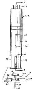
In general terms, as shown in one preferred form is Figs. 1-5, the injector 10
comprises a cylindrical forward barrel 28 (or device housing) having a plunger
30 (or carrier
body) mounted therein for longitudinal sliding movement within a hollow bore
between a
forward advanced position (Fig. S) and a rearward retracted position (Fig. 9).
The plunger 30
has a head 32 at a forward end thereof for releasibly receiving and retaining
the subcutaneous
insertion set 14, in a manner to be described in more detail. A rear end of
the plunger 30
2 0 cooperates with a trigger-type actuator assembly 34 mounted on the rear
end of the barrel 28.
The trigger actuator assembly 34 (or driver) is adapted to hold the plunger 30
in a retracted
position, against the force of a compressed drive spring 36. A trigger button
38 of the
actuator assembly 34 is adapted for fingertip depression to release the
plunger 30 for spring-
loaded travel toward the advanced position, and corresponding transcutaneous
placement of
the insertion needle 12 through the patient's skin.
Figs. 2-5 illustrate construction details of the injector barrel 28, wherein
the forward
or nose end thereof defines a flat and generally planar surface for placement
against the skin
of a patient (Fig. 1) with a longitudinal axis of the barrel 28 oriented
generally perpendicular
to the patient's skin 16. The barrel 28 has a size and shape for substantially
mated sliding fit
3 0 reception of the infusion set 14, with the insertion needle 12 and related
cannula 26
projecting in a direction for placement on a patient. In this regard, the nose
end of the barrel
28 defines an opposed pair of relatively wide and open-ended cut outs 40 for
slide-fit
-12-
CA 02484271 1998-12-18
w0 99J33504 PCTlUS98I26978
reception of the oppositely projecting base wings 24. A narrower slot 42 is
also formed in
the barrel nose end, at a location for slide-fit reception of the infusion
tubing 22 attached to
the infusion set 14. Thus, the forward or nose end of the barrel 28
accommodates sliding
reception of the subcutaneous insertion set 14 therein for movement along the
cut outs 40
and the slot 42 between the advanced position (Fig. 5) disposed substantially
at the
forwardmost end of the barrel 28, and the retracted position (Fig. 9) with the
base wings 24
and infusion tubing 22 positioned substantially at the inboard ends of the cut
outs 40 and the
associated slot 42.
The plunger 30 includes the head 32 of generally cylindrical shape for slide-
fit
reception within the injector barrel 28. A forward end of the head 32 includes
a cylindrical
counterbore recess 44 for receiving the hub 18 and housing 20 of the insertion
set 14, with
the enlarged base wings 24 fitted against a pair of outwardly protruding
backstop flanges 46
adapted for slide-fit reception within the cut outs 40 in the barrel nose end.
A pair of track
arms 48 (Fig. 5) protrude rearwardly from the plunger head 32 and include out-
tanned latch
fingers 50 for guided reception within longitudinally extending track slots 52
formed in the
barrel 28 at a location spaced aft from the barrel nose end. These track arms
48 thus
cooperate with the barrel track slots 52 to guide the plunger 30 between the
advanced
position (Figs. 5 and 7) and the retracted position (Fig. 9).
The plunger 30 also includes a central drive stem 54 (Fig. 5) which protrudes
2 0 rearwardly from the plunger head 32 within the barrel interior. The
rearward end of the drive
stem 54 is longitudinally split to define a pair of trigger anus 56 which have
out-turned
trigger fingers 58 on the rearward ends thereof.
The trigger-type actuator assembly 34 is mounted on the rearward end of the
injector
barrel 28, and generally functions to releasibly retain the plunger 30 in a
retracted and cocked
2 5 position, ready for rapid and spring-loaded actuation upon depression of
the trigger button 38
to place the insertion set 14 on the patient. More particularly, as shown best
in Figs. 5-9, the
trigger assembly 34 comprises a main support cap 60 having a mounting sleeve
62 protruding
in a press-fit and preferably adhesively connected manner into the rear or aft
end of the
injector barrel 28. The drive spring 36 comprises a coil spring positioned
about the drive
3 0 stem 54 on the plunger 30 and reacts between a rearward face 64 of the
plunger head 32, and
a shoulder 66 on the support cap 60. The drive spring 36 normally biases the
plunger 30
toward the advanced position (Figs. 5 and 7). However, an insertion set 14
seated in the
-13-
CA 02484271 1998-12-18
WO 99!3350-t PCTlUS98I26978
plunger head 32 can be pressed rearwardly against the plunger 30 to move the
plunger to the
retracted position, as viewed in Fig. 9, with the trigger fingers 58 passed
through a conical or
tapered latch bore 68 formed in the support cap 60 to engaging a shoulder 70
on an opposite
side of the support cap 60. In this regard, the trigger fingers 58 have
rarnped outboard faces
to accommodate movement of the forgers 58 radially toward each other as they
pass through
the latch bore 68. When the trigger fingers 58 pass entirely through the bore
68, the spring
resilience of the trigger arms 56 is sufficient to spread the trigger forgers
58 so that they
engage the shoulder 70. In this retracted plunger position, the drive spring
36 is retained in a
compressed and cocked condition, with the insertion set 14 including the
insertion needle 12
and related cannula 26 withdrawn into the interior of the barrel 28, in spaced
relation to the
patient's skin 16.
The trigger actuator assembly 34 additionally includes an actuator pin 72
mounted
within a noncircular bore 74 (Figs. 6 and 7) formed in the support cap 60 for
longitudinal
sliding movement through a short stroke, relative to the plunger 30. In this
regard, the
7.5 actuator pin 72 includes one or more noncircular lands 76 for slide-fit
reception within the
bore 74, to prevent actuator pin rotation therein. The actuator pin 72 is held
within the bore
74 by a stepped lock ring 78 which is retained against a rearward end of the
support cap 60
by a press-fit outer retainer sleeve 80 having an inturned rim 82 at the
rearward end thereof.
Importantly, as shown best in Fig. 8, an oblong land 84 is formed on the
actuator pin 72 for
mated slide-fit reception through an oblong recess 85 formed in the lock ring
78. A return
spring 86 (Fig. 7) is carried within the support cap bore 74 and reacts
between the shoulder
70 and a nose end of the actuator pin 80 for biasing the actuator pin 80
rearwardly within the
support cap.
The rearmost end of the actuator pin 72 defines the trigger button 38. As
shown in
Figs. 1 I and 13, the trigger button 38 can be depressed with a fingertip to
move the actuator
pin ?2 through a short stroke against the return spring 86 in a direction
toward the trigger
fingers 58 at the rear end of the plunger 30. As shown best in Fig. 13, the
actuator pin 72 has
a hollowed cylindrical forward tip 88 with a diametric size for engaging and
squeezing the
trigger fingers 58 together at the rear end of the plunger 30, in a manner
enabling those
3 0 trigger fingers 58 to pass back through the tapered conical latch bore 68.
As soon as the
trigger fingers 58 thus release from engagement with the shoulder 70 on the
support cap 60,
the drive spring 36 translates the plunger 30 with the insertion set 14
thereon with a rapid and
-14-
CA 02484271 1998-12-18
w0 99/3350.1 PCT/US98I26978
controlled force and speed toward the advanced position, resulting in
transcutaneous
placement of the needle 12 and cannula 26, as viewed in Fig. 15. Importantly,
the spring rate
characteristics of the drive spring 36 and the distance of plunger stroke are
chosen for a
substantially optimized and proper transcutaneous placement of the needle 12
and cannula
26, all in a manner which minimizes patient discomfort during the needle
placement
procedure. Moreover, by forming the nose end of the injector barrel 28 with a
squared-off
shape as shown, the injector 10 can be easily oriented substantially
perpendicular to the skin
16 for proper placement of the insertion set.
Depression of the actuator pin 72 by means of the trigger button 38 requires
the lock
ring 78 to be rotatably oriented in a position aligning the oblong recess 85
therein with the
oblong land 84 on the actuator pin. Accordingly, when these oblong structures
are
rotationally aligned (Figs. 13-14), the injector 10 is armed for trigger
button depression and
corresponding release of the retracted and cocked plunger. However, the lock
ring 78 can be
rotated relative to the actuator pin 72 to misalign these oblong structures,
as shown in Figs.
9-I2, whereupon the actuator pin 72 is locked in a rearward position against
depression and
actuation. A set pin 90 on the lock ring 78 may be provided and received
within an accurate
notch 92 formed in the retainer sleeve flange rim 82, to permit lock ring
rotation back-and-
forth through a part circle increment, on the order of about 90 degrees.
Appropriate indicia
may be applied to the retainer sleeve rim 82, such as the letter "L" for
"locked" and the letter
"A" for "armed", as viewed in Figs. I2 and 14, to provide a visual indication
of the setting of
the trigger assembly 34.
In accordance with one aspect of the invention, the plunger head 32
additionally
includes a safety lock mechanism in the form of a pair of safety lock arms 94
pivotally
carried in narrow slots 96 formed in the plunger head 32. These safety lock
arms 94 have
2 5 rearward ends connected to the head 30 by pivot pins 98, and forward ends
defining
contoured lock fingers 100 which protrude into the plunger head recess 44. As
shown in Fig.
7, the safety lock arms 94 and their associated lock fingers 100 have a size
and shape so that
the fingers 100 can engage and retain the hub 18 of the. insertion set 14, for
example, by
fitting into a recess 101 defined between the hub 18 and housing 20 of the
insertion set.
3 0 Importantly, the locations of the lock arm pivot points are chosen to
insure that the lock arms
94 engage and retain the insertion set 14 whenever the plunger 30 is moved
from the
advanced position (Fig. 7) toward and to the retracted position (Fig. 9). When
the plunger 30
-15-
CA 02484271 1998-12-18
WO 99J33~Oa PCT/US98126978
reaches the fully advanced position, the safety lock arms 94 including their
respective pivot
pins 98 are disposed within the wide cut outs 40 and are therefore free to
swing outwardly,
relative to the insertion set 14, to accommodate separation of the insertion
set from the
injector 10 with a substantially minimum separation force. This configuration
has been
found to be highly effective as a safeguard to prevent the insertion set 14
from being thrown
as a projectile from the injector 10, in the event that the trigger assembly
34 is activated
without prior placement of the injector 10 firmly against the patient's skin
16.
In use, the subcutaneous insertion set 14 can be placed quickly and easily
into the open nose
end of the injector barrel 28, within the recess 44 formed in the plunger head
32. Such
assembly of the insertion set 14 with the injector 10 requires simple
alignment of the base
wings 24 and infusion tubing 22 with the appropriate cut outs and slots 40, 42
formed in the
nose end of the barrel 28. The insertion set 14 and plunger 30 can then be
manually retracted
rearwardly, against the drive spring 36, to the retracted position with the
plunger 30 cocked
and latched as viewed in Figs. 9 and 11. The injector 10 can then be placed
firmly against
the patient's skin 16, with the insertion set 14 supported in the proper
orientation and at a
predetermined distance from the skin 16. Simple depression of the trigger
button 38 releases
the cocked plunger 30 for spring-loaded travel rapidly albeit with a
controlled speed and
force of insertion, to ensure penetration of the patient's skin with minimal
discomfort, and in
a manner which properly places the insertion needle and cannula. The safety
lock arms 94
2 0 prevent accidental projection of the insertion set 14 through free space,
in the event that the
trigger button 38 is prematurely depressed. When the insertion set 14 is
properly placed,
however, the safety lock arms 94 release from the insertion set with minimal
force, for easy
separation of the injector 10 from the insertion set 14.
In preferred embodiments, the controlled speed and force of the insertion
device is
2 5 obtained by selecting a spring constant of a spring to propel and insert
the insertion set at the
proper speed and force to ensure penetration with minimal discomfort. In
alternative
embodiments, as shown in Figs. 48a-48d, there is the need to vary the speed
and force, from
one insertion cycle to the next, to accommodate different insertion sets (such
as finer needles,
sensor sets fragility or the like) and insertion site conditions (such as
overweight,
3 0 underweight, children or the like). As shown in Fig. 48a, a force changing
mechanism 1000
having a spring 1002 enclosed in a sealed compartment 1004 is used with a set
(or
adjustable) orifice 1006 to allow equalization of internal and ambient
pressures during the
-16-
CA 02484271 1998-12-18
WO 99/3350.1 PCT/US98/26978
insertion stroke of the insertion device. The sealed compartmentll$$es sealing
O-
rings 1008 and 1010 to seal the sealed compartment 1004. The O-ring 1008 seals
the
insertion set carrier body 1012, and the O-ring 1010 seals the actuator
housing 1014 (which
contains the orifice 1006) of the force generating mechanism 1000. The force
changing
mechanism 1000 may be activated by, for example, a trigger 1016 that is biased
by a spring
1018 to close off the orifice 1006 until depressed. The limiting flow through
the office 1006
acts as a dampening force, counteracting the spring force from the spring
1002, thereby
allowing control of insertion speed and force. The orifice size can be
adjustably attained
through a number of approaches, such as bearing 1020 and spring 1022 that
blocks the
orifice 1006 and resists air flow based on the tension of the spring 1022 on
the bearing 1020
(see Fig. 48b); while presenting a lower resistance during retraction as the
air contained in
the sealed compartment 1004 is compressed, forcing bearing 1020 against spring
1022 to
unseat the bearing 1022 from the orifice 1006 to present the maximum orifice
size for
escaping air during compression of spring 1002. This structure minimizes the
force needed
to compress spring 1022 by allowing air in the sealed compartment 1004 to
escape freely and
quickly through the orifice 1006; rather than be compressed within the sealed
compartment
1004 because the orifice 1006 is restricted by bearing 1002. In another
alternative, as shown
in Fig. 48c, a disk 1024 has a plurality of various sized holes 1026(1) to
1026(n). The disk
1024 is rotateable over the orifice 1006 to sequentially obstruct, completely
obstruct or
2 0 partially obstruct the orifice 1006 flow path and changes the effective
size of the orifice 1006
by blocking the orifice 1006 with the various sized holes 1026( 1 ) through
1026(n). In
another embodiment, as shown in Fig. 48d, a tapered valve plug 1028 is
threaded into
position relative to the orifice 1006 to change the effective size of the
orifice 1006. Other
orifice 1006 size changing methods may be used. In addition, other methods of
controlling
the insertion speed and force may be used, such as controlled friction, change
in spring
tension, hydraulics, pneumatics or the like may be used.
Following separation of the injector 10 from the placed insertion set 14, the
insertion
needle 12 can be withdrawn quickly and easily from the cannula as viewed in
Fig. 16.
Thereafter, the insertion set 14 can be used in a normal manner to deliver a
selected
3 0 medication through the infusion tubing 22 and cannula 26 to the patient.
An alternative preferred form of the invention is shown in Figs. 17-29,
wherein
components corresponding in structure and function to those described
previously with
-17-
CA 02484271 1998-12-18
WO 99/33504 PC'f/11S98l26978
respect to Figs. 1-16 are identified by common reference numerals increased by
100. The
embodiment of Figs. 17-29 show a modified injector 110 constructed from a
reduced number
of parts and including an alternative safety lock mechanism for preventing
undesired
projection of the insertion set 14 through free space in the event of injector
operation without
placing the nose end thereof firmly against the skin 16 of a patient. However,
the alternative
safety lock mechanism again permits quick and easy separation of the injector
110 from the
insertion set 14, with minimal separation force. Once again, although an
insertion set for
infusing medical fluids to a patient will be shown and described, it will be
understood that
alternative insertion sets such as transcutaneous sensor insertion sets and
the like as
previously referenced herein may be used with the injector 110.
In general, the modified injector 110 comprises a plunger 130 and a trigger-
type
actuator 134 assembled with a generally cylindrical hollow barrel 128. The
plunger I30 has
a generally cylindrical plunger head 132 which defines a counterbore recess
144 for receiving
and retaining the hub 18 of the infusion set 14. As shown best in Figs. 27-29,
a radially
inwardly projecting rim 202 is formed on the plunger head 132 generally at a
leading or nose
end of the recess I44, wherein this rim 202 has a noncircular and preferably
oval or elliptical
shape (Fig. 28) to accommodate reception of the hub 18 into the recess 144
provided that the
hub 18 is oriented angularly relative to a central longitudinal axis of the
plunger 130 and
barrel 128. Similar angular orientation of these components accommodates quick
and easy
2 0 separation thereof. However, when the insertion set 14 is oriented with
the medical needle
12 aligned coaxially with the barrel center axis, a portion of the rim 202
projects into the
insertion set recess 101 to prevent release of the insertion set 14 from the
injector 110.
More specifically, with reference to Figs. 17-20, the barrel 128 again has a
forward or
nose end defining a flat and generally planar surface for firm placement
against the skin of a
patient. The nose end of the barrel 128 has a pair of relatively wide and
generally opposed
cut outs 140 formed therein for slide-fit reception of the base wings 24 of
the insertion set 14,
in combination with a narrower slot 142 for slide-fit reception of the
infusion tubing 22.
This slot 142 may be formed in one or both sides of the barrel nose end.
The plunger 130 is slidably fitted into the barrel 128 for movement between an
3 0 advanced position shown in Figs. 17, 18, 20 and 21, and a retracted
position shown in Fig.
23. The plunger 130 includes the modified plunger head 132 of generally
cylindrical shape,
formed preferably to include a shallow notch or groove 133 in one side thereof
for slide-fit
-18-
CA 02484271 1998-12-18
WO 99/3350 PCT/US98/26978
reception of the infusion tubing 22 on the insertion set 14. In thWregxr~, the
plunger head
groove 133 is formed in a position aligned with the narrow slot 142 in the
nose end of the
barrel.
The plunger head 132 is formed integrally with a drive stem 154 which projects
rearwardly within the barrel interior. As shown best in Fig. 22, the drive
stem 154 is flanked
by and formed integrally with a pair of rearwardly projecting track arms 148
which have
latch fingers 150 formed at the rear ends thereof. As shown in Figs. 21 and
23, these latch
fingers 150 are received slidably within longitudinally extending track slots
152 formed in
the barrel 128, and function to guide the plunger 130 between the advanced and
retracted
positions. Cushioning material (not shown) may be included at the leading ends
of the track
slots 152 to form a combined stop upon spring driver advancing motion of the
plunger 130,
as will be described.
The plunger 130 additionally includes a pair of trigger arms 156 which project
generally rearwardly from a rear end of the drive stem 154 and have out-turned
trigger
fingers 158 at the rear ends thereof (Fig. 22). These trigger fingers 158 are
adapted and sized
for partial radial compression toward each other as they ride within the
barrel base when the
plunger 130 is displaced from the advanced position (Fig. 21) to the retracted
position (Fig.
23). As the retracted position is reached, the trigger fingers 158 are spring-
loaded by the
resiliency of the trigger arms 156 to move outwardly for partial reception
into relatively short
2 0 trigger slots 159 formed in the barrel 128. In this position, as shown in
Fig. 23, the triggers
fingers 158 retain the plunger 130 in the retracted position.
A drive spring 136 is mounted within the barrel 128 to react between the
trigger-type
actuator 134 and the plunger 130, in the same manner as previously described
with respect to
Figs. 1-16. In this regard, the trigger actuator 134 comprises a generally
cylindrical actuator
2 5 sleeve 188 mounted slidably within the barrel 128 at the rear or upper end
thereof. This
actuator sleeve 188 has a tapered or ramped leading edge face 188' (Figs. 22,
23 and 25) for
engaging matingly shaped ramped outer faces of the trigger fingers 158, to
radially compress
the trigger arms 156 and release the plunger 130 for spring-loaded travel from
the retracted
and cocked position to the advanced position. A trigger button 138 is formed
integrally with
3 0 the actuator sleeve 188 and is exposed for fingertip depression at the
rear or top of the barrel
128 to move the actuator sleeve 188 into releasing engagement with the trigger
fingers 158.
As shown best in Figs. 22 and 24-26, the triggers button 138 extends through
an
-19-
CA 02484271 1998-12-18
WO 9913350 PCT/US98/26978
opening formed in the rear of the barrel 128, generally within a lock sleeve
1?8 formed
integrally with the barrel 128. The lock sleeve 178 defines an oppositely
formed pair of
guide slots 192 for aligned reception of a pair of outwardly radiating lock
tabs 184 formed on
the trigger button 138. When the tabs 184 and rotationally aligned with the
guide slots I 92,
the trigger button 138 can be depressed to actuate the spring-locked plunger,
as described.
However, the lock tabs 184 have sufficient length to pcrmit fingertip rotation
of the actuator
134 to re-position the tabs 184 within shallow lock grooves 193 formed
adjacent the guide
slots 192. When the tabs 184 are seated in the lock grooves 193, the lock
sleeve 178 blocks
depression of the triggers button 138 and thereby locks the injector 110
against actuation.
Return rotation of the actuation 134 to re-align the tabs 184 with the guide
slots 192 is
required before the injector can be activated.
In accordance with one aspect of the invention, the plunger head 132 includes
the
safety lock mechanism in the form of the noncircular rim 202 at the leading
end of the recess
144 in the plunger head. As shown in Figs. 27 and 28, the rim 202 has a
generally elliptical
shape defining a major axis that is greater than the diameter of the hub 18 on
the insertion set
14, and a minor axis that is less than the hub diameter. With this geometry,
and by providing
sufficient axial depth to the plunger head recess 144, the hub 18 can be
fitted into the plunger
head by angularly orienting the components to permit slide-fit of the hub 18
through the
major axis portion of the rim 202. Subsequent re-orientation of the components
to align the
2 0 medical needle 12 generally coaxiaIly with plunger head 32 enables the
minor axis portion of
the rim 202 to project into the insertion set recess 101, thereby locking the
components
together. Thereafter, when the insertion set 14 is placed on the patient (Fig.
29), the
components are easily separated by lifting the injector 110 off the insertion
set 14 at the same
angle to allow the hub 18 to press freely through the major axis center of the
rim 202.
2 5 Importantly, such engagement and disengagement of the components occurs
with essentially
no resistance force to separation. The infusion set 14 can be oriented
angularly relative to the
glunger 130 only when the plunger is in the advanced position, with the
adjacent barrel 128
precluding such angular orientation when the plunger 130 is moved rearwardly
from the
restricted position.
3 0 In an alternative mode of operation, subsequent to actuation of the
injector 110 to
place the insertion set 14 of the patient, the injector 1 I O can be simply
withdrawn or retracted
in a direction away from the patient's skin 16, in which case the rim 202 at
the nose end of
-20-
CA 02484271 1998-12-18
WO 9913350 PCT1US98/26978
the plunger head 132 will engage the needle hub 18 and thereby gently withdraw
the medical
needle 12 from the insertion set 14, In this manner, the needle 12 is
retracted from the
cannula 26 which remains at the desired transcutaneous insertion site.
A further alternative preferred form of the invention is shown in Figs. 30-34,
wherein
a further modified injector 210 is constructed and operated generally as shown
and described
in Figs. 17-29, but wherein an alternative configuration for a plunger head
232 is provided.
Figs. 30-32 show the injector 210 with the plunger head 232 in the advanced
position within
the front or nose end of the barrel 128 which includes the wide cut outs 140
and the narrow
slot 142 for respective slide-fit reception of the base wings 24 and the
tubing 22 of the
insertion set 14. As shown, the modified plunger head 232 has a laterally open
recess 244
formed therein of undercut geometry and laterally exposed through the cut outs
140 when the
plunger is in the advanced position. The insertion set 14 can be slide-fit
assembled with the
plunger head 232, by fitting the hub 18 into the wider upper region of the
recess 144, with an
interned rim 302 at the leading end of the plunger head fitting into the
insertion set recess
101. A laterally open gap 303 (Fig. 34) in the rim 302 permits slide-fit
reception of the hub
18 into the recess 244, and a short carrier post 304 (Fig. 32) may be provided
at the base of
the recess 244 to seat within a shallow detent in the top of the hub.
With the insertion set 14 assembled with the plunger head 232, as viewed in
Fig. 32,
the plunger can be retracted and cocked as previously shown and described with
respect to
2 0 Figs. 17-29. The cut outs 140 and slot 142 accommodate sliding movement of
the insertion
set 14 with the plunger 232 during such retraction. Thereafter, the front or
nose end of the
injector 210 can be placed fu-mly against the patient's skin (Fig. 33) and the
trigger button
138 depressed to release the plunger so that the medical needle 12 is
transcutaneously placed
with the controlled drive force and speed. During forward drive motion of the
plunger, the
2 5 forward rim 302 on the plunger head 232 prevents projectile release of the
insertion set.
After placement of the insertion set on the patient, the injector 210 can be
laterally displaced
relative to the insertion set for quick and easy separation therefrom.
Alternately, as viewed
in Fig. 34, the injector 210 can be withdrawn or retracted from the insertion
set 14 to slidably
withdraw the medical needle 12 while leaving the insertion set in place on the
patient.
30 Figs. 35-40g illustrate an insertion device 500 in accordance with a second
embodiment of the present invention. The insertion device S00 includes a
barrel 502 (or
device housing) having a surface seat 501 and an assembly port 503, a carrier
body 504 (or
-21-
CA 02484271 1998-12-18
WO 99i3350.t PCT/US98/26978
plunger or the like) having an assembly rim 505 and a seating flange 506, a
drive spring 507
(or driver), a release button 508, and dual spring triggers S 10 and 512. As
shown in Fig. 35,
the barrel 502 performs as a housing to hold the carrier body 504. The carrier
body 504 is
connected to the barrel 502 by the carrier body being inserted through an
opening in the
surface seat S01 of the barrel 502, and then passing the assembly rim 505 of
the carrier body
504 through the assembly port 503 of the barrel 502. The section of the
carrier body 504
with the assembly rim 505 compresses slightly, as it passes through the
assembly port 503,
due to the presence of compression slots 509, and then essentially restores to
its original
shape to prevent the carrier body 504 from sliding out of the barrel 502,
since the assembly
rim 505 of the carrier body engages with the assembly port 503 of the barrel
502.
The carrier body 504 is driven to an advanced position from a retracted
position by
the drive spring 507 and held in the retracted position (or released to move
to the advanced
position) by the trigger buttons 510 and 512. This embodiment of the insertion
device 500 is
primarily adapted for insertion of insertion sets 400 (as exemplary shown in
Fig. 39 as an
infusion set), or the like, that are inserted with the piercing member 402 (or
needle) at 90
degrees (or perpendicular) to the skin surface after insertion. In preferred
embodiments, the
carrier body S04 is permanently coupled to the barrel 502 and new insertion
sets 400 are
attached to the carrier body 504 for each new insertion. However, in
alternative
embodiments, the carrier body 504 may be a disposable that is replaced after
each insertion
2 0 so that, for instance, a carrier body 504 may be shipped with a pre-
installed insertion set 400
and then loaded into the barrel 502 of the insertion device 500.
The insertion device S00 features a low profile compact package that tends to
minimize the effects of hand movement during insertion of the insertion set
400. In this
embodiment, the release button 508 is depressed to release the insertion set
400, or the like,
2 5 from the carrier body 504 of the insertion device 500; rather than
engaging or disengaging
the insertion set 14 using a lateral slot as shown in Figs. 31-34 above. The
release button S08
can be used before or after insertion of the insertion set 400, or the like.
To facilitate
insertion of an insertion set 400, or the like, the insertion device 500
utilizes dual trigger
buttons S 10 and 512, which provide an extra margin of safety and
substantially prevents
3 0 accidental activation of the insertion device 500 upon contact with the
skin surface. This
obviates the need for a Lock and unlock position on the activation buttons (or
triggers) of the
earlier insertion devices shown in Figs. 1-34. The insertion device S00 also
includes another
-22-
CA 02484271 1998-12-18
WO 99!3350.1 PCT/tJS98i26978
rim on the carrier body 504 that forms the seating flange 506 to hold a rim
404 (or wing) of
the insertion set 400, or the like, that carries an adhesive 406 for adhering
the insertion set
400 to the surface of the skin. Upon activation of the insertion device 500 to
move the
carrier body to the advanced position, the seating flange 506 presses the
adhesive 406 and
rim 404 of the insertion set 400 firmly against the skin surface to provide
positive seating and
attachment of the insertion set 400 to the skin. This may make it unnecessary
to require
placement of an additional adhesive patch prior to or after inserting an
insertion set 400 to
secure the insertion set 400 at the insertion site. The insertion device 500
further includes an
automatic release of the piercing member (or needle) hub 408 and piercing
member 402 (or
needle) from the insertion set 400, or the like, after the insertion set 400,
or the like, has been
inserted. This permits the insertion set 400 to be left on the skin surface
without the piercing
member hub 408 and piercing member 402 (or needle) remaining by simply
removing the
insertion device 500 from the skin surface. This automatic release feature
also minimizes
potential patient contact with the piercing member 402 (or needle) of the
insertion set 400, or
the like.
In preferred embodiments, the insertion set 400, or the like, is adapted to
tightly fit
within a cavity 514 (or receiving structure) of the carrier body 504. The
cavity 514 of the
carrier body 504 includes guides 516 to orient the insertion set in a
particular orientation and
an expansion member 518 in the center bottom interior of the cavity 514 of the
carrier body
2 0 504 to engage with the piercing member hub 408 (or needle hub) of the
insertion set 400, or
the like. The piercing member hub 408 of the insertion set 400, or the like,
includes a center
section 410 that engages with the expansion member 518 with a slight
interference fit. The
interference fit expands the center section 410 of the piercing member hub 408
slightly to
expand and press the piercing member hub 408 against the sides of the cavity
514 of the
2 5 cannier body 504 to firmly secure the insertion set 400, or the like,
within the cavity 514 of
the carrier body 504. The tight fit of the insertion set 400,or the like, in
the carrier body 504
substantially prevents the insertion set 400, or the like, from being
dislodged when the
insertion device 500 is activated to improve insertion of the insertion set
400, or the like, on
the skin. However, the tight fit also prevents the insertion set 400, or the
Like, from being
3 0 ejected if the insertion device 500 is inadvertently activated when it is
not pressed against the
skin surface. In preferred embodiments, the insertion device 500 is configured
to have
guides 516 and an expansion member 518 to work with existing insertion sets
400, or the
-23-
CA 02484271 1998-12-18
WO 99/3350 PCT/US98/269?8
Like. However, in alternative embodiments, the insertion set 400, or the like,
may be
modified to have a piercing member base, housing or the like that includes
slots (not shown)
for receiving guides and expanding members of the insertion device 500 to
improve the
connection between the insertion device 500 and the insertion set 400, or the
like. In further
alternative embodiments, the guides and expansion members may be formed on the
insertion
set 400, or the like, and the corresponding guide slots and expanding sections
may be formed
on the insertion device 500.
The illustrated embodiment employs a dual trigger activation structure to
minimize
the ability of the insertion device 500 to be unintentionally activated. As
illustrated, the
barrel 502 of the insertion device 500 includes two outwardly extending guide
channels 520
and 522 on the side of the barrel 502. The guide channels 520 and 522 extend
from the base
524 of the barrel 502 up to portal openings 526 and 528 in the side of the
barrel 502. The
dual trigger buttons 510 and 512 are carried on opposite sides of the seating
flange 506 at the
end of the carrier body 504. Each trigger button 510 and 512 is biased outward
from the side
of the seating flange 506 by a trigger spring 530 and 532 between the end of
the trigger
buttons 510 and 5I2 and the side of the seating flange 506. When the Garner
body 504 of the
insertion device 500 is locked in the firing position (or retracted position),
the trigger buttons
510 and 512 are pushed out by the trigger springs 530 and 532 to extend out of
the portal
openings 526 and 528. In this position, the trigger buttons 510 and 512 extend
beyond the
2 0 bottom of the guide channels 520 and 522, which prevents the trigger
buttons 510 arid 512
from moving down the guide channels 520 and 522. To activate the insertion
device 500, the
user must depress both trigger buttons 510 and 512 so that the trigger buttons
510 and 512
can then slide along the bottom of the guide channels 520 and 522, which in
turn allows the
carrier body 504 to move down along the barrel 502 until the insertion set
400, or the like, is
2 5 inserted. In preferred embodiments, the portal openings 526 and 528 and
the end of the
guide channels 520 and 522 terminating at the portal openings 526 and 528 are
rounded to
match the shape of the trigger buttons 510 and 512. This tends to minimize the
resisting
pressure on the trigger buttons 510 and 512 during depression of the trigger
buttons 510 and
512. However, in alternative embodiments other portal opening and guide
channel end
3 0 shapes, such as beveled, squared, polygonal, or the like, may be used.
The end of the carrier body 504 having the assembly rim 505 is connected to a
release
button 508 that can be depressed or slightly extended relative to the carrier
body 504. The
-24-
CA 02484271 1998-12-18
WO 99/3350a PCT/US98I2G978
release button 508 includes engagement tabs 550 and lock teeth 552 (see Figs.
35 and 36)~
that engage with carrier slots 554 and carrier locks 556 (see Figs. 36 and 38)
to lock the
release button 508 to the carrier body 504. The lock teeth 552 engage with the
carrier locks
556 (see Figs. 36 and 38) to permit an amount of movement of the lock teeth
552 along the
carrier locks 556 to allow the release button 508 to be depressed to release
an insertion set
from the carrier body 504. The release button 508 is also slightly extended
away from the
carrier body 504 when an insertion set 400 is placed in the interior cavity
514 of the carrier
body 504 to permit seating of the insertion set 400. Engaging the release
button 508 with the
carrier body substantially prevents the compression slots 509 and assembly rim
505 from
compression and inhibits release of the carrier body 504 from the barrel 502
of the insertion
device 500.
The release button 508 is depressed to release the insertion set 400, or the
like, from
the carrier body 504 of the insertion device 500. The release button 508
pushes the insertion
set 400, or the like, out of the cavity 514 of the carrier body 504
sufficiently enough to
release the insertion set 400, or the like, from the guides 516 and the
expanding member 51
in the cavity 514 and leave the inserted insertion set 400, or the like, on
the skin.
Alternatively, the release button 508 may be activated to release an insertion
set 400, or the
like, from the carrier body 504 prior to the insertion set 400, or the like,
being inserted by the
insertion device 500. The release button 508 also includes a ramp portion 534
(or other
2 0 trigger mechanism) that is adapted to bend or adjust the piercing member
hub 408 (or needle
hub) of the insertion set 400, or the like, to allow the piercing member hub
408 and piercing
member 402 (or needle) to be released and separated from the insertion set
400, or the like,
when the insertion set 400, or the like, has been inserted and the insertion
device 500 is lifted
off the skin. This can be accomplished by separating the elements of the
insertion set 400, or
2 5 the like, so that only the insertion set, or the like, housing and tubing
(or wiring or the like)
are left in contact with the skin. The ability to remove the piercing member
hub 408 and
piercing member 402 is preferably facilitated by the adhesive 406 of the
insertion set 400, or
the like, that attaches to the skin to provide sufficient tension to allow for
separation of the
piercing member hub 408 and the piercing member 402 from the rest of the
insertion set 400,
3 0 or the like, without dislodging the insertion set 400, or the like. In
preferred embodiments,
the insertion device 500 is adapted to work with an existing piercing member
hub 408 on an
insertion set 400, or the like. However, in alternative embodiments, the
piercing member
-25-
,._,~.,...._..._...._. .._~.r~. .___ _ _ _.
CA 02484271 1998-12-18
WO 9913350a PCTlUS98/26978
hub 408 and the connection between the piercing member hub ~hd' insertion set
housing, or the like, is modified to work with the release mechanism of the
insertion device
500.
In preferred embodiments, the release button 508 is biased in position by a
plastic or
metal spring. However, in alternative embodiments, the release button 508 may
be manually
reset by engaging and disengaging detents or using other elastomeric materials
to bias the
release button 508 in position relative to the barrel 502 and the carrier body
504. In preferred
embodiments, pulling up the release button 508 (or extending it away from the
assembly port
503 of the barrel 502) pulls the carrier body 504 to the retracted position in
the barrel 502,
where it is locked in place by triggers 510 and 512 engaging the portal
openings 526 and 528.
1'kiis procedure separates the piercing member hub 408 and piercing member 402
from the
housing of the insertion set 400, or the like. This has the advantage of
removing the piercing
member 402 and piercing member hub 408 to minimize the opportunity of a user
being stuck
by the piercing member 402.
Figs. 40a-40g illustrate one method of insertion of an insertion set 400 with
the
insertion device 500 in accordance with an embodiment of the present
invention. The user
first cleans and sterilizes an insertion site on the skin. Next, the user
makes sure the insertion
device 500 has the carrier body 504 in the advanced position to avoid
unintentional
activation of the insertion device 500 before placement on the skin. As shown
in Fig. 40a,
2 0 the user places the insertion set 400 in the cavity 514 of the carrier
body by aligning the
tubing (or wire leads or the like) with the slot 536 in the carrier body 504
and the slot 538 in
the barrel 502 of the insertion device 500. The user presses against the
piercing member
guard 414 (or needle guard) to seat the piercing member hub 408 (or needle
hub) and the
insertion set 400 in the cavity 514 of the carrier body 504. As shown in Fig.
40b, the user
2 5 removes the adhesive backing 416 covering the adhesive 406 on the rim 404
of the insertion
set 400. It is preferred that the piercing member guard 414 is not removed at
this point to
avoid unintentional sticks by the piercing member 402, and minimize or avoid
contact with
the adhesive 406. As shown in Fig. 40c, the user presses against the piercing
member guard
414 to move the carrier body 504 from the advance position to the retracted
position, at
3 0 which point the trigger buttons 510 and 512 will extend out of the portal
openings 526 and
528 to extend beyond the guide channels 520 and 522 to lock the carrier body
504 in the
retracted position. Next, as shown in Fig. 40d, the user removes the piercing
member guard
-26-
CA 02484271 1998-12-18
414 (normally by twisting) to expose the piercing member 402 while
ina~ntaining the
insertion set 400 within the carrier body 504. Then, as shown in Fig. 40e, the
user places the
surface seat 501 of the barrel 502 of the insertion device 500 with the
insertion set 400 over
the insertion site on the skin. The user depresses the two trigger buttons 510
and 512 through
the portal openings 526 and 528 sufficiently for the trigger buttons 510 and
512 to slide
down along the guide channels 520 and 522 to insert and install the insertion
set 400 at the
insertion site on the skin. As shown in Fig. 40f, the user depresses the
release button 508 to
release the insertion set from the cavity 514 of the earner body 504. Finally,
as shown in Fig.
40g, the user removes the insertion device 500, while maintaining installation
of the insertion
set 400. In alternative embodiments, the user may extend the release button
508 to lift off the
piercing member hub 408 and piercing member 402, and maintain the remainder of
the
insertion set 400 at the insertion site on the skin. If the piercing member
hub 408 and
piercing member 402 are lifted off the device, the user should re-install the
piercing member
guard 414 prior to removal of the remaining set from the insertion device 500.
Figs. 41-46 illustrate an insertion device 600 in accordance with a third
embodiment
that is similar to the insertion devices shown in Figs. 1-34. The insertion
device 600 includes
a device housing end 601 and a carrier body 602 that has angled insertion
contact surfaces
603 and 604. The angled insertion contact surfaces 603 and 604 enable the user
to properly
angle the insertion device 600 to insert an insertion set 700, or the like, at
the proper insertion
angle relative to the skin.
Preferred embodiments of the insertion device 600 have angled insertion
contact surfaces 603 and 604 that permit insertion of insertion sets, or the
like, that are
angled from 89.9 degrees to 25 degrees relative to the skin surface. In
further embodiments,
the angled insertion contact surfaces 603 and 604 may handle even shallower
angles down to
approximately 10 degrees relative to the skin surface.
The key is the angled insertion contact surfaces 603 and 604 minors the
insertion
angle of the insertion set 700, or the like, so that the piercing member 702
(or needle) of the
insertion set 700, or the like, is in axial alignment in the direction of
movement of the carrier
-27-
CA 02484271 1998-12-18
body 602 of the insertion device 600. This permits an insertion device
designed primarily for
use with a 90 degree insertion set, or the like, to be modified to work with
angled insertion
sets 700, or the like, by modification of the angle of the angled insertion
contact surfaces 603
and 604. In addition, it is preferred that the piercing member ?02 of the
insertion set ?00, or
the like, be slightly off-center from the center axis of the carrier body 602
to permit easy
removal of the insertion device 600 once the insertion set 700, or the like,
has been inserted.
Preferred embodiments of the present invention include a carrier body 602 with
a receiving
stricture that includes a recess 606 and bore 608 on one side of the carrier
body 602. The
recess 606 is adapted to hold the piercing member hub 704 by a slight
interference fit and the
bore 608 is adapted to hold the insertion tubing or transmitter hub 706 of the
insertion set
700, or the like. In other embodiments for the insertion sets such as infusion
sets with tubing
(or sensor sets with wire leads already connected to a sensor) the bore 608
may be open on
one side (not shown) to permit insertion and removal of the infusion tubing
(or wire leads),
but closed of sufficiently to securely hold and grasp the insertion tubing or
transmitter hub
706 that connects the tubing or wire leads to the housing of the insertion set
700, or the like.
Fig. 47 illustrates an insertion device 800 that is adapted for inserting
insertion sets,
or the like, at angles that are generally less than or equal to 10 degrees
relative to the skin
surface after insertion of the insertion set, or the like. This embodiment
includes a pair of
pinchers 802 and 804 that grasps the skin. The pinchers 802 and 804 pinches
(or bunches)
2 0 up the skin in front of a carrier body 806 holding an insertion set, or
the like. Once the skin
is pinched (or bunched up), the user depresses an activation button and the
insertion set, or
the like, is inserted into the skin. In alternative embodiments, the user
presses the pinchers
802 and 804 closer together to activate the insertion device 800. After
insertion, the user
releases the pinchers 802 and 804 and removes the insertion device 800 from
the insertion
set, or the like. The effect of this embodiment is to raise the skin so that
the actual insertion
angle of the piercing member relative to the side of the raised (or pinched)
area of skin
ranges from 10 degrees to 90 degrees so that the piercing member is inserted
in manner
sinular to the embodiments described above. However, when the pinched skin is
released,
the piercing member is left in the skin at a shallow angle between 0 and 10
degrees. The
3 0 amount of pinching and the height of the pinch must be carefully
controlled to assure that the
insertion set, or the like, is inserted at the proper depth and location in
the skin tissue.
-28-
CA 02484271 1998-12-18
While the description above refers to particular embodiments of the present
invention, it will be understood that many modifications may be made without
departing
from the spirit thereof. The accompanying claims are intended to cover such
modifications
as would fall within the true scope and spirit of the present invention.
The presently disclosed embodiments are therefore to be considered in al!
respects as
illustrative and not restrictive, the scope of the invention being indicated
by the appended
claims, rather than the foregoing description, and all changes which come
within the meaning
and range of equivalency of the claims are therefore intended to be embraced
therein.
-29-