Note: Descriptions are shown in the official language in which they were submitted.
CA 02484875 2011-01-12
METHOD AND SYSTEM FOR OPTICALLY DETECTING
BLOOD AND CONTROLLING A GENERATOR DURING
ELECTROSURGERY
BACKGROUND
1. Technical Field
The disclosure relates to electrosurgey combined with optical detection of
blood, and more particularly the automatic control of the level of
electrosurgical energy
to be delivered to tissue in accordance with the amount of blood optically
detected.
2. Description of the Related Art
Electrosurgery involves the application of radio frequency energy to achieve a
tissue effect. An electrosurgical generator is used in surgical procedures to
deliver
electrical energy to the tissue of a patient. An electrosurgical generator
often includes a
radio frequency generator and its controls. When an electrode is connected to
the
generator, the electrode can be used for cutting or coagulating the tissue of
a patient
with high frequency electrical energy. During normal operation, alternating
electrical
current from the generator flows between an active electrode and a return
electrode by
passing through the tissue and bodily fluids of a patient.
The electrical energy usually has its waveform shaped to enhance its ability
to
cut or coagulate tissue. Different waveforms correspond to different modes of
operation of the generator, and each mode gives the surgeon various operating
advantages. Modes may include cut, coagulate, a blend thereof, or desiccate. A
surgeon can eas
ily select and change the different modes of operation as the surgical
procedure progresses.
In each mode of operation, it is important to regulate the electrosurgical
energy
delivered to the patient to achieve the desired surgical effect. This can be
done, for
example, by controlling the output energy from the electrosurgical generator
for the
type of tissue being treated.
CA 02484875 2004-11-04
WO 03/092520 PCT/US03/14155
2
Different types of tissues will be encountered as the surgical procedure
progresses and each unique tissue requires more or less energy in terms of
voltage,
current or power as a function of frequently changing tissue impedance and
other
factors, such as the level of vascularization, i.e., blood flow within the
tissue.
Therefore, the same tissue will present different load impedance as the tissue
is
desiccated.
Two conventional types of energy regulation are used in commercial
electrosurgical generators. The most common type controls the DC power supply
of the
generator by limiting the amount of power provided from the AC mains to which
the
generator is connected. A feedback control loop regulates output voltage by
comparing
a desired voltage or current with the output voltage or current supplied by
the power
supply. Another type of power regulation in commercial electrosurgical
generators
controls the gain of the high-frequency or radio frequency amplifier. A
feedback
control loop compares the output power supplied from the RF amplifier for
adjustment
to a desired power level.
U.S. Patent Nos. 3,964,487; 3,980,085; 4,188,927 and 4,092,986 have circuitry
to reduce the output current in accordance with increasing load impedance. In
those
patents, constant voltage output is maintained and the current is decreased
with
increasing load impedance.
U.S. Patent No. 4,126,137 controls the power amplifier of the electrosurgical
unit in accord with a non-linear compensation circuit applied to a feedback
signal
derived from a comparison of the power level reference signal and the
mathematical
product of two signals including sensed current and voltage in the unit.
U.S. Patent No. 4,658,819 has an electrosurgical generator which has a
microprocessor controller based means for decreasing the output power as a
function of
changes in tissue impedance.
U.S. Patent No. 4,727,874 includes an electrosurgical generator with a high
frequency pulse width modulated feedback power control wherein each cycle of
the
generator is regulated in power content by modulating the width of the driving
energy
pulses.
CA 02484875 2004-11-04
WO 03/092520 PCT/US03/14155
3
U.S. Patent No. 3,601,126 has an electrosurgical generator having a feedback
circuit that attempts to maintain the output current at constant amplitude
over a wide
range of tissue impedances.
None of the aforementioned U.S. patents include optical detection of blood for
regulating or controlling the output energy or output waveforms of the
electrosurgical
generator during different operational modes over a finite patient tissue
impedance
range. Optical detection of blood during electrosurgery also allows surgeons
with color
blindness to effectively perform electrosurgery. In a study that was published
in 1997,
18 of 40 physicians with color blindness reported difficulties in detecting
blood in body
products. Spalding, J. Anthony B., "Doctors with inherited colour vision
deficiency:
their difficulties in clinical work," Cavonius CR, ed., Colour Vision
Deficiencies, XII:
Proceeding of the International Research Group for Colour Vision Deficiencies,
1995,
Norwell, Mass.: Kluwer Academic Publishers, pages 483-489, 1997.
Accordingly, there exists a need for a method and system for optically
detecting
blood during electrosurgery and controlling the output energy or output
waveforms of
an electrosurgical generator in accordance with the amount of blood optically
detected.
SUMMARY
A method and electrosurgical system for optically detecting blood and
controlling an electrosurgical generator are provided. An optical blood
detection
system is used for optically detecting blood and may be included as an
integral part of
the overall electrosurgical system's circuitry, or may be designed as a
separate unit that
connects to, and controls, an electrosurgical generator. The optical blood
detection
system may be embodied through a variety of analog, digital and/or optical
circuit
components or arrangements, including software running on computational and
memory circuitry.
The optical blood detection system controls the output energy of the
electrosurgical generator in accordance with the amount of blood detected.
This allows
for a surgeon to perform electrosurgery without having to stop and observe the
condition of the tissue to determine if additional electrosurgery is needed.
CA 02484875 2004-11-04
WO 03/092520 PCT/US03/14155
4
More particularly, the optical blood detection system automatically controls
the
output waveform generated by the electrosurgical generator during
electrosurgery using
a feedback signal received from the optical blood detection system. For
example, if
coagulation of the tissue is desired, the optical blood detection system
continuously
analyzes the tissue for the presence of blood and controls the output waveform
accordingly.
While the optical blood detection system may be used to control
electrosurgical
generators of varying designs, it is preferred that the electrosurgical
generator includes a
power selection system wherein the user may initialize, set, monitor, and/or
control the
operation of the electrosurgical generator. The preferred electrosurgical
generator need
not be limited to these four functional elements, for example the
electrosurgical
generator could also include additional safety, monitoring, signal
modification/conditioning, and/or feedback circuitry or functional
elements/processes.
The actual electrosurgical generator's design may include the use of digital
components
and signaling, analog components and signaling, and/or optical components and
signaling, or may be embodied, completely or partially within a software
process
running on hardware components.
The optical blood detection system includes an optical light beam generating
circuit having optical components for generating and focusing a light beam in
close
proximity to and/or on an electrode of an electrosurgical instrument; a
circuit having
optical components for capturing reflected light energy, such as a
photosensitive
detector; a blood detection circuit for analyzing the reflected light energy
and/or other
characteristics and determining the amount of blood present in proximity to
and/or on
the electrode; and a feedback correction circuit.
The feedback correction circuit which is electrically connected to receive a
signal from the blood detection circuit functions to produce a feedback
control signal
which it then supplies to the power selection system, within the
electrosurgical
generator, so as to cause the power selection system to control the amount of
electrosurgical energy created and/or the type of output waveform generated in
accordance to the amount of blood present in proximity to and/or on the
electrode. The
system can also detect the presence of any blood vessels in proximity to the
distal end
CA 02484875 2004-11-04
WO 03/092520 PCT/US03/14155
of the electrode and control the electrosurgical generator accordingly or
alert the
surgeon to prevent, for example, the severing of major blood vessels.
Preferably, the optical light beam is focused in front of the distal end of
the
electrode to detect blood present on tissue which is being cut or coagulated
by the
5 electrosurgical instrument. The optical light beam may have light energy
within the
visible, near-infrared and infrared light spectrum wavelengths.
It is provided that one or more of the above-mentioned circuits can be
implemented by one or more sets of programmable instructions configured for
being
executed by at least one processor of the electrosurgical system or at least
one processor
remotely located from the electrosurgical system. For example, the data
corresponding
to the reflected light energy can be transmitted, either wirelessly or non-
wirelessly, over
a network, such as a LAN, WAN, or the Internet, to a remote server or control
station
for analyzing the data using a set of programmable instructions for
determining the
amount of blood present in proximity to and/or on the electrode.
In accordance with the analysis performed, the remote server or control
station
then generates using the same or another set of programmable instructions the
feedback
control signal and supplies the signal to the power selection system. It is
contemplated
that another form of electromagnetic energy can be used to detect for the
presence of
blood besides the optical beam of light.
In one embodiment of the present invention an electrosurgical system is
provided which includes a handpiece having a proximal end and a distal end
from
which light energy is emitted therefrom; at least one electrosurgical
electrode on the
handpiece and extending from the distal end from which electrosurgical energy
is
emitted there from; a source of light energy for generating the light energy
and
transmitting the same to the distal end via at least one waveguide; a source
of
electrosurgical energy for generating the electrosurgical energy and
transmitting the
same by at least one electrically conductive element to the electrode; and
means for
analyzing light energy characteristics for determining the amount of blood
present in
proximity to the electrode and for controlling the source of electrosurgical
energy
accordingly.
CA 02484875 2004-11-04
WO 03/092520 PCT/US03/14155
6
In another embodiment of the present invention an electrosurgical system is
provided which includes means for generating and directing light energy on
tissue;
means for generating electrosurgical energy and transmitting the same via an
electrode
to the tissue; and means for analyzing characteristics of the light energy for
determining
the amount of blood present in proximity to the electrode and for controlling
the means
for generating electrosurgical energy accordingly.
Further, in another embodiment of the present invention, a method is provided
for performing electrosurgery. The method includes the steps of supplying
light energy
and electrosurgical energy to tissue via at least one instrument having a
distal end; and
analyzing characteristics of the light energy for determining the amount of
blood
present in proximity to the at least one instrument and for controlling the
delivery of
electrosurgical energy accordingly.
Finally, in another embodiment of the present invention, a surgical method is
provided which includes the steps of providing a surgical instrument
configured for
insertion within a patient; providing a source of light energy for generating
light energy
and delivering the same via the surgical instrument; and analyzing light
energy
characteristics for determining the amount of blood present in proximity to
the surgical
instrument.
Further features of the disclosure will become more readily apparent to those
skilled in the art from the following detailed description taken in
conjunction with the
drawings.
CA 02484875 2004-11-04
WO 03/092520 PCT/US03/14155
7
BRIEF DESCRIPTION OF THE DRAWINGS
Various embodiments will be described hereinbelow with reference to the
drawings wherein:
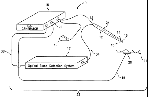
FIG. 1 is a perspective diagram of one embodiment of the present
electrosurgical system;
FIG. 2 is cut-away, schematic diagram of an electrosurgical handpiece
instrument of the electrosurgical system of FIG. 1;
FIG. 3 is a block diagram of the optical blood detection system;
FIG. 4 is a flow chart showing the operation of the optical blood detection
system according to a first method;
FIG. 5 is a flow chart showing the operation of the optical blood detection
system according to a second method; and
FIG. 6 is a cut-away, schematic diagram of another embodiment for the
electrosurgical handpiece instrument.
DETAILED DESCRIPTION OF A PREFERRED EMBODIMENT
An electrosurgical system 10 is shown in perspective in FIG. 1 and allows a
surgeon to provide cutting, coagulating, and/or a combination thereof on
tissue of a
patient 11. The electrosurgical system 10 has a handpiece 12 with a proximal
end 13 to
be held and controlled by the surgeon. A distal end 14 on the handpiece 12 has
a port
15 from which an optical light beam is directed to the patient 11. An
electrosurgical
electrode 16 extends from the distal end 14 of the handpiece 12.
An optical blood detection system 17 for generating the optical light beam is
connected to the proximal end 13 of the handpiece 12 via waveguide/wires 34.
The
optical blood detection system 17 can be manually controlled by the surgeon or
automatically controlled for delivering the optical light beam from the distal
end 14 of
the handpiece 12 toward the patient 11. An electrosurgical generator 18 for
generating
the electrosurgical energy is electrically connected to the proximal end 13 of
the
handpiece 12 and may be manually controlled by the surgeon or automatically
controlled for transmitting electrosurgical energy from the electrosurgical
electrode 16
toward the patient 11. The optical blood detection system 17 and the
electrosurgical
CA 02484875 2004-11-04
WO 03/092520 PCT/US03/14155
8
generator 18 are connected by a cable 38 for providing data communications
there
between and a feedback control signal from the optical blood detection system
17 to the
generator 18 for controlling the generator 18.
While the optical blood detection system 17 may be used to control
electrosurgical generator 18, it is preferred that the electrosurgical
generator 18 includes
a power selection system wherein the user may initialize, set, monitor, and/or
control
the operation of the electrosurgical generator 18. The preferred
electrosurgical
generator need not be limited to these four functional elements, for example
the
electrosurgical generator 18 could also include additional safety, monitoring,
signal
modification/conditioning, and/or feedback circuitry or functional
elements/processes.
The actual electrosurgical generator's design may include the use of digital
components
and signaling, analog components and signaling, and/or optical components and
signaling, or may be embodied, completely or partially within a software
process
running on hardware components.
A return path 19 is provided for the electrosurgical energy; the return path
19
may be in a monopolar or bipolar circuit. FIG. 1 illustrates a monopolar
circuit having
a return pad 20, in lieu of a return electrode in the case of a bipolar
circuit. The return
path 19 is connected to receive at least a portion of the transmitted
electrosurgical
energy from the source of electrosurgical energy 18 and then the patient 11. A
return
input 22 for the source of electrosurgical energy 18 is connected to the
return path 19
for furnishing a complete circuit 23 between the electrosurgical electrode 16,
the patient
11, and the electrosurgical generator 18.
A manually-actuated control button 24 is provided on the handpiece 12 for the
selective control by the surgeon of the electrosurgical generator 18 for
controlling the
electrosurgical energy delivered from the distal end 14. The control button 24
may also
be located at a foot pedal 26.
It is provided that the surgeon can utilize the optical beam emanating from
port
15 to pinpoint the target tissue to be treated if the optical light beam has
light energy
within the visible spectrum. It is envisioned that the optical light beam may
have light
3o energy within the visible, near-infrared and infrared light spectrum
wavelengths.
CA 02484875 2004-11-04
WO 03/092520 PCT/US03/14155
9
With reference to FIG. 3, the optical blood detection system 17 includes an
optical light beam generating circuit 52 having optical components for
generating and
focusing a light beam, such as a laser light beam, as known in the art, in
close proximity
to and/or on the electrode 16 of the handpiece 12. The wave guide 34, shown in
FIG. 1,
is used to deliver the light energy from the proximal end 13 to beyond the
distal end 14.
The optical blood detection system 17 further includes at least one optical
component
54 positioned at the distal end 14 of the handpiece 12, for capturing
reflected light
energy as known in the art. The at least one optical component 54 returns
signals
indicative of the reflected light energy to the system 17 via waveguide/wires
34 to at
least one photosensitive detector.
The optical blood detection system 17 further includes a blood detection
circuit
56 for analyzing the reflected light energy and determining the amount of
blood present
in proximity to and/or on the electrode 16; and a feedback correction circuit
58.
The reflected light energy preferably includes data corresponding to light
reflections indicative of two different wavelengths, a first and a second
wavelength.
First, a first optical light beam having the first wavelength is generated and
emanated
from the handpiece 12. The reflected light energy indicative of the first
optical light
beam is captured and analyzed by the optical blood detection system 17 for
measuring
various parameters, such as photon counts. Second, a second optical light beam
having
the second wavelength is generated and emanated from the handpiece 12. The
reflected
light energy indicative of the second optical light beam is captured and
analyzed by the
optical blood detection system 17 for measuring various parameters, such as
photon
counts.
Alternatively, a broadband optical light beam is generated and emanated from
the handpiece 12. The reflected light energies indicative of two separate and
distinct
wavelengths are captured and analyzed by the optical blood detection system 17
for
measuring various parameters, such as photon counts. Preferably, in either
method, the
first wavelength is in the range of 620-700 nanometers and the second
wavelength is in
the range of 540-610 nanometers or 950-1050 nanometers.
A ratio is then obtained using two measured values corresponding to a
particular
parameter; one measured value is indicative of the first optical light beam or
CA 02484875 2004-11-04
WO 03/092520 PCT/US03/14155
wavelength and one measured value is indicative of the second optical light
beam or
wavelength. A look-up table or other data structure is then used by a
processor or by an
individual to correlate the ratio with a particular amount or level of blood
present in
proximity to the electrode 16.
5 The reflected light energy can also be analyzed for determining the amount
of
blood present using one of several known methods, such as Near Infrared
Spectroscopy
(LAIRS), Infrared Spectroscopy (IRS), Fluorescence Spectroscopy, Raman
Spectroscopy,
Photoacoustic Spectroscopy (where the system 10 is equipped with a microphone
for
measuring an acoustic pressure wave created by the optical beam rapidly
heating the
10 tissue), laser Doppler flowmetry, light scatter change measurements, and
polarization
change measurements. These methods determine the light intensity level, light
scattering effects, level of fluorescent energy, and other characteristics of
the reflected
light energy. The determined light intensity level, light scattering effects,
level of
fluorescent energy, and/or other characteristics of the reflected light energy
are then
used to compute using mathematical equations, algorithms, and/or programmable
instructions executed by at least one processor the amount of blood present in
proximity
to the electrode 16.
By knowing the optical signal characteristics of the generated light beam and
the
determined light intensity level, light scattering effects, level of
fluorescent energy, and
other characteristics of the reflected light energy, the system 17 is able to
determine
using a look-up table or other data structure the amount of blood present in
proximity to
the electrode 16. If the analysis indicates that there is a high amount of
blood present in
proximity to the electrode 16, one can conclude that the tissue has not
coagulated (in
the case of a coagulation procedure) or has been cut (in the case of a cutting
procedure).
If the analysis indicates that there is a low amount of blood present in
proximity to the
electrode 16, one can conclude that the tissue has coagulated (in the case of
a
coagulation procedure) or has not been adequately cut (in the case of a
cutting
procedure).
The system can also detect the presence of any blood vessels in proximity to
the
distal end of the electrode 16 and control the electrosurgical generator 18
accordingly or
alert the surgeon to prevent, for example, the severing of major blood
vessels.
CA 02484875 2004-11-04
WO 03/092520 PCT/US03/14155
11
The feedback correction circuit 58 which is electrically connected to receive
a
signal from the blood detection circuit 56 fiulctions to produce a feedback
control
signal which it then supplies to the power selection system, within the
electrosurgical
generator 18, via wire 3 8 so as to cause the power selection system to
control the
amount of electrosurgical energy created and/or the type of output waveform
generated
(coagulation or tissue division waveform) in accordance to the amount of blood
present
in proximity to and/or on the electrode 16.
FIG. 4 is a flow chart illustrating an exemplary method of operation of the
optical blood detection system 17. In step 400, the optical light beam and
electrosurgical energy are generated. The reflected light energy is captured
in step 402
and analyzed in step 404 to determine the amount of blood present in proximity
to the
electrode 16 at step 406. In step 408 it is determined whether the sensed
level of blood
in proximity to the electrode 16 is above a predetermined threshold (the
predetermined
threshold value is dependent on the method being used to detect the amount of
blood
present). If the sensed level of blood is not above the predetermined
threshold value, it
is then determined at step 410 whether the procedure being performed is a
coagulation
procedure. If a coagulation procedure is not being performed, i.e., a cutting
procedure
is being performed, the cutting procedure is continued at step 412, and the
process
returns to step 408.
If at step 410, it is determined that a coagulation procedure is being
performed,
the process proceeds to step 414 where a signal is transmitted by the feedback
correction circuit ' 58 to the electrosurgical generator 18 to control the
amount of
electrosurgical energy and/or the type of output waveform generated or to shut-
off the
electrosurgical generator 18, since the coagulation procedure has been
adequately
performed. If at step 408, it is determined that the sensed level of blood is
above the
predetermined threshold value, it is then determined at step 416 whether the
procedure
being performed is a cutting procedure. If a cutting procedure is not being
performed,
i.e., a coagulation procedure is being performed, the coagulation procedure is
continued
at step 418, and the process returns to step 408.
If at step 416, it is determined that a cutting procedure is being performed,
the
process proceeds to step 414 where a signal is transmitted by the feedback
correction
CA 02484875 2004-11-04
WO 03/092520 PCT/US03/14155
12
circuit 58 to the electrosurgical generator 18 to control the amount of
electrosurgical
energy and/or the type of output waveform generated or to shut-off the
electrosurgical
generator 18, since the cutting procedure has been adequately performed.
FIG. 5 is a flow chart illustrating another exemplary method of operation of
the
optical blood detection system 17. In step 500, the optical light beam and
electrosurgical energy are generated. The reflected light energy is captured
in step 502
and analyzed in step 504 to determine the amount of blood present in proximity
to the
electrode 16 at step 506. Step 506 determines the amount of blood present by
calculating the ratio value as determined by dividing the photon counts at
wavelength 1
by the photon counts at wavelength 2. The ratio value is analyzed at step 508.
If the ratio value is low (lower than a predetermined ratio value)then the
process
proceeds to step 510 where a signal is transmitted by the feedback correction
circuit 58
to the electrosurgical generator 18 to control the mode of operation, namely,
selecting a
tissue division (cut) mode. Also, the amount of electrosurgical energy may be
adjusted.
If at step 508, it is determined that the ratio value is high (greater than
the
predetermined ratio value), the process proceeds to step 512 where a signal is
transmitted by the feedback correction circuit 58 to the electrosurgical
generator 18
selecting a hemostasis (coagulation) mode. The amount of electrosurgical
energy may
also be adjusted.
If at step 508, it is determined that the ratio value is at an intermediate
value
(approximately equal to the predetermined ratio value), the process proceeds
to step
514 where a signal is transmitted by the feedback correction circuit 58 to the
electrosurgical generator 18 selecting a blended mode that is in proportion to
the
detected ratio value. Following either step 510, 512, or 514, the process
returns to
capture reflected light energy in step 502 in a continuous loop.
It is provided that depending on which of the above spectroscopy and other
methods is used by the optical blood detection system 17 to determine the
amount of
blood present, the optical blood detection system 17 is controlled accordingly
using
known blood-related optical measurement parameters for each method, in order
to
generate and focus an optical light beam having characteristics suitable for
the method.
The optical blood detection system 17 can change the wavelength of the optical
light
CA 02484875 2004-11-04
WO 03/092520 PCT/US03/14155
13
beam within the visible, near-infrared and infrared light spectrum wavelengths
depending on which of the above methods is being used for determining the
amount of
blood present in proximity to the electrode 16. For example, if the LAIRS
method is
used, the optical light beam needs to have a wavelength just above the visible
spectrum.
The wavelength of the optical light beam can be manually selected using a
control knob or other control means on the optical blood detection system 17.
If the
wavelength of the optical light beam is in a particular range, the light
energy of the
optical light beam can be used to create an ionized conductive pathway along
which the
electrosurgical energy can be guided.
When the light energy is being used to create an ionized pathway, the light
energy must be controlled using the control means in order to avoid undesired
tissue
effects. The duty cycle of the light beam should be kept in the range of 10-5
to 10-8.
Energy density delivered to any single area of tissue from the light beam
should not
exceed 26 J/cm2 for wavelengths between 1.06 and 10.6 microns, and 17 J/cm2
for
wavelengths around and below 0.53 microns. For creating the ionized pathway,
the
wavelength of the optical beam should be in the range of 0.3 to 10.6 microns.
It is further provided that one or more of the above-mentioned circuits 52, 56
and 58 can be implemented by one or more sets of programmable instructions
configured for being executed by at least one processor of the electrosurgical
system 10
or at least one processor remotely located from the electrosurgical system 10.
For
example, the data corresponding to the reflected light energy can be
transmitted, either
wirelessly or non-wirelessly, over a network, such as a LAN, WAN, or the
Internet, to a
remote server or control station for analyzing the data using a set of
programmable
instructions for determining the amount of blood present in proximity to
and/or on the
electrode 16 and/or the presence of blood vessels in proximity to the distal
end of the
electrode 16.
In accordance with the analysis performed, the remote server or control
station
then generates using the same or another set of programmable instructions the
feedback
control signal and supplies the signal to the power selection system. It is
contemplated
that another form of electromagnetic energy can be used to detect for the
presence of
blood besides the optical beam of light.
CA 02484875 2004-11-04
WO 03/092520 PCT/US03/14155
14
Another embodiment for a handpiece for the electrosurgical system 10 is
depicted by FIG. 6 and designated generally by reference numeral 12A. The
handpiece
12A includes a proximal end 13A which is held and controlled by the surgeon. A
distal
end 14A on the handpiece 12A has a port 15A from which an optical light beam
is
directed to the patient 11. An electrosurgical electrode 16A extends from the
distal end
14A of the handpiece 12A. The at least one optical component 54 at the distal
end 14A
of the handpiece 12A returns signals indicative of the reflected light energy
to the
optical blood detection system 17 via waveguide/wires 34 to at least one
photosensitive
detector.
A manually-actuated variable control button 24A is provided on the handpiece
12A for the real-time, selective control by the surgeon of the intensity or
level of the
current, i.e., intensity of the output waveform, provided by the
electrosurgical generator
18 in accordance with the amount of blood detected by the optical blood
detection
system 17. Accordingly, the handpiece 12A provides the surgeon with the
ability to
control the amount of tissue cutting, coagulating, etc. as the system 10
concurrently
detects the amount of blood.
In another preferred embodiment with continued reference to FIG. 6, the
optical
detection of the presence of blood controls the mode of the electrosurgical
generator
output in real-time or on-the-fly. For illustrative purposes, if a large
amount of blood is
detected adjacent to the electrode 16A then the electrosurgical generator
output mode is
automatically set for a high-level "hemostasis" (coag) waveform. If no blood
is
detected, then a "tissue division" (cut) waveform is automatically selected
for the
electrosurgical generator output. If an intermediate amount of blood is
detected, then a
"blend" is selected in proportion to the amount of blood detected.
Simultaneously, the
surgeon can use the manually-actuated variable control button 24A for real-
time,
selective control of the intensity or level of current.
The surgeon selects the intensity that provides an operational speed within
his
individual comfort zone. So the selection of the mode is automatically
controlled by
the blood detection circuit 56 and the surgeon controls the intensity of the
output in
real-time or on-the-fly. This embodiment greatly simplifies the surgeon-
equipment
interface by providing an automated mode select to assist the surgeon. As a
result there
CA 02484875 2004-11-04
WO 03/092520 PCT/US03/14155
is an improvement in the surgical outcome, because the appropriate mode is
selected in
real-time, thereby reducing thermal spread within the tissue. Additionally,
since the
surgeon maintains control of the intensity of the current, there is a built-in
safety
feature.
5 The above-described control scheme can be offered as a selectable feature or
option. That is, a selectable switch would allow the surgeon to choose between
operating the system of the present invention in a fully automatic mode or in
a mode
which enables the surgeon to control the intensity of the current.
It is contemplated that the control button 24A may also be located at the foot
10 pedal 26. It is further contemplated that the functions of the variable
control button
24A can be automated, in order for the system 10 to automatically control the
intensity
of the current in accordance with the amount of blood detected by the optical
blood
detection system 17.
It is provided that the surgeon can utilize the optical beam emanating from
port
15 15A to pinpoint the target tissue to be treated if the optical light beam
has light energy
within the visible spectrum. It is envisioned that the optical light beam may
have light
energy within the visible, near-infrared and infrared light spectrum
wavelengths.
As shown by FIGS. 2 and 6, the electrosurgical system 10 is configured so the
distal end 14, 14A and the electrosurgical electrode 16, 16A are preferably
arranged
geometrically relative to the handpiece 12, 12A to provide the light energy
from the
distal end 14, 14A. This geometry provides for the combined concurrent
application of
the light energy and the electrosurgical energy. The ionized pathway is formed
by the
light energy from the distal end 14, 14A to the patient 11 to direct the
electrosurgical
energy there along.
A method for providing cutting, coagulating, and/or a combination thereof on
tissue of the patient 11 with the electrosurgical system 10 includes the
following step of
directing light energy and electrosurgical energy from the handpiece 12, 12A
with its
proximal and distal ends, 13, 13A and 14, 14A, along a longitudinal axis of
the
handpiece 12, 12A by aiming the distal end 14, 14A thereof along the
longitudinal axis
from which light energy and electrosurgical energy may be at least in part
concurrently
directed.
CA 02484875 2004-11-04
WO 03/092520 PCT/US03/14155
16
Preferably, as shown by FIGS. 2 and 6, the optical light beam is focused in
front
of the distal end 14, 14A of the electrode 16, 16A to detect blood present on
tissue
which is being cut or coagulated by the handpiece 12, 12A. The light energy is
emanated continuously from the distal end 14, 14A of the handpiece 12, 12A.
Or,
alternatively, the surgeon activates the electrosurgical generator 18 using
the control
button 24, 24A on the handpiece 12, 12A or the footswitch 26. When activation
is
initiated, first, light energy is emitted from the distal end 14, 14A of the
handpiece 12,
12A, then after a brief time delay in which the presence of blood is detected,
the
transmission of electrosurgical energy from the electrosurgical electrode 16,
16A at the
distal end 14, l4Aof the handpiece 12, 12A is enabled.
In the case of encountering a bleeding vessel that has created a pool of
blood,
this method provides detection of the pool of blood and automatic select of a
hemostatic (coagulation) waveform by the electrosurgical generator 18 in order
to affect
a "spot coag" procedure.
Likewise, if no blood is present, the detection system selects a tissue
division
(cut) waveform. In this way, the thermal damage to the tissue is reduced
creating a
superior tissue effect.
The method includes the additional step of guiding the electrosurgical energy
by
arranging the distal end 14, 14A and the electrosurgical electrode 16, 16A
geometrically
relative to the handpiece 12, 12A for providing the optical light beam from
the distal
end 14, 14A for the combined concurrent application of the optical light beam
and the
electrosurgical energy. Then the added step of ionizing a conductive pathway
with light
energy from the distal end 14, 14A to the patient 11 to direct the flow of
electrosurgical
energy is performed.
The method also includes the additional step of providing an elongate
electrosurgical electrode support for supporting the electrode 16, 16A for
endoscopic or
laparoscopic use where a cannula is placed through the patient's body wall.
The claims which follow seek to cover the described embodiments and their
equivalents. The concept in its broadest scope covers the system and methods
for
optically detecting the presence of blood and/or determining the amount of
blood
detected during electrosurgery. It is to be understood that the concept is
subject to
CA 02484875 2004-11-04
WO 03/092520 PCT/US03/14155
17
many modifications without departing from the spirit and scope of the claims
as recited
herein.
Although the subject invention has been described with respect to preferred
embodiments, it will be readily apparent to those having ordinary skill in the
art to
which it appertains that changes and modifications may be made thereto without
departing from the spirit or scope of the subject apparatus as defined by the
appended
claims.