Note: Descriptions are shown in the official language in which they were submitted.
CA 02488337 2004-12-03
WO 03/103476 PCT/US03/17715
PATENT FORAMEN OVALE (PFO) CLOSURE DEVICE
WITH RADIAL AND CIRCUMFERENTIAL SUPPORT
Field of the Invention
[0001] The present invention relates generally to an occlusion device for the
closure of physical anomalies like septal apertures, such as patent foramen
ovule and
other septal and vascular defects.
Background of the Invention
[0002] A patent foramen ovule (PFO), illustrated in Figure l, is a persistent,
one-
way, usually flap-like opening in the wall between the right atrium 11 and
left atrium 13
of the heart 10. Because left atrial (LA) pressure is normally higher than
right atrial (RA)
pressure, the flap usually stays closed. Under certain conditions, however,
right atrial
pressure can exceed left atrial pressure, creating the possibility that blood
could pass from
the right atrium 11 to the left atrium 13 and blood clots could enter the
systemic
circulation. It is desirable that this circumstance be eliminated.
[0003] The foramen ovule serves a desired purpose when a fetus is gestating in
utero. Because blood is oxygenated through the umbilical chord, and not
through the
developing lungs, the circulatory system of a heart in a fetus allows the
blood to flow
through the foramen ovule as a physiologic conduit for right-to-left shunting.
After birth,
with the establishment of pulmonary circulation, the increased left atrial
blood flow and
pressure results in functional closure of the foramen ovule. This functional
closure is
subsequently followed by anatomical closure of the two over-lapping layers of
tissue:
septum primum 14 and septum secundum 16. However, a PFO has been shown to
persist
in a number of adults.
CA 02488337 2004-12-03
WO 03/103476 PCT/US03/17715
2
[0004] The presence of a PFO is generally considered to have no therapeutic
consequence in otherwise healthy adults. Paradoxical embolism via a PFO is
considered
in the diagnosis for patients who have suffered a stroke or transient ischemic
attack (TIA)
in the presence of a PFO and without another cause of ischemic stroke. While
there is
currently no definitive proof for a cause-effect relationship, many studies
have confirmed
a strong association between the presence of a PFO and the risk for
paradoxical embolism
or stroke. In addition, there is significant evidence that patients with PFO
who have had a
cerebral vascular event are at increased risk for future, recurrent
cerebrovascular events.
[0005] Accordingly, patients with an increased future risk are considered for
prophylactic medical therapy to reduce the risk of a recurrent embolic event.
These
patients are commonly treated with oral anticoagulants, which have the
potential for
adverse side effects, such as hemorrhaging, hematoma, and interactions with a
variety of
other drugs. The use of these drugs can alter a person's recovery and
necessitate
adjustments in a person's daily living pattern.
[0006] In certain cases, such as when anticoagulation is contraindicated,
surgery
may be necessary or desirable to close the PFO. The surgery would typically
include
suturing a PFO closed by attaching septum secundum to septum primum. This
sutured
attachment can be accomplished with either an interrupted or a continuous
stitch and is a
common way a surgeon shuts a PFO under direct visualization.
[0007] Umbrella devices and a variety of other similar mechanical closure
designs, developed initially for percutaneous closure of atrial septal defects
(ASDs), have
been used in some instances to close PFOs. These devices have the potential to
allow
patients to avoid the potential side effects often associated with
anticoagulation therapies
CA 02488337 2004-12-03
WO 03/103476 PCT/US03/17715
3
and the risks of invasive surgery. However, umbrella devices and the like that
are
designed for ASDs are not optimally suited for use as a PFO closure device.
[0008] Currently available designs of septal closure devices present
drawbacks,
including technically complex implantation procedures. Additionally, there are
not
insignificant complications due to thrombus, fractures of the components,
conduction
system disturbances, perforations of heart tissue, and residual leaks. Many
devices have
high septal profile and may include large masses of foreign material, which
may lead to
unfavorable body adaptation of a device. Since ASD devices are designed to
occlude a
hole, many lack anatomic conformability to the PFO flap-like anatomy. That is,
when
inserting an ASD device to close a PFO, the narrow opening and the thin flap
may form
impediments to proper deployment. Even if an occlusive seal is formed, the
device may
be deployed in the heart on an angle, which could leave some components not
securely
seated against the septum, thereby risking thrombus formation due to
hemodynamic
disturbances. Finally, some septal closure devices are complex to manufacture,
which
may result in lack of consistency in product performance.
[0009] The present invention is designed to address these and other
deficiencies
of the prior art septal closure devices.
Summary of the Invention
[0010] The present invention provides a device for occluding an anatomical
aperture, such as a septal defect or a PFO. This occluder includes two sides
connected by
an intermediate joint. Each of the sides includes at least one wire or other
elongate
element for structural support (referred to collectively as "wire"), which is
arranged to
form non-overlapping loops. Each loop has at least one radially-extending
segment that
CA 02488337 2004-12-03
WO 03/103476 PCT/US03/17715
4
is adjacent to a radially-extending segment of another loop. In at least some
embodiments, at least one pair of adjacent radially-extending segments is
connected. The
loops of the device may be of various shapes and sizes. In at least some
embodiments,
the loops have rounded peripheries. The configuration of the loops and sides
of the
occluder are varied according to different embodiments of the invention. In
some
embodiments, at least one of the sides includes a tissue scaffold.
[0011] The wires forming the occluders of the present invention may be
constructed of various biocompatible materials. In some embodiments, the wires
are
formed of shape memory materials, e.g. nitinol. In other embodiments, the
wires are
formed of polymers, bioabsorbable polymers, or combinations thereof.
[0012] The occluder according to the present invention is designed such that,
when deployed in vivo, the two sides are disposed on opposite sides of the
septal tissue
surrounding the aperture, i. e. septum primum and septum secundum. Thus, the
two sides
exert a compressive force on the septal tissue that is distributed along both
the outer
periphery of the occluder and the radially-extending segments. In at least
some
embodiments, the radially-extending segments increase the stiffness of the
occluder,
thereby preventing the occluder from becoming dislodged from its intended
delivery site.
In at least some embodiments, the flexible, rounded peripheries of the loops
prevent the
occluder from inflicting trauma upon the septal tissue as the heart contracts.
In at least
some embodiments of the present invention, the occluder is repositionable
and/or
retrievable. These and other advantageous features of the present invention
will be
explained in more detail in connection with the following illustrations.
Brief Descriution of the Drawings
CA 02488337 2004-12-03
WO 03/103476 PCT/US03/17715
[0013] Figures 1 is a schematic representation of a human heart including a
septal
defect;
[0014] Figure 2 is a top view of an occluder according to one embodiment of
the
invention;
[0015] Figure 3 is a front elevational view of the distal side of the occluder
of
Figure 2;
[0016] Figure 4 is a front elevational view of the proximal side of the
occluder of
Figure 2;
[0017] Figure 5 is a front elevational view of the occluder of Figure 2;
[0018] Figures 6A and 6B are a side view and a front elevational view,
respectively, of an occluder according to another embodiment of the present
invention;
[0019] Figure 7 is a side elevational view of the occluder of Figures 6A and
6B
deployed in vivo;
[0020] Figure 8 is a front elevational view of the distal side of an occluder
according to a further embodiment of the present invention;
[0021] Figure 9 is a front elevational view of the proximal side of an
occluder
according to still another embodiment of the present invention;
[0022] Figures l0A-lOD are front elevational views of various embodiments of
the proximal side of an occluder according to the present invention;
[0023] Figure 11 is a front elevational view of the distal side of an occluder
according to yet another embodiment of the present invention;
CA 02488337 2004-12-03
WO 03/103476 PCT/US03/17715
6
[0024] Figure 12 is a front elevational view of a proximal side of an occluder
according to the present invention that includes a tissue scaffold;
[0025] Figures 13A, 13B, and 13C are perspective, side elevational, and side
elevation in vivo views, respectively, of an occluder according to yet a
further
embodiment of the present invention;
[0026] Figures 14A-14E are side elevational views of one method for delivering
an occluder according to the present invention to a septal defect;
[0027] Figures 15A-15E are side elevational views of a second method for
delivering an occluder according to the present invention to a septal defect;
[0028] Figure 16 is a side elevational view of a partially-deployed occluder
according to the present invention;
[0029) Figures 17A-17D are side elevational views of one method for retrieving
an occluder according to the present invention from a septal defect; and
[0030] Figure 18 is a side elevational view of a second method for retrieving
an
occluder according to the present invention from a septal defect.
Detailed Description of the Invention
[0031] The present invention provides a device for occluding an aperture
within
body tissue. In particular and as described in detail below, the occluder of
the present
invention may be used for closing a PFO in the atrial septum of a heart.
Although the
embodiments of the invention are described with reference to a PFO, one
skilled in the art
will recognize that the device and method of the present invention may be used
to treat
CA 02488337 2004-12-03
WO 03/103476 PCT/US03/17715
7
other anatomical conditions. As such, the invention should not be considered
limited to
any particular anatomical condition.
[0032] Figure 1 illustrates a human heart 10, having a right atrium 11 and a
left
atrium 13. The atrial septum 12 includes septum primum 14, septum secundum 16,
and a
passage 18 between the right 11 and left 13 atria. The anatomy of the septum
varies
widely within the population. In some people, septum primum 14 extends to and
overlaps
with septum secundum 16. The septum primum 14 may be quite thin. When a PFO is
present, there is a chance that blood could travel through the passage 18
between septum
primum 14 and septum secundum 16 (referred to as "the PFO tunnel")
[0033] An occluder according to one embodiment of the present invention is
shown in Figures 2 through 7. As shown in Figure 2, the occluder 20 includes a
distal
side 30 (Figure 3) and a proximal side 40 (Figure 4). In this application,
"distal" refers to
the direction away from a catheter insertion location and "proximal" refers to
the
direction nearer the insertion location. Distal side 30 and proximal side 40
are connected
by intermediate joint 22. As shown in Figure 7, the occluder 20 may be
inserted into the
septal tissue 12 to prevent the flow of blood through the passage 18, i.e. the
occluder may
extend through the PFO tunnel 18 such that the distal side 30 is located in
the left atrium
13 and the proximal side 40 is located in the right atrium 11. Various
features of the
occluder 20 will be described with reference to Figures 2 through 7.
[0034] The occluder 20 is constructed of wire or other elongate element for
structural support, referred to collectively as "wire" 25. The wire is
arranged to form
loops in both the distal 30 and proximal 40 sides of the occluder 20.
According to some
embodiments of the present invention, several wires 25 are used to construct
the occluder
CA 02488337 2004-12-03
WO 03/103476 PCT/US03/17715
8
20. According to other embodiments, the occluder may be formed of a tube
using, for
example, an etching or cutting process to create elongate members. The
elongate
members have the general structure of a wire, i.e. long and thin, but are not
necessarily
round. As used herein, the term "wire" is intended to encompass wires and
elongate
members (whether or not formed by an etched tube).
[0035] The wires) 25 may be formed of various biocompatible materials. In at
least some embodiments, the occluder 20 is formed of shape memory material
(e.g.
nitinol). The thermal shape memory andlor superelastic properties of shape
memory
materials, e.g. nitinol, permit the occluder 20 to resume and maintain its
intended shape in
vivo despite being distorted during the delivery process. In particular
embodiments, the
occluder 20 is formed of nitinol that is austenitic at body temperature.
Alternatively, or
additionally, the occluder 20 may be formed of other high-strength super-
alloys, such as
Hastelloy~ (available from Haynes International), Elgiloy~, or MP35N. In still
other
embodiments, occluder 20 may be formed of a polymer (e.g. plastics),
bioabsorbable
polymer, or combination of the foregoing.
[0036] The distal side 30 of the occluder 20 (also called the "anchor
portion") is
shown in Figure 3. The distal side 30 includes three loops 32a, 32b, and 32c,
collectively
referred to as loops 32. As illustrated, the loops 32 are evenly distributed
about and held
together at center joint 22. Each of loops 32 has six sides of roughly the
same linear
dimension. Each of loops 32 has at least one segment that is adjacent to a
segment of
another of loops 32. Specifically, segment 33a of loop 32a is adjacent to
segment 31b of
loop 32b; segment 33b of loop 32b is adjacent to segment 31c of loop 32c; and
segment
33c of loop 32c is adjacent to segment 31a of loop 32a.
CA 02488337 2004-12-03
WO 03/103476 PCT/US03/17715
9
[0037] Although the distal side 30 of the occluder 20 shown in Figure 3
includes
three loops 32, occluders according to the present invention may include any
number of
loops 32 necessary for a given application. Occluders having less than or
equal to ten
loops 32 may be formed without requiring significant adjustments. In general,
the
stiffness of the occluder 20 increases as the number of loops 32 increases.
However,
occluders having more than ten loops 32 may be complicated to manufacture and
deliver
through the vasculature. Whatever the number of loops chosen, the loops 32 may
be of
varied sizes to facilitate delivery, e.g. to improve collapsibility of the
occluder 20 or to
enhance securement at the delivery site. For example, loops 32 sized to better
conform
with anatomical landmarks will provide enhanced securement of the occluder 20
to the
septal tissue 12 in vivo.
[0038] Regardless of the number of loops included in distal side 30, the outer
shape of the loops 32 may vary. For example, as illustrated in Figure 3, the
loops 32 may
be hexagonal with 120 degree angles at their bends (i.e. "blunt loops").
Alternatively, or
additionally, the non-adjacent wire segments may be rounded to provide for a
smoother
perimeter. As the number of loops 32 in the distal side 30 of occluder 20
increases, it
becomes desirable to round the outer perimeters of the loops 32 so as to
prevent the
infliction of trauma on the surrounding septal tissue 12. The loops 32 may
also be formed
as concave structures, such that the outermost portions of the loops 32 of the
distal side
30 oppose the outermost portions of the loops 42 of the proximal side 40, as
described in
more detail below, thereby creating a desirable opposing force that secures
the occluder
20 at its desired location in vivo.
CA 02488337 2004-12-03
WO 03/103476 PCT/US03/17715
[0039] As previously mentioned, the wires 25 forming loops 32 are attached at
center joint 22. The adjacent segments extend radially outward from center
joint 22 at a
spacing of approximately 120 degrees apart. The area of septal tissue enclosed
by loops
32 provides support for the distal side 30 once the occluder 20 is deployed in
vivo. In at
least one embodiment of the present invention, a connection is provided
between the
adjacent segments, e.g. between segments 33a and 31b, between segments 33b and
31c,
and between segments 33c and 31a. For example, as shown in Figure 3, the
adjacent
segments may be connected by welds 38. Such connections provide additional
stiffness
to the occluder 20 and help secure the occluder 20 at its desired location in
vivo, as
described in more detail below.
[0040] The adjacent segments may be connected in a variety of ways. As
previously indicated, the adjacent segments may be welded. The length of the
welds 38
may extend along less than the entire radial distance of the adjacent
segments.
Alternatively, the adjacent segments may be connected with a tube, e.g. a hypo
tube,
having a smaller diameter than the diameter of the coupled adjacent segments.
In such a
configuration, the tube holds the segments together by exerting a compressive
force
against the wires. Numerous additional means of connecting the segments will
be
apparent to those skilled in the art, e.g. glue, clips, sutures, polymer
sleeves, etc., and are
considered to be within the scope of the present invention.
[0041] As previously indicated, the connections, e.g. welds 38, between
adjacent
segments provide stiffness to the distal side 30 of the occluder 20. As
illustrated in
Figure 3, the welds 38 may extend a significant distance along the length of
the adjacent
segments or may extend along only a portion of the adjacent segments. Without
CA 02488337 2004-12-03
WO 03/103476 PCT/US03/17715
11
connections between the adjacent segments, a force on any of loops 32 will be
borne by
that loop alone, and the stiffness of the distal side 30 is diminished. The
capacity to vary
the stiffness of the distal side 30 using various numbers and types of
connections provides
significant advantages. Thus, for some applications of the present invention,
it may be
desirable to include connections between some adjacent segments but not others
or to
vary the radial distance that the connections extend and/or the placement of
the
connections relative to the center joint 22. As the distance that the
connections, e.g.
welds 38, extend increases, the distal side 30 becomes stiffer. When the
connections
extend along less than half of the radial distance, the stiffness of the
distal side 30 is
diminished. The location of welds 38 also affects the stiffness of the
occluder 50. For
example, a shorter weld 38 placed at a more radially outward location along
the adjacent
segments will increase the stiffness and dislodgement resistance of the
occluder 20. In at
least some embodiments of the present invention, the connections, e.g. welds
38, extend
along the entire length of the adjacent segments.
[0042] It should be noted that the inclusion of connections, e.g. welds 38, to
increase the stiffness of the distal side 30 necessitates the use of a greater
force to
maintain the occluder 20 in reduced profile (i.e. in delivery configuration).
The delivery
system for an occluder 20 including distal side 30 having connections, e.g.
welds 38,
must, therefore, possess greater radial strength to contain such a
configuration.
[0043] The proximal side 40 of the occluder 20 is shown in Figure 4. The
proximal side 40 includes six loops, 42a-42f, collectively referred to as
loops 42. The
loops 42 are evenly distributed about tip 44. Tip 44 may be a weld, solder, or
tube into
which the wires would fit. Each of loops 42 is formed of wire segments that
extend
CA 02488337 2004-12-03
WO 03/103476 PCT/US03/17715
12
radially outward from tip 44, bend approximately 180 degrees, and then extend
back to
intermediate joint 22. Thus, one end of each of loops 42 is attached to tip
44, while the
other end of each of loops 42 is attached to intermediate joint 22. As a
result, the axial
position of each of loops 42 in proximal side 40 is slightly offset.
[0044] The wires forming each of loops 42 do not overlap, i. e. they are not
intertwined or weaved. In at least one embodiment, illustrated in Figure 5,
the radially-
extending segments of the proximal side 40 are rotated, for example, 60
degrees with
respect to the radially-extending segments of the distal side 30. Thus, as
shown in Figure
5, the proximal radially-extending segments 41a, 43b, 41c, 43d, 41e, and 43f,
which
depart from intermediate joint 22 are rotated 60 degrees (as indicated by
angle cp) with
respect to distal radially-extending segments 31a, 33a, 31b, 33b, 31c, and
33c. Further,
the loops 42 of proximal side 40 may be flat, while the loops 32 of distal
side 30 may be
concave, as previously described. Upon deployment in vivo, the opposing
compressive
forces exerted by the sides 30 and 40 on the septal tissue 12 are particularly
advantageous.
[0045] Although the proximal side 40 of the occluder 20 shown in Figure 4
includes six loops 42, one skilled in the art will recognize that the proximal
side 40 of an
occluder according to the present invention may include any number of loops 42
required
for a given application. However, in view of the fact that the loops 42 are
non-
overlapping, it may not be practical to include more than ten loops 42 in
proximal side 40.
[0046] In a manner similar to that described above with regard to the distal
side
30, loops 42 of proximal side 40 also include adjacent segments that may be
connected.
CA 02488337 2004-12-03
WO 03/103476 PCT/US03/17715
13
Specifically, segment 43a of loop 42a is adjacent to segment 41b of loop 42b;
segment
43b of loop 42b is adjacent to segment 41c of loop 42c; segment 43c of loop
42c is
adjacent to segment 41d of loop 42d; segment 43d of loop 42d is adjacent to
segment 41e
of loop 42e; segment 43e of loop 42e is adjacent to segment 41f of loop 42f;
and segment
43f of loop 42f is adjacent to segment 41a of loop 42a. Connections may be
included
between any or all of the adjacent segments. The adjacent segments may be
connected
using any of the connection means previously described, e.g. welds 48. For
example, as
shown in Figure 4, welds 48 are located between each pair of adjacent
segments.
Alternatively, as shown in Figure 9, welds 98 are located between adjacent
segments that
are spaced 120 degrees apart, i.e. between segments 43b and 41c, between
segments 43d
and 41e, and between segments 43f and 41a. In preferred embodiments, welds are
typically located on those adjacent segments extending from intermediate joint
22, such
that the segments contacting the septal tissue 12 in the right atrium 11 are
stiffest.
Furthermore, including connections between at least those adjacent segments
that contact
the septal tissue minimizes fretting and the possibility of corrosion due to
metal rubbing
against metal.
[0047] As indicated previously and shown in Figure 2, distal side 30 and
proximal
side 40 of occluder 20 are connected by intermediate joint 22. The
intermediate joint 22
secures the wires of the device and, according to some embodiments, may be a
weld,
solder or tube. If a tube is used, the tube may have a diameter slightly less
than that of the
collected wires, such that the tube may be expanded during delivery and then
returned to
its reduced diameter following deployment of the occluder 20 in vivo. The
reduced
diameter tube will secure the wires forming loops 32 and 42 into the tube. A
tube capable
CA 02488337 2004-12-03
WO 03/103476 PCT/US03/17715
14
of expanding and reducing may be constructed of a shape memory material, e.g.
nitinol.
Alternatively, the intermediate joint 22 may be a tube having a diameter
larger than that
of the collected wires; following deployment of the occluder 20 in vivo, this
tube may be
crimped to secure the wires forming loops 32 and 42.
[0048] In other embodiments of the present invention, the intermediate joint
22
may be a spring, e.g. a coil spring. According to these embodiments, the
spring is
designed to pull the proximal side 40 of occluder 20 closer to the distal side
30, thereby
compressing the septal tissue 12 between the distal 30 and proximal 40 sides
in vivo. The
tension of the spring may be selected such that the occluder 20 accommodates
septal
tissue of varying thicknesses. When considering the characteristics of the
spring, the
need to accommodate septal tissue of varying thicknesses and the need to
provide
sufficient (but not too much) compressive force must be balanced. One skilled
in the art
will be capable of selecting a spring meeting these criteria for a given
application.
[0049] In still further embodiments of the present invention, intermediate
joint 22
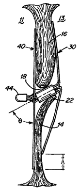
is positioned at an angle 8, as shown in Figure 6. Often, anatomical anomalies
have non-
perpendicular apertures and are sometimes quite significantly non-
perpendicular. Thus,
the occluder 20 may include an angled intermediate joint 22, such that the
angle of the
anatomical aperture is more closely matched by the pre-formed angle 8 of the
occluder
20. Accordingly, the distal 30 and proximal 40 sides of occluder 20 are more
likely to be
seated against and minimize distortion to the septal tissue 12 surrounding the
passage 18.
A well-seated occluder 20 is less likely to permit blood leakage between the
right 11 and
left 13 atria, and the subject into which the occluder 20 has been placed is,
therefore, less
likely to suffer embolisms and other adverse events. Advantageously, angled
CA 02488337 2004-12-03
WO 03/103476 PCT/US03/17715
1S
intermediate joint 22 also facilitates delivery of occluder 20, as described
in more detail
below, because it is angled toward the end of the delivery catheter. In at
least some
embodiments, the angle 8 is about 0-45 degrees off the plane created by the
proximal side
40. One skilled in the art will recognize that the concept of an angled
intermediate joint
may also be applied to septal occluders other than those disclosed herein.
[0050] When intermediate joint 22 is positioned at angle A, distal side 30 and
proximal side 40 of occluder 20 may be configured such that they are either
directly
opposing or, as shown in Figures 6A and 6B, offset by distance A. One skilled
in the art
will, of course, recognize that the configuration of either or both of distal
side 30 and
proximal side 40 may be adjusted such that the compressive forces applied by
the distal
30 and proximal 40 sides of occluder 20 are as directly opposing as possible.
However,
in some clinical applications, an occluder 20 having an offset of distance A
may be
particularly desirable. For example, as shown in Figure 7, if the septal
tissue 12
surrounding passage 18 includes a disproportionately thick portion (e.g.
septum
secundum 16 as compared to septum primum 14), the offset may be used to seat
occluder
20 more securely upon septal tissue 12. Moreover, the offset A allows each of
sides 30
and 40 to be centered around each side of an asymmetric defect.
[0051] When an intermediate joint 22 at angle 8 is included in occluder 20, a
marker is required to properly orient the occluder 20 in its intended in vivo
delivery
location. For example, platinum wire may be wrapped around one of loops 32 or
42 so as
to permit visualization of the orientation of the occluder 20 using
fluoroscopy.
Alternatively, other types of markers may be used, e.g. coatings, clips, etc.
As will be
readily understood by one skilled in the art, the orientation of a non-
symmetrical occluder
CA 02488337 2004-12-03
WO 03/103476 PCT/US03/17715
16
20 during delivery is of great importance. Of course, when a non-symmetrical
occluder
20 is used, the periphery of the occluder 20 may be configured such that the
clamping
force applied by the proximal side 40 is directly opposed to that applied by
the distal side
30.
[0052] Upon deployment in vivo (a process described in detail below), an
occluder according to the present invention applies a compressive force to the
overlapping layers of septal tissue 12, i.e. septum primum 14 and septum
secundum 16.
Distal side 30 is seated against the septal tissue 12 in the left atrium 13;
joint 22 extends
through passage 18; and proximal side 40 is seated against the septal tissue
12 in the right
atrium 11. As illustrated in Figures 2, 5, and 7, the proximal 40 and distal
30 sides of
occluder 20 overlap significantly, such that septum primum 14 and septum
secundum 16
are "sandwiched" between them once the occluder 20 is deployed. The connected,
adjacent segments provide a radially-extending compressive force, while the
peripheral
loops 32 and 42 provide a circumferential compressive force. Thus, the
compressive
forces are more evenly and more widely distributed across the surface of the
septal tissue
12 surrounding the PFO. The unique combination of radially-extending,
connected,
adjacent segments and peripheral loops 32 and 42, therefore, provides the
occluder 20
with superior dislodgement resistance as compared to prior art devices. As
used herein,
"dislodgement resistance" refers to the ability of an occluder 20 to resist
the tendency of
the force applied by the unequal pressures between the right 11 and left 13
atria (i. e. the
"dislodging force") to separate the occluder 20 from the septal tissue 12.
Generally, a
high dislodgement resistance is desirable.
CA 02488337 2004-12-03
WO 03/103476 PCT/US03/17715
17
[0053] Moreover, loops 32 and 42 are configured to provide occluder 20 with
adequate surface area to seal the PFO. For example, the broad configuration of
loops 32
and 42 increases the surface area of occluder 20. Thus, loops 32 and 42
provide sealing
along a large circumference around the passage 18 (i.e. the PFO), thereby
minimizing the
possibility of leakage between the right 11 and left 13 atria.
[0054] While configured to provide sufficient circumferential sealing, loops
32
and 42 are also configured to minimize the trauma they inflict on the septal
tissue 12
surrounding the PFO. Specifically, two features of loops 32 and 42 achieve
this. First,
the peripheries of loops 32 and 42 may be rounded. Second, the peripheries of
loops 32
and 42 are formed of a single wire and are, therefore, more flexible than the
interiorly-
located, connected, adjacent segments, which are formed of two wires. These
features
minimize the overall trauma inflicted by occluder 20 on the septal tissue 12
surrounding
the PFO. Accordingly, occluder 20 has a low compression resistance. As used
herein,
"compression resistance" refers to the ability of an occluder 20 to resist the
lateral
compressive force applied by the heart as it contracts during a heartbeat.
Generally, an
occluder that resists compressive force, i.e. has high compression resistance,
is
undesirable because its rigid configuration may cause trauma to the septal
tissue 12, the
right atrium 11, and/or the left atrium 13.
[0055] In heretofore known occluder designs, dislodgement resistance must
usually be sacrificed in order to improve, i.e. minimize, compression
resistance.
However, the occluder 20 according to the present invention possesses both
increased
dislodgement resistance and minimized compression resistance. These desirable
attributes are achieved by the unique combination of radially-extending,
connected,
CA 02488337 2004-12-03
WO 03/103476 PCT/US03/17715
18
adjacent segments and peripheral loops 32 and 42 discussed above. The radially-
extending, connected, adjacent segments (i.e. struts) increase the stiffness
and,
correspondingly, the dislodgment resistance of the occluder 20. The atraumatic
shape of
the peripheral loops 32 and 42 decreases the compression resistance of the
occluder 20.
In effect, because the struts are formed of double-stranded wire and the
peripheries of the
loops 32 and 42 are formed of single-stranded wire, the center of the occluder
20 is twice
as strong as its parameter. This, correspondingly, produces the advantageous
combination of increased dislodgement resistance and minimized compression
resistance
in occluder 20.
[0056] The dislodgement resistance of occluder 20 may be further increased
without increasing the compression resistance by the inclusion of additional
struts. As
illustrated in Figure 8, additional struts 85a-85c, collectively referred to
as additional
struts 85, may be included between loops 32a-32c, i.e. between adjacent
segments 33a
and 31b, 33b and 31c, and 33c and 31a. Additional struts 85 may be of any
suitable
diameter, and, according to some embodiments, the diameter of additional
struts 85 may
vary along their length. For example, the diameter of additional struts 85 may
increase as
the additional struts 85 extend from intermediate joint 22 to the periphery of
loops 32.
Although Figure 8 depicts additional struts 85 between loops 32 of distal side
30,
additional struts 85 may additionally or alternatively be included between
loops 42 of
proximal side 40 of occluder 20.
[0057] The configuration of the occluder 20 according to the present invention
provides several further advantages. First, broad loops 32 and 42 create a
large surface
area for occluder 20 and thereby anchor the occluder 20 more securely in vivo.
In
CA 02488337 2004-12-03
WO 03/103476 PCT/US03/17715
19
contrast, many previously known occluders include narrow loops, which afford
less
surface area for exertion of compressive forces and secure placement of the
occluder 20.
Second, the loops 32 and 42 create an occlusion perimeter that likely extends
significantly
beyond the passage 18. Third, loops 32 and 42 are non-overlapping, i.e. the
wires are not
intertwined or weaved. This non-overlapping configuration reduces the
occurrence of
fretting corrosion, which frequently occurs in prior art devices containing
overlapping
wires.
[0058] Occluder 20 may be modified in various ways. According to some
embodiments of the present invention, loops 32 of distal side 30 and loops 42
of proximal
side 40 may be formed in a variety of shapes. Four examples are illustrated in
Figures
l0A-IOD. For convenience, only the proximal side 40 of each of these modified
embodiments is depicted. However, the distal side 30 of occluder 20 may be
similarly
modified. The star-shaped pattern 100a shown in Figure l0A includes four large
loops,
referred to collectively as loops 102a. Loops 102a are centered and
approximately
equally spaced around tip 44. Any or all of loops 102a may include a smaller
loop,
collectively referred to as loops 104a, at their radial extent. Smaller loops
104a may be
capable of receiving a suture to facilitate retrieval of the occluder 20.
[0059] An alternative, diamond pattern 100b is shown in Figure IOB. Diamond
pattern 100b includes six diamond-shaped loops, referred to collectively as
loops 102b,
which are equally spaced around tip 44. Diamond pattern 100b is asymmetrically
oriented, such that two of loops 102b extend further in the radial direction
than the other
loops 102b. This asymmetry may provide more complete and secure coverage of
passage
CA 02488337 2004-12-03
WO 03/103476 PCT/US03/17715
18 than that provided by a symmetric occluder 20. The asymmetric pattern 100b
may
also facilitate the compact, percutaneous delivery of occluder 20.
[0060] Still a further alternative, rectangular pattern 100c, is shown in
Figure IOC.
Rectangular pattern 100c includes four rectangular-shaped loops, referred to
collectively
as loops 102c, which are equally spaced around tip 44. Rectangular pattern
100c provides
extended coverage in two directions. Such a rectangular shape may be
particularly suited
for coverage of certain passages 18. Loops 102c may extend further in either
the
horizontal or vertical direction. As shown in Figure lOC, loops 102c extend
further in the
horizontal direction.
[0061] Yet a further alternative, diamond pattern 100d, is shown in Figure
lOD.
Diamond pattern 100d includes four diamond-shaped loops, referred to
collectively as
loops 102d. Two of loops 102d are larger than the other two loops 102d. Thus,
an
extended amount of coverage may be provided across the passage 18 in either
the
horizontal or vertical direction. As shown in Figure lOD, extended coverage is
provided
in the horizontal direction.
[0062] Of course, distal 30 and proximal 40 sides of occluder 20 may be
configured in a combination of shapes and sizes depending on clinical needs
presented by
a given PFO. If required, the loops 102 in the illustrative patterns provided
in Figures
l0A-IOD, may be rounded. The number of loops in embodiments of either the
distal 30 or
proximal 40 sides may be varied as necessary. As previously described, loops
102 in the
illustrative patterns provided in Figures l0A-lOD include adjacent segments,
which may
be connected by, e.g., welds 108a-108d, respectively. One skilled in the art
will be able
to identify the configurations) appropriate for a given clinical application.
CA 02488337 2004-12-03
WO 03/103476 PCT/US03/17715
21
[0063] According to further embodiments of the present invention, smaller
loops
may be included on distal side 30 and/or proximal side 40 of occluder 20 to
increase the
compressive force applied in close proximity to passage 18 (i.e. the PFO). As
illustrated
in Figure 11, three smaller loops 115a-115c, referred to collectively as
smaller loops 115,
are located on distal side 30. Smaller loops 115 are centered and equally
spaced around
intermediate joint 22. Although smaller loops 115x-115c in Figure 11
correspond in
number and alignment with loops 32a-32c, respectively, such correspondence is
not
required. Moreover, smaller loops 115 need not lie entirely in the same plane
as loops 32
or 42. Thus, smaller loops 115 may bend in a direction generally perpendicular
to the
plane in which loops 32 or 42 lie. Smaller loops 115 may be attached only to
intermediate joint 22 or, alternatively, may also be connected to the adjacent
segments of
loops 32. In still other embodiments, smaller loops 115 may be located at the
peripheries
of loops 32 rather than connected to intermediate joint 22. When the smaller
loops 115
are located at the peripheries of loops 32, additional wire segments may be
included
within loops 32 to connect the smaller loops 115 to the intermediate joint 22.
One skilled
in the art will be able to determine the precise configuration of smaller
loops 115
appropriate for a given clinical application.
[0064] According to still further embodiments of the present invention and as
illustrated in Figure 12, distal side 30 and/or proximal 40 side of occluder
20 may include
a tissue scaffold 125. Tissue scaffold 125 ensures more complete coverage of
passage 18
and promotes encapsulation and endothelialization of septal tissue 12, thereby
further
encouraging anatomical closure of septum primum 14 and septum secundum 16.
Tissue
scaffold 125 may be formed of any flexible, biocompatible material capable of
promoting
CA 02488337 2004-12-03
WO 03/103476 PCT/US03/17715
22
tissue growth, including but not limited to polyester fabrics, Teflon-based
materials,
ePTFE, polyurethanes, metallic materials, polyvinyl alcohol (PVA),
extracellular matrix
(ECM) or other bioengineered material, synthetic bioabsorbable polymeric
scaffolds,
other natural materials (e.g. collagen), or combinations of the foregoing
materials. For
example, tissue scaffold 125 may be formed of a thin metallic film or foil,
e.g. a nitinol
film or foil, as described in United States Patent Appln. No. 2003/0059640
(the entirety
of which is incorporated herein by reference).
[0065] Adjacent segments may be stitched to tissue scaffold 125 so as to
securely
fasten the scaffold 125 to occluder 20. For example, Figure 12 shows tissue
scaffold 125
affixed to proximal side 40 of an occluder according to the present invention.
Proximal
side 40 includes six loops 42a-42f, collectively referred to as loops 42.
Adjacent
segments 43a and 41b, 43b and 41c, 43c and 41d, 43d and 41e, 43e and 41f, and
43f and
41a are attached to tissue scaffold 125 by stitches 127. Stitches 127 increase
the stiffness
of occluder 20 without welding or soldering. Additionally, when the adjacent
segments
of loops 42 are connected to tissue scaffold 125, the adjacent segments of
loops 42 may
be spaced apart a small distance (i.e. they need not necessarily be
connected). Altering
the spacing of the adjacent segments of loops 42 adjusts the stiffness of the
occluder 20,
which may be desirable in certain circumstances. One skilled in the art will
be able to
determine those clinical applications in which the use of stitches 127 and/or
spaced,
adjacent segments is appropriate.
[0066] According to yet further embodiments of the present invention, the
configuration of occluder 20 may be modified to produce the low-profile
occluder 130
shown in Figure 13A. In this embodiment, the manufacturing process is modified
to
CA 02488337 2004-12-03
WO 03/103476 PCT/US03/17715
23
increase the force with which the distal 30 and proximal 40 sides urge toward
one
another. Specifically, during manufacture, distal 30 and proximal 40 sides of
occluder 20
may be crossed over each other (as shown in Figure 13B) prior to connecting
the adjacent
segments of loops 32 and 42 (i.e. while the occluder 20 is in an
"unconstrained" state).
This crossed-over configuration may be achieved by, for example, using the
shape
memory properties of a shape memory material, such as nitinol, i.e. forcing,
e.g., loops
42d and 42e of proximal side 40 through loop 32c of distal side 30 or vice
versa and heat-
setting the crossed-over shape. The crossed-over shape, therefore, becomes the
predisposed position of occluder 20. Occluder 20 is then returned to its
original, non-
crossed-over state, and the adjacent segments of loops 32 and 42 are
connected. The
connected, adjacent segments prevent loops 42d and 42e from passing through
loop 32c,
and occluder 20 is, consequently, no longer capable of assuming its
predisposed position.
However, loops 42d and 42e of proximal side 42 still tend to bend toward
distal side 30.
The resulting occluder 130, shown in Figure 13A, is of low profile. Further,
occluder 130
exerts a greater compressive force on the septal tissue 12 when deployed in
vivo (as
shown in Figure 13C) then at least some of the previously-described
embodiments of
occluder 20. This increased compressive force may be desirable in applications
where the
septal tissue 12 is particularly thin in one area, i.e. septum primum 14. The
profile of
occluder 130 may be lowered even further by angling tip 44 such that it is
substantially
parallel to proximal side 40 of occluder 130, as shown in Figure 13A. Angled
tip 44 also
facilitates catheter delivery of occluder 130 because angled tip 44 points
toward the end
of the delivery catheter.
CA 02488337 2004-12-03
WO 03/103476 PCT/US03/17715
24
[0067] Finally, although occluders according to the present invention have
been
heretofore described as including distal 30 and proximal 40 sides having
different
configurations, an occluder 20 according to the present invention may,
alternatively,
include distal 30 and proximal 40 sides having identical configurations. This
identical
design may provide several advantages, including ease of manufacture.
Furthermore, any
of the configurations described herein for either distal side 30 or proximal
side 40 may be
applied to either or both of distal side 30 and proximal side 40 of occluder
20.
[0068] An occluder as described herein may be delivered to a septal defect
using
any of several suitable delivery techniques, two of which will be described
herein. In the
first delivery technique, shown in Figures 14A-14E, a delivery catheter 140 is
used to
deliver, e.g., occluder 20. Catheter 140 contains occluder 20 in its
distorted, elongated
form. As previously mentioned, in at least some embodiments, occluder 20 is
formed of a
shape memory material, e.g. nitinol, such that occluder 20 will resume its
intended shape
following deployment in vivo. As shown in Figure 14A, delivery catheter 140 is
first
inserted into the right atrium 11 of the subject's heart. Catheter 140 is next
inserted
between septum primum 14 and septum secundum 16 (i.e. through passage 18,
which, in
this embodiment, is the PFO tunnel) and into the left atrium 13 (Figure 14B).
Distal side
30 of occluder 20 is then deployed into the left atrium 13, as shown in Figure
14C.
Following deployment of distal side 30, the catheter 140 is withdrawn through
the PFO
tunnel and into the right atrium 11, such that intermediate joint 22 is
deployed through the
PFO tunnel (Figure 14D). Finally, proximal side 40 of occluder 20 is deployed
into the
right atrium 11, and catheter 140 is withdrawn from the heart (Figure 14E).
Once
deployed, occluder 20 rests within the septal defect, and the distal 30 and
proximal 40
CA 02488337 2004-12-03
WO 03/103476 PCT/US03/17715
sides exert a compressive force against septum primum 14 and septum secundum
16 in
the left 13 and right 11 atria, respectively, to close the PFO.
[0069] In a second delivery technique, shown in Figures 15A-15E, delivery
catheter 150 includes a needle 151 capable of puncturing septum primum 14. As
illustrated in Figure 15A, septum primum 14 is long and thin and extends over
septum
secundum 16 in the left atrium 13. In some clinical applications, it may be
advantageous
to access the left atrium 13 by puncturing septum primum 14 rather than
inserting the
occluder 20 through the passage 18 between septum primum 14 and septum
secundum
16. For example, some anatomical configurations include an extremely oblique
passage
18 between the right atrium 11 and the left atrium 13. Thus, according to this
second
delivery technique, delivery catheter 150 includes a needle 151 on its distal
end and
contains occluder 20 in its distorted, elongated form. Catheter 150 is first
inserted into
the right atrium 11 of the subject's heart (Figure 15A). Next, as shown in
Figure 15B,
needle 151 punctures septum primum 14, and catheter 150 enters the left atrium
13.
Needle 151 is then retracted, and distal side 30 of occluder 20 is deployed
into the left
atrium 13 (Figure 15C). Following deployment of distal side 30, catheter 150
is
withdrawn through septum primum 14 and into the right atrium 11, such that
intermediate
joint 22 is deployed through septum primum 14, as shown in Figure 15D.
Finally,
proximal side 40 of occluder 20 is deployed into the right atrium 11, and
catheter 150 is
withdrawn from the heart (Figure 15E). Once deployed, the distal 30 and
proximal 40
sides of occluder 20 exert a compressive force against septum primum 14 and
septum
secundum 16 in the left 13 and right 11 atria, respectively, to close the PFO.
When using
this second delivery technique to deploy occluder 20, intermediate joint 22
should not be
CA 02488337 2004-12-03
WO 03/103476 PCT/US03/17715
26
angled, i.e. intermediate joint 22 should be perpendicular to both the distal
30 and
proximal 40 sides of the occluder 20.
[0070] Figure 16 provides a more detailed representation of occluder 20 in its
intermediate configuration between its compressed and fully-deployed states.
As
previously described, proximal side 40 of occluder 20 includes wires) 25,
which form
connected, adjacent radially-extending segments and loops 42, and tip 44.
During
delivery of occluder 20, tip 44 is attached to a delivery wire 161, in a
manner known to
those skilled in the art. When the proximal side 40 of occluder 20 is being
deployed in
the right atrium 11, the wires) 25 exit catheter 140 or 150 first, followed by
tip 44, and,
finally, delivery wire 161. Once occluder 20 has been positioned, delivery
wire 161 is
then fully retracted into the catheter 140 or 150 and the catheter is
retracted out of the
right atrium 11.
[0071] Delivery wire 161 may be used to reposition and/or retrieve occluder 20
as
shown in Figures 17A-17D. If, following partial or complete deployment, the
clinician
desires to reposition or retrieve occluder 20, tip 44 may be recaptured with
delivery wire
161 in catheter 170, as shown in Figure 17A. As delivery wire 161 and tip 44
are pulled
back into catheter 170, loops 42 of proximal side 40 fold back into their
delivery (i.e.
compressed) configuration (Figure 17B) and are constrained by catheter 170.
Catheter
170 is then advanced through passage 18 and delivery wire 161 is further
retracted, such
that loops 32 of distal side 30 fold into their delivery configuration (Figure
17C) and are
constrained by catheter 170. Catheter 170 containing retrieved occluder 20 is
then
withdrawn through passage 18, into the right atrium 11 (Figure 17D), and out
of the heart.
CA 02488337 2004-12-03
WO 03/103476 PCT/US03/17715
27
[0072] In some embodiments according to the present invention, occluder 20 may
be repositioned and/or retrieved using the alternative technique shown in
Figure 18. As
previously described, an occluder 20 according to the present invention may
include
identical distal 30 and proximal 40 sides. Thus, for example, occluder 20 may
include
both distal 30 and proximal 40 sides as depicted in Figure 3. In such an
embodiment,
proximal side 40 will not include a tip 44 for recovery by a delivery wire. An
alternative
method of retrieving the occluder is, therefore, required. In Figure 18,
occluder 20 has
been delivered (according to either of the delivery techniques described
above) to the
extent that proximal side 40 has been deployed in the right atrium 11 but not
released
from catheter 140. A thread 181, such as a suture, is attached to each of
loops 42 on
proximal side 40 of occluder 20. If the occluder 20 requires repositioning,
then thread
181 may be retracted and loops 42 will fold back into their delivery
configuration, such
that occluder 20 may be repositioned or, even, completely retrieved. Once
occluder 20
has been deployed correctly, thread 181 may be cut and removed via catheter
140.
[0073] One skilled in the art would recognize that the occluders described
herein
may be used with anti-thrombogenic compounds, including but not limited to
heparin and
peptides, to reduce thrombogenicity of the occluder and/or to enhance the
healing
response of the septal tissue 12 following deployment of the occluder in vivo.
Similarly,
the occluders described herein may be used to deliver other drugs or
pharmaceutical
agents (e.g. growth factors, peptides). The anti-thrombogenic compounds,
drugs, and/or
pharmaceutical agents may be included in the occluders of the present
invention in
several ways, including by incorporation into the tissue scaffold 125, as
previously
described, or as a coating, e.g. a polymeric coating, on the wires) forming
the distal 30
CA 02488337 2004-12-03
WO 03/103476 PCT/US03/17715
28
and proximal 40 sides of the occluder. Furthermore, the occluders described
herein may
include cells that have been seeded within tissue scaffold 125 or coated upon
the wires)
forming the distal 30 and proximal 40 sides of the occluder.
[0074] One skilled in the art would recognize that occluders according to this
invention could be used in occluding other vascular and non-vascular openings.
For
example, the device could be inserted into a left atrial appendage or other
tunnels or
tubular openings within the body.
[0075] Having described preferred embodiments of the invention, it should be
apparent that various modifications may be made without departing from the
spirit and
scope of the invention, which is defined in the claims below.