Note: Descriptions are shown in the official language in which they were submitted.
WO 2004/028347 CA 02499976 2005-03-23 PCT/US2003/030383
Title of the Invention
[0001] Implantable Materials Having Engineered Surfaces and Method of Making
Same
Background of the Invention
[0002] The present invention relates generally to implantable medical devices
and
more particularly to controlling surface properties of implantable bio
compatible materials
suitable for fabrication of implantable medical devices. Implantable medical
devices are
fabricated of materials that are sub-optimal in terms of the biological
response they elicit in
vivo. Many conventional materials used to fabricate implantable devices, such
as titanium,
polytetrafluoroethylene, silicone, carbon fiber and polyester, are used
because of their
strength and physiologically inert characteristics. However, tissue
integration onto these
materials is typically slow and inadequate. Certain materials, such as
silicone and polyester,
elicit a significant inflammatory, foreign body response that drives fibrous
encapsulation of
the synthetic material. The fibrous encapsulation may have significant adverse
effects on the
implant. Moreover, conventional biomaterials have proved inadequate in
eliciting a
sufficient healing response necessary for complete device integration into the
body. For
example, in devices that contact blood, such as stents and vascular grafts,
attempts to modify
such devices to promote endothelial cell adhesion may have a concomitant
effect of making
the devices more thrombogenic.
[0003] When implanted, conventional blood-contacting implantable devices, such
as
stents, stent-grafts, grafts, valves, shunts and patches, fail to develop a
complete endothelial
layer, thereby exposing the device material to thrombus formation or smooth
muscle cell
proliferation, and ultimate failure of the implanted device. It has been
recognized that, when
implanted into the body, metals are generally considered to have superior
biocompatibility
than polymers used to fabricate commercially available polymeric grafts.
[0004] In investigating cellular interactions with prosthetic material
surfaces, it has
been found that cell adhesion to the material surface is mediated by integrins
present on cell
membranes that interact with the prosthetic surface. Integrins are the most
prominent
member of a class of extracellular matrix (ECM) adhesion receptors. Integrins
are a large
family of heterodimeric transmembrane proteins with different a and 13
subunits. Integrins are
regulated at several levels. Modulation of the affinity of the adhesion
receptor for ligand,
WO 2004/028347 CA 02499976 2005-03-23 PCT/US2003/030383
termed affinity modulation, is a mechanism for activation of platelet
aggregation and is
believed to underlie activation of leukocyte adhesion. Adhesive strengthening
by clustering
of adhesion receptors or by cytoskeletal-dependent processes such as cell
spreading has been
shown to be crucial for strong cellular attachment, control of cell growth and
cell motility.
Under high shear forces present in flowing blood, leukocytes first tether,
then roll along the
vessel surface. When a local signal, e.g., a cytokine, is released in their
vicinity, the
leukocyte arrests, develops a firm adhesion then migrates across the
endothelium. Tethering,
rolling, arrest and adhesion tightening are all known to result from
activation of leukocyte
integrins.
[0005] Once adhered to a surface, cell spreading and migration are associated
with
assembly of focal adhesion junctions. Cell migration entails the coordination
of cytoskeletal-
mediated process extension, i.e., filopodia and lamellopodia, formation of
adhesive contacts
at the leading edge of a cell, breaking adhesive contacts, and cytoskeletal
retraction at the
trailing edge of the cell. Focal adhesions are comprised of integrins as the
major adhesion
receptors along with associated cytoplasmic plaque proteins. Assembly of focal
adhesions is
regulated by extracellular ligand binding events and by intracellular
signaling events. Ligand
binding controls localization of (31- and 133-containing integrins into focal
adhesions. The
cytoplasmic domains of the 13 subunits have intrinsic signals for focal
adhesion localization,
but incorporation of the integrins into focal adhesions is prevented by the a
subunits of the
heterodimers. Ligand binding, however, relieves this inhibition and allows the
subunit
cytoplasmic tail signals to recruit the integrin dimmer into the focal
adhesion.
[0006] Attempts at coating implanted metal devices, such as stents, with
proteins that
contain the Arg-Gly-Asp (RGD) attachment site have been made with some
success. The
RGD sequence is the cell attachment site of a large number of adhesive
extracellular matrix,
blood, and cell surface proteins and many of the known integrins recognize the
RGD
sequence in their adhesion protein ligands. Integrin-binding activity may also
be reproduced
by synthetic peptides containing the RGD sequence. However, bare metal
implanted
materials will not, of course, have native RGD attachment sites. Thus, metal
implantable
devices, such as stents, have been derivitized with polymers having RGD
attachment sites
bound to the polymer matrix.
10007] It has been found that when prosthetic materials are implanted,
integrin
receptors on cell surfaces interact with the prosthetic surface. When cells
come into contact
with the extracellular matrix, such as a prosthetic surface, their usual
response is to extend
filopodia, and integrins at the tip of the filopodia bind to the extracellular
matrix and initiate
-2-
CA 02499976 2013-03-22
WO 2004/028347 PCT/1JS2003/030383
the formation of focal adhesions. Actin-rich lamellipodia are generated, often
between
filopodia, as the cell spreads on the extracellular matrix. Fully developed
focal adhesions and
associated actin stress fibers ensue. These same events occur during cell
migration as cells
extend lamellipodia and form focal adhesions to derive the traction necessary
for movement.
Giancotti, E.G., et al. Science, 285:13 August 1999, 1028-1032.
[0008] The integrin receptors are specific for certain ligands in vivo. If a
specific
protein is adsorbed on a prosthetic surface and the ligand exposed, cellular
binding to the
prosthetic surface may occur by integrin-ligand docking. It has also been
observed that
proteins bind to metals in a more permanent fashion than they do to polymers,
thereby
providing a more stable adhesive surface. The conformation of proteins coupled
to surfaces
of most medical metals and alloys appears to expose greater numbers of ligands
and attract
endothelial cells having surface integrin clusters to the metal or alloy
surface, preferentially
over leukocytes.
[0009] Because of their greater adhesive surface profiles, metals are also
susceptible
to short-term platelet activity and/or thrombogenicity. These deleterious
properties may be
offset by administration of pharmacologically active antithrombogenic agents
in routine use
today. Surface thrombogenicity usually disappears 1-3 weeks after initial
exposure.
Antithrombotic coverage is routinely provided during this period of time for
coronary
stenting. In non-vascular applications such as musculoskeletal and dental,
metals have also
greater tissue compatibility than polymers because of similar molecular
considerations. The
best article to demonstrate the fact that all polymers are inferior to metals
is van der Giessen,
WI. et al. Marked inflammatory sequelae to implantation of biodegradable and
non-
biodegradable polymers in porcine coronary arteries, Circulation,
1996:94(7):1690-7.
[0010] Normally, endothelial cells (EC) migrate and proliferate to cover
denuded
areas until confluence is achieved. Migration, quantitatively more important
than
proliferation, proceeds under normal blood flow roughly at a rate of 25 pm/hr
or about 2.5
times the diameter of an EC, which is nominally 10um. EC migrate by a rolling
motion of
the cell membrane, coordinated by a complex system of intracellular filaments
attached to
clusters of cell membrane integrin receptors, specifically focal contact
points. The integrins
within the focal contact sites are expressed according to complex signaling
mechanisms and
eventually couple to specific amino acid sequences in substrate adhesion
molecules. An EC
has roughly 16-22% of its cell surface represented by integrin clusters.
Davies, P.F.,
Robotewskyi A., Griem M.L. Endothelial cell adhesion in real time.
J.Clin.Invest.1993;
91:2640-2652, Davies, P.F., Robotewski, A., Griem, M.L., Qualitiative studies
of endothelial
-3-
WO 2004/028347 CA 02499976 2005-03-23 PCT/US2003/030383
cell adhesion, J.Clin.Invest.1994; 93:2031-2038. This is a dynamic process,
which involves
more than 50% remodeling in 30 minutes. The focal adhesion contacts vary in
size and
distribution, but 80% of them measure less than 6 ptm2, with the majority of
them being about
1 ium2, and tend to elongate in the direction of flow and concentrate at
leading edges of the
cell. Although the process of recognition and signaling to determine specific
attachment
receptor response to attachment sites is not completely understood,
availability of attachment
sites will favorably influence attachment and migration. It is known that
materials
commonly used as medical grafts, such as polymers, do not become covered with
EC and
therefore do not heal after they are placed in the arteries. It is therefore
an object of this
invention to replace polymer grafts with metal grafts that can potentially
become covered
with EC and can heal completely. Furthermore, heterogeneities of materials in
contact with
blood flow are preferably controlled by using vacuum deposited materials.
[0011] There have been numerous attempts to increase endothelialization of
implanted medical devices such as stents, including covering the stent with a
polymeric
material (U.S. Patent No. 5,897,911), imparting a diamond-like carbon coating
onto the stent
(U.S. Patent No. 5,725,573), covalently binding hydrophobic moieties to a
heparin molecule
(U.S. Patent No. 5,955,588), coating a stent with a layer of blue to black
zirconium oxide or
zirconium nitride (U.S. Patent No. 5,649,951), coating a stent with a layer of
turbostratic
carbon (U.S. Patent No. 5,387,247), coating the tissue-contacting surface of a
stent with a
thin layer of a Group VB metal (U.S. Patent No. 5,607,463), imparting a porous
coating of
titanium or of a titanium alloy, such as Ti-Nb-Zr alloy, onto the surface of a
stent (U.S. Patent
No. 5,690,670), coating the stent, under ultrasonic conditions, with a
synthetic or biological,
active or inactive agent, such as heparin, endothelium derived growth factor,
vascular growth
factors, silicone, polyurethane, or polytetrafluoroethylene (U.S. Patent No.
5,891,507),
coating a stent with a silane compound with vinyl functionality, then forming
a graft polymer
by polymerization with the vinyl groups of the silane compound (U.S. Patent
No. 5,782,908),
grafting monomers, oligomers or polymers onto the surface of a stent using
infrared
radiation, microwave radiation or high voltage polymerization to impart the
property of the
monomer, oligomer or polymer to the stent (U.S. Patent No. 5,932,299).
However, all these
approaches do not address the lack of endothelialization of polymer grafts.
[0012] In accordance with the present invention, the capacity for complete
endothelialization of conventional implantable materials, including metals and
polymers, may
be enhanced by imparting a pattern of chemically and/or physiochemically
active features
onto a blood contacting surface of the implantable material. The inventive
implantable metal
-4-
WO 2004/028347 CA 02499976 2005-03-23 PCT/US2003/030383
devices may be fabricated of polymers, pre-existing conventional wrought
metallic materials,
such as stainless steel or nitinol hypotubes, or may be fabricated by thin
film vacuum
deposition techniques. In accordance with the present invention, it is
preferable to fabricate
the inventive implantable materials and resulting devices by vacuum deposition
of either or
both of the base implant material and the chemically and/or physiochemically
active features.
Vacuum deposition permits greater control over many material characteristics
and properties
of the resulting material and formed device. For example, vacuum deposition
permits control
over grain size, grain phase, grain material composition, bulk material
composition, surface
topography, mechanical properties, such as transition temperatures in the case
of a shape
memory alloy. Moreover, vacuum deposition processes will permit creation of
devices with
greater material purity without the introduction of large quantities of
contaminants that
adversely affect the material and, therefore, the mechanical and/or biological
properties of the
implanted device. Vacuum deposition techniques also lend themselves to
fabrication of more
complex devices than those that are manufactured by conventional cold-working
techniques.
For example, multi-layer structures, complex geometrical configurations,
extremely fine
control over material tolerances, such as thickness or surface uniformity, are
all advantages of
vacuum deposition processing.
[0013] In vacuum deposition technologies, materials are formed directly in the
desired geometry, e.g., planar, tubular, etc. The common principle of vacuum
deposition
processes is to take a material in a minimally processed form, such as pellets
or thick foils,
known as the source material and atomize them. Atomization may be carried out
using heat,
as is the case in physical vapor deposition, or using the effect of
collisional processes, as in
the case of sputter deposition, for example. In some forms of deposition a
process such as
laser ablation, which creates microparticles that typically consist of one or
more atoms, may
replace atomization; the number of atoms per particle may be in the thousands
or more. The
atoms or particles of the source material are then deposited on a substrate or
mandrel to
directly form the desired object. In other deposition methodologies, chemical
reactions
between ambient gas introduced into the vacuum chamber, i.e., the gas source,
and the
deposited atoms and/or particles are part of the deposition process. The
deposited material
includes compound species that are formed due to the reaction of the solid
source and the gas
source, such as in the case of chemical vapor deposition. In most cases, the
deposited
material is then either partially or completely removed from the substrate, to
form the desired
product.
-5-
CA 02499976 2005-03-23
WO 2004/028347 PCT/US2003/030383
[0014] A first advantage of vacuum deposition processing is that vacuum
deposition
of the metallic and/or pseudometallic films permits tight process control and
films may be
deposited that have a regular, homogeneous atomic and molecular pattern of
distribution
along their fluid-contacting surfaces. This avoids the marked variations in
surface
composition, creating predictable oxidation and organic adsorption patterns
and has
predictable interactions with water, electrolytes, proteins and cells. In
particular, EC
migration is supported by a homogeneous distribution of binding domains that
serve as
natural or implanted cell attachment sites in order to promote unimpeded
migration and
attachment.
[0015] Secondly, in addition to materials and devices that are made of a
single metal
or metal alloy layer, the inventive grafts may be comprised of a layer of
biocompatible
material or of a plurality of layers of bio compatible materials formed upon
one another into a
self-supporting multilayer structure because multilayer structures are
generally known to
increase the mechanical strength of sheet materials, or to provide special
qualities by
including layers that have special properties such as superelasticity, shape
memory, radio-
opacity, corrosion resistance etc. A special advantage of vacuum deposition
technologies is
that it is possible to deposit layered materials and thus films possessing
exceptional qualities
may be produced (cf., H. Holleck, V. Schier: Multilayer PVD coatings for wear
protection,
Surface and Coatings Technology, Vol. 76-77 (1995) pp. 328-336). Layered
materials, such
as superstructures or multilayers, are commonly deposited to take advantage of
some
chemical, electronic, or optical property of the material as a coating; a
common example is an
antireflective coating on an optical lens. Multilayers are also used in the
field of thin film
fabrication to increase the mechanical properties of the thin film,
specifically hardness and
toughness.
[0016] Thirdly, the design possibilities for possible configurations and
applications of
the inventive graft are greatly realized by employing vacuum deposition
technologies.
Specifically, vacuum deposition is an additive technique that lends itself
toward fabrication
of substantially uniformly thin materials with potentially complex three
dimensional
geometries and structures that cannot be cost-effectively achieved, or in some
cases achieved
at all, by employing conventional wrought fabrication techniques. Conventional
wrought
metal fabrication techniques may entail smelting, hot working, cold working,
heat treatment,
high temperature annealing, precipitation annealing, grinding, ablation, wet
etching, dry
etching, cutting and welding. All of these processing steps have disadvantages
including
contamination, material property degradation, ultimate achievable
configurations, dimensions
-6-
CA 02499976 2013-03-22
WO 2004/028347 PCT/US2003/030383
and tolerances, biocompatibility and cost. For example conventional wrought
processes are
not suitable for fabricating tubes having diameters greater than about 20mm,
nor are such
processes suitable for fabricating materials having wall thicknesses down to
about 1 um with
sub-am tolerances.
[0017] Overall rate to reach confluence for the endothelial cells on the blood
contact
surface of implanted medical device is mainly determined by two factors, the
rate of cell
movement and rate of cell proliferation, with the first being more important.
The rate of cell
movement further comprises three interrelated steps. Initially, a cell forms
lamellipodia and
filopodia that protrude outward. This step involves reassembly of actins in
the forefront of
lamellipodia. After protrusion of lamellipodia from one or multiple points
from the cell
membrane, the front end of the lamellipodia will form a close attachment,
called focal
adhesion point, to the substratum through the interaction of integrin on the
cell membrane
and extrcellular matrix binding site. The final step of cell movement involves
the contraction
of the posterior end through the action of myosin II. The formation of a focal
adhesion point
is critical for the cell movement because the protruding lamellipodia will
otherwise fold back.
Without the tension force from the focal adhesion point, a cell loses the
contraction from the
posterior end and hence stops moving.
[0018] Availability of attachment sites on the substratum is not only
important for the
focal adhesion point formation, but also important for propagation. It has
been shown that
cells are forced to spread, survive better and proliferate faster than cells
that are confined to
the same amount of surface area (Science 276:1425-1428, 1997). This may
explain why
spreading of neighbor cells stimulate a cell to proliferate, after cells are
lost from epithelium.
[0019] The formation of extracellular matrix (ECM) is, to much extent,
determined
by the cells within it because molecules which form ECM are secreted by the
cells.
Subsequently, the structure of the ECM, and hence the distribution of
attachment sites on the
ECM for the integrin binding, determines the focal adhesion point formation,
the critical step
in cell movement. Therefore, proper distribution of integrin binding sites on
the surface of an
implanted medical device substantially determines the speed of
reendothelialization from the
ends surrounding the device.
[0020] There still remains a need for a medical device that stimulates
endothelial
proliferation and movement when implanted in order to form an endothelial
layer over the
medical device. Furthermore, there is a remaining need for a method of
fabricating such a
medical device.
-7-
WO 2004/028347 CA 02499976 2005-03-23 PCT/US2003/030383
SUMMARY OF THE INVENTION
[0021] In accordance with an aspect of the present invention, there is
provided an
implantable material having at least one blood contact surface comprising an
evenly
distributed geometric feature for cell attachment. The evenly distributed
feature on the blood
contact surface of the medical device includes: circle dots, square dots,
rectangular dots,
triangle dots, parallel lines and intersecting lines, or any combination
thereof. Additionally,
another aspect of the present invention provides methods of making a device
that has evenly
distributed geometric features on the blood contact surface.
BRIEF DESCRIPTION OF THE FIGURES
[0022] FIG. 1 is a perspective view of an embodiment of the present invention
including evenly distributed elevated geometric features on the surface of an
implantable
material.
[0023] FIG. 2 is cross-sectional view of FIG. 1 along line 2-2.
[0024] FIG. 3 is a perspective view of an embodiment of the present invention
inculuding evenly distributed chemically defined geometric features on the
surface of an
implantable material.
[0025] FIG. 4 is a cross-sectional view of FIG. 3 along line 4-4.
[0026] FIG. 5 is a photomicrograph showing an embodiment of the present
invention
including geometric features as carbon coated silicon.
[0027] FIGS. 6a 6c are photomicrographs showing cellular migration on the
surface
with no inventive geometric features versus on the surface with inventive
features.
[0028] FIG. 7 is a photomicrograph showing the stained focal adhesion points
close
to the geometric features.
[0029] FIGS. 8a ¨ 8b are photomicrographs showing the formation of multiple
focal
adhesion points of a migrating cell and its attachment to the inventive
geometric features.
[0030] FIGS. 9a¨ 9d are cross-sectional diagrammatic views of an embodiment of
the present invention, the combination of a-d representing the steps to make
an inventive
implantable material with elevated geometric features.
[0031] FIGS. 10a ¨ 10d are cross-sectional diagrammatic views of an embodiment
of
the present invention, the combination of a-d representing the steps to make
an inventive
implantable material with chemically defined geometric features.
-8-
WO 2004/028347 CA 02499976 2005-03-23 PCT/US2003/030383
DETAILED DESCRIPTION OF THE PREFERRED EMBODIMENTS
[0032] The present inventions takes advantage of the discovered relationship
between
chemically or physiochemically-active geometric features defined and
distributed on a blood
contact surface enhanced endothelial cell binding, proliferation and migration
over the blood
contact surface of the implantable material. The present invention involves
focal adhesion
point formation during cellular movement and the well-established observation
known as
anchorage dependence, that spreading cells proliferate faster than non-
spreading cells. It has
been found the addition of a patterned array of ultra-thin features having a
hydrophobic,
hydrophilic or surface energy difference relative to the surface onto which
the ultra-thin
features are added, enhances the binding, proliferation and migration of
endothelial cells to
and between the features and across the surface. Use of the term "ultra-thin"
is intended to
include material thicknesses between about 0.1gm and 3p.m. It has been found
that below
about 3 gm the interactions between endothelial cells and the ultra-thin
features is primarily
chemical and electrochemical. Features having thicknesses greater than 3 gm
and up to about
20 gm may also be employed in the present invention, it being understood that
as the
thickness of the feature increases there is a decreasing chemical and/or
electrochemical
interaction between the feature and the endothelial cells and an increasing
physical
interaction.
[0033] Additionally, it has been found that by employing UV irradiation to
oxidized
titanium or titanium-alloy surfaces, photochemical alteration of the surface
titanium oxides
alter the hydrophobicity of the exposed titanium oxides and act as affinity
binding and
migration sites for endothelial cell attachment and proliferation across a
titanium or titanium-
alloy surface. Where UV irradiation is employed, the thickness of the
photochemically
altered regions of titanium oxide are, for all practical purposes, 0 p.m.
Thus, within the
context of the present application, the term "geometric features" is intended
to include both
physical members and photochemically-altered regions having thicknesses having
thicknesses down to 0 gm.
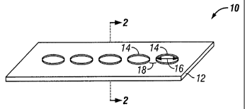
[00341 In FIG. 1, a portion of an implantable material 10 showing the surface
material
12 with described elevated geometric features 14 is illustrated. The geometric
features are
elevated from the surface of the implantable material to a height ranging from
about sub-
micron to about 20 gm. Preferably, the height of the geometric feature 14
ranges from about
sub-micron to about 3 gm. The shape of geometric features can be either
circular, square,
rectangle, triangle, parallel lines, straight or curvilinear lines or any
combination thereof.
-9-
CA 02499976 2013-03-22
WO 2004/028347 PCT/US2003/030383
Each of the geometric features is preferably from about 10 um to about 75 gm,
and
preferably from about 15 gm to 50 gm in feature width 16, or feature diameter
if the
geometric feature is circular. A gap distance 18 between each of the geometric
features
should generally be the same as the feature width 16, i.e., between about 10
um to about 75
um edge-to-edge.
[0035] FIG. 2 is a cross-sectional view along line 2-2 in FIG. 1. One of the
elevated
geometric features 14 is shown on the surface 12 of the implantable material.
[00361 In FIG. 3, a titanium or titanium-alloy material 20 is heated to
oxidize and
form titanium dioxide on the surface of the material 20, then features 24 are
formed by
exposing the material 20 to UV through a pattern mask. UV irradiation alters
the titanium
oxides in the areas of features 24, thereby chemically altering the geometric
features 24
relative to the surrounding the surrounding surface area 22 of material 20.
The shape of
geometric features can be circular, square, rectangle, triangle, parallel
lines, intersecting lines
or any combination. Each of the geometric features is from about 10 gm to
about 75 pm, and
preferably from about 15 um to 50 i.tm in feature width 26, or feature
diameter if the
geometric feature is circular. The gap distance 28 between each component of
the geometric
features is in the same magnitude as the feature width 26.
[00371 FIG. 4 is a cross-sectional view of FIG. 3 along line 4-4. The
described
geometric features 24 are indicated by the dotted lines, which indicates that
the geometric
features 24 are at the same level of the surrounding surface 22.
[0038] FIG. 5 shows geometric features that are evenly distributed across the
at least
one surface of the implantable material that contacts body fluid, preferably
blood. As
disclosed in FIG. 1 and FIG. 2, the geometric features are elevated from the
rest of the
surface to a height ranging from about sub-micron to about 20 micrometer.
Preferably, the
height of the geometric feature ranges from about sub-micron to about 3
micrometer. The
shape of the geometric features is not confined within the shape that is
shown. The shape of
the chemically defined domain can also be any of circle, square, rectangle,
triangle, parallel
lines, intersecting lines or any combination of the above.
[0039] FIG. 6A shows the cell 32 spreading on the surface of hydrophilic
treated Si.
FIG. 6B shows the cell 32 spreading on the surface of hydrophilic treated Si
with circular
dots that are 15 microns in diameter. Cells in FIG. 6B appear to have much
more focal
adhesion points 36 than those in FIG. 6A. Because these geometric features
provide for cell
attachment, acting as affinity domains, the size of each of these affinity
domains relative to
-10..
CA 02499976 2013-03-22
WO 2004/028347 PCT/US2003/030383
the size of an endothelial cell determines the availability of affinity
domains to the
subsequent round of cell movement. According to the present invention, the
preferred size of
each of the individual component of the geometric features is about 10 grn to
about 75 gm,
and preferably from about 15 gm to 50 gm in feature width, or diameter if the
geometric
feature is circular. As described in the background section, focal adhesion
point formation is
the critical step in cell movement and cell proliferation, therefore,
geometric features such as
carbon dots on the hydrophilic Si surface promote cell movement. It is known
to the person
skilled in the art that spreading of cells promotes cell proliferation.
Promoting cell movement
and cell proliferation ultimately accelerates covering of the implanted
implantable material
with endothelial cells on exposed surfaces having the geometric features.
Although the
geometric features shown in FIG.6B are circular, the shape of the geometric
features are not
limited to this particular embodiment.
[0040] FIG. 6C is a magnification of a portion of the image of FIG. 6B.
Multiple
focal adhesion points 36 are again shown. Wide spreading of the cell is
primarily due to the
formation of multiple focal adhesion points on the circular geometric
features. Extensive
spreading of the cells is beneficial towards endothelialization because it
promotes cell
movement and cell proliferation.
[0041] FIG. 7 shows the stained focal adhesion points 36 of human aortic
endothelial
cells (HAEC) on the surface of an implantable material with geometric features
14 that are in
the form of carbon dots. The focal adhesion points are located at or very
close to the
geometric features 14. As described in the background section, these focal
adhesion points
serve as tension points for the cell to contract from the opposite end of the
cell and hence
promote cell movement.
[0042] FIG. 8A shows the wide spreading of cells 32 and focal multiple focal
adhesion points 36 on the surface of an implantable material with geometric
features that are
in the form of NiTi dots of 25 micrometers in diameter. The NiTi dots are
invisible due to the
weak contrast between the NiTi dots and surrounding Si surface.
[0043] FIG. 8B shows a magnified slide of a human aortic epithelial cell 32,
as shown
in FIG. 8A. Multiple focal adhesion points 36 are shown to encapsulate the
NiTi dots
patterned on the hydrophilic Si surface.
[0044] Referring to FIG. 9A, a portion of an implantable material 46 with
surface 42
and 44 is shown.
-11-
CA 02499976 2012-05-22
WO 2004/028347 PCT/US2003/030383
[00451 Referring to FIG. 98, according to the present invention, a machined
mask 48
having laser-cut holes 40 of defined size ranging from about 10 pun to about
75 um, and
preferably from about 15 gm to 50 pm, patterned throughout coats at least one
surface 42 of
the implantable material 46 and is tightly adhered to the covered surface 42.
[00461 Referring to FIG 9C, a thin film of material 14 was deposited into the
space as
defined by the holes 40, as seen in FIG. 9B, in the mask 48 by thin film
deposition
procedures.
(00471 Referring to FIG 9174 after deposition, the mask is removed to reveal
the
geometric features 49 patterned across the at least one surface 42 of the
implantable material
46.
100481 As described above, the shape of the holes in the mask could be in any
of the
shapes described for the geometric features including: circle, square,
rectangle, triangle,
parallel lines and intersecting lines, or any combination thereof. In the thin
film deposition
embodiment of the manufacturing the geometric features, the geometric features
are elevated
from the surface of the implantable material. The thickness of the geometric
features is based
upon the thickness of the holes in the mask, the thickness ranging from about
sub-micron to
about 20 micrometer. Preferably, the thickness of the holes in the mask range
from about
sub-micron to about 3 micrometer.
[0049I In accordance with an alternate embodiment of the present invention,
the
substrate for the implantable medical device is formed of titanium, nickel-
titanium alloy or
other titanium-rich alloy metals, which is oxidized to convert surface
titanium to titanium
dioxide, then covered with a pattern-mask and exposed to high intensity UV
irradiation. It is
well-known that titanium dioxide (Ti02) absorbs UV radiation and has been used
in a variety
of applications as a UV inhibitor to prevent UV transmission across a T102
bather layer. It
has been discovered that upon exposure to IN irradiation, an orientally
hydrophobic and
oleophilie titanium oxide layer becomes amphiphilie. The effect of UV
irradiation on a
titanium oxide surface is believed to occur because of unsymmettial cleavage
of the Ti-0
bond to leave Till' ions on the surface in some regions. Presently, these
amphiphilic surfaces
are being used in a range of technological applications, such as self-cleaning
paints and anti-
misting glasses. It has been recognized that these arophipluIc titanium oxide
layers have use
in medical applications. Zarbakhsh, A., Characterization of photon-controlled
titanium oxide
surfaces t ISIS Experimental Report, Rutherford Appelton Laboratory,
5/16/2000.
-12-
CA 02499976 2005-03-23
WO 2004/028347 PCT/US2003/030383
[0050] It has been recognized by the present inventors that the amphiphilic
state of
the UV irradiated titanium oxide may be advantageously employed as an
alternative to
depositing patterned features onto the implantable substrate surface. An
implantable
substrate fabricated of titanium or a titanium alloy is masked with a pattern
mask having a
plurality of openings passing there through. As with the above-described
embodiment, the
plurality of openings preferably have a size and special array selected to
define affinity
binding domains and cellular migration cites for promoting endothelial cell
binding and
proliferation across the substrate surface. The open surface area of each of
the plurality of
openings in the pattern mask is preferably in the range of between about 10 to
75 gm, and
with adjacent pairs of openings being in a spaced apart relationship such that
a distance of
about 10 to about 75 Itm exists between the openings, the inter-opening
distance
corresponding to the size of the opening. By interposing the pattern mask
between a UV
source and the substrate surface, a pattern of UV irradiated regions is
imparted to the
substrate surface, thereby altering the titanium dioxides present at the
irradiated regions and
forming affinity domains at the substrate surface.
[0051] Referring to FIG. 10A, a portion of an implantable material 56 made of
titanium or a titanium-alloy is shown having at least one surface 52 and 54
that is oxidized by
heating or an equivalent known by the person skilled in the art.
[0052] Referring to FIG. 10B, according to the present invention, a machined
mask
48 that had laser-cut holes 40 of defined size from 10 gm to about 75 gm, and
preferably
from about 15 gm to 50 gm, patterned throughout to coat the at least one
surface 52 of the
implantable material 56 and is tightly adhered to the covered surface 52.
[0053] Referring to FIG. 10C, the implantable material 56 covered with the
mask 48
is then illuminated by the ultraviolet rays. Because TiO2 is sensitive to
ultraviolet, the
chemical composition in holes 58 is different from the area that is covered by
the mask. In
contrast to the geometric features illustrated in FIG. 9C, the geometric
features 59 in FIG.
10C is not elevated relative to the surrounding surface of the implantable
material.
[0054] Referring to FIG. 10D, after ultraviolet irradiation, the mask is
removed to
reveal the surface 52 that surrounds the geometric features 59 formed by
ultraviolet
irradiation. As described above, because the shape of the holes 58 in the mask
48 could be in
any of the shapes described for the geometric features including: circle,
square, rectangle,
triangle, parallel lines and intersecting lines, and combinations thereof, the
geometric features
58 accordingly adopts such shapes also.
-13-
WO 2004/028347 CA 02499976 2005-03-23 PCT/US2003/030383
[0055] Example 1: Nickel-titanium sheets were heated to oxidize titanium
present at
the surface of the sheet. Pattern masks fabricated from machined metal were
laser drilled a
pattern of holes having diameters ranging from 15 tim to 50 [tm, with a single
diameter of
holes on each pattern mask. A single pattern mask was placed over a single
nickel-titanium
sheet and the assembly was exposed to high intensity ultra-violet irradiation.
After UV
irradiation, the irradiated nickel-titanium sheet was placed on a fully
endothelialized test
surface and maintained at 37 C under simulated in vivo flow conditions and
under static flow
conditions. Qualitative observations were periodically made and it was found
that
endothelial cells bound to the pattern of UV irradiated affinity domains and
migrated across
the nickel-titanium sheet by proliferating across the pattern of affinity
domains, eventually
fully forming an endothelium on the nickel-titanium sheet.
-14-