Note: Descriptions are shown in the official language in which they were submitted.
CA 02512283 2005-06-30
WO 2004/062520 PCT/US2003/041813
DENTAL IMPLANT SYSTEM
Background of the Invention
Field of the Invention
The present invention relates generally to dental implants and more
particularly to
dental implants systems.
Description of the Related Art
Implant dentistry involves the restoration of one or more teeth in a patient's
mouth
using artificial components. Such artificial components typically include a
dental implant
and a prosthetic tooth and/or a final abutment that is secured to the dental
implant. The
process for restoring a tooth may be carried out in three stages.
Stage I involves implanting the dental implant into the bone of a patient's
jaw. The
oral surgeon first accesses the patient's jawbone through the patient's gum
tissue and
removes any remains of the tooth to be replaced. Next, the specific site in
the patient's jaw
where the implant will be anchored is widened by drilling and/or reaming to
accommodate
the width of the dental implant to be implanted. Then, the dental implant is
inserted into
the hole in the jawbone, typically by screwing, although other techniques are
known for
introducing the implant in the jawbone.
The implant itself is typically fabricated from pure titanium or a titanium
alloy.
Such materials are known to produce osseointegration of the fixture with the
patient's
jawbone. The dental implant fixture also typically includes a hollow threaded
bore through
at least a portion of its body and extending out through its proximal end
which is exposed
through the crestal bone for receiving and supporting the final tooth
prosthesis and/or
various intermediate components or attachments.
After the implant is initially installed in the jawbone, a cover screw is
secured over
the exposed proximal end in order to seal the internal bore. The patient's
gums are then
sutured over the implant to allow the implant site to heal and to allow
desired
osseointegration to occur. Complete osseointegration typically takes anywhere
from four to
ten months.
During stage II, the surgeon reaccesses the implant fixture by making an
incision
through the patient's gum tissues. The cover screw is then removed, exposing
the proximal
end of the implant. The interior of the implant is thoroughly cleaned and
dried. The
-1-
CA 02512283 2005-06-30
WO 2004/062520 PCT/US2003/041813
surgeon then attaches a temporary healing abutment or a final abutment to the
implant.
Typically, the healing or final abutment includes a threaded post, which is
screwed directly
into the hollow threaded bore of the implant. To accurately record, the
position the
orientation and the shape of the final abutment, the surgeon may take a mold
or impression
of the patient's mouth. The impression is used to create a plaster model or
analogue of the
mouth and the abutment and provides the information needed to fabricate the
prosthetic
replacement tooth and any required intermediate prosthetic components. Stage
II is
typically completed by securing a protective cap to the abutment with
temporary cement.
Alternatively, a conventional temporary restoration may be attached to the
abutment.
Stage III involves fabricating and placement of a cosmetic tooth prosthesis to
the
implant fixture. The plaster analogue provides laboratory technicians with a
model of the
patient's mouth and the final abutments. Based on this model, the technician
constructs a
final restoration. The final step in the restorative process is attaching the
final restoration to
the abutment.
Summary of the Invention
One embodiment of the invention includes the recognition that the body's
natural
defense mechanisms tend to provide approximately a 1-3 millimeter zone of soft
tissue
between the abutment-implant interface (i.e., microgap) and the alveolar
crest. This zone is
referred to as the "biological width" and is present around natural teeth as
well as dental
implants. The biological width typically extends 360 degrees around the
implant and lies
coronal to the alveolar crest and apical to the prosthetic crown margin
(approximately 2.5-3
millimeters). The biological width consists of approximately 1 millimeter
gingival sulcus,
1 millimeter epithelial attachment and 1 millimeter connective tissue zone. In
prior art
implants, the abutment-implant interface typically lies flush with the
alveolar crest. As
such, the bone tissue is reabsorbed and the alveolar crest retreats until the
proper biological
width may be reestablished. This bone loss is undesirable both aesthetically
and
structurally.
Accordingly, in one embodiment, a one-piece dental implant includes an implant
body portion and an abutment portion. The implant body portion is located at a
distal end
of the combination and is configured to lie at least partially below a crest
of a patient's
jawbone. The abutment portion is located at a proximate end of the combination
and is
configured to lie at least partially above the crest of the patient's jawbone.
The abutment
-2-
CA 02512283 2011-02-03
portion comprises a flared portion, a shoulder portion and a final restoration
portion. The
shoulder portion lies between the flared portion and the final restoration
portion.
The present invention, as claimed, is more particularly directed to a dental
implant system, comprising:
a dental implant including a body portion and an abutment portion that is
integrally formed with the body portion, the implant body portion located at a
distal
end and configured to lie at least partially below a crest of a patient's
jawbone, the
abutment portion located at a proximate end of the implant and configured to
lie at
least partially above the crest of the patient's jawbone, the abutment portion
comprising a flared portion, a shoulder portion and a final restoration
portion, the
shoulder portion lying between the flared portion and the final restoration
portion, the
dental implant further including a bore that extends generally along the
longitudinal
axis of the dental implant from a top surface of the abutment portion, the
bore
including a notch configured to releasably receive one or more lever arms or
prongs
on a mating component; and
a mating component including one or more lever arms or prongs configured to
engage the notch;
wherein the bore of the dental implant further includes an anti-rotational
chamber that extends from the top surface and includes one or more anti-
rotation
features and a threaded portion, wherein the notch is positioned between the
anti-
rotational chamber and the threaded portion.
Moreover, the present invention also concerns a method for installing a
prosthetic tooth, comprising the steps of:
inserting a distal end of a body portion of a single stage dental implant
having
a body portion and an abutment portion into a patient's jawbone during a first
stage
surgery;
coupling one or more lever arms or prongs of a mating component to a notch
disposed in a bore of the abutment portion, the notch being positioned between
an
anti-rotational chamber and a threaded portion of the bore of the abutment
portion,
3
CA 02512283 2011-02-03
such that a tissue retraction flange of the mating component extends below a
shoulder portion of the abutment portion;
removing the mating component from the abutment portion during a second
stage surgery; and
taking an impression of the combination during the second stage surgery after
the healing cap has been removed from the abutment portion.
3a
CA 02512283 2005-06-30
WO 2004/062520 PCT/US2003/041813
FIG. 5A is a bottom plan view of an exemplary embodiment of a coping that may
be
used with the dental implant of FIG. lA;
FIG. 5B is a cross-sectional view taken along line B-B of FIG. 5A;
FIG. 5C is a cross-sectional view taken along line C-C of FIG. 5A;
FIG. 5D is a side elevational view of the coping of FIGS. 5A-C placed over the
dental implant of FIG. 1A;
FIG. 6A is a bottom plan view of another exemplary embodiment of a coping that
maybe used with the dental implant of FIG. 1A;
FIG. 6B is a cross-sectional view taken along line B-B of FIG. 6A;
FIG. 6C is a cross-sectional view taken along line C-C of FIG. 6A;
FIG. 7A front view of another exemplary embodiment of a dental implant;
FIG. 7B is a side view of the dental implant of FIG. 7A;
FIG. 8A is a cross-sectional view of an exemplary embodiment of a healing cap
that
may be used with the dental implant of FIG. 7A;
FIG. 8B is a bottom view of the healing of FIG. 8A;
FIG. 9A front view of another exemplary embodiment of a dental implant; and
FIG. 9B is a side view of the dental implant of FIG. 8A.
Detailed Description of the Preferred Embodiments
FIGS. lA-1C illustrate an exemplary embodiment of single stage dental implant
10.
As is laiown in the art, with a single stage implant, stage I and stage II
surgery may be
combined into a single procedure. The implant 10 is preferably sized and
dimensioned to
receive and support one or more dental attachments or components, which will
be described
in detail below. In particular, the dental implant 10 is sized and dimensioned
to support a
final restoration. The implant 10 is preferably made of a dental grade
titanium alloy,
although other suitable materials may also be used.
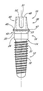
As best seen in FIG. IA, the implant 10 includes a body portion 12, a neck 14,
and a
collar 16. The body portion 12 is preferably generally cylindrical with a
tapered distal end
and includes threads 18 that may be configured to mate with a preformed
threaded hole or
osteotomy formed in the patient's jawbone (not shown). However, it should be
appreciated
that the body portion 12 may also be configured so as to be self-tapping. It
should also be
appreciated that although the illustrated body portion 12 has tapered or
conical portions, the
body portion 12 may be substantially cylindrical or completely tapered.
Finally, it should
-4-
CA 02512283 2005-06-30
WO 2004/062520 PCT/US2003/041813
be appreciated that the body portion 12 may be unthreaded if the surgeon
prefers to use an
unthreaded implant. In one particular embodiment, the body 12 has a shape
substantially
similar to the Brdemark System line of implants sold by Nobel BiocareTM. In
such an
embodiment, the lower portion may be substantially cylindrical, including
threads, and self-
tapping features as is well known in the art.
The collar 16 of the implant is substantially cylindrical and is defined in
part by a
vertical side wall 26 that, in the preferred embodiment, is approximately 2
millimeters in
axial length. In modified embodiments, the implant 10 may be formed without
the neck 14
and/or the collar 16. Similarly, the neck 14 and/or collar 16 may have
dimensions that are
smaller or larger than the exemplary embodiment.
In the illustrated embodiment, the body 12 is preferably covered with a bone
apposition surface 21, which is configured to promote osseointegration. In one
embodiment, the bone apposition surface 21 increases the surface area of the
body 12. In
such an embodiment, to increase surface, the bone apposition surface 21 may be
formed by
roughening the lower portion 12 in several different manners, such as, for
example, acid-
etching (e.g., to apply an oxidized titanium surface such as the oxidized
surface
manufactured by Nobel Biocare under the trademark TiUniteTM), grit blasting,
and/or
machining. Alternatively, the bone apposition surface 21 may be formed by
coating the
lower surface with a substance that increases the surface area of the body 21.
Calcium
phosphate ceramics, such as tricalcium phosphate (TCP) and hydroxyapatite (HA)
are
examples of suitable materials. In other embodiments, the bone apposition
surface 21 may
comprise macroscopic structures, such as, for example, threads, micro-threads,
indentations, grooves that are configured to promote osseointegration and may
be used
alone or combined with the roughening and/or the coatings described above.
With continued reference to FIGS. 1A and 1B, in the exemplary embodiment, a
top
edge 23 of the bone tissue apposition surface 21 preferably extends above
through the neck
14 and onto the collar 16. In modified embodiments, the top edge 34 may have a
curved or
scalloped shape with at least one and more preferably two peaks and valleys
that follow or
at least closely approximate the shape of the naturally occurring contours of
a patient's
bone-tissue morphology. It should also be appreciated that in other
embodiments, the peaks
and valleys may be approximated by various combinations of straight and/or
curved lines
-5-
CA 02512283 2005-06-30
WO 2004/062520 PCT/US2003/041813
that follow or at least closely approximate the shape of the naturally
occurring contours of a
patient's bone-tissue morphology.
The surface 35 of the collar 16 above the top edge 23 may be polished to
reduce
accumulation of plaque and calculus. In a modified embodiment, the surface 35
may be
treated to promote, enhance or maintain soft-tissue attachment. Such
treatments may
include applying growth factor, applying protein, roughening and/or the
application of
coatings that increase surface area. In addition, the surface 35 may be
modified or covered
with a coating that changes the color of the collar 16. For example, in one
embodiment the
surface 35 is coated with a material hydroxyapatite (HA) or other ceramic
coatings that are
generally white or "tooth-like" in color.
With continued reference to FIGS. 1A-C, exemplary implant 10 includes an upper
portion or abutment 38, which is integrally formed with or permanently
attached to the
collar 16. In this manner, there is preferably no "microgap" between the
abutment 38 and
the collar 16. In a preferred embodiment, the body 12, collar 16 and the
abutment 38 are
machined from a single piece of material (e.g., dental grade alloy). As will
be explained in
more detail below, the abutment 38 is sized and dimensioned to support a final
restoration
and other dental components.
As best seen in FIGS. IA and 1B, the outer surface of the final abutment 38
preferably includes an upper region 40 and a flared region 42. In the
illustrated
embodiment, the upper region 40 is substantially smooth and tapered. The upper
region 40
also has a top surface 48 that is substantially flat. Towards the bottom of
the upper region
(i.e., the portion nearest the flared region 42) is a flared portion 45 that
flares outward
towards a shoulder or ridge 47. The flared region 42 extends from the ridge 47
and
connects the upper region 40 to vertical side wall 26 of the collar 16.
In the illustrated embodiment, the upper region 40 also preferably includes a
plurality of grooves 51 (see also FIG. 1C). These grooves 51 help orient and
prevent the
rotation of a final restoration as described below. Accordingly, the final
restoration may
have an inner surface that matches or engages the shape of the upper region 40
of the
abutment 38. However, those skilled in the art will readily appreciate that
the upper region
40 and the grooves 51 may be formed into a variety of other shapes that may
also provide
an anti-rotational interface between the final restoration 54 and the abutment
38.
-6-
CA 02512283 2005-06-30
WO 2004/062520 PCT/US2003/041813
In general, the illustrated dental implant 10 has a generally circular cross-
sectional
shape. However, it should be appreciated that in modified embodiments the
cross-sections
may be non-round. For example, the cross-section of the upper region and
flared region
may have a non-round (e.g., oval) cross-section that resembles the cross-
section of a natural
tooth.
To permanently secure the final restoration, cement may be applied to the
upper
region 40 of the abutment 38. Alternatively, the final restoration 52 may be
coupled to the
final abutment 38 by a screw (not shown). In such an arrangement, a screw hole
(not
shown) may be provided on the side of the abutment 3 8.
As best seen in FIG. 1D, the abutment 38 advantageously includes an inner bore
52
that may include a threaded portion 53. As will be explained in more detail
below, the
inner bore 52 is configured to receive a coupling screw, which may be used to
couple
various components to the implant 10. The inner bore 52 may also include an
anti-rotation
chamber 55, which includes one or more anti-rotation features, such as, for
example, flat
sides, grooves, and/or indentations. A driving tool (not shown) with
corresponding anti-
rotational features may be inserted into the anti-rotational chamber so as to
transmit torque
from the driving tool to the dental implant 10 and or prevent rotation between
the implant
10 and a mating component (e.g., a healing cap, impression coping or dental
restoration).
In one embodiment, the anti-rotation chamber 55 may comprise a hexagonal
recess
configured to receive a hexagonally shaped tool such as a conventional Allen
wrench. In
another embodiment, the chamber 55 may include a tapered recess comprising
plurality of
concave side portions interconnected by flat or slightly curved side portions
(see e.g., the
internal connection marketed under the trademark UnigripTM by Nobel Biocare
AB).
The illustrated inner bore 52 may also include an annular recess or notch 57.
The
notch 57 may be configured for receiving the prongs or snapping elements on a
mating
component or driver. In this manner, the driver or mating component may be
releaseably
engaged with the implant 10. Any of a variety of complementary surface
structures may be
provided, to create a releasable retention force between the system and the
mating
component or driver. For example, the mating component or driver may include
one or
more lever arms or prongs that cooperate with the notch 57. In other
embodiments, the
driver or mating component may include a band of resilient material configured
to produce
a friction or mechanical interference fit retention force. In the illustrated
embodiment, the
-7-
CA 02512283 2005-06-30
WO 2004/062520 PCT/US2003/041813
releasable retention force is added by providing the notch 57. However, in
modified
embodiments, the complementary surface structures may be configured to engage
a bore 52
without a notch 57.
FIGS. 2A-2D illustrate an exemplary embodiment of a healing cap 76 that may be
used in combination with the dental implant 10 described above. The healing
cap 76 may
be made of a synthetic polymer, such as, for example, polyester or Nylon.
However, it
should be appreciated that other suitable materials may also be used. The
healing cap 76 is
preferably white or close to natural tooth color so that it has a natural
appearance when it is
placed in the patient's mouth.
The healing cap 76 includes an inner surface 77 which defines an internal
cavity 78.
The inner surface 77 also defines a top opening 80 and a bottom opening 82.
The inner
surface 77 is sized and dimensioned such that the that healing cap fits over
the upper region
40 of the abutment 38. With particular reference to FIG. 2C, the inner surface
77 preferably
includes a stop for limiting advance of the healing cap 76 onto the abutment
38, such as, a
base surface 84 that is sized and dimensioned to rest against the flanged
portion 45 of the
final abutment 3 8.
With continued reference to FIG. 2C, the healing cap 76 also preferably
includes a
tissue retraction flange 86. The tissue retraction flange 86 is sized and
dimensioned such
that when the healing cap 76 is placed upon the abutment 38 it extends beyond
at least the
upper limit of the shoulder 47 of the abutment 38. The purpose and function of
the tissue
retraction flange 86 will be described below.
With reference to FIG. 2B, the top opening 80 is preferably defined by top and
bottom portions 88, 90. The diameter of the top portion 88 is slightly larger
than the
diameter of the second portion 90. Accordingly, a seat 92 is formed between
the first and
second portions 88, 90. The seat 92 provides support for a healing cap screw
94 (see FIG.
3). Alternatively, and/or in addition, the opening 80 may be flared or
chamfered to provide
a flared seating surface.
As with the abutment 38, it should be appreciated that although the
illustrated cross-
sections of the healing cap 76 are round in modified arrangements the cross-
sections may
be non-round. For example, the cross-sections may have a non-round cross-
section that
resembles the cross-section of a natural tooth.
-8-
CA 02512283 2005-06-30
WO 2004/062520 PCT/US2003/041813
Turning now to FIG. 3, the healing cap screw 94 will now be described. The
healing cap screw 94 is sized and dimensioned so as extend through the healing
cap 76 and
to couple the healing cap 76 to final abutment 38. The healing cap screw 94 is
preferably
made of a dental grade titanium alloy; although, other suitable materials may
be used. The
healing cap screw 94 includes a flange 96, an anti-rotational recess 98, a
barrel 99 and
lower threads 100. The flange 96 preferably has a diameter that is slightly
smaller than the
diameter of the upper portion 88 of the healing cap 76. The recess 98 extends
through the
flange 96 and allows for the insertion of, for example, hexagonally shaped
tool such as a
conventional Allen O wrench or the tools sold under the trademark UnigripTM by
Nobel
Biocare AB, which may be used to rotate the healing cap screw 94. The threads
100 are
sized and dimensioned to match the threaded bore 52 of the implant 10 (see
FIG. 1D).
Preferably, the barrel 99 has a diameter that is slightly larger than the
inner diameter
of the bottom portion of the healing cap 76. The barrel 99 preferably includes
a groove
101, which is located below the flange 96 and has a diameter that is slightly
smaller than
the inner diameter of the bottom portion 90 of the healing cap. As such, the
healing cap
screw 94 may be press-fit into the healing cap 76 such that the bottom portion
90 fits into
the groove 101 and the top portion 97 is flush with the top of the healing cap
76. In this
manner, the healing screw 94 is captured by the healing cap 76 and may rotate
freely inside
the healing cap 76. Of course, in a modified arrangement, the healing cap
screw 94 may be
configured without the capture feature.
In use, the surgeon first places the implant 10 into the patient's jawbone
during
Stage I surgery with the top edge 23 of the bone apposition surface being
approximately
equal or slightly above the upper most bone surface. The surgeon then places
the healing
cap 76 over the abutment 38 and uses the captured healing cap screw 94 to
couple the
healing cap 76 to the abutment 38. Specifically, the surgeon rotates the
healing cap screw
94 so that the threads 100 engage the inner bore 52 of the implant 10.
Accordingly, the
healing cap 76 is held securely against the abutment 38. As will be explained
in more
detail below, the healing cap 76 helps to control the healing and growth of
the patient's
gum tissue around the implant site. The healing cap 76 also improves the
appearance of the
patient's mouth and provides the patient with a temporary chewing surface. If
desired, the
healing cap 76 may also be used to support a temporary restoration and/or may
itself be
shaped in the form of a temporary restoration.
-9-
CA 02512283 2005-06-30
WO 2004/062520 PCT/US2003/041813
The patient then returns home and the implant is allowed to osseointegrate
with the
jawbone and the patient's gums are allowed to heal. Once the implant
osseointegrates and
the gums heal, the patient returns to the surgeon who takes an impression of
the patient's
mouth. The surgeon loosens the healing cap screw 94 and removes the healing
cap 76 from
the final abutment 38. As will be described in more detail below, at this
point, the surgeon
takes the impression of the patient's mouth to record the position,
orientation and shape of
the dental abutment within the mouth.
As will be described below, the impression is used to make a model of the
patient's
mouth and to form the final restoration. As mentioned above, the final
restoration has an
inner surface that matches the upper region 40 of the abutment 38.
Accordingly, in a final
procedure, the surgeon may attach the final restoration by slipping it onto
the final
abutment 38 cementing it in place and/or securing it with a screw.
As best seen in FIG. 2D, the tissue retraction flange 86 controls the healing
and
growth of the patient's gum tissue around the abutment 38. In contrast, prior
art protection
caps would rests upon the shoulder region if the abutment. This allows the gum
tissue
during a healing period grows near and above the shoulder region during
healing periods.
This may cause several problems. For example, when such a protection cap is
removed, the
gum tissue tends to relax and fall over the shoulder region. When an
impression is taken of
the abutment, this fallen gum tissue may compromise the accuracy of the
impression.
Moreover, if an impression cap such as the one disclosed in U.S. Patent No.
5,688,123 is
used, the fallen gum tissue may become pinched between the impression cap and
the
shoulder region when the impression cap is snapped over the shoulder region.
This may
cause discomfort to the patient. In addition, when a final restoration is
attached to the final
abutment and implant, the gum tissue may also become pinched in between the
final
restoration and the shoulder region.
In contrast, in the illustrated embodiment of the healing cap 76 includes a
tissue
retraction flange 86 that extends below the shoulder 47 of the final abutment
38. The tissue
retraction flange 86 pushes the gum tissue down and away from the shoulder 47.
The tissue
retraction flange 86 also pushes the gum tissue laterally away from the
shoulder 47.
Accordingly, a gap is formed between the gum tissue and the shoulder 47 of the
final
abutment 38. Thus, when the healing cap 76 is removed, the gum tissue is less
likely to fall
over the shoulder 47. This arrangement tends to prevent patient's gums from
falling over
-10-
CA 02512283 2011-02-03
the shoulder 47 of the abutment when (i) the impression is taken, (ii) an
impression cap is
being attached to the abutment and/or when the final restoration is attached
to the abutment
38. This results in more accurate impressions and minimal discomfort to the
patient.
The tissue retraction flange 86 is sized and dimensioned to hold the gum
tissue far
enough away from the shoulder 47 to achieve some or all the results described
above.
Generally, the tissue retraction flange 86 holds the gum tissue at least about
0.25
millimeters below the shoulder, in some embodiments about 0.5 millimeters, in
other
embodiments 1 millimeter or greater. Additional embodiments and more details
concerning the healing cap 76 may be found in U.S. Patent No. 6,431,866,
entitled "HEAL
IN-PLACE ABUTMENT SYSTEM", issued August 13, 2002.
FIGS. 4A-E illustrate an impression cap 174, which may be used to take an
impression of the dental implant 10 as mentioned above. In this exemplary
embodiment,
the impression cap 174 is configured to engage the dental implant 10 with a
releasable
retention force. The illustrated impression cap 178 comprises a body 180 with
a proximal
end 182 and a distal end 184. The body 122 is preferably made of resilient
moldable plastic
and/or polymer, such as, for example, polycarbonate. The body 180 defines an
inner
surface 186, which forms an inner cavity 188. The inner cavity 188 is
configured such that
the impression cap 178 may fit over the upper region 40 of the abutment 3 8.
111 the illustrated embodiment, the impression cap 178 is preferably
configured to
engage the abutment 38 of the implant 10 in a snap fit. In the illustrated
embodiment, this
snap fit is achieved by providing the proximal end 182 with a notch or groove
190, which is
best seen in FIG. 4D. The groove 190 is configured to snap over the shoulder
47 of the
abutment 38. That is, in the engaged position, the groove 190 fits around the
shoulder 47 of
the abutment 38 such that the impression cap 178 is coupled to the abutment
38. In the
illustrated embodiment, the groove 190 is generally V-shaped with an distal
portion 192, an
apex 194 and a proximal portion 196. In the engaged position, the proximal
portion 196
lies generally below the shoulder 47 of the abutment 38, the apex 136 lies
generally parallel
11
CA 02512283 2011-02-03
to the shoulder 47 and the distal portion 192 lies generally above the
shoulder 47.
Advantageously, in the illustrated embodiment, the distal portion 192 is
oriented such that
it may lie flush with the flared portion 45 of the abutment 152. The distal
portion 192
preferably blends into the radius of the apex 194. In one embodiment, the apex
194 has a
7/
11a
CA 02512283 2005-06-30
WO 2004/062520 PCT/US2003/041813
radius of about .004" to .002" and, in a preferred embodiment, the apex has a
radius of
about.003".
Preferably, the groove 190 is sized and dimensioned such that in the engaged
position the impression cap 178 may be rotated with respect to the final
abutment 158.
That is, in a preferred embodiment, the space defined by the groove 192 is
slightly larger
than the corresponding portions of the flared portion 45, the shoulder 47 and
the notch 172
of the final abutment 152. As such, in the engaged position, the proximal
portion 196 of
the impression cap 178 is not in a stressed (e.g., in a flexed and/or
compressed state). Of
course, in one modified embodiment, the groove 192 may be sized and
dimensioned such
that in the engaged position the proximal portion is stressed and thus exerts
a positive
holding force on the final abutment 152.
With reference back to FIG. 4A, in the illustrated embodiment, the side wall
186
extends from the proximal portion to a roof 187. Preferably, a junction 142
between the
side wall 186 and the roof 187 is located at about the same elevation as the
top surface of
the abutment 38 when the impression cap 178 is in an engaged position. In the
illustrated
embodiment, the side wall 186 is substantially smooth and has a substantially
cylindrical
shape. However, in modified embodiments, the side wall 186 may be textured or
roughened so as to enhance retention of impression material, which, as will be
explained
below, is injected into the cavity 188. The substantially cylindrical shape of
the side wall
186 is generally preferred because it provides a large amount of space for the
impression
material near the top surface of the abutment 38, which as will be explained
below may be
modified by the dental surgeon. Correspondingly, it provides also provides
less space for
the impression material near the shoulder 47 of the abutment 38. This
arrangement
therefore creates a thin or featheredge of impression material which fades
away at the
shoulder 47 of the abutment 38.
In the illustrated embodiment, the roof 187 is funnel shaped. That is, the
roof 187
tapers from the most distal end 184 to the side walls 186. Advantageously, the
roof 187
defines a transition space, which is located above the top surface of the
abutment 38 when
the impression cap 120 is in the engaged position. The transition space
facilitates the flow
of impression material above the abutment 38 to the sides and shoulder 47 of
the abutment
38.
-12-
CA 02512283 2005-06-30
WO 2004/062520 PCT/US2003/041813
With particular reference to FIG. 4A, the impression cap 178 also includes an
injection port 150, which provides a pathway for injecting impression material
into the
internal cavity 188. In the illustrated embodiment, the injection port 150 is
positioned at
the distal end 184 on a top surface 152 of the impression cap 120 and
communicates with
the transition space. The illustrated injection port 150 includes a tapered
portion 152 and a
cylindrical portion 154. The cylindrical portion 154 preferably has a diameter
that is
approximately equal to a gap between the top of the abutment 38 and the side
wall 186,
when the impression cap 178 is engaged on the implant 10. This arrangement is
preferred
because it ensures that impression material injected into the impression cap
is directed
towards space between the side of the abutment 38 and the side wall 186. In
one
embodiment, the cylindrical portion has a diameter of about .06 inches and the
most distal
portion of the tapered section 152 has a diameter of about .09.
As best seen in FIGS. 4A and 4C, the impression cap 178 includes a plurality
of
vent holes 156, which extend through the main body 122 into the cavity 188. In
the
illustrated embodiment, the vent holes 156 are arranged in three rows. Each
row comprises
three vent holes 156, which are aligned vertically. The rows are spaced about
120 degrees
apart around the periphery of the impression cap 178. As will be explained in
detail below,
the vent holes 156 provide a vent for air and excess impression material. In
one
embodiment, the vent holes 156 have a diameter of about .2 inches. In the
illustrated
embodiment, the vent holes 156 are generally cylindrical but in modified
embodiments may
be funneled shaped with the end exposed to the inner cavity 188 having a
smaller diameter
than the other end.
With reference back to FIG. 4A, the impression cap 178 preferably includes one
ore
more embedment features 160. As will be explained in more detail below, the
embedment
features 160 facilitate the gripping and retention of the impression cap 178
within an
impression tray. The one or more embedment features preferably define at least
one
interference surface 162, which faces lies generally transverse to a
longitudinal axis 164 of
the impression cap. In the illustrated embodiment, the embedment feature 160
comprises a
flange 166, which is positioned the distal end 184 of the main body 122. The
illustrated
flange 166 includes a plurality of through holes 168, which extends through
the four
corners of the flange 166. In one embodiment, each hole 168 preferably has a
diameter of
about .050". In FIG. 4E, the impression cap 178 includes a pair of flanges
166.
-13-
CA 02512283 2005-06-30
WO 2004/062520 PCT/US2003/041813
In use, the impression cap 178 may be used to take an impression of the
abutment
38 and/or record the orientation of the implant 10. Such an impression may be
taken during
stage one, two or stage three as deemed effective by the dental practitioner.
In some
embodiments, a block out plug (not shown) may be first inserted into the bore
52 of the
abutment 38 to prevent impression material from entering the bore 52.
After the block out plug is in place, the surgeon then snaps the impression
cap 178
onto the abutment 38 as shown in FIG. 4E. After the impression cap 178 is in
place, the
surgeon uses a syringe (not shown) with a small nozzle to inject under
pressure a
impression material, such as, for example, polyvinylsiloxane or polyether into
the cavity
188. Preferably, this involves placing tip of the small nozzle into the
internal cavity 188
through the injection port 150.
As the impression material is forced into the impression cap 178, air and
excess
impression material 186 is forced out of the vent holes 156. Preferably, the
surgeon
continues to inject impression material into the impression cap 178 until
impression
material extrudes from most and more preferably all of the vent holes 156.
This ensures
that the impression material has completely filed the internal cavity 188. As
such, the
impression material within the impression cap 178 will provide a precise
impression of the
upper region 40 of the abutment 38 without voids or tears in the impression
material. The
excess material that is forced into the vents 156 becomes locked or trapped
within the vents
156. As mentioned above, in some embodiments, the vents 156 are funnel shaped.
Advantageously, this increases the interlocking of impression cap 178 with the
impression
material and helps to prevent separation of the impression material from the
impression cap
178.
After injecting the impression material into the impression cap 178, an
impression is
preferably taken of the whole arch or quadrant if the patient's mouth. This is
typically
involves using a U-shaped impression tray not shown that is filled with a
second impression
material. The tray is inserted into the mouth over the impression cap 178. As
such, the
impression cap 178 becomes embedded in the second impression material. The
interference surface 162 of the impression cap 178 facilitates mechanically
interlocking
between the impression material and the impression cap 178. Such interlocking
is further
enhanced by the holes 156.
-14-
CA 02512283 2011-02-03
Once the second impression material is set, the tray is removed from the
mouth.
The impression cap 178 remains embedded in the second impression material and
is thus
uncoupled from the final abutment 38 as the tray is removed. The tray is then
sent to a
dental laboratory and is used by a dental technician to fabricate a final
restoration (i.e., a
dental prosthesis). An analog (not shown) of the abutment may be placed within
the
impression cap, with the same axial orientation as the abutment 38 and the
implant 10 in
the patient's mouth. The impression tray is then filled or covered with dental
stone or any
modeling material. After the modeling material has set the model is separated
from the
impression. The model is an accurate reproduction of the implant site and
allows the dental
technician to fabricate the final restoration for the patient in the proper
position in axial and
rotational alignment.
The stone or plaster analogue may then be used to form the final restoration
(not
shown), using conventional techniques that may involve using a coping and/or
modifying
the abutment on the stone model. In other embodiments, various commercially
available
productions CAD/CAM systems may also be used to scan the stone or plastic
model and to
guide the design and creation of the final restoration (e.g., the system
marketed and used by
Nobel Biocare under the trademark ProceraTM ) (see also e.g., U.S. Patent Nos.
6,062,861,
5,938,446, 5,880,962, 5,742,828, 5,733,126, 5,652,709, 5,587,912, 5,440,496).
In
other embodiments, prefabricated copings and/or final restorations may also be
used.
In some instances the dental surgeon may choose to modify the shape of the
upper
region 40 of the abutment 38. For example, the upper region 40 may be modified
to refine
the occulusal length and axial draw. By way of example, the upper region 40
may be
modified using a high-speed dental handpiece with carbide burs.
One advantage of the impression cap 178 is that it may be used to record the
shape a
modified abutment. That is, after the abutment 38 has been modified the
impression cap
178 may snapped into place. The impression cap 178 is then filled as described
above and
CA 02512283 2011-02-03
an impression is taken of the patient's mouth. The impression tray is then
sent to a dental
laboratory. At the laboratory, the impression cap 178 is filled with dental
stone or any
modeling material, thereby reproducing the shape of the upper region 40 of the
abutment
38, which was stored in the first impression material.
In modified embodiments, the impression cap 178 may be configured such that it
does not engage the dental implant 10 with a realeasable retention force. In
such
embodiments, the cap 178 may be configured to rest on the shoulder 47 of the
abutment. In
this ma ner, the modified impression cap may also be used as a pick-up coping
as is known
in the art.
Additional embodiments and further details of the impression cap 178 can be
found
in co-pending U.S. Patent Publication No. 2002/0106610 Al published on August
8,
2002 and entitled "IMPRESSION CAP".
FIGS. 5A-5E illustrate an exemplary embodiment of a coping 600 that may be
used
with the implant 10 described above to form a final restoration. The
illustrated coping 600
is configured to mate with the abutment 38 of the implant 10 of FIG. 1A or an
analogue of
the abutment 38.
The illustrated coping 600 comprises a main body 602. The main body 602
includes an inner surface 604, that defines an internal cavity 606. The inner
surface 604 is
configured such that the coping 600 may fit over the abutment 38.
The inner surface includes one or more feet or standoffs 610. Each standoff
640
preferably extends from the inner surface 604 towards the center of the cavity
606 at least
about 10 microns and often approximately 25-50 microns. The inner surface 604
preferably
also includes a flanged portion 612, which is configured to rest upon shoulder
47 of the
abutment 38. Preferably, the flanged portion 612 is sized and configured such
that the
coping 600 is centered on the abutment 38 or analogue and a top surface 614 of
the inner
surface 604 lies a desired distance (e.g., at least about 10 microns and often
approximately
25-50 microns) above the abutment 38 or analogue.
In the illustrated arrangement, the standoffs 610 preferably extend from the
top
surface 615 of the inner surface 604. Moreover, the coping 600 preferably
includes six
16
CA 02512283 2011-02-03
standoffs 610, which are preferably arranged around the perimeter of the inner
surface 604
at approximately 60 degrees from each other. This arrangement is preferred
because for
any angular orientation of the illustrated coping 600 with respect to the
abutment 38 do not
lie within the recesses or grooves 51 (see FIG. 1A). As such, at least one
standoff 610
contacts the outer surface of the abutment 38. In this manner, the standoffs
610 and the
flanged portion 612 cooperate to produce a substantially uniform gap between
the coping
600 and the abutment 38.
i
16a
CA 02512283 2005-06-30
WO 2004/062520 PCT/US2003/041813
FIGS. 6A-6C illustrate another arrangement of a coping 700. The illustrated
coping
700 is also configured to mate with the abutment 38 of the implant 10. The
illustrated
coping 700 comprises a main body 702. The main body 702 includes an inner
surface 704
that defines an internal cavity 706. The inner surface 704 is configured such
that the coping
700 may fit over the upper region abutment 38 described above. The inner
surface 704
includes one or more feet or standoffs 710. In this arrangement, the standoffs
710 are
configured to fit within the grooves or recesses 51 of the abutment 38 (see
FIG. IA). As
such, the standoffs 710 help to orient and prevent the rotation of the coping
700 with
respect to the abutment 38. The standoffs 710 are also configured such that
the inner
surface 704 of the coping lies at least about 10 microns and often
approximately 25-50
microns above the outer surface of the final abutment or analogue 550. That
is, the
standoffs 710 are configured to extend from the inner surface 604 at least and
additional 10
microns and often approximately 25-50 microns beyond the depth of the grooves
or
recesses 51.
The inner surface 704 preferably also includes a flanged portion 712, which is
configured to rest upon a lower portion or shoulder 47 of the abutment 38.
Preferably, the
flanged portion 712 is sized and configured such that the coping 700 is
centered on the
analogue and a top surface 715 of the inner surface 704 lies a desired
distance (e.g., at least
about 10 microns and often approximately 25-50 microns) above the abutment 38.
The
standoffs 710 and the flanged portion 712 cooperate to produce a uniform gap
between the
coping 700 and the abutment.
Several methods for creating a final restoration from the copings 600, 700
described
above. One such method utilizes investment casting techniques to create a
metal coping
with an inner surface substantially similar to the inner surface 604, 704 of
the coping 600,
700. Tn such a method, the coping 600 may be made of plastic or another
material suitable
for investment casting. The technician applies, by way of example, wax to the
outer surface
of the coping 600 to form a model of a metal coping. The technician removes
the wax and
the coping 600 from an analogue of the implant 10 and encases the combination
in an
investment material. The investment material is then heated to remove the wax
and coping
600. The technician fills the investment material with a metal, such as, for
example, gold
or another suitable materials. Once the metal solidifies, the investment
material is broken
to release a metal coping.
-17-
CA 02512283 2011-02-03
The metal coping will have an inner surface that is substantially the same
shape and
size as the inner surface 604 of the plastic coping 600. Accordingly, the
metal coping will
includes standoffs that are substantially the same size as the standoffs 610
describe above.
Moreover, the inner surface of the metal coping will include a top surface and
a lower
flange that are the same distance from each other as the top surface 615 and
lower flange
612 of the plastic coping 610.
To form the final restoration, a porcelain cover or other suitable tooth-like
material
is attached to the metal coping using well known techniques. The metal coping
provides
structural strength and rigidity to the final restoration. When the final
restoration is placed
upon the abutment 38 of the implant 10, the standoffs and the lower flange
create a uniform
gap for the cement between the metal coping and the abutment 38. Moreover, the
standoffs
help to center the final restoration on the abutment 38. Accordingly, the
final restoration
rests squarely and evenly upon the final abutment 10.
In one modified embodiment, the coping is made from a material that is
suitable
forming at least part of the final restoration Such materials may include gold
or a ceramic
material. In such, an embodiment, the final restoration may be attached
directly or built
upon the coping.
Further details on the coping and other modified embodiments can be found in
U.S.
Patent Publication No. 2002/0028425 Al published on March 7, 2002, entitled
"COPINGS WITH STANDOFFS".
FIGS. 7A and 7B illustrate a modified implant 800. This implant 800 has a
lower
body 812 that may be configured as described above with reference to FIGS. IA-
1D. In
this embodiment, the abutment 838 comprises a sidewall 839 which tapers
inwardly from
the collar 816 to provide the abutment 838 with a generally conical shape with
a taper of
about 5 degrees. Grooves 850 preferably extend partially around a top segment
852 of the
abutment 838, except for a smooth flat surface 854. The smooth surface 854 is
preferably
characterized by a flat plane or face intersecting and truncating the outer
surface of the
18
CA 02512283 2011-02-03
otherwise conical shape of the top segment 852. This flat surface 854 provides
for
engagement with a wrench or other torque providing tool and also provides anti-
rotation
relative to any mating components.
As shown in FIGS. 7A and 7B, in the illustrated embodiment, the top edge 823
of
the bone apposition surface 821 may have a curved or scalloped shape with. at
least one and
/
18a
CA 02512283 2005-06-30
WO 2004/062520 PCT/US2003/041813
more preferably two peaks and valleys that follow or at least closely
approximate the shape
of the naturally occurring contours of a patient's bone-tissue morphology. It
should also be
appreciated that in other embodiments, the peaks and valleys may be
approximated by
various combinations of straight and/or curved lines that follow or at least
closely
approximate the shape of the naturally occurring contours of a patient's bone-
tissue
morphology. The surface above the top edge 823 may be smooth or polished.
The implant 800 of FIGS. 7A and 7B may be used to replace narrow, smaller
diameter teeth, such as, for example, the anterior teeth and, in particular,
the incisors. In
such applications, the edentulous spaces are particularly narrow. Accordingly,
the sidewall
839 preferably has a maximum diameter that is no greater and, more preferably
smaller than
the diameter of the maximum diameter of the collar 816. In one embodiment, the
collar
816 has a maximum diameter of about 3.0 millimeters.
In use, the upper portion of the implant 800 may be with a healing cap. An
exemplary embodiment of such a healing cap 860 is shown in FIGS. 8A-8B. The
healing
cap 860 is formed from body 862 having an outer surface 864 and an inner
surface 866. In
the illustrated embodiment, the outer surface 864 has generally vertical
sidewalls 868 and a
horizontal top surface 870 that is connected to the sidewalls 868 by a rounded
top edge 872.
The inner surface 866 forms a cavity 874. The inner surface 866 is preferably
configured to
substantially match in shape and size the outer surface of the abutment 838.
Accordingly,
the inner surface 866 includes a flat 876 that corresponds to the flat 854
formed on the
abutment 838. To secure the cap 860 to the abutment 838, an adhesive may be
applied to
the outer surface of the abutment 838 and/or the inner surface 866 of the cap
860 before the
cap 860 is inserted onto the abutment 838. After a healing period, the cap 860
may be
removed from the abutment 38 with a dental pick, an impression may be taken of
the
patient's mouth to record the position of the implant 800, a final restoration
may formed
and attached to the abutment 838.
FIGS. 9A and 9B illustrate another exemplary embodiment of an implant 900.
This
implant 900 is a substantially similar to the previous embodiment. However,
this
embodiment is configured for larger diameter teeth. In one embodiment, the
collar 916 has
a diameter of about 4.3 millimeters.
With reference to FIGS. 9A and 9B, as compared to the previous embodiment, the
distal end of the abutment 938has been removed or truncated leaving the
abutment with a
-19-
CA 02512283 2005-06-30
WO 2004/062520 PCT/US2003/041813
substantially flat top surface 950. As with the previous embodiment, the top
edge 923 of
the bone apposition surface 921 may have a curved or scalloped shape with at
least one and
more preferably two peaks and valleys that follow or at least closely
approximate the shape
of the naturally occurring contours of a patient's bone-tissue morphology. It
should also be
appreciated that in other embodiments, the peaks and valleys may be
approximated by
various combinations of straight and/or curved lines that follow or at least
closely
approximate the shape of the naturally occurring contours of a patient's bone-
tissue
morphology. The surface above the top edge 823 may be smooth or polished.
In use, after the implant 900 is installed into the patient's mouth, an
impression may
be taken of the implant 900 to record the position of the implant 900 and/or
any
modifications made to the shape of the abutment 938 by the dental surgeon. A
final
restoration may then be formed and attached to the abutment 938 with an
adhesive (e.g.,
dental cement).
Certain objects and advantages of the invention have been described above for
the
purpose of summarizing the invention and the advantages achieved over the
prior art. Of
course, it is to be understood that not necessarily all such objects or
advantages may be
achieved in accordance with any particular embodiment of the invention. Thus,
for
example, those skilled in the art will recognize that the invention may be
embodied or
carried out in a manner that achieves or optimizes one advantage or group of
advantages as
taught herein without necessarily achieving other objects or advantages as may
be taught or
suggested herein.
Furthermore, although this invention has been disclosed in the context of
certain
preferred embodiments and examples, it will be understood by those skilled in
the art that
the present invention extends beyond the specifically disclosed embodiments to
other
alternative embodiments and/or uses of the invention and obvious modifications
and
equivalents thereof. Thus, it is intended that the scope of the present
invention herein
disclosed should not be limited by the particular disclosed embodiments
described above,
but should be determined only by a fair reading of the claims that follow.
-20-