Note: Descriptions are shown in the official language in which they were submitted.
CA 02514651 2005-07-27
WO 2004/066853 PCT/GB2004/000338
DETECTION APPARATUS AND METHOD
The present invention relates to an apparatus for, and a method of, locating a
tip of a
tubular element, in particular a needle, within a region, in particular the
epidural
space and the peritoneal cavity, of a body of a subject.
Epidural anaesthesia is becoming increasingly popular for a wide variety of
surgical,
obstetric and analgesic procedures, with indications including surgical
anaesthesia,
analgesia during labor and prolonged post-operative pain relief. Epidural
anaesthesia
requires the identification of the epidural space, which currently is
performed by
inserting a needle, in particular a Touhy needle, into the appropriate
location in the
spinal column. The epidural space is located beyond the ligamentum flavum
indenting the dura, and is entered after the tip of the needle passes through
the
ligamentum flavum. The most commonly employed technique is the °loss of
resistance" technique in which a Touhy needle, which is connected to a loss or
resistance (LOR) syringe, is advanced through the layers of back tissue while
actuating the syringe to apply a pressure to the contained fluid, with the
location of
the tip of the needle in the epidural cavity being detected by the user
"feeling" the
resistance to actuation of the syringe. This technique, however, requires
considerable experience in order to avoid advancing the needle through the
epidural
space and into the dura, and is made particularly difficult by the user having
to
concentrate separately both on actuating the syringe and inserting the needle.
Puncturing of the dura has been identified inter alia as. the cause of chronic
back
pain.
Laparascopy is also being increasingly utilized, and requires the
identification of the
peritoneal cavity, which currently is performed by the introduction of a
Veress needle
through the abdomen and into the peritoneal cavity for insufflation to produce
pneumoperitoneum. As with the identification of the epidural space, the
successful
identification of the peritoneal cavity requires considerable experience.
Identification
of the peritoneal cavity is particularly difficult as the sharpness of the
Veress needle
does not readily allow for differentiation between layers of high and low
resistance
when passing the Veress needle through the abdominal layers. It is not
uncommon
to require very many attempts to identify the peritoneal cavity, often more
than ten
attempts. Furthermore, successful insertion of the Veress needle can only be
determined on insufflation of the peritoneal cavity.
CONFIRMATION COPY
CA 02514651 2005-07-27
WO 2004/066853 PCT/GB2004/000338
2
Apparatuses have been devised for use in detecting the epidural space, such as
disclosed in US-A-5024662, but these apparatuses, in utilizing elastic
elements to
bias the plunger of a delivery syringe do not allow for reliable
identification of the
epidural space.
It is thus an aim of the present invention to provide an improved detection
apparatus
and method which allows for the reliable location of a tip of a tubular
element in the
epidural space, and also a detection apparatus and method which allows for the
reliable location of a tip of a tubular element in the peritoneal cavity.
In one aspect the present invention provides a detection apparatus for use in
locating
a tip of a tubular element, in particular a needle, within a region, in
particular one of
the epidural space and the peritoneal cavity, of a body of a subject, the
delivery
apparatus comprising: a biasing unit operative to maintain a biasing force to
a fluid
contained in a delivery unit connected in use to the tubular element, such as
to cause
the delivery unit to deliver fluid to the tubular element when the biasing
force is
greater than a resistance to flow of the fluid from the tip of the tubular
element as
when located within the region.
In another aspect the present invention provides a detection apparatus for use
in
locating a tip of a tubular element, in particular a needle, within a region,
in
particular one of the epidural space and the peritoneal cavity, of a body of a
subject,
the detection apparatus comprising: a biasing unit operative to maintain a
substantially constant biasing force to any volume of fluid contained in a
delivery unit
connected in use to the tubular element, such as to cause the delivery unit to
deliver
fluid from the tubular element when the biasing force is greater than a
resistance to
flow of the fluid from the tip of the tubular element as when located within
the
region.
In a further aspect the present invention provides a detection apparatus for
use in
locating a tip of a tubular element, in particular a needle, within a region,
in
particular one of the epidural space and the peritoneal cavity, of a body of a
subject,
the detection apparatus comprising: a delivery unit for containing a volume of
fluid
and connected in use to the tubular element; and a spring element operative to
maintain a biasing force to the fluid contained in the delivery unit, such as
to cause
the delivery unit to deliver fluid from the tubular element when the biasing
force is
CA 02514651 2005-07-27
WO 2004/066853 PCT/GB2004/000338
3
greater than a resistance to flow of the fluid from the tip of the tubular
element as
when located within the region.
In a yet further aspect the present invention provides a detection apparatus
for use
in locating a tip of a tubular element, in particular a needle, within a
region, in
particular one of the epidural space and the peritoneal cavity, of a body of a
subject,
the detection apparatus comprising: a delivery unit comprising a body which
includes
a cavity and an outlet through which a fluid is deliverable and to which the
tubular
element is connected, and a plunger which is movably disposed in the cavity
such
that fluid is drawn into the fluid chamber on withdrawal of the plunger and
expelled
therefrom on depression of the plunger, wherein the plunger includes a shaft
which
includes an elongate cavity therein for housing a needle element of a needle
unit for
use with the apparatus.
In yet another aspect the present invention provides a detection apparatus for
use in
locating a tip of a tubular element, in particular a needle, within a region,
in
particular one of the epidural space and the peritoneal cavity, of a body of a
subject,
the detection apparatus comprising: a delivery unit comprising a body which
includes
a cavity and an outlet through which a fluid is deliverable and to which the
tubular
element is connected, and a plunger which is movably disposed in the cavity
such
that fluid is drawn into the fluid chamber on withdrawal of the plunger and
expelled
therefrom on depression of the plunger, wherein the plunger includes a
delivery
channel extending to a forward end thereof to allow for the delivery of a
substance to
the outlet of the body through the plunger, and a valve unit for providing
only for
one-way delivery of a substance through the delivery channel to the outlet of
the
body.
In still another aspect the present invention provides a detection apparatus
for
locating a tip of a tubular element, in particular a needle, within a region,
in
particular one of the epidural space and the peritoneal cavity, of a body of a
subject,
the detection apparatus comprising: a tubular element having a tip for
insertion into
body tissue; a sensing element extending through the tubular element and
having a
blunt tip; and a biasing unit for biasing the sensing element with a biasing
force such
that the tip of the sensing element is biased outwardly of the tip of the
tubular
element, whereby, when the resistance at the tip of the tubular element is
greater
than the biasing force, the sensing element remains stationary relative to the
tubular
element, and, when the resistance at the tip of the tubular element is less
than the
CA 02514651 2005-07-27
WO 2004/066853 PCT/GB2004/000338
4
biasing force, the tip of the sensing element extends from the tip of the
tubular
element.
In still yet another aspect the present invention provides a method of
locating a tip of
a tubular element, in particular a needle, within a region, in particular one
of the
epidural space and the peritoneal cavity, of a body of a subject, the method
comprising the steps of: connecting a detection apparatus to the tubular
element,
wherein the detection apparatus comprises a delivery unit which contains a
fluid and
is connected to the tubular element, and a biasing unit which is operable to
maintain
a biasing force on the fluid contained in the delivery unit which is such as
to cause
the delivery unit to deliver fluid therefrom when the biasing force is greater
than a
resistance to flow of the fluid from the tip of the tubular element as when
located
within the region; operating the biasing unit to maintain the biasing force on
the fluid
contained in the delivery unit; and progressively inserting the tubular
element into
the body of the subject until the biasing force acts to cause fluid to be
delivered from
the delivery unit through the tubular element, at which position the tip of
the tubular
element is within the region and the biasing force is greater than a
resistance to flow
of the fluid from the tip of the tubular element.
In a still yet further aspect the present invention provides a method of
locating a tip
of a tubular element, in particular a needle, within a region, in particular
one of the
epidural space and the peritoneal cavity, of a body of a subject, the method
comprising the steps of: providing a detection apparatus comprising a tubular
element having a tip for insertion into body tissue, a sensing element
extending
through the tubular element and having a blunt tip, and a biasing element for
biasing
the sensing element with a biasing force such that the tip of the sensing
element is
biased in a direction outwardly of the tip of the tubular element, the biasing
element
being such that, when the resistance at the tip of the tubular element is
greater than
the biasing force, the sensing element remains stationary relative to the
tubular
element, and, when the resistance at the tip of the tubular element is less
than the
biasing force, the tip of the sensing element extends from the tip of the
tubular
element; and progressively inserting the tubular element into the body of the
subject
until the biasing force acts to cause the tip of the sensing element to extend
from the
tubular element, at which position the tip of the tubular element is within
the region.
The present invention assists in significantly improving both the speed and
reliability
of positioning the tip of a tubular element at a desired region in the body of
a
CA 02514651 2005-07-27
WO 2004/066853 PCT/GB2004/000338
subject, notably the epidural space and the peritoneal cavity, and thereby
provides
for more successful identification of such regions.
Preferred embodiments of the present invention will now be described
hereinbelow by
way of example only with reference to the accompanying drawings, in which:
Figure 1(a) illustrates a longitudinal sectional view of a detection apparatus
in
accordance with a first embodiment of the present invention;
Figure 1(b) illustrates a longitudinal sectional view of the detection
apparatus of
Figure 1(a) in the operative state;
Figure 2(a) illustrates a longitudinal sectional view of a detection apparatus
in
accordance with a second embodiment of the present invention where co-packaged
with a needle unit;
Figure 2(b) illustrates a longitudinal sectional view of the detection
apparatus of
Figure 2(a) with the needle unit removed therefrom;
Figure 3(a) illustrates a longitudinal sectional view of a detection apparatus
in
accordance with a third embodiment of the present invention;
Figure 3(b) illustrates a longitudinal sectional view of the detection
apparatus of
Figure 3(a) with a connector connected to the valve unit thereof for the
delivery of a
substance through the detection apparatus;
Figure 4(a) illustrates a longitudinal sectional view of a detection apparatus
in
accordance with a fourth embodiment of the present invention;
Figure 4(b) illustrates a longitudinal sectional view of the detection
apparatus of
Figure 4(a) in the operative state;
Figure 5(a) illustrates a longitudinal sectional view of a detection apparatus
in
accordance with a fifth embodiment of the present invention;
Figure 5(b) illustrates a longitudinal sectional view of the detection
apparatus of
Figure 5(a) in the operative state;
CA 02514651 2005-07-27
WO 2004/066853 PCT/GB2004/000338
6
Figure 6(a) illustrates a longitudinal sectional view of a detection apparatus
in
accordance with a sixth embodiment of the present invention;
Figure 6(b) illustrates a longitudinal sectional view of the detection
apparatus of
Figure 6(a) in the operative state;
Figure 7(a) illustrates a longitudinal sectional view of a detection apparatus
in
accordance with a seventh embodiment of the present invention;
Figure 7(b) illustrates a longitudinal sectional view of the detection
apparatus of
Figure 7(a) in the operative state;
Figure 8(a) illustrates a longitudinal sectional view of a detection apparatus
in
accordance with an eighth embodiment of the present invention;
Figure 8(b) illustrates a longitudinal sectional view of the detection
apparatus of
Figure 8(a) in the operative state;
Figure 9(a) illustrates a longitudinal sectional view of a detection apparatus
in
accordance with a ninth embodiment of the present invention;
Figure 9(b) illustrates a longitudinal sectional view of the detection
apparatus of
Figure 9(a) in the operative state;
Figure 10(a) illustrates a longitudinal sectional view of a detection
apparatus in
accordance with a tenth embodiment of the present invention; and
Figure 10(b) illustrates a longitudinal sectional view of the detection
apparatus of
Figure 10(a) in the operative state.
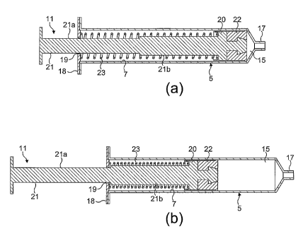
Figures 1(a) and (b) illustrate a detection apparatus in accordance with a
first
embodiment of the present invention for detecting the location of the tip of a
tubular
element, in particular a needle, within a region, in particular the epidural
space and
the peritoneal cavity, of a body of a subject.
CA 02514651 2005-07-27
WO 2004/066853 PCT/GB2004/000338
7
The apparatus comprises a body 5 which includes an elongate cavity 7, in this
embodiment having a diameter of 0.6 inches, and a plunger 11 which is movably
disposed in the cavity 7 and together with the cavity 7 defines a fluid
chamber 15
forward of the plunger 11 for containing a fluid, in this embodiment a liquid,
typically
a saline solution. In this embodiment the body 5 and the plunger 11 are
configured
as a loss of resistance (LOR) syringe and define a delivery unit.
The body 5 includes an outlet 17 at a forward end thereof through which a
fluid is
deliverable and to which a needle (not illustrated) is connected, and a stop
member
18 at the rear end thereof which includes an aperture 19 through which extends
the
plunger 11.
The plunger 11 comprises a head 20, an elongate shaft 21 which extends
rearwardly
of the head 20, and a gasket seal 22 which is attached to the head 20 for
providing a
fluid-tight seal with the cavity 7 of the body 5, such as to enable fluid to
be drawn
into the fluid chamber 15 on withdrawal of the plunger 11 and expelled from
the fluid
chamber 15 on depression of the plunger 11.
In this embodiment the shaft 21 includes a first, rear section 21a of a
diametral
dimension which is smaller than the aperture 19 of the stop member 18 such as
to
allow the rear section 21a to freely pass through the aperture 19 in the stop
member
18, and a second, forward section 21b which has a diametral dimension greater
than
the aperture 19 in the stop member 18 such as to prevent the plunger 11 from
being
withdrawn beyond a predetermined extent from the body 5.
The apparatus further comprises a biasing element 23, in this embodiment a
resilient
element, here a compression spring, which acts to bias the plunger 11 such as
to act
to depress the same with a predetermined biasing force, and thereby maintain a
fluid
contained in the fluid chamber 15 at a predetermined pressure. The pressure is
preferably not greater than about 8.5 Ibin'~, more preferably from about 0.5
Ibin-Z to
about 8.5 Ibin-2, still more preferably from about 0.5 Ibin-~ to about 5.66
Ibin-z, yet
more preferably from about 2.0 Ibin-~ to about 5.66 Ibin-z, and still yet more
preferably from about 2.0 Ibin-~ to about 3.5 Ibin-~.
In this embodiment the biasing element 23 is disposed about the shaft 21 of
the
plunger 11 within the cavity 7 of the body 5 and acts between the head 20 of
the
plunger 11 and the stop member 18 of the body 5. In this embodiment the
biasing
CA 02514651 2005-07-27
WO 2004/066853 PCT/GB2004/000338
8
element 23 is configured such as to provide a substantially constant biasing
force
over the entire operative stroke of the plunger 11, in this embodiment to
maintain a
fluid contained in the fluid chamber 15 at a pressure of about 2.83 Ibin'~.
Through
providing a constant biasing force, the present inventors have identified that
the
detection apparatus provides for the more reliable detection of expandable
body
regions. In providing a constant biasing force, the user can himself/herself
select the
volume of fluid to be delivered, and ensure that that volume of fluid is
delivered in its
entirety. For epidural application, by virtue of this complete delivery of the
fluid, the
fluid can be the epidural anaesthetic.
With this configuration, the biasing element 23 maintains the predetermined
biasing
force on the plunger 11 such that, when the resistance at the outlet 17 of the
body 5
is greater than the biasing force, such as when the tip of the needle is
located in
body tissue which provides a relatively high flow resistance to the contained
fluid, the
plunger 11 remains stationary, but, when the resistance at the outlet 17 of
the body
is less than the biasing force, such as when the tip of the needle is located
in an
expandable region in the body of a subject, the plunger 11 is depressed by the
biasing force into the body 5 such as to deliver fluid from the fluid chamber
15.
In operation, the plunger 11 is first withdrawn from the body 5 such as to
draw a
volume of fluid, typically from 2 to 7 ml, and preferably from 2 to 3 ml, into
the fluid
chamber 15 ahead of the plunger 11, as illustrated in Figure 1(b). Whilst
holding the
shaft 21 of the plunger 11 in the withdrawn position, the outlet 17 of the
body 5 is
connected to a needle, which has been partially pre-inserted into a subject.
The
needle is typically a Touhy needle for epidural application and a Veress
needle for
peritoneal application. Once the outlet 17 of the body 5 is connected to the
needle,
the shaft 21 of the plunger 11 is released. At this point, the plunger 11
remains
stationary, as the tip of the needle will be located in body tissue and the
flow
resistance at the tip of the needle will be greater than the biasing force of
the biasing
element 23. The needle is then slowly advanced, typically a millimetre at a
time,
until such point as the tip of the needle enters an expandable region in the
body of
the subject, at which point the flow resistance at the tip of the needle is
less than the
biasing force of the biasing element 23 and the biasing element 23 acts to
depress
the plunger 11 into the cavity 7 of the body 5 and expel fluid from the fluid
chamber
into the expandable region. This expulsion of fluid from the fluid chamber 15
is
indicative of the tip of the needle being located in a desired expandable
region of the
body of the subject, such regions including the epidural space and the
peritoneal
CA 02514651 2005-07-27
WO 2004/066853 PCT/GB2004/000338
9
cavity. Following location of the tip of the needle in the desired expandable
region,
the detection apparatus is then disconnected from the needle to allow for the
delivery
of the appropriate substance through the needle to the body region, typically
an
epidural anaesthetic for epidural application and an insufflation gas, such as
carbon
dioxide, for peritoneal application.
In alternative embodiments the extent to which the plunger 11 can be withdrawn
from the body 5 could be determined by full compression of the biasing element
23
or engagement with the head 20 of the plunger 11.
Figures 2(a) and (b) illustrate a detection apparatus in accordance with a
second
embodiment of the present invention for detecting the location of the tip of a
tubular
element, in particular a needle, within a region, in particular the epidural
space, of a
body of a subject.
The apparatus of this embodiment is very similar to the apparatus of the above-
described first-embodiment, and thus, in order to avoid unnecessary
duplication of
description, only the differences will be described in detail, with like parts
being
designated by like reference signs.
The apparatus of this embodiment differs from that of the above-described
first
embodiment in that the shaft 21 of the plunger 11 includes an elongate cavity
25
which is open at the rear end of the shaft 21, and in further comprising a
needle unit
27 which is located in the cavity 25 when packaged.
The needle unit 27 comprises a tubular needle 29, in this embodiment a Touhy
needle, which, when packaged, is located in the cavity 25 in the shaft 21 of
the
plunger 11 and a connector element 31 which is attached to one, the rear, end
of the
needle 29 and includes a first connector 33 by which the needle unit 27 is
connected
to the shaft 21 when packaged and a second connector 35, in this embodiment a
catheter connector, which in use provides for connection to the outlet 17 of
the body
5.
The apparatus of this embodiment is particularly advantageous in allowing for
the co-
packaging of the needle unit 27, and also enabling co-sterilization.
Traditionally, the
needle unit 27 would be sterilized and packaged independently, with the
packaging
be relatively expensive in requiring a rigid, shielding component to shield
the needle.
CA 02514651 2005-07-27
WO 2004/066853 PCT/GB2004/000338
Operation of the apparatus of this embodiment is the same as for the above-
described first embodiment, where the needle unit 27 is removed from the shaft
21
of the plunger 11 on use, as illustrated in Figure 2(b).
Figures 3(a) and (b) illustrate a detection apparatus in accordance with a
third
embodiment of the present invention for detecting the location of the tip of a
tubular
element, in particular a needle, within a region, in particular the epidural
space and
the peritoneal cavity, of a body of a subject.
The apparatus of this embodiment is very similar to the apparatus of the above-
described first-embodiment, and thus, in order to avoid unnecessary
duplication of
description, only the differences will be described in detail, with like parts
being
designated by like reference signs.
The apparatus of this embodiment differs from that of the above-described
first
embodiment in that the plunger 11 includes a delivery channel 37 which extends
therethrough from the forward end of the head 20 to the rear end of the shaft
21,
and a valve 39 which provides for the one-way delivery of a substance through
the
plunger 11 to the outlet 17 of the body 5, thereby allowing for the delivery
of a
substance, other than the detection fluid as first contained in the fluid
chamber 15,
through an attached needle without requiring disconnection of the apparatus.
Such
substances include an epidural anaesthetic for epidural application and an
insufflation
gas, such as carbon dioxide, for peritoneal application.
In this embodiment the valve 39 is a one-way gasket valve, which allows for
the
delivery of a substance through the delivery channel 37 to the outlet 17 of
the body
5, but prevents the back-flow of a substance through the plunger 11 in the
opposite
direction, that is, from the rear end of the shaft 21 of the plunger 11. In an
alternative embodiment the valve 39 could comprise a rupturable element which
is
ruptured in fitting a connector to deliver a substance through the delivery
channel 37
to the outlet 17 of the body 5.
The apparatus of this embodiment is particularly advantageous in enabling the
delivery of a substance to the detected body region without requiring
disconnection
of the apparatus from the needle when the tip of the needle is located in the
desired
location in the body region.
CA 02514651 2005-07-27
WO 2004/066853 PCT/GB2004/000338
11
Operation of the apparatus of this embodiment is the same as for the above-
described first embodiment, except that the apparatus is not disconnected from
the
needle following insertion. Rather, following insertion of the needle, a
substance is
delivered through the delivery channel 37 of the apparatus to the needle
without
disconnection of the apparatus, as illustrated in Figure 3(b).
Figures 4(a) and (b) illustrate a detection apparatus in accordance with a
fourth
embodiment of the present invention for detecting the location of the tip of a
tubular
element, in particular a needle, within a region, in particular the epidural
space and
the peritoneal cavity, of a body of a subject.
The apparatus of this embodiment is quite similar to the apparatus of the
above-
described first embodiment, and thus, in order to avoid unnecessary
duplication of
description, only the differences will be described in detail, with like parts
being
designated by like reference signs.
The apparatus of this embodiment differs from that of the above-described
first
embodiment in that the plunger 11 does not include a gasket seal 22, and in
further
comprising a separate variable-volume container 41 which is disposed forwardly
of
the head 20 of the plunger 11 in the cavity 7 of the body 5 and fluidly
connected to
the outlet 17 of the body 5. In this embodiment the container 41 defines the
fluid
chamber 15 and together with the body 5 and the plunger 11 defines a delivery
unit,
with a fluid being drawn into the container 41 on expansion thereof and
expelled
from the container 41 on compression thereof.
In this embodiment the container 41 comprises a flexible, tubular bellows
structure
which is attached to the forward end of the head ZO of the plunger 11, such as
to be
operable by the same between a compressed state, as illustrated in Figure
4(a), and
an expanded state, as illustrated in Figure 4(b).
In one alternative embodiment the container 41 could comprise a resilient,
tubular
bellows structure which is configured to expand to the expanded state in the
absence
of the application of a biasing force thereto by the plunger 11, with the
effective
biasing force of the main biasing element 23 being configured such as to
achieve the
required fluid pressure at the outlet 17 of the body 5. In this embodiment the
bellows structure need not be attached to the plunger 11.
CA 02514651 2005-07-27
WO 2004/066853 PCT/GB2004/000338
12
In another alternative embodiment the container 41 could comprise a flexible,
tubular bellows structure and a separate biasing element which is configured
to
expand the bellows structure to the expanded state in the absence of the
application
of a biasing force thereto by the plunger 11, with the effective biasing force
of the
main biasing element 23 being configured such as to achieve the required fluid
pressure at the outlet 17 of the body 5. In this embodiment the bellows
structure
need not be attached to the plunger 11.
Operation of the apparatus of this embodiment is the same as for the above-
described first embodiment, where Figure 4(b) illustrates the plunger 11 when
withdrawn as ready for use in detecting the location of the tip of a needle,
with the
container 41 being in the expanded state.
Figures 5(a) and (b) illustrate a detection apparatus in accordance with a
fifth
embodiment of the present invention for detecting the location of the tip of a
tubular
element, in particular a needle, within a region, in particular the epidural
space and
the peritoneal cavity, of a body of a subject.
The apparatus comprises a body 105 which includes a cavity 107 and an outlet
109
which is fluidly connected to the cavity 107, through which a fluid, in this
embodiment a liquid, typically a saline solution, is deliverable and to which
a needle
(not illustrated) is connected, and a variable-volume container 111 which is
fluidly
connected to the cavity 107 and together with the cavity 107 defines a fluid
chamber
115 for containing a fluid, with a fluid being drawn into the fluid chamber
115 on
expansion of the container 111 to an expanded state and expelled from the
fluid
chamber 115 on contraction of the container 111 to a contracted state. In this
embodiment the body 105 and the container 111 define a delivery unit.
The apparatus further comprises a grip member 117 which is attached to the
container 111, in this embodiment the rear end thereof, and is gripped by a
user to
expand the container 111 to the expanded state and draw a fluid into the fluid
chamber 115, with the grip member 117 enabling the user to maintain the
container
111 in the expanded state until the outlet 109 of the body 105 is connected to
a
needle. In this embodiment the grip member 117 comprises an annular sleeve
which
encloses the container 111 and is slideably disposed to the body 105.
CA 02514651 2005-07-27
WO 2004/066853 PCT/GB2004/000338
13
In this embodiment the container 111 comprises a resilient, tubular bellows
structure
which is configured such as normally to be under tension and acts to contract
to the
contracted state, thereby acting as a biasing unit and applying a
predetermined
biasing force to a fluid contained in the fluid chamber 115, with the biasing
force
being such that a fluid contained in the fluid chamber 115 is at a
predetermined
pressure. The pressure is preferably not greater than about 8.5 Ibin-a, more
preferably from about 0.5 Ibin-Z to about 8.5 Ibin-~, still more preferably
from about
0.5 Ibin-~ to about 5.66 Ibin-z, yet more preferably from about 2.0 Ibin-Z to
about 5.66
Ibin-2, and still yet more preferably from about 2.0 Ibin-z to about 3.5 Ibin-
Z.
In this embodiment the container 111 is configured such as to provide a
substantially
constant biasing force over the entire operative extension thereof, in this
embodiment to maintain a fluid contained in the fluid chamber 115 at a
pressure of
about x.83 Ibin-~. Through providing a constant biasing force, the present
inventors
have identified that the detection apparatus provides for the more reliable
detection
of expandable body regions.
In an alternative embodiment the container 111 could comprise a flexible,
tubular
bellows structure and a separate biasing element, such as tension spring,
which is
configured to contract the bellows structure to the contracted state, with the
biasing
force of the separate biasing element being such that a fluid contained in the
fluid
chamber 115 is at a predetermined pressure.
With this configuration, the biasing force applied to a fluid contained in the
fluid
chamber 115 is such that, when the resistance at the outlet 109 of the body
105 is
greater than the biasing force, such as when the tip of the needle is located
in body
tissue which provides a relatively high flow resistance to the contained
fluid, the
container 111 remains in the expanded state, but, when the resistance at the
outlet
109 of the body 105 is less than the biasing force, such as when the tip of
the needle
is located in an expandable region in the body of a subject, the container 111
contracts such as to deliver fluid from the fluid chamber 115 through the
outlet 109
of the body 105.
In operation, the grip member 117 is first withdrawn such as to draw a volume
of a
fluid, typically 2 to 7 ml, and preferably 2 to 3 ml, into the fluid chamber
115, as
illustrated in Figure 5(b). In a preferred embodiment at least 1 ml of fluid
is to be
delivered. Whilst holding the grip member 117 in the withdrawn position, the
outlet
CA 02514651 2005-07-27
WO 2004/066853 PCT/GB2004/000338
14
109 of the body 105 is connected to a needle, which has been partially pre-
inserted
into a subject. The needle is typically a Touhy needle for epidural
application and a
Veress needle for peritoneal application. Once the outlet 109 of the body 105
is
connected to the needle, the grip member 117 is released. At this point, the
container 111 remains in the expanded state, as the tip of the needle will be
located
in body tissue and the flow resistance at the tip of the needle will be
greater than the
biasing force as applied by the container 111. The needle is then slowly
advanced,
typically a millimetre at a time, until such point as the tip of the needle
enters an
expandable region in the body of the subject, at which point the flow
resistance at
the tip of the needle is less than the biasing force as applied by the
container 111,
and the container 111 contracts, expelling fluid from the fluid chamber 115
through
the outlet 109 of the body 105 and into the expandable region. This expulsion
of
fluid from the fluid chamber 115 is indicative of the tip of the needle being
located in
a desired expandable region of the body of the subject, such regions including
the
epidural space and the peritoneal cavity. Following location of the tip of the
needle in
the desired expandable region, the detection apparatus is then disconnected
from the
needle to allow for the delivery of the appropriate substance through the
needle to
the body region, typically an epidural anaesthetic for epidural application
and an
insufflation gas, such as carbon dioxide, for peritoneal application.
Figures 6(a) and (b) illustrate a detection apparatus in accordance with a
sixth
embodiment of the present invention for detecting the location of the tip of a
tubular
element, in particular a needle, within a region, in particular the epidural
space and
the peritoneal cavity, of a body of a subject.
The apparatus comprises a delivery unit 203 which is actuatable to deliver a
fluid, in
this embodiment a liquid, typically a saline solution.
The delivery unit 203, in this embodiment as a loss of resistance (LOR)
syringe,
comprises a body 205 which includes an elongate cavity 207, and a plunger 211
which is movably disposed in the cavity 207 and together with the cavity 207
defines
a fluid chamber 215 forward of the plunger 211 for containing a fluid, in this
embodiment a liquid, typically a saline solution.
The body 205 includes an outlet 217 at a forward end thereof through which a
fluid is
deliverable and to which a needle (not illustrated) is connected.
CA 02514651 2005-07-27
WO 2004/066853 PCT/GB2004/000338
The plunger 211 comprises a head 220, an elongate shaft 221 which extends
rearwardly of the head 220, and a gasket seal 222 which is attached to the
head 220
for providing a fluid-tight seal with the cavity 207 of the body 205, such as
to enable
a fluid to be drawn thereinto on withdrawal of the plunger 211 and expelled
therefrom on depression of the plunger 211.
The apparatus further comprises a biasing unit 225 which is coupled to the
delivery
unit 203 such as to bias the plunger 211 of the delivery unit 203 with a
predetermined biasing force into the body 205 thereof.
The biasing unit 225 comprises a body 227 which is coupled to the body 205 of
the
delivery unit 203, a drive member 229 which is movably disposed to the body
227
and connected to the plunger 211 of the delivery unit 203 such as to provide
for
movement of the plunger 211 on movement of the drive member 229, and a biasing
element 231, in this embodiment a resilient element, such as a compression
spring,
which is such as normally to bias the drive member 229, and hence the plunger
211
of the delivery unit 203, with a predetermined biasing force, and thereby
maintain a
fluid contained in the fluid chamber 215 at a predetermined pressure. The
pressure
is preferably not greater than about 8.5 Ibiri a, more preferably from about
0.5 Ibin-~
to about 8.5 Ibin-Z, still more preferably from about 0.5 Ibin-z to about 5.66
Ibin-z, yet
more preferably from about 2.0 Ibin-~ to about 5.66 Ibin-~, and still yet more
preferably from about 2.0 Ibin-Z to about 3.5 Ibin-Z.
In this embodiment the biasing element 231 is configured such as to provide a
substantially constant biasing force over the entire stroke of the plunger
211, in this
embodiment to maintain a fluid contained in the fluid chamber 215 at a
pressure of
about 2.83 Ibin-2. Through providing a constant biasing force, the present
inventors
have identified that the detection apparatus provides for the more reliable
detection
of expandable body regions.
With this configuration, the biasing element 231 maintains a predetermined
biasing
force on the plunger 211 of the delivery unit 203 such that, when the
resistance at
the outlet 217 of the delivery unit 203 is greater than the biasing force,
such as when
the tip of the needle is located in body tissue which provides a relatively
high flow
resistance to the contained fluid, the plunger 211 remains stationary, but,
when the
resistance at the outlet 217 of the delivery unit 203 is less than the biasing
force,
such as when the tip of the needle is located in an expandable region in the
body of a
CA 02514651 2005-07-27
WO 2004/066853 PCT/GB2004/000338
16
subject, the plunger 211 is driven by the biasing force into the body 205 of
the
delivery unit 203 such as to deliver fluid from the fluid chamber 215 and
through the
outlet 217 of the delivery unit 203.
In this embodiment the drive member 229 is fixed to the plunger 211 of the
delivery
unit 203 and includes a grip 233 which allows for manual operation by a user,
and, in
particular, movement of the drive member 229 such as to withdraw the plunger
211
from the body 205 of the delivery unit 203 and thereby enable fluid to be
drawn into
the fluid chamber Z15 of the delivery unit 203 ahead of the plunger 211, and
also
allow for the biasing unit 225 to be held in an inoperative position until the
outlet 217
of the delivery unit 203 is connected to a needle.
In operation, the biasing unit 225 is coupled to the delivery unit 203, and
the drive
member 229 of the biasing unit 225 is withdrawn such as to draw a volume of a
fluid,
typically 2 to 7 ml, and preferably 2 to 3 ml, into the fluid chamber 215 of
the
delivery unit 203 ahead of the plunger 211, as illustrated in Figure 6(b).
Whilst
holding the drive member 229 in the withdrawn position, in this embodiment by
holding the grip 233, the outlet 217 of the delivery unit 203 is connected to
a needle,
which has been partially pre-inserted into a subject. The needle is typically
a Touhy
needle for epidural application and a Veress needle for peritoneal
application. Once
the outlet 217 of the delivery unit 203 is connected to the needle, the drive
member
229 of the biasing unit 225 is released. At this point, the drive member 229
of the
biasing unit 225 remains stationary, as the tip of the needle will be.located
in body
tissue and the flow resistance at the tip of the needle will be greater than
the biasing
force of the biasing unit 225. The needle is then slowly advanced, typically a
millimetre at a time, until such point as the tip of the needle enters an
expandable
region in the body of the subject, at which point the flow resistance at the
tip of the
needle is less than the biasing force of the biasing unit 225 and the biasing
unit 225
acts to drive the plunger 211 of the delivery unit 203 into the cavity 207 of
the body
205 of the delivery unit 203 and expel' fluid from the fluid chamber 215
through the
outlet 217 of the delivery unit 203 and into the expandable region. This
expulsion of
fluid from the fluid chamber 215 is indicative of the tip of the needle being
located in
a desired expandable region of the body of the subject, such regions including
the
epidural space and the peritoneal cavity. Following location of the tip of the
needle in
the desired expandable region, the detection apparatus is then disconnected
from the
needle to allow for the delivery of the appropriate substance through the
needle to
CA 02514651 2005-07-27
WO 2004/066853 PCT/GB2004/000338
17
the body region, typically an epidural anaesthetic for epidural application
and an
insufflation gas, such as carbon dioxide, for peritoneal application.
In this embodiment the biasing unit 225 is configured such as to be detachable
from
the delivery unit 203 through a quick-fit coupling, and thereby provides for
the use of
a replacement delivery unit 203 with each procedure.
Figures 7(a) and (b) illustrate a detection apparatus in accordance with a
seventh
embodiment of the present invention for detecting the location of the tip of a
tubular
element, in particular a needle, within a region, in particular the epidural
space and
the peritoneal cavity, of a body of a subject.
The apparatus comprises a delivery unit 303 which is actuatable to deliver a
fluid, in
this embodiment a liquid, typically a saline solution.
The delivery unit 303, in this embodiment as a loss of resistance (LOR)
syringe,
comprises a body 305 which includes an elongate cavity 307, and a plunger 311
which is movably disposed in the cavity 307 and together with the cavity 307
defines
a fluid chamber 315 forward of the plunger 311 for containing a fluid.
The body 305 includes an outlet 317 at a forward end thereof through which a
fluid is
deliverable and to which a needle (not illustrated) is connected.
The plunger 311 comprises a head 320, an elongate shaft 321 which extends
rearwardly of the head 320, and a gasket seal 322 which is attached to the
head 320
for providing a fluid-tight seal with the cavity 307 of the body 305, such as
to enable
a fluid to be drawn thereinto on withdrawal of the plunger 311 and expelled
therefrom on depression of the plunger 311.
The apparatus further comprises a biasing unit 325 which is coupled to the
delivery
unit 303 such as to bias the plunger 311 of the delivery unit 303 with a
biasing force
into the body 305 thereof.
The biasing unit 325 comprises a body 327 which is coupled to the body 305 of
the
delivery unit 303, a drive member 329 which is movably disposed to the body
327
and connected to the plunger 311 of the delivery unit 303 such as to provide
for
movement of the plunger 311 on movement of the drive member 329, and an
CA 02514651 2005-07-27
WO 2004/066853 PCT/GB2004/000338
18
electromagnetic drive 331 which is operable to bias the drive member 329, and
hence the plunger 311 of the delivery unit 303, with a predetermined biasing
force,
and thereby maintain a fluid contained in the fluid chamber 315 at a
predetermined
pressure. The pressure is preferably not greater than about 8.5 Ibin-a, more
preferably from about 0.5 Ibin-~ to about 8.5 Ibin-Z, still more preferably
from about
0.5 Ibin-~ to about 5.66 Ibin-Z, yet more preferably from about 2.0 Ibin-~ to
about 5.66
Ibin-~, and still yet more preferably from about 2.0 Ibin-z to about 3.5 Ibin-
~.
In this embodiment the electromagnetic drive 331 is configured such as to
provide a
substantially constant biasing force over the entire operative stroke of the
plunger
311, in this embodiment to maintain a fluid contained in the fluid chamber 315
at a
pressure of about 2.83 Ibin-2. Through providing a constant biasing force, the
present inventors have identified that the detection apparatus provides for
the more
reliable detection of expandable body regions.
The electromagnetic drive 331 comprises an energizable coil 333 which is
disposed
within the body 327 of the biasing unit 325, a magnet 335, in this embodiment
an
annular magnet, which is fixed to the drive member 329 and disposed within the
coil
333 such as to be biased forwardly on the energization of the same, and a
switch-
operated power source 337, in this embodiment a battery, for energizing the
coil
333.
In an alternative embodiment the energizable coil 333 could be disposed to the
drive
member 329 of the biasing unit 325 and the magnet 335 disposed to the body 327
of
the biasing unit 325.
In another alternative embodiment the energizable coil 333 could be replaced
by a
magnet such as to provide a magnetic drive.
With this configuration, the electromagnetic drive 331 is operative to
maintain a
predetermined biasing force on the plunger 311 of the delivery unit 303 such
that,
when the resistance at the outlet 317 of the delivery unit 303 is greater than
the
biasing force, such as when the tip of the needle is located in body tissue
which
provides a relatively high flow resistance to the fluid at the outlet 317 of
the delivery
unit 303, the plunger 311 of the delivery unit 303 remains stationary, but,
when the
resistance at the outlet 317 of the delivery unit 303 is less than the biasing
force,
such as when the tip of the needle is located in an expandable region in the
body of a
CA 02514651 2005-07-27
WO 2004/066853 PCT/GB2004/000338
19
subject, the plunger 311 of the delivery unit 303 is driven by the biasing
force into
the cavity 307 in the body 305 of the delivery unit 303 to expel fluid from
the fluid
chamber 315 through the outlet 317 of the delivery unit 303 and into the
expandable
region.
In this embodiment the drive member 329 of the biasing unit 325 is fixed to
the
plunger 311 of the delivery unit 303 and includes a grip 339 which allows for
manual
operation by a user, and, in particular, movement of the drive member 329 such
as
to withdraw the plunger 311 from the body 305 of the delivery unit 303 and
thereby
enable fluid to be drawn into the cavity 307 in the body 305 of the delivery
unit 303
ahead of the plunger 311.
In operation, the biasing unit 325 is coupled to the delivery unit 303, and
the drive
member 329 of the biasing unit 325 is withdrawn such as to withdraw the
plunger
311 of the delivery unit 303 and draw a volume of a fluid, typically 2 to 7
ml, and
preferably 2 to 3 ml, into the fluid chamber 315 of the delivery unit 303. The
outlet
317 of the delivery unit 303 is then connected to a needle, which has been
partially
pre-inserted into a subject. The needle is typically a Touhy needle for
epidural
application and a Veress needle for peritoneal application. Once the outlet
317 of the
delivery unit 303 is connected to the needle, the electromagnetic drive 331 is
actuated. At this point, the drive member 329 of the biasing unit 325 remains
stationary, as the tip of the needle will be located in body tissue and the
flow
resistance at the tip of the needle will be greater than the biasing force of
the
electromagnetic drive 331. The needle is then slowly advanced, typically a
millimetre
at a time, until such point as the tip of the needle enters an expandable
region in the
body of the subject, at which point the flow resistance at the tip of the
needle is less
than the biasing force of the electromagnetic drive 331 and the
electromagnetic drive
331 acts to drive the plunger 311 of the delivery unit 303 into the cavity 307
in the
body 305 of the delivery unit 303 and expel fluid from the fluid chamber 315
through
the outlet 317 of the delivery unit 303 and into the expandable region. This
delivery
of fluid from the fluid chamber 315 is indicative of the tip of the needle
being located
in a desired expandable region of the body of the subject, such regions
including the
epidural space and the peritoneal cavity. Following location of the tip of the
needle in
the desired expandable region, the detection apparatus is then disconnected
from the
needle to allow for the delivery of the appropriate substance through the
needle to
the body region, typically an epidural anaesthetic for epidural application
and an
insufflation gas, such as carbon dioxide, for peritoneal application.
CA 02514651 2005-07-27
WO 2004/066853 PCT/GB2004/000338
In this embodiment the biasing unit 325 is configured such as to be detachable
from
the delivery unit 303 through a quick-fit coupling, and thereby provides for
the use of
a replacement delivery unit 303 with each procedure.
In an alternative embodiment the biasing unit 325 could be integrally formed
with
the delivery unit 303 such as to provide a single disposable/re-usable
apparatus.
Figures 8(a) and (b) illustrate a detection apparatus in accordance with an
eighth
embodiment of the present invention for detecting the location of the tip of a
tubular
element, in particular a needle, within a region, in particular the epidural
space and
the peritoneal cavity, of a body of a subject.
The apparatus comprises a delivery unit 403 which is actuatable to deliver a
fluid, in
this embodiment a liquid, typically a saline solution.
The delivery unit 403, in this embodiment as a loss of resistance (LOR)
syringe,
comprises a body 405 which includes an elongate cavity 407, and a plunger 411
which is movably disposed in the cavity 407 and together with the cavity 407
defines
a fluid chamber 415 forward of the plunger 411 for containing a fluid.
The body 405 includes an outlet 417 at a forward end thereof through which a
fluid is
deliverable and to which a needle (not illustrated) is connected.
The plunger 411 comprises a head 420, an elongate shaft 421 which extends
rearwardly of the head 420, and a gasket seal 422 which is attached to the
head 420
for providing a fluid-tight seal with the cavity 407 of the body 405, such as
to enable
a fluid to be drawn thereinto on withdrawal of the plunger 411 and expelled
therefrom on depression of the plunger 411.
The apparatus further comprises a biasing unit 425 which is coupled to the
delivery
unit 403 such as to bias the plunger 411 of the delivery unit 403 with a
predetermined biasing force into the body 405 thereof.
The biasing unit 425 comprises a body 427 which is coupled to the body 405 of
the
delivery unit 403, a drive member 429, including an annular piston 430
disposed at
the forward end thereof, which is movably disposed in the body 427 and
connected
CA 02514651 2005-07-27
WO 2004/066853 PCT/GB2004/000338
21
to the plunger 411 of the delivery unit 403 such as to provide for movement of
the
plunger 411 on movement of the drive member 429, and a pneumatic drive 431
which is operable to apply a predetermined pressure to the piston 430 of the
drive
member 429 and bias the drive member 429, and hence the plunger 411 of the
delivery unit 403, with a predetermined biasing force, and thereby maintain a
fluid
contained in the fluid chamber 415 at a predetermined pressure. The pressure
is
preferably not greater than about 8.5 Ibin-~, more preferably from about 0.5
Ibin-a to
about 8.5 Ibin-Z, still more preferably from about 0.5 Ibin-z to about 5.66
Ibin-~, yet
more preferably from about 2.0 Ibin-~ to about 5.66 Ibin-Z, and still yet more
preferably from about 2.0 lbin-2 to about 3.5 Ibin Z.
In this embodiment the pneumatic drive 431 is configured such as to provide a
substantially constant biasing force over the entire stroke of the plunger
411, in this
embodiment to maintain a fluid contained in the fluid chamber 415 at a
pressure of
about 2.83 Ibin-2. Through providing a constant biasing force, the present
inventors
have identified that the detection apparatus provides for the more reliable
detection
of expandable body regions.
The pneumatic drive 431 comprises a switch-operated gas supply 437, in this
embodiment a battery-powered gas supply, for supplying a gas at a
predetermined
pressure to the piston 430 of the drive member 429, and thereby bias the drive
member 429, and hence the plunger 411 of the delivery unit 403, at the
predetermined biasing force.
With this configuration, the pneumatic drive 431, when actuated, maintains a
predetermined biasing force on the plunger 411 such that, when the resistance
at the
outlet 417 of the delivery unit 403 is greater than the biasing force, such as
when the
tip of the needle is located in body tissue which provides a relatively high
flow
resistance to the fluid at the outlet 417 of the delivery unit 403, the
plunger 411
remains stationary, but, when the resistance at the outlet 417 of the delivery
unit
403 is less than the biasing force, such as when the tip of the needle is
located in an
expandable region in the body of a subject, the plunger 411 is driven by the
biasing
force.
In this embodiment the drive member 429 of the biasing unit 425 is fixed to
the
plunger 411 of the delivery unit 403 and includes a grip 439 which allows for
manual
operation by a user, and, in particular, movement of the drive member 429 of
the
CA 02514651 2005-07-27
WO 2004/066853 PCT/GB2004/000338
22
biasing unit 425 such as to withdraw the plunger 411 of the delivery unit 403
from
the body 405 of the delivery unit 403 and thereby enable a fluid to be drawn
into the
cavity 407 in the body 405 ahead of the plunger 411.
In operation, the biasing unit 425 is coupled to the delivery unit 403, and
the drive
member 429 of the biasing unit 425 is withdrawn such as to draw a volume of a
fluid,
typically 2 to 7 ml, and preferably 2 to 3 ml, into the fluid chamber 415 of
the
delivery unit 403, as illustrated in Figure 8(b). The outlet 417 of the
delivery unit
403 is theh connected to a needle, which has been partially pre-inserted into
a
subject. The needle is typically a Touhy needle for epidural application and a
Veress
needle for peritoneal application. Once the outlet 417 of the delivery unit
403 is
connected to the needle, the pneumatic drive 431 is actuated. At this point,
the
drive member 429 of the biasing unit 425 remains stationary, as the tip of the
needle
will be located in body tissue and the flow resistance at the tip of the
needle will be
greater than the biasing force of the pneumatic drive 431. The needle is then
slowly
advanced, typically a millimetre at a time, until such point as the tip of the
needle
enters an expandable region in the body of the subject, at which point the
flow
resistance at the tip of the needle is less than the biasing force of the
pneumatic
drive 431 and the pneumatic drive 431 acts to drive the plunger 411 of the
delivery
unit 403 into the cavity 407 in the body 405 of the delivery unit 403 and
expel fluid
from the fluid chamber 415 through the outlet 417 of the delivery unit 403 and
into
the expandable region. This expulsion of fluid from the fluid chamber 415 is
indicative of the tip of the needle being located in a desired expandable
region of the
body of the subject, such regions including the epidural space and the
peritoneal
cavity. Following location of the tip of the needle in the desired expandable
region,
the detection apparatus is then disconnected from the needle to allow for the
delivery
of the appropriate substance through the needle to the body region, typically
an
epidural anaesthetic for epidural application and an insufflation gas, such as
carbon
dioxide, for peritoneal application.
In this embodiment the biasing unit 425 is configured such as to be detachable
from
the delivery unit 403 through a quick-fit coupling, and thereby provides for
the use of
a replacement delivery unit 403 with each procedure.
In an alternative embodiment the biasing unit 425 could be integrally formed
with
the delivery unit 403 such as to provide a single disposable/re-usable
detection
apparatus.
CA 02514651 2005-07-27
WO 2004/066853 PCT/GB2004/000338
23
In another embodiment the gas supply 437 could be a manually-primed supply,
for
example, as provided by a manually-operated piston.
Figures 9(a) and (b) illustrate a detection apparatus in accordance with a
ninth
embodiment of the present invention for detecting the location of the tip of a
tubular
element, in particular a needle, within a region, in particular the epidural
space and
the peritoneal cavity, of a body of a subject.
The apparatus comprises a delivery unit 503 which is actuatable to deliver a
fluid, in
this embodiment a liquid, typically a saline solution.
The delivery unit 503, in this embodiment as a loss of resistance (t-OR)
syringe,
comprises a body 505 which includes an elongate cavity 507, and a plunger 511
which is movably disposed in the cavity 507 and together with the cavity 507
defines
a fluid chamber 515 forward of the plunger 511 for containing a fluid.
The body 505 includes an outlet 517 at a forward end thereof through which a
fluid is
deliverable and to which a needle (not illustrated) is connected.
The plunger 511 comprises a head 520, an elongate shaft 521 which extends
rearwardly of the head 520, and a gasket seal 522 which is attached to the
head 520
for providing a fluid-tight seat with the cavity 507 of the body 505, such as
to enable
a fluid to be drawn thereinto on withdrawal of the plunger 511 and expelled
therefrom on depression of the plunger 511.
The apparatus further comprises a biasing unit 525 which is coupled to the
delivery
unit 503 such as to bias the plunger 511 of the delivery unit 503 with a
predetermined biasing force into the body 505 thereof.
The biasing unit 525 comprises a drive motor 527, in this embodiment a battery-
powered DC motor, a geared connection 529, which operably couples the drive
motor
527 to the plunger 511 of the delivery unit 503 such as to drive the same with
a
predetermined biasing force on operation of the drive motor 527, and control
circuitry 531 for controlling the operation of the drive motor 527.
CA 02514651 2005-07-27
WO 2004/066853 PCT/GB2004/000338
24
In this embodiment the biasing force applied to the plunger 511 maintains a
fluid
contained in the fluid chamber 515 at a predetermined pressure. The pressure
is
preferably not greater than about 8.5 Ibin'Z, more preferably from about 0.5
Ibin'~ to
about 8.5 Ibin'Z, still more preferably from about 0.5 Ibin'z to about 5.66
Ibin'z, yet
more preferably from about 2.0 Ibiri z to about 5.66 Ibin'~, and still yet
more
preferably from about 2.0 Ibin'~ to about 3.5 Ibin'2.
In this embodiment the biasing unit 525 is configured such as to apply a
substantially
constant biasing force to the plunger 511, in this embodiment to maintain a
fluid
contained in the fluid chamber 515 at a pressure of about 2.83 Ibin'2. Through
providing a constant biasing force, the present inventors have identified that
the
detection apparatus provides for the more reliable detection of expandable
body
regions.
In this embodiment the geared connection 529 comprises a rack 533 mounted to
the
shaft 521 of the plunger 511 and a pinion 535 mounted to the shaft of the
drive
motor 527.
In this embodiment the control circuitry 531 includes a drive circuit which
comprises
a PIC microcontroller (as supplied by Microchip, Inc, USA) which controls the
drive
motor 527 to develop a predetermined maximum torque corresponding to the
required biasing force, such that, when the resistance at the outlet 517 of
the
delivery unit 503 is greater than the biasing force, such as when the tip of
the needle
is located in body tissue which provides a relatively high flow resistance to
the fluid
at the outlet 517 of the delivery unit 503 and the plunger 511 remains
stationary, the
drive motor 527 remains in a stall condition and draws a high current, but,
when the
resistance at the outlet 517 of the delivery unit 503 is less than the biasing
force,
such as when the tip of the needle is located in an expandable region in the
body of a
subject, the drive motor 527 enters a drive condition in which the plunger 511
is
driven by the drive motor 527, with the drive motor 527 drawing only a low
current
in the drive condition. In this embodiment the drive circuit is configured to
de-
actuate the drive motor 527 following the onset of the drive condition, as
detected by
the drive current being below a predetermined threshold value, so as thereby
advantageously to require only a minimum delivery of fluid from the fluid
chamber
515 into the expandable body region.
CA 02514651 2005-07-27
WO 2004/066853 PCT/GB2004/000338
The control circuitry 531 further comprises an indicator circuit which
includes an
indicator 537, in this embodiment a LED, for indicating the status of location
of the
tip of the needle. In this embodiment the indicator circuit drives the
indicator 537 to
flash where the drive motor 527 is in the stall condition, that is, where the
tip of the
needle is located in body tissue, and be continuously illuminated where the
drive
motor 527 is in the drive condition, that is, where the tip of the needle is
located in
the required body region.
In operation, the biasing unit 525 is first actuated to withdraw the plunger
511 of the
delivery unit 503 and draw a volume of a fluid, typically 2 to 7 ml, and
preferably 2
to 3 ml, into the fluid chamber 515 of the delivery unit 503, as illustrated
in Figure
9(b). The outlet 517 of the delivery unit 503 is then connected to a needle,
which
has been partially pre-inserted into a subject. The needle is typically a
Touhy needle
for epidural application and a Veress needle for peritoneal application. Once
the
outlet 517 of the delivery unit 503 is connected to the needle, the biasing
unit 525 is
actuated to drive the plunger 511 of the delivery unit 503. At this point, the
drive
motor 527 is in the stall condition and the plunger 511 of the delivery unit
503
remains stationary, as the tip of the needle will be located in body tissue
and the flow
resistance at the tip of the needle will be greater than the biasing force,
with the
indicator 537 flashing to indicate to the user that the tip of the needle is
located
within body tissue. The needle is then slowly advanced, typically a millimetre
at a
time, until such point as the tip of the needle enters an expandable region in
the
body of the subject, at which point the flow resistance at the tip of the
needle is less
than the biasing force and the drive motor 527 enters the drive condition and
acts to
drive the plunger 511 of the delivery unit 503 into the cavity 507 in the body
505 of
the delivery unit 503 and expel fluid from the fluid chamber 515 through the
outlet
517 of the delivery unit 503 and into the expandable region. This expulsion of
fluid
from the fluid chamber 515 is indicative of the tip of the needle being
located in a
desired expandable region of the body of the subject, such regions including
the
epidural space and the peritoneal cavity. On the drive motor 527 entering the
drive
condition, as detected by the drive current being less than a predetermined
threshold
value, the drive motor 527 is de-actuated and the indicator 537 is
continuously
illuminated to indicate to the operator that the needle is correctly inserted.
In another embodiment the geared connection 529 could comprise a ball
screw/lead
screw arrangement, where the ball screw/lead screw has a large pitch such as
to
CA 02514651 2005-07-27
WO 2004/066853 PCT/GB2004/000338
26
provide for a significant differential in the drive current when the drive
motor 527 is
in the stall and drive conditions.
Figures 10(a) and (b) illustrate a detection apparatus in accordance with a
tenth
embodiment of the present invention for detecting the location of the tip of a
tubular
element, in particular a needle, within a region, in particular the epidural
space and
the peritoneal cavity, of a body of a subject.
The apparatus comprises an elongate tubular element 605, in this embodiment a
needle, here embodied as a Touhy needle, an elongate sensing element 607 which
extends through the tubular element 605, and a biasing unit 609 for biasing
the
sensing element 607 with a predetermined biasing force such that the tip of
the
sensing element 607 is biased outwardly of the tip of the tubular element 605.
In this embodiment the sensing element 607 comprises a plastic element having
a
blunt tip, but could be a metal element having a blunt tip or a metal element
fitted
with a blunt plastic tip.
The biasing unit 609 comprises a first body 611 to which the tubular element
605 is
attached, in this embodiment at the rearward end thereof, a second body 615 to
which the sensing element 607 is attached, in this embodiment at the rearward
end
thereof, and a biasing element 617 which couples the first and second bodies
611,
615 and biases the sensing element 607 relative to the tubular element 605
such
that the tip of the sensing element 607 is biased outwardly of the tip of the
tubular
element 605. Through providing a constant biasing force, the present inventors
have
identified that the detection apparatus provides for the more reliable
detection of
expandable body regions.
In this embodiment the biasing element 617 comprises a resilient bellows
structure,
but can comprise any biasing means capable of providing a predetermined
biasing
force, such as a spring element, a magnetic drive, an electromagnetic drive, a
pneumatic drive or a motorized drive.
With this configuration, the biasing element 617 maintains a predetermined
biasing
force on the sensing element 607 such that, when the resistance at the tip of
the
tubular element 605 is greater than the biasing force, such as when the tip of
the
tubular element 605 is located in body tissue which provides a relatively high
CA 02514651 2005-07-27
WO 2004/066853 PCT/GB2004/000338
27
resistance to the movement of the sensing element 607, the sensing element 607
remains stationary relative to the tubular element 605, but, when the
resistance at
the tip of the tubular element 605 is less than the biasing force, such as
when the tip
of the tubular element 605 is located in an expandable region in the body of a
subject, the tip of the sensing element 607 extends from the tip of the
tubular
element 605, which extension of the sensing element 607 from the tip of the
tubular
element 605 is reflected by relative movement of the sensing element 607 and
the
tubular element 605.
The apparatus further comprises a detection unit 619 for detecting a
predetermined
movement of the sensing element 607 relative to the tubular element 605.
In this embodiment the detection unit 619 is a capacitance detector and
comprises a
first capacitance plate 621 of fixed position relative to the tubular element
605, in
this embodiment attached to the rear end of the first body 611, a second
capacitance
plate 623 of fixed position relative to the sensing element 607, in this
embodiment
attached to the rear end of the sensing element 607, and detection circuitry
625 for
detecting a predetermined proximity of the first and second capacitance plates
621,
623 corresponding to a predetermined extension of the tip of the sensing
element
607 from the tip of the tubular element 605.
In alternative embodiments the detection unit 619 could be a resistive
detector, an
inductive detector, an optical detector or a switched detector.
In operation, the tip of the tubular element 605 is inserted into body tissue
of a
subject. While the distal end of the tubular element 605 is located in body
tissue and
the resistance as experienced by the blunt tip of the sensing element 607 at
the tip
of the tubular element 605 is greater than the biasing force of the biasing
element
617, the sensing element 607 is disposed within the tubular element 605
against the
bias of the biasing force, as illustrated in Figure 10(b). The tubular element
605 is
slowly advanced, typically a millimetre at a time, until such point as the tip
of the
tubular element 605 enters an expandable region in the body of the subject, in
this
embodiment the epidural space, at which point the resistance as experienced by
the
blunt tip of the sensing element 607 at the tip of the tubular element 605 is
less than
the biasing force of the biasing element 617 and the biasing element 617 acts
to
extend the tip of the sensing element 607 from the tip of the tubular element
605.
This extension of the sensing element 607 from the tip of the tubular element
605 is
CA 02514651 2005-07-27
WO 2004/066853 PCT/GB2004/000338
28
reflected by relative movement of the tubular element 605 and the sensing
element
607, and hence relative movement of the first and second capacitance plates
621,
623 of the detection unit 619. Where the extension of the tip of the sensing
element
607 from the tip of the tubular element 605 is greater than a predetermined
extent,
corresponding to a predetermined proximity of the first and second capacitance
plates 621, 623 of the detection unit 619, the detection circuitry 625 signals
the
location of the tip of the tubular element 605 in an expandable body region.
At this
point, the sensing element 607 is withdrawn from the tubular element 605 to
allow
for connection to the tubular element 605.
Finally, it will be understood that the present invention has been described
in its
preferred embodiments and can be modified in many different ways without
departing from the scope of the invention as defined by the appended claims.
For example, in the described embodiments, the delivered detection fluid is a
liquid,
such as a saline solution, but in other embodiments could be a gas, such as
air, or
even a mixture of a liquid and a gas.
In ones of the above-described embodiments, fluid is drawn into the fluid
chambers
15, 115, 215, 315, 415, 515 at the time of operation, but in alternative
embodiments
the fluid chambers 15, 115, 215, 315, 415, 515 could be pre-filled.
In ones of the described embodiments, the movable parts are configured to be a
sliding fit. For example, ones of the plungers 11, 211, 311, 411, 511 are
configured
to be a sliding frictional fit in the respective bodies 5, 205, 305, 405, 505
and in a
preferred embodiment configured to be a sliding fit with uniform frictional
characteristics such as to enable continuous movement of the plungers 11, 211,
311,
411, S11 on the application of a biasing force. Similarly, in the above-
described fifth
embodiment, the grip member 117 is a sliding frictional fit over the body 105,
and in
a preferred embodiment configured to be a sliding fit with uniform frictional
characteristics such as to enable a smooth, continuous movement of the
container
1i1 in the application of a biasing force to a fluid as contained in the fluid
chamber
115. In an alternative embodiment all movable parts could be guided using
bearings, typically linear bearings.
CA 02514651 2005-07-27
WO 2004/066853 PCT/GB2004/000338
29
In a modification of the above-described ninth embodiment, the drive motor 527
of
the biasing unit 525 could be a linear motor which directly drives the plunger
511 of
the delivery unit 503.
In addition, the detection apparatuses of the above-described first to eighth
embodiments could be modified such as to deliver a predetermined volume of
fluid
from the fluid outlet 17, 117, 217, 317, 417 into the identified expandable
body
region. In the above-described seventh and eighth embodiments, the biasing
units
325, 425 could be de-actuated at the onset of the loss of resistance at the
fluid outlet
317, 417 or following detection of a predetermined movement of the plunger
311,
411 in the manner of the above-described tenth embodiment.
In one embodiment the detection apparatus of the present invention can be
employed in a fully-automated system, typically a robotic system, whereby a
region
in a body of a subject can be located without requiring initial intervention.