Note: Descriptions are shown in the official language in which they were submitted.
CA 02521196 1994-06-09
78406-lE
1
BONE CURRING DEVICE
This application is a divisional of Canadian
Patent Application No. 2,164,859 filed June 9, 1994.
BACKGROUND OF THE INVENTION
1. Field of the Invention
The present invention relates to artificial fusion
implants to be placed into the intervertebral space left
remaining after the removal of a damaged spinal disc and
specifically to the apparatus for and method of, inserting
the implants.
2. Description of the Prior Art
For the purpose of achieving long term stability
to a segment of injured spine, a fusion (the joining
together of two or more bones via a continuous bridge of
incorporated bone) may be performed. Well-known to those
skilled in such art is the interbody fusion wherein the disc
is partially excised and bone placed within that space
previously occupied by that disc material (between adjacent
vertebrae) for the purpose of restoring a more normal
spatial relationship, and to provide for stability; short
term by mechanical support, and long term by the permanent
cross bonding of bone from vertebra to vertebra. For fusion
to occur within the disc space, it is necessary to
CA 02521196 1994-06-09
PCTlUS94106345
W~~~'2164859
2
prepare the vertebrae to be fused by breaking through, or
cutting into, the hardened outside plates of bone (the
endplates) to allow the interposed bone graft to come into
direct contact with the more vascular cancellous (spongy)
bone, and to thereby trick the body into attempting to heal
this induced, but controlled, "fracturing" by both bone
production and the healing of the grafts to both opposed
vertebral surfaces such that they become one continuous
segment of bone.
The purpose of the present invention is to
provide an implant, and the apparatus and method of
inserting the implant within the intervertebral space left
after the removal of the disc material and permanently
eliminate all motion at that location. To do so, the
device of the present invention is space occupying within
the disc interspace, rigid, self-stabilizing to resist
dislodgement, stabilizing to the adjacent spinal vertebrae
to eliminate local motion, and able to intrinsically
participate in a vertebra to vertebra bony fusion so as to
assure the permanency of the result.
At present, following the removal of a damaged
disc, either bone or nothing is placed into the remaining
space. Placing nothing into this space allows the space to
collapse which may result in damage to the nerves; or the
space may fill with scar tissue and eventually lead to a
reherniation. The use of bone to fill the space is less
than optimal in that bone obtained from the patient
requires additional surgery and is of limited availability
suasTlrurF sNE~r ~~u~ zs~
CA 02521196 1994-06-09
PCTIU594/06345
216459
3
in its most useful form, and if obtained elsewhere, lacks
living bone cells, carries a significant risk of infection,
and is also limited in supply as it is usually obtained
from accident victims. Furthermore, regardless of the
source of the bone, it is only marginal structurally and
lacks a means to either stabilize itself against
dislodgement, or to stabilize the adjacent vertebrae.
a. Prior Art Implants
There have been an extensive number of attempts
to develop an acceptable disc prosthesis (an artificial
disc). Such devices by design would be used to replace a
damaged disc and seek to restore the height of the
interspace and to restore the normal motion of that spinal
joint. No such device has been found that is medically
acceptable. This group of prosthetic or artificial disc
replacements, seeking to preserve spinal motion and so are
different from the present invention, would include:
U.S. Patent No. 3,867,728 to STUBSTAD -
describing a flexible disc implant.
2O U.S. Patent No. 4,349,921 to KUNTZ - describing
a flexible disc replacement with file-like surface
projections to discourage device dislocation.
U.S. Patent No. 4,309,?77 to PATIL - describing
a motion preserving implant with spiked outer surfaces to
resist dislocation and containing a series of springs to
urge the vertebrae away from each other.
U.S. Patent No. 3,875,595 to FRONING - describing
s~~as~~n~ s~~~r t~u~.F Zs~
CA 02521196 1994-06-09
WO 3W128824
216 4 8 5 9 ~T~S94106345
4
a motion preserving bladder-like disc replacement with two
opposed stud-like projections to resist dislocation.
Patent No. 2,372,622 to FASSIO (France)
describing a motion preserving implant comprising
complimentary opposed convex and concave surfaces.
In summary, these devices resemble the present
invention only in that they are placed within the
intervertebral space following the removal of a damaged
disc. In that they seek to preserve spinal motion, they
l0 are diametrically different from the present invention
which seeks to permanently eliminate all motion at that
spinal segment.
A second related area of prior art includes those
devices utilized to replace essentially wholly removed
vertebrae. Such removal is generally necessitated by
extensive vertebral fractures, or tumors, and is not
associated with the treatment of disc disease. While the
present invention is to be placed within the disc space,
these other vertebral devices cannot be placed within the
disc space as at least one vertebra has already bean
removed such that there no longer remains a "disc space".
Furthermore, these devices are limited in that they seek to
perform as temporary structural members mechanically
replacing the removed vertebrae (not a removed disc), and
do not intrinsically participate in supplying osteogenic
material to achieve cross vertebrae bony fusion.
Therefore, unlike the present invention which provides for
a source of osteogenesis, use of this group of devices must
SUBSTITUTE SHEET (RULE 26)
CA 02521196 1994-06-09
WO 94!Z8824 PCTlt3S94l06345
2164859
be accompanied by a further surgery consisting of a bone
fusion procedure utilizing conventional technique. This
group consisting of vertebral struts rather than disc
replacements would include the following:
5 U.S. Patent No. 4,553,273 to WU - describing a
turnbuckle-like vertebral strut.
U.S. Patent No. 4, 401,112 to REZAIAN - describing
a turnbuckle- like vertebral strut with the addition of a
long stabilizing staple that spans the missing vertebral
l0 body.
U.S. Patent No. 4,554,914 to KAPP - describing a
large distractible spike that elongates with a screw
mechanism to span the gap left by the removal of an entire
vertebra and to serve as an anchor for acrylic cement which
is then used to replace the missing bone (vertebrae).
U.S. Patent No. 4,636,217 to OGILVIE - describing
a vertebral strut mechanism that can be implanted after at
Least one vertebrae has been removed and consists of a
mechanism for causing the engagement of screws into the
vertebrae above and the vertebrae below the one removed.
In summary, this second group of devices differs
from the present invention in that they are vertebral
replacements struts, do not intrinsically participate in
the bony fusion, can only be inserted in the limited
circumstances where an entire vertebra has been removed
from the anterior approach, and are not designed for, or
intended to be used for the treatment of disc disease.
A third area of prior art related to the present
SUBSTITUTE SHEET (RULE 26)
CA 02521196 1994-06-09
WO 94128824
2 l 6 4 8 5 9 ~T~S~a~os3as
6
invention includes all devices designed to he applied to
one of the surfaces of the spine. Such devices include all
types of plates, struts, and rods which are attached by
hooks, wires and screws. These devices differ
significantly from the present invention in that they are
not inserted within the disc space and furthermore do not
intrinsically participate in supplying osteogenic material
for the fusion.
Therefore, where permanent spinal immobilization
is desired, an additional surgery, consisting of a spinal
fusion performed by conventional means or the use of
supplemental methylmethacrylate cement is required. Such
devices applied to the spine, but not within the disc
space, would include the following:
U.S. Patent No. 4,604,995 to STEPHENS -
describing a "U" shaped metal rod attached to the posterior
elements of the spine with wires to stabilize the spine
over a large number of segments.
U.S. Patent No. 2, 677, 369 to KNOWLES - describing
a metal column device to be placed posteriorly along the
lumbar spine to be held in position by its shape alone and
to block pressure across the posterior portions of the
spinal column by locking the spine in full flexion thereby
shifting the maximum weight back onto the patient s own
disc.
Other devices are simply variations on the use of
rods (e. g. Harrington, Luque, Cotrel-Dubosset, Zielke),
wires or cables (Dwyer), plates and screws (Steffee), or
SUBSTITUTE SHEET (RULE 26j
CA 02521196 1994-06-09
VSO 94!28814 2 1 6 4 8 5 9
7
struts (Dunn, Knowles)
In summary, none of these devices are designed to
be nor can be used within the disc space. Moreover, these
devices do not replace a damaged disc, and do not
intrinsically participate in the generation of a bony
fusion.
Another area of related prior art to be
considered is that of devices designed to be placed within
the vertebral interspace following the removal of a damaged
to disc, and seeking to eliminate further motion at that
location.
Such a device is contained in Patent No.
4,501,269 issued to BAGGY which describes an implantable
device and limited instrumentation. The method employed is
as follows: a hole is bored transversely across the joint
and a hollow metal basket of larger diameter than the hole
is then pounded into the hole and then the hollow metal
basket is filled with the bone debris generated by the
drilling.
While the present invention (device,
instrumentation, and method) may appear to bear some
superficial resemblance to the BAGGY invention, it is
minimal, while the differences are many fold and highly
significant. These differences include the following:
1. Safet - The present invention provides for
a system of completely guarded instrumentation so that all
contiguous vital structures (e. g. large blood vessels,
neural structures) are absolutely protected. The
SUBSTITUTE SHEET (RULE 26)
CA 02521196 1994-06-09
WO 94!2887.4
216 4 8 5 9 PCTIUS94106345
8
instrumentation of the present invention also makes
overpenetration by the drill impossible. Such
overpenetration in the cervical spine, for example, would
result in the total paralysis or death of the patient. In
the thoracic spine, the result would be complete
paraplegia. In the lumbar spine, the result would be
paraplegia or a life-threatening perforation of the aorta,
vena cava, or iliac vessels.
The present invention is atraumatically screwed
l0 into place while the BAGBY device, in contradistinction, is
pounded into position. BAGBY describes that its implant is
significantly larger in size than the hole drilled and must
be pounded in. This is extremely dangerous and the
pounding occurs directly over the spinal cord which is
Z5 precariously vulnerable to percussive injury. Furthermore,
while it is possible, for example in the lumbar spine, to
insert the present invention away from the spinal cord and
nerves, the BAGBY device must always be pounded directly
towards the spinal cord.
20 Furthermore, since the BAGBY device is pounded
into a smooth hole under great resistance, and lacking any
specific design features to secure it, the device is highly
susceptible to forceful ejection which would result in
great danger to the patient and clinical failure. The
25 present invention, in contradistinction, is securely
screwed into place, and possesses highly specialized
locking threads to make accidental dislodgement impossible.
Because of the proximity of the spinal cord, spinal nerves,
SUBSTITUTE SHEET (RULE 25~
CA 02521196 1994-06-09
WO 94IZ8824 21 b 4 8 5 9 ~T~594106345
9
and blood vessels, any implant dislodgement as might occur
with the BAGBY device might have catastrophic consequences.
2. Broad apulicabilitv - The BAGBY device can
only be inserted from the front of the vertebral column,
however, in contrast, the present invention can be utilized
in the cervical, thoracic, and lumbar spine, and can be
inserted from behind (posteriorly) in the lumbar spine.
This is of great importance in that the purpose of these
devises is in the treatment of disc disease and probably
greater than 99 percent of all lumbar operations for the
treatment of disc disease are performed from behind where
the present invention can easily be utilized, but the BAGBY
device, as per BAGBY~S description, cannot.
3. Disc removal - The BAGBY invention requires
the complete removal of the disc prior to the drilling
step, whereas the present invention eliminates the
laborious separate process of disc removal and efficiently
removes the disc and prepares the vertebral end plates in
a single step.
4. Time reguired - The present invention saves
time over the BAGGY invention since time is not wasted
laboring to remove the disc prior to initiating the fusion.
Also, with the present invention the procedure is performed
through a system of guarded instrumentation, time is not
wasted constantly placing and replacing various soft tissue
retractors throughout the procedure.
5. Implant stabilitv - Dislodgement of the
implant would be a major source of device failure (an
SUBSTITUTE SHEET (RULE 26)
CA 02521196 1994-06-09
wo 9arzssza
216 ~ 8 5 9 ~~S94I06345
unsuccessful clinical result), and might result in patient
paralysis or even death. As discussed, the BAGBY device
lacks any specific means of achieving stability and since
it is pounded in against resistance to achieve vertebral
5 distraction, and is susceptible to forceful dislodgement by
the tendency of the two distracted vertebrae, to return to
their original positions squeezing out the device. The
present invention, however, is screwed into place. As
there is no unscrewing force present between the vertebrae,
10 compression alone cannot dislodge the implant. The implant
is inherently stable by its design. Furthermore, the
threads of the present invention are highly specialized in
that they are periodically interrupted so that the tail
ends of each of the tabs so formed are blunted and twisted
so as to resist accidental unscrewing. The removal of an
implant with such "locking threads" requires the use of a
special extractor included within the instrumentation. The
stability of the present invention is still further
enhanced, again in contradistinction to the BAGGY device,
by the presence of a "bone ingrowth" surface texturing,
Which both increases the friction of the fit and allows for
the direct growth of the vertebral bone into the casing of
the implant itself.
6. Spinal stability - The present invention is
not only self-stabilizing, it also provides stability to
the adjacent vertebrae in at least three ways that the
BAGGY device cannot. First, the BAGBY device is placed
transversely across the joint in the center, leaving both
SUBSTITUTE SHEET (RULE 26)
CA 02521196 1994-06-09
WO 94128824
216 4 8 5 9 ~T~S94/06345
zz
vertebrae free to rock back and forth over this round
barrel shaped axis, much like a board over a barrel, being
used for a seesaw.
Secondly, as the BAGBY device lacks any specific
design features to resist sliding, it may actually behave
as a third body allowing the translation of the vertebrae
relative to the device and to each other.
Thirdly, any device can only provide stability if
it remains properly, seated. The present invention is
l0 inherently stable, and therefore assures that it will
stabilize the adjacent vertebrae, rather than, as with the
BAGBY, the instability of the spine to be treated may cause
a dislocation of the BAGBY implant, with further loss of
spinal stability.
7. The collapse of the interspace - While both
the present invention and the BAGGY device can be
fabricated to withstand the compression forces within the
interspace, the interspace may nevertheless collapse under
the superincumbent body weight as the implant settles into
the vertebral bone. This is related to the load per unit
area. Again the present invention is superior to the BAGBY
device in at least four ways.
First, the present invention offers considerably
greater surface area to distribute the load. Secondly,
while the BAGGY device is placed centrally, the present
device is placed bilaterally where the bone tends to be
more cortical and much stronger out towards the rim.
Thirdly, the present invention supports the load achieving
SUBSTITUTE SHEET (RULE 26)
CA 02521196 1994-06-09
WO 94128824
2 1 6 4 8 5 9 ~~594/06345
12
an "I" beam effect, whereas the BAGGY implant does not.
Fourthly, it is not pressure alone that causes the collapse
of the bone adjacent to the implant, but also bony erosion
that is caused by the motion under pressure of the implant
against the bone. As discussed in item 6 above, the
present invention alone is highly resistant to such motion,
again diminishing the likelihood of erosion and interspace
collapse.
8 . BoneincLrowth surface texturing - The
present invention has a surface treatment of known and
conventional technology to induce the growth of bone from
the vertebrae directly into the casing material of the
implant itself. The BAGGY device has no similar feature.
{L. A. - we may want to list examples of these bone growth
factors .
9. Fusion mass - The BAGBY invention calls for
removing the disc and then drilling a hole between the
adjacent vertebrae. The bony debris so generated is then
put into the device. The present invention takes a core
of pure bone producing marrow from the iliac crest, and
then by use of a special press, forcibly injects the
implant device with an extremely dense compressed core of
that osteogenic material until the material itself
virtually extrudes from every cell of the implant.
I0. The probability of achieving fusion - The
fusion rate within the spine is known to be related
directly to the amount of exposed vascular bone bed area,
the quality and quantity of the fusion mass available, and
SUBSTITUTE SHEET (RULE 26)
CA 02521196 1994-06-09
' 216 4 8 5 9 PCT~S~106345
13
the extent of the stabilization obtained with all other
factors being half constant. It would then be anticipated,
that the fusion rate would be superior with the present
invention as compared to the BAGBY device, because of
optimal implant stability (#5), optimal spinal stability
6), bone ingrowth surface treatment (~8), superior fusion
mass (#9), and the greater exposed vertebral bony surface
area (~7).
The last area of prior art possibly related to
the present invention and therefore, to be considered
related to "bony ingrowth", are patents that either
describe methods of producing materials and or materials or
devices to achieve the same. Such patents would include:
U.S. Patents No. 4,636,526 (DORMAN), No.
4,634,720 (DORMAN), No. 4,542,539 (ROWE), No. 4,405,319
(COSENTINO), No. 4,439,152 (SMALL), No. 4,168,326
(BROEMER), No. 4,535,485 (ASHMAN), No. 3,987,499
(SCHARBACH) , No. 3,605,123 (HAHN), No. 4,655,777 (DUNK),
No, 4,645,503 (LIN), No. 4,547,390 (ASHMAN), No. 4,608,052
(VAN KAMPEN), No. 4,698,375 (DORMAN), No. 4,661,536
(DORMAN), No. 3,952,334 (BOKROS), No. 3,905,047 (LONG), No.
4,693,721 (DUCHEYNE), No. 4,070,514 (ENTHERLY).
However, while the implant of the present
invention would utilize bone ingrowth technology, it would
do so with conventional technology.
b. prior Art Instrumentations And Methods
The following is a history of the prior art
SUBSTITUTE SHEET (RULE 26~
CA 02521196 1994-06-09
WO 9411~81A
216 4 8 5 9 pCT~S94/06345
14
apparatus and methods of inserting spinal implants:
In 1956, Ralph Cloward developed a method and
instruments which he later described for preparing the
anterior aspect (fronty of the cervical spine, and then
fusing it. Cloward surgically removed the disc to be fused
across and then placed a rigid drill guide with a large
foot plate and prongs down over an aligner rod and embedded
said prongs into the adjacent vertebrae to maintain the
alignment so as to facilitate the reaming out of the bone
adjacent the disc spaces. As the large foot plate sat
against the front of the spine, it also served as a fixed
reference point to control the depth of drilling. The
reaming left two opposed resected arcs, one each, from the
opposed vertebral surfaces. The tubular drill guide, which
was placed only preliminary to the drilling, was thereafter
completely removed. A cylindrical bony dowel,
significantly larger in diameter than the hole formed, was
then pounded into the hole already drilled. Cloward's
method of instrumentation was designed for, and limited to,
use on the anterior aspect and in the region of the
cervical spine only. The hole was midline, which would
preclude its use posteriorly where the spinal cord would be
in the way.
As the bone graft to be inserted in Cloward's
method was necessarily larger in diameter than the hole
drilled, the graft could not be inserted through the drill
guide. This mandated the removal of the drill guide and
left the graft insertion phase completely unprotected.
SUBSTITUTE SHEET (RULE 26)
CA 02521196 1994-06-09
WO 94!288Z4
216 4 8 5 9 ~~94105345
Thus Cloward's method and instrumentation was inappropriate
far posterior application.
In addition, the failure to provide continuous
protection to the delicate neural structures from the
5 instruments, as well as the bony and cartilaginous debris
generated during the procedure, made Cloward's method
inappropriate for posterior application. Also, the drill
guide described by Cloward could not be placed posteriorly
within the spinal canal, as the foot plate would crush the
to nerves. Modifying Cloward's drill guide by removing the
foot plate completely, would still leave the instrument
unworkable as it would then lack stability, and would not
be controllable for depth of seating.
Nevertheless, Wilterberger, (Wilterberger, B.R.,
15 Abbott, K.Ii., ''Dowel Intervertebral Fusion as Used in
Lumbar Disc Surgery," The Journal of Bone and Job
Surgery, Volume 39A, pg. 234-292, 195?) described the
unprotected drilling of a hole from the posterior into the
lumbar spine between the nerve roots and across the disc
space, and then inserting a stack of button-like dowels
into that space. While Wilterberger had taken the Cloward
concept of circular drilling and dowel fusion and applied
it to the lumbar spine from a posterior approach, he had
not provided for an improved method, nor had he advanced
the instrumentation so as to make that procedure
sufficiently safe, and it rapidly fell into disrepute.
Crock (Crock, A.V., "Anterior Lumbar Interbody
Fusion - Indications for its Use and notes on Surgical
SUBSTITUTE SHEET (RULE 26~
CA 02521196 1994-06-09
W4 94128824
2164859
16
Technique," Clinical Orthopedics, Volume 165, pg. 157-163,
1981) described his technique and instrumentation for
Anterior Interbody Fusion of the lumbar spine, wherein he
drilled two large holes side by side across the disc space
from anterior to posterior essentially unprotected and then
pounded in two at least partially cylindrical grafts larger
than the holes prepared.
A review of the prior art is instructive as to a
number of significant deficiencies in regard to the method
l0 and instrumentation for the performance of Interbody Spinal
Fusion utilizing drilling to prepare the endplates.
As the great majority of spinal surgery is
performed in the lumbar spine and from posteriorly, a
review of the prior art reveals a number of deficiencies in
regard to the spine in general, and to the posterior
approach to the lumbar spine specifically. These
deficiencies include the:
1. Failure to protect the surrounding tissues
throughout the procedure, specifically, prior to drilling
and until after the insertion of the graft;
2. Failure to contain the debris, bony and
cartilaginous, generated during the procedure;
3. Failure to optimize the contact of the
cylindrical drill hole and bone graft, the mismatch in
their diameters resulting in incongruence of fit;
4. Failure to determine the optimal drill size
prior to drilling;
5. Failure to determine the optimal amount of
SUBSTITUTE SHEET RULE 26)
CA 02521196 1994-06-09
WO 94128824 PCT/U594f06345
2164859
distraction prior to drilling;
6. Inability to optimize the amount of
distraction so as to restore the normal spatial
relationships between adjacent vertebrae;
7. Inability to create sufficient working space
within the spinal canal (between the nerve roots and the
ducal sac) to make the procedure safe;
8. Absent a foot plate on the drill guide, as
necessitated by the close tolerances posteriorly, the
1o inability to reliably insure that the drilling is parallel
to the vertebral endplates;
9. The inability to insure equal bone removal
from the opposed vertebral surfaces; and
I0. The inability to determine within the spinal
canal, the proper side by side positioning for dual drill
holes.
B~E~ SUM[~ARY OF THE INVENT ON
The present invention comprises a series of
artificial implants, the purpose of which is to participate
in, and directly cause bone fusion across an intervertebral
space following the excision of a damaged disc. Such
implants are structurally load bearing devices, stronger
than bone, capable of withstanding the substantial forces
generated within the spinal interspace. The devices of the
present invention have a plurality of macro sized cells and
openings, which can be loaded with fusion promoting
materials, such as autogenous bone, for the purpose of
SUBSTfTUTE SHEET (RULE 26~
CA 02521196 1994-06-09
_ WO 94n88Z4 PCT/US94106345
2164859
18
materially influencing the adjacent vertebrae to perform a
bony bond to the implants and to each other. The implant
casing may be surface textured or otherwise treated by any
of a number of known technologies to achieve a "bone
ingrowth surface" to further enhance the stability of the
implant and to expedite the fusion.
The devices of the present invention are
configured and designed so as to promote their own
stability within the vertebral interspace and to resist
being dislodged, and furthermore, to stabilize the adjacent
spinal segments.
The apparatus and method of the present invention
for preparing the vertebrae for insertion of the implant
allows for the rapid and safe removal of the disc,
preparation of the vertebrae, performance of the fusion,
and internal stabilization of the spinal segment.
The present invention is a method for Interbody
Spinal Fusion utilizing novel instrumentation, whereby a
protective tubular member is placed prior to the drilling
part of the procedure and is left in place until the graft
is fully seated.
In the preferred embodiment two distractors are
used to separate two adjacent vertebrae to a preferred
distance. A hollow Outer Sleeve having teeth at one end is
driven into the adjacent vertebrae on one side to hold the
vertebrae in position when the distractor is removed, a
diameter reducing hollow Inner Sleeve is introduced into
the Outer Sleeve, a drill having a drill stop is passed
SUBSTITUTE SHEET (RULE 26~
CA 02521196 2006-07-06
.78406-lE
19
through the hollow Inner Sleeve to drill a hole to a desired
depth, and an implant is inserted in the hole. The method
is repeated on the other side of the disc.
In summary then, the present invention,
instrumentation, and method, provides for a single surgery
providing for an integrated discectomy, fusion, and
interbody internal spinal fixation.
According to one aspect the invention provides a
bone-cutting device adapted for use in forming an
implantation space in between and at least in part into two
adjacent vertebral bodies adjacent a disc space formed by
the removal of disc material from a disc between the
adjacent vertebral bodies of a human spine, said bone-
cutting device comprising: a shaft having a distal end and
a proximal end; a forward projecting portion proximate said
distal end of said shaft being configured to fit into the
disc space, said forward projecting portion being adapted to
contact each of the adjacent vertebral bodies from within
the disc space and urge apart the adjacent vertebral bodies;
and a cutting portion proximate said distal end of said
shaft and proximate said forward projecting portion, said
cutting portion having at least two opposed sharpened
cutting surfaces adapted to cut bone from each of the
adjacent vertebral bodies during advancement of said cutting
portion into the spine behind said projecting portion, said
forward projecting portion and said cutting portion being
fixed relative to one another to permit simultaneous
advancement of said forward projecting portion and said
cutting portion into the spine.
According to another aspect the invention provides
a bone-cutting device adapted for use in forming an
implantation space in between and at least in part into two
CA 02521196 2006-07-06
78406-lE
19a
adjacent vertebral bodies adjacent a disc space formed by
the removal of disc material from a disc between the
adjacent vertebral bodies of a human spine, said bone-
cutting device comprising: a shaft having a longitudinal
axis, a distal end, and a proximal end; a cutting portion
proximate said distal end of said shaft having a maximum
height transverse to the longitudinal axis of said shaft and
at least two opposed forward facing sharpened cutting
surfaces adapted to cut through bone of each of the adjacent
vertebral bodies; and a forward projecting portion extending
distally beyond a plane perpendicular to the longitudinal
axis that represents the furthest distal extent of said
cutting portion, said forward projecting portion being
adapted to contact each of the adjacent vertebral bodies
from within the disc space and urge apart the adjacent
vertebral bodies, said forward projecting portion having a
maximum height transverse to the longitudinal axis of said
shaft that is greater than one half of the maximum height of
said cutting portion.
According to another aspect the invention provides
a bone-cutting device adapted for use in forming an
implantation space in between and at least in part into two
adjacent vertebral bodies adjacent a disc space formed by
the removal of disc material from a disc between the
adjacent vertebral bodies of a human spine, said bone-
cutting device comprising: a shaft having a longitudinal
axis, a distal end, and a proximal end; a cutting portion on
said shaft having a height transverse to the longitudinal
axis of said shaft and at least two opposed forward facing
sharpened cutting surfaces adapted to cut bone from each of
the adjacent vertebral bodies; and a forward projecting
portion on said cutting portion, said forward projecting
portion being adapted to urge apart the adjacent vertebral
CA 02521196 2006-07-06
78406-lE
19b
bodies, said forward projecting portion having a height
transverse to the longitudinal axis of said shaft, the
height of said forward projecting portion being
approximately the height of a restored disc space between
the adjacent vertebral bodies and being less than the height
of said cutting portion.
According to another aspect the invention provides
a bone-cutting device adapted for use in forming an
implantation space in between and at least in part into two
adjacent vertebral bodies adjacent a disc space formed by
the removal of disc material from a disc between the
adjacent vertebral bodies of a human spine, said bone-
cutting device comprising: a shaft having a distal end and
a proximal end; and a working end attached to said distal
end of said shaft, said working end comprising a forward
projecting portion being configured to fit into the disc
space, said forward projecting portion being adapted to
contact each of the adjacent vertebral bodies from within
the disc space and urge apart the adjacent vertebral bodies,
said working end further comprising a cutting portion
proximate said forward projecting portion, said cutting
portion having at least two opposed sharpened cutting
surfaces adapted to cut bone from each of the adjacent
vertebral bodies during advancement of said cutting portion
into the spine behind said forward projecting portion, said
forward projecting portion and said cutting portion being
fixed relative to one another to permit simultaneous
advancement of said forward projecting portion and said
cutting portion into the spine.
According to another aspect the invention provides
an apparatus for use in spinal surgery for positioning two
adjacent vertebral bodies in selected relationship to each
other, said apparatus comprising: a body having cutting
CA 02521196 2006-07-06
78406-lE
19c
flutes along an outer surface of said body; and a disc space
penetrating extension extending from said body for insertion
into a disc space in between the two adjacent vertebral
bodies and for bearing against adjacent endplates of the two
adjacent vertebral bodies, said disc penetrating extension
having a first portion for bearing against one of the
adjacent endplates and a second portion for bearing against
the other of the adjacent endplates, said disc penetrating
extension having a tapered front end to facilitate insertion
of said disc penetrating extension into the disc space.
According to another aspect the invention provides
a bone removal device for use in performing spinal surgery
for forming an opening in a spine, said bone removal device
comprising a shaft having a longitudinal axis and
terminating in a cutting end for removing bone, said cutting
end having end surfaces perpendicular to the longitudinal
axis and having a center which extends inwardly from the
rest of said cutting end, said shaft having a proximal end
opposite said cutting end, said end surfaces of said cutting
end and said shaft having cutting flutes over at least a
portion of a length of said shaft, said center having an
indented portion and at least one of said flutes
intersecting said indented portion.
Discussion of the Instrumentation
The apparatus and method of the present invention
provide the following advantages:
1. The present invention is safer by providing
protection of the surrounding tissues. An Outer Sleeve
places all of the delicate soft tissue structures, nerves,
blood vessels, and organs outside of the path of the various
CA 02521196 2006-07-06
78406-lE
19d
sharp surgical instruments and the implant. Further, it is
an improvement upon hand held retractors in that it occupies
the least possible amount of area, avoids the stretching
associated with manual retraction, provides for the
retraction and shielding of the surrounding tissues in all
directions circumferentially and simultaneously, and it does
so exclusively with smooth, curved surfaces.
2. The present invention is safer by providing
protection against the danger of instrument or implant over
penetration.
3. The present invention is safer as the surgical
site and wound are protected from the debris generated
during the procedure.
CA 02521196 1994-06-09
WO 94128824 216 4 8 5 9 ~T~S94I06345
4. The present invention is safer because the
method provides for absolute protection to the soft tissues
directly and from indirect injury by overpenetration. It
makes safe the use of power instrumentation which is both
5 more effective and efficient.
5. The present invention maintains the
vertebrae to be fused rigid throughout the procedure.
6. The present invention holds the vertebrae to
be fused aligned throughout the procedure.
10 7. The present invention holds the vertebrae to
be fused distracted throughout the procedure.
8. The present invention assures that all
instruments introduced through the Outer Sleeve are coaxial
and equally centered through the disc space and parallel
15 the endplates.
9. The present invention facilitates the
implant insertion by countering the high compressive forces
tending to collapse the interspace, which if left unchecked
would resist the introduction and advancement of the
20 implant and make stripping more likely.
10. The present invention extends the range and
use of the procedure and similarly the interbody spinal
implant itself by making the procedure safe throughout the
spine.
11. The present invention increases the ability
to use a specifically sized implant.
12. In the present invention the end of all the
penetrating instrumentation is blunt faced.
SUBSTITUTE SHEET (RULE 26~
CA 02521196 1994-06-09
WO 94/Z88Z4
216 4 8 5 9 ~~594106345
21
13. In the present invention all of the
instruments have been stopped at a predetermined depth to
avoid overpenetration.
14. The design of the Outer Sleeve in the
present invention conforms to the spacial limitations of
the specific surgical site.
15. The design and use of a second or Inner
Sleeve in the present invention allows for the difference
in size between the inside diameter of the Outer Sleeve,
and the outside diameter of the drill itself. This
difference being necessary to accommodate the sum of the
distraction to be produced, and the depth of the
circumferential threading present of the implant.
16. In the present invention a specially
designed drill bit with a central shaft recess allows for
the safe collection of the drilling products, which can
then be removed without disturbing the Outer Sleeve by
removing the drill bit and Inner Sleeve as a single unit.
17. In the present invention a specially
designed trephine for removing a core of bone slightly
smaller in diameter than the internal diameter of the
implant cavity itself, however of a greater length.
18. In the present invention a specially
designed press for forcefully compressing and injecting the
long core of autogenous bone into the implant, such that it
extrudes through the implant itself.
19. In the present invention a specially
designed driver extractor, which attaches to the implant
SUBSTITUTE SHEET (RULE 26'~
CA 02521196 1994-06-09
216 4 8 5 9 ~T~~~45
22
and allows the implant to be either inserted or removed
without itself dissociating from the implant, except by the
deliberate disengagement of the operator.
Z0. In the present invention predistraction
increases the working space.
21. The Distractor in the present invention is
self-orienting acting as a directional finder.
22. The Distractor in the present invention is
self-centralizing between the opposed vertebral surfaces
acting as a centering post for the subsequent bone removal.
23. In the present invention predistraction
assures the equal removal of bone from the adjacent
vertebral surfaces.
24. In the present invention predistraction
assures the exact congruence between the hole drilled and
the device.
25. in the present invention predistraction
assures that the drilling is parallel to the vertebral
endplates.
2s. In the present invention predistraction
allows for the determination of the optimal distraction
prier to drilling.
27. In the present invention predistraction
allows for the verification of the correct prosthesis size
prior to drilling,
28. In the present invention predistraction
facilitates device insertion by relieving the compressive
loads across the interspace which would resist
SUBSTITUTE SHEET (RULE 26~
CA 02521196 1994-06-09
PCTIUS94106345
21b4859
23
implantation.
29. In the present invention predistraction
decreases the likelihood of stripping the bone during
insertion.
30. In the present invention predistraction
provides for the side by side positioning, spacing, and
parallelism required prior to the irrevocable event of
drilling.
31. In the present invention predistraction
provides for the rigid stabilization of the vertebrae
opposed to the disc space throughout the surgical
procedure.
32. In the present invention predistraction
provides for an implant easier to insert as the compressive
loads of the opposed vertebrae are held in check so that
the device itself need not drive the vertebrae apart to be
inserted.
33. In the present invention predistraction
allows for the insertion of a more effective implant as
more of the implant can be dedicated to its intended
purpose and be full diameter, whereas without the benefit
of predistraction and the ability to maintain the same, a
significant portion of the forward end of the implant would
need to be dedicated to the purpose of separating the
opposing vertebrae.
34. The present invention allows for the use of
an implant with a sharper thread or surface projections as
there is no danger to the surrounding tissues.
SUBSTITUTE SHEET (RULE 26~
CA 02521196 1994-06-09
WO 94n88Z4 PCT/U594106345
2164859
24
35. The present invention allows for the implant
to be fully preloaded as provided to the surgeon, or for
the surgeon to load it with the material of his choice at
the time of surgery.
36. The present invention allows f or the loading
of a spinal implant outside of the spinal canal and prior
to implantation.
OBJECTS OF THE PRESENT INVENTION
It i~ an object of the present invention to
provide an improved method of performing a discectomy, a
fusion, and an,internal stabilization of the spine, and
specifically, all three of the above simultaneously and as
a single procedure.
It is another object of the present invention to
provide an improved method of performing a discectomy, a
fusion, and an internal stabilization of the spine, which
is both quicker and safer than is possible by previous
methods.
It is another object of the present invention to
2o provide an improved method of performing a discectomy, a
fusion and an internal stabilization of the spine, to
provide for improved surgical spinal implants.
It is another object of the present invention to
provide an improved method of performing a discectomy, a
fusion, and an internal stabilization of the spine, which
provides for an improved system of surgical instrumentation
SUBSTITUTE SHEET (RULE 26~
CA 02521196 1994-06-09
WO 94/28824
216 4 8 5 9 ~'~S~~°~4~
to facilitate the performance of the combined discectomy,
fusion, and internal spinal stabilization.
It is another object of the present invention to
provide an improved method of performing a discectomy, a
5 fusion, and an internal stabilization of the spine
procedures.
It is an object of the present invention to
provide instrumentation and a method of spinal interbody
arthrodesis that is faster, safer, and more efficacious
10 than prior methods, and can effectively be performed in the
cervical, thoracic, and lumbar spine anteriorly, as well as
in the lower lumbar spine posteriorly.
It is a further object of the present invention
to provide a means for inserting a spinal implant between
15 adjacent vertebrae while maintaining their optimal spacing,
positioning, and alignment.
These and other objects of the present invention
will be apparent from review of the following specification
and the accompanying drawings.
20 BRIEF DESCRIPTION t~F THE DRAWINGS
Figure 1 is a side view of the Long Distractor,
of the present invention inserted into the intervertebral
space.
Figure 2 is a side view of a Convertible
25 Distractor assembly in relation to the spine.
Figure 3 is a perspective view of a high
SUBSTITUTE SHEET (RULE 26)
CA 02521196 1994-06-09
WO 94/28824 PCT/US94J06345
21b4859
26
retention Short Distractor of Figure 2.
Figure 3A is a side view of the high retention
Short Distractor of Figure 2.
Figure 3B is a side view of an alternative Short
Distractor with circumferential forward facing ratcheting.
Figure 3C is a top view of the alternative Short
Distractor of Figure 3B.
Figure 3D is a perspective view of an alternative
embodiment of a Short Distractor.
Figure 3E is a top view of the alternative
distractor of Figure 3D.
Figure 3F is a side view of a further alternative
rectangularized Short Distractor with knurled surfaces.
Figure 4 is a perspective view of a spinal
segment (two vertebrae and an interposed disc) with a Short
Distractor in place, with a portion of the upper vertebrae
and disc cut away to show the Short Distractor on one side
of the spine and the Long Distractor about to be placed
contralaterally.
Figure 5 shows a side view of the Outer Sleeve in
place over the Long Distractor, and about to receive the
Driver Cap in preparation for being seated.
Figure 6 shows the Long Distractor, Outer Sleeve,
and Driver Cap following the proper seating of the Outer
Sleeve into the two adjacent vertebrae.
Figure 7A is a side view of the cervical outer
Sleeve being placed over a Long Distractor which is in
place within the disc space anteriorly.
SUBSTITUTE SHEET (RULE 26~
CA 02521196 1994-06-09
WO 94/28824
216 4 8 5 9 ~T~S94/06345
27
Figure 7B is a bottom view of the single Outer
Sleeve of Figure 7A.
Figure 7C is a bottom view of a Dual Outer
Sleeve.
Figure 7D is an enlarged side view of the
proximal portion of Figure 7C.
Figure 7E is a bottom view of a Dual Driver Cap
for driving two distracters.
Figure 7F is a side sectional view showing the
l0 Dual Outer Sleeve of Figures ?C and 7D, Distracters and
Dual Cap of Figure 7E seated.
Figure 8 is a side view of the Outer Sleeve of
Figure 7A centered on the Long Distracter and fully seated
on the anterior aspect of the cervical spine.
Figure 9 is a perspective view of the Distracter
Pulley.
Figure 10 is a cutaway partial side view of the
Proximal Pullet engaging the extraction ring of the Long
Distracter over the end of the Outer Sleeve.
2o Figure lOA is a side view of the Pullet coupled
to the Long Distracter just prior to its extraction.
Figure lOB is a posterior view of the proximal
Outer Sleeve and a Short Distracter in place in regard to
the vertebrae, disc and nerves.
Figure 11A is a side sectional view of the Drill
and Inner Sleeve within the Outer Sleeve and drilling
across the intervertebral space and cutting partially
cylindrical arcs from the adjacent vertebrae.
SUBSTITUTE SHEET (RItLE 26'~
CA 02521196 1994-06-09
216 4 8 5 9 ~TNS94106345
28
Figure 11B is a sectional side view of
preparation of the intervertebral space by the alternative
"Trephine Method" showing the Distractor, Trephine, Inner
Sleeve, and Outer Sleeve in place.
Figure 11C is a sectional side view as in Figure
11A, but showing the use of an alternative drilling
conformation wherein the extended proximal portion is both
distracting and self-centering.
Figure 11D is a side view of an instrument for
removing arcs of bone from vertebrae following drilling.
Figure 12 is a perspective view of the surgical
Tap.
Figure 13 is a side view of the Outer Sleeve and
the surgical Tap fully threaded within the interspace.
Figure 14A is a side view of the bone harvesting
Trephine and motor adapter.
Figure 14B is a perspective view of the implant
Bone Loading Device.
Figure 14C is a perspective view of the Corkscrew
bone freeing and extracting instrument.
Figure 15 is a partial perspective view of the
Hone Loading Device in operation.
Figure 16 is a perspective view of the Implant
Driver about to engage the spinal implant.
Figure 17 is a side view of the spinal implant
being fully seated within the intervertebral space by means
of the Driver apparatus in place within the Outer Sleeve.
Figure 18 is a side view of the lumbar spine
SUBSTITUTE SHEET (RULE 26)
CA 02521196 1994-06-09
WO 94128824
216 4 8 5 9 ~T/US94/06345
29
showing the end result of the device implantation via the
posterior route.
Detailed Description of the Drawinas And
Detailed Description of Method of Insertion
The following discussion will be in regard to
application in the lumbar spine via the posterior approach.
In its simplest farm, the method of the present invention
involves the following steps. The patient is placed on a
spinal surgery frame, which allows for the distraction and
alignment of the disc space to be fused. A bilateral
posterior exposure of the interspace, with or Without
partial discectomy is then performed. Utilizing
distractors the disc space is distracted, and a hollow
Outer Sleeve is fitted over one of the distractors. The
end of the Outer Sleeve has teeth for engaging the two
adjacent vertebrae. The Outer Sleeve is driven into the
vertebrae and the distractor is then removed. A hollow
Inner Sleeve is then inserted into the Outer Sleeve and a
stopped Drill is utilized to prepare the opposed vertebral
surfaces. The Drill and the Inner Sleeve are removed as a
single unit. The space is tapped if so required. The
prepared spinal implant is then inserted via the outer
Sleeve utilizing a stopped inserter. The instruments are
then removed and the procedure repeated on the
contralateral side of the spine.
SUBSTITUTE SHEET (RULE 26)
CA 02521196 1994-06-09
PCT/US94106345
~''~ 94'~ 2 i 6 4 8 5 9
Detailed Description of the Preferred Embodiment
Step la. Prior to surgery, translucent implant
templates appropriately adjusted for scale are superimposed
on AP, lateral, and axial images of the interspace to be
5 fused, for the purpose of selecting the optimal implant
size and to determine the desired distraction.
Step 1b. The patient is preferably planed onto
a spinal surgery frame capable of inducing both distraction
and vertebral alignment.
10 Step 2. In the preferred embodiment, a standard
bilateral (partial) discectomy is performed and any
posterior lipping of the vertebral bodies adjacent the
interspace is removed. Alternatively, no disc material
need be removed. In the preferred embodiment, the
15 interspace is exposed by performing bilateral paired
semihemilaminotomies and resecting the inner aspects of the
facet joints adjacent the spinal canal while preserving the
supra and interspinous ligaments.
Step 3. Beginning on the first side, the dural
20 sac and traversing nerve root at that level are retracted
medially and a Long Distractor then inserted and impacted
flush to the posterior vertebral bodies adjacent that
interspace. Long Distractors with working ends of
increasing diameter are then sequentially inserted until
25 the optimal distraction is obtained. This optimal
distraction not only restores the normal height of the
interspace, but further achieves a balance wherein the
tendency for the space to collapse is resisted, which in
SlJBSTITUTE SHEET (RULE 26)
CA 02521196 1994-06-09
WO 94128824 PCT/LTS94106345
216859
31
urging the vertebral bodies apart is being equally resisted
by the powerful soft tissue structures about the spinal
segment including the outer casing of the disc (the annulus
fibrosus), various ligaments, capsular structures, as well
as the muscles and other soft tissue structures. This
balanced distraction not only provides for the spatial
restoration of the height of the interspace, but for
considerable stability as the space now resists further
distraction or collapse.
1o In the preferred embodiment, as the desired
distraction is approached, the use of the solid bodied Long
Distractors is terminated and a disassemblable Convertible
Distractor is placed with tactile and/or radiographic
confirmation of ideal distraction. The Convertible
Distractor is then disassembled such that the Short
Distractor portion is left in place and the ultra-low
profile head portion being positioned adjacent to the canal
floor and safely away from the neural structures. To
insure that the Short Distractor remains in place until its
2o removal is desired, various embodiments of the Short
Distractor are available with varying degrees of resistance
to dislodgment. In the preferred embodiment of the
procedure, attention is then directed to the contralateral
side of the spine.
Step 4. On the contralateral side of the same
interspace the Long Distractor having at its working end
the diameter matching the Short Distractor already in
place, is then inserted. If however, due to an
SUBSTITUTE SHEET (RULE 26)
CA 02521196 1994-06-09
WO 94iZ8824
21 b 4 8 5 9 ~TN~4106345
32
asymmetrical collapse of the interspace it is then
determined that greater distraction is required on the
second side to achieve the optimal stability, then the
appropriate Short Distractor would be placed on the second
side. Then the Short Distractor would be removed from the
first side and replaced with a larger Long Distractor so as
to bring the interspace into balance.
In an alternative embodiment, the entire
procedure is performed on the one side of the spine
utilizing only the Long Distractor prior to repeating the
procedure on the contralateral side of the spine. While
this method can be performed in accordance with the
remaining steps as described in the preferred embodiment,
when utilized it is best performed using a Trephine which
allows the Long Distractor to remain in place, thereby
allowing for interspace distraction otherwise provided in
the first method by the Short Distractor. This alternative
method then requires the use of a Trephine over the Long
Distractor in lieu of a reamer and is therefore called the
"Trephine Method", Which will be discussed in detail later.
Step 5. With the Short Distractor in place on
the first side of the spine, and the matching Long
Distractor in place on the second side of the spine, and
with the ducal sac and traversing nerve root safely
retracted, the Outer Sleeve is placed over the Lang
Distractor and firmly impacted to its optimal depth using
the Impaction Cap and a mallet. The Long Distractor is
then removed.
SUBSTITUTE SHEET (RULE 26)
CA 02521196 1994-06-09
PCT/US94I06345
2164859
33
Step 6. An Inner Sleeve is then placed within
the outer Sleeve, and the interspace is then prepared on
that side by utilizing a Drill, Endmill, Reamer, or
Trephine to drill, ream, or cut out the bone to be removed
to either side, as well as any remaining interposed discal
material. In the preferred method, utilizing a specially
designed Endmill-Drill, it and the Inner Sleeve are removed
as a unit, safely carrying away the bone and disc debris
trapped within them from the spinal canal.
Step 7. If required, a thread forming Tap with
penetration limiting means to control the depth of
insertion, is then inserted through the Outer Sleeve.
Step 8. The prepared implant is then inserted
utilizing the specialized Driver unit. It should be noted
that the implant may be coated with, made of, and/or loaded
with substances consistent with bony fusion. However, in
the preferred embodiment, the implant is treated with bone
promoting and inducing substances, but is loaded with
materials suitable for participating in a fusion.
While substances both natural and artificial are
covered by the present invention, the preferred embodiment
is in regard to the use of the patient's own bone by the
following method. A hollow Trephine is utilized to harvest
a core of bone from the posterior superior aspect of the
iliac crest adjacent the sacroiliac joint. This core of
bone is at its outside diameter, slightly smaller than the
inside diameter of the spinal implant to be loaded, but
longer than the spinal implant. Utilizing an instrument
SUBSTITUTE SHEET (RULE 26'~
CA 02521196 1994-06-09
W4 94128824 2 1 b 4 8 5 9 ~~Sg4106345
34
designed for that purpose, the core of bone is then
injected from within the Trephine into the central cavity
of the implant causing a superabundance of the bone
material within the implant such that the bone material
tends to press out through the openings communicating with
the outside surface of the implant.
Step 9. Using the Driver Extractor instrument,
the prepared implant is threaded into the prepared
interspace. The instrumentation is removed from that side
of the spine and attention is then redirected to the first
side of the spine. A small retractor is utilized to move
the dural sac and traversing nerve root medially and to
protect them and allowing the direct visualization of the
retained Short Distracter unit. Without removing the Short
Distracter, it is reassembled to its shaft portion,
essentially reconstituting itself into a Long Distracter.
With the inserted implant now acting as the distracter on
the opposite side, the Long Distracter is utilized to guide
the Outer Sleeve down where it is impacted as described in
Step 5.
Steps 6 & 7 are then repeated, completing the
procedure at that level. The wound is then irrigated and
closed in the routine manner.
Representative Example of The Preferred Method
Through preoperative templating of the patient's
anterior posterior, lateral, and axially imaged MRI scan in
conjunction with translucent overlays of the various sized
SUBSTITUTE SHEET (RULE 26)
CA 02521196 1994-06-09
WO 94JZ88Z4
216 4 8 5 9 ~T~S94/06345
implants, the correct implant diameter and length are
accurately assessed, as well as the correct amount of
distraction needed to restore the interspace to its
premorbid height. The patient is then properly positioned
5 and a bilateral partial discectomy performed via paired
semihemilaminotomies.
For the purpose of this example, it will be
assumed that by preoperative assessment it was determined
that the correct implant would have an external diameter of
10 l8mm and be 26mm long. Further, the distraction necessary
to restore the height of the interspace would be
approximately lomm. The dural sac and traversing nerve
root would then be retracted medially and protected, while
a Long Distractor having an outside diameter to the barrel
15 portion corresponding to the implant to be inserted, that
is l8mm, and having a diameter at the working end of
perhaps 8mm, would be inserted. This then being found to
be slightly smaller than optimal by direct observation, a
Convertible Distractor having in its barrel portion an l8mm
20 outside diameter, but having in its working portion a lOmm
diameter would then be inserted. Direct observation and/or
x-ray then confirming the ideal distraction, the
Convertible Distractor would then be disassembled, the
barrel and head portion removed, and the Short Distractor
25 portion left deeply embedded and with its~f langed head flat
against the canal floor and deep to the neural structures.
It would then be safe to allow the dural sac and nerve root
to return to their normal positions, which would be
SUBSTITUTE SHEET (RULE 26)
CA 02521196 1994-06-09
WU 94/2884
216 4 8 5 9 pCT~S94106345
36
superficial to the flanged portion of the Short Distractor.
Attention would then be directed to the
contralateral side. The dural sac and nerve root would
then be retracted medially on this second side, and a Long
Distractor with an l8mm diameter barrel portion and a lOmm
working portion would then be inserted into the interspace
and driven flush to the bone if necessary, such impaction
imploding any osteophytes not already removed, and assuring
that the shoulder portion of the barrel comes to lie flat
against the posterior aspects of the adjacent bodies. With
the dural sac and nerve root still safely retracted, the
Outer Sleeve would then be placed over the Long Distractor
and utilizing the Driver Cap and a mallet, seated to the
optimal depth.
In the preferred embodiment, the Long Distractor
is then removed and the Inner Sleeve is inserted into the
Outer Sleeve. Since the purpose of the Inner Sleeve is to
support the drill and allow for the increased size of the
implant over the size of the drill, thus making it possible
for the insertion of the implant to occur through the Outer
Sleeve, the Inner Sleeve therefore measures l8mm in its
outside diameter, and 16.6mm in its inside diameter. This
allows it to fit within the Outer Sleeve, the diameter of
which is 18.1 mm and to admit the drill bit which is 16.5mm
in diameter.
Following the drilling procedure, the Drill and
Inner Sleeve are removed as a single unit with the trapped
SUBSTITUTE SHEET (RULE 26)
CA 02521196 1994-06-09
WO 94/28824
216 4 8 5 9 P~~s94106345
37
interposed cartilaginous and bony debris. The depth of
drill penetration is preset and limited by the fixed rigid
column of the Outer Sleeve. In this example, the space
will be prepared to a depth of 28mm in anticipation of
countersinking a 26mm long implant at least 2mm. If a Tap
were to be utilized, it would be inserted at this time and
be appropriate to the minor and major diameters of the
implant to be inserted and as with the Drill, controlled
for its depth of penetration. The spinal implant would
then be prepared for implantation by utilizing a Trephine
to harvest a core of posterior iliac bone greater than 3omm
long and approximately 14.5mm in diameter.
Using the Bone Loading Device, this core of bone
would be forcefully injected into the internal chamber of
the spinal implant which would then be capped. Cap end
forward, the fully loaded implant would then be attached to
the Insertion Driver, down the Outer Sleeve and screwed
into place with the depth of penetration limited by the
Insertion instrument. The Insertion Driver is then
unscrewed from the implant and removed from the Outer
Sleeve. With the dural sac and nerve root retracted and
protected, the Outer Sleeve would then be removed. This
would complete the fusion procedure on that side, and then
as described, the procedure would be repeated on the other
(first) side of the same interspace.
Alterna,~ive Methods
An alternative and extremely useful method is the
SUBSTITUTE SHEET (RULE 26)
CA 02521196 1994-06-09
PCT/US94/06345
~''°'~'~'' 216 4 8 5 9
38
"Trephine Method". Its advantages include that it may be
used in conjunction with the preferred embodiment
substituting the use of a hollow, tubular cutter, called a
Trephine for the use of the Drill in Step 5 of the
preferred embodiment. Additionally, it may be utilized so
as to obviate the need for the placement of the short
Distractor and to allow the procedure to be effectively
performed from start to finish on one side prior to
initiating the procedure on the opposite side, and while
nevertheless maintaining distraction at the site of the
bone removal.
The following is a description of the "Trephine
Method".
Having completed the exposure of the interspace on at least
one side, the dural sac and nerve root are retracted. A
Long Distractor differing from the Long Solid Bodied
Distractor of the preferred embodiment only in that the
barrel portion is of a precisely lesser diameter than the
spinal implant. As in the preferred embodiment, the Outer
2o sleeve has an inner diameter only slightly greater than the
implant to be inserted. Therefore, at this time, a first
Inner Sleeve is inserted into the Outer Sleeve to make up
the difference between the outside diameter of the Long
Distractor and the inside diameter of the outer Sleeve.
With the Outer Sleeve and first Inner Sleeve thus
assembled, they are placed over the Long Distractor and the
Outer Sleeve is optimally seated using the Impaction Cap.
The Cap and first Inner Sleeve are removed, but the Long
SUBSTITUTE SHEET (RULE 26)
CA 02521196 1994-06-09
WO 94128$1A
216 4 8 5 9 ~~~~345
39
Distracter and Outer Sleeve are left in place.
With the Long Distracter maintaining optimal
distraction and with the Outer Sleeve locking the vertebrae
together so as to resist any movement of the vertebrae, a
hollow, tubular cutter known as a Trephine is then inserted
over the Long Distracter and its barrel portion and within
the Outer Sleeve. The Trephine, which is stopped out to
the appropriate depth, can then be utilized to cut equal
arcs of bone from the opposed vertebral endplates.
Alternatively, a second Inner Sleeve may be
placed within the outer Sleeve prior to placing the
Trephine over the Long Distracter and within that second
sleeve. This second Inner Sleeve would be just greater in
its internal diameter than the Long Distracter and just
Z5 smaller in its outside diameter than the inner diameter of
the Outer Sleeve. While it would provide enhanced
stability to the Trephine, provision would then need to be
made in the way of large flutes passing longitudinally or
obliquely along the outer surface of the Distracter to its
barrel portion to accommodate the bony and cartilaginous
debris generated during the cutting procedure.
Following the use of the Trephine to the
appropriate depth by either of these methods, the Trephine,
the Long Distracter, and the second Inner Sleeve, if
utilized, are all removed. Since the Trephine cuts two
arcs of bone but does not ream them out, a shafted
instrument with a perpendicular cutting portion at its
working end is then inserted parallel to the disc space and
SUBSTITUTE SHEET (RULE 26)
CA 02521196 1994-06-09
WO 94/28824 2 1 6 4 8 5 9 ~T~S94106345
then rotated through an arc of motion cutting the bases of
the two longitudinally cut arcs, thus freeing them for
removal through the Outer Sleeve. The space may then be
tapped if required, and the implant is inserted as per the
5 preferred method. As already mentioned, the "Trephine
Method" can be used with or without the use of the Short
Distractor on the contralateral side.
Applications of Method in Other Areas of the St~ine
The following method is the preferred embodiment
10 for performing anterior interbody fusion in the thoracic
and lumbar spines. It is also appropriate in the cervical
spine when the width of the spine anteriorly is sufficient
so that it is possible to place two implants side by side
and such that each intrudes at least several millimeters
15 into the substance of the opposed vertebrae and for the
length of the implants.
The interspace to be fused is adequately exposed
and the soft tissues and vital structures retracted and
protected to either side. Visualization of the broad width
20 of the interspace anteriorly is made possible by the
absence of the neurological structures in relation to this
aspect of the spine. The center line of the anterior
aspect of the interspace is noted and marked. The disc is
removed using first a knife and then curettes and rongeurs
25 as needed. Alternatively, the disc may be left intact to
be removed during the drilling stage of the procedure.
However, as per the preferred embodiment of the procedure,
SUBSTITUTE SHEET (RULE 26)
CA 02521196 1994-06-09
WO 94/28824 PGTIU594l06345
2164859
41
having removed the great mass of the nucleus and the
greater portion of the annulus anteriorly, Long Distractors
with progressively increasing diameters to their working
ends are inserted into the interspace at a point midway
between the central marking line and the lateral extent of
the anterior aspect of the spine as visualized.
The Dual Outer Sleeve with its common Foot Plate
and Retention Prongs is then inserted over either a singly
placed Kong Distractor and then the second Distractar
placed, or is placed over both Distractors if already
placed. The Dual Outer Sleeve is then seated firmly
against the anterior aspect of the spine. Any spurs which
would interfere with the flush seating of the Foot Plate to
the anterior aspect of the spine should be removed prior to
inserting the Long Distractors. Once the Outer Sleeve has
been optimally seated, one of the Long Distractors is
removed and in its place is inserted an Inner Sleeve and
drill bit. The drill bit has as its outside diameter the
minor diameter of the implant to be inserted. The Inner
Sleeve is essentially equal in thickness to the difference
between the minor and major diameters of the threaded
implant.
A Stopped Drill is then utilized to prepare the
opposed vertebral surfaces and to remove any remaining disc
material interposed. If required, a Stopped Tap may be
inserted through the Outer Sleeve and into the interspace
to create a thread form. The properly prepared implant is
then affixed to the Insertion Driver and passed through the
SUBSTITUTE SKEET (RULE 26~
CA 02521196 1994-06-09
WO 94I288Z4 , 2 1 6 4 8 5 9 ~~S94I46345
42
Outer Sleeve down into the interspace and inserted until
its depth of penetration is limited by the stop on the
Insertion Driver. With the implant itself now in a
position to act as a distractor, the Long Distractor is
then removed from the contralateral side and the procedure
repeated. When both implants are firmly in place, the
outer sleeve may then be removed. The amount of
countersinking of the implants may then be adjusted under
direct vision.
Detailed Description of the Preferred Embodiment
Method and Instrumentation
In the preferred embodiment, the disc (D) between
adjacent vertebrae (V) is approached via bilateral paired
semihemilaminotomies of the adjacent vertebrae. In the
preferred embodiment the supraspinous ligament, the
interspinous ligament, the spinous process, portions of the
lamina, and most of the facet joints are preserved.
However, while less desirable, these structures may be
removed.
In the preferred method, a bilateral partial
nuclear discectomy is then performed through bilateral
openings created through the posterior aspect of the
annulus fibrosus. While considered less desirable, disc
excision can be delayed and performed simultaneously with
the vertebral bone resection during the drilling procedure.
Starting on the first side a dural nerve root retractor is
placed such that the dural sac and lower nerve root are
SUBSTITUTE SHEET (RULE 26)
CA 02521196 1994-06-09
WO 94128824 PCT/LT594I06345
2164859
43
retracted medially allowing exposure to one side of a
portion of two adjacent vertebral bodies and the interposed
disc posteriorly.
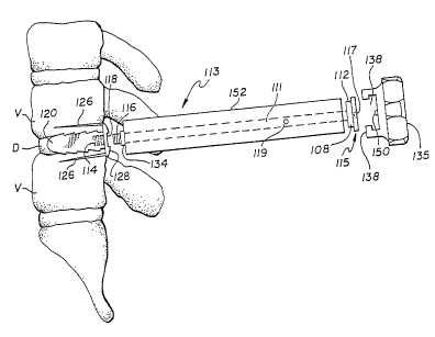
Referring now to Figure 1, preferably after
removing some portion of nuclear disc material, a Long
Distractor 100 is inserted under direct vision into the
intervertebral space. The disc penetrating portion 102 is
essentially cylindrical with a bullet-shaped front end 103
and a shoulder portion 104 where the penetrating portion
102 extends from barrel 106. The penetrating portion 102
urges the vertebral bodies apart, facilitating the
introduction of the instruments. Long Oistractors with
sequentially increasing diameter penetrating portions 102
are then introduced. As the optimal diameter of
penetrating portion 102 is achieved, the vertebral bodies
to either side are forced into full congruence and thus
become parallel, not only to the penetrating portion 102,
but to each other. At this time, any remaining
excrescences of bone of the posterior vertebral bodies
adjacent the posterior disc which have not already been
removed are flattened flush to the vertebral body by the
forced impaction, such as by hitting with a hammer flat
surface 109 of crown 110, driving the shoulder 104 against
the lipped portions of vertebrae V. Because of the forced
opposition of the vertebral endplates to portion 102 with
optimal distraction, unit 100 will then come to lie
absolutely perpendicular to the plane of the posterior
bodies and absolutely parallel to the vertebral endplates,
SUBSTITUTE SHEET (RULE 26)
CA 02521196 1994-06-09
WO 94128814
216 4 8 5 9 ~~4J06345
44
allowing optimal alignment for the procedure to be
performed.
Penetrating portion 102 is available in various
diameters, but all are of a constant length, which is less
than the known depth of the interspace. This combined with
the circumferential shoulder 104, which is too large to fit
within the interspace, protects against the danger of
overpenetration. Barrel 106 is of the same diameter as the
external diameter of the device to be implanted. A
recessed portion 108 below the crown 110 allows for the
Long Distractor 100 to be engaged by an extractor unit
shown in Figure 9.
In the preferred embodiment, a Convertible Long
Distractor 113 is used on the first side of the spine. As
shown in Figures 2, the Convertible Long Distractor 113 has
a barrel portion 152 separable from the Short Distractor
portion 120. While the initial distraction may be
performed with a solid Long Distractor, as the optimal
distraction is approached the appropriate Convertible Long
Distractor is utilized. The Convertible Long Distractor
113 consists of a Short Distractor portion 120 and a barrel
152 having a rectangular projection 134 at one end. The
Short Distractor 120 has an increased diameter head 128, a
rectangular slot 118 and an internal threaded opening 114.
The barrel 152 is hollow and has an internal shaft 111
terminating in a large diameter hexagonal crown 115 at one
end and a reduced diameter portion 112. The crown has a
detent portion 117 in its flat surface. The other end of
SUBSTITUTE SHEET (RULE 26)
CA 02521196 1994-06-09
WO 94128824
PCTIUS94/06345
the shaft 111 has a threaded small member 116 that
corresponds to threaded opening 114. The shaft 111 is
prevented from removal from the barrel 152 by set pin 119
passing through the wall of barrel 152 in a convenient
5 manner. The Short Distractor portion 120 is removably
attached to the barrel portion 152 via the mating of female
rectangular slot 118 and the male mating member 134. The
mating held together by utilizing knob 136 to drive the
crown 110 connected to interior shaft 111 having a threaded
l0 working end screw 116 that threads into the female aperture
118 of the Short Distractor portion 120.
Cap 136 has an open socket 138 for fitting around
crown 115 and engages the reduced diameter hexagonal
portion 112 so as to permit the rotation of shaft 111 and
15 threaded male member 116. A detent ball 150 in the inside
of the socket 138 engages detent 117 in the crown 115,
holding them together.
The Short Distractor portion 120 of Figures 2, 3,
and 3A-3F are designed to provide for high stability when
2o temporarily situated so as to resist inadvertent migration
while the surgeon is working on the second side. To that
end, the embodiment of the Short Distractor 120 shown in
Figures 3 and 3A has a pair of sharp pegs 126, to embed
into the opposing vertebral bodies and forward facing
25 ratchetings 124, that further resist backward movement.
Figures 3B and 3C, which show the preferred embodiment, are
side and top views of an alternative embodiment of the
distractor portion such that the distractor portion to be
SUBSTITUTE SHEE? (RULE 26)
CA 02521196 1994-06-09
WO 94!Z8824 216 4 8 5 9 ~T~S94106345
46
interposed between the vertebrae is essentially
cylindrical, but with circumferential forward facing
ratchetings 124.
A further alternative embodiment is shown in
Figures 3D and 3E. This is a more rectangularized design,
with forward facing ratchetings, without the sharp prongs
126 of Figure 3. Figure 3F is a side view of a further
embodiment of the Short Distractor 120 shown with knurling,
to increase the interference with the bone surface so as to
add stability to the unit and to resist dislodgment. To
this end, it is apparent that the working ends of both the
Long and Short Distractors can have a variety of
configurations consistent with their purpose, and that
surface irregularities as well as the shape of the ends
themselves, with or without prongs 126, may be utilized to
make the Short Distractor 120 more resistant to migration.
Once the ideal distraction has been achieved an
the first side of the spine, the Convertible Distractor is
dissociated, leaving Short Distractor 120 in place with its
rounded external end 128, safely on the canal floor and
deep to the dural sac and nerve root.
As shown in Figure 4, the surgeon then moves to
the other side of the spine at the same disc (D) level, and
retracts the dural sac and nerve root medially, exposing
the disc on that side. Long Distractors 100 are then
sequentially inserted into the disc space until the
diameter of the distractor on the second side is at least
as big as that on the first side. If because of some
SUBSTITUTE SHEET (RULE 26~
CA 02521196 1994-06-09
WO 94/28824 PCTIUS94/06345
2164859
47
asymmetry of the interspace a larger diameter distracter is
required on the second side to achieve the ideal
distraction as compared to the first side, then the second
side is fitted with a Short Distracter of the larger
diameter, and the surgeon would then return back to the
first side. In that event, the first side Short Distracter
would then be removed and the Long Distracter 100
corresponding to the increased diameter of the already
placed Short Distracter 120 would then be inserted. In
either event, the operation is continued by working on the
one side where the Long Distracter is in place. In this
regard, it should be noted, that by the use of such a
device as the Michelson Spinal Surgery Frame, it may be
possible to obtain adequate distraction preoperatively such
that the surgeon is either disinclined to use a distracter,
or to simply place the correct Long Distracter on the f first
side and then proceed with the surgical procedure on that
side before moving to the opposite side. These variations
are within the scope of the present invention.
The Long Distracter now serves as both a
centering post and an alignment rod for the hollow Outer
Sleeve 140 shown in Figure 5 which is fitted over the Long
Distracter 100, shown by phantom lines 101 in Figure 5.
The Outer Sleeve 140 is metal and has a sharp toothed front
end 142 that is capable of penetrating into and holding
fast the two adjacent vertebrae (V). Interrupting the
circumferential sharp teeth of 142 are flat, planar areas
152 which serve to resist the further insertion of the
SUBSTITUTE SHEET (RULE 26)
CA 02521196 1994-06-09
WO 94/Z8824
2 i 6 4 8 5 9 ~"'~4~~~45
48
sharp teeth into the vertebral bodies. The toothed front
end 142 of the Outer Sleeve 140 is a continuation of the
tubular shaft 144, which in turn is connected to
circumferentially enlarged tubular back end 146 having a
knurled outer surface 148 for easier manipulation. An
alternative embodiment of an outer Sleeve incorporates an
expansile key hole and slot configuration 154 to either
side of shaft 144 along the mid-plane of the interspace and
parallel to it such that the end 142 resists the collapse
of the vertebrae (V) to either side of the disc (D) , but
may nevertheless allow for their further distraction, in
the event the only diameter or the root diameter of the
implant is larger than the hole drilled.
A Driver Cap 160 in the form of an impaction cap
has at its far end a flat, closed-back surface 162 and at
its other end a broad, circular opening. The Driver Cap
16o fits over both the Outer Sleeve 140 and the Long
Distractor 100. As the Driver Cap 160 is seated, interior
surface 170 circumferentially engages portion 146 of the
Outer Sleeve until the back end 172 engages the internal
shoulder 164. As mallet blows are applied to surface 162,
that force is transmitted via the internal shoulder 164 to
the Outer Sleeve 140 via its far end 172, seating teeth 142
into the vertebral bodies adjacent the disc space D and to
the depth of the teeth 142 to the flat portions 152. As
the Outer Sleeve 140 is advanced forward, crown portion 110
of the Long Distractor is allowed to protrude within the
Driver Cap 160 unobstructed until it contacts the interior
SUBSTITUTE SHEET (MULE 26y
CA 02521196 1994-06-09
WO 94!?.8824 PCT/US94/06345
2164859
49
flat surface 168. Once crown 110 comes into contact with
the flat interior surface 168, then further taps of the
mallet will not advance the outer Sleeve, any further
motion being resisted by the flat shoulder portion 104 of
the Long Distractor abutting the hard surfaces of the
posterior vertebral bodies. In this way, the Outer Sleeve
140 is safely and assuredly inserted to its optimal depth
and rigidly securing the two opposed vertebrae as shown in
Figure 6.
The Cap 160 is then removed and the Distractor
Fuller 200 of Figure 9 utilized to remove the Long
Distractor 100 from the spine leaving the Outer Sleeve 140
in place. The Distractor Fuller 200 has front portion 202,
a mid portion 204, and a back handle portion 206. At the
front portion 202 of the Distractor Pullet 200, a socket
208 is connected to one end of shaft 210 which at its far
end is connected to back handle portion 206. The socket
208 has defined within it a cavity 212 that is open at its.
front end and funnelized on the interior aspect of its
sides. The Cavity 212 is constructed so that the head of
the Distractor Fuller 200 and the partially circumferential
flange 218 engages the circumferential recess 108 of the
Distractor 100. The entrance to cavity 212 is slightly
funnelized, and the leading edges of flange 218 slightly
rounded to facilitate the engagement of recess 108 and head
110 of Distractor 100, which is further facilitated in that
the Driver Cap 160 leaves portion 108 of Distractor 100
precisely flush with the back surface 172 of the Outer
SUBSTITUTE SHEET (RULE 26)
CA 02521196 2006-07-06
,78406-lE
Sleeve 140. This provides a large, flat surface 172 to
precisely guide surface 230 of socket 208, and open portion
212 around head 1i0 while flange 218 engages recess 108.
The springloaded detent hall 228 engages hemispherical
5 depression 112 in the crown 110, shown in Figure 2. This
springloaded detent 228 in engagement with complimentary
indent 218 protects against the inadvertent dissociation of
the Long Distractor from the Fuller 200 after the
Distractor has been removed from within the Outer Sleeve
10 14D and prior to its removal from the wound. Once out of
the body, the two instruments are easily disassociated by
freeing the crown portion 110 from cavity 212 by a manual
force applied perpendicular to their relative long axes at
this location.
15 A cylindrical and free removable weight 216 is
fitted around shaft 210 between the front portion 202 and
the rear handle portion 206. Gently, but repeatedly
sliding the weight 216 along shaft 210 and driven
rearwardly against a flat surface, transmits a rearward
20 vector to proximal end 202 and thereby to the Long
Distractor 100 to which it is engaged.
Paired extended handle 224 and 226, allow the
surgeon to resist any excessive rearward motion as the
instrument is used to liberate the Long Distractor 100.
25 Paired handles 224 and 226 are also useful in that they
allow a rotational directing of portion 208, via the shaft
210. This allows the surgeon to control and manipulate
rotationally the orientation of the opening of cavity 212
CA 02521196 1994-06-09
WO 94128824
216 4 8 5 9 p~~S94106345
51
to facilitate its application, to the head 110 of the
distracter 100.
The Distracter Puller 200 is a significant
improvement over the alternatives of striking a remover
instrument with an independent hammer over the exposed
surgical wound, or manually extracting the distracter by
forcefully pulling. The use of a free hammer over the open
wound is dangerous because the neural structures can be
impacted on the back swing which is made even more likely
by the effects of gravity on the mallet head. Manual
extraction by pulling is dangerous because of the
significant interference fit of portion 102 within the
spine such that significant farce would be required to
remove the Distracter 100, and if force were not coaxial
then the Outer Sleeve might be dislodged or misaligned.
Further, once the flat portion 102 became free of the
interspace, all resistance to withdrawal would be lost and
in the face of the considerable force necessary to free it,
the Distracter 100 might easily become projectile imparting
2o injury to the patient and/or the surgeon.
Once the Long Distracter 100 has been fully
removed from the Outer Sleeve 140, the toothed end 142 of
the Outer Sleeve 140, working in conjunction with the Short
Distracter 120 on the contralateral side rigidly maintains
the relative position of the adjacent vertebrae V.
Further, since the remainder of the procedure on that side
of the spine occurs entirely through the protective Outer
Sleeve 140, and as the nerves and dural sac are external to
SUBSTIT1JTE SHEET (RULE 26~
CA 02521196 1994-06-09
1~V0 94!288I4
21 b 4 8 5 9 ~T~~4/06345
52
that outer Sleeve and superficial to the toothed end 142 of
the outer Sleeve 140, which is firmly embedded into the
adjacent vertebrae V, the Outer Sleeve 140 serves to insure
the safety of these delicate neural structures. Further,
since the Outer Sleeve 140 is of a fixed length and rigid,
its flat rearward surface 172 may be used as a stop to the
advancement of all instruments placed through the Outer
Sleeve 140, thus protecting against accidental
overpenetration. Further, the Outer Sleeve 140 assures
that the further procedure to be performed will occur
coaxial to the disc space D and further, be symmetrical in
regard to each of the opposed vertebral surfaces.
Figure 10B is a posterior view of the spine at
this stage of the procedure, showing a short Distractor 120
in place on one side of the spine and the bottom portion of
Outer Sleeve 140 in place on the opposite side of the
spine.
Referring to Figure 11A, an Inner Sleeve 242 is
inserted from the rear within the Outer Sleeve 140. This
Inner Sleeve has a collar portion 244 of a known thickness
which seats against the top edge surface 172 of Outer
Sleeve 140. The cylindrical barrel portion of Inner Sleeve
242 comes to approximate the posterior aspect of the
vertebral bodies interior the Outer Sleeve when fully
seated. A Drill 240, having a known selected length is
then introduced through the rearward aperture of the Inner
Sleeve 242 and utilized to ream out the arcs of bone which
it engages from the opposed vertebral endplates as well as
SUBSTITUTE SHEET (RULE 26)
CA 02521196 1994-06-09
WO 94128821
216 4 8 5 9 ~~S94106345
53
any discal material within its path down to its
predetermined and limited depth. The Drill 240, has a
narrow engagement portion 246, which allows it to be
affixed to a drill mechanism which may be either a manual
or a power unit. A circumferential collar 248 of an
increased diameter serves to limit the depth of penetration
of the drill 240 and may be fixed, or lockably adjustable.
Not shown here, but well known to those skilled
in the art, are various mechanisms to lockably adjust such
l0 instruments as drills. Such mechanisms include, but are
not limited to, the use of collets, threaded shafts with
lock nuts, and flanges engaging grooves forced therein by
either a cap pulled over the flanges or screwed down upon
them.
In the preferred embodiment, the forward cutting
edge 252 of Drill 24o is a modification of a large fluted
drill design such that the end resembles an end cutting
mill which may contain any workable number of cutting
surfaces, but preferably four or more, and such cutting
surfaces being relatively shallow such that the advancement
of the instrument occurs more slowly. The outside diameter
of the Drill 240 corresponds to the minor diameter of the
threaded spinal implant. The Inner Sleeve 242 has an inner
diameter slightly greater than that dimension and its outer
diameter is slightly smaller than the inside diameter of
the outer Sleeve 14o which has the same outer diameter as
the major diameter of the threaded implant.
SUBSTITUTE SHEET (RULE 26)
CA 02521196 2006-07-06
78406-lE
54
The drill shaft of drill 240 comprises an upper
portion 243, a central recessed portion 256 of a smaller
diameter and a lower cutting drill portion 250. The upper
portion 243 and lower portion 250 of the drill 240 have the
same outside diameter.
The Inner Sleeve 242 serves many functions.
First, it provides a more intimate drill guide for drill
240 in the event a smaller diameter hole is to be drilled
than that of the inside diameter of the Outer Sleeve 140.
Second, since it now guides the Drill, it allows for the
Outer Sleeve 140 to have an internal diameter large enough
to admit the threaded spinal implant, which is indeed
considerably larger in diameter than the Drill 240 itself.
If a larger outer Sleeve 140 were utilized absent
the Inner Sleeve 242, then the Drill 240 would be free to
wander within the confines of that greater space and would
not reliably make parallel cuts removing equal portions of
bone from the adjacent vertebrae V. Further, the bone
removal not only needs to be equal, but must be correctly
oriented in three dimensions. That is, the path of the
Drill 240 must be equally centered within the disc space,
parallel the endplates, and parallel to the sagittal axis
dissecting the interspace.
A further purpose of the Inner Sleeve 242 is that
it may be. removed simultaneously with the Drill 240,
thereby trapping the debris, both cartilaginous and bony
generated during the drilling procedure, which are guided
rearward by the large flutes 251 of Drill portion 250,
CA 02521196 1994-06-09
WO 94/2882~i
216 4 8 5 9 p~~S94106345
where they are collected around recessed portion 256
between the recessed portion 256 and the inner wall of the
Inner Sleeve 242 are there contained therein. Thus, by
removing the Drill 240 in conjunction with the Inner Sleeve
5 242, all of the debris generated by the reaming procedure
is safely removed from the spinal canal and wound area.
Further, if the disc tissue in the area to be
reamed has been removed previously, as per the preferred
method, then the patient's own bone of good quality and
l0 useful within the operation will then be contained between
the Inner Sleeve 242 and the shaft portion 256. Once away
from the surgical wound, this material may be used to load
the spinal implant or placed deep within the interspace to
participate in the fusion.
15 The method of actually producing the surgical
hole within the spine is variable. As shown in Figure 11C,
in an alternative embodiment Drill end 250 has a forward
projecting nipple 260, which itself is bullet-shaped in its
leading aspect so as to ease its entrance into the disc
20 space and to urge the vertebrae apart. Nipple 26o is
distracting, stabilizing as it resists any tendency of the
vertebrae to move together, is self-centering to the Drill
portion 250 when working in conjunction with Sleeves 140
and 242, and virtually assures the symmetrical resection of
25 bone from the opposed vertebral surfaces.
The alternative "Trephine Method" referred to
earlier in this application, is shown in Figure 11B. In
this alternative, a Long Distractor 100 is left in place
SUBSTITUTE SHEET (RULE 26)
CA 02521196 1994-06-09
WU 94/28824
2 ~ b 4 8 5 9 ~~~4106345
56
after the Outer Sleeve 140 is seated. The Long Distractor
100 in this case differs from the Long Distractor of the
preferred embodiment in that its outside diameter of the
barrel 106 is of a smaller diameter than in the prior
version. This is made necessary because regardless of the
method, the hole to be formed corresponds to the minor
diameter of the spinal implant. Trephine 270, a hollow,
tubular member with sharp cutting teeth 251 at its proximal
end, has a wall thickness and since the outside diameter of
that trephine 270 must correspond to the root diameter of
the implant, then the wall thickness of the trephine 270
must be allowed for by a corresponding reduction in the
diameter of the Long Distractor 100.
A further modification of the Long Distractor 100
to the "Trephine Method" would use longitudinal grooves
(not shown) along the barrel surface 106 for the purpose of
transmitting any debris generated during the cutting
procedure, rearward. Since the cutting element is both
centered and aligned by the Long Distractor, the use of the
Inner Sleeve 242 is not mandatory, but may once again be
useful in controlling the path of the debris. To that end,
little debris is generated in the "Trephine Method" as the
bony arcs are not so much being reamed out and removed as
they are simply being cut into the bone where these arcs of
bone are left connected at their far ends: Thus, when the
Trephining Method has been completed and the Trephine 270
and Inner Sleeve 242 removed, unlike in the preferred
embodiment where the hole is drilled out, it remains
S~!!?STITu : E SHEET (RULE 26
CA 02521196 2006-07-06
78406-lE
57
necessary to remove both the two arcs of bone, and any
interposed material. Nevertheless, this is very easily
performed by various means, one of which is depicted in
Figure 11D.
Instrument 272 consisting of a shaft 276 attached
off center to the lower surface 273 handle 274. The shaft
276 terminates in a cutting arm 278. The instrument 272 is
inserted through Outer Sleeve 140 where the lower surface
273 of handle 274 abuts the top 172 of the Outer Sleeve
140, both stopping downward motion of instrument 272 and
precisely placing the perpendicularly cutting arm 278 of
instrument 272 so that as handle portion 274 is rotated,
the cutting arm 278 is also rotated, cutting the arcs of
bone and liberating them from their last attachments.
These portions of bone are then removed utilizing this
instrument or a long forceps, and then placed within the
implants or otherwise used to participate in the fusion.
4~hile in the preferred embodiment of the present
invention the spinal implant I, is essentially
self-tapping, if the bone is unusually hard it may be
desirable to form the thread pattern within the interspace
prior to the insertion of the implant I. To that end, as
shown in Figure 12, Tap 280 has a threadcutting portion 282
connected by a shaft 286 to a handle portion 292, which has
been designed to give mechanical advantage to the rotation
of the instrument for the purpose of cutting threads. The
lower portion of handle 290 has a forward facing flat
surface 288 too large to fit through the opening of outer
CA 02521196 1994-06-09
WO 94128824 216 4 8 5 9 PCT~S94106345
58
Sleeve 140 which thus safely limits the depth of
penetration of the cutting element 282. This tap 280 is
further made safe by blunt end 294 which will engage the
uncut portions of the vertebral bone just prior to the
engagement of shoulder 288 against surface 172. This
feature allows the surgeon to appreciate a less harsh
resistance as the blunt nose 294 encounters the remaining
unresected bone for the drill hole and prior to the sudden
increase in resistance caused by the seating of shoulder
288 against top edge 172, which first resistance serves as
a warning to the surgeon to discontinue the tapping
procedure. Thus, the surgeon has both visual (as shoulder
288 approaches top edge 172) and tactile warnings to avoid
stripping the thread form. Tap end 2B2 is highly
specialized for its specific purpose. Rearward to the
- specialized blunt tip 294 is a truncated bullet-shaped area
298 Which ramps up to the constant diameter intermediate
the cutting ridges 296. Ramp portion 298 urges the opposed
vertebral bodies apart, which motion is resisted by Outer
Sleeve 140, thus progressively driving the sharp leading
edges of thread forms 296 into the vertebral bodies. The
periodic longitudinal grooves 284 interrupting the thread
forms, which may number 1 to 8, but preferably 4, function
to accumulate the bony material which is removed during the
thread cutting process. In that regard, in the ideal
embodiment, the thread cutting form is designed to compress
the bone to be formed rather than to trough through it.
Further, while both the major and minor diameters of the
SUBSTITUTE SHEET (RULE 26)
CA 02521196 1994-06-09
WO 941288?R PCT/US94/06345
2164859
59
Tap 280 may be varied, in the preferred embodiment, the
minor diameter corresponds to the minor diameter of the
implant I, but the major diameter is slightly less than the
major diameter of the implant.
With Tap 280 now removed, and Sleeve 140 still in
place, the surgical site is now fully prepared to receive
the spinal implant I. In the preferred embodiment of the
spinal implant, the implant has been enhanced by the use
of, application to, and filling with fusion promoting,
enhancing, and participating substances and factors. Thus,
the implant may be fully prepared for insertion as provided
to the operating surgeon. However, at the present time,
human bone is most commonly used as the graft material of
choice, with the patient's own bone being considered the
best source.
Figure 14a shows a trephine 300 with an
exceedingly sharp front cutting edge 302 for quickly and
cleanly coring into the patient's posterior iliac crest, or.
any other bony tissue, and for the purpose of producing a
core of bone then contained within the hollow 304 of the
trephine 300. Trephine 300 has a rear portion 306 with a
pair of diametrically opposed slots 310, and disposed
clockwise from their longitudinally oriented rearward
facing openings so as to engage diametrically and opposing
members 312 of Drive unit 308, by which trephine 300 may be
attached to either a hand or power drill. It can be
appreciated that engagement mechanism 312 is stable during
the clockwise cutting procedure, and yet allows for the
SUBSTITUTE SHEET (RULE 25~
CA 02521196 2006-07-06
78406-lE
rapid disconnection of the two components once the cutting
is completed.
Because of the high interference between the
graft and the inner wall of hollow portion 304, and the
5 relative weakness of the cancellous bone being harvested,
it is possible to remove the Trephine 300 while still
drilling, and to have it extract the core of bone with it.
However, in the highly unlikely event that the core of bone
would remain fixed at its base, then with the drive
l0 mechanism 308 removed, a corkscrew 408 shown in Figure 14C
is introduced though the central opening of rear portion
306 and threaded down and through the core of bone within
304 and to the depth of teeth 302. The tip 318 of the
corkscrew 408, which extends substantially on line with the
15 outer envelope of the corkscrew, then cuts radially through
the base of the bone core. As the handle portion 314 of
the corkscrew 408 abuts the flat, rearward surface of
portion 306 and it can no longer advance. As corkscrew 408
is continued to be turned further, it will cause the core
20 of bone to be pulled rearward, as in removing a cork from
a wine bottle. Trephine 300 has a barrel portion 304
continuous With sharp toothed portion 302 having an inner
diameter just less than the inner diameter of the spinal
implant I to be loaded.
25 The Trephine 300 with its core of harvested bone
is then placed as shown in Figure 14B, through an open end
of Implant Bone Loading device 320, where the barrel
portion 304 then passes through and is stopped by circular
CA 02521196 2006-07-06
78406-lE
61
flange 344. The plunger shaft 326 of instrument 320 is
then prepared for attachment by rotating knob 332
counterclockwise such that the plunger 372 is pulled via
the long threaded shaft portion 328 back to the base of
collar 330 at its proximal end. In this position, knob 332
is considerably extended rearward from collar 330. With
plunger shaft 326 in this position, the plunger head 372 is
inserted into the central hollow of portion 306 of Trephine
300 as the proximal cylindrical portion of collar 330 then
follows it, such that the plunger 372 then occupies the
rearward portion of barrel 304 and the proximal cylindrical
portion of collar 330 occupies the central hollow of
portion 306. A pair of diametrically opposed radially
projecting arms 346 on collar 330 are then advanced
longitudinally into diametrically opposed paired L slots
341 and then rotated clockwise to complete this assembly.
At the other end of instrument 320, a spinal
implant I is engaged through its female rectangular slot
364 by a rectangular protruding bar extending from rearward
facing surface of end plug 324, (not shown) and secured
there by knob 334 which extends as a rod through a central
aperture within end plug 324 to extend at the far end as a
small bolt which threads to a female aperture centered
within the female slot 364 of the spinal implant. With the
spinal implant I secured to end plug 324 and the opposite
end of the implant I presenting as a hollow, tubular
opening, end plug 324 is advanced into device 320 where it
CA 02521196 1994-06-09
WO 94128824
216 4 8 5 9 ~~594106345
62
is secured by rotationally engaging diametrically opposed
L-shaped slots 321. With device 320 fully assembled, end
302 of trephine 300 lies coaxial and opposed to the open
end of implant I.
As shown in Figure 15, as knob 332 is then
rotated clockwise, the plunger 372 proximal the threaded
shaft 328 is then forcibly, but controllably driven forward
down the barrel 304 ejecting the bone graft directly into
the spinal implant I. As the bone graft is greater in
l0 length than the interior of the spinal implant, with
further compression the bone is forced into the radially
disposed apertures through the wall of the device
communicating from the central cavity to the exterior.
End plug 324 is then removed from apparatus 320.
Using end plug 324 as a handle, end cap 374 shown in Figure
16 is secured to the open end of the spinal implant I. The
implant is then disassociated from end plug 324 by rotating
knob 334 counterclockwise.
Figure 16 shows a.n Implant Driver instrument
which may be used to either insert or to remove said
implant I. Driver 350 has at its far end 362, a
rectangular protrusion 398, which protrusion intimately
engages the complimentary rectangular slot 3.64 of implant
I. Protruding from slot 398 of end 362 is threaded portion
353, which extends as a rod through hollow shaft 358 and
hollow hand barrel 360 to knob 354 where it can be
rotationally controlled. Threaded portion 353 screws into
a female aperture central slot 364, urging 353 into 364,
~JB~TITLITE SHEtT (RULE 26)
CA 02521196 1994-06-09
WO 94128824 PCTIUS94106345
21b4859
63
and binding them together such that instrument 350 can be
rotated via paired and diametrically opposed extending arms
366 and in either direction while maintaining contact with
the implant.
Affixed to the Driver 350, the implant is then
introduced through the Outer Sleeve 140 and screwed into
the interspace opposed between the two prepared vertebrae
V until such time as the leading edge of the Implant Cap
374 reaches the depth of the prepared hole at which time
its forward motion is impeded by the bone lying before it
which had not been drilled out. This allows for a
progressive feel to the surgeon as the implant is screwed
home.
As described previously, with the use of the Tap
280, this tenainal resistance to further seating provides
significant tactile feedback to the surgeon. Again, as
with the Tap 280, visual monitoring of the depth of
insertion of the implant is provided to the surgeon by
observing the progressive approximation of the forward
surface 370, of barrel portion 360, as it approaches the
rearward facing surface 172 of outer Sleeve 140.
Nevertheless, a final safety mechanism, when the full depth
of insertion has been achieved, surface 370 of instrument
350 will abut surface 172 of the Outer Sleeve 140,
prohibiting any further installation of the spinal implant.
Once the implant has been fully installed, the
Driver 350 is dissociated from the implant by turning knob
354 in a counterclockwise direction. The Driver 350 is
S~JBSTITUTE SHEt~ RULE 2fi~
CA 02521196 2006-07-06
,78406-lE
64
then withdrawn from the outer sheath, then the outer Sleeve
140 is removed. This leaves the implant fully installed
and inset to the determined depth as shown in Figure 18.
Attention is then redirected to the other, or
first, side of the spine. A dural nerve root retractor is
used to retract the neural structures medially, bringing
into full view the head 128 of the Short Distractor 120,
lying flush on the canal floor. Utilizing apparatus 152,
extended screw portion 116 is inserted into the female
threaded portion 114 of the Short Distractor 120 as the
extended rectangular portion 134 of apparatus 152 is
engaged to. the female rectangular portion 118 of the Short
Distractor 120. Then turning rearward facing portion 108
and head 110, utilizing the knob 136 of Figure 2, the bong
Distractor configuration is restored.
With the dural sac and nerve roots still
retracted and protected, the Outer Sleeve 140 is slipped
over the reconstituted Long Distractor and seated using the
Driver Cap 162. The entire sequence of events as described
for the implantation of the spinal implant I as already
placed, is then repeated such that both spinal implants
come to lie side by side within the interspace. Though not
necessary, circlage or other internal fixation of the
levels to be fused may additionally be performed, and then
the wound is closed in the routine manner.
Brief Discussion With Reference To The Drawinqsof The
Preferred Method And Instrumentation For Anterior Interbod
Fusion
CA 02521196 1994-06-09
WO 94128824 216 4 8 5 9 pCT~S94/06345
Incorporating Intercorporeal
Predistraction And Utilizing A Guarded Sleeve
stem Is Disclosed
Because of the absence of the spinal cord and
5 nerve roots, it is generally possible to visualize in one
instance the entire width of the disc space from side to
side throughout the cervical, thoracic, or lumbar spine.
In the preferred embodiment of the anterior interbody
fusion, implants are placed side by side from anterior to
l0 posterior parallel to the interspace and extending through
into the adjacent vertebral bodies. Where the transverse
width of the disc space is insufficient to allow far the
use of two implants, each of which would be large enough to
protrude to the required depth into the adjacent vertebrae,
15 then a singular and significantly larger implant may be
placed centrally. With this in mind, and in light of the
very detailed description of the technique and
instrumentation already provided in regard to the method of
posterior lumbar interbody fusion, a brief discussion of
20 anterior spinal interbody fusion with dual implant
installation will suffice, and the method for installation
of a large, singular midline graft will become obvious.
The interspace to be fused is exposed anteriorly.
The soft tissues are withdrawn and protected to either
25 side, and if necessary, above and below as well. It is
then possible to visualize the entire width of the
vertebrae anteriorly adjacent that interspace. As
discussed above, the surgeon has already templated the
SUB~STITUTt SHEET (R~L~ ~6)
CA 02521196 1994-06-09
PCT/LTS94106345
Wo 94'4 216 4 8 5 9
66
appropriate patient radiographs to determine the requisite
distraction and optimal implant size. In the preferred
method, the surgeon then broadly excises the great bulk of
the nuclear disc portion. (Alternatively, the disc can be
left to be removed via the drill later.) The surgeon then
notes and marks a point midway from side to side
anteriorly. He then inserts Long Distractor IOD centering
it on a point midway between the point just noted and the
lateral extent of the intervertebral space visualized
anteriorly. The outer barrel portion 106 of the Distractor
100 utilized, will correspond to the outside diameter of
the implants to be installed. The Distractor tips 102
inserted are sequentially larger in diameter until the
optimal distraction is achieved. This optimal distraction,
although suggested by the initial templating, may be
visually and tactilely confirmed as performed. When the
optimal distraction is achieved, the vertebral endplates
will come into full congruence and parallel to the forward
shaft portion 102 of the Distractor 100, causing an
alteration in the alignment of the vertebrae and a
significant increase in the interference fit and
pressurization at the tip, such that the instrument becomes
exceedingly stable.
There is a sensation imparted to the surgeon of
the tissues having moved through their elastic range to the
point where the two adjacent vertebrae V begin to feel and
move as if a single solid. These changes are easily
appreciated visually as the vertebrae realign to become
SUBSTITUTt SHEEP (RULE 26)
CA 02521196 1994-06-09
WO 94/28824
216 4 8 5 9 ~~594106345
67
congruent to tip 102, and can also easily be appreciated
via lateral Roentgenography. However, should the surgeon
fail to appreciate that optimal distraction has been
achieved and attempt to further distract the interspace, he
would find that extremely difficult to do because of the
increased resistance as the tissues are moved beyond their
range of elastic deformation. Further, there would be no
elasticity left to allow the vertebrae to move further
apart and the sensation to the surgeon should he attempt to
gently tap the oversized Distractor forward with a mallet,
would be one of great brittleness.
Returning now to the procedure, when the correct
intercorporeal Distractor 100 producing the ideal
interspace distraction having its barrel portion 106
corresponding to the implant to be installed has been
inserted, then its exact duplicate is inserted anteriorly
equidistant to the other side of the spine. As the barrel
portion 106 of Long Distractor 100 is exactly of the same
major diameter as the spinal implant I looking coaxially on
end, the surgeon can then asses the anticipated side by
side relationship of the dual implants when implanted.
As shown in Figures 7C and 7D, a Dual Outer
Sleeve 340 consisting of a pair of hollow tubes is then
introduced over the side by side Long Distractors
protruding anteriorly from the spine. The Dual Outer
Sleeve 340 is comprised of two hollow tubular members
identical in size displaced from each other ideally the sum
of the difference between the minor and major diameters of
SUBSTITUTE SHEET (RULE 2~3
CA 02521196 2006-07-06
78406-lE
68
both implants combined, but not less than that difference
f or one implant, as it is possible to have the threads of
one implant nest interposed to the threads of the other,
such that they both occupy a common area between them.
However, while the preferred embodiment is slightly greater
than two times the difference between the major and minor
diameters of the implant (the sum of both) the distance may
be considerably greater. Whereas in the preferred
embodiment extending tubular portions 348 of instrument 340
are parallel, when the area between them 351, is
sufficiently great, these elements may be inclined or
declined relative to each other such that they either
converge or diverge at their proximal ends. Paired tubular
structures 348, may be bridged in part or wholly throughout
their length, but are rigidly fixed by Foot Plate 344. In
its preferred embodiment, a top view shows the Foot Plate
to be essentially rectangular, but Without sharp corners.
Other shapes can be utilized. In side view 7D it
can be appreciated, that Foot Plate 344 is contoured so as
to approximate the shape of the vertebrae anteriorly.
Extending forward from Foot Plate 344 are multiple sharp
prongs 342 sufficiently long to affix them to the
vertebrae. The prongs 342 are limited in length so as to
not penetrate too far posteriorly and number from 2 to 10,
but preferably 6. As the Dual Outer Sleeve 340 is driven
forward utilizing Dual Driver Cap 420, of Figure 7E,
engaging the rearward end 352, the prongs 342 extending
CA 02521196 1994-06-09
~'~'O g4PCT/US941D6345
2164859
69
from Foot Plate 344 are embedded into the opposed vertebral
bodies until their forward motion is inhibited by the
curved Foot Plate 344 becoming congruent to and being
stopped by, the anterior aspect of the vertebral bodies.
As already taught in Figure 5, the Dual Driver
Cap 420 is of the same design as Single Driver Cap 160, in
that there is a recess 354 as per 168, allowing the outer
Sleeve to be fully seated without impeding the rearward
projection of the Long Distractor unit. However, unlike in
Cap 160, area 354 is more relieved as it is unnecessary for
the Dual Cap 420 to contact the Long Distractor through
portion 110 to inhibit its forward motion, as the Foot
Plate 344 functions to that effect. Further, the Dual Cap
420 for the Dual Outer Sleeve 340 is correspondingly dual
itself and engages the rearward facing dual tubular portion
352. Once the Dual Outer Sleeve has been fully seated, the
vertebrae adjacent the interspace to be fused are rigidly
held via Foot Plate 344 and the prongs 342. Thus, it is
possible to remove either one, or if desired, both of the
Long Distractor rods utilizing Long Distractor puller 200,
as per the method already described. It is then the
surgeon's choice to work on one or both sides of the spine.
As per previous discussion, the surgeon may drill the
interspace utilizing the Inner Sleeve 242 or leave the Long
Distractors in place as per the "Trephine Method".
Tapping, if necessary, and the insertion of the
implants then occurs through the protective outer Sleeve
340. Once the implants have been fully inserted, the Outer
suesTfruTF s~~ ~RU« ~~
CA 02521196 1994-06-09
WO 94128824
216 4 8 5 9 P~~S94106345
sleeve is removed.
Having utilized the Drill method, or "Trephine
Method", with or without an Inner Sleeve to prepare the
fusion site, it is the preferred embodiment to leave the
5 Outer Sleeve 340 in place as it provides for the ideal
placement and aligrunent of the Tap 2s0 and implant I.
It is anticipated that the surgeon wishing to
work deep within the interspace, or preferring the ability
to directly visualize the tap being used, or the implant
10 being inserted, may choose to remove the Outer Sleeve after
the insertion of the first prosthesis to maintain
stability, or prior to that, which while not the preferred
embodiments, are nevertheless within the scope of the
present invention.
15 Alternatiye Methods To The Preferred Embodiment For
Method Of Anterior Interbody Fusion
As previously described for the posterior lumbar
spine, alternatively, one can employ the "Trephine Method"
as has been described in detail.
20 As a further alternative, it should be noted that
the key element in the anterior method is the use of the
predistraction principle, where such distraction is
maintained by the Outer Sleeve with or without the Long
Distractor. Therefore, once the preparation of the
25 interspace has been completed, while not the preferred
embodiment, it is nevertheless within the scope of this
invention that one could remove the Outer Slesve as there
SUBSTITUTE SHEEN' (RUB 26~
CA 02521196 2006-07-06
,78406-lE
71
are no neural structures requiring protection, and insert
the implants directly rather than through the Outer Sleeve.
As yet a further alternative of this method,
where the height of the distracted interspace is such that
the diameter of the implant required to span that height
and to embed with sufficient depth into the opposed
vertebral bodies is such that it is not possible to place
two such implants side by side, then only a single implant
which may be of significantly increased diameter, is used
and placed centrally within the interspace rather than to
either side. The placement of a singular central graft via
the present invention method and instrumentation is in
keeping with the methods already described and can be
performed using either a drill or the "Trephine Method".
Referring to Figures 16-18, a cylindrical
embodiment of the spinal implant I of the present invention
is shown. In Figure 16 the implant I is shown attached to
the insertion device 350. In Figures 17 and 18 the implant
I is shown installed in the disc space D, between the
adjacent vertebrae.
The cylindrical implant I comprises a hollow
tubular member which in the preferred embodiment is made of
an ASTM surgically implantable material, preferably
Titanium. The cylindrical implant I is closed at one end
and open at the other end covered by a cap 374~ The
cylindrical implant I has a series of macro-sized openings
390 through the side walls of the cylindrical implant I.
A series of external threads 392 are formed on the
CA 02521196 1994-06-09
~''~ '216 4 8 5 9 ~T~S94/06345
72
circumference of the cylindrical implant I. Any variety of
threads may be used on the implant. The cap 374 has a
hexagonal opening 394 for tightening the cap 374.
While the present invention has been described in
association with the implant of a threaded spinal implant,
it is recognized that other forms of implants may be used
with the present method. For example, dowels, made from
bone or artificial materials, knurled or irregularly shaped
cylinders or spheres, or any other shaped implants that can
ZO be introduced through the outer sleeve may be used. Being
able to perform the procedure through the outer sleeve
permits the procedure to be performed safely and quickly,
and more accurately.
SIJBS~'ITtITE SHEET ~R~1.E ~~