Note: Descriptions are shown in the official language in which they were submitted.
CA 02521567 2005-10-05
WO 2004/096033 PCT/GB2004/001780
MEASUREMENT OF DISTRIBUTION OF MACULAR PIGMENT
Field of the invention
This invention relates to apparatus for use in inspecting the density and
spatial distribution
of macular pigment in an eye, and to a method of determining said density and
spatial
distribution.
Background to the invention
Macular pigment is a yellow pigment situated in the central portion of the
human retina.
The absorption spectrum for the pigment has a peak for light of a wavelength
of 460nm
and zero for light for a wavelength of 540nm, so that the pigment absorbs
significant
amounts of the shorter wavelength light, whilst having little or no effect on
light of the
longer wavelength.
The highest concentrations of macular pigments are'to be found in the region
of the retina
which has a very high number density of cone receptors, and is coupled with a
disproportionately large area of the visual cortex, giving that region a high
degree of visual
acuity .
It has been proposed that the macular pigment protects the retina against
harmful effects of
short wavelength radiation, and accordingly much work has been devoted to
measuring the
optical density, and spatial distribution, of macular pigment in various
subjects in order to
determine whether there is any correlation between irregularities in the
amount of macular
pigment present and certain defects.
CA 02521567 2005-10-05
WO 2004/096033 PCT/GB2004/001780
2
A flicker photometer is an instrument that enables a subjective measurement of
macular
pigment density to be made. The flicker photometer projects green and blue
light in an
alternating sequence into a subject's eye, and the subject is able to vary the
relative
intensity of light of one of those colours until a minimum or no flickering is
perceived.
Photographic methods have also been used to obtain an objective indication of
the macular
pigment density/spatial distribution, but in order to be effective, have
involved dilating the
subjects pupil, bleaching photo pigments to minimise their contributions and
then
photographing the fundus twice, once in blue light and once in green light.
Those images
are then digitised (if not already captured by a CCD camera), combined in
registration
with each other, logarithmically transformed and then subtracted.
However, ensuring that the images are precisely registered, is a time
consuming step
which places high demands on image processing software and hardware.
Summary of the invention
According to a first aspect of the invention, there is provided apparatus for
use in
measuring the density and spatial distribution of macular pigment in an eye,
the apparatus
comprising a camera for capturing a colour image of the retina of an eye under
examination, filter means for filtering light reaching camera, the filter
means having a
transmission spectrum which has a peak in the region of the .wavelength of
light absorbed
by the pigment and another peak in a region at which no such absorption occurs
A conventional colour camera can obtain a colour image from a single exposure,
but this
image, whilst providing a representation of the colour of the photographed
features, does
not have sufficient colour resolution for use in the measurements of macular
pigment/spatial distribution. However, the filter of the present invention
increases the
sensitivity of the apparatus to said macular pigment since the filter will
pass light having a
component at the peak of absorption of the macular pigment and another which
will be
CA 02521567 2005-10-05
WO 2004/096033 PCT/GB2004/001780
3
unaffected by the pigment, so that the captured image has a component which is
greatly
affected by macular pigment density and another, reference component which is
not.
Since both components are present in a single image, there is no need for
separate images
to be obtained, and the invention therefore also avoids the problem of
achieving image
alignment. In addition, a conventional camera can be used, so that apparatus
in
accordance with the invention may be relatively cheap to produce.
In order to provide good resolution, the filter means preferably has a
transmission
spectrum which is substantially zero between said two peaks. . To that end the
transmission
spectrum may to advantage not exceed 0.001 % between said peaks. Preferably
each peak
is no more than 40nm wide.
The filter means may be placed anywhere in the path of light which illuminates
the eye and
travels to an image capture device, for example one or more CCD arrays in the
camera.
Preferably, however, the filter means is situated in between the eye and an
illuminating
light source, so that the spectrum of light which illuminates the eye has said
peaks. Thus,
for example, a conventional 3 CCD array retinal camera, which typically has a
flash lamp
and an associated and interchangeable filter for the flash lamp, can be
converted into
apparatus according to the invention, simply by replacing the existing filters
with said filter
means.
Since the advantages of the invention can be achieved by selecting an
appropriate spectrum
of illuminating light, there is provided, in accordance with the second aspect
of the
invention, apparatus for use in the measuring of the density and spatial
distribution of
macular pigment in an eye under examination, the apparatus comprising
illumination
means for illuminating said eye and a camera for capturing a colour image of
the eye,
when so illunninated, wherein the illumination means is operable to illuminate
the eye with
light the spectrum of which has a first peak at a wavelength of light which is
absorbed by
the macular pigment and a second peak at a wavelength at which substantially
no such
absorption occurs.
CA 02521567 2005-10-05
WO 2004/096033 PCT/GB2004/001780
4
Preferably, the spectrum of said illuminating light falls to substantially
zero between these
two peaks.
The filter means preferably comprises a single alter having both said peaks in
its
transmission spectrum.
Preferably, one of said peaks is at the wavelength corresponding to blue
light, the other at
that corresponding to red light.
Preferably, said first peak is at 460nm, the second at 600nm.
The filter may conveniently be a triple bandpass filter, the transmission
spectrum of which
has a further peak and a wavelength corresponding to green light (e.g. 540nm).
The filter may be a proprietary item available from, for example, OMEGA
OPTICAL.
Preferably, the apparatus includes an image processor for processing the image
captured
by the camera, wherein the image processor is programmed to subtract the
reference
component of the image from the component in the absorption spectrum of the
macular
pigment, thereby to remove the contribution to the image of pigments other
than the
macular pigment.
Preferably, the processor is operable to display the results of the
subtraction as a macular
pigment map.
Preferably, said subtraction is of the logs of the intensities of the two
components.
Preferably the image processor is operable to take the logs of three images,
each
corresponding to a respective peak of the triple bandpass filter's
transmission spectrum,
CA 02521567 2005-10-05
WO 2004/096033 PCT/GB2004/001780
S
and to combine these so as to eliminate any contributions from non uniform
distributions of
both melanin and photopigments.
If, however, the haemoglobin and melanin are uniformly distributed in the
retina, they
will cause a uniform reduction in image intensity, which leaves only three
unknown
pigment distributions: macular pigment, rod photopigment and cone
photopigment.
In this case, the image processor is preferably operable to determine, from
the three
images, the distributions macular pigment, rod photopigment and cone
photopigment
across the retina.
According to a third aspect of the invention, there is provided a method of
measuring
macular pigment density and spatial distribution in an eye, the method
comprising the steps
of,
a) capturing a colour image of the retina of the eye, the image having a first
and
second colour component, the first colour component having a spectrum the peak
of
which is' at a wavelength at which the absorption by macular pigment is at a
maximum and a second peak at which substantially no such absorption occurs;
b) subtracting one of the image components. from the other, at each region of
. the
image, to remove at least some colour contributions not arising from the
macular
pigment and
c) providing an output representative of the contribution of the macular
pigment to the
image.
Preferably the step of capturing the image involves illuminating the eye with
light, the
spectrum of which has said first and second peaks.
CA 02521567 2005-10-05
WO 2004/096033 PCT/GB2004/001780
6
The image may be captured by means of a camera and a filter which has a first
and second
peak its transmission spectrum, corresponding to the two peaks of the
components, and
which filters the light forming the image captured by the camera. The filter
may be in the
path of light from the eye under examination to the camera, but is preferably
in the path of
light from a source of illumination to the eye.
Brief Description of Drawings
The invention will now be described, by way of example only, with reference to
the
accompanying drawings in which:
Figure 1 is an external view of apparatus in accordance with the invention;
Figure 2 is a simplified schematic view of optical elements and paths within
the apparatus;
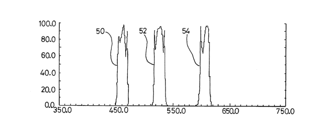
Figure 3 is the transmission spectrum of a triple bandpass filter used in the
apparatus;
Figure 4 is the optical density plot for the triple bandpass filter;
Figure 5 is a plot of calculated macular pigment density against position
along a vertical
line passing through the fovea in the retina of an eye under examination;
Figure 6 is a surface plot showing calculated macular pigment density across a
retina;
Figure 7 is an image of a retina photographed using apparatus according to the
invention;
and
CA 02521567 2005-10-05
WO 2004/096033 PCT/GB2004/001780
7
Figure 8 shows a spectral response from 3 CCDs used by the apparatus to
provide an
electrical output signal representative of a captured image of a retina.
Detailed Description
The camera shown in figure 1 is a modified version of a non mydriatric retinal
camera, in
this case the TOPCON TRC-NW6SF camera. The camera comprises a housing 1
containing illumination and imaging optics and a flash lamp. At one end of the
housing 1
there is an objective lens assembly 2, and at the other end a 3CCD (charge
coupled device)
camera 4 for generating a three component colour output signal representative
of a
captured image obtained via the imaging optics in the housing 1. The rear of
the housing 1
is also provided with an LCD view finder screen 6, and supports a shutter
control 8.
Attached to the front of the housing 1 is a head support 10 comprising a
headband 12 and a
chin rest 14. The head support 10 locates the head of the subject to
facilitate the correct
positioning of the eye under examination relative to the objective lens
assembly 2.
Figure 2 shows, in simplified form, the illumination and imaging optics within
the housing
l, as well as an eye under examination 16, the camera's flash lamp 18 and a
focusing
lamp 20. The focusing lamp 20 is used to illuminate the eye 16 while the
operator is
setting up the camera to photograph that eye. The illumination provided by the
lamp 20
enables the image of the retina of eye 16 to be viewed on the view finder
screen 6 so that
the operator can correctly position the eye and focus the camera. Light from
the lamp 20
passes through a focusing lens system 22 to a beam splitter 24 in the form of
a half
silvered mirror, from which it is reflected through a filter assembly 26. The
assembly 26
comprises a holder 28 which holds four filters, respectively referenced 30,
32, 34, and 36,
and which is rotatable about an axis parallel to the beam of light from the
focusing lamp 20
to bring any selected one of those filters into registry with that beam. It
will be
appreciated that a holder capable of carrying different numbers (more or
fewer) filters
could be used in the camera. In the present case, the filters 30-34 are used
for standard
retina photography, whilst the filter 36 is a triple bandpass filter,
described below, which
CA 02521567 2005-10-05
WO 2004/096033 PCT/GB2004/001780
8
enables the image captured by the camera to be used to measure macular pigment
density
and spatial distribution on the retina of the eye 16.
Light passing through the filter 36 then passes to an annular mirror 38 via a
reflecting
mirror 40 and focusing lenses 42, 44 and 46. The mirror 38 reflects that light
via the
objective lens assembly 2 into the eye 16 to illuminate the retina of that
eye. That light is
reflected from the retina and some of it passes back through the lens 2 which
directs the
light through the aperture (referenced 48) in the mirror 38, through a further
system of
lenses 50, 52, 54 and 56 which focus an image of the illuminated retina onto
the image
plane of the CCD camera 4. . The TOPCON TRC-NW6 camera is supplied with a
neutral
filter for use in normal colour photography (for example for use in diabetic
screening) and
an exciter filter for use in fluorescein angiography. These filters may be
interchanged with
other filters, and modification to the camera necessary to convert it into
apparatus
according to the invention is achieved by replacing one of those filters with
the triple
bandpass filter 36. In reality, the camera has a more complex arrangement of
optical
elements than is indicated by figure 2, but since these are, save for the
filter 36, identical
to those used in the known camera, they have not been described in detail.
The output of the camera 4 is connected to a computer 5 which has a video
capture card
for enabling the output to be recorded onto the computer's hard drive for
subsequent
processing.
The CCD camera 4 has three CCD arrays and associated red, green and blue
colour filters.
Each CCD array is positioned behind a respective one of the three filters, and
the camera
includes a beam splitter for projecting the image of the retina of the eye 16
onto each of
the 3 CCD arrays through its respective filter. The output of each array will
therefore
represent an array of grey scale pixel values which itself constitutes an
intensity map of the
filtered light received from the retina. The output of the CCD arrays
therefore constitutes
red, green and blue channels.
CA 02521567 2005-10-05
WO 2004/096033 PCT/GB2004/001780
9
Figure ~ illustrates the spectral response of the blue (B) green (G) and red
(R) channels in
the camera 4. Were white light to be used to illuminate the retina under
inspection, the
blue green and red channels of the camera output would not provide sufficient
colour
resolution to enable macular pigment density to be measured. However, the
spectral
responses from the three CCD arrays in the camera 4 will be shaped into
narrower wave
bands by the filter 36, since the transmission spectrum of this filter has
three relatively
narrow bands, referenced 50, 52 and 54 in figure 3, in its transmission
spectrum. The
width of each of these bands is considerably narrower than that of the three
bands, B, G
and R, the transmission spectrum between adjacent bands is substantially zero,
as is
illustrated in the optical density map of figure. 4 in which the vertical axis
is minus one
multiplied by the log (to base ten) of the transmittance. Thus, the
transmittance of the
triple bandpass filter 36 between the transmission bands does not exceed
0.00001 (i.e an
optical density of 5). A filter having these spectral characteristics is
available from Omega
Optical. The interaction between the triple bandpass filter 36 and the filters
in the CCD
camera 4 is such that, of the light transmitted through the filter 36, the
light within the
band 50 will only affect the blue output channel for the camera 4, all light
in the band 52
will affect the green channel whilst light in the band 54 only appears in the
red channel.
Thus, light transmitted in each of the three bands of the bandpass filter 36
will only affect
the output from a respective one of the 3 CCD arrays in the camera 4.
The method of operation of the apparatus, and the analysis of the retinal
image captured by
the apparatus, will now be described.
Initially, the subject places his or her head against the head support 10, and
the focusing
lamp 20 and camera 4 are activated respectively to illuminate the eye 16 and
to capture a
video image thereof. That image is displayed on the display 6 and the operator
adjusts the
controls of the camera to focus and align that image. The manner of this
adjustment is the
same as for the known retinal camera on which the present apparatus is based.
The operator then activates the shutter switch, causing the lamp 1~ to flash
and a shutter
(not shown) in the camera 4 to operate, so that the camera 4 captures the
colour image of
CA 02521567 2005-10-05
WO 2004/096033 PCT/GB2004/001780
the retina of the eye 16 when the latter is being illuminated by the lamp 18
through the
filter 36, i. e. with light having a spectrum corresponding to the
transmission spectrum of
figure 3.
The camera supplies R, G and B signals to the computer 5, said signals
representing an
array of grey scale pixel values for each of the 3 CCD arrays.
Image analysis software (for example ImagePro Plus) which has been pre-
installed on the
computer 5 is then used to analyse the captured image. This is a powerful
application
capable of performing many operations, including those needed to generate an
optical
density map of the macular pigment of the retina. However, it is envisaged
that other,
simpler software packages could be used to achieve the same end, using an
analysis
technique developed from the underlying theory summarised below.
We will assume a general .situation of non-uniform illumination of the retina
by the
camera's flash lamp. Let the incident intensities by IF,B, IF,R, IP,B and
IP,R, where the
subscripts F and P refer to a foveal and peripheral retinal location (no
macular pigment),
and the additional subsripts B and R refer to the blue (460 nm) and red
wavelength bands,
respectively of the light source (i.e flash lamp 18 and filter 36). The
analysis would not be
affected if the green wavelength band had been chosen instead of the red.
Similarly let
RF,B, RF,R, RP,B and RP,R be the corresponding reflectances of all retinal
layers posterior to
the macular pigment. Finally, T is the 460 nm transmittance of the macular
pigment at the
foveal location, and the logarithms/log differences in this description are to
base ten.
For the blue illumination, the log difference in reflected intensities between
the foveal and
peripheral locations will be given by
LD = to I TZR - to I R = to IF,BTzRF,B ,
B g F,B F,B g P,B P,B g
IP,BRP,B
CA 02521567 2005-10-05
WO 2004/096033 PCT/GB2004/001780
11
and for red illumination by LDR = logIF,RRF,R - logIP,RRP,R = log IF,RRF,R
IP,RRP,R
The factor TZ in the first equation is due to the double passage of the light
through the
macular pigment.
LD - LD = to IF,RRF,RIP,BRP,B
Subtracting, R B g z
IP RRP RIF BT RF B
......................................(1)
The spectral distributions of light on the fovea and periphery will be the
same,
IF,R IP,R
..I -I
It will also be assumed that the reflectance spectrum is the same in each
location,
RF, R RP, R
. ~ RF. B RP, B
Equation (1) then becomes LDR - LD$ = log TZ = 2D
where D (_ - log T) is the optical density of the macular pigment at 460 run.
Thus
D = ~ (LDR - LDB) .................................(2)
Using ImagePro Plus, the spatial distribution of D is obtained from a single
retinal image
as follows:
CA 02521567 2005-10-05
WO 2004/096033 PCT/GB2004/001780
12
1. Individual grayscale images are extracted from the original image,
corresponding to
the modified blue and red (and green) channels of the camera 4.
2. The greyscale images are transformed to floating point format to minimise
loss of
information in the subsequent steps.
3. The "red" and "blue" images are logarithmically transformed.
4. The "log blue" image is subtracted from the "log red" image.
5. The resulting image is halved, in accordance with equation (2).
The result will be a grayscale image, an example of which is shown in Figure
7, in which
the light area 56 is the area of macular pigment. A variety of options is
available for
further analysis or presentation. The image rnay be rendered as a surface plot
as in Figure
6 in which the area of macular pigment is shown as a "hill" in the centre of
the image. A
density scan may be made along a line through the fovea, for example along
horizontal or
vertical meridians. An example is shown in figure 5. From such a plot, the
peak macular
pigment optical density will be obtained as the difference between the pixel
values at the
peak and at a peripheral location, such as 8° above the fovea.
Alternatively, a circular
"area of interest" corresponding to, say, 1.5° may -be defined. The
average pixel value
along the circular line, or the average pixel value within the enclosed area,
may be
obtained. There is evidence that flicker photometry determines the macular
pigment
density at the edge of the stimulus rather than the average value over the
stimulus area.
Thus, if a comparison is to be made between flicker photometry and
reflectometry, .
determining the average pixel value along the circular line may be more
appropriate.
The new method offers several advantages over traditional reflectometry, which
requires
the acquisition of separate blue and green images that must be precisely
registered with
each other. Such alignment is possible with ImagePro, but it would be too time-
consuming
for large-scale screening. With the proposed procedure, the blue and red
images will be
extracted from a single image and will be perfectly registered. Also, when
separate
images are acquired, there is the problem of non-uniform illumination of the
retina that
may be different in the two images. As can be seen in the derivation of
equation (2), any
CA 02521567 2005-10-05
WO 2004/096033 PCT/GB2004/001780
13
non-uniformity is the same in both images, if these are extracted from a
single image, and
is self cancelling.
There remains the question of whether to use a red or green image as the
reference image.
Either fulfils the requirement of showing zero or near zero macular pigment
optical
density. However, the green image shows a darkening in the same region as the
macular
pigment due to the presence of long and medium wavelength cone photopigments.
To
minimise the contribution of these photopigments, they would normally have to
be
bleached ( approx.5.6 log Td for approx. 3 minutes) prior to the acquisition
of the image.
However, with a method in accordance with the invention a triple bandpass
filter 36 with
the red transmitting band centred at approx. 600nm is used. At this
wavelength, the
optical density of the cone photopigments is approximately the same as at 460
nm, the
centre of the blue transmitting band. This photopigment optical density will
contribute
equally to the red and blue images and will be eliminated by the subtraction
process. At
600 nm, rod photopigment optical density is approx. zero, but this is not the
case at 460
nm and could affect the comparison between the foveal and peripheral sites in
the blue
image. However, the optical density at 500 nm has been estimated to be about
0.016 at 7°
to 10° from the fovea (Brindley G.S. and Williner E.N.(1952). The
reflexion of light
from the macular and peripheral fundus oculi in man. J. Fhysiol. 116, 350-
356). This
would correspond to roughly 0.01 at 460 nm and is comparable with the estimate
of
"Delori F.C., Goger D.G., Hammond B.R., Snoddlerly D.M., Burns S.A. (2001)
Macular pigment density measured by autofluorescence spectrometry: comparison
with
reflectometry and heterochromatic flicker photometry. J. Opt. Soc. Am., A,
Optics, Image
Science, & Vision' 18, 1212-30. Assuming no rods at the foveal site, macular
pigment
optical density would be underestimated by only about 2 to 4% in the average
subject.
Apart from photopigments, melanin and oxyhaemoglobin can potentially influence
macular
pigment measurements obtained by reflectometry. Oxyhaemoglobin can probably be
ignored because its density is the same in the fovea and periphery
(12°). Melanin may
pose a problem since it has been shown to have a non-uniform distribution in
the retina,
peaking in the macula. Also it has an absorbance spectrum that decreases with
increasing
CA 02521567 2005-10-05
WO 2004/096033 PCT/GB2004/001780
14
wavelength. Thus the blue image would be the most affected, the green image
would be
moderately affected, and the red image would be least affected. This would
tend to cause
the macular pigment optical density to be overestimated by a factor that would
be larger if
the red image is used as the reference rather than the green. In principle,
the effects of
melanin can be removed. To achieve this, theory indicated that equation (2)
would need to
be replaced by
D = 1/a (rLDR - LDB) .... . .. ...... ...... ............ ........ . . (3)
where r is the ratio of the melanin extinction coefficients at 460 and 600 nm
(approx. 4) .
Hence the "log red" image would need to be multiplied by r prior to
subtracting the "log
blue" image. However, it should be noted that equation (3) assumes uniform
illumination
of the retina and a spectrally flat reflector. In addition , the value D given
by (3) will be
affected by any non-uniform distribution of photopigment across the retina.
B~ploiting
the green image, as well as the blue and red images, we can eliminate the
contributions
from non-uniform distributiions of both melanin and photpi mg~~ ants. The
appropriate
equation for D is then
D = 1/a LDR rZr~(r~ - r3) _ LDB + LD~ r~r'(r< - r2) ..... . .... .. . .. ... .
.. ... ... . . . (4)
r,r4 - r~r3) r,r4 - rzr3
where the coefficients, rn, are the ratios of melanin or photopigment
extinction coefficients
at different pairs of wavelengths. More specifically the r factors are as
follows:
r1 = ext. coeff. at the blue wavelength/ext.coeff. at the green wavelength for
melanin
r2 = ext. coeff. at the blue wavelength/ext. coeff. at the red wavelength for
melanin
r3 =ext. coeff. at the blue wavelength/ext. coeff. at the green wavelength for
cone
photopigment
r4 - ext.coeff. at the blue wavelength/ext. coeff. at the red wavelength for
cone
photopigment
CA 02521567 2005-10-05
WO 2004/096033 PCT/GB2004/001780
The ratios are obtainable from the literature. To put equation (4) into
practice, the "log
red" , "log green" and "log blue" images will be linearly combined using the
appropriate
multipliers shown in the equation.
Here, D is the optical density of the macular pigment at the wavelength of the
blue filter
band (460 nm) and LDR, etc are the logarithmically transformed red, green and
blue
grayscale images. The software (ImagePro Plus) is Windows-based and performs
each of
the following steps.
1. Individual grayscale images are extracted from the original image,
corresponding to
the filter-modified blue and red and green channels of the camera.
2. The "red" "green" and "blue" grayscale images are transformed to floating
point
format to minimise loss of information in the subsequent steps.
3. The three grayscale images are logarithmically transformed.
4. The 3 logarithmically transformed images are combined according to equation
(4).
The result is an image of the retina that shows a lighter area (higher
intensity/higher pixel
value) in the region of the macula. A "value" of macular pigment density may
be found
by taking the average of a set of pixel values within a circular region (e.g.
1 degree in
diameter) centred on the centre of the macula, and subtracting the average of
a similar set
centred at a reference location at, say, 8 degrees from the centre of the
macula (where
macular pigment density ~ 0). This would provide the average macular pigment
density in
the central 1 degree.
It will be appreciated that in the maps/plots of figures 5-7, each individual
pixel represents
a mathematical combination of the amounts of light transmitted through each
band of the
triple bandpass filter, subsequently reflected from the retina, and modified
in the central
part of the retina by the transmitting properties of the macular pigment. Thus
the macular
CA 02521567 2005-10-05
WO 2004/096033 PCT/GB2004/001780
16
pigment optical density, D at any point within this central part of the retina
is obtained by
subtracting from the corresponding pixel value the pixel value at some non-
central retinal
location, such as at an eccentricity of 8°, where macular pigment
density is known to be
negligible. For example, in figure 5, the peak optical density D is obtained
by subtracting
from the peak ordinate value the ordinate value at pixel number 95, this
representing a
point on the retina approximately 8 ° from the centre of the fovea.
Notwithstanding the above comments on the distribution of rod photopigments,
it is
believed that the effect of such pigments on the macular pigment measurement
may be
eliminated by using an image of the retina illuminated by light at a fourth
wavelength. In
order to obtain the second image, the triple bandpass filter 36 is exchanged
for a filter with
peak transmittance at 680nm and a bandwidth of 20nm and the eye under
examination is
photographed a second time. The first photographs yields the 'red' green' and
'blue'
images, one from each respective CCD array, whilst the second photograph
yields a
second 'red' image (at a wavelength longer than that of the first 'red'
image). There are
therefore 4 images at difference wavelengths, and these can be used to obtain
the macular
pigment optical density in a way which eliminates the (small) effect of rod
photopigment.
Here, briefly , is how we would obtain the macular pigment optical density
distribution,
including this new refinement:
1. Obtain an image using the triple bandpass filter. Use image analysis
software to
extract the grayscale images corresponding to the red, green and blue
channels, as
before, and concert these to logs (LDR,LD~,LDB).
2. Obtain a second image using a filter with peak transmittance at 680nm and a
bandwidth
of 20nm, for example. This is a longer wavelength than the red band of the
triple
bandpass filter. At 680nm, the only pigment with a significant absorption is
melanin.
Again extract the grayscale image (from the red channel), and convert to logs,
LDR.
CA 02521567 2005-10-05
WO 2004/096033 PCT/GB2004/001780
17
3. Use image analysis software to align the LDR, image with the LDR, LDP and
LDB
rxnages .
4. Obtain the macular pigment optical density distribution by combining the 4
images in a
linear fashion -
D = -0.525*LDB + 0.355*LDG - 0.882*LDR + 2.60*LDR~
The numerical factors are different combinations of extinction coefficients of
the 4
pigments at the 4 wavelengths; similar to those shown symbolically (4) of the
specification.
Since there are four different images and four unknown pigment distributions,
the cone and
rod distributions can also be determined using the following. equations:
Doone = -0.391 *LDR + 0.654*LDR,
Drod = 0.0254*LDB = -0.355LD~ + 1.081LDR - 0.826*LDR~