Note: Descriptions are shown in the official language in which they were submitted.
CA 02522956 2011-03-01
Ophthalmic Microsurgical Instruments
Background of Invention:
Glaucoma is a disease condition of the eye in which increased intraocular
pressure
(IOP) is created by blockage of the drainage mechanism for the aqueous fluid
produced in the anterior portion of the eye. Such conditions are usually
treated by
topical drugs in the form of eye drops, but may result in surgical treatment
if drug
treatment becomes ineffective or if patient compliance is an issue.
Traditional
glaucoma surgery such as trabeculectomy, involves a flap dissection of the eye
and
the removal of a portion of the trabecular meshwork (TM) or the corneo-scleral
junction. The aqueous fluid is directed posteriorly under the surgical flap
and to a
sub-conjunctival lake known as a bleb. Post-surgical complications and bleb
management are significant issues with trabeculectomy and similar procedures.
Furthermore, the control of the aqueous outflow is achieved through the
management of the integrity of the surgical flap rather than controlling the
opening
into the anterior chamber. Other procedures involving laser energy to create
holes in
the TM are partially successful, however long term results are limited as
compared to
trabeculectomy.
Recently developed surgical treatments for glaucoma involve surgically
accessing
Schlemm's Canal by manner of a surgical flap or flaps and subsequently
dilating or
expanding the canal to increase aqueous humor drainage into the natural
drainage
pathway. Current procedures and instruments can only access a short passage of
Schlemm's Canal from either side of the surgical site. US 5,486,165 to
Stegmann et
al. in discloses a microcannula designed for delivery of substances to
Schlemm's
Canal during such a procedure. EP 0898947A2 to Grieshaber et al. discloses an
improvement to the Stegmann apparatus to deliver substances or stents for
maintaining the passage of fluid in the canal. Other inventions disclose the
use of
microcatheters to introduce water-jet type cutting apparatus or bladed
mechanisms to
1
CA 02522956 2005-10-20
WO 2004/093761 PCT/US2004/011783
the canal for disruption of the TM. However these methods cut the TM network
open
in a non-controlled manner and do not remove tissue or debris from the
operative
field.
The treatment of glaucoma usually involves patient specific requirements for
the
amount of drainage increase desired by the physician. It is therefore of
advantage to
be able to treat or remove a controlled amount of the TM or associated
juxtacanalicular tissues in order to be able to titrate drainage rates and
control the
disease process on a patient specific basis. Furthermore, it is desired to
perform the
controlled treatment or removal of tissues from within Schlemm's Canal in
order to
facilitate the restoration of natural aqueous drainage system without the
requirement
for blebs and the concomitant complications, and to enable less invasive
surgical
methods. It is also advantageous to physically stabilize the tissues in order
to
facilitate control of the amount of tissues being treated or removed.
This invention is directed at ophthalmic microsurgical instruments which may
be
directly inserted into Schlemm's Canal to allow controlled treatment or
removal of
adjacent tissues such as the TM or the juxtacanalicular tissues to effect the
reduction
of intra-ocular pressure. It is a further object of this invention to describe
an
instrument which allows the directed access to Schlemm's Canal by a flexible
microcannula. The instrument is useful in allowing controlled guidance by the
surgeon while viewing through a surgical microscope or by non-invasive medical
imaging.
Known prior art:
United States Patent 4,501,274
Skjaerpe February 26, 1985
Microsurgical instrument
United States Patent 5,486,165
Stegmann January 23, 1996
Method and appliance for maintaining the natural intraocular pressure
2
CA 02522956 2005-10-20
WO 2004/093761 PCT/US2004/011783
United States Patent 6,142,990
Burk November 7, 2000
Medical apparatus, especially for reducing intraocular pressure
United States Patent 6,221,078
Bylsma April 24, 2001
Surgical implantation apparatus
United States Patent 6,283,940
Mulholland September 4, 2001
Catheter
United States Patent 6,375,642 B1
Grieshaber, et al. April 23, 2002
Method of and device for improving drainage of aqueous humor within the eye
United States Patent 6,494,857 B1
Neuhann December 17, 2002
Device for improving in a targeted manner and/or permanently ensuring the
ability of
the aqueous humor to pass through the trabecular meshwork
United States Patent Application 20020013546
Grieshaber, Hans R. ; et al. January 31, 2002
Method and device to improve aqueous humor drainage in an eye
United States Patent Application 20020111608
Baerveldt, George ; et al. August 15, 2002
Minimally invasive glaucoma surgical instrument and method
United States Patent Application 20020082591
Haefliger, Eduard June 27, 2002
Device for the treatment of glaucoma
3
CA 02522956 2005-10-20
WO 2004/093761 PCT/US2004/011783
United States Patent Application 2003014092
Inventor(s): Neuhann Thomas (De)
Apparatus for the treatment of glaucoma
Patent Number: EP0898947 A2
Inventor(s): Grieshaber Hans R (Ch); Stegmann Robert Prof M D (Za)
Method and apparatus to improve the outflow of the aqueous humor of an eye
Patent Number: EP1114627 Al
Inventor(s): Grieshaber Hans R (Ch); Stegmann Robert Prof M D (Za)
Method and apparatus to improve the outflow of the aqueous humor of an eye
Patent Number: W00064389
Inventor(s): Brown Reay H (Us); Lynch Mary G (Us); King Spencer B Iii (Us)
Trabeculotomy device and method for treating glaucoma
Patent Number: W002056805
Inventor(s): Roy Chuck; Baerveldt George
Minimally invasive glaucoma surgical instrument and method
Patent Number: W002074052
Inventor(s): Smedley Gregory T; Gharib Morteza; Tu Hosheng
Applicator and methods for placing a trabecular shunt for glaucoma treatment
Patent Number W003045290
Inventor(s): Conston Stanley R; Yamamoto Ronald K
Ophthalmic Microsurgical System
Brief Description of the Drawings
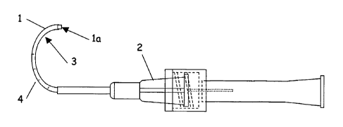
Figure 1 illustrates a sheath microcannula with an inner member.
Figure 2 illustrates a microcannula with expandable segments.
Figure 3 illustrates a microcannula with a signaling beacon tip.
Figure 4 illustrates a microcannula with a side connection fitting.
4
CA 02522956 2005-10-20
WO 2004/093761 PCT/US2004/011783
Figure 5 illustrates a microcannula with an open distal tip with a side
channel for
application of suction.
Figure 6 illustrates a microcannula with fenestrations and an inner member for
controlled tissue removal.
Figure 7 illustrates a microcannula with a single fenestration for controlled
tissue
removal.
Figure 8 illustrates a microcannula with a rotating inner member for tissue
cutting.
Figure 9 illustrates a microcannula with a side fenestration and tissue
cutting flap.
Figure 10 illustrates a microcannula with a side fenestration and inner member
for
directed tissue abrasion.
Description of Invention:
Schlemm's Canal is a channel in the corneo-scleral junction of the eye and is
the
primary pathway for the drainage of aqueous humor. The inner wall of the Canal
comprises the TM and juxtacanalicular tissues through which the aqueous humor
drains from the anterior chamber. The outer wall of is comprised of scleral
tissue
with openings to collector channels for the passage of aqueous humor from the
Canal to the venous system. Due to its relative positioning to the TM, the
Canal
forms a circular channel that encircles the anterior chamber. The Canal is
approximately 10 to 15 mm in diameter and 200 microns by 50 microns in cross-
section. The drainage of aqueous humor through the TM and juxtacanalicular
tissues
into Schlemm's Canal is believed to be the predominant route for aqueous
drainage.
In open surgery for glaucoma, surgical treatment of the inner wall of
Schlemm's
Canal and removal of associated tissue such as the TM and juxtacanalicular
tissues
has demonstrated an increase in aqueous outflow and reduction of intraocular
pressure. It is an object of the present invention to enable treatment and
removal of
tissues in these specific regions by use of minimally invasive surgical
instruments. It
is also an object of the invention to treat a specific segment of the tissue
tract and
also to treat specific regions of the selected segment to minimize surgical
trauma and
post-surgical scarring.
The ophthalmic microsurgical instruments of the present invention comprise a
thin
walled outer sheath microcannula with a connector at the proximal end, a
distal tip
5
CA 02522956 2005-10-20
WO 2004/093761 PCT/US2004/011783
and a communicating channel therebetween, as shown in Figure 1. The
microcannula lumen provides a fluid and gas tight, sealed passage from the
proximal
end to the distal tip of the instruments. An inner member which fits and
slides or
rotates within the sheath may also be incorporated, the inner member
comprising at
least a proximal end and a distal tip. The distal end of the instruments may
be
curved in a manner to approximate the curvature of Schlemm's Canal. The
instruments may also comprise a guidance means to effect proper advancement of
the distal portion. Furthermore the instruments may comprise means to
mechanically
stabilize the target tissues. The tissues may be held in tension or
compression for
controlled treatment or removal of tissue. The instruments may also comprise
cutting means to excise targeted tissues. The instruments may also be used to
deliver drugs or implants to the tissue tract to treat adjacent tissues.
The microcannula may be introduced into Schlemm's Canal manually or as part of
a
system to provide surgical support or guidance. Once inserted into Schlemm's
Canal, the microcannula may be progressively advanced to the appropriate areas
for
treatment. The distal end is preferably sized and curved or compliant enough
to
access at least one half the length of Schlemm's Canal, approximately 15 to 25
mm.
Treatment of the entire Canal may be effected by inserting the instrument in
the
opposite direction from the first treatment at the surgical access point. The
positioning of the instrument in the Canal can be verified by several means
including
a fiber-optic beacon tip inner member, a change in pressure or vacuum
resistance in
the surrounding environment as the system enters the Canal, a change in tissue
color, direct visual location during surgical cut-down or by external image
guidance
such as ultrasound or optical coherence tomography. Features of the instrument
can
aid accurate positioning within the Canal.
The selective treatment or removal of tissues adjacent to Schlemm's Canal such
as
TM or juxtacanalicular tissues may be accomplished by various means. One means
incorporates the use of side holes or fenestrations on the outer sheath
directed at the
target tissues adjacent to the inner radius. The outer sheath may be
configured to
allow for tissue treatment or removal separately or in conjunction with an
inner
member that works in alignment with the side holes or fenestrations. Another
means
6
CA 02522956 2005-10-20
WO 2004/093761 PCT/US2004/011783
for selective treatment of the TM or juxtacanalicular tissues may be
accomplished by
the use of suction through the microcannula, which has been observed to act
predominantly on the inner wall of the Canal. Both means may also be combined,
such as the use of suction to pull a region of the target tissue into a side
hole or
fenestration of the outer sheath for subsequent treatment or excision.
Suction or vacuum may also be incorporated to clear the operative field and
the
microcannula lumen, either concurrent with tissue treatment or subsequent to
tissue
treatment since the sheath also functions to provide a disposal path for the
excised
tissues and surgical debris. Furthermore the ability of the cannula to remove
particles and debris may be used by itself or in conjunction with other
treatment
methods such as laser trabeculoplasty in order to enhance the outcome by
removal
of waste particles.
The microcannula may comprise a thin walled polymer or metallic tube I of
sufficient
stiffness to allow it to be advanced into Schlemm's Canal, and of sufficient
flexibility
or compliance to follow the curvature of the Canal. It is preferable that the
distal tip
1a be beveled or radiused so as to provide for atraumatic advancement into the
Canal. The proximal connector 2 may be of a Luer type or similar system for
the
attachment or introduction of secondary elements or may be designed for
attachment
only to specific components. Due to the small size of Schlemm's Canal,
approximately 200 microns in diameter, the microcannula must be appropriately
sized. Typically, the microcannula is sized in the range of 100 to 350 microns
outer
diameter with a wall thickness from 10 to 100 microns to allow cannulation of
Schlemm's Canal. However, Schlemm's Canal may be expanded prior to insertion
of
the microcannula with for example, the injection of a surgical viscoelastic
material.
With prior expansion of the Canal, cannulation becomes much easier to perform
without damaging tissues. Expansion of Schlemm's Canal also allows a
microcannula of up to 500 microns outer diameter to be used to access the
Canal.
Due to the curvature of Schlemm's Canal, the microcannula should be flexible
in the
appropriate dimensions. In some embodiments, a predetermined curvature 3 may
be
applied to the inner member and/or the outer sheath during fabrication. The
7
CA 02522956 2005-10-20
WO 2004/093761 PCT/US2004/011783
curvature is preferably slightly greater than the curvature of the Canal in
order to
prevent the instrument from perforating the inner wall while advancing the
microcannula. It is also desirable for a portion of the instrument to be able
to be
swiveled at least 1800 around to provide for handedness to the curved
microcannula.
This allows the surgeon to cannulate the entire circumference of Schlemm's
Canal
from a comfortable working position.
Suitable materials for the microcannula sheath include metals,
polyetheretherketone
(PEEK), polyimide, polyamide, polysulfone, or similar materials. The sheath
may
also comprise surface treatments such as lubricious coatings to assist in
cannulation
and ultrasound or light interactive coatings to aid in location and guidance.
The
microcannula may also have markings 4 on the exterior for assessment of depth
in
the tissue tract. The external markings allow user assessment of the length of
the
tissue tract accessed by the microcannula, and the approximate location of the
microcannula tip.
The microcannula 5 may also comprise a segment or series of segments capable
of
being expanded in a radial direction in order to place tension on the target
tissues for
treatment, as shown in Figure 2. The segments may comprise means such as stent-
like structures, balloons or elastomeric sections 6 which may be inflated or
deformed
in a radial manner 7. Multiple expandable segments may be used to stabilize
and
isolate segments of Schlemm's Canal for surgical or drug treatment through the
microcannula lumen. Furthermore, the expandable segments may be slidably
disposed about the central axis such that the segments may be translated
axially
apart from each other to provide further tension on the tissues. The
expandable
segments may comprise polymers and elastomers such as latex, silicone rubber,
urethane, vinyl, polyether block amide (Pebax) or may be a metallic structure
comprised of shape-memory or superelastic alloy, stainless steel, tungsten or
similar
materials. Alternatively, another outer member may be disposed about the
microcannula as a tissue stabilization means. The expandable structure would
be
activated or mechanically released to expand during the procedure and then
retracted or compressed for removal.
8
CA 02522956 2005-10-20
WO 2004/093761 PCT/US2004/011783
Depending on the application, the inner member may be used to guide the
positioning of the microcannula, surgical tools and instrumentation or act as
a
surgical tool. The inner member may comprise a guide wire, hollow needle or
tube,
micro-trocar, cutting tool or similar element and comprises a proximal end and
a
distal tip, and may contain a communicating channel between. The inner member
may also comprise sensing means such as a pressure transducer or fiber optic
to aid
in determining location, local fluid pressure, blood flow or other parameters.
The inner
element is sized correspondingly to fit slidably within the microcannula and
therefore
will be in the range of 90 to 450 microns in outer diameter. If hollow, the
inner
diameter will be in the range of 40 to 400 microns. The inner member may be
removed during the surgical procedure and replaced sequentially with other
inner
members acting as instruments or tools.
A first inner member used for initial placement may comprise a signaling
beacon to
identify the location of the microcannula tip relative to the target tissues,
as shown in
Figure 3. The beacon may comprise an echogenic material for ultrasound
guidance,
an optically active material for optical guidance or a light source for visual
guidance.
In one embodiment, a plastic optical fiber (POF) 8 is used to provide a bright
visual
light source at its distal tip 9. The distal tip of the POF 10 may be
positioned at or
slightly beyond the end of the microcannula sheath 11 and the emitted signal
may be
detected through the scleral tissues visually or using sensing means such as
infrared
imaging. The POF may also comprise a tip which is beveled or mirrored or
otherwise
configured to provide for a directional beacon. If the emitted directional
light is
directed toward the inner radius at the TM, the surgeon may view the
illuminated spot
in the anterior angle using a goniometer lens, and verify placement of the
operative
instrument at the targeted tissues. The beacon may be illuminated by a high
intensity
light source, laser, laser diode or light-emitting diode 12, which may be
powered by
batteries 13 or standard AC power. Upon arrival of the microcannula distal end
at the
target tissues, the beacon assembly and POF may be removed, leaving the
microcannula sheath at the desired location for treatment. The connection
point
between the outer microcannula sheath and the inner member may be sealed with
a
cap or preferably with a self-sealing mechanism such as a one-way valve or an
elastomer seal.
9
CA 02522956 2005-10-20
WO 2004/093761 PCT/US2004/011783
In one embodiment, the instrument set also comprises a fitting as the
connection
point for the illumination package. Additionally, as shown in Figure 4, the
instrument
may contain a central section 14 comprising a single or multiple side fittings
15 to
allow the attachment of ancillary equipment such as syringes, vacuum or
pressures
sources, sensing means and the like. The attachment fittings may comprise
standard
designs such as Luer fittings or may be designed to only accept connection
with
specific components.
The operative function of the invention is an instrument to treat or remove
specific
tissues adjacent to Schlemm's Canal such as the TM in such a manner that the
area
of the treatment or removal is controlled and repeatable. In some
applications, the
instrument may be used to remove a controlled layer of adjacent target tissue,
such
as the juxtacanalicular tissues at the inner wall of Schlemm's Canal.
Furthermore,
the procedure can be performed at multiple sites within the eye to effect
treatment
per the patient's requirements by using the microcannula sheath for
repositioning to
other target locations from within the Canal.
In one embodiment the microcannula 16 alone is used to remove portions of the
adjacent tissue using suction means 17, as shown in Figure 5. The microcannula
is
advanced into Schlemm's Canal 18. A vacuum syringe, vacuum or aspiration pump
is used to provide suction and a portion of the inner wall is pulled into the
lumen 19
and removed. Due the large difference in mechanical properties between the
thick
scleral outer wall of the Canal and the flexible tissues of the inner wall,
suction
applied by a microcannula has demonstrated preferential ability to manipulate
the
inner wall. Control of the suction characteristics may be used to control the
amount
of tissue treated or removed from the inner wall. In some cases, suction alone
may
be applied to the TM to remove tissue debris and improve aqueous outflow,
without
removing a portion of the TM.
In another embodiment, shown in Figure 6, the distal tip of the microcannula
20 is
closed off 21. A fenestration or series of fenestrations 22 are disposed along
the
inner radius wall 23 of the microcannula, directed toward the TM. Suction is
applied
CA 02522956 2005-10-20
WO 2004/093761 PCT/US2004/011783
24 to pull a small amount of TM into the lumen and apply tension to the target
tissue.
An inner member 25, comprised of a thin hollow shaft, is then extended through
the
microcannula 20 and may be rotated or axially advanced, to cut off the
intruding
tissues. The inner member may comprise a beveled or sharpened leading edge to
facilitate tissue cutting. The excised tissue may be removed by a suction
mechanism
through the lumen. The amount of tissue removal may be controlled through the
sizing of the ingress holes and the amount of suction applied. The outermost
layer of
the TM, including the juxtacanalicular tissues, interfacing Schlemm's Canal
may be
removed by minimal application of suction, or alternatively openings through
the TM
of controlled geometry may be formed with greater amounts of suction.
In a similar embodiment, Fig 7, a single fenestration 26 is created along the
inner
radius wall of the microcannula 29. The distal tip 27 is closed and is fully
radiused to
produce a ball-end tip. A single cutting element 28 is disposed in the lumen
at the
distal end, with the cutting edge oriented proximally. The target tissues are
pulled
into the lumen, and the cannula is withdrawn which allows the cutting element
to
remove tissue to a determined depth. The cutting depth may be set and adjusted
by
the cutting element design, the dimensions of the fenestration and the amount
of
suction applied.
Furthermore, the microcannula may contain stabilization means in conjunction
with
cutting means thereby applying traction to the tissues to improve cutting
efficiency
and control. The microcannula may comprise a multilumen tube such that each
lumen is connected separately to a hole or a series of holes along the inside
radius
facing the target tissues. For example, a two-lumen microcannula may be
constructed comprised with three holes a set distance apart along the inner
radius
wall. The outermost two holes are connected to one lumen of the microcannula
and
the central hole to the second lumen. In this manner, a low suction pressure
may be
applied to the outermost holes, providing tissue stabilizing forces, while a
higher
suction pressure may be applied to the center hole, removing a controlled
portion of
tissue.
11
CA 02522956 2005-10-20
WO 2004/093761 PCT/US2004/011783
In another embodiment shown in Figure 8, the microcannula lumen is open 30 and
a
rotating hollow inner member 31 is employed. The distal tip of the inner
member may
be beveled or sharpened and is extended just slightly beyond the end of the
cannula
32 or adjacent to a fenestration along the inner radius. Suction is applied to
the
cannula and the inner member is rotated 33 to provide a cutting action. As the
instrument is advanced, the TM tissues are preferentially pulled 34 toward the
microcannula axis allowing the inner member to cut away portions as required.
The
amount of tissue removal is controlled by extent of advancement and applied
suction
during the cutting process.
In another embodiment shown in Figure 9, the instrument distal end is
comprised of
two concentric thin-walled tubes. The outer tube 35 contains a window or
fenestration 36 near the distal end and aligned along the inner radius wall of
the tube
37 which interfaces the adjacent TM. The inner tube 38 contains an angled slit
39
partially through the tube which creates a sharp pointed flap 40 directed
proximally
and also aligned with the window in the outer tube and the TM. The flap 40 is
pre-
bent to allow it to project outward from the tubing 38, in the direction of
the TM and is
used as a piercing and cutting member. The outer tube 35 is slidably disposed
about
the inner tube. During insertion into Schlemm's Canal, the outer tube is
positioned
such that the window is not adjacent to the flap and the flap is thereby
constrained
within the outer tube. At the operative target position, the outer tube is
advanced so
that the window is over the flap, allowing the flap to protrude from the
assembly. The
instrument is retracted slightly allowing the flap to pierce the TM and then
retracted a
specified amount such that the full length of the flap has pierced the
tissues. The
outer tube is then retracted, moving the window proximally, causing the flap
to be
pulled back and thereby cutting a portion of the TM approximating the geometry
of
the flap and constraining the excised tissue within the inner tube for
disposal.
Suction may be used to remove the tissue from the lumen and the procedure
repeated as required.
In another embodiment shown in Figure 10, the inner wall of Schlemm's Canal
may
be removed by the application of controlled abrasion. The inner member 41 may
comprise a brush or rasp like tool 42 on the distal end which abrades the
tissue
12
CA 02522956 2005-10-20
WO 2004/093761 PCT/US2004/011783
surface. The abrading tool may be used by passing the distal portion of the
inner
member past the distal tip of the microcannula with concurrent suction, or by
positioning it in a window or fenestration 43 in the side of the microcannula
44 near
the distal tip 45. The use of a side opening allows a controlled portion of
the tissue
tract, such as the TM adjacent to Schlemm's Canal to be treated selectively.
Suction
may also be applied concurrently through the microcannula lumen to stabilize
the
tissues during treatment and remove resultant tissue debris.
The microcannula may also be used to deliver a fiber optic for laser ablation
of the
tissues from within Schlemm's Canal. The microcannula may be used to provide
suction to remove the ablative residue and any tissue debris from the site and
deliver
treatment adjuvants or medications to minimize fibrosis during wound healing.
Examples:
Example 1: A single element microcannula was fabricated with polyimide tubing
(MicroLumen, Inc.), 0.0101" (256p) inner diameter by 0.0141" (358p) outer
diameter.
The distal end was sealed with epoxy to create a ball end. The distal portion
was
curved with a radius of approximately 15mm for a distance of 2 cm. A
fenestration
approximately 1.2 mm long was cut into the inner wall of the curvature and
extending
inward to 1/2 the diameter. A Luer fitting was bonded to the proximal end. The
microcannula was attached to a collection bottle and then to a vacuum pump
generating up to 27 inches of Hg.
An enucleated human eye was prepared for the experiment by inflating the
posterior
chamber to a pressure of 10mm Hg with phosphate buffered saline (PBS). A
scleral
flap was surgically excised and Schlemm's Canal unroofed. The microcannula was
inserted into Schlemm's Canal and vacuum was applied. Suction was confirmed by
observing fluid flow within the microcannula.
Subsequently, the globe was hemisected and the vitreous, ciliary body, lens
and iris
removed allowing visualization of the TM and Schlemm's Canal from inside. The
microcannula was advanced into the Canal to a point approximately 1000 from
the
13
CA 02522956 2005-10-20
WO 2004/093761 PCT/US2004/011783
surgical site. Suction was applied and the results observed visually under the
surgical microscope. Upon application of vacuum, the inner wall of Schlemm's
Canal
at the fenestration site was seen to be pulled into the lumen of the
microcannula.
The vacuum level was varied from 1 to 27 inches Hg. In each case the inner
wall
was observed being pulled into the lumen at approximately 4 inches Hg or
greater,
while the outer wall was not noticeably deformed. The microcannula was
withdrawn
under vacuum and upon examination, excised tissue was observed adhered to the
distal edge of the fenestration. An open ended microcannula of approximately
the
same size, without side fenestration, was placed in Schlemm's Canal and the
suction
experiments repeated at various vacuum levels. The inner wall of the Canal was
observed to be preferentially deflected toward the microcannula tip at
approximately
4 inches of Hg or greater.
Example 2: A microcannula with an inner member and outer sheath was
fabricated.
The outer sheath was fabricated with a single fenestration as in Example 1 but
with a
polyimide tube of 0.0087" inner diameter and 0.0117" outer diameter. The inner
member was comprised of polyimide tubing 0.0049" inner diameter by 0.0067"
outer
diameter and was slidably disposed within the outer member.
An enucleated human eye was prepared as in Example 1. The microcannula was
placed with the fenestration toward the inner wall of Schlemm's Canal. The
vacuum
was applied and tissue was seen being pulled into the lumen. The inner member
was then advanced until it stopped against the closed distal tip of the outer
member.
Upon removal of the microcannula, excised tissue was observed attached to the
inner member.
Example 3: A microcannula with an inner member and outer sheath was
fabricated.
The outer sheath was similar to the outer sheath in Example 2. An inner member
designed to abrade the tissues was fabricated comprised of a stainless steel
wire
0.006" diameter to which the distal end was roughened using a grinding wheel.
The
inner member was slidably disposed within the outer sheath.
14
CA 02522956 2005-10-20
WO 2004/093761 PCT/US2004/011783
An enucleated human eye was prepared as in Example 1 with the addition of
placing
a 27 gauge needle into the cornea, and attaching the needle to a flow meter
and
reservoir of PBS. The reservoir was raised to provide constant pressure flow
into the
anterior chamber, and the flow meter used to observe changes in flow.
The microcannula was advanced into Schlemm's Canal. Suction was applied to
pull
the inner wall of the Canal into the lumen and then the inner member was slid
back
and forth across the tissues. The microcannula was removed and surgical flap
sealed. An increase in aqueous outflow was observed after the procedure.
Example 4: A microcannula was fabricated similar to the outer sheath in
Example 2.
A cutting element inner member was fabricated from Nitinol wire, incorporating
a flat
blade situated at the axis of the wire. The cutting element was bonded into
the distal
lumen of the microcannula with the cutting blade facing proximally and
extending into
the fenestration area.
Enucleated human eyes were prepared as in Example 3. The microcannula was
advanced into Schlemm's Canal. Suction was applied, drawing the inner wall of
the
Canal into the lumen, and the microcannula was retracted while still under
vacuum.
Upon removal from the eye, the cutting element was observed to have excised
tissue
attached. Subsequently aqueous outflow was seen to increase.
Example 5: A signaling means for determining the location of the microcannula
was
fabricated and incorporated into a microcannula instrument. A single strand
plastic
optical fiber (POF) (Biogeneral, Inc.) 100 microns in diameter was used with a
flat
distal tip. The fiber was disposed within an instrument assembly comprising a
polyimide microcannula 110 microns ID and 160 microns OD (MicroLumen, Inc.),
which was bonded to a needle assembly. The needle assembly consisted of a base
section of 18 gauge hypodermic tubing, with a 14 gauge tubing guide tube
fabricated
so as to slide forward and backward along the 18 gauge tube for a fixed
distance of
15 mm. The distal tip of the guide tube was comprised of a 28 gauge tube to
direct
the microcannula and POF during insertion. The POF was illuminated using a
battery powered red laser diode (Digikey Corp.). A second POF was also
fabricated
CA 02522956 2005-10-20
WO 2004/093761 PCT/US2004/011783
with a distal tip cut at approximately 600 and the jacket removed opposite the
bevel.
This provided a partially directed illumination spot toward the inner radius.
An ex-vivo human eye was placed in a soft holding cup stage under a
stereomicroscope. A surgical flap was created at the limbus and the flap
removed to
access Schlemm's Canal. The tip of the guide tube was placed at the ostium of
the
Canal. The microcannula and POF were advanced into the canal with the light
source on. The illuminated tip of the fiber was seen through the scleral
tissues in the
case of the flat tipped POF. Using the beveled POF, illumination could be
viewed
from within the anterior chamber of the eye depending on the rotation of the
microcannula, allowing the appropriate surgical tissues such as the TM to be
targeted.
Example 6: In another example, Schlemm's Canal of an eye is cannulated with
the
microcannula described in example 3. The signaling beacon inner member is used
to verify the position of the tip of the microcannula in the desired location
of the eye
and with proper rotational alignment with respect to the TM. The signaling
beacon
inner member is removed and a surgical tool inner member to remove tissue from
the
TM is guided into the lumen of the microcannula and advanced to the distal
tip. The
inner member also incorporates suction to remove tissue debris. After removal
of TM
tissue, the surgical tool inner member is exchanged for the signal beacon
inner
member. The microcannula may be positioned to another area of Schlemm's Canal
to repeat the process as needed to increase aqueous outflow to an appropriate
level.
While the present invention has been described herein with respect to the
exemplary
embodiments and the best mode for practicing the invention, it will be
apparent to
one of ordinary skill in the art that many modifications, improvements and
subcombinations of the various embodiments, adaptations and variations can be
made to the invention without departing from the spirit and scope thereof.
16