Note: Descriptions are shown in the official language in which they were submitted.
CA 02525777 2005-11-14
WO 2004/109717 PCT/US2004/017081
HIGH ENERGY POLYENERGETIC ION SELECTION SYSTEMS, ION BEAM
THERAPY SYSTEMS, AND ION BEAM TREATMENT CENTERS
CROSS REFERENCE TO RELATED APPLICATIONS
This patent application claims the benefit of U.S. provisional patent
application serial no.
60/475,027, filed June 2, 2003, the entirety of which is incorporated by
reference herein.
GOVERNMENT RIGHTS
The work leading to the disclosed invention was funded in whole or in part
with Federal
funds from the National Institutes of Health. The Government may have certain
rights in the
invention under NIH contract number CA7~331.
FIELD OF THE INVENTION
The present invention is related to the field of devices and methods for
generating high
energy ion beams. The present invention is also related to uses of high energy
ion beams for
radiation therapy. In addition, the present invention is related to the field
of treating patients in
cancer treatment centers using high energy ion beams.
BACKGROUND OF THE INVENTION
Radiation therapy is one of the most effective tools for cancer treatment. It
is well known
that the use of proton beams provides the possibility of superior dose
conformity to the treatment
target as well as providing a better normal tissue sparing, as a result of the
Bragg peak effect,
compared to photons (e.g., X-rays) and electrons. See, e.g., T. Bortfeld, "An
analytical
approximation of the Bragg curve fog therapeutic py~oton beams", Med. Phys.,
2024-2033
-1-
CA 02525777 2005-11-14
WO 2004/109717 PCT/US2004/017081
(1997). While photons show high entrance dose and slow attenuation with depth,
protons have a
very sharp peak of energy deposition as a function of beam penetration. As a
consequence, it is
possible for a larger portion of the incident proton energy to be deposited
within or very near the
3D tumor volume, thus avoiding radiation-induced injury to surrounding normal
tissues that
commonly occurs with x-rays and electrons.
Despite the dosimetric superiority characterized by the sharp proton Bragg
peak,
utilization of proton therapy has lagged behind that of photon therapy. This
lag is apparently due
to the operating regime (the total operating cost for accelerator maintenance,
energy
consumption, and technical support) for proton accelerators being at least an
order of magnitude
higher compared to electron/X-ray medical accelerators. Currently, proton
therapy centers
utilize cyclotrons and synchrotrons. See, e.g., Y. A. Jongen et al., "Proton
therapy system for
MGH's NPTC: equipment description and progress report ", Cyclotrons and their
Applications,
J. C. Cornell (ed) (New Jersey: World Scientific) 606-609 (1996); "Initial
equipment
commissioning of the North Proton Therapy Center ", Proc. of the 1998
Cyclotron Conference
(1998); and F. T. Cole, "Accelef°ator Considerations in the Design of a
Proton Therapy Facility",
Particle Acceleration Corp. Rep (1991). Despite a somewhat limited number of
clinical cases
from these facilities, treatment records have shown encouraging results
particularly for well
localized radio-resistant lesions. See, e.g., M. Fuss et al., "Proton
radiation therapy (PRT) for
pediatric optic pathway gliomas: Comparison with. 3D planned conventional
photons and a
standard photon technique ", Int. J. Radiation Oncology Biol. Phys., 1117-1126
(1999); J.
Slater et al., "Conformal proton therapy for prostate carcinoma ", Int. J.
Radiation Oncology
Biol. Phys., 299-304 (1998); W. Shipley et al., "Advaneed prostate cancer: the
results of a
randomized comparative trial of high dose irradiation boosting with conformal
protons
compared with conventional dose irradiation using photons alone ", Int. J.
Radiation Oncology
Biol. Phys., 3-12 (1995); and R. N. Kjellberg, "Stereotactic Bragg Peak Proton
Radiosurgery
for Cerebral Arteriovenous Malformations" Ann Clin. Res., Supp.47, 17-25
(1986). This
situation could be greatly improved by the availability of a compact,
flexible, and cost effective
proton therapy system, which would enable the widespread use of this superior
beam modality
and therefore bring significant advances in the management of cancer.
Thus, there remains the problem of providing a practical solution for a
compact, flexible
and cost-effective proton therapy system. See, e.g., C.-M. Ma et al., "Laser
accelerated proton
beams for radiatioya therapy ", Med. Phys., 1236 (2001); and E. Fourkal et
al., "Particle in cell
simulation of laser-accelerated proton beams for radiation therapy ", Med.
Phys., 2788-2798
(2002). Such a proton therapy system will require three technological
developments: (1) laser-
_2_
CA 02525777 2005-11-14
WO 2004/109717 PCT/US2004/017081
acceleration of high-energy protons, (2) compact system design for ion
selection and beam
collimation, and (3) the associated treatment optimization software to utilize
laser-accelerated
proton beams.
U.S. Patent Application Pub. No. US 2002/0090194 A1 (Tajima) discloses a
system and
method of accelerating ions in an accelerator to optimize the energy produced
by a light source.
It is disclosed that several parameters may be controlled in constructing a
target used in the
accelerator system to adjust performance of the accelerator system.
Simulations of the laser acceleration of protons reported by Fourkal et al.,
showed that,
due to their broad energy spectrum, it is unlikely that laser accelerated
protons can be used for
therapeutic treatments without prior proton energy selection. If such an
energy distribution is
achieved, however, it should be possible to provide a homogeneous dose
distribution through the
so-called Spread Out Bragg's Peak ("SOBP"). Using multiple beams (beamlets) it
should also
be possible to conform the dose distribution to the target laterally
(intensity modulation).
Intensity-modulated radiation therapy ("IIVVIRT") using photon beams could
deliver more
conformal dose distribution to the target while minimizing the dose to
surrounding organs
compared to conventional photon treatments. In "On the role of ihtefZSity-
modulated radiation
therapy ih radiation oncology ", Med. Phys., 1473-1482 (2002), R. J. Shultz,
et al. addressed the
role of the intensity-modulated radiation therapy in treatments of specific
disease sites. This
topic of research is still in its latent stage requiring accumulation and
analysis of more data, but
the findings of Shultz et al. suggest that at least there could be an
advantage of using IIVVIRT for
treatments of such sites as the digestive system (colorectal, esophagus,
stomach), bladder and
kidney.
Giving a homogeneous dose distribution in the target's depth direction may be
possible;
see, e.g., C. Yeboah et al., "Intensity and energy modulated radiotherapy with
protoya beams:
hariables affecting optimal prostate plafa ", Med. Phys., 176-189 (2002); and
A. Lomax,
"Intensity modulation methods for proton radiotherapy ", Phys. Med. Biol., 185-
205 (1999).
Accordingly, Energy- and Intensity- Modulated Proton Therapy ("EIMPT") should
further
improve target coverage and normal tissue sparing effects. In recent years,
the planning and
delivery of X-rays has improved considerably so that the gap between the
conventional proton
techniques and X-ray methods has decreased dramatically. The main pathway of
research has
been toward the optimization of individual beamlets and the calculation of
optimal intensity
distributions (for each beamlet) for intensity modulated treatments. See,
e.g., E. Pedroni,
"Therapy plahraihg system for the SIN pion therapy facility ", in Treatment
Plafaraifzg for External
Beayra Therapy with Neutrons, ed. G. Burger, A. Breit and J. J. Broerse
(Munich: Urban and
-3-
CA 02525777 2005-11-14
WO 2004/109717 PCT/US2004/017081
Schwarzenberg); and T. Bortfeld et al., "Methods of image reconstruction from
projections
applied to conformation radiotherapy", Phys. Med. Biol., 1423-1434 (1990).
Unfortunately, the
implementation of intensity modulation for proton beams has lagged behind that
of photons due
to the design limitations of conventional beam delivery methods in proton
therapy. See, e.g., M.
Moyers "Proton Therapy ", The Modern Technology of Radiation Oncology, ed. J.
Van Dyk
(Medical Physics Publishing, Madison, 1999). Thus, there remains the problem
of providing a
combination of a compact proton selection and collimation device and treatment
optimization
algorithm to make EIMPT possible using laser-accelerated proton beams.
Laser acceleration was first suggested in 1979 for electrons (T. Tajima and J.
M. Dawson,
"Laser electron accelerator", Phys. Rev. Lett., 267-270 (1979)), and rapid
progress in laser-
electron acceleration began in the 1990's after Chirped Pulse Amplification
("CPA") was
invented (D. Strickland, G. Mourou, "Compression of amplified chirped optical
pulses, " Opt.
Comm., 219-221 (1985)) and convenient high fluence solid-state laser materials
such as
Tiaapphire were discovered and developed. The first experiment that has
observed protons
generated with energy levels much beyond several MeV (58 MeV) is based on the
Petawatt
Laser at Lawrence Livermore National Laboratory ("LLNL"). See, e.g., M. H. Key
et al.,
"Studies of the Relativistic Electron Source and related Phenomena in Petawatt
Laser Matter
Interactions ", in First International Conference on Inertial Fusion Sciences
and Applications
(Bordeaux, France, 1999); and R. A. Snavely et al., "Intense high. energy
proton beams from
Petawatt Laser irradiation of solids ", Phys. Rev. Lett., 2945-2948 (2000).
Until then, there had
been several experiments that observed protons of energy levels up to 1 or 2
MeV. See, e.g., A.
Maksimchuk et al., "Forward Ion acceleration in thin films driven by a high
inteyasity laser",
Phys. Rev. Lett. 4108-4111, (2000). Another experiment at the Rutherford-
Appleton
Laboratory in the U.K. has been reported recently with proton energy levels of
up to 30 MeV.
See, e.g., E. L. Clark et al., "Energetic heavy ion and pf°oton
generation from ultraintense laser-
plasma interactions with solids ", Phys. Rev. Lett., 1654-1657 (2000).
It has long been understood that ion acceleration in laser-produced plasma
relates to the
hot electrons. See, e.g., S. J. Gitomer et al., "Fast ions and hot electrons
in the laser plasma
interaction ", Phys. Fluids , 2679-2686 (1986). A laser pulse interacting with
the lugh density
hydrogen-rich material (plastic) ionizes it and subsequently interacts with
the created plasma
(collection of free electrons and ions). The commonly recognized effect
responsible for ion
acceleration is a charge separation in the plasma due to high-energy
electrons, driven by the laser
inside the target (see, e.g., A. Maksimchuk et al., Id., and W. Yu et al.,
"Elects°on Acceleration
by a Short Relativistic Laser Pulse at the Front of Solid Targets ", Phys Rev.
Lett., 570-573
-4-
CA 02525777 2005-11-14
WO 2004/109717 PCT/US2004/017081
(2000)) or/and an inductive electric field as a result of the self generated
magnetic field (see,
e.g., Y. Sentoku et al., "Bursts of Superreflected Laser Light from
Inhomogeneous Plasmas due
to the Generation ofRelativistic Solitary Waves", Phys. Rev. Lett., 3434-3437
(1999)), although
a direct laser-ion interaction has been discussed for extremely high laser
intensities, on the order
of 1O22Wlcm2 ; see, e.g., S. V. Bulanov et al., "Generation of Collimated
Beams of Relativistic
Ions in Laser-Plasma Interactions ", JETP Letters, 407-411 (2000). These
electrons can be
accelerated up to multi-MeV energy levels (depending on laser intensity) due
to several
processes, such as ponderomotive acceleration by propagating laser pulse (W.
Yu et al., Id.);
resonant absorption in which a part of laser energy goes into creation of a
plasma wave which
subsequently accelerates electrons (S. C. Wilks and W. L. Kruer, "Absorption
of Ultrashort,
ultf°a-intense laser light by solids and overdense plasmas " IEEE J.
Quantum Electron., 1954-
1968 (1997)); and "vacuum heating" due to the v x B component of the Lorentz
force (W. L.
Kruer and K. Estabrook, "J x B heating by very intense laser light, " Phys.
Fluids , 430-432
(1985)). Because of the number of mechanisms for electron acceleration and the
corresponding
electric field generation, different regimes of ion acceleration are possible.
Understanding the
mechanisms of ion acceleration in the interaction of laser pulse with a solid
target and
quantification of the ion yield in terms of the dependencies on the laser
pulse and the plasma
parameters are useful for designing laser proton therapy systems.
Having the quantified ion yield of a laser-accelerated proton ion beam alone
is typically
insufficient for preparing a therapeutically-suitable proton ion dose. Such
proton ion beams have
a wide energy distribution that further require energy distribution shaping
(i. e., the resulting high
energy polyenergetic ion beam) to be therapeutically suitable. In addition to
needing to shape
the polyenergetic beam's energy distribution, beam size, direction and overall
intensity need to
be controlled to provide proton beams that are therapeutically sufficient for
irradiating a target in
a patient. Lower-energy protons typically treat shallower regions in a
patient's body, whereas
higher-energy protons treat deeper regions. Thus, there remains the problem of
providing
systems and methods for forming therapeutically-suitable polyenergetic ion
beams from sources
of laser-accelerated high energy protons that are capable of treating a
predetermined three
dimensional conformal region within a body. Such ion selection systems are
presently needed to
provide low-cost, compact, ion therapy systems to enable the greater
availability of positive ion
beam therapy to society.
SUMMARY OF THE INVENTION
The present inventor has now designed ion selection systems for forming
therapeutically-
suitable polyenergetic ion beams. In a first aspect of the present invention
there are provided ion
-5-
CA 02525777 2005-11-14
WO 2004/109717 PCT/US2004/017081
selection systems, having a collimation device capable of collimating a laser-
accelerated high
energy polyenergetic ion beam, the laser-accelerated high energy polyenergetic
ion beam
including a plurality of high energy polyenergetic positive ions; a first
magnetic field source
capable of spatially separating the high energy polyenergetic positive ions
according to their
energy levels; an aperture capable of modulating the spatially separated high
energy
polyenergetic positive ions; and a second magnetic field source capable of
recombining the
modulated high energy polyenergetic positive ions.
The present inventor has also designed methods of forming high energy
polyenergetic
positive ion beams from laser-accelerated high-energy polyenergetic ion beam
sources that are
suitable for ion beam therapy. Thus, in a second aspect of the present
invention there are
provided methods of forming a high energy polyenergetic positive ion beam,
including the steps
of forming a laser-accelerated high energy polyenergetic ion beam including a
plurality of lugh
energy polyenergetic positive ions, the high energy polyenergetic positive
ions characterized as
having a distribution of energy levels; collimating the laser-accelerated ion
beam using a
collimation device; spatially separating the high energy positive ions
according to their energy
levels using a first magnetic field; modulating the spatially separated high
energy positive ions
using an aperture; and recombining the modulated high energy polyenergetic
positive ions using
a second magnetic field.
Within additional aspects of the invention there are provided laser-
accelerated high
energy polyenergetic positive ion therapy systems that are capable of
delivering therapeutic
polyenergetic beams to a three-dimensional conformal target in a body. In
these aspects of the
invention there are provided laser-accelerated high energy polyenergetic
positive ion therapy
systems, including: a laser-targeting system, the laser-targeting system
having a laser and a
targeting system capable of producing a high energy polyenergetic ion beam,
the high energy
polyenergetic ion beam including high energy positive ions having energy
levels of at least about
50 MeV; an ion selection system capable of producing a therapeutically
suitable high energy
polyenergetic positive ion beam from a portion of the high energy positive
ions; and an ion beam
monitoring and control system.
In another aspect of the invention, there are provided methods of treating
patients with a
laser-accelerated high energy polyenergetic positive ion therapy system,
including the steps of
identifying the position of a targeted region in a patient; determining the
treatment strategy of the
targeted region, the treatment strategy including determining the dose
distributions of a plurality
of therapeutically suitable high energy polyenergetic positive ion beams for
irradiating the
targeted region; forming the plurality of therapeutically suitable high energy
polyenergetic
-6-
CA 02525777 2005-11-14
WO 2004/109717 PCT/US2004/017081
positive ion beams from a plurality of high energy polyenergetic positive
ions, the high energy
polyenergetic positive ions being spatially separated based on energy level;
and delivering the
plurality of therapeutically suitable high energy polyenergetic positive ion
beams to the targeted
region according to the treatment strategy.
In a related aspect of the invention, there are provided laser-accelerated ion
beam
treatment centers, including: a location for securing a patient; a laser-
accelerated high energy
polyenergetic positive ion therapy system capable of delivering a
therapeutically suitable
polyenergetic positive ion beam to a patient at the location, the ion therapy
system having a
laser-targeting system, the laser-targeting system having a laser and at least
one target assembly
capable of producing a high energy polyenergetic ion beam, the high energy
polyenergetic ion
beam including high energy polyenergetic positive ions having energy levels of
at least about 50
MeV; an ion selection system capable of producing a therapeutically suitable
high energy
polyenergetic positive ion beam using the high energy polyenergetic positive
ions, the high
energy polyenergetic positive ions being spatially separated based on energy
level; and a
monitoring and control system for the therapeutically suitable high energy
polyenergetic positive
ion beam.
In additional aspects of the present invention there are provided methods of
producing
radioisotopes using the laser-accelerated high energy polyenergetic ion beams
provided herein.
In these aspects of the present invention there are provided methods of
producing radioisotopes,
including the steps of forming a high energy polyenergetic positive ion beam,
including forming
a laser-accelerated ion beam having a plurality of high energy positive ions,
the high energy
polyenergetic positive ions characterized as having an energy distribution;
collimating the laser-
accelerated high energy polyenergetic ion beam using at least one collimation
device; spatially
separating the lugh energy polyenergetic positive ions according to energy
using a first magnetic
field; modulating the spatially separated high energy polyenergetic positive
ions using an
aperture; recombining the spatially separated high energy polyenergetic
positive ions using a
second magnetic field; and irradiating a radioisotope precursor with the
recombined spatially
separated high energy polyenergetic positive ions.
Other aspects of the present invention will be apparent to those skilled in
the art in view
of the detailed description of the invention as provided herein.
BRIEF DESCRIPTION OF THE DRAWINGS
The foregoing summary, as well as the following detailed description, is
further
understood when read in conjunction with the appended drawings. For the
purpose of
illustrating the invention, there is shown in the drawings exemplary
embodiments of the
7_
CA 02525777 2005-11-14
WO 2004/109717 PCT/US2004/017081
invention; however,,the invention is not limited to the specific methods and
instrumentalities
disclosed. In the drawings:
FIG.1 is a schematic diagram of one embodiment of the polyenergetic ion
selection
system of the present invention. E represents th'e electric field of the pulse
polarized along the y-
axis. k is the wave vector of the pulse directed along the x-axis. The pulse
is initialized to the
left of the target and propagates from the left to the right side of the
diagram.
FIG. 2(a) shows the energy distribution of protons at t = 400/wPe . wp~ =1.18
* 1015 rad/s.
N represents the number of protons in a given energy range when the total
number of protons
used in the simulation is 1048576.
FIG. 2(b) shows the angular distributions of accelerated protons at t =
400/wP~ . The
solid line shows the distribution for protons in the energy range 95 <- E <_
105 MeV, the dotted
line represents the protons in the energy range 145 <_ E <-155 MeV, and the
dashed line represent
protons in the energy range 245 <- E <_ 255 MeV. The laser pulse length and
intensity are 14 fs
and I =1.9 * 1022 Wlcm2 correspondingly. The error bars represent one-standard
deviation
statistical uncertainty.
FIG. 3 shows the proton spatial distributions N = N(y) per laser pulse for a
given
number of the total protons simulated versus y - axis at the plane x=40 cm,
z=0 cm, for a
primary collimator opening of 1 x 1 cm2 defined at 100 cm source to surface
distance ("SSD").
N represents the number of protons in a given range of spatial y-coordinate.
The solid line
represents protons in the, energy range 80 <_ E <_ 90 MeV, the dotted line
represents protons in the
energy range 110 <_ E <_ 120 MeV, the dashed line represents protons in the
energy range
140 _< E <_ 150 MeV, the dashed-dotted line represents protons in the energy
range 190 <- E <_ 200
MeV and the dashed-two dotted line represents protons in the energy range 250
<_ E <_ 260 MeV.
FIG. 4 shows the proton energy distributions N = N(E) per laser pulse for a
given
number of the total protons simulated versus energy at plane x=40 cm, z=0 cm,
for a primary
collimator opening of 1 x 1 cmz defined at 100 cm SSD. The solid line
represents protons with
energy disti=ibution peaked at E = 81 MeV, the dotted line represents protons
with energy
distribution peaked at E =114 MeV, the dashed line represents protons with
energy distribution
peaked at E =145 MeV, the dashed-dotted line represents protons with energy
distribution
peaked at E =190 MeV and the dashed-two dotted line represents protons with
energy
distribution peaked at E = 250 MeV.
FIG. 5 shows the depth dose distributions for protons with energy spectra
shown in FIG.
4 normalized to the initial proton fluence. The solid line represents the dose
distribution
_g_
CA 02525777 2005-11-14
WO 2004/109717 PCT/US2004/017081
calculated using the proton spectrum peaked at E = 81 MeV, the dotted line
represents the dose
distribution calculated using the proton spectrum peaked at E =114 MeV, the
dashed line
represents the dose distribution calculated using the proton spectrum peaked
at E =145 MeV,
the dashed- dotted line represents the dose distribution calculated using the
proton spectrum
peaked at E =190 MeV, and the dashed-two dotted line represents the dose
distribution
calculated using the proton spectrum peaked at E = 240 MeV. The primary
collimator opening
is 1 x 1 cm2 defined at 100 cm SSD. One-standard deviation associated with the
calculations is
on the order of 1 ~ .
FIG. 6(a) shows the proton spatial distributions N = N(y) versus y - axis at
the plane
x=40 cm, z=0 cm, for a primary collimator opening of 5 x 5 cm2 defined at 100
cm SSD. The
solid line represents protons in the energy range 80 <_ E _< 90 MeV, the
dotted line represents
protons in the energy range 110 <_ E <_ 120 MeV, the dashed line represents
protons in the energy
range 140 <_ E <_ 150 MeV, the dashed-dotted line represents protons in the
energy range
180 <_ E <-190 MeV and the dashed-two dotted line represents protons in the
energy range
245 _< E <_ 255 MeV.
FIG. 6(b) shows the proton energy distributions N = N(E) versus energy at
plane x=40
cm, z=0 cm, for a primary collimator opening of 5 x 5 cmz defined at 100 cm
SSD. The solid
line represents protons with energy distribution peaked at E = 76 MeV, the
dotted line
represents protons with energy distribution peaked at E = 95 MeV, the dashed
line represents
protons with energy distribution peaked at E =133 MeV, the dashed-dotted line
represents
protons with energy distribution peaked at E =190 MeV and the dashed-two
dotted line
represents protons with energy distribution peaked at E = 208 MeV.
.FIG. 7 shows the energy spread versus the primary collimator opening. The
solid line
corresponds to the protons peaked at energy 103 MeV, the dashed line
corresponds to the protons
peaked at energy 124 MeV and dashed-dotted line corresponds to the protons
peaked at energy
166 MeV.
FIG. 8 shows the depth dose distributions for protons with energy spectra
shown in
FIG.6(b) normalized to the initial proton fluence. The solid line represents
the dose distribution
calculated using the proton spectrum peaked at E = 76 MeV, the dotted line
represents the dose
distribution calculated using the proton spectrum peaked at E =133 MeV, the
dashed line
represents the dose distribution calculated using the proton spectrum peaked
at E =190 MeV,
the dashed- dotted line represents the dose distribution calculated using the
proton spectrum
-9-
CA 02525777 2005-11-14
WO 2004/109717 PCT/US2004/017081
peaked at E = 208 MeV. The primary collimator opening is 5 x 5 cmz defined at
100 cm SSD.
One-standard deviation associated with the calculations is on the order of 1 %
.
FIG. 9(a) shows the modulated proton energy distribution based on 1 x 1 cmz
primary
collimator opening defined at 100 cm SSD. ~ represents the number of protons
in a given
energy range normalized to the number of protons with energy E=152 MeV. The
solid line
represents the energy spectrum calculated using polyenergetic proton beams.
The dashed line
represents the energy spectrum calculated using mono-energetic protons.
FIG. 9(b) shows the SOBP dose distribution with a 4x 4 cmz field normalized to
the
initial fluence of protons. The solid line represents the dose distribution
calculated using 16 1 x 1
cm2 beamlets with the spectrum shown in FIG. 9(a) (solid line). The dashed
line represents the
dose distribution calculated using a spectrum of ideal mono-energetic protons.
One-standard
deviation associated with the calculations is on the order of 1 %
FIG.10 shows the temporal evolution of the proton cloud's size. The solid line
represents the numerical solution to Equation 7. The points represent the
results of PIC
simulations. z represents time in units of ion plasma frequency, z = ~p~t .
FIG.11 shows dose distributions of various radiation modalities as a fwction
of depth in
water.
FIG.12 shows the JanLTSP laser system and target chambers.
FIG.13 shows the angular distribution of laser-accelerated protons, relative
number per
radian (top) and maximum proton energy as a fixnction of laser pulse length
(bottom) for a laser
intensity of 1021 W/cm2.
FIG. 14 shows Laser-accelerated proton energy spectra collimated by a small
aperture
(top) and dose distributions from these spectra (bottom) for a laser intensity
of 10x1 W/cm~' and
50 fs pulse length.
FIG.15 shows depth dose curves of protons of different energy levels and
intensities to
form a SOBP (top) using monoenergies (solid) or the spectra in FIG. 14
(dashed), and the
weight of each energy spectrum for the spectrum-based SOBP (bottom).
FIG.16 shows isodose distributions for a 8-field EIlVVIPT plan (a) and a 8-
field photon
IMRT plan (b), and DVHs for the target (c) and the rectum (d) for the same
patient geometry
using 4 different treatment modalities. The prescribed target (PTV) dose is 50
Gy. The isodose
lines represent 5, 15, 25, 35, 40, 45, 50 and 55 Gy.
FIG.17 shows a schematic diagram of one embodiment of a laser-accelerated
positive
ion beam treatment center (e.g., laser-proton therapy unit, the laser is not
shown) having a laser
- 10-
CA 02525777 2005-11-14
WO 2004/109717 PCT/US2004/017081
beam line and beam scanning mechanism of the laser-driven proton therapy
system of the
invention.
FIG.18 shows a schematic of one embodiment of the ion selection system of the
present
invention showing tracks calculated for 50, 150 and 250 MeV protons in 3 T
magnetic fields
(moving from left to right). Protons having energy levels within an energy
range pass by the
beam stoppers and recombine through an exit collimator and the primary monitor
chamber
(PMC). The high-energy proton stopper also serves as a photon stopper and the
electrons are
deflected downward and terminated by the electron stopper. The secondary
monitor chamber
(SMC) measures both the energy spread and intensity change.
FIG.19 shows (a) angular distributions of protons in a raw beam (each curve
represents
one energy); (b) spatial spread of protons after going through magnet fields
(each circle
represents one energy) and a rectangular aperture to select desired energy
levels; (c) Energy
spectrum of raw protons (solid) and selected protons (dashed); and (d) Depth
dose curve of raw
protons (solid) and selected protons (dashed).
FIG. 20 depicts (a) angular distributions for different energy protons in the
raw laser-
proton beam; (b) spatial spreads of protons of different energy levels after
going through a
square collimator and the magnets. A square aperture on the right hand side of
(b) is used to
select a desired energy.
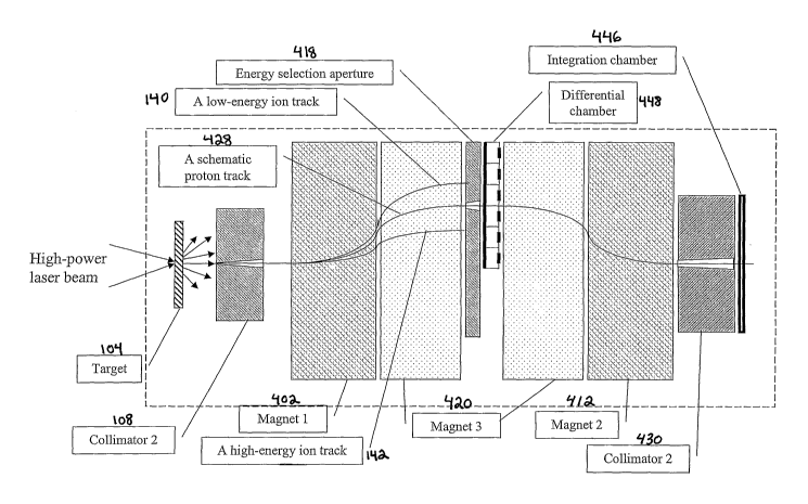
FIG. 21 depicts a sectional view of an embodiment of an ion selection system
of the
present invention.
FIG. 22 depicts a perspective view of an embodiment of an ion selection system
of the
present invention.
FIG. 23 depicts a sectional view of an ion selection system depicted in FIG.
22.
FIG. 24 depicts a sectional view of an embodiment of an ion selection system
of the
present invention.
FIG. 25 depicts a sectional view of an embodiment of an ion selection system
of the
present invention.
FIG. 26 depicts a sectional view of an embodiment of an ion selection system
of the
present invention.
FIG. 27 depicts a sectional view of an embodiment of an ion selection system
of the
present invention.
FIG. 28 depicts a sectional view of an embodiment of an ion selection system
of the
present invention.
-11-
CA 02525777 2005-11-14
WO 2004/109717 PCT/US2004/017081
FIG. 29 depicts a sectional view of an embodiment of an ion selection system
of the
present invention.
FIG. 30 depicts a sectional view of an embodiment of an ion selection system
of the
present invention.
FIG. 31(a) depicts a sectional view of an embodiment of an ion selection
system of the
present invention.
FIG. 31(b) depicts a schematic illustration of a collimator 2 (i.e., a
multileaf collimator)
in the x-z plane showing openings in the collimator for selecting positive
ions of a particular
energy.
FIG. 32 depicts a schematic illustration of an energy selection aperture.
FIG. 33 depicts a schematic illustration of a multileaf collimator in the x-z
plane: (a)
shows openings in the multileaf collimator for selecting low energy ions; (b)
shows openings in
the multileaf collimator for selecting lugh energy ions.
FIG. 34 depicts a sectional view of an embodiment of an ion selection system
of the
present invention.
FIG. 35 depicts a sectional view of an embodiment of an ion selection system
of the
present invention.
FIG. 36 depicts a sectional view of an embodiment of an ion selection system
of the
present invention.
FIG. 37 depicts a sectional view of an embodiment of an ion selection system
of the
present invention.
FIG. 38 depicts a sectional view of an embodiment of an ion selection system
of the
present invention.
FIG. 39 depicts a sectional view of an embodiment of an ion selection system
of the
present invention.
FIG. 40 depicts a sectional view of an embodiment of an ion selection system
of the
present invention.
FIG. 41 depicts a sectional view of a laser-accelerated high energy
polyenergetic positive
ion therapy system of the present invention.
FIG. 42(a) depicts a perspective view of an embodiment of a laser-accelerated
high
energy polyenergetic positive ion beam treatment center.
FIG. 42(b) depicts a perspective view of an embodiment of a laser-accelerated
high
energy polyenergetic positive ion beam treatment center that includes an
optical monitoring and
control system.
-12-
CA 02525777 2005-11-14
WO 2004/109717 PCT/US2004/017081
FIG. 42(c) depicts a perspective view of an embodiment of a laser-accelerated
high
energy polyenergetic positive ion beam treatment center that includes more
than one ion therapy
system.
FIG. 42(d) depicts a perspective view of an embodiment of a laser-accelerated
high
energy polyenergetic positive ion beam treatment center that includes more
than one ion therapy
system, with each of the ion therapy systems having an optical monitoring and
control system.
FIG. 43 depicts a flow chart of an embodiment of a method of treating a
patient using
polyenergetic high energy positive ions.
DETAILED DESCRIPTION
OF ILLUSTRATIVE
EMBODIMENTS
The following
abbreviations
and acronyms
axe used herein:
CORVUS a treatment optimization system for photon
IIVVIRT from NOMOS
CPA ~~ chirped pulse amplification
CT computer-aided tomography
DICOM Digital Imaging and Communications in Medicine
DICOM RT DICOM Radiation Therapy Supplement
DVH dose-volume histogram
EIMPT energy- and intensity-modulated proton therapy
EGS4 Electron Gamma Shower (version 4) Monte Carlo
code system
GEANT(3) a Monte Carlo system for radiation (proton,
neutron, etc) simulation
IlVVIRT intensity-modulated (photon) radiation therapy
JanUSP a high power (1019-1021W/cma) laser at LLNL
LLNL Lawrence Livermore National Laboratory
LLUMC Loma Linda University Medical Center, Loma
Linda, CA
MCDOSE an EGS4 user-code for dose calculation in a
3-D geometry
MGH Massachusetts General Hospital, Boston, MA
MLC multileaf collimator
NOMOS NOMOS Corp., Sewickley, PA
NTCP normal tissue complication probability
PC personal computer
PIC particle-in-cell (simulation technique for
laser plasma physics)
PMC primary monitor chamber
PSA prostate-specific antigen
PTV planning target volume
PTR_AN a Monte Carlo code system for proton transport
simulation
-13-
CA 02525777 2005-11-14
WO 2004/109717 PCT/US2004/017081
RTP radiotherapy treatment planning
SMC secondary monitor chamber
SOBP spread out Bragg peak (for proton/ion beams)
SSD source-surface distance
TCP tumor control probability
MeV million electron volts
GeV billion electron volts
T Tesla
As used herein, the term "protons" refers to the atomic nuclei of hydrogen
(Hl) having a
charge of +1.
As used herein, the teen "positive ions" refers to atoms and atomic nuclei
having a net
positive charge.
As used herein, the teen "polyenergetic" refers to a state of matter being
characterized as
having more than one energy level.
As used herein, the term "high energy" refers to a state of matter being
characterized as
having an energy level greater than 1 MeV.
As used herein, the term "beamlet" refers to a portion of a high energy
polyenergetic
positive ion beam that is spatially separated, or energetically separated, or
both spatially and
energetically separated.
The terms "primary collimator", "primary collimation device", "initial
collimator", and
"initial collimation device" are used interchangeably herein.
The terms "energy modulation system" and "aperture" are used interchangeably
when it
is apparent that the aperture referred to is capable of modulating a spatially
separated high
energy polyenergetic positive ion beam.
All ranges disclosed herein are inclusive and combinable.
In one embodiment of the present invention there is provided a laser-
accelerated
polyenergetic ion selection system for radiation therapy. The design of this
system typically
includes a magnetic field source that is provided to spatially separate
protons of different energy
levels. A magnetic field source is also provided to separate out plasma
electrons that initially
travel with the protons. While these two magnetic field sources are typically
provided by the
same magnetic field source, two or more separate magnetic field sources may be
provided to
carry out these functions. After the protons have been spatially separated,
one or more apertures
are typically provided to select an energy distribution needed to cover the
treatment target in the
depth direction for a given beamlet. The form of an aperture is dictated by
the location as well as
- 14-
CA 02525777 2005-11-14
WO 2004/109717 PCT/US2004/017081
the depth dimension of the target, as described more fully below. Once the
spatial position and
the target size are known, the proton energy spectrum needed to cover the
target for a given
beamlet in the depth direction is calculated by combining the depth dose
curves of different
proton energy levels, as described more fully below. Due to the angular
distribution of protons,
a primary collimation device is typically employed to reduce spatial mixing of
different energy
protons. The primary collimation device is typically employed to collimate the
positive ions into
a magnetic field that separates the ions by energy levels. As a result of this
spatial mixing, the
proton energy spectrum in a given spatial location typically has a small
spread that depends on
the energy of the protons. The depth dose curves are typically calculated
using the spread out
(i.e., polyenergetic) proton spectrum. In this regard, the depth dose curves
for the proton energy
modulation are typically modified to account for this polyenergetic spreading
effect, as described
more fully below.
Description of a proton selection and collimation system: In one embodiment of
the
present invention there is provided an ion selection and collimation device
needed for proton
energy modulation. Using the 2D particle in cell simulation code (PIC),
described by C.K.
Birdsall and A.B. Langdon in Plasma Physics via Computer Simulation (McGraw-
Hill Book
Company, Singapore 1985), the interaction of a petawatt laser pulse with a
thin dense foil
(hydrogen rich) was simulated, yielding protons with energy well beyond 200
MeV and
maximum energy reaching 440 MeV. The simulations were performed for a 3.6 ,um
(in the
radial direction) full width at half maximum (FWHM), 14 femtosecond (fs)
linearly polarized
laser pulse with a wavelength, ~, = 0.8 ,um and intensity I =1.9 x 10²² Wlcm2
, normally
incident onto a thin dense plasma slab (ionized foil) with a density thirty
times higher than the
critical density n~r = 4~c2mec2sol(e2~,2) and thickness d ~ l,um . Such laser
intensity is within the
reach of the recent technological developments, as described by G. A. Mourou
et al., in
"Ultrahigh-Intensity Lasers: Physics of the Extreme on a Tabletop ", Physics
Today, 22-28
(1998). The basic configuration of such as laser light source system is
described in U.S. Pat. No
5,235,606, issued Aug. 10, 1993 to Mourou et al., which is incorporated by
reference herein.
U.S. Patent Appl. No. 09/757,150 filed by Tajima on Jan. 8, 2001, Pub. No.
U.S. 2002/0090194
Al, Pub. Date July 11, 2002, "Laser Driven Ion Accelerator" discloses a system
and method of
accelerating ions in an accelerator using such a laser light source system,
the details of which are
incorporated by reference herein in their entirety.
The protons coming from a thin foil are typically accelerated in the forward
direction by
the electrostatic field of charge separation induced by the high intensity
laser. Further details of
this process are described by V. Yu. Bychenkov et al., in "High energy iora
generation in
-15-
CA 02525777 2005-11-14
WO 2004/109717 PCT/US2004/017081
interaction of short laser- pulse with solid derasity plasma ", Appl. Phys. B,
207-215 (2002).
Over a period of several tens of plasma frequency wp = ne2/m~Eo cycles,
protons are typically
accelerated to relativistic energy levels. The maximum value of the proton
energy levels
typically depend on several factors, including laser pulse length and
intensity, and plasma foil
thickness. The late time dynamics can be discerned by PIC code, which shows
that protons
reach a stationary distribution (energy, angular) and move in a formation
together with the
electrons. This reassures the preservation of the low proton emittance,
shielding proton space
charge, which otherwise could be detrimental to the emittance. The angular
distribution of
protons exhibits the spread which depends on the energy. Typically, the
general trend is such
that the higher the energy of the accelerated protons, the more they are
emitted in the forward
direction. The depth dose distribution calculated using the laser-accelerated
proton spectrum
shows that the polyenergetic positive ion spectrum emitted from the target
typically cannot be
readily used for radiation treatments. A high energy deposition to the area
beyond the effective
Bragg peak typically arises from the high entrance dose to the superficial
structures and the long
tails in the polyenergetic dose distributions. Thus, in one embodiment of the
present invention,
one delivers a homogeneous dose to the tumor volume to minimize the dose to
the surrounding
healthy tissues. This is achieved by providing an ion (e.g., proton) selection
and collimation
device that generates the desired polyenergetic proton energy distribution.
This device separates
polyenergetic positive ions (e.g., protons) into spatial regions according to
their energy. The
spatially separated regions of the positive ions are subsequently controlled
using at least one
magnetic field. The spatially separated positive ions are controllably
modulated using an
aperture to provide the desired dose. Optionally, the device also includes a
magnetic field source
for generating a magnetic field to eliminate the plasma electrons that travel
with the positive
ions. This optional magnetic field source can be the same or a different
magnetic field as the one
spatially separating the polyenergetic positive ions. This magnetic field is
also useful for
eliminating plasma electrons traveling together with the laser-accelerated
positive ions.
A schematic diagram of one embodiment of the ion selection system (100) is
provided in
FIG.1. Referring to this figure, there is provided a series of magnetic field
sources that produce
a magnetic field pattern B = B(z)eZ , the z-direction being perpendicular to
the page. A first
magnetic field source provides a first magnetic field (102), listed as "5.0 T
into page", at a
distance from 5 cm to 20 cm from a plasma target (104) located at 0 cm along
the x (primary
beam) axis (114). High energy polyenergetic positive ions (110) are generated
by the interaction
between the plasma target (104) with a suitable laser pulse (not shown). A
beam of high energy
polyenergetic positive ions (e.g., protons) (106) enter the first magnetic
field (102) after exiting
-16-
CA 02525777 2005-11-14
WO 2004/109717 PCT/US2004/017081
an initial collimation device (108). The protons are shown exiting the initial
collimation device
(108) into the first magnetic field (102), the protons being characterized as
having an angular
spread. A second magnetic field (112) source provides a second magnetic field
listed as "5.0 T
into page" at a distance from 60 cm to 75 cm from the plasma target (104)
along the x (primary
beam) axis (114). High energy polyenergetic positive ions (116) (protons in
certain
embodiments) enter the second magnetic field (112) after exiting an aperture
(118). Also shown
in FIG. 1 is a third magnetic field source providing a third magnetic field
(120), which is listed
as "5.0 T out of page" at a distance from 25 cm to 55 cm from the plasma
target (104) located at
0 cm along the x axis (114). The x axis as drawn is parallel to the beam axis
(114) of the laser in
this embodiment. Other coordinate orientations and coordinate systems, such as
cylindrical and
spherical coordinate systems, can be suitably used. High energy polyenergetic
positive ions
(126) enter the third magnetic field (120) after exiting the first magnetic
field (102). The first
magnetic field (102) is shown spatially separating the trajectories (128) of
the high energy
polyenergetic positive ions by energy level. The third magnetic field (120) is
shown bending the
trajectories of spatially separated ions (130) towards the aperture (118). The
aperture modulates
the ion beam by controllably selecting a portion of the spatially separated
ions, as described
further herein. The third magnetic field (120) is also shown bending the
trajectories of the
spatially separated polyenergetic positive ions (132) towards the beam axis
and towards the
second magnetic field (112). The second magnetic field (112) recombines the
spatially separated
and modulated ions (134) to form a recombined ion beam (136). The recombined
ion beam
(136) is shown entering a secondary collimation device (138). Upon exiting the
secondary
collimation device (138), a high energy polyenergetic positive ion beam is
provided that is
suitable for use in high energy polyenergetic positive ion radiation therapy.
Suitable magnetic
field sources for this and various embodiments of the present invention
typically have a magnetic
field strength in the range of from about 0.1 to about 30 Tesla, and more
typically in the range of
from about 0.5 to about 5 Tesla. The Lorentz force of the magnetic field
typically spreads out
the polyenergetic protons. The lower energy protons (140) typically are
deflected more from
their original trajectories exiting the initial collimation device 108)
("initial collimator") than are
the high energy protons (142).
As described herein, many of the embodiments of the present invention use
magnetic
field sources to provide magnetic fields for manipulating the positive ion
beams. In additional
embodiments of the present invention one or more of the magnetic field sources
are replaced by,
or combined with, one or more electrostatic field sources for manipulating the
positive ion
beams.
-17-
CA 02525777 2005-11-14
WO 2004/109717 PCT/US2004/017081
The initial collimator (108) typically defines the angular spread of the
incoming beam
(106) entering the first magnetic field (102). The tangent of the angle of the
beam spread of the
beam (106) exiting the initial collimator (108) is typically about the ratio
of one half the distance
of the initial collimator exit opening (144) where the beam exits the
collimator to the distance of
the collimator exit opening (144) to the proton beam source (i.e., the plasma
target, 104).
Typically, this angle is less than about 1 radian. The emitting angle is the
angle of the initial
energy distribution exiting the target system (i. e., target, 104 and initial
collimation device, 108).
Electrons (146) are typically deflected in the opposite direction from the
positive ions by the first
magnetic field and absorbed by a suitable electron beam stopper (148).
Suitable electron
stoppers (148) include tungsten, lead, copper or any material of sufficient
thickness to attenuate
the electrons and any particles they generate to a desired level. The aperture
(118) is typically
used to select the desired energy components, and the matching magnetic field
setup (in this
embodiment, the second magnetic field, 112) is selected that is capable of
recombining the
selected protons (134) into a polyenergetic positive ion beam. Suitable
apertures typically can be
made from tungsten, copper or any other materials of sufficient tluckness that
are capable of
reducing the energy levels of positive ions. This energy level reduction is
typically carried out to
such a degree that the positive ions can be differentiated from those ions
that do not go through
the aperture. In various embodiments of the present invention, the aperture
geometry can be a
circular, rectangular, or irregular-shaped opening (150)(or openings) on a
plate (152)(or slab),
which when placed in a spatially separated polyenergetic ion beam, is capable
of fluidically
communicating a portion of the ion beam therethrough. In other embodiments,
the aperture
(118) can be made from a plate that has multiple openings that are
controllably selected, such as
by physical translation or rotation into the separated ion beam to spatially
select the desirable
energy level or energy levels to modulate the separated ion beam. The
modulation of the ion
beam gives rise to a therapeutically suitable high energy polyenergetic
positive ion beam (136)
as described herein. Suitable apertures include multi-leaf collimators. In
addition to controllably
selecting the spatial position of the openings that fluidically communicate
the spatially separated
ion beams, the aperture openings may also be controllably shaped or multiply
shaped, using
regular or irregular shapes. Various combinations of openings in the aperture
(118) are thus used
to modulate the spatially separated ion beam (130). The spatially separated
positive ions (132)
are subsequently recombined using the second magnetic field (134).
The high and low energy positive ion (e.g., proton beam) stoppers (154 and
156,
respectively) typically eliminate unwanted low-energy particles (140) and high-
energy particles
(not shown). Because of the broad angular distribution of the accelerated
protons (which
-18-
CA 02525777 2005-11-14
WO 2004/109717 PCT/US2004/017081
depends on a given energy range), there is typically a spatial mixing of
different energy positive
ions after they pass through the first magnetic field. For example, a portion
of the low energy
protons may go to regions where the high energy particles reside, and vice
versa. Reducing the
spatial mixing of protons is typically carried out by introducing a primary
collimation device,
such as the initial collimation device 108 of the embodiment depicted in
FIG.1. A primary
collimation device is typically used to collimate protons to the desired
angular distribution.
As described fuxther below, proton spatial differentiation is typically
carried out by
passing the positive ions through a small collimator opening prior to their
entering the first
magnetic field. An example of a small collimator opening is depicted in FIG. 1
as the initial
collimator opening (144). Typically, the collimator exit opening (144) is not
arbitrarily small,
since smaller openings typically lower the dose rate and increase the
treatment time. As a result
of the finite size of the collimator opening (144), the protons are typically
spatially mixed.
Accordingly, any given spatial location for a collimator opening (however
small) typically
provides a polyenergetic proton energy distribution. While not being bound by
any particular
theory of operation, the energy modulation calculations take into account the
polyenergetic
characteristics of the positive ions entering the ion selection device to
provide the needed depth
dose curves. The polyenergetic characteristics of these positive ions is
understood through the
influence of the magnetic field on the dynamics of the positive ions. The
following description
is directed to the dynamics of protons, as one illustrative embodiment.
Additional embodiments
to other positive ions in addition to protons are also envisioned.
To describe the proton's dynamics in the magnetic field, a numerical code is
written
which solves the following equation of motion,
dp' = evt x B (1)
dt
where p = mpvl 1-v2/c2 , B is the magnetic induction vector, mP is the proton
rest mass and i
signifies the particle number. For one embodiment of the present invention,
this equation was
solved using a symplectic integration algorithm developed by J. Candy and W.
Rozmus in "A
Symplectic Integration Algorithm fog Separable Hamiltonian Functions ", J.
Comp. Phys. 230-
239 (1991). The initial conditions ,[ (ro, vo) ] were obtained from the PIC
simulation data, wluch
provided the phase-space distribution for protons. The contribution of the
self consistent fields
on the proton dynamics were neglected, since the Lorentz force created by the
external magnetic
field to separate the electrons from the protons is greater for the magnetic
field induction used in
the calculations than the Coulomb force in the region beyond the initial
collimation device.
Using the equation of balance between the Lorentz and the inter-particle
Coulomb forces, one
-19-
CA 02525777 2005-11-14
WO 2004/109717 PCT/US2004/017081
arrives at a condition for particles spatial separation distance for which the
magnetic force
prevails over the Coulomb force,
liz
~~ > a (2)
4~s°Bv
where B is the magnitude of the magnetic field, v is the particle velocity and
a is an elementary
charge. The average inter-particle distance r can be obtained from the
particle density ~ = fZ-'~3 ,
thus the inequality (2) can be rewritten in the form:
h < 47L8°BV 3/2 (3)
a
Providing the lowest therapeutic energy protons of about 50 MeV, which
corresponds to
proton velocity of v = 0.3c , and the magnetic field induction B = 5 T, the
condition (3) gives,
n < 2 ~ 102° cm-3 . The particle density in the region beyond the
initial collimation device can be
estimated using the arguments presented by E. Fourkal et al. in "Particle iu
cell simulation of
laser-accelerated proton beams for radiation therapy ", Id. (2002). In this
region the particle
density is n = 4 ~ 1013 cm-3 , which is far below the estimated threshold
value of 2 ~ 102° cm-3 .
This estimate validates the assumption of the insignificant contribution of
the self consistent
electrostatic field on the proton dynamics in the external magnetic field.
The calculations of the proton dynamics in the magnetic field have also
neglected such
boundary effects as edge focusing due to the influence of the fringing field
patterns at the edge of
a sector field. These effects are expected to be small in the bulk of the
selection system due to
the canceling action of alternating magnetic field patterns (with the same
absolute value of the
field induction). As the positive ions (e.g., protons) leave the final field
section, the boundary
fringe field can introduce some focusing effect. This effect can be accounted
for by using the
magnetic field distribution at the boundary.
Monte Carlo calculations: While not being bound by any particular theory of
operation,
the GEANT3 Monte Carlo radiation transport code is used for dose calculations.
GEANT3 is
used to simulate the transport and interactions of different radiation
particles in different
geometries. The code can run on different platforms. A detailed description of
the operation and
usage of GEANT3 has been given by R. Brun et al., in GEANT3-Detector
description and
simulation tool Refereface Manual (1994). GEANT3 is equipped with different
user selectable
particle transport modes. Being more versatile than most Monte Caxlo codes
concerning the
production of secondaries, GEANT3 has three options to deal with these rays.
An important user
controlled variable for these options is DCUTE below which the secondary
particle energy losses
are simulated as continuous energy loss by the incident paxticle, and above it
they are explicitly
-20-
CA 02525777 2005-11-14
WO 2004/109717 PCT/US2004/017081
generated. In the first option, the secondary particles are produced over the
entire energy range
of the incident particle. This mode is termed as "no fluctuations". The second
mode of energy
loss is "fixll fluctuations", in which secondaries are not generated, and the
energy loss straggling
is sampled from a Landau ( "Oh the energy loss of fast particles by ionization
", J. Phys. USSR, ,
201-210 (1944)), Vavilov ( "lohisation losses of high energy heavy particles
", Soviet Physics
JETP, , 749-758 (1957)) or Gaussian distribution each according to its
validity limits (R. Brun et
al., Id.). The third is "restricted fluctuations", with generation of
secondaries above DCUTE and
restricted Landau fluctuations below DCUTE. In principle, choosing energy loss
fluctuations
typically carries am advantage if energy deposited is scored in voxel sizes
larger than the range of
secondaries. This results in great savings of computation time and avoids
tracking a large
number of secondaries generated below DCUTE. Typically, a continuous energy
loss by the
incident particle is assumed according to the Berger-Seltzer formulae.
Moliere multiple scattering theory is used by default in GEANT3. Multiple
scattering is
well described by Moliere theory. See, e.g., G. Z. Moliere, "Theoy~ie der
St~euung schfzeller
geladeheY TeilclaefZ L~ Eihzelstf~euung am abgeschi~mten Coulomb-Feld ", Z.
Naturforsch., a,
133-145 (1947); and G. Z. Moliere, "Theorie der St~euung schheller geladener
Teilche~c Il.
Meh~fach- ufZd T~ielfachstreuuhg", Z. Naturforsch., a, 78-85 (1948). A
limiting factor in the
Moliere theory is the average number of Coulomb scatters S~o for a charged
particle in a step.
When S2o < 20 , the Moliere theory is typically not applicable. According to
E. Keil et al. in
"Zur Eiy fach- uud MehYfachstreuung geladehe~ Teilchen ", Z. Naturforsch, a,
1031-1048 (1960),
the range 1 < SZo <_ 20 is called the plural scattering regime. In this range
a direct simulation
method is used for the scattering angle in GEANT3 (R. Brun et al., Id.). A
simplification of the
Moliere theory by a Gaussian form is also implemented in GEANT3. The Gaussian
multiple
scattering represents Moliere scattering to better than 2 % for 10 < SZo <_
10$ .
The hadronic interactions in matter (elastic, inelastic, nuclear fission,
neutron nuclear
capture) are described by two software routines, GHEISHA and FLUKA, which are
available to
users of LEANT. The GHEISHA code generates hadronic interactions with the
nuclei of the
current tracking medium, evaluating cross-sections and sampling the final
state kinematics and
multiplicity, while the LEANT philosophy is preserved for the tracking
purposes. A number of
routines that exist in GHEISHA are responsible for generating the total cross-
sections for
hadronc interactions, calculating the distance to the next hadronic
interaction according to the
total cross-sections and finally the main steering routine for the type of
occurred hadronic
interaction. FLUKA is a simulation program, which as a standalone code
contains transport and
-21 -
CA 02525777 2005-11-14
WO 2004/109717 PCT/US2004/017081
the physical processes for hadrons and leptons and tools for geometrical
description. In LEANT,
only the hadronic interaction part is included. As with the GHEISHA package,
the FLUI~A
routines can compute the total cross-sections for hadronic processes, and
perform the sampling
between elastic and inelastic processes. The cross-sections for both types of
interactions are
computed at the same time as the total cross-section. Subsequently, a particle
is sent to the
elastic or inelastic interaction routines. After the interaction, the eventual
secondary particles are
written to the LEANT stack.
The following control parameters were used to calculate the depth dose
distributions for
proton beams in the example presented herein: The cutoff energy for particles
was 20 keV, the
Rayleigh effect was considered, 8 -ray production was turned on, continuous
energy loss for
particles below cutoff energy levels sampled directly from the tables, Compton
scattering was
turned on, pair production with generation of a /e+ was considered,
photoelectric effect was
turned on, and positron annihilation with generation of photons was
considered.
Results and Discussion: The PIC simulations show that the maximum proton
energy of
the polyenergetic proton beam is a function of many variables including the
laser pulse intensity
and duration, as well as the target density and its thickness. The
quantitative dependence of the
maximum proton energy on laser/plasma target parameters can be found in
Fourkal et al. The
overall results of this study showed that the maximum proton energy increases
with decreasing
thickness of the plasma target reaching the plateau for the target thicknesses
on the order of the
hot electron Debye length (for a given laser intensity). In the same time, the
proton energy is a
non-monotonous function of the laser pulse length, reaching the maximum value
for the laser-
pulse length of the order of 50 femtoseconds. Thus, depending on the
simulation parameters,
one can obtain a broad spectrum of energy distributions for the accelerated
protons.
FIGS. 2(a) and 2(b) show the energy and angular distributions for the protons
accelerated by the laser pulse described above. For the laser/plasma
parameters chosen in the
simulation, the maximum proton energy reaches the value of 440 MeV, which is
much higher
than typically needed for radiotherapy applications. To reduce the unwanted
protons, as well as
to collimate them to a specific angular distribution, a primary collimation
device is provided. Its
geometrical size and shape is typically tailored to the energy and angular
proton distributions.
For example, in one embodiment of the present invention there is provided a 5
cm long tungsten
collimator that absorbs the unwanted energy components. Because of its density
and the
requirement for the compactness of the selection system, tungsten is a
favorable choice for
collimation purposes. A suitable primary collimator opening provides a 1 x 1
c~ra2 field size
defined at 100 cm SSD. Protons that move into aa1 angle larger than tlus are
typically blocked.
-22-
CA 02525777 2005-11-14
WO 2004/109717 PCT/US2004/017081
With the magnetic field configuration shown in FIG.1, for example, the
solution to the equation
of motion (1) with the initial conditions given by the proton phase space
spectra obtained from
the PIC simulations, yields the proton spatial distributions N = N(y) at the
plane x = 40 cm, z =
0 cm, as shown in FIG. 3. This shows that the magnetic field spreads the
polyenergetic protons
into spatial regions according to their energy and angular distributions.
Their spatial distribution
is such that the lower energy particles are deflected at greater distances
away from the central
axis, and as the proton energy increases the spatial deflection decreases.
Therefore, the
contribution of both the magnetic field and the primary collimator (with a
specific collimator
opening) creates such a spatial proton distribution that allows the energy
selection or proton
energy spectrum reformation, using an aperture. The geometric shape of an
aperture typically
determines the energy distribution of the therapeutic protons.
As mentioned above, due to the presence of the angular spread, there is
typically a spatial
mixing of different energy protons. As a result of this mixing, the proton
energy distribution in a
given spatial location is typically no longer monochromatic, but has a spread
around its peak.
FIG. 4 shows the proton energy distributions at different spatial locations.
These distributions
were calculated by counting the number of protons in the given spatial
location of width Dy = 3
mm as a function of energy. This figure shows that the lower energy particles
have a much
smaller spread than the high energy particles. Without being bound to a
particular theory of
operation, this result is apparently due to the higher energy protons not
being deflected as much
in the magnetic field as are the lower energy particles. Because of the energy
spread effect, the
depth dose curves needed for the energy modulation calculations typically are
modified to
include the effect of the energy spread in the calculations, since mono-
energetic protons are not
typically for the depth dose calculations. Using the GEANT3 Monte Carlo
transport code the
dose distributions for the proton energy spectra shown in FIG. 4 for a 4 x 4
cm2 field size was
calculated. The results of the simulation are shown in FIG. 5. The presence of
an energy spread
in the proton spectra leads to the broadening of the dose distributions, which
leads to a less sharp
falloff of the energy-modulated Bragg peals as compared to the case of mono-
energetic beams.
See, e.g., T. Bortfeld. The broadening is typically most profound for the
higher energy protons.
FIGS. 6(a) and 6(b) show the spatial distribution of protons N = N(y) at the
plane x=40
cm, z=0 cm for the magnetic field configuration shown in FIG.1, using a
primary collimator
opening of 5 x 5 czzz2 defined at 100 cm SSD and the proton energy
distributions N~ = Ni (E) ,
where index i denotes the energy levels of the polyenergetic proton beams.
Comparing FIG. 5,
6(a) and 6(b) to FIGS. 3 and 4 the spatial separation of protons at larger
openings is less
effective leading to the higher order of spatial mixing and the larger spread
in the energy
- 23 -
CA 02525777 2005-11-14
WO 2004/109717 PCT/US2004/017081
distributions. The energy spread as used herein is defined as the difference
between the
maximum and the minimum energy in the distribution. FIG. 7 shows the energy
spread as a
function of a collimator opening for several proton energy levels; the energy
spread increases
with increasing aperture opening and is more profound for higher energy
particles.
As a result of the energy spread effect, the depth dose curves will typically
have less
sharp falloff beyond the effective Bragg peak region for wider apertures as
compared to the cases
of narrower collimator openings. FIG. 8 shows the dose distributions for the
proton energy
spectra shown in FIG. 6(b), which corresponds to a primary collimator of 5 x 5
crn2 defined at
100 cm SSD, normalized to the incident proton fluence. Comparing FIG. 5 with
FIG. 8 shows
that desirable dosimetric characteristics from the laser accelerated protons
are typically obtained
for smaller primary collimator openings. Suitable primary collimator openings
are typically
smaller than about 2000 cm2, more typically smaller than about 100 cm2, and
even more
typically smaller than about 1 cmz, when defined at 100 cm SSD. Typically
there is a lower limit
on the size of the collimator opening, which is suitably determined by the
field size, dose rate, or
both, that the system can yield after beam collimation. The geometry of the
collimator opening
typically influences the treatment time.
Once the depth dose distributions for polyenergetic proton beamlets are
determined, a
proton energy distribution that provides a homogeneous dose along the target's
depth direction is
calculated using the target location and volume. In one embodiment, the
following steps are
carried out to calculate the desired proton energy distribution:
1. The geometrical size of the target (in the depth direction) determines the
proton energy
range for radiating the target. Using the depth dose distributions for a given
energy range, the
weights for the individual polyenergetic beamlet are computed, with the
assumption that the
weight for the beamlet with the energy distribution, which gives the effective
Bragg peak at the
distal edge of the target, is set to one. The weights W = W (E) are computed
based. on the
requirement of the constancy of the dose along the depth direction of the
target.
2. Once the weights are lcnown, the proton energy distribution N(E) for
providing a
suitable dose along the target's depth dimension are calculated by convolving
the weights W,. (E)
with the energy distributions Nl (E) of polyenergetic proton beamlets to give
N(E) _ ~ W (E)N~ (E) (4)
r
where index i runs through energy levels of the polyenergetic proton beamlets
for radiating the
area of interest (in depth direction). A suitable energy modulation
prescription for protons is
provided by the formulation of the absorbed dose distribution for electrons
introduced by
-24-
CA 02525777 2005-11-14
WO 2004/109717 PCT/US2004/017081
Gustafsson, A., et al., in "A generalized pencil beam algorithm for
optimization of radiation
therapy ", Med. Phys., 343-356 (1994), in which the incident particle
differential energy fluence
integrated over the surface and solid angle corresponds to the energy
distribution defined in Eq.
(4). As an example, a hypothetical target with spatial dimensions 4 x 4 x 5
cm3 , located at depth
lying between 9 cm and 14 cm is considered. The energy range of polyenergetic
protons
required to cover this taxget is 110 MeV < E < 152 MeV. Using both the depth
dose
distributions for polyenergetic proton beamlets with the spread out energy
spectra discussed
earlier and the condition of a constancy of the resultant dose along the
target's depth direction,
the weights W for each individual beamlet, that are indicated in Table 1 are
readily obtained.
Table 1
~s21OOy~490.25W,460.15
1'11430.12y~400.10W370.095
yy340.09X310.085y~280.08
yy25O.O7yY220.06yyl90.05
~ls0.04W130.035Wlo0.03
Distribution of weights corresponding to protons with a different
characteristic
energy: In one embodiment of the present invention, a procedure for finding
the weights is
provided. This procedure is mathematically similar to minimizing the following
functional
r(z) _ ~ W 7~~ (z) -Do, for9cm <_ z <_ l4cm (5)
where i denotes energy bins, DI is the depth-dose distribution corresponding
to the ith
polyenergetic energy bin and Do is a constant corresponding to a specific dose
level (typically
larger than the distant Bragg peak in view of the contribution from the
adjacent depth-dose
distributions). The physical meaning of the weights are described further. The
absolute value of
each individual weight is correlated to the physical method associated with
the actual energy
modulation process in the selection system. The design of the energy
modulation system (i.e.,
the aperture) is achieved by either using an aperture whose geometric shape is
correlated to the
weights or by using a slit, which can move along the y -axis in the region
where the protons are
spread according to their energy levels, and the time spent in a given region
will be proportional
to the value of the weight for the given energy. Convolving the weights of the
Table (1) with the
energy distributions for each individual beamlet according to equation (4),
one obtains the actual
modulated energy distribution that will deliver the SOBP for the given
target's depth dimension.
- 25 -
CA 02525777 2005-11-14
WO 2004/109717 PCT/US2004/017081
This energy distribution differs from that calculated using monoenergetic
proton beams (for
which the weights themselves represent the actual energy distribution) because
of the presence of
particles with energy levels beyond the ones associated with the weights,
which typically arises
from a consequence of a finite primary collimator. The presence of these
"extra particles"
typically makes the dose distribution beyond the SOBP fall off less sharply
than that obtained
using mono-energeticebeams.
FIG. 9 shows the proton energy spectrum (a) and the corresponding dose
distribution
(normalized to the incident proton fluence) (b) for a target considered in the
calculations. The
resultant dose distribution shows the quick fall off of the dose beyond the
distal edge of the
target although not as dramatic as for an ideal case of convolving mono-
energetic protons shown
also in FIG. 9(b). The entrance dose is still significant compared to the dose
to the target. In
order to reduce the entrance dose, several proton beams coming from different
directions but
converging at the target could be used, so that the target receives the
prescribed dose and the
surrounding healthy tissue receives much less dose. Therefore, the energy and
intensity
modulated proton therapy is expected to further improve target coverage and
normal tissue
sparing.
Dose Rate Determination: As mentioned earlier, it is important to determine
the
absolute dose rate that the ion selection system can yield. This quantity is
closely related to the
absolute number of accelerated protons. From the PIC simulations it was
determined that for a
laser intensity of about I =1.9 x 10²² YYlcm2 and pulse length of about 14 fs,
the number of
protons accelerated to energy levels higher than about 9 MeV is about 4.4 x
105 when the total
number of protons used in PIC simulation is 1048576. Without being bound by a
particular
theory of operation, not all of the protons in the plasma slab are believed to
interact with the
laser. Only those protons that are located in the laser's propagation path
typically experience the
strongest interaction.
In simulation studies, the laser occupies an area of about 3/5 of the total
size of the
simulation box (in a direction perpendicular to the propagation), which
provides about 6.3 x 10⁵
protons (out of 1048576) that will "sample" the laser. This means that about
70 io of the
effective number of protons are accelerated to energy levels higher than about
9 MeV. On the
other hand, the total number of protons in a plasma slab that subtends the
laser pulse can be
estimated using the proton density of the foil n f as well as the laser focal
area S and the
thickness of the foil d to give N = S x n f x d ~ 2 x 10'2 . Finally this
gives about
-26-
CA 02525777 2005-11-14
WO 2004/109717 PCT/US2004/017081
N = 0.7 * 2 x 10'z =1.4 x 10¹² protons that will be typically accelerated to
energy levels greater
than about 9 MeV.
With the above in mind, the absolute dose delivered to the target is estimated
in the
following way. The polyenergetic beams needed to cover the target in depth
direction (9 cm
< ~ < 14 cm) will typically have an energy range of about 110-152 MeV. The
number of
protons in the energy range of about 147 MeV < E < 157 MeV moving into the
angle of 0.01
radian (approximately 2.6 % of the total number of protons in the energy range
147 MeV < E <
157 MeV) is N = 2.6 x 1 O$ , which corresponds to ~o = 2.6 x 1 O$ 1/cmz ( 1 x
1 cm2 field size) per
laser pulse for the initial fluence of protons at a distance of about 100 cm
from the source.
FIG. 9(b) shows that the dose deposited by protons in the Monte Gaxlo
simulations
(normalized to the initial fluence) at depths 9 cm <_ d <_ 14 cm is about Do
=1.6 x 10⁻⁹ Gy * cm2 .
This gives D = Do * ~o ~ 0.43 Gy per laser shot. Typical lasers operating in a
10 Hz repetition
rate yield D ~ 256 Gy per minute for the pencil beam of 1 x 1 cm2 . The dose
rate is typically
not only a function of laser-plasma parameters but also depends on the
location and volume of
the target. This leads to D ~ 64 Gy/min for the target located at depth z = 25
cm (the distal
edge of the target) with volume of 1 x 1 x 5 cm3 . While not being bound to
any particular theory
of operation, the reduction of the dose rate in this case is apparently due to
both the smaller
number of protons in the energy range needed to cover the deeply seated
target, as well as the
less energy deposited within the target (the height of the Bragg peak gets
smaller as the proton
energy increases). The calculation presented above estimates the absolute dose
rate for 1 x 1 cmz
pencil beam. More typically, the cross-section of the treatment volume is
larger in area than 1 x 1
cm2 and the "effective" dose rate becomes smaller and comparable to that of
conventional linear
accelerators. Larger targets can be effectively treated by scanning the high
energy polyenergetic
positive ion beam over the target. In an alternative embodiment, treatment
target volumes larger
than the cross section of the beam is irradiated by varying the field size to
cover the cross
sectional depth at the field volume using different proton energy levels in
individual beams.
Multiple beams varying in energy, area, location and shape can be combined to
conform to the
targeted volume. For example, for the hypothetical target considered in the
energy-modulation
calculations with spatial dimensions of 4 x 4 x 5 cm3 , the dose rate becomes
D=256/16=16
Gy/min. The same estimations would give D=4 Gy/min for a target located at
depth z=25 cm
and a volume of 4 x 4 x 5 cm3 . The calculations presented above can also be
used to estimate the
treatment time needed for a given target. Assuming the 2 Gy treatment
regiment, the time
needed to deliver this dose to a target with a volume of 4 x 4 x 5 cna3
located at depth of 14 cm is
_27_
CA 02525777 2005-11-14
WO 2004/109717 PCT/US2004/017081
t=2/16=0.125 minute. This is carried out using a laser-accelerated high energy
polyenergetic
positive ion beam treatment center (200), such as the one described in FIG.17.
Referring to the laser-accelerated high energy polyenergetic positive ion beam
treatment
center (200) in FIG.17, there is provided a main laser beam line (202) that is
reflectively
transported using a series of beam reflectors, e.g., mirrors (204, a-f), to a
target and ion selection
system (100). The target and ion selection system (100) includes the target
system for generating
high energy polyenergetic ions and an ion separation system, such as depicted
schematically in
FIGS. 1 (with target) and 18 (without target). The proton beam exiting the
target and ion
selection system includes therapeutically suitable high energy polyenergetic
positive ions that are
generated as described above. As shown, the proton beam exiting the target and
ion selection
system are directed in the direction parallel to the direction of the laser
beam entering the target
and ion selection system. The proton beam (206) is shown directed towards a
couch (208),
which locates the patient and the patient's target. The mirrors (204 a-f) and
target and ion
selection system (100) are capable of being rotated (here shown capable of
being rotated in the x-
z plane, the z direction being perpendicular to the x-y plane) around the axis
of the main laser
beam line using a gantry. Typically, the final mirror (204, f) from which the
laser beam is
reflected into the target and ion selection system (100) is fixed to the
target and ion selection
system. The distance between the final mirror (204, f) and mirror (204, e) and
ion selection
system is shown adjustable along the y direction to permit scanning of the
proton beam (206)
along the y direction. The distance between mirror (e) and mirror (d) is shown
adjustable along
the x direction to permit scanning of the proton beam along the x direction.
Suitable target and
ion selection systems (100) are compact (i.e., less than about 100 to 200 kg
in total mass, and
less than about 1 meter in dimension). The compactness of the target and ion
selection systems
permit their positioning with robotically-controlled systems to provide rapid
scanning of the
proton beam (206) up to about 10 cm/s.
One embodiment of the high energy polyenergetic positive ion beam radiation
treatment
centers of the present invention includes the components as shown in FIG. 17,
along with a
suitable laser (such as described with respect to FIG. 12 below) and a system
for monitoring and
controlling the therapeutically suitable high energy polyenergetic positive
ions. Suitable lasers
are typically housed in a building, such as in the same building as the
positive ion beam
treatment center, or possibly in a nearby building connected by a conduit for
containing the laser
beam. The main laser beam line (202) is typically transported through the
building within
shielded vacuum conduit using a series of mirrors (e.g., 204) to direct the
laser beam (202) to the
target and ion selection system (100). The target and ion selection system
(100) is typically
_28_
CA 02525777 2005-11-14
WO 2004/109717 PCT/US2004/017081
mounted on a gantry, which is placed in a treatment room. In additional
embodiments of the
present invention, the main laser beam (202) is split using a beam splitter
into a plurality of laser
beams emanating from a single laser. Each of the laser beams emanating from
the beam splitter
is directed to an individual target and ion selection system (100) for
treating a patient. In this
fashion, high energy polyenergetic positive ion radiation treatment centers
are provided using
one laser source and a plurality of ion therapy systems to treat a plurality
of patients. In certain
embodiments of the high energy polyenergetic positive ion radiation treatment
centers of the
present invention, there are provided a plurality of treatment rooms, each
treatment room having
an individual target and ion selection system, a location for a patient, and a
proton beam
moiutoring and controlling system. A plurality of treatment rooms equipped
this way enables a
greater number of patients that can be treated with the investment of one high
power laser for
providing therapeutically suitable high energy polyenergetic positive ions.
Laser-accelerated proton beams also typically generate neutrons, which may
contaminate
the ion beam. The energy modulation process leads to a large portion of proton
energy being
deposited within the beam stoppers as well as the aperture and collimators. As
described earlier,
N =1.4 x 10'2 protons have energy levels higher than 9 MeV. In this regard,
these protons cam be
accelerated by the laser, and only 0.02 % of the total proton energy is
allowed to go through the
final collimator and be deposited within the target. Proper shielding is
typically provided to
prevent the "waste" protons and unselected particles and their descendants
from leaking out of
the treatment uiut. There is a finite probability that some of the contaminant
particles may pass
through the final (or secondary) collimating device (138) or leak out through
the shielding.
Determining the number of contaminant particles is typically considered in the
shielding
calculations.
Coulomb Expansion of Proton beam: Without being bound by a particular theory
of
operation and refernng to FIG.1, it is believed that as the protons go through
the aperture (118),
the subsequent recombining magnetic field configuration (112), and through the
secondary
collimation device (138), the protons (134) form a non-neutral proton plasma
with
uncompensated charge, which typically tends to spread apart due to a repulsive
force arising
from the Coulomb interaction among the protons. This repulsive force typically
introduces an
extra divergence to the proton beam in addition to the initial divergence. The
initial divergence
is typically due to the angular spread of the laser-accelerated protons, which
is typically
controlled by the geometry of the primary collimation device. The magnitude of
the repulsive
force depends on the proton density at the exit region. Both the theoretical
description as well as
the particle in cell simulations can be used to estimate the rate at which the
given distribution of
-29-
CA 02525777 2005-11-14
WO 2004/109717 PCT/US2004/017081
protons will expand. For simplicity, a spherically symmetrical distribution of
protons with a
given initial density and size is assumed to correspond to the size and
density of the proton cloud
at the exit region. Due to the spherical symmetry of the problem considered,
the subsequent time
evolution of the system typically maintains its symmetry. The equation of
motion for the outer
most protons, which can approximate the size of a proton cloud, is, in the non-
relativistic limit,
m d - Q (6)
dtz 4~~° ~3
where m is the proton mass and Q is the charge of the proton cloud. It is
convenient to
introduce the dimensionless units z = twPl , r = RR° , where R°
is the initial radius of the proton
cloud, c~pi = yae2/mp~° is the proton plasma frequency and f2 is the
initial proton density. In
these units, the equation governing the evolution of the outer part of the
proton cloud is,
d2R _ R (7)
d2z 3R3
The numerical solution to this equation with the initial conditions R =1,
dRldz = 0
when z- = 0 is plotted in FIG.10. To convert these results to the real space-
time variables, the
value for the proton plasma frequency wpi is used, which in turn typically
requires the l~nowledge
of the initial proton density in a cloud. The total number of protons in a
cloud can be estimated
using the arguments presented earlier. Through suitable calculations, the
number of protons
accelerated to energy levels higher than about 9 MeV is determined to be about
N ~ 1.4 * 1012 . A
small fraction ( ~ 0.03) of these protons typically pass through the initial
collimation device,
giving N ~ 4 * 101° . In one embodiment of the present invention
described in FIG. 1, where an
exit point of the particle selection system is at 70 cm away from the source,
the volume that the
accelerated protons occupy is determined as the product V = tILxOLY~LZ , where
~Lx , tlLy and
OLZ are the spatial dimensions of the proton cloud. For a 0.7 x 0.7 cm2 field
size, OLy = 0.7
cm, tILZ = 0.7 cm. OLx can be found by calculating the spatial extent, at the
exit point, between
the fastest and the slowest particles used for the therapeutic purposes
(typically about 50 MeV
< E < about 500 MeV; and more typically about 80 MeV < E < about 250 MeV). For
these
energy levels, OLx = L * (1- vs /v f ) ~ 25 cm. With that in mind, the average
proton density and
the proton plasma frequency are ra = NlY ~ 3.5 x 101° crn-3 , wp; ~ 6 x
10' s-1. Providing a
patient location 1 meter ("m") away from the secondary collimation device, the
average time
required for a proton beam to reach a patient is t ~ 7 * 10'9 s, giving z~ =
c~P=t = 0.4 . FIG. 10
-30-
CA 02525777 2005-11-14
WO 2004/109717 PCT/US2004/017081
shows that at z = 0.4, a two to three percent increase in the size of the
proton cloud is expected
to wise primarily from the electrostatic repulsion. FIG.10 also shows the
results of PIC
simulations of the non-neutral proton plasma dynamics with the initial
conditions corresponding
to those used in this description. As shown here, there is a good agreement
between the two
approaches. The calculations shown above represent an upper limit for the rate
of proton
divergence due to the electrostatic repulsion. Typically, due to the energy
modulation process,
the total number of particles will be less than that used in the calculations
(since many of the
initial protons will be discarded), thus a lower beam divergence rate due to
the electrostatic
repulsion typically results.
In one embodiment of the present invention there is provided a proton
selection system.
The calculations provided herein show that ion selection systems of the
present invention that
utilize a magnetic field along with a collimation device can generate proton
beams with energy
spectra suitable for radiation treatment. Due to the broad energy and angular
distributions of the
laser-accelerated protons, the ion selection system provides polyenergetic
positive ion (e.g.,
proton) beams with energy distributions that have an energy spread in them,
leading to broader
dose distributions as compared to the case of monoenergetic protons. A design
of tlus
embodiment provides for a collimator opening of about 1 x 1 cmz defined at
about 100 cm SSD,
the energy spread for about 80 MeV proton beam is about 9 MeV, and the energy
spread for
about 250 MeV proton beam is about 50 MeV. In this system, as the primary
aperture opening
increases, the spread in proton energy distributions increases as well. The
calculated depth-dose
distributions for collimator openings of about 1 x 1 cm2 , about 5 x 5 cm2 and
about 10 x 10 cm2
show the preference of using narrower apertures. The aperture opening cannot
be arbitrarily
small, since it would decrease the effective dose rate for larger targets. A
collimator opening of
about 1 x 1 cm2 defined at about 100 cm SSD typically provides an adequate
treatment time as
well as typically provides satisfactory depth-dose distributions for energy-
modulated proton
beams.
The proton selection systems provided by the various embodiments of the
present
invention open up a way for generating small beamlets of polyenergetic protons
that can be used
for inverse treatment planning. Due to the dosimetric characteristics of
protons, the energy and
intensity modulated proton therapy can significantly improve the conformity of
the dose to the
treatment volume. In addition, healthy tissues are spared using the methods of
the present
invention compared to conventional treatments. Overall results suggest that
the laser accelerated
protons together with the ion selection system for radiation treatments will
bring significant
advances in the management of cancer.
-31-
CA 02525777 2005-11-14
WO 2004/109717 PCT/US2004/017081
Radiation therapy is one of the most effective treatment modalities for
prostate cancer. In
external beam radiation therapy, the use of proton beams provides the
possibility of superior
dose conformity to the treatment target and normal tissue sparing as a result
of the Bragg peak
effect. FIG.11 shows the energy deposition (or dose) as a function of the
penetration depth for
protons, photons (X-rays), electrons, and neutrons. While neutrons and photons
(X-rays) show
high entrance dose and slow attenuation with depth, monoenergetic protons have
a very sharp
peak of energy deposition as a function of the beam penetration just before
propagation through
tissue stops. As a consequence, it is possible for almost all of the incident
proton energy to be
deposited within or very near the 3D tumor volume, avoiding radiation-induced
injury to
surrounding normal tissues. Protons have a higher linear energy transfer
component near the end
of their range, and are expected to be more effective biologically for
radiotherapy of deep-seated
tumors than conventional medical accelerator beams or cobalt-60 sources.
In spite of the dosimetric superiority characterized by the sharp Bragg peak,
utilization of
proton therapy has lagged far behind that of photons for prostate treatment.
This is because the
operating regime for proton accelerators is at least an order of magnitude
higher in cost and
complexity, which results in their being too expensive for widespread clinical
use compared to
electron/photon medical accelerators. Conventional proton accelerators are
cyclotrons and
synchrotrons, of which only two such medical facilities exist in the U.S.,
those of Massachusetts
General Hospital (MGH) (Jongen 1996, Flanz et al. 1998) and Loma Linda
University Medical
Center (LLUMC) (Cole 1991). Both occupy a very large space (entire floor or
building).
Although they are growing in number, only several such clinical facilities
exist in the world
(Sisterson 1999). Despite a somewhat limited number of clinical cases from
these facilities,
treatment records have shown encouraging results particularly for well-
localized radio resistant
lesions (Sisterson 1989, 1996; Austin-Seymour et al., Duggan and Morgan 1997;
Seddon et
al.1990; Kjellberg 1986). The degree of clinical effectiveness for a wide
variety of malignancies
has not been quantified due to limited treatment experience with this beam
modality. This
situation will be greatly improved by the availability of a compact, flexible,
and cost-effective
proton therapy system, as provided by the present invention. The present
invention enables the
widespread use of this superior beam modality and therefore bring significant
advances in the
management of cancers, such as brain, lung, breast and prostate cancers.
In one embodiment of the present invention there is provided a compact,
flexible and
cost-effective proton therapy system. This embodiment relies on three
technological
breal~throughs: (1) laser-acceleration of high-energy polyenergetic protons,
(2) compact system
design for ion selection and beam collimation, and (3) treatment optimization
software to utilize
-32-
CA 02525777 2005-11-14
WO 2004/109717 PCT/US2004/017081
laser-accelerated proton beams. As described above, laser-proton sources have
been developed
to accelerate protons using laser-induced plasmas. U.S. Patent Appl. No.
09/757,150 filed Jan. 8,
2001, Pub. No. U.S. 2002/0090194 Al, Pub. Date July 11, 2002, "Laser Driven
Ion
Accelerator", discloses a system and method of accelerating ions in an
accelerator using such a
laser light source system, the details of which are incorporated by reference
herein in their
entirety. Such laser-proton sources are compact for the reason that the
accelerating gradient
induced by the laser is far greater, and the beam emittance is far smaller,
than current radio-
frequency and magnet technology based cyclotrons and synchrotrons (IJmstadter
et al. 1996).
One embodiment of the present invention provides an ion-selection system in
which a
magnetic field is used to spread the laser-accelerated protons spatially based
on their energy
levels and emitting angles, and apertures of different shapes are used to
select protons within a
therapeutic window of energy and angle. Such a compact device eliminates the
need for the
massive beam transportation and collimating equipment that is common in
conventional proton
therapy systems. The laser-proton source and the ion selection and collimating
device of the
present invention are typically installed on a treatment gantry (such as
provided by a
conventional clinical accelerator) to form a compact treatment unit, which can
be installed in a
conventional radiotherapy treatment room.
A treatment optimization algorithm is also provided to utilize the small
pencil beams of
protons generated with ion selection systems of the present invention to
obtain conformal dose
distributions for cancer therapy, such as for prostate treatment. In various
embodiments of the
present invention there are provided optimal target configurations for laser-
proton acceleration
and methods for ion selection and beam collimation. In this embodiment of the
present
invention, dose distributions of laser-accelerated protons for cancer
treatment are typically
determined by dose calculation of proton beamlets, optimization of beamlet
weights and delivery
of beamlets using efficient scan sequence. Commercial software is available
for carrying out
intensity modulation of photon beams for targeting. Such software can be
adapted for use with
laser-accelerated proton beams by the following steps: calculating dose
needed; optimizing the
weights of the beam; and determining the sequence of the therapeutically
suitable high energy
polyenergetic positive ion beams. As a specific example, the treatment of
prostate cancer is
carried out by selecting beam incident angles based on the target volume and
its relationship with
the critical structures (rectum, bladder and femurs), preparing positive ion
beams with different
shapes, sizes and/or energies, optimizing the weights of individual beamlets,
generating a scan
sequence based on the beam weights, and verifying the final dose distribution
by Monte Carlo
calculations or by measuring with a suitable monitoring device.
-33-
CA 02525777 2005-11-14
WO 2004/109717 PCT/US2004/017081
Laser acceleration was first suggested in 1979 for electrons (Tajima and
Dawson 1979)
and rapid progress in laser-electron acceleration began in the 1990's after
chirped pulse
amplification (CPA) was invented (Strickland et al.1985) and convenient high
fluence solid-
state laser materials such as Tiaapphire were discovered and developed. The
first experiment
that has observed protons generated with energy levels much beyond several MeV
is based on
the Petawatt Laser at the Lawrence Livermore National Laboratory (LLNL) (Key
et al. 1999,
Snavely et al. 2000). Until then there had been several experiments that
observed protons of
energy levels up to 1 or 2 MeV, which were considered to be 'standard'
(Maximchuclc et al.
2000). Another experiment at the Rutherford-Appleton Laboratory in the U.K.
has been reported
recently with proton energy levels of up to 30 MeV (Clark et al. 2000). The
Petawatt Laser is a
specially modified arm of large NOVA Laser at LLNL. The pulse is shortened by
the CPA
technique (Strickland et al.1985) into several hundred fs (femtosecond, fs=10-
15 sec), but it is not
ultrashort (i. e. in the range of tens of fs). In the latest Petawatt Laser
experiments, high-energy
protons of 58 MeV were observed (Key et al. 1999, Snavely et al. 2000). A
surprisingly large
fraction of laser energy (of the order of 10%) was converted into proton
energy in these
experiments. Without being bound by a particular theory of operation, the
electrostatic field
generated by electrons driven by the laser is generally considered to be the
main initiator (Wilks
et al. 1999). Hydrogen atoms and thus protons, which are quickly generated
from ionization of
hydrogen, are typically accelerated from the back surface of the metal due to
the electronic space
charge to high energy levels. There are several relevant theoretical and
computational studies of
proton acceleration at high laser intensities (Rau et al. 1998; Bulanov et al.
1999; Wilks et al.
1999; Ueshima et al. 1999, Fourkal et al. 2002a).
Experimental investigations on laser-proton acceleration using a short pulsed
CPA
intense Tiaapphire laser (JanUSP) have been carried out. This technology is
different from that
of the Petawatt Laser (based on a glass laser). The short-pulsed Tiaapphire
laser can be much
more compact and have higher repetition than the glass laser. This is
particularly useful for
radiotherapy applications as multiple shorts are typically needed for one
treatment. The JanUSP
laser system is shown in FIG.12. A continuous train of 800 nm sub-100fs pulses
is emitted
from a commercial mode locked oscillator pumped by 8 Watts of 530 nm light.
The time-
frequency transform limited oscillator output is stretched in a folded
diffraction grating pulse
stretcher to approximately 250 ps. The stretched 4 nJ pulse is then amplified
in a regenerative
amplifier to 8 mJ and then to 220 mJ in a 5 pass amplifier in a bow-tie
configuration. Isolation
from amplified spontaneous emission and pre-pulse leakage from the
regenerative amplifier is
provided by three stages of glen polarizes Pockel cell pulse slicers. The
portion of the laser
-34-
CA 02525777 2005-11-14
WO 2004/109717 PCT/US2004/017081
operates at 10 Hz and 90 mJ energy, allowing both rapid setup and timing of
diagnostics at
intensities up to 1019 W/cm2. Two additional stages of amplification are
pumped by a frequency
doubled Nd:Silicate glass amplifier. These final amplifiers raise the
stretched beam energy to
greater than 21 J. A vacuum compressor employing two 40 cm diameter gratings
is used for
pulse recompression to 80 fs. The 200 TW compressed pulse is routed in vacuum
to the target
chamber, where it is focused onto the target by a 15 cm diameter F/2 off axis
parabola to provide
focal intensities on target of > 2 x 10²¹ W/cm2. The Gaussian focal spot is
approximately 2 qm
in diameter. Because of its high focal intensity, the JanUSP laser is a
suitable laser that is
coupled to a targeting system for generating high energy polyenergetic ion
beams in accordance
with the invention.
A facility for a laser-accelerated ion therapy system can be designed using
previous
neutron treatment suites in existing cancer treatment facilities, which
provide adequate space and
shielding. A typical laser useful in the ion therapy system has a similar
construction as the
JanUSP laser. The laser pulse repetition rate is typically designed at a rate
of from 1-100 Hz,
but typically is about 2 to 50 Hz, and most typically about 10 Hz. Laser
intensity is typically in
the range of from about 1017 W/cm2 to about 1024 W/cm2, more typically in the
range of from
about 1019 W/cm2 to about 1023 W/cm2, and even more typically in the range of
from about 1020
W/cm2 to about 1022 W/cm2, and most typically about 1021 W/cm2, which is
commercially
available.
It has been found that the target configuration plays an important role in
laser-proton
acceleration. At an intensity of 1021 W/cm2, recent theoretical and
computational results (Tajima ,
1999; Ueshima et al. 1999) show that under favorable conditions protons can be
accelerated up
to about 400 MeV (Table 2). It was found (Tajima 1999) that the innovation of
the target and
judicious choice of laser and target parameters can yield a large number of
protons with energy
levels > 100 MeV. Depending on the details of the target preparation and
geometry, as well as
the pulse length and shape, the average a~ld maximum energy levels of protons
(and other ions)
vary. In Case 3, with the most sophisticated target, the average proton energy
is in excess of
100MeV and the maximum is 400 MeV. The energy converted into ions amounts to
14% of the
incoming laser energy. This efficiency is consistent with the Petawatt Laser,
where about 10%
conversion efficiency into protons was observed although parameters and
preparations differed
from Case 3.
Table 2: Particle-in-cell (PIC) Results (Ueshima et al. 1999) on proton and
electron acceleration by laser
irradiation on three thin targets. A laser intensity of 102' W/cm2on the
target surface is applied.
-35-
CA 02525777 2005-11-14
WO 2004/109717 PCT/US2004/017081
Case 1 Case 2 Case 3
Energy conversion50% 24% 31%
Ion 4% 8% 14%
Electron 48% 16% 17%
Peak energy H~ 200 MeV 400 MeV 400 MeV
Peak energy Al"'~1 GeV 2 GeV 2 GeV
Peak energy electron25 MeV 15 MeV 20 MeV
Average energy 58 MeV 95 MeV 115 MeV
HT
--
Average energy 130 MeV 500 MeV 500 MeV
Al'+
Without being bound to a particular theory of operation, a high laser
intensity in the range
of from about 1017 W/cm2 to about 1024 W/cm2 is believed to be an important
parameter in the
generation and acceleration of positive ions to energy levels suitable for
radiation therapy. An
other important parameter is the design of suitable targets that generate
polyenergetic protons.
Various suitable targets for generating high energy polyenergetic positive
ions are known.
Suitable targets have been designed using various materials, dimensions, and
geometry. Laser
irradiation fashion, e.g., intensity and spot size, is also known to influence
the generation of
positive ions. According to preliminary PIC simulations of the optimized laser
target interaction
(IJeshima et al. 1999; Tajima 1999, Fourkal et al. 2002a), the charge
separation distance of a few
microns with the electrostatic field on the order of 100 GeV/mm is expected to
develop upon the
irradiation of high Z materials (electron density of about 1024/cm3). With
this field over this
distance, protons can be accelerated to energy levels greater than 100 MeV.
With proper
geometry and dimensions of the target, the average proton energy levels may be
increased by
several times over a simple target. U.S. Patent Appl. No. 09/757,150 filed
Jan. 8, 2001, Pub. No.
U.S. 2002/0090194 A1, Pub. Date July 11, 2002, "Laser Driven Ion Accelerator",
is incorporated
by reference herein for the disclosures pertaining to taxget construction used
in a laser-proton
accelerator systems. Such targets are suitably used in various embodiments of
the present
invention.
In Table 2, Case 3, with a particular target shape, an average proton energy
greater than
100 MeV and the maximum energy at 400 MeV are provided. Various target
configurations are
readily tested for higher energy proton generation.
Based on these laser specifications, particle-in-cell (PIC) simulations have
also been
performed to investigate the effect of target shape, material and laser pulse
length on the energy
of laser-accelerated protons (Fourkal et al. 2002a). These results show that
using a laser
intensity of 1021 W/cm2 and a pulse length of 50 fs, protons can be
accelerated to 310 MeV.
-36-
CA 02525777 2005-11-14
WO 2004/109717 PCT/US2004/017081
FIG.13 shows the angular distributions of these protons and the maximum proton
energy as a
function of the laser pulse length for the same laser intensity. The raw
proton beams from a
laser-driven proton accelerator have a broad energy spectrum and variable beam
profiles for
different energy levels; they typically cannot be used directly for
therapeutic applications. One
solution to this problem is to design a compact ion selection and collimation
device in order to
deliver small pencil beams (beamlets) of protons with desired energy spectra
to cover the
treatment depth range, as described earlier above acid further below.
As shown in FIG. 14, lcm x lcm beamlet depth dose curves are provided for
different
polyenergetic protons, described above. By combining the depth dose curves of
different
spectra, a spread out Bragg peak (SOBP) is achieved that covers the treatment
taxget in the depth
direction (FIG.15). This process is termed herein, "energy modulation".
Although the
spectrum-based (polyenergetic) SOBP is not as clean as the monoenergetic SOBP,
the weights of
individual proton beamlets can be varied through an optimization routine to
conform the dose
distribution to the target laterally. As used herein, this process is termed "
intensity modulation",
which is commonly used for photon beam treatments. The estimated dose rate for
the laser
proton beams shown in FIGS. 14 and 15 is 1-20 Gy per minute for field sizes
from lcm x lcm to
20cm x 20cm. Intensity-modulated radiation therapy (IMRT) using photon beams
typically can
deliver more conformal dose distributions to the prostate target (and the
associated nodes)
compared to conventional 4-6 photon field treatments. Modulation of the dose
distribution of
photon beams in the depth direction is essentially impossible, however, this
is not the case with
proton beams (Verhey and Munzenrider 1982). Accordingly, energy- and intensity-
modulated
proton therapy (EIIVIPT) further improves target coverage and normal tissue
sparing for radiation
treatments, such as for the treatment of prostate cancer. The combination of a
compact ion
selection and collimation device and an associated treatment optimization
algorithm typically
makes EIMPT possible using laser-accelerated proton beams. Without being bound
to a
particular theory of operation, the polyenergetic nature of a laser proton
beam makes it ideal for
EIMPT since it is convenient for both energy modulation (using a spectrum) and
intensity
modulation (through beam scanning).
To demonstrate the superiority of EIMPT for prostate treatment, dose
distributions of
prostate plans using different treatment modalities were compared (Ma et al.
2001 a, Shahine et
al. 2001). FIG. 16 shows dose volume histograms (DVH) of the target and the
rectum for a
prostate treatment. The proton isodose distribution is also shown. The photon
IIVVIRT plan was
derived from a commercial treatment optimization system, CORVUS (NOMOS Corp.,
Sewickley, PA) using eight 15 MeV photon beams. The gantry angles were 45, 85,
115, 145,
-37-
CA 02525777 2005-11-14
WO 2004/109717 PCT/US2004/017081
215, 245, 275, and 315 degrees. The 8-field conventional proton plan included
energy
modulation but did not have intensity modulation. The proton beams were
incident at the same
gantry angles as the photon MZT plan. The 8-field EMT included both energy
modulation
and intensity modulation with the same gantry angles. The 4-field conventional
proton plan was
derived using only 45, 115, 245, and 315 degrees ports. This shows that target
coverage can be
significantly improved using both energy- and intensity-modulation in a proton
treatment. The
rectum dose is much lower with the 8 field EMT compared to other beam
modalities. The 8-
field conventional proton plan is better than the 4-field proton plan and the
latter is better than
the 8-field photon IMRT plan in terms of the rectum dose. The results of Ma et
al. 2001 a are
consistent with the findings of Cella et al. (2001), who compared 5-field
intensity-modulated
proton beams with 5-field IMRT (the Memorial Sloan-Kettering Cancer Center
technique,
Burman et al. 1997), 2-field conventional protons (the LLUMC teclnuque, Slater
et al. 1998),
and the conventional 6-field photon treatment for prostate. EMT plans are
consistently
superior to conventional treatments and IMRT plans in target coverage and
normal tissue sparing
(lower doses to rectum, bladder and femoral heads).
The results of Ma et al. 2001 a described above assumed ideal energy selection
and beam
collimation for the proton beamlets. The actual beamlet dose distributions of
realistic proton
spectra generated by the ion radiation system of the present invention will
typically not be the
same as the ideal dose distributions used in the preliminary calculations of
Ma et al. 2001a,
which also used a 2D patient geometry to generate these plans.
The present inventor has demonstrated that different beamlet dose
distributions ca~i be
combined through beamlet optimization to obtain ideal dose distributions. In
one embodiment of
the present invention, PIC simulations are performed to derive optimal target
configurations and
laser parameters and then use the simulated proton beam data to design an
efficient ion selection
and beam collimation device. The simulated proton phase space data is used for
the Monte Carlo
simulations to obtain accurate dose distributions using the proton beamlets
from the proton
therapy unit to achieve optimal target coverage and normal tissue sparing.
Through energy- and intensity-modulation, high-energy protons generated by a
laser-
accelerated proton source are developed into an effective modality for
radiation therapy. The
positive ion therapy systems of the present invention are comparable to
conventional photon
clinical accelerators both in size and in cost. Therefore, the widespread use
of this compact,
flexible and low-cost proton source will result in significant benefits for
cancer patients.
Methods
-38-
CA 02525777 2005-11-14
WO 2004/109717 PCT/US2004/017081
System Design: As described above, the raw proton beams accelerated by laser
induced
plasmas typically cannot be used directly for radiotherapy treatment. An
important component
of a laser proton radiotherapy system is a compact ion selection and beam
collimation device,
which is coupled to a compact laser-proton source to deliver small pencil
beams of protons of
different energy levels and intensities. hi one embodiment of the present
invention there is
provided an overall design of a laser- proton therapy system, which includes
system structure
and layout, mechanisms of the major components and research strategies for the
experiment
work (Ma 2000). FIG.17 shows a schematic diagram of one embodiment of a laser-
accelerated
positive ion beam treatment center (e.g., laser-proton therapy unit, the laser
not shown). The
laser and the treatment unit are typically placed on the same suspension bench
to ensure laser
beam alignment (negligible energy loss due to the small distance). This also
keeps the whole
system compact. The target assembly and the ion selection device are placed on
a rotating gantry
and the laser beam is transported to the final focusing mirror 204(f) through
a series of mirrors
204(a-e). The distances between mirrors 204(d) and 204(e) and mirrors 204(e)
and 204(f) are
adjusted to scan the proton beam along x- and y-axis, respectively, which
generates a parallel
scanned beam. An alternative method is to swing the target and ion selection
device about the
laser beam axis defined by mirrors 204(d) and 204(e) and that defined by
204(e) and 204(f),
respectively, to achieve a scan pattern. This generates a divergent scan beam.
The treatment
couch is adjusted to perform coplanar and noncoplanar, isocentric and SSD
(source-to-surface
distance) treatments.
PIC study of proton acceleration: PIC simulations of target configurations and
laser
parameters are carried out for optimizing laser proton acceleration. The PIC
simulation method
computes the motions of a collection of charged particles (e.g., ions)
interacting with each other
and with externally applied fields. Chaxged plasma species are modeled as
individual
macroparticles (each macroparticle represents a large number of real
particles). Since the spatial
resolution is limited by the size of the particle, the spatial grid (cell) is
introduced across the
simulation box. The size of the grid is approximately equal to the size of the
macroparticle. The
charge densities as well as the electric currents are calculated at each grid
position by assigning
particles to the grid according to their position employing a weighting
scheme. Once the charge
density and the current density at the grid positions are known, the electric
and magnetic fields at
the same grid points are calculated using Poisson's and Maxwell's equations.
These equations
are typically solved using Fast Fourier Transforms (FFT). Fields at the
particle positions are
subsequently determined using an inverse weighting scheme in which the fields
at the grid points
are interpolated to the points of particle locations to yield the fields at
particle locations.
-39-
CA 02525777 2005-11-14
WO 2004/109717 PCT/US2004/017081
Particles are then moved via Newton's equations, using a leap-frog finite
differencing method
(positions and fields are calculated at integer time-steps, velocities at half
time-steps). This
procedure is repeated to give the time evolution of the system. A two-
dimensional,
electromagnetic relativistic PIC code is typically used for carrying out these
optimization
experiments. At each time step, the coordinates and momenta of the particles
and
electromagnetic field are calculated for the given initial and boundary
conditions. All the
variables to be calculated are functions of time and two spatial coordinates x
and y. Different
laser parameters and target geometry are simulated. Further details of our PIC
simulations are
described further herein and in Fourkal et al., 2002a.
PIC simulations axe performed using the codes developed by Tajima (1989).
These one
to two-and-one-half dimensional, first-principle, full dynamics physics tools
are particularly
effective for ultrafast intense laser matter interaction. Those skilled in the
art are experienced
with high field science analyses (for example, Tajima et al. 2000) and with
PIC simulations in
plasma physics (Fourkal et al. 2002a). These skills can be applied to simulate
previous
experiments and the experimental setups currently used to confirm the
experimental laser-proton
acceleration results. The experimental situations are analyzed and the
configurations and
parameters are optimized to guide further experiments. Suitable targets used
are typically simple
freestanding planar foils and composite planar foils of plastic and other
materials. Dense gas
targets are also suitable targets. PIC simulations of these target
configurations using different
laser intensities, focal spot sizes and pulse lengths can be performed of the
ion radiation facility
of the present invention. An optimal set of laser parameters is found using
these simulations that
can produce protons of energy levels up to at least 250 MeV with small angular
distribution and
high dose rate. These PIC simulation results are used for further analytical
studies on the ion
selection and beam collimation system.
Characterization of laser-accelerated proton beams: Accurate determination of
the
characteristics of all the particle components in a laser-accelerated proton
beam is particularly
important. This knowledge assists the design and operation of the ion
selection and beam
collimation system. The energy, angular and spatial distributions of laser-
accelerated protons are
evaluated from the PIC simulations. Beam characterization studies are carried
out for source
modeling and beam commissioning for further dosimetric studies. Several Monte
Carlo codes
have been installed, expanded and extensively used for radiation therapy dose
calculation
including EGS4 (Nelson et al. 1985), PENELOPE (Salvat et al. 1996), PTR.AN
(Berger 1993),
and LEANT (Loosens et al. 1993). The codes typically run on a PC network
consisting of 16
Pentium III (866 MHz) microprocessors. Magnetic field distributions axe
simulated using
-40-
CA 02525777 2005-11-14
WO 2004/109717 PCT/US2004/017081
commercial software, which is suitable for 3-dimensional field simulation and
the results are
compared with measurements of an ion radiation system of the present
invention. Radiation
transport in a magnetic field has been extensively simulated for electron
beams (Ma et al. 2001b,
Lee and Ma 2000). Software is implemented and verified for protons to obtain
proton energy,
angular and spatial distributions at the exit window of the laser-proton
device. The geometry of
an ion radiation system of the present invention is used in the simulations.
The characteristics of
the anticipated beams are studied to evaluate their advantages and
disadvantages for radiation
oncology application.
Analytical study of ion selection and beam collimation: To use the proton
beams for
treatment, one typically removes the contaminant photons, neutrons and
electrons from the beam
using any of a variety of beam stopping and shielding materials. In preferred
embodiments of
the ion selection systems of the present invention, low-field magnets are used
to separate the four
major radiation components. As shown schematically in FIG. 18, several 3 Tesla
magnetic
fields (220, 222, 224) are used to deflect protons a small angle. A photon
beam stopper (228) is
placed on the beam axis (230). Suitable beam stoppers (228, 234) are used to
remove unwanted
low- and high-energy protons. The matching magnetic field setup in this
embodiment assists the
recombining of the selected protons, and the final beam is collimated by the
primary and
secondary collimators 242 and 240, respectively. The opening of the collimator
is typically
small (about 0.5 cm x 0.5 cm), and the collimators are typically greater than
about 10 cm in total
thickness. Scattered protons from the beam stoppers 228, 234 and the protons
missing the
opening of the aperture are not transmitted through the collimator opening. As
the
bremsstrahlung photons and neutrons are also forward directed, a 1-2 cm wide,
10 cm thick
tungsten stopper typically stops all the direct particles and the scattered
particles are terminated
by the shielding materials (not shown). Electrons typically are deflected
downward by the
magnetic field (220) and absorbed by an electron stopper. FIG.19(a) shows the
proton energy
and angular distributions before and after ion selection. Lower energy protons
(140) typically
have larger angular spread compared to higher energy protons (142). In
FIG.19(b), lower
energy protons (140) they typically spread over a larger area (244) spatially
after going through
the magnets compared to the spatial spread (246) of higher energy protons. An
aperture (238)
typically is used to select the desired energy components. FIG. 19(c) shows
the energy spectrum
of raw protons (solid line) and that of the resulting selected protons (dashed
line). FIG. 19(d)
shows the depth dose curve of raw protons solid line) and that of the
resulting selected protons
(dashed line). A secondary monitor chamber (240) ("SMC" in FIG. 18) measures
the intensity
of each energy component. A primary monitor chamber (242) ("PMC" in FIG.18) is
also
-41 -
CA 02525777 2005-11-14
WO 2004/109717 PCT/US2004/017081
provided. Various ways of monitoring ion beams and control systems are
disclosed in U.S.
Patent Appl. No. 09/757,150 filed Jan. 8, 2001, Pub. No. U.S. 2002/0090194 Al,
Pub. Date July
11, 2002, "Laser Driven Ion Accelerator", the portion of which pertaining to
monitoring ion
beams and control systems is incorporated by reference herein. A suitable
laser-proton beam, as
selected by the ion selection system (100) of the present invention, typically
has an energy
spectrum suitable for a desired treatment depth range (uniform dose over that
range). By using a
plurality of beams, a conformal and uniform dose coverage in the beam
direction is achieved for
essentially any target shape and depth.
The design parameters for the ion selection and collimating system using the
experimental setup described above can be optimized by those skilled in the
art. Because the
proton beams are very small in cross-section, suitable magnetic field ("B-
field") sources for
providing high magnetic fields within a small space are used. Suitable magnets
for providing
such magnetic fields are readily available to those skilled in the art. The
ion selection system of
the present invention does not require strict B-field spatial distribution,
for example, the fields
may have a slow gradient or a fast gradient. Likewise, the opposing B-fields
may be matched or
mismatched. One skilled in the art can perform theoretical optimization
studies on different
magnets to determine various compact geometries. A suitable compact geometry
is illustrated in
FIG. 18, ',which provides dimensions of less than 50 cm in length and less
than 40 cm in
diameter. The properties of the primary beam for treatment and the leakage
through the
collimating system together with other contaminant particles can be
investigated using a
numerical simulation program for further treatment planning dose calculations.
Criteria for
proton spectra and beamlet dose distributions are determined based on the
minimum
requirements for beam penumbra laterally and in the depth direction for
treatment optimization.
The results are used to guide further optimization work on collimator design
and proton energy
selection and modulation studies. Source models for the proton beams are also
investigated so
that for patient simulation, the phase-space information can be reconstructed
from the source
models rather than using large phase-space data files (inefficient for
simulation and large disk
space, Ma 1998, Ma et al. 1997) or simulating the laser proton device every
time. Beam
commissioning procedures axe also established by one skilled in the art for
validating the source
model parameters and the beam reconstruction accuracy.
FIG. 20 illustrates one set of design principles of the present invention of
the ion
selection mechanism. Since different laser-protons have different angular
distributions (three
energy levels are shown in FIG. 20(a)), a collimator (e.g. 108, FIG. 1) is
typically used (i.e.,
positioned at the distance along beam axis 0 cm in FIG.18) to define the field
size. When the
-42-
CA 02525777 2005-11-14
WO 2004/109717 PCT/US2004/017081
initial collimator (108) has a square opening, and the polyenergetic
collimated protons of
different energy levels have passed through the magnet fields, the collimated
protons will reach
different transverse locations (250) (as shown at the distance 30 cm in FIG.
18). FIGS. 20 (a
and b) shows the square fields of 50, 150 and 250 MeV protons, which are well
separated
spatially. The transverse plane is referred to as "the energy space (plane)"
as different proton
energy levels typically occupy different transverse locations. Because of the
finite size of the
initial collimator there typically is some overlap of proton energy levels,
which typically depends
on the size of the initial collimator, the magnetic field strength and the
distance from the energy
plane to the initial collimator. For selecting the desired energy of this
embodiment, a second
collimator is typically used, which is typically positioned at the
corresponding transverse
location. As shown in FIG. 20(b), a square aperture (248) (on the right hand
side) is used to
select either the 50, 150 or the 250 MeV field. A differential transmission
chamber (the
secondary monitor chamber, SMC in FIG. 18) is used to measure the intensity of
each energy
component. Multiple laser pulses are typically provided to produce a
combination of protons to
provide a desired spectrum. The desired proton energy spectrum is used to
produce a
therapeutically high energy polyenergetic positive ion beam, which provides
uniform dose
distributions over a desired depth range.
Another embodiment of the ion selection system of the present invention is to
use
variable aperture sizes at the energy space (plane) to select both an energy
and the total number
of protons of that energy (intensity) simultaneously. This embodiment
typically requires fewer
laser pulses to achieve a desired proton spectrum compared to the preceding
embodiment. This
variable aperture size embodiment preferably uses an elongated aperture at the
energy space with
variable widths at different transverse (energy) locations. Without being
bound by a particular
theory of operation, this design allows for energy and intensity selection
simultaneously from the
same laser pulse. This appears to be a highly efficient way to use a
polyenergetic laser-proton
beam to achieve a uniform dose over a depth range for radiation therapy. A
variable energy
aperture size typically uses a subsequent differential magnetic system to
recombine the fields of
different proton energy levels to a similar field size.
In certain embodiments, a secondary collimation device (138) (FIG. 1) is
typically
provided to define the final field size and shape of the positive ions that
form the therapeutically
suitable high energy polyenergetic positive ion beam. Small shaped beams
(e.g., squares, circles,
rectangles, and combinations thereof) are provided in to modulate the
intensity of individual
beamlets so that a conformal dose distribution to the target volume can be
achieved. Since the
individual proton beams can have variable energy spectra for providing a
uniform dose
- 43 -
CA 02525777 2005-11-14
WO 2004/109717 PCT/US2004/017081
distribution over the depth range of the target volume, EM'T can be used to
produce a more
uniform proton dose distribution in.the target than photon M2T (Lomax 1999, Ma
et al. 2001).
Another method of modulating the spatially separated high energy polyenergetic
positive
ion beam is to deliver EIMRT using a plurality of individual narrow energy
polyenergetic proton
beams at a time with a relatively large field that covers at least a portion
of the cross-section of
the target volume at the corresponding depth (i.e., the depth of the Bragg
Peak). In this
embodiment, there is provided a modulatable secondary collimation device that
is capable of
modulating the spatially separated beam. The modulatable secondary collimation
device may
have a variable shape, which can be realized using an aperture, as described
earlier, such as a
multileaf collimator (MLC). A number of laser pulses are typically provided
using this
embodiment to treat a target volume. While the aperture that modulates the
energy levels
typically moves in the transverse direction to select a desired energy
spectrum to cover the depth
range of at least a portion of the entire target volume, the modulatable
secondary collimation
devices (e.g., the MLC) are capable of changing the field shape of the
recombined beam to
enclose at least a portion of the cross-section of the target volume at the
corresponding depths.
The methods described herein for the ion selection systems (100) of the
present invention
may suitably be performed using the devices and instrumentalities described
herein. Because the
proton beams are typically small in cross-section, it is possible to establish
a high magnetic field
within a small space. Certain embodiments of the present invention do not
require strict B-field
spatial distribution, rather, the magnetic fields may have a slow gradient,
they may be spatially
overlapping, or both. Suitable embodiments of the present invention will
include at least two
magnetic field sources that have matching, opposite, B-fields. For example,
the ion selection
system geometry provided in FIG. 18, which is less than 50 cm in length and
less than 40 cm in
diameter, includes a first magnetic field source (220) of 3.0 T into the page,
a second magnet
field source (224) of 3.0 T into the page, and a third magnetic field source
(222) of 3.0 T out of
the page. The geometry may be further reduced in the beam direction by using
higher magnetic
fields, smaller photon beam stoppers, or both.
Improvement of Monte Carlo dose calculation tools: Dose calculation tools for
EIMPT are also provided in accordance with the invention. Dose calculation is
performed in
treatment optimization for laser accelerated proton beam therapy because the
dose distributions
of small proton beamlets are significantly affected by the beam size and
heterogeneous patient
anatomy. Patient dose calculations are estimated using the GEANT3 system. The
code is
designed as a general purpose Monte Carlo simulation. The dose distributions
shown in FIG. 16
(a-d) took about 100 hours of CPU time on a Pentium III 450 MHz PC. Much
faster computers
-44-
CA 02525777 2005-11-14
WO 2004/109717 PCT/US2004/017081
that are currently available should be able to reduce this computation time by
at least about one
or two orders of magnitude. For accelerating dose calculation, a fast proton
dose calculation
algorithm has been developed based on conventional photon and electron Monte
Carlo dose
calculation algorithms (Ma et al. 1999a-b, 2000ab, Deng et al. 2000ab, Jiang
et al. 2000a, 2001,
Li et al. 2000, 2001). Various variance reduction techniques have been
implemented in the code
to speed up the Monte Carlo simulation. These include "deterministic sampling"
and "particle
track repeating" (Ma et al. 2000b, Li et al. 2000), wluch are very efficient
for charged particle
simulations. The implementation of this fast Monte Carlo code is tested using
the GEANT3
code. The source models are also implemented to reconstruct the phase-space
parameters
(energy, charge, direction and location) for the proton pencil beams emerging
from the laser
proton therapy device during a Monte Carlo dose calculation. Suitable software
is available
(Moyers et al. 1992, Ma et al. 1999b) that can be adapted for use in treating
patients with laser-
accelerated polyenergetic positive ions. Such software first converts the
patient CT data into a
simulation phantom consisting of air, tissue, lung and bone. Based on the
contours of the target
volume and critical structures, the software computes the dose distributions
for all the beamlets
of different spectra, incident angles (e.g., gantry angles specified by the
planner), and incident
locations (e.g., within a treatment port/field). The final dose array for all
the beamlets is
provided to the treatment optimization algorithm, as described further below.
Improvement of treatment optimization tools: In certain embodiments, improved
treatment optimization tools for EIMI'T are also provided. A treatment
optimization algorithm
has been developed based on typical polyenergetic proton beams generated from
a typical laser
proton accelerator and actual' patient anatomy. Commonly used "inverse-
planning" techniques
include computer simulated annealing (Webb 1990,1994), iterative methods
(Holines and
Mackie 1994a, Xing and Chen 1996), filtered back projection and direct Fourier
transformation
(Brahme 1988, Holmes and Mackie 1994b). Considering the calculation time and
the possible
complexity with proton beams, the iterative optimization approach (based on a
gradient search)
is suitably adopted. This is based on iterative optimization algorithms for
photon and electron
energy- and intensity-modulation (Pawlicki et al. 1999; Jiang 1998, Ma et al.
2000b, Jiang et al.
2000b). Improved algorithms for energy- and intensity-modulated proton beams
are tested.
Further improvements of the algorithm is carned out in view of the special
features of the
realistic proton beams. The "optimizer" performs the following tasks: (1)
takes the beamlet dose
distributions from the dose calculation algorithm (see above), (2) adjusts the
beamlet weights
(intensities) to produce the best possible treatment plan based on the
target/critical structure dose
- 45 -
CA 02525777 2005-11-14
WO 2004/109717 PCT/US2004/017081
prescriptions, and (3) outputs the intensity maps (beamlet weighting factors)
for all the beam
ports and gantry angles for beam delivery sequence studies.
Treatment plan comparison: The present invention has been evaluated for the
treatment modality for prostate cancer. Comparisons are made of treatment
plans generated by
EIMPT using laser-accelerated proton beams with those generated by existing
beam modalities
such as conventional photon and proton beams a~Zd photon IMRT. ' A group of 20
clinical cases
for prostate alone, prostate + seminal vesicles, and prostate + seminal
vesicles + lymph nodes
have been performed using EMT under the same conditions as for conventional
radiotherapy
treatments using conventional photons and protons and photon 1MRT. The
treatment plans are
compared with those using a commercial RTP system for conventional photon
beams with 4 or 6
photon fields (the FOCUS system) and a commercial treatment optimization
system for IMRT
with 5-9 intensity modulated photon fields (the CORVUS system). These cases
are also planned
using the proton treatment planning module in the FOCUS system, for
conventional proton
treatments with 2-6 fields.
The plans are evaluated using isodose distributions, DVHs, TCP, NTCP and other
biological indices with emphasis on target coverage, target dose homogeneity
and normal tissue
sparing. The same objective (penalty) functions are used for both proton
EIIVIPT and photon
IMRT, under similar conditions. The "goodness" of a treatment plan is judged
based on the
appearance of the isodose distributions and on DVH, TCP, NTCP and other
biological indices.
A significantly improved plan is considered to possess one or more of the
following: (a) more
uniform (5 - 10%) dose within the target volume, much less (moderate vs. high
or low vs.
moderate) dose to the immediately adjacent normal structures, (b) a
significantly reduced
exit/scatter dose (by a factor of two or more) to remote organs, and (d) an
unambiguously
improved dose distribution. Furthermore, a physician typically makes a
clinical judgment as to
whether a particular plan would be used and provide reasons justifying this
decision.
Production of Radioisotopes. The present invention also provides methods of
producing radioisotopes using the laser-accelerated high energy polyenergetic
ion beams
provided herein. The production of 2-deoxy-2-18F fluoro-D-glucose ("[18F]FDG")
is carried out
by proton bombardment of the chemical precursors leading to the radioisotopes.
These processes
use proton beams generated using traditional cyclotron and synchrotron
sources. For example, J.
Medema, et al. [http://www.kvi.nl/~agorcalc/ecpm3l/abstracts/medema2 html]
have reported on
the production of [18F] Fluoride and [18F] FDG by first preparing [18F]
fluoride via the 180(p, n)
[18F] nuclear reaction in 180 enriched water, and producing the [18F]FDG by
recovering the
[18F]fluoride via the resin method and the cryptate drying process. The
present invention
-46-
CA 02525777 2005-11-14
WO 2004/109717 PCT/US2004/017081
provides high energy polyenergetic ion beams suitable for use in this process
of preparing
radioisotopes. Thus, the process of producing radioisotopes includes the steps
of forming a high
energy polyenergetic proton beam as described herein to provide an appropriate
particle, target
and beam current. A target precursor is filled with H2180. The high energy
polyenergetic proton
beam irradiates the target precursor until a preselected integrated beam
current or time is
reached. The target pressure is typically monitored by a pressure transducer.
When the
integrated beam current or the time is reached the [18F]fluoride is used for
chemically
synthesizing [18F] FDG. The final product is isotonic, colorless, sterile, and
pyrogen free and is
suitable for clinical use.
Various alternate embodiments of the present invention are further depicted in
FIGS. 21-
44, in which the ion tracks are illustrated to provide a general position and
orientation of the
ions. For example, FIGS. 21, 23 (schematic cross sections) and 22
(perspective) depicts an
embodiment of an ion selection system (100) composed of a collimation device
(408) capable of
collimating a laser-accelerated high energy polyenergetic positive ion beam,
the laser-accelerated
high energy polyenergetic ion beam having a plurality of high energy
polyenergetic positive
ions; a first magnetic field source (magnet 202) capable of spatially
separating the high energy
polyenergetic positive ions according to their energy levels; an aperture
(418) capable of
modulating the spatially separated high energy polyenergetic positive ions;
and a second
' magnetic field source (magnet 412) capable of recombining the modulated high
energy
polyenergetic positive ions.
FIG. 24 depicts a schematic of an embodiment of an ion selection system
similar to that
provided in FIG. 21 that further includes a third magnetic field source
(magnet 420), the third
magnetic field source capable of bending the trajectories (428) of the
spatially separated high
energy polyenergetic positive ions towards the aperture (418).
FIG. 25 depicts a schematic of an embodiment of an ion selection system
similar to that
provided in FIG. 24 that shows the aperture (418) being placed inside the
magnetic field of the
third magnetic field source (magnet 420).
FIG. 26 depicts a schematic of an embodiment of an ion selection system
similar to that
provided in FIG. 24 that shows the aperture (418) being placed outside of the
magnetic field of
the third magnetic field source (magnet 420), where the third magnetic field
source is separated
into two portions.
FIG. 27 depicts a schematic of an embodiment of an ion selection system in
which the
magnetic field of the third magnetic field source (magnet 420) is capable of
bending the
-47-
CA 02525777 2005-11-14
WO 2004/109717 PCT/US2004/017081
trajectories (428) of the modulated high energy polyenergetic positive ions
towards the second
magnetic field source (magnet 412).
FIG. 28 depicts a schematic of an embodiment of an ion selection system in
which the
second magnetic field source (magnet 412) is capable of bending the
trajectories (428) of the
modulated high energy polyenergetic positive ions towards a direction that is
not parallel to the
direction of the laser-accelerated high energy polyenergetic ion beam.
FIG. 29 depicts a schematic of an embodiment of an ion selection system in
which the
second magnetic field source (magnet 412) is capable of bending the
trajectories (428) of the
modulated high energy polyenergetic positive ions towards a direction that is
parallel to the
direction of the laser-accelerated high energy polyenergetic ion beam.
FIG. 30 depicts a schematic of an embodiment of an ion selection system that
further
shows a secondary collimation device (430) capable of fluidically
communicating a portion of
the recombined high energy polyenergetic positive ions therethrough.
FIG. 31 depicts an embodiment of an ion selection system that shows a
secondary
collimation device (430) that is capable of modulating the beam shape of the
recombined high
energy polyenergetic positive ions.
FIG. 32 depicts details of a rotatable wheel (440) with an aperture (418)
having a
plurality of openings (442, 444), each of the openings capable of fluidically
commmucating high
energy polyenergetic positive ions therethrough.
FIG. 33 depicts details of an aperture that is a multileaf collimator (408)
having openings
(444, 442) that are capable of passing low energy ions, high energy ions,
respectively, or a
combination thereof.
FIG. 34 depicts how an ion selection system in accordance with the invention
manipulates ion beams. This figure depicts the forming of a laser-accelerated
high energy
polyenergetic ion beam including a plurality of high energy polyenergetic
positive ions (110), the
high energy polyenergetic positive ions (110) characterized as having a
distribution of energy
levels. The collimating of the laser-accelerated ion beam (110) is performed
using a collimation
device (collimator 408), and the positive ions (140, 142) are spatially
separated according to their
energy levels using a first magnetic field (magnet 402). The spatially
separated high energy
polyenergetic positive ions are modulated using an energy selection aperture
(418) and the
modulated high energy polyenergetic positive ions are recombined (428) using a
second
magnetic field (magnet 412). In this embodiment, a portion of the positive
ions are transmitted
through the aperture, e.g., having energy levels in the range of from about 50
MeV to about 250
MeV, and other portions are blocked by the energy selection aperture (418).
-48-
CA 02525777 2005-11-14
WO 2004/109717 PCT/US2004/017081
FIG. 35 depicts the bending of the trajectories of the positive ions (140,
142) in a
direction away from the beam axis of the laser-accelerated high energy
polyenergetic ion beam
(110) using the first magnetic field (magnet 402).
FIG. 36 depicts the bending of the trajectories of the spatially separated
positive ions
(140, 142) in a direction towards aperture (444) using the third magnetic
field (magnet 420).
FIGs. 37 and 38 depict the spatially separated high energy positive ions being
modulated
by energy level (low energy (140) and high energy (142), respectively) using a
location-
controllable opening in aperture(442, 444).
FIG. 39 depicts an embodiment of an ion selection system in which the third
magnetic
field (magnet 420) is capable of bending the selected positive ions towards
the second magnetic
field (magnet 412), as in FIG. 28.
FIG. 40 depicts an embodiment of an ion selection system in which the high
energy
polyenergetic positive ions are spatially separated over distances up to about
50 cm.
FIG. 41 depicts an embodiment of an ion therapy system that includes a laser-
targeting
system, the laser-targeting comprising a laser and a targeting system (104)
capable of producing
a high energy polyenergetic ion beam (110), the high energy polyenergetic ion
beam inlcuding
high energy polyenergetic positive ions having energy levels of at least about
50 MeV. The high
energy polyenergetic positive ions are spatially separated (428) based on
energy level (140, 142),
and an ion selection system capable of producing a therapeutically suitable
high energy
polyenergetic positive ion beam from a portion of the high energy
polyenergetic positive ions is
provided. Also provided is a differential chamber (448) and an integration
chamber (446).
Positive ions of different energies will typically pass through different
parts of the differential
chamber (448) that measures the differences in energies of the ions, which
monitors the energy
of the selected ions. Typically, the differential chamber (448) does not
control the energy
selection aperture, The integration chamber is provided to generate a signal
that is analyzed (e.g.,
by a computer or suitable data processor, not shown) to determine the position
of the aperture
(418) and the aperture openings.
FIGS. 42(a-d) depicts perspective diagrams of a variety of laser-accelerated
high energy
polyenergetic positive ion beam treatment centers (200), that each suitably
include at least one of
the ion therapy systems depicted in FIGS. 21-41 and a location for securing a
patient (i. e., a
couch, 208). For example, FIG. 42(a) depicts a suitable treatment center of
the type described
above with respect to FIG.17 in which the laser beam (202) is reflectively
transported to the
taxget assembly (100) using a plurality of mirrors (204). FIG. 42(b) depicts a
suitable treatment
center that includes an optical monitoring and control system (450) for the
laser beam (202).
-49-
CA 02525777 2005-11-14
WO 2004/109717 PCT/US2004/017081
FIG. 42(c) depicts a suitable treatment center in which at least one beam
splitter or mirror (452)
is provided to split the laser beam (202) into split or reflected laser beams
454 to each of at least
two target assemblies (100) or to reflect the laser beam to one of the target
assemblies (100).
Depicted is a suitable treatment center that shows the laser-targeting system
having two target
assemblies and two ion selection systems each capable of individually
producing a
therapeutically suitable high energy polyenergetic positive ion beam from each
of the individual
high energy polyenergetic positive ion beams. An individual polyenergetic ion
beam monitoring
and control system is also provided for each of the therapeutically suitable
high energy
polyenergetic positive ion beams. This embodiment depicts a mirror (452) that
is capable of
being positioned in and out of the main laser beam to direct the beam to one
of the ion therapy
systems. Alternatively, a beam sputter can be used when a sufficiently
powerful laser beam is
provided so that'split beams can be used simultaneously by two or more ion
therapy systems.
For providing patient privacy, typical ion therapy centers having two or more
ion therapy
systems will have an individual treatment room for each of the ion therapy
systems. In such
embodiments, the laser beam source is suitably located in a separate room or
building. FIG.
42(d) depicts an embodiment of the treatment center that further includes an
optical monitoring
system (450). In this embodiment, the optical monitoring system (450) permits
the operator to
lenow, and control, which of the ion therapy systems is -being activated.
FIG. 43 is a flow-chart(500) of a method of treating a patient in accordance
with the
invention. This method includes the steps (502-508) of identifying the
position of a targeted
region in a patient, determining the treatment strategy of the targeted
region, the treatment
strategy comprising determining the dose distributions of a plurality of
therapeutically suitable
high energy polyenergetic positive ion beams for irradiating the targeted
region (e.g.,
determining the energy distribution, intensity and direction of a plurality of
therapeutically
suitable high energy polyenergetic positive ion beams); forming the plurality
of therapeutically
suitable high energy polyenergetic positive ion beams from a plurality of high
energy
polyenergetic positive ions, the high energy polyenergetic positive ions being
spatially separated
based on energy level; and delivering the plurality of therapeutically
suitable polyenergetic
positive ion beams to the targeted region according to the treatment strategy.
Thus, methods and systems providing high energy polyenergetic positive ion
radiation
therapy have been provided. While the present invention has been described in
connection with
the exemplary embodiments of the various figures, it is to be understood that
other similar
embodiments may be used or modifications and additions may be made to the
described
embodiment for performing the same function of the present invention without
deviating
-50-
CA 02525777 2005-11-14
WO 2004/109717 PCT/US2004/017081
therefrom. For example, one skilled in the art will recognize that the present
invention as
described in the present application may apply to any configuration of
magnets, apertures and
collimators that selects positive ions based on energy from a source of laser-
accelerated high
energy polyenergetic positive ions. Therefore, the present invention should
not be limited to any
single embodiment, but rather should be construed in breadth and scope in
accordance with the
appended claims.
References
M. Austin-Seymour, et al., "Considey°ations in F~actiofzated
Pr°oton Radiotherapy:
Clinical Potential and Results ", Radiother. Oncol, 17, 29 (1990).
M. J. Berger (1993), "P~oton Monte Canlo transport p~og~am PTRAN", NISTIR 5113
(Gaithersburg, MD: NIST) (1993).
Brahme A., "Optimization of stationary and moving beam radiation
thef°apy tec7zniques ",
Radiotherapy and Oncology, (1988) 12:129-140.
S. V. Bulanov, V. A. Vshivkov, G. I. Kudnikova, T. Z. Eriskepov, F. Caligano,
F. F.
Kamenets, T. V. Liseikina, N. M. Naumova, and F. Pegoraro, "Interaction of
petawatt laser
pulses with underdense plasmas ", Plasma Phys. Rep. 25, 701 (1999).
C. Burman, C. S. Chuff, G. Kutcher, et al., "Planning delivery, and quality
assurance of
intensity modulated radiotherapy using dynamic multileaf collirraato~: A
strategy for lasge-scale
implementation for the treatment of carcinoma of the prostate ", Int J Radiat
Oncol Biol Phys 39:
863-73 (1997).
L. Cella, A. Lomax and R. Miralbell, "Potential ~°ole of intensity
modulated proton
beams in prostate cahce~ ~adiothe~apy ", Int. J. Radiation Oncology Biol.
Phys. 49: 217-223
(2001 ).
E. L. Clark, K. Krushelnick, M. Zepf, F. N. Beg, M. Tatarakis, A. Machacek, M.
I. K.
Santala, I. Watts, P. A. Norreys, and A. E. Dangor, "Energetic Heavy Ion and
Proton
Generation fi~on2 Ult~~aihterase LaseY-Plasma Interactions with Solids ",
Phys. Rev. Lett. 85
1654-57 (2000).
F. T. Cole, "Accele~~atoy~ Cohside~atioyzs in the Design of a P~~oton Therapy
Facility", in
Particle Acceleration Corp Rep (1991).
J. Deng, S. B. Jiang, J. S. Li, T. Pawlicki and C.-M. Ma, "Photon beam.
characterization
and modeling for Monte Cay~lo t~eatmerat planning", Phys. Med. Biol. (2000a)
45: 411-27.
J. Deng, S. B. Jiang, T. Pawlicki, J. Li and C.-M. Ma, "Election beam
conZmissioning fog
Monte Ca~lo dose calculation ", Phys. Med. Biol. (2000b) submitted.
-51-
CA 02525777 2005-11-14
WO 2004/109717 PCT/US2004/017081
J. L. Duggan and I. L. Morgan, Eds., "Application of Accelerators in Research
and
Industry ", (AIP Press, New York, 1997), p. 1261.
Fourkal E., Tajima T., Ding M. and Ma C.M., "PIC simulation of laser
pf°oton
acceleration for radiotherapy ", Med. Phys. (2002a) conditionally accepted.
J. B. Flanz, S. G. Bradley, M. Goitein, A. Smith, Y. Jongen, J. Bailey, M.
Ladeuze, S.
Schmidt, J. Schubert, A. vanMeerbeeck, T. Hurn and R. Junge, "Initial
equipment
commissioning of the North Proton Therapy Center", Proc. the 1998 Cyclotron
conference.
(1998).
M. Goosens, S. Giani, S. Ravndal, "GEANT.~ detector dexcriptiorz and
simulation tool ",
Technical Report CERN Program Library, long writeup, CERN, Geneva, Switzerland
W5013
(1993).
Holines T. W. and Mackie T. R.. "A comparison of three inverse treatment
playaning
algorithms" Phys. Med. Biol., 39:91-106, (1994a).
Holines T. W. and Mackie T. R.. "A filtered backprojection dose calculation
method for
inverse treatmentplanning", Med. Phys, 21:303-313, (1994b).
S. B. Jiang, "Intensity modulated radiation therapy using compensators ",
Ph.D. Thesis,
Medical College of Ohio, Toledo, OH (1998).
S. B. Jiang, A. Kapur and C.-M. Ma, "Electron beam modelling and commissioning
for
Monte Carlo treatmentplanning", Med. Phys. 27:180-191 (2000a).
S. B. Jiang, J. Deng, J. S. Li, T. Pawlicki, A. L. Boyer and C.-M. Ma, "An
aperture based
optimization method for modulated electron radiotherapy ", Proc. AAPM 2000
Annual Meeting
(Chicago, IL, 2000b) in press.
S. B. Jiang, J. Deng, A. L. Boyer and C.-M. Ma, "An extrafocal source model
for photon
beam dose calculation ", Med. Phys. 2f: 55-66 (2001).
Y. A. Jongen, et al., "Proton therapy system for MGH's NPTC: equipment
description
and progress s°epof°t, In Cyclotrons and Their Applications ",
J.C. Cornell (ed) (New Jersey:
World Scintific) 606-609 (1996).
M. H. Key, et al., "Studies of the Relativistic Electron Source and Related
Plaenomerta in
Petawatt Laser Matter Interactions ; in "First International Conferetace on
Inertial Fusion
Sciences arad Applications" (Bordeaux, France, 1999).
R. N. Kjellberg, "Stereotactic Bragg Peak Proton Radiosurgery for Cerebral
Arteriovenous Malformations", Ann. Clin. Res. 18, Supp.47,17 (1986).
M. C. Lee and C.-M. Ma, "Monte Carlo investigation of electrora beam dose
distributions
in a transverse magnetic field ", Phys. Med. Biol. (2000) submitted.
-52-
CA 02525777 2005-11-14
WO 2004/109717 PCT/US2004/017081
J. S. Li, T. Pawlicki, J. Deng, S. B. Jiang and C.-M. Ma, "Simulation of beam
modifiers
for Monte Carlo treatment playzning ", Proc. ICCR XIIIth (Heldelberg, Germany,
2000) 437-39.
C.-M. Ma, B. A. Faddegon, D. W. O. Rogers and T. R. Mackie, "Accurate
characterization of Monte-Carlo calculated electron beams for radiotherapy ",
Med. Phys. 24
(1997) 401-416 (1997).
C.-M. Ma, "Characterization of computer simulated radiotherapy beams for Monte
Carlo treatment planning", Radiation Phys. Chemistry 53 (1998) 329-44 (1998).
C.-M. Ma, "A compact laser proton radiotherapy system ", Int. Report SU-RADONC-
PHYS-0006, Stanford University School of Medicine, Stanford, CA (2000).
C.-M. Ma, J. S. Li, T. Pawlicki, S. B. Jiang, Deng, S. Brain and A. L. Boyer,
"A Monte
Carlo dose calculation tool for radiotherapy treatment planning", Med. Phys.
26, 1084. (1999a).
C.-M. Ma, E. Mok, A. Kapur, D. Findley, S. Brain, K. Forster and A. L. Boyer,
"Clinical
implementation of a Monte Carlo treatment planning system ", Med. Phys.
26:2133-43 (1999b).
C.-M. Ma and S. B. Jiang, "Monte Carlo modelling of electron accelerators ",
Phys. Med.
Biol., 44: 8167-212 (1999).
C.-M. Ma, A compact laser proton radiotherapy system, Internal Report, SU-
RADONC-
PHYS-0006, Stanford University School of Medicine, Stanford, CA (2000).
C.-M. Ma, T. Pawlicki, S. B. Jiang, E. Mok, A. Kapur, L. Xing, L. Ma and A. L.
Boyer,
Monte Carlo verification of IMRT dose distributions from a coznmercial
treatment plantzing
optimization system, Phys. Med. Biol., 45:2483-95 (2000a).
C.-M. Ma, T. Pawlicki, M. C. Lee, S. B. Jiang, J. S. Li, J. Deng, E. Mok, B.
Yi, G.
Luxton & A.L. Boyer, Energy- and intensity-modulated electron beam
radiotherapy for breast
cancer, Phys. Med. Biol. 45: 2293-2311 (2000b).
C.-M. Ma, J. S. Li, T. Pawlicki, S. B. Jiang and J. Deng, MCDOSE - a Monte
Caf°lo dose
calculation tool for radiotherapy treatment planning, Proc. ICCR XIIIth
(Hiedelberg, Germany,
123-25 (2000c).
C.-M. Ma, T. Tajima, B. Shahine, M. C. Lee, T. Guerrero and A. L. Boyer, Laser-
accelerated proton beams for radiation therapy, AAPM 2001 Annual Meeting (Salt
Lake City)
(2001 ).
C.-M. Ma, T. Pawlicki, M. C. Lee, S. B. Jiang, J. S. Li, J. Deng, and A. L.
Boyer (2001b),
Electron beam modulation with transverse magnetic fields for radiatioyz
therapy, AAPM 2001
Annual Meeting (Salt Lake City) submitted.
-53-
CA 02525777 2005-11-14
WO 2004/109717 PCT/US2004/017081
Maximchuck, S. Gu, K. Flippo, and D. Umstadter, V. Yu. Bychenkov., Forward Ion
Acceleration in Thin Finns Driven by a High Intensity Laser, Phys. Rev. Lett
84 4108-4111
(2000).
M. F. Moyers, D. W. Miller, J. V. Siebers, R. Galindo, S. Sun, M. Sardesai and
L. Chan,
Water equivalence of various materials for 155 to X50 MeV protons, (abstract)
Med. Phys.
19:892 (1992).
T. A. Pawlicki, S. B. Jiang, J. Deng, J. S. Li and C.-M. Ma, "Monte Carlo
calculated
beanZlets for photon beam inverse planning", Med. Phys. 26: 1064-65 (1999).
Rau and T. Tajima, 'Strongly nonlinear magnetosonic waves and ion
acceleratiort', Phys.
Plasma 5, 3575 (1998).
F. Salvat, J. M. Fernandez-Vera, J.'Baro and J. Sempau, PENELOPE, An Algorithm
and
Computer Code For Monte Carlo Simulation of Electron-Photon Showers (Spain:
Informes
Tecnicos Ciemat) (1996).
J. M. Seddon, 'Relative Survival Rates after Alternative Therapies for Uveal
Melanoma',
Ophtalinol. 97, 769(1990).
Shahine, M. C. Lee, J. S. Li, J. Deng, T. Guerrero, A. L. Boyer and C-M. Ma,
Monte
Carlo dose calculation for energy- and intensity-modulated proton therapy,
AAPM 2001 Annual
Meeting (Salt Lake City) submitted.
J. M. Sisterson, 'Clinical Use of Protons and Ion Beams from a Worldwide
Perspective',
Nucl. Instr. Methods B40,1350 (1989).
J. M. Sisterson, "Proton Therapy in 1996".
J. M. Sisterson, World wide charged particles patient totals, Particles, 23, 1
(1999).
J. D. Slater, L. T. Yonemoto, C. J. Rossi, et al. Conformal proton therapy for
prostate
carcinoma, Int J Radiat Oncol Biol Phys 42: 299-304 (1998).
R. A. Snavely, et al., Intense high energy proton. beams from Petawatt Laser
irradiation
of solids, Phys. Rev. Lett. 85:2945-48 (2000)
Strickland, G. Mourou, Opt. Comm. 56, 219 (1985)
T. Tajima, 'Compact Laser Proton Accelerator beyond 100 Mehfor Medicine',
(LLNL,
Livermore,1999).
T. Tajima, "Computational Plasma Physics", (Addison-Wesley, Reading, MA,
1989).
T. Tajima and J. M. Dawson, 'Laser electron accelerator', Phys. Rev. Lett. 43,
267
(1979).
T. Tajima, K. Mima, and H. Baldis, Eds. "High Field Science" (Plenum, New
York,
2000).
-54-
CA 02525777 2005-11-14
WO 2004/109717 PCT/US2004/017081
Y. Ueshima, Y. Sentoku, Y. Kishimoto, and T. Tajima, 'Simulation oh
interaction of a
relativistically intense shot pulse laser with. solid thin film', in Proc.
JIFT Workshop, K. Mima
and T. Tajima, Eds. (JIFT, Tokai, 1999).
Umstadter, S. Y. Chen, A. Maksimchuk, G. Mourou, and G. Mourou, 'Nonlinear
Optics
in Relativistic Plasrnas and Laser Wakefield Acceleration of Electrons',
Science 273, 472(1996).
D. Umstadter, S. Y.Chen, A. Maksimchulc, G. Mourou, and R.Wagner, Science 273,
606
(1996).
L. J. Verhey and J. E. Munzenrider, Proton beans therapy, Ann Rev. Biophys.
Bioeng.
11:331-57 (1982).
Webb S., Optimization of conformal radiotherapy dose distributions by
simulated
annealing. Phys. Med. Biol., 34:1349-1370 (1990).
Webb S. Optimizing the planning of intensity-modulated radiotherapy. Phys.
Med.
Biol., 39:2229-2246 (1994).
S. C. Wilks, W. L. Kruer, T. Cowan, S. Hatchett, M. Key, A. B. Langdon, B.
Lasinski, A.
McKinnon, P. Patel, T. Phillips, M. Roth, P. Springer, R. Snavely, Bull. Amer.
Phys. Soc. 44,
229 (1999).
Xing L and Chen G. T. Y. Iterative methods for inverse treatment planning.
Phys. Med.
Biol, 41:2107-2123, (1996).
-55-