Note: Descriptions are shown in the official language in which they were submitted.
CA 02525780 2005-11-14
WO 2004/103220 PCT/US2004/015280
MEDICAL DEVICES AND METHODS OF MAKING THE SAME
TECHNICAL FIELD
The invention relates to medical devices, such as, for example, stems and stem-
grafts,
and methods of making the devices.
BACKGROUND
The body includes various passageways such as arteries, other blood vessels,
and other
body lumens. These passageways sometimes become occluded or weakened. For
example, the
passageways can be occluded by a tumor, restricted by plaque, or weakened by
an aneurysm.
o When this occurs, the passageway can be reopened or reinforced, or even
replaced, with a
medical endoprosthesis. An endoprosthesis is typically a tubular member that
is placed in a
lumen in the body. Examples of endoprostheses include stems and covered stems,
sonnetimes
called "stmt-grafts".
Am endoprosthesis can be delivered inside the body by a catheter that supports
the
~5 ! endoprosthesis in a compacted or reduced-size form as the endoprosthesis
is transported to a
desired site. Upon reaching the site, the endoprosthesis is expanded, for
example, so that it can
contact the walls of the lumen.
When the endoprosthesis is advanced through the body, its progress can be
monitored,
e.g., tracked, so that the endoprosthesis can be delivered properly to a
target site. After the
2o endoprosthesis is delivered to the target site, the endoprosthesis can be
monitored to determine
whether it has been placed properly and/or is functioning properly.
One method of monitoring a medical device is magnetic resonance imaging (MRI).
MRI
is a non-invasive technique that uses a magnetic field and radio waves to
image the body. In
some MRI procedures, the patient is exposed to a magnetic field, which
interacts with certain
25 atoms, e.g., hydrogen atoms, in the patient's body. Incident radio waves
are then directed at the
patient. The incident radio waves interact with atoms in the patient's body,
and produce
characteristic return radio waves. The return radio waves are detected by a
scanner and
processed by a computer to generate an image of the body.
CA 02525780 2005-11-14
WO 2004/103220 PCT/US2004/015280
SUMMARY
In one aspect, the invention features a method of making a medical device,
such as a
stmt. W some embodiments, the stmt includes one or more electrically
conductive layers that
s are unable to carry an electrical current in a closed loop. As explained
below, this lacy of
electrical continuity can enhance the visibility of material present in the
lumen of the stmt during
MRI. At the same time, the stmt can be made relatively strong, e.g., the stmt
is capable of
supporting a body lumen.
In azlother aspect, the invention features a method of malting a medical
device, such as a
1 o stmt, including providing a body having an electrically insulating first
member defining an
elongated lumen, and an electrically conducting second member on a first
surface of the first
member, removing a portion of the second member and forming the body into the
device, e.g.,
stmt. The medical device can be, for example, a catheter, a marlter band, a
hypotube, or a
guidewire.
~5 Embodiments of aspects of the invention may include one or more of the
following
features. The method includes removing the portion of the second member to
expose a portion
of the first member. The portion of the second member is removed by
electropolishing. The
second member defines a non-centric lumen. The first member includes a
polymer, a cement, or
a ceramic. A thinnest portion of the second member is removed. The method fiu-
ther includes
2o providing an electrically conducting third member on a second surface of
the first member. The
third member defines a non-centric lumen. The second member defines a non-
centric lumen, and
the lumens of the second and third members are spaced relative to each other
about a perimeter
of the body. The second member defines a non-centric lumen, and the lumens of
the second and
third members are spaced about 180° relative to each other about a
perimeter of the body The
25 second member defines a lumen having a non-circular cross section. The
lumen of the second
member has an oval cross section or a polygonal cross section. The second
member defines a
lumen having a circular cross section.
In another aspect, the invention features a method of malting a stmt,
including providing
an electrically insulating first tubular member, providing an electrically
conducting second
3o tubular member on a surface of the first tubular member, the second tubular
member defining a
2
CA 02525780 2005-11-14
WO 2004/103220 PCT/US2004/015280
non-centric lumen, removing a portion of the second tubular member to expose a
portion of the
first tubular member, and forming the first and second tubular members into
the stmt.
The method can further include providing an electrically conducting third
tubular
member on a second surface of the first tubular member, and removing a portion
of the third
tubular member to expose a portion of the first tubular member.
In another aspect, the invention features a medical device, such as a stmt,
including a
body defining a lumen (e.g., a tubular body) including an electrically
insulating first member
defining a lumen, and an electrically conducting second member on a first
surface of the first
member, the second member defining a lmnen and having multiple thicl~nesses.
The medical
1 o device case be, for example, a catheter, a marlcer band, a hypotube, or a
guidewire.
Embodiments of aspects of the invention may include one or more of the
following
features. The second member defines a non-centric lumen. The second member
defines a
circulax lumen. The second member defines a non-circular lumen. The first
member includes a
cement, a polymer, and/or a ceramic. The second member includes a non-ferrous
material. The
~ 5 stmt fizrther includes an electrically conducting third member on a second
surface of the first
member, the third member defnung a lumen. The lumens of the second and third
members are
displaced relative to each other about a circumference of the body. The third
member has
multiple thicknesses. The stmt further includes a strut having only a portion
of the insulating
first member and a portion of the conducting third member. The stmt fizrther
includes a strut
2o having only a portion of the insulating first member and a portion of the
conducting second
member.
In another aspect, the invention features a method of malting a device, such
as a stmt,
including forming a member having an electrically insulating coating into a
first structure
defining a lumen, the first structure having edges spaced from each other,
contacting the edges
25 together, and forming the first structure into the device, e.g., stmt.
Embodiments of aspects of the invention may include one or more of the
following
features. The edges are contacted together by drawing the first structure. The
method further
includes providing a second structure on a first surface of the first
structure, the second structure
definng a lumen and having an electrically insulating coating, the second
structure further
3o including edges spaced from each other. The edges of the first and second
structures are spaced
relative to each other about a perimeter.
-3
CA 02525780 2005-11-14
WO 2004/103220 PCT/US2004/015280
In another aspect, the invention features a method of making a device, e.g.,
stmt,
including forming an electrically conducting first tubular body, removing a
first portion of the
first tubular body, depositing an electrically insulating material in the
first portion, and forming
the first tubular body into the device, e.g., stent.
Embodiments of aspects of the invention may include one or more of the
following
features. The first portion is a seam portion of the first tubular body. The
method further
includes forming an electrically insulating layer on the first tubular body
The method further
includes drawing the first tubular body. The method further includes providing
a second tubular
body on a surface of the first tubular body. The first and second tubular
bodies include seams
o spaced relative to each other about a perimeter. The seams are spaced about
180° relative to each
other.
Embodiments may have one or more of the following advantages. The methods
described below can be used to make other medical devices, such as those that
include tubes or
other enclosing structures, to enhance visibility of material in the devices.
The medical devices
~5 can be, for example, catheters, marker bands, or hypotubes.
Other aspects, features and advantages of the invention will be apparent from
the
description of the preferred embodiments and from the claims.
DESCRIPTION OF DRAWINGS
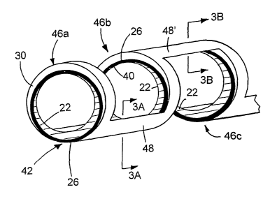
2o Fig. 1 illustrates a method of making a stmt.
Fig. 2 is a detailed illustration of a portion of the stmt of Fig. 1.
Fig. 3A is a cross-sectional view of a strut, taken along line 3A-3A of Fig.
2; and Fig. 3B
is a cross-sectional view of a strut, taken along line 3B-3B of Fig. 2.
Fig. 4 illustrates a portion of a method of malting a stem.
25 Fig. 5 illustrates a method of malting a stmt.
DETAILED DESCRIPTION
Referring to Fig. 1, a method 20 of making a stmt 100 is illustrated. Method
20 is
capable of providing a stmt that includes electrically conductive portions
that are unable to carry
so an electrical current in a closed loop, e.g., around the circumference of
the stmt. Consequently,
CA 02525780 2005-11-14
WO 2004/103220 PCT/US2004/015280
as described more below, the visibility of material, such as blood or a
stenosis, present in the
lumen of stmt 100 during magnetic resonance imaging (MRI) can be enhanced.
Method 20 provides a mechanically strong stmt having at least one electrically
conductive portion (e.g., layer) interrupted by an electrical insulator.
Method 20 includes
providing an electrically conductive inner tubular member 22. Inner tubular
member 22 has a
non-centric lumen 24 such that along a radial cross section, the inner tubular
member has a
relatively thin portion 25 and a relatively thick portion 27. Next, a layer of
electrically insulating
material 26 is formed over imler tubular member 22 (step 28), and
subsequently, an electrically
conductive outer tubular member 30 is formed or placed over layer 26 (step 32)
to yield a three-
layer tubular member 34. As shown, three-layer tubular member 34 is formed
such that inner
tubular member 22 and layer 26 are non-centric with respect to outer tubular
member 30, e.g.,
diametrically opposed to lumen 24. As a result, similar to inner tubular
member 22, outer
tubular member 30 has a relatively thin portion 36 and a relatively thick
portion 37.
Next, in step 38, portions of firmer tubular member 22 and outer tubular
member 30 are
removed. As shown, thin portions 25 and 36, are removed to reveal an inner
portion 40 and an
outer portion 42 of electrically insulative layer 26, respectively. The result
is a tubular member
44 having inner tubular member 22 and outer tubular member 30 separated by
electrically
insulative layer 26, and each member 22 and 30 is interrupted by the
electrically insulative layer
at portions 40 and 42, respectively. As a result, neither inner tubular member
22 nor outer
2o tubular member 30 can carry an electrical current circumferentially (arrow
A) around tubular
member 44.
Tubular member 44 is then formed, e.g., by laser cutting, into stmt 100 having
bands 46
and struts 48 connecting the bands (step 50). In particular, referring to
Figs. 2 and 3, struts 48
are formed at selected locations of bands 46 such that there is no electrical
continuity between
the bands for an electrical current to flow in a closed loop. As shown, one
strut 48 is formed at
portion 42 (Fig. 2). Starting at any starting reference point of inner tubular
member 22 of band
46a, electrical current can flow to inner tubular member 22 of band 46b via a
section of tubular
member 22 in strut 48 (Fig. 3A). I3owever, the electrical current cannot flow
back to the starting
point to close a loop because inner tubular member 22 of band 46b is
interrupted by insulative
layer 26 at portion 40. Electrical current also cannot flow from outer tubular
member 30 of
bands 46a or 46b through strut 48 because the strut does not include a portion
of the outer tubular
CA 02525780 2005-11-14
WO 2004/103220 PCT/US2004/015280
member. Similarly, alternatively or in addition to strut 48 shown in Fig. 2, a
strut including a
portion of insulative layer 26 and a portion of outer tubular member 30 can be
formed at portion
40 (as exemplified by strut 48' between band 46b and 46c). Current cannot flow
to form a loop.
because outer tubular member 30 of bands 46b and 46c axe interrupted by
insulative layer 26 at
portion 42.
Thus, electrical current cannot flow in a loop within a band because
conductive tubular
members 22 and 30 are interrupted by insulative layer 26. Current also cannot
form a closed
loop by flowing between bands because struts 48 are formed at selected
positions to prevent an
electrical current loop from forming.
1 o The laclc of electrical continuity within a band and between bands 46 can
enhance the
MRI visibility of material in the lumen of stmt 100. Without wishing to be
bound by theory,
during MRI, an incident electromagnetic field is applied to a stmt. The
magnetic environment of
the stmt can be constant or variable, such as when the stmt moves within the
magnetic field
(e.g., from a beating heart) or when the incident magnetic field is varied.
When there is a change
15 in the magnetic environment of the stmt, which can act as a coil or a
solenoid, an induced
electromotive force (emf) is generated, according to Faraday's Law. The
induced emf in turn can
produce an eddy current that induces a magnetic field that opposes the change
in magnetic field.
The induced magnetic field can interact with the incident magnetic field to
reduce (e.g., distort)
the visibility of material in the lumen of the stmt. A similar effect can be
caused by a
2o radiofrequency pulse applied during MRI.
By forming stmt 100 to include electrically conductive portions that camlot
form a closed
current loop, the occurrence of an eddy current is reduced (e.g., eliminated).
Accordingly, the
occurrence of an induced magnetic field that can interact with the incident
magnetic field is also
reduced. As a result, the visibility of material in the lumen of stmt 100
during MRI can be
25 enhanced.
Method 20 is described in more detail below.
Referring again to Fig. l, inner tubular member 22 can be formed of any
biocompatible
material suitable for MRI, e.g., non-ferromagnetic materials. The
biocompatible material can be
suitable for use in a self expandable stmt, a balloon-expandable stmt, or
both. For self
3o expandable stents, inner tubular member 22 can be formed of a continuous
solid mass of a
relatively elastic biocompatible material, such as a superelastic or pseudo-
elastic metal alloy.
CA 02525780 2005-11-14
WO 2004/103220 PCT/US2004/015280
Examples of superelastic materials include, for example, aNitinol (e.g., 55%
nickel, 45%
titaniiun), silver-cadmium (Ag-Cd), gold-cadmium (Au-Cd), gold-copper-zinc (Au-
Cu-Zn),
copper-aluminum-nickel (Cu-Al-Ni); copper-gold-zinc (Cu-Au-Zn), copper-
zinc/(Cu-Zn),
copper-zinc-aluminum (Cu-Zn-Al), copper-zinc-tin (Cu-Zn-Sn), copper-zinc-xenon
(Cu-Zn-Xe),
indium-thallium (In-Tl), nickel-titanium-vanadium (Ni-Ti-V), and copper-tin
(Cu-Sn). See, e.g_,
Schetsky, L. McDonald, "Shape Memory Alloys", Encyclopedia of Chemical
Technology (3rd
ed.), John Wiley & Sons, 1982, vol. 20. pp. 726-736 for a full discussion of
superelastic alloys.
Other examples of materials suitable for inner tubular member 22 include one
or more precursors
of superelastic alloys, i.e., those alloys that have the same chemical
constituents as superelastic
alloys, but have not been processed to impart the superelastic property under
the conditions of
use. Such alloys are further described in PCT application US91/02420.
In other embodiments, inner tubular member 22 can include one or more
materials that
can be used for a balloon-expandable stmt. Suitable examples of materials
include noble metals,
such as platinum, gold, and palladium, refractory metals, such as tantalum,
tungsten,
~ 5 molybdenum and rhenium, and alloys thereof. Suitable materials include
radiopaque materials,
such as metallic elements having atomic numbers greater than 26, e.g., greater
than 43, and/or
those materials having a density greater than about 9.9 g/cc. In certain
embodiments, the
radiopaque material is relatively absorptive of X-rays, e.g., having a linear
attenuation coefficient
of at least 25 cm 1, e.g., at least 50 cm 1, at 100 keV. Some radiopaque
materials include
2o tantalum, platinum, iridium, palladium, tungsten, gold, ruthenium, and
rhenium. The radiopaque
material can include an alloy, such as a binary, a ternary or more complex
alloy, containing one
or more elements listed above with one or more other elements such as iron,
nickel, cobalt, or
titanium. Other examples of stmt materials include titanium, titanium alloys
(e.g., alloys
containing noble and/or refractory metals), stainless steels, stainless steels
alloyed with noble
25 and/or refractory metals, nickel-based alloys (e.g., those that contained
Pt, Au, and/or Ta), iron-
based alloys (e.g., those that contained Pt, Au, and/or Ta), and cobalt-based
alloys (e.g., those
that contained Pt, Au, and/or Ta).
W ner tubular member 22 can include a mixture of two or more materials listed
above, in
any arrangement or combination.
3o Inner tubular member 22 including non-concentric lumen 24 can be formed by
conventional techtuques. For example, inner tubular member 22 can be formed
from a solid rod
CA 02525780 2005-11-14
WO 2004/103220 PCT/US2004/015280
of a selected material, and lumen 24 can be mechanically formed, e.g., by
drilling. Alternatively,
inner tubular member 22 can be extruded to include a non-concentric lumen. The
size of lumen
24 can be determined, for example, by the final thickness desired for inner
tubular member 22
after thin portion 25 is removed (step 38).
Next, insulative layer 26 is formed on inner tubular member 22 (step 32).
Insulative
layer 26 can include any electrically non-conductive and MRI compatible
material. Suitable
materials include polymers, such as thermoplastics or thermosetting materials.
The polymer can
enhance the flexibility of stmt 100. Examples of polymers include polyolefms,
polyesters,
polyethers, polyamides and nylons, polyvinyl chlorides, copolymers and
terpolymers thereof, or
o mixtures thereof. Other suitable materials include ceramics, such as
titanium oxides, hafiiium
oxides, iridium oxides, chromium oxides, aluminum oxides (e.g., oc -A1203 or
yttria-stabilized
alumina), glass ceramic (e.g., MacorTM, a blend of fluorophlogopite mica and
borosilicate glass
from Corning, or BioglassTM from USBiomaterials), calcium phosphate (e.g.,
hydroxylapatite),
zirconium oxide (e.g., transformation toughened zirconia, fully stabilized
zirconia, or partially
stabilized zirconia with magnesium or yttrium), feldspathic porcelain, and
silicon nitride. Other
suitable materials include cements. Examples include glass ionomers (e.g.,
GlasscorTM or
GlassbaseTM available from Pulpdent), resin reinforced glass ionomers (e.g.,
VitrebondTM from
3M), polycarboxylates (e.g., Tylol~PlusTM from L.D. Caulk), cyanoacrylates,
zinc phosphates,
resin composite cements (e.g., filled bisphenol-A-glycidyldimethacrylate resin
combined with
2o methacrylics, or RelyX ARC from 3M), and cements used in the field of
dentistry. Insulative
layer 26 can include a mixture of two or more materials listed above, in any
arrangement or
combination.
In some embodiments, insulative layer 26 can include an insulating form of the
material
of inner tubular member 22. For example, inner tubular member 22 can include
tantalum or
tungsten, and insulative layer 26 can include tantalum oxide or tungsten
oxide, respectively.
Such embodiments can have relatively low interfacial differences (e.g.,
stress), which can
provide good adhesion between the materials.
The thiclazess of insulative layer 26 can vary. Generally, insulative layer 26
is
sufficiently thick to electrically isolate inner tubular member 22 from outer
tubular member 30,
so andlor to prevent members 22 and 30 from carrying a continuous loop of
electrical current.
Insulative layer 26 is preferably sufficiently thick to withstand processing
tolerances, e.g.,
CA 02525780 2005-11-14
WO 2004/103220 PCT/US2004/015280
handling during manufacturing or removal of portions 25 and 36 without damage.
Tn some
embodiments, the thickness of insulative layer 26 can range from about 5 to
about 200
nanometers for ceramics or cements,: or about 0.1 to about 50 micrometers for
polymers.
Insulative layer 26 can be formed on inner tubular member 22 according to a
variety of
teclmiques. In some cases, the choice of technique is a function of the
materials of insulative
layer 26 and/or inner tubular member 22. For example, in embodiments in which
insulative
layer 26 includes a polymer, an adhesive can be used to bond the polymer to
inner tubular
member 22. In embodiments in which insulative layer 26 includes an insulating
form of a
material of inner tubular member 22, techniques, such as plasma ion
implantation or heating the
1 o inner tubular member in an appropriate (e.g., oxidizing) atmosphere, can
be used. Other suitable
techniques include thermal spraying techniques, such as plasma arc spraying,
chemical vapor
deposition, physical vapor deposition, or dipping. In certain embodiments,
inner and outer
tubular members 22 and 30 can be co-drawn, and insulative layer 26, for
example, a polymer,
can be formed, e.g., by pouring the liquid or molten polymer into the space
defined between the
members.
After insulative layer 26 is formed, outer tubular member 30 is formed over
the insulative
layer to form three-layer tubular member 34 (step 32). In general, materials
suitable for inner
tubular member 22 are also suitable materials for outer tubular member 30.
Outer tubular
member 30 can be provided as described above for inner tubulax member 22.
Stent 100 can
2o include the same or different materials for inner and outer tubular members
22 and 30.
Outer tubular member 30 can be joined to inner tubular member 22.and
insulative layer
26 using a variety of methods. For example, similar to inner tubular member
22, outer tubular
member 30 can include a non-concentric lumen (not shown) into which inner
tubular member 22
and insulative layer 26 are inserted. Members 22 and 30 can be joined together
by co-drawing
the members. Alternatively or in addition, members 22 and 30 can be joined
together using
magnetic pulse forming or welding. The use of magnetic forces to deform a work
piece is
described, for example, in Batygin Yu et al., "The Experimental Investigations
of the Magnetic
Pulse Method Possibilities for Thin-walled Metal Plates Deformation",
Technical Electro-
dynamics, 1990, #5, p. 15-19; and commonly assigned U.S.S.N. 10J192,253, filed
July 10, 2002.
3D In some embodiments, an adhesive can be applied between insulative layer 26
and outer tubular
member 30.
CA 02525780 2005-11-14
WO 2004/103220 PCT/US2004/015280
As shown in Fig. l, tubular member 34 is formed such that lumen 24 of inner
tubular
member 22 and the lumen defined by outer tubular member 30 are offset (as
shown,
diametrically offset) relative to the circumference of tubular member 34.
Expressed another
way, thin portions 25 and 36 are about 180 degrees apart about the
circumference of tubular
member 34. By offsetting the lumens of inner and outer tubular members 22 and
30, when thin
portions 25 and 36 are removed to form tubular member 44 (described below),
tubular member
44 can be formed with relatively unform wall thickness and good structural
integrity. In other
embodiments, lumen 24 and the lumen defined by outer tubular member 30 (or
thin portions 25
and 36) are less than about 180 degrees, e.g., between zero and 180 degrees,
apart about the
1 o circumference of tubular member 34.
After tubular member 34 is formed, portions of inner and outer tubular members
22 and
30 are removed to prevent the members from carrying an electrical current
circumferentially
around tubular member 34 (step 38). In certain embodiments, thin portions 25
and 36 are
removed such that inner and outer tubular members 22 and 30, respectively, are
interrupted by
insulative layer 26. Since lumen 24 and the lumen of outer tubulax member 30
are offset, the
portion of inner tubular member 22 that is removed (e.g., thin portion 25) is
compensated by
relatively thiclc portion 37 of the outer tubular member. Similarly, the
portion of outer tubular
member 30 that is removed (e.g., thin portion 36) is compensated by relatively
thick portion 27
of inner tubular member 22. As a result, tubular member 44 has relatively
uniform wall
2o thickness and good strength.
Portions of inner and outer tubular members 22 and 30 can be removed by a
variety of
methods. For example, portions of inner and outer tubular members 22 and 30
can be removed
by electropolishing, in which both portions can be removed simultaneously.
Since thin portions
and 36 are thinner than other portions of members 22 and 30, respectively,
techniques, such
25 as electropolishing, that uniformly remove layers of members 22 and 30 will
eliminate the thin
portions first to expose insulative layer 26. Electropolishing is described,
for example, in U.S.
Patent No. 6,375,826. Other suitable methods for removing portions of inner
and outer tubular
members 22 and 30 include laser cutting, mechanical machining (e.g.,
drilling), andlor chemical
etching combined with a suitable mashing technique.
3o Subsequently, tubular member 44 is formed into stmt 100 (step 50). For
example,
selected portions of tubular member 44 can be removed for the tubulax member
to define bands
CA 02525780 2005-11-14
WO 2004/103220 PCT/US2004/015280
46 and struts 48. The portions can be removed by laser cutting, for example,
using an excimer
laser and/or an ultrashort pulse laser. Laser cutting is described, for
example, in U.S. Patent Nos.
5,780,807 and 6,517,888. In certain embodiments, during laser cutting, a
liquid carrier, such as a
solvent or an oil, is flowed through lumen 24. The tamer can prevent dross
formed on one
portion of tubular member 44 from re-depositing on another portion (possibly
providing
electrical continuity), andlor reduce formation of recast material on the
tubular member. Other
methods of removing portions of tubular member 44 include mechanical machining
(e.g., micro-
machining), electrical discharge machining (EDM), photoetching (e.g., acid
photoetching),
and/or chemical etching.
1 o In some oases, tubular member 34 can be formed into a stmt before portions
of inner and
outer tubular members 22 and 30 are removed. For example, laser cutting
tubular member 34
into a stmt can precede electropolishing tubular member 34.
Stent 100 can further be finished, e.g., clectropolished to a smooth finish,
according to
conventional methods. In some embodiments, about 0.0001 inch of material can
be removed
~5 from the interior and/or exterior surfaces by chemical milling and/or
electropolishing. Stent 100
can be annealed at predetermined stages of method 20 to refine the mechanical
and physical
properties of the stmt.
In use, stmt 100 can be used, e.g., delivered and expanded, according to
conventional
methods. Suitable catheter systems are described in, for example, Wang U.S.
5,195,969, and
20 Hamlin U.S. 5,270,086. Suitable stems and stmt delivery are also
exemplified by the Radius~
or Symbiot~ systems, available from Boston Scientific Scimed, Maple Grove, MN.
Generally, stmt 100 can be of any desired shape and size (e.g., coronary
stems, aortic
stems, peripheral vascular stems, gastrointestinal stems, urology stems, and
neurology stems).
Depending on the application, stem 100 can have a diameter of between, for
example, 1 mm to
25 46 mm. In certain embodiments, a coronary stmt can have an expanded
diameter of from about
2 mm to about 6 mm. In some embodiments, a peripheral stmt can have an
expanded diameter
of from about 4 mm to about 24 rmn. In certain embodiments, a gastrointestinal
and/or urology
stmt can have an expanded diameter of from about 6 mm to about 30 mm. In some
embodiments, a neurology stmt can have an expanded diameter of from about 1 mm
to about 12
so mm. An abdominal aortic aneurysm (AAA) stmt and a thoracic aortic aneurysm
(TAA) stmt
can have a diameter from about 20 n~rn to about 46 mm. Stent 100 can be
balloon-expandable,
11
CA 02525780 2005-11-14
WO 2004/103220 PCT/US2004/015280
self expandable, or a combination of both (e.g., U.S. Patent No. 5,366,504).
Stent 100 can be
delivered by other actuating mechanisms, such as those that include an
electroactive polymer or
a pneumatic action.
Stent 100 can also be a part of a stmt-graft. In other embodiments, stmt 100
can include
and/or be attached to a biocompatible, non-porous or semi-porous polymer
matrix made of
polytetrafluoroethylene (PTFE), expanded PTFE, polyethylene, urethane, or
polypropylene. The
endoprosthesis can include a releasable therapeutic agent, drug, or a
pharmaceutically active
compound, such as described in U.S. Patent Nos. 5,674,242 and 6,517,888;
U.S.S.N. 09/895,415,
filed July 2, 2001; and U.S.S.N. 101232,265, filed August 30, 2002. The
therapeutic agents,
o drugs, or pharmaceutically active compounds can include, for example, anti-
thrombogenic
agents, antioxidants, anti-inflammatory agents, anesthetic agents, anti-
coagulants, and
antibiotics.
Still numerous other embodiments are possible.
For example, while described above as tubular, inner member 22, insulative
layer 26,
~ 5 and/or outer member 30 can have non-circular cross sections, e.g., non-
circular inner and/or
outer perimeters. The cross sections can be oval, elliptical, or regularly or
irregularly polygonal,
having three or more sides. The lumens of imier member 22, insulative layer
26, and/or outer
member 30 can be relatively concentric. Furthermore, other arrangements of
struts 48 axe
possible.
2o For example, referring to Fig. 4, three-layer member 34a (similar to member
34) includes
an imier member 22a, an insulative layer 26a, and an outer member 30a, each
having an oval
cross section. Inner member 22a, insulative layer 26a, and outer member 30a
are generally the
same as member 22, layer 26, and member 30, respectively. Three-layer member
34a can be
processed as described above (step 38) to remove portions of members 22a and
30a and to
25 prevent members 22a and 30a from carrying a closed loop of electrical
current. As a result, a
member 44a is formed having member 22a interrupted by insulative layer 26a at
two locations
(A and B), and member 30a interrupted by the insulative layer at two locations
(C and D).
Member 44a can be formed into a stem as described above. Struts 48 can be
formed in any
arrangement at locations A, B, C, and/or D.
12
CA 02525780 2005-11-14
WO 2004/103220 PCT/US2004/015280
While stmt 100 is shown including wide, substantially solid bands 46, in other
embodiments, bands 46 include a wire shaped in an undulating pattern (as
described, e.g., U.S.
Patent No. 6,419,693).
Stent 100 can have fewer or more than the three layers shown in Fig. 1. For
example,
stmt 100 can include insulative layer 26, and inner member 22 or outer member
30.
In some embodiments, stmt 100 includes a protective coating on the exterior
surface
and/or on the interior surface. The coating can be used to enhance the
biocompatibility of the
stmt and/or to protect the stmt from corrosion if, for example, the stmt
includes two different
metals. The protective coating can include one or more of the ceramic,
polymer, and/or cement
1 o described above. More than one protective coatings can be applied.
Other methods for malting a stmt unable to carry electrical current in a
closed loop are
possible. Referring to Fig. 5, method 60 includes starting with a first sheet
62 of electrically
conductive material having an insulative layer 64 on the sheet and on the
edges 66 of the sheet.
First sheet 62 is then rolled (e.g., around a mandrel) to form a tube 68
having edges 66 spaced
apart (step 70). A second sheet 72 (similar to first sheet 62) is formed into
a tube and placed
over tube 68 to form tubular member 76 (step 74). As shown, the edges 78 of
second sheet 72
are spaced apart from each other, and spaced from edges 66, e.g., about 180
degrees. Next,
tubular member 76 is reduced in sized (e.g., by drawing) to join edges 66
together, edges 78
together, and sheets 62 and 72 together (step 80). The result is tubular
member 82, which can be
2o used to form a stmt, as described above (e.g., step 50). Struts 48 can be
formed where edges 66
and 78 meet. Sheets 62 and 72 can include the same materials as member 22, and
insulative
layer 64 can include the same materials as layer 26.
In other embodiments, edges 66 and 78 can be joined together (e.g., by
welding) to form
tubular member 76 having two seams. After tubular member 76 is reduced in
sized (e.g., drawn)
to form tubular member 82, the seams can be preferentially removed, e.g., by
chemical etclung.
The removed material can be subsequently replaced with an insulative material.
Tubular
member 82 can then be formed into a stmt as described above.
Method 20 and the embodiments described above can be used to form medical
devices
other than stems and stmt-grafts. For example, method 20 can be used to form
filters, such as
ao removable thrombus filters described in Kim et al., U.S. 6,146,404; in
intravascular filters such
as those described in Daniel et al., U.S. 6,171,327; and in vena cava filters
such as those
13
CA 02525780 2005-11-14
WO 2004/103220 PCT/US2004/015280
described in Soon et al., U.S. 6,342,062. Method 20 can be used to form
guidewires, such as a
Meier steerable guidewire, catheters, and hypotubes. Method 20 can be used to
form vaso-
occlusive devices, e.g., coils, used to treat intravascular aneurysms, as
described, e.g., in Bashiri
et al., U.S. 6,468,266, and Wallace et al., U.S. 6,280,457. Method 20 can also
be used in surgical
instnunents, such as forceps, needles, clamps, and scalpels.
All publications, applications, references, and patents referred to in this
application are
herein incorporated by reference in their entirety.
Other embodiments are within the claims.
14