Note: Descriptions are shown in the official language in which they were submitted.
CA 02529390 2005-12-13
WO 2005/061009 PCT/US2004/019337
Microparticles For Microarterial Imaging And Radiotherapy
CROSS REFERENCE TO RELATED APPICATION
[01] This application claims benefit of priority of U.S. provisional
application
number 601479,532, entitled "Instant Microparticles for Microarterial Imaging
and
Radiotherapy", filed on June 20, 2003, which is incorporated by reference
herein in its
entirety, and claims benefit of priority of U.S. application number
10/762,507, entitled
"Microparticles for Microarterial Imaging and Radiotherapy", filed on January
23,
2004, which is incorporated by reference herein in its eniirety.
BACKGROUND OF THE INVENTION
[OZ] Over 100,000 patients develop primary or metastatic cancer to the liver
in the
United States yearly. The majority of patients have surgically unresectable
lesions
that are poorly responsive to chemotherapy. Externally delivered conventional
radiotherapy can cause hepatic tumors to regress or be destroyed, but
selective
delivery of radiation to predominately the tumor cells in the liver is almost
impossible. Moreover, the dose of radiation required to destroy hepatic tumors
far
exceeds the tolerance of the normal liver cells immediately adjacent to the
tumor.
Thus despite the significant potential of radiation as an important anti-
cancer therapy,
the problems stemming from the indiscriminate nature have not been overcome,
thereby leading to ineffective treatment and/or excessive radiation dosages
being
applied to healthy tissue and cells. The present invention overcomes these
limitations
by offering a site-directed internal radiation therapy for treatment of cancer
cells,
solid tumors, and rheumatoid arthritis.
1
CA 02529390 2005-12-13
WO 2005/061009 PCT/US2004/019337
Therapy of Hepatic carcinoma
[03] In the past decade significant resources have been expended in clinical
trials
testing hepatic artery infusion of chemotherapy, typically FUDR or SFU.
(Kemeny
et al., New England Journal of Medicine 1999; 341:2039-2048; Kennedy et al.,
Proceedings of the 14th W ternational Congress on Anti-Cancer Treatment
2003:156;
Kennedy et al., Int J Cancer 2002; 513:226-227.)
[04] Despite eight prospective randomized trials in patients with colorectal
cancer
liver metastases, a consensus has not been reached as to the efficacy and
optimal use
of such infusion of chemotherapy. With other diseases such as carcinoid,
hepatoma,
breast, lung and sarcoma, non-surgical management of liver lesions is the
mainstay,
producing palliation at best with short prolongation of survival. It has long
been
understood that chemosensitization for many solid tumors is beneficial.
However, it is
unclear how to optimally deliver hepatic radiation while respecting normal
tissue
tolerance. Brachytherapy offers the hope of delivering tumorcidal doses.
Surgical
resection is obviated with diffuse hepatic involvement or where extra-hepatic
failure
is likely. Additionally, alternative approaches are being explored, such as
radioactive
seed placement which predate advancements in 3D external beam treatment
planning.
(Kennedy et al., Regulatory Peptides 2002; 108:32.)
[OS] Other localized therapies, such as radiofrequency ablation are known to
be
minimally invasive, but only effective in cases of limited, focal tumors
amenable to a
seed implant approach. (Murthy et al., J Vasc Interv Radiol 2002;
13:S2;~Murthy et
al., Proceedings of American Association for Cancer Research 2002; 43:485;
Murthy
et al., J Vasc Interv Radiol 2002; 13:52; Sarfaraz et al., International
Journal of
Radiation Biology and Physics 2001; S 1:32-33.)
[06] Despite recent advances with chemotherapy, surgery and interventional
radiology, solid tumors in the liver remains a significant and common site of
2
CA 02529390 2005-12-13
WO 2005/061009 PCT/US2004/019337
refractory disease in solid tumors. Conservative estimates of liver disease in
colorectal cancers alone represent at least 77,500 cases in the USA yearly.
(Kemeny
et al., New England Journal of Medicine 1999; 341:2039-2048.) Considering
pancreas, carcinoid, stomach and other solid tumors, the total number of
patients
having malig~zant liver tumors exceeds 150,000. (Fong et al., CA Cancer J Clin
1995;
45:50-62) In addition, the incidence of hepatocellular cancer in the United
States and
elsewhere is increasing, with a US rate of 2.4 per 100,000 between 1991 and
1995.
(EI-Serag et al., N Engl 3 Med 1999; 340:745-50)~ Unfortuntely, most of these
patients will not be candidates for curative surgical therapy and require
alternative
therapy options. Although radiation therapy provides potential benefits to
these
patients, delivering sufficiently high doses of radiation via an external beam
to
destroy rnetastatic or primary liver tumors is usually not practical because
the
relatively low radiation tolerance of hepatocytes. Tn seminal work by
Lawrence, a
conformal three-dimensional treatment with concurrent hepatic artery
chemotherapy
demonstrated that radiation given in the same dose ranges as is delivered to
non-
hepatic sites produces durable hepatic tumor control, without loss of hepatic
function.
(Lawrence et aL, Oncology (Huntingt) 1993; 7:51-7; discussion 57-8, 63;
Lawrence et
al., Front Radiat Ther Oncol 1996; 29:221-8; Lawrence et al., Int J Radiat
Oncol Biol
Phys 1991; 20:555-61; McGinn et al., J Clin Oncol 1998; 16:2246-52; McGinn et
al.,
Semin Radiat Oncol 1997; 7:313-323.) However, most patients are not candidates
for
this or other localized therapies such as radiofrequency ablation, cryotherapy
or
chemoembolization. Therefore, there is a long-felt need in the art for a
technique
capable of delivering therapeutic doses of radiation specifically to tumors
concomitantly sparing normal, healthy surrounding tissue and cells. One area
of
particular need for such invention is in the treatment of hepatic tumors.
3
CA 02529390 2005-12-13
WO 2005/061009 PCT/US2004/019337
[07] Embolization is a process wherein a material is injected into a blood
vessel to
at least partially fill or plug the blood vessel and/or encourage clot
formation so that
blood flow through the vessel is reduced or stopped. Embolization of a blood
vessel
can be useful for a variety of medical reasons, including preventing or
controlling
bleeding due to lesions (e.g., organ bleeding, gastrointestinal bleeding,
vascular
bleeding, and bleeding associated with an aneurysm), or to ablate diseased
tissue (e.g.,
tumors, vascular malformations, hemorragic processes, etc.), by cutting off
blood
supply. Embolization may also be used to prevent blood loss during or
immediately
following surgery. Embolization of tumors may be performed preoperatively to
shrink
tumor size, to aid in visualization of a tumor, and to prevent blood loss
related to
surgical procedures.
[08] Pioneering efforts in vascular embolic agents with and without radiation
can
be traced back to Prinzmetal (Van Echo et al., Amer Soc Clin Oncol 2001;
260a:1038.), who first showed the utility of glass sphere infusion via
arterial routes in
animal studies and human subjects in 1947. Shortly after, Muller in 1951 used
intravenously injected radioactive gold in charcoal to treata patient with
bilateral lung
cancer. Moreover, multiple researchers in the early 1960's, who were treating
highly
vascular neuroendocrine tumors in the liver, reported efficacy with hepatic
artery
infusion of 90Yttrium-resin spheres of approximately 35 ~,m. However, the
infusions
led to fatal toxicity levels, which were linked to the unintended deposition
of spheres
in the stomach leading to ulceration and hemorrhage. Others have also
described fatal
pulmonary toxicity from radiation pneumonitis due to the shunting of
radioactive
spheres from the liver to the lungs, the next capillary bed after the liver.
Recent
research using glass microspheres, resin spheres, and as described herein,
polymeric
4
CA 02529390 2005-12-13
WO 2005/061009 PCT/US2004/019337
microspheres, indicates the incidence of gastrointestinal (G~ bleed, biliary
sclerosis,
or pulmonary toxicity is relatively low. (Burton et al., 1989; Anderson et
al., 1992;
Yan et al., 1993; Andrews et al., 1994; Lau et al., 1994; Leung et al., 1994;
Kennedy et al., 2001; Kennedy et al., 2002; Coldwell et al., 2001; Wright et
al.,
2002; Mourtzikos et al., 2002; Hisley et al., 2002; Hafeli et al., 1999.)
(09] In the early 1960's, use of beta radiation in the liver was attempted for
delivery of 90Y or 32P attached to resin or ceramic microspheres. (Ariel IM.,
1965;
Ariel et a1.,1967; Simon et al., 1968; Caldarola et al., 1965; Blanchard et
al., 1964;
Blanchard et al., 1965; Kim et al., 1962.) However, a trend towards using 90Y
later
occurred due to the isotopes relatively high beta energy compared to other
isotopes.
Yttrium-90 (9°Y) is a pure beta emitter, which decays to stable
zirconium-90 with an
average energy of 0.94 MeV via a half life of 2.67 days (64.2 hours). Yttrium-
90 is
produced by neutron bombardment of 89Y in a commercial reactor, yielding
9°Y beta
radiation with a tissue penetration of 2.5 rnm, and a maximum range of 1.1 cm.
One
GBq (27 mCi) of 9°Y delivers a total dose of about 50 Gy/Kg in
tissue.
Radioactive microparticles
[10] Previous attempts have been made to locally administer radioactive
materials
to patients with cancer as a form of radiation therapy. In some cases, the
radioactive
materials were incorporated (embedded) into small particles, seeds, wires and
similar
related configurations that are directly implanted into a cancer site (tumor).
Radioactive materials have also been formulated into microspheres for
injection into
an axterial blood supply of a target organ. Administering radioactive
particles or
microspheres into a blood supply of a target organ is known as Selective
hlternal
Radiation Therapy (SIRT).
CA 02529390 2005-12-13
WO 2005/061009 PCT/US2004/019337
[1l] There are many potential advantages of SIRT over conventional, external
beam radiotherapy. Firstly, the radiation is delivered preferentially to the
cancer
within the target organ. Secondly, the radiation is slowly and continually
delivered as
the radionuclide decays. Thirdly, by manipulating the arterial blood supply
with
vasoactive pharmaceuticals, such as vasodilators like Angiotensin-2, it is
possible to
enha~zce the percentage of radioactive microspheres that go to the cancerous
part of
the organ, as compared to the healthy normal tissues. The effect is
preferential
increase in the radiation dose to the cancer site while maintaining the
radiation dose to
the normal tissues at a significantly lower level (Burton, M. A. et al.;
1988.).
[12] In the earliest clinical use of yttrium-90-containing microspheres, the
yttrium
was incorporated (embedded) into a polymeric matrix that was formulated into
microspheres. While these microspheres were of an appropriate density to
ensure
good distribution characteristics in the liver, the radioactive agent, yttrium-
90, leached
Beverly from the microspheres, causing inappropriate radiation of other non-
targeted
tissues, i.e., non-specific irradiation.
[13] In one attempt to overcome the problem of leaching, a radioactive
microsphere
comprising a biologically compatible glass material containing a beta- or
gamtna-
radiation emitting radioisotope, such as yttrium-90, uniformly distributed
(embedded)
throughout the glass, was disclosed. (International Patent Publication No. WO
86/03124). Additionally, these glass microspheres required neutron activation
prior to
use.
[14] Production of light polymeric ion-exchange microspheres have been
developed to address the serious problem of leaching of yttrium upon injection
into
the body. Ahigh objective response rate for patients with secondary liver
cancer was
obtained upon injection of the microspheres into the hepatic artery (Gray, B.
N. et al.
6
CA 02529390 2005-12-13
WO 2005/061009 PCT/US2004/019337
1992.). One disadvantage of such polymeric ion exchange microspheres is the
yttrium-90 radionuclide must be added to the microsphere after neutron
activation of
the stable isotope of yttrium-89. This requires the use of specialised
facilities and
represents a hazard to manufacturing personnel. Furthermore, tie polymeric ion
exchange microspheres contain only a low percentage of yttrium, which
adversely
impacts dosage levels available for administration.
[l5] There have been modifications of the 90Y carrier which include resin-
based
and ceramic tmaterials. Hafeli and Day reported a modification of a glass
microsphere (magnesium alumino borate glass, 25-32p,m) to replace 9°Y
with
Rhenium (natural isotopes ls6Re and ls~Re). These have large cross-sections
for
neutrons, and more easily yield therapeutic amounts of ~eta-emitters 186Re and
lssRe,
which have maximal energies of 1.1 MeV and 2.1 MeV respectively, compared with
~°Y of .97 MeV. The purpose of using them, however, was that the y-rays
released
were 9.5% (186Re) and 15%(issRe). They reported its use in Sprague-Dawley rats
with Novikoff hepatoma, a highly chemo- and radioresistant tumor.
Interestingly, the
y-ray production made imaging possible,however, the very short half life of
lss+issRe
(17 hours) compared to 9°Y (65 hours) made lss+isaRe undesirable for
clinical
practice.
[16] Clinical studies have been reported involving the use of solid glass
radioactive
microspheres. For example, ten patients with primary hepatocellular carcinoma
were
treated by Shepherd et al with solid glass radioactive microspheres, yet none
of the
subjects exhibited therepeutic response.. (Shepherd, F. et al., 1992.).
[17] Radioactive microspheres that have been used clinically (TheraSphere~)
(MDS
Nordion, Inc., 447 March Road, Ontario, Canada K2K 1X8) are composed of glass
impregnated with 9°Y. Each sphere has a diameter of 25 ~10 pm so they
are trapped
7
CA 02529390 2005-12-13
WO 2005/061009 PCT/US2004/019337
mainly within tumor terminal arterioles, which are estimated to have a
diameter of 8-
p,m. It is estimated that each milligram contains between 22,000 and 73,000
microspheres. The 9°Y does not leach from the glass spheres in the
patient, because it
is permanently trapped in the matrix of the microsphere.
[18] There are significant deficiencies in the two products currently
available for
microsphere therapy. First, there is no source on the spheres that allow for
imaging
and identification of the spheres location in the body. This also complicates
attempts
to develop radiation treatment planning software. Clinical trials and broad
use of this
type of device will require accurate dosimetry and localizing within the
liver, as is
current state of the art for any brachytherapy product. Second, the process of
generating 9°Y spheres is cumbersome, requiring shipments from nuclear
reactor,
which can result in significant time delays. Further production is often
limited in
terms of maximum doses. Thus, there is a need for a more efficient production
system, which would allow more patients' to receive therapy in a timely
manner.
Because the radioactivity is at a fixed activity at the time of manufacture,
there is only
a 4-hour window for therapeutic use of radioactive glass spheres, and <24
hours for
resin spheres.
[19] A non-surgical approach, performed in an outpatient setting, could
provide
therapy to a laxge number of patients safely while using available
interventional
radiology techniques and catheters. The present invention fulfills the long-
felt need in
radiation therapy by providing a novel therapeutic radioactive microparticle
capable
of site-specific treatment of a tumor. The microparticles of the present
invention
exploit the properties of a beta-emitting isotope, which allows for local
irradiation of
a site, such as a tumor, and a sharp dose decline in regional tissue to enable
sparing of
adjacent normal cells. Recognizng that short-range electrons (such as those
provided
s
CA 02529390 2005-12-13
WO 2005/061009 PCT/US2004/019337
by beta-emitting isotopes) cannot be imaged or confirmed in anyway as to their
location post-administration to a subject, in certain embodiments fo the
present
invention in which diagnostic and/or imaging capabilities are desired, the
microparticles comprise a targeting entity.,
BRIEF SUMMARY OF THE INVENTION
[20] In one embodiment of the present invention there is a microparticle
comprising a core and at least one readioactive therapeutic agent attached,
indirectly
or directily, to a surface of the core. The microparticle may be introduced by
intravascular administration for imaging and/or diagnostic and/or therapeutic
intervention. The therapeutic compositions of the present invention include a
suspension of the radioactive microparticles (particulate material) in a
physiologically
acceptable liquid for injection into humans.
[21] A core and a linking Garner is employed (such as, by way of a non-
limiting
example, poly(methyl methacrylate)), which comprises biocompatible
microspheres
having a diameter in the range of from about 5 to about 200 microns. The
material
may have attached an alpha, beta- or gamma-emitting radionuclide, or any
combination thereof, depending upon the clinical need.
[22] In certain embodiments, the present invention is directed to a
microparticle
comprising a core, at least one linking carrier on said core, wherein said
linking
carrier comprises a biocompatible polymer, and at least one radioactive
therapeutic
agent covalently bonded to said linking carrier; wherein said microparticle
has a
diameter in the range of from about 5 to about 200 microns and said
mieroparticle is
non-biodegradable.
9
CA 02529390 2005-12-13
WO 2005/061009 PCT/US2004/019337
[23] In specific embodiments of the present invention, the radioactive
therapeutic
agent comprises an alpha-emitting radionuclide, a beta-emitting radionuclide,
a
gamma-emitting radionuclide, or a combination thereof, including, for example,
an
alpha-emitting radionuclide and a beta-emitting radionuclide, and/or a beta-
emitting
radionuclide and a gamma-emitting radionuclide, and/or an alpha-emitting
radionuclide and a gamma-emitting radionculide.
[24] In certain preferred embodiments of the present invention, the
radioactive
therapeutic agent comprises a therapeutic radionuclide and an imaging or
diagnostic
radionuclide. More specifically, the therapeutic radionuclide comprises a beta-
emitting radionuclide and the imaging or diagnostic radionuclide comprises a
gamma-
emitting radionuclide.
[25] Non-limiting examples of therapeutic radionuclides contemplated in the
inventive microparticles include, but are not limited to, Y-90, Bi-213, At-
211, I-123,
I-125, I-131, At-211, Cu-67, Sc-47, Ga-67, Rh-105, Pr-142, Nd-147, Pm-151, Sm-
153, Ho-166, Gd-159, Tb-161, Eu-152, Er-171, Re-186 and Re-188. Non-limiting
a
examples of the imaging or diagnostic radionuclide contemplated in the
inventive
microparticles include, but are not limited to, Tc-99m, In-111, Ga-67, Rh-105,
I-123,
Nd-147, Pm-151, Sm-153, Gd-159, Tb-161, Er-171, Re-186, Re-188, and Tl-201. In
preferred embodiments, the therapeutic radionuclide comprises yttrium-90 and
the
imaging or diagnostic radionuclide comprises indium-111 or Tc-99m.
[26] In other embodiments, the radioactive therapeutic agent is a radionuclide
or a
radiopharmaceutical. The radionuclides useful in the present invention
include, but
are not limited to, one or more of iridium, radium, cesium, phosphorus,
yttrium,
rhenium, actinium, bismuth, astatine, technetium, indium, iodine, and carbon,
nitrogen, fluorine, sodium, magnesium, aluminum, silicon, potassium, vanadium,
to
CA 02529390 2005-12-13
WO 2005/061009 PCT/US2004/019337
manganese, gallium, niobium, iodine, lead, Y-90, Bi-213, At-211, I-123, I-125,
I-131,
At-211, Cu-67, Sc-47, Ga-67, Rh-105, Pr-142, Nd-147, Pm-151, Sm-153, Ho-166,
Gd-159, Tb-161, Eu-152, Er-171, Re-186, Re-188, Tc-99m, In-11 l, Ga-67, Rh-
105, I-
123, Nd-147, Pm-151, Sm-153, Gd-159, Tb-161, Er-171, Re-186,. Re-188, and Tl-
201.
[27] The radioactive therapeutic agent is attached to the core either
indirectly or
directly. Examples of indirect attachments of the linking carrier to the core
include
attachment through one or more spacer groups or through a chelator group.
Chelator
groups that are contemplated comprise at least one of
cyclohexyldiethylenetriaminepentaacetic acid ligand (CHX-DTPA),
diethylenetriaminepentaacetic acid (DTPA), ethylenediarninetetraacetic acid
(EDTA),
1,4,7,10-tetraazacyclododecane-N,N', N,"N"' tetraacetate (DOTA),
tetraazacyclotetradecane-N,N", N''N''-tetraacetic acid (TETA), cyclohexyl 1,2-
diamine tetra-acetic acid (CDTA), ethyleneglycol-O,O'-bis(- 2-aminoethyl)-
N,N,N',N'-tetra-acetic acid (EGTA), N,N-bis(hydroxybenzyl)-e- thylenediamine-
N,N'-diacetic acid (HBED), triethylene tetramine hexa-acetic acid (TTHA),
hydroxyethyldiamine triacetic acid (HEDTA), hydroxyethylidene diphosphonate
(HEDP), dimercaptosuccinic acid (DMSA),
diethylenetriaminetetramethylenephosphonic acid (DTTP) and 1-(p-aminobenzyl)-
DTPA, 1,6-diamino hexane-N,N,N',N'-tetraacetic acid, DPDP, and ethylenebis
(oxyethylenenitrilo)-tetraacetic acid. In a preferred embodiment, the chelator
group
comprises DOTA.
[28] Alternative examples of attachment of the linking Garner to the core
comprise,
for example, a bifunctional linker, carbodiimide condensation, or a disulfide
bond.
11
CA 02529390 2005-12-13
WO 2005/061009 PCT/US2004/019337
[29] The microparticles of the present invention comprise a core comprising a
polymer. The polymer may include, but is not limited to, poly(methyl
methacrylate),
polyacrylate, ethylene-vinyl acetate polymer, an acyl substituted cellulose
acetate,
polyurethane, polystyrene, polyvinylchloride, polyvinyl flouride, polyvinyl
imidazole), chlorosulphonate polyolefin, polyethylene oxide, blends thereof,
and
copolymers thereof, a polyphosphazine, a polyvinyl alcohol), a polyamide, a
polycarbonate, a polyalkylene, a polyacrylamide, a polyalkylene glycol, a
polyallcylene oxide, a polyalkylene terephthalate, a polyvinyl ether, a
polyvinyl ester,
a polyvinyl halide, polyvinylpyrrolidone, a polyglycolide, a polysiloxane, and
copolymers thereof, a alkyl cellulose, an hydroxyalkyl cellulose, a cellulose
ether, a
cellulose ester, a nitrocellulose or a combination thereof. In all
compositions of the
present invention, the core is non-radioactive until the therapeutic agent is
attached
thereto. In specific embodiments, the core comprises poly(methyl methacrylate)
and/or polystyrene.
[30] In certain embodiments, at least one linking Garner comprises a linear
polymer, a branched polymer and/or a dendritic polymer. In specific
embodiments
which include the dendritic polymer, the dendrirner comprises a disulfide bond
in its
core and/or has a final external layer capped with a reactive group. In a
preferred
specific embodiment, the reactive group is a targeting entity or a therapeutic
entity. In
another preferred specific embodiment, the reactive group comprises an amine
or a
carboxyl group.
[31] In other specific embodiments, the dendrimer has at least one terminal
functional group accessible to a chelator capable of interacting with said at
least one
functional group. The terminal functional group comprises an ester, an ether,
a thiol,
a carbonyl, a hydroxyl, an amide, a carboxyl, and/or am imide.
12
CA 02529390 2005-12-13
WO 2005/061009 PCT/US2004/019337
[32] In specific embodiments involving multiple dendrimers, the dendrimers are
monodispersed.
[33] The microparticles of the present invention advantageously do not leach
radionuclide. In certain embodiments, the microparticles have a density in the
range
of from 1 to 4 gm/cm3, or more preferably from 1 to 2 gm/cm3.
[34] In another embodiment, the microparticle further comprises a second
therapeutic agent, wherein said at least one radioactive therapeutic agent is
a first
therapeutic agent and said second therapeutic agent is not the same
therapeutic agent
as the second therapeutic agent. In a specific embodiment, the second
therapeutic
agent includes at least one of a metal chelate complex, a drug, a prodrug, a
radionuclide, a boron addend, a labeling compound, a toxin, a cytolcine, a
lymphokine, a chemokine, an immunomodulator, a radiosensitizer, an
asparaginase, a
radioactive halogens, a chemotherapy drug and a contrast agent.
[35] Another microparticle of the present invention comprises a core, and at
least
two radioactive therapeutic agents attached to said core.
[36] In specific embodiments, the radioactive therapeutic agents of the
present
invention comprise an alpha-emitting radionuclide, a beta-emitting
radionuclide
and/or a gamma-emitting radionuclide. In other specific embodiments, the
radioactive therapeutic agents are independently selected from the group
consisting of
a therapeutic radionuclide and a targeting radionuclide. In a preferred
specific
embodiment, the therapeutic radionuclide comprises a beta-emitting
radionuclide and
the targeting radionculide comprises a gamma-emitting radionuclide. More
specifically, the beta-emitting radionuclide comprises yttrium-90 and the
gamma-
emitting radionuclide comprises indium-111 or Tc-99m. In certain embodiments,
the
radioactive therapeutic agents are each attached to the core through a
covalent bond.
13
CA 02529390 2005-12-13
WO 2005/061009 PCT/US2004/019337
[37] In one embodiment, there is a microparticle comprising a core, at least
one
radioactive targeting entity attached to said core, wherein said targeting
entity
comprises a garmna-emitting radionuclide, and the microparticle has a diameter
in the
range of about 5 to about 200 microns and is non-biodegradable. In a further
embodiment, the radioactive therapeutic entity further comprises a beta-
emitting
radionuclide. In another further embodiment, the microparticle further
comprises at
least one linking carrier on said core, wherein said linking carrier comprises
a
biocompatible polymer.
[38] In another embodiment, the present invention provides a particulate
material
comprising microparticles having: a core, at least one linking carrier on said
core,
wherein said linking carrier comprises a biocompatible polymer, and at least
one
radioactive therapeutic agent covalently bonded to said linking carner;wherein
said
microparticles have a diameter in the range of from about 5 to about 200
microns and
said microparticles are non-biodegradable. In specific embodiments, the
microparticles have a diameter in the range of from 8-100 microns, more
preferably, a
diameter in the range of from 25-50 microns, most preferably, a diameter in
the range
of from 20-30 microns. In all cases, the microparticles of the particulate
material are
sufficiently large so as to avoid phagocytosis.
[39] The present invention also provides methods of using the compositions for
radiation therapy.
[40] One embodiment is a method of treating a patient with radiation therapy,
comprising administering to the patient in need of radiation therapy a
plurality of
radioactive microparticles, wherein each of said plurality of radioactive
microparticles
have a diameter in the range of from about 5 to about 200 microns, are non-
biodegradable and comprise a core, at least one linking carrier on said core,
wherein
14
CA 02529390 2005-12-13
WO 2005/061009 PCT/US2004/019337
said linking carrier comprises a biocompatible polymer, and at least one
radioactive
therapeutic agent covalently bonded to said linking carrier, wherein the
plurality of
radioactive microparticles provide the radiation therapy to the patient. In
specific
embodiments, the plurality of radioactive microparticles are administered
parenterally, intravenously, intravascularly via a vascular catheter, into an
arterial
vascular system supporting a tumor iii the patient, and/or at or near a target
site, such
as a tumor. The administration may be as a single dose and/or as a continous
infusion
or as multiple doses administered over a period of time. In certain
embodiments, the
aspects of embolization are exploited and the plurality of radioactive
microparticles
are immobilized at a site of the administration, such as a target site, such
as a tumor or
in the arterial vasculature supporting a tumor.
[41] In another embodiment of the present invention, there are methods of
embolizing a blood vessel comprising administering a plurality of the
micropaxticles
of the present invention. In specific embodiments, the embolization includes
delivery
of an embolic agent composition to a blood vessel to fill or plug the blood
vessel
ancUor to encourage clot formation so that blood flow through the vessel is
reduced,
decreased, blocked and/or stopped.
[42] The methods of the present invention are useful in radiation therapy,
including
in treating a cancer patient and/or a tumor, in imaging a target site of a
patient, such
asm for example, a tumor, andlor in diagnosing a subject suspected ofhavin~ a
cancer
or a tumor. The methods of the present invention are particularly suited for
subjects
having primary or seconday stage of liver cancer, rheumatoid arthritis, a
solid cancer,
liver cancer, brain cancer, breast cancer, ovarian cancer, a renal cell
carcinoma, a
hepatoma, a sarcoma, a cancer of the head or neck, or a central nervous system
tumor.
is
CA 02529390 2005-12-13
WO 2005/061009 PCT/US2004/019337
[43] One method of imaging a target organ or a tumor in a patient comprises
admhustering to the patient at a target site in the patient a plurality of
radioactive
microparticles, wherein each of said plurality of radioactive microparticles
have a
diameter in the range of from about 5 to about 200 microns, are non-
biodegradable
and comprise a core, at least one linking carrier on said core, wherein said
linking
Garner comprises a biocompatible polymer, and at least one radioactive
therapeutic
agent covalently bonded to said linking carrier, wherein said radioactive
therapeutic
agent comprises a gamma-emitting radionuclide; and detecting said plurality of
radioactive microparticles, wherein said detection provides the image of the
target
organ or the tumor. The detection may be during the lifetime of the radiation
or,
alternatively, post-life of the radiation. In a further embodiment, the method
further
comprises determining a location of the plurality of radioactive
microparticles in the
patient. It is contemplated that the plurality of microparticles are
immobilized at the
target site, which includes the target organ or tumor.
[44] In certain embodiments, the methods of the present invention are directed
to
diagnosis of a cancer and/or tumor in a patient. Such methods comprise
administering
a pluralilty of radioactive microparticles of the present invention to the
patient,
preferably at a taxget site such as a site suspected of being a tumor or
primary or
secondary site of cancer, detecting the plurality of radioactive
microparticles, and
determining from the detection whether the patient has the cancer and/or
tumor,
wherein detection of said tumor and/or cancer indicates a positive diagnosis.
Such
diagnostic methods are contemplated for diagnosis of hepatic cancer, a solid
cancer,
brain cancer, breast cancer, ovarian cancer, a renal cell carcinoma, a
hepatoma, a
sarcoma, a central nervous system tumor, and/or a cancer in the head and/or
neck.
16
CA 02529390 2005-12-13
WO 2005/061009 PCT/US2004/019337
[45] In other embodiments, there is a kit for preparing the particulate
material of
the present invention comprising: a non-radioactive core, at least one linking
carrier
for attaching at least one radionuclide to said particle core, and
instructions or a
means for obtaining instructions for preparing said microparticle treatment
dose. In
specific embodiments, the kit further comprises a radionuclide, which may by
provided separately from the kit. In a further specific embodiment, the kit
further
comprises at least one component selected from the group consisting of an
inert
pharmaceutically acceptable carrier, a formulating agent, an adjuvant, an
active agent,
water, saline, a transfer ligand, a reducing agent, a lyophilization aid, a
stabilization
aid, a solubilization aid, a bacteriostat, a buffer, an X-ray contrast agent,
an ultrasound
contrast agent, and a metallopharmaceutical. In yet another further specific
embodiment, the kit further comprises at least one component selected from the
group
consisting of a syringe, shielding equipment, and imaging equipment. In yet
another
further specific embodiment, the kit further comprises at least two chemically
different non-radioactive cores or at least two chemically different linking
carriers.
[46] In other embodiments, methods of using a kit of the present invention are
provided. One method of using the kit described herein is to prepare a
microparticle
treatment dose for a patient in need thereof, comprising: determining the type
and
dosimety of rnicroparticle treatment needed from a prescription for said
patient and
preparing said microparticle treatment dose from said instructions or said
means for
obtaining instructions.
[47] Another method of using the kits of the present invention to prepare a
microparticle treatment dose for a patient in need thereof, comprises:
determining the
type and dosimety of microparticle treatment needed from a prescription for
said
patient, selecting a type of non-radioactive core from the cores included in
said lut,
17
CA 02529390 2005-12-13
WO 2005/061009 PCT/US2004/019337
selecting a type of linker from the linkers included in said kit, selecting a
radionuclide
and preparing said microparticle treatment dose from said instructions or said
means
for obtaining instructions.
[48] Another aspect of the present invention relates to a method for
embolization
including delivery of an embolic agent composition to a blood vessel to fill
or plug
the blood vessel and/or encourage clot formation so that blood flow through
the vessel
is reduced or stopped. The embolic agent composition comprises the
particulatee
mateerial of the present invention and a pharmaceutically acceptable carrier.
BRIEF DESCRIPTION OF THE DRAWINGS
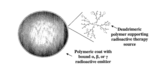
[49] Figure 1: A typical example of microsphere construction from a
biocompatible
core chemically linked to a chelator that tightly binds a radioactive emitter.
The
radioactive isotope can be supported by a dendrimeric interface to increase
the
amount of bound chelator.
[50] Figure 2. Synthesis of PMMA-PAMAM dendrimer conjugate.
[51] Figure 3. Synthesis of PMMA-DOTA microspheres.
[52] Figure 4. Microparticle.
DETAILED DESCRIPTION OF THE INVENTION
I. Definitions.
[53] To ensure clarity of the description that follows, the following
definitions are
provided
[54] By "naic~oparticles" or "fnicrospheres" is meant particles that support
an
effector substance over its surface. The microparticle is non-biodegradable
and
biocompatible.
is
CA 02529390 2005-12-13
WO 2005/061009 PCT/US2004/019337
[55] By "hofz-biodegradable" is meant a material that should not degrade by
bodily
processes to a significant extent over the period of therapy.
[56] By "biocompatible" is meant not toxic to the body, is pharmaceutically
acceptable, is not carcinogenic, and does not significantly induce
inflammation in
body tissues.
[57] A "liszkizzg carrier", as used herein, is a molecule that is used to join
the
effector molecule to the microparticle. The linker is capable of forming
covalent
bonds to both the effector and the microparticle matrix.
(58] An "effector" is a molecular construct that may involve a chelator that
carnes
out a useful biological function within the body. As used herein, the term
therapeutic
effector is used to mean any compound or molecule or isotope that will either
cause,
elicit or initiate a cellular or physiological response witlun the targeted
tissue.
[59] A "clzelator" or "bonding unit" is the moiety or group on a reagent that
binds
to a molecular such as a metal ion through the formation of chemical bonds
with one
or more donor atoms.
[60] As used herein, "body" preferably refers to the human body, but it should
be
understood that body can also refer to a non-human animal body.
II. Tlie Present Invention
(61] The present invention is directed to therapeutic radioactive compositions
and
uses thereof and overcome three major limitations of non-specific radiation
observed
in extenal radiation therapies: 1) utilizing beta radiation as a therapeutic
agent spares
normal nearby hepatocytes; 2) delivering directly to a tumor vasculature where
the
radioactive microparticles will then reside within the tumor, spares normal
cells
and/or tissue regional to the tumor; and 3) enabling delivery of significantly
increased
19
CA 02529390 2005-12-13
WO 2005/061009 PCT/US2004/019337
radiation doses to achieve the range of radiation known to be effective in
destroying
virtually all solid tumors (e.g., therapeutically effective amount of
radiation).
[62] The present invention relates to the radioactive complex formed by
labeling a
biocompatible _polymer, with radionuclide, a microsphere/pax-ticle core that
supports
the polymer-radioactive complex forming micro particles, and a "kit" of the
necessary
components for preparing the radioactive complex. In addition, the present
invention
relates the process of preparation thereof and methods and use thereof for an
internal
radiation diagnostic and/or therapeutic agent.
Microparticle
The therapeutic compositions of the present invention are directed to
microparticles. In certain embodiments, the microparticles of the present
invention
comprise a core, at least one linking carrier on said core, wherein said
linking carrier
comprises a biocompatible polymer, and at least one radioactive therapeutic
agent
covalently bonded to said linking carrier; wherein said microparticle has a
diameter in
the range of from about 5 to about 200 microns and said microparticle is non-
biodegradable.
[63] The microsphere is comprised of a non-ceramic,' non-radioactively labeled
core material that serves as a support for a polymeric coating comprised of a
linear,
branched, or dendromer biocompatible polymer to which a suitable binding agent
is
attached. The binding agent is selected from a number of chemically stable
compounds that bind radioactive or non-radioactive therapeutic agents, as
described
in more detail herein.
[64] In the present invention, locally deposited polymer depots on the surface
of
the microsphere core are used as a vehicle for the immobilization and local
delivery of
a radionuclide or radiopharmaceutical.
CA 02529390 2005-12-13
WO 2005/061009 PCT/US2004/019337
[65] Standard radionuclides that have been used for local radiotherapy
(brachytherapy) may be used, such as radionuclides of rhenium, iodine,
iridium,
radium, cesium, yttrium or other elements.
[66] Suitable therapeutic and diagnostic agents include those whose efficacy
within
the body is predicated on their ability to remain within or to be carned
within the
vascular compartment. Accordingly the methods involving administering the
compositions of the present invention are readily adapted for treating several
diseases
and disorders, including cancer and/or a tumor, and/or for imaging selected
regions of ,
a mammal by various imaging techniques, and/or for diagnosing a subject
suspected
of having a cancer andlor tumor, particularly in the liver.
[67] In specific embodiments, microspheres or mircroparticles comprise, via
chelate attachment to a linear, branched or dendrimeric polymer coat, any or
all of the
following: phosphorus, yttrium, rhenium, andlor other beta emitting isotopes;
actinium, bismuth, astatine and other alpha emitting isotopes; technetium,
indium,
iodine and/or other gamma emitting isotopes; and carbon, nitrogen, fluorine,
sodium,
magnesium, aluminum, silicon, potassium, vanadium, manganese, gallium,
niobium,
iodine and/or lead.
[68] The microspheres may be chosen for a longer time of degradation or
elimination of greater than 320 hours, when five half lives of the implanted
yttrium-
90 have expired and the vast majority of radioactive decay has occurred.
[69] The invention described herein takes advantage of the expertise and
equipment
available in hospitals with nuclear medicine. The present invention is also
directed to
a "kit" comprising polymer spheres, linkers, and a radioactive isotope, that
is mixed
onsite or in a local radiopharmacy. The advantage entails increased
flexibility, with a
dose tailored to individual patient needs based on preplanning dosimety. The
21
CA 02529390 2005-12-13
WO 2005/061009 PCT/US2004/019337
invention provides for single or multiple tracer tags with gamma and beta or
alpha
sources. The hallmark of present invention remedies two problematic issues
with
existing microspheres, i.e. inability to image and, consequently, locate the
microspheres, and limited distribution of product.
[70] The present invention provides microparticle constructs comprising a
biocompatible microparticulate core, an optional linking carrier, and a
molecular
effector coupled directly or indirectly to the biocompatible core. A preferred
form of
the effector is a radioisotope bound to the linking carrier by a chelator
group. In
addition, the present invention includes the process of preparation of a "kit"
formulation thereof and the use thereof for an internal radiation diagnostic
and/or
therapeutic agent.
[71] In certain embodiments, the biocompatible microspheres have a diameter in
the range of from 10 to 200 microns.
[72] In certain embodiments, the microparticle core comprises a non-ceramic,
non-
radioactively labeled material that serves as a support for a polymeric
coating on the
surface of the core (see, Figure 1) comprised of a linear, branched, or
dendritic
biocompatible polymers to which a suitable binding agent is attached. The
binding
agent is selected from a number of chemically stable compounds that bind
radioactive
or non-radioactive therapeutic agents. The biocompatible polymer may have
attached
thereto an alpha, beta- and/or gamma-emitting radionuclide, or any combination
thereof, depending upon the clinical need.
[73] In order to overcome the problem of leaching of radionuclide from ceramic
microspheres, while at the same time maintaining the microspheres with a low
density, the present invention provides microspheres with improved physical
characteristics. The microparticles (microspheres) of the present invention
can be
22
CA 02529390 2005-12-13
WO 2005/061009 PCT/US2004/019337
formulated to be of such a size, shape and density that they have improved
distribution characteristics when administered into the arterial supply of
target organs
to be treated. In addition, each microsphere may deliver a higher amount of
ionising
radiation than prior art microspheres. This, in turn, means that a relatively
lesser
number (less product) is administered to the target organ in order to deliver
the same
'radiation dose. In alternative embodiment, the microspheres are labeled after
manufacture, thereby improving the manufacture process.
[74] The chemical durability of the microspheres is such that they do not
release a
significant amount of radiation emitting radioisotope into the circulatory
system upon
administration.
Microparticulate core
[75] The inventive augmentation material comprises smooth rounded,
substantially
spherical, particles of a matrix material, preferably of a biocompatible
polymer. The
term "substantially spherical" refers to the fact that while some of the
present particles
may be spheres, most of the particles of the present invention are sphere-like
in their
shape, i.e., they are spheroidal. The terms "rounded" or "smooth, rounded" as
used
herein refers to the fact even though the present particles .are not perfect
spheres, they
do not have any sharp or angular edges. The particles must be sufficiently
large so as
to avoid phagocytosis.
[76] As used herein, the term "microparticles" refers to particles having, a
number
median diameter of greater than 5 microns. In a particular embodiment, the
microparticles have a number median diameter of greater than about 10 microns.
For
example, the core diameter may be from about 10 microns to about 200 microns.
23
CA 02529390 2005-12-13
WO 2005/061009 PCT/US2004/019337
Preferably also, the microspheres have a diameter in the range of from about
20 to
about 80 microns.
[77] However, it is understood that for introduction by injection the upper
limit on
particle size will be dictated by the particular injection equipment employed.
That is,
the particles must be sufficiently small so as to avoid aggregation and
clogging of the
syringe when being injected. A typical range for injection is from about 10 to
150
microns, preferably in a narrow particle size range extending not more than
about 35
microns, and more preferably extending not more than about 20 to 30 microns,
and
most preferably having substantially equivalent particle sizes.
[78] The micropaxticle diameter may be from about 10 microns to about 200
microns. In one embodiment, the microspheres have a diameter in the range of
from
8, to about100 microns. In another embodiment, the microspheres have a
diameter of
from about 20 to about 30 microns.
[79] These are meant to be exemplary and not limiting. Other narrow particle
size
ranges within the overall size range of 10 to 150 microns can also be used. In
discussing these ranges, it should be understood that as a practical matter, a
small
amount of particles outside the desired range may be present in a sample of
the
present augmentation material. However, most of the particles in any given
sample
should be within the desired range. Preferably, 90% of the particles are
within the
desired range and most preferably 95-99% are within the range.
[80] ..As used herein, the term "particle size" refers to a number median
diameter as
determined by conventional particle size measuring techniques known to those
skilled
in the art, such as, laser diffraction, photon correlation spectroscopy,
sedimentation
field flow fractionation, disk centrifugation or electrical sensing zone
method. Laser
diffraction is preferred. The "number median diameter" reflects the
distribution of
24
CA 02529390 2005-12-13
WO 2005/061009 PCT/US2004/019337
particles (by number) as a function of particle diameter. An alternative
designation of
particle size often used in the art is the "volume median diameter". The
volume
median diameter is the median diameter of the volume weighted size
distribution.
The volume median diameter reflects the distribution of volume as a function
of
particle diameter.
[81] In a~preferred embodiment, the microparticle has a diameter selected
based on
a size that lodges in a desired region of the body. Use of microspheres that
lodge
within an organ or region of the body is common in studies of blood flow
(Flaim et al,
J Pharmacol. Meth. 11:1-39, 1984; Heymann et al, Prog. Cardiovasc. Dis. 20:55-
79,
1977). For example, a microparticle selected to lodge in a capillary typically
has a
diameter between 15 to 35 microns. Microparticles can be fabricated from
different
polymers using a variety of different methods known to those skilled in the
art.
Numerous methods are known for preparing microparticles of any particular size
range. Synthetic methods for microparticles from molten materials, are known,
and
include polymerization in emulsion, in sprayed drops, and in separated phases.
For
solid materials or preformed gels, known methods include wet or dry milling or
grinding, pulverization, classification by air jet or sieve, and the like.
[82] A further preferred feature of the particulate material of the present
invention
is that the microspheres have a density in the range of from 1 to 4 gm/cm3,
more
preferably in the range of from 1 to 2 gmlcm3.
[83] In the present invention, locally deposited polymer depots on the surface
of
the microsphere core are used as a vehicle for the immobilization and local
delivery of
a radionuclide or radiopharmaceutical.
[84] In one embodiment, the microparticle is not water swellable.
CA 02529390 2005-12-13
WO 2005/061009 PCT/US2004/019337
[85] The interior of said core preferably does not contain radioactive
therapeutic
agent. Alternatively, the microparticle of the present invention comprises a
gamma-
emitting radionuclide attached, indirectly or directly, to the core. In a
specific
embodiment, it is contemplated that the gamma-emitting radionuclide is
attached by
molding into a sphere, such as, for example, during preparation thereof,
preferably
attached at, a surface. In a specific embodiment the surface is a surface of a
resin
particle, and a plurality of resin particles are admixed together with the
ganuua-
emitting radionuclide and the core, are molded, such as, for example, a an
elevated
temperature and/or pressure, and said molding forms a resin-radionuclide layer
on a
surface of the core, thus providing the radioactive microparticle.
Biocompatible microparticulate core materials
[86] The preferred microparticulate cores of the invention are polymers that
are
biocompatible. Suitable biocompatible polymers can be either slowly
biodegradable
or non-biodegradable polymers or blends or copolymers thereof, as described
herein.
The biocompatible polymers suitable fox use in the invention can therefore be
water-
insoluble or minimally water-soluble.
[87] A polymer is biocompatible if the polymer and any degradation products of
the polymer are non-toxic to the recipient and also possess no significant
deleterious
or untoward effects on the recipient's body, such as an imrnunological
reaction at the
inj action site.
[88] Suitable biocompatible, non-biodegradable polymers include ~ iion-
biodegradable polymers selected from the group consisting of polyacrylates,
polymers
of ethylene-vinyl acetates and other acyl substituted cellulose acetates, non-
degradable polyurethanes, polystyrenes, polyvinylchloride, polyvinyl flouride,
26
CA 02529390 2005-12-13
WO 2005/061009 PCT/US2004/019337
polyvinyl imidazole), chlorosulphonate polyolefins, polyethylene oxide, blends
thereof, and copolymers thereof.
[89] Representative synthetic polymers include polyphosphazines, polyvinyl
alcohols), polyamides, polycarbonates, polyalkylenes, polyacrylamides,
polyalkylene
glycols, polyalkylene oxides, polyalkylene terephthalates, polyvinyl ethers,
polyvinyl
esters, polyvinyl halides, polyvinylpyrrolidone, polyglycolides,
polysiloxanes,
polyurethanes and copolymers thereof. Synthetically modified natural polymers
include alkyl celluloses, hydroxyalkyl celluloses, cellulose ethers, cellulose
esters,
and nitrocelluloses. Other polymers of interest include, but are not limited
to, methyl
cellulose, ethyl cellulose, hydroxypropyl cellulose, hydroxypropyl methyl
cellulose,
hydroxybutyl methyl cellulose, cellulose acetate, cellulose propionate,
cellulose
acetate butyrate, cellulose acetate phthalate, carboxymethyl cellulose,
cellulose
triacetate, cellulose sulfate sodium salt, poly(methyl methacrylate),
poly(ethyl
methacrylate), poly(butyl methacrylate), poly(isobutyl methacrylate),
poly(hexyl
methacrylate), poly(isodecyl methacrylate), poly(lauryl methacrylate),
poly(phenyl
methacrylate), poly(methyl acrylate), poly(isopropyl acrylate), poly(isobutyl
acrylate),
poly(octadecyl acrylate) polyethylene, polypropylene, polyethylene glycol),
polyethylene oxide), poly (ethylene terephthalate), polyvinyl acetate),
polyvinyl
chloride, polystyrene, polyvinyl pyrrolidone, and polyvinylphenol.
[90] These polymers can be obtained from sources such as Sigma r'.hP"~ical
Co.,
St. Louis, Mo., Polysciences, Warrenton, Pa., Aldrich, Milwaukee, Wis., Rluka,
Ronkonkoma, N.Y., and BioRad, Richmond, Calif. or else synthesized from
monomers obtained from these suppliers using standard techniques.
(91] Suitable polymer compositions preferably have intrinsic and controllable
biodegradability, so that they persist for about a week to about six months;
are non-
27
CA 02529390 2005-12-13
WO 2005/061009 PCT/US2004/019337
toxic, containing no significant toxic monomers and degrading into non-toxic
components; are biocompatible; are chemically compatible with the substances
to be
delivered, are able to remain at the site of application by adherence or by
geometric
factors, such as by being trapped at a desired location; are capable of being
delivered
by techniques of minimum invasivity, such as by catheter.
[92] Acceptable molecular weights for polymers used in this invention can be
determined by a person of ordinary skill in the art taking into consideration
factors
such as the desired polymer degradation rate, physical properties such as
mechanical
strength, and rate of dissolution of polymer in solvent. Typically, an
acceptable range
of molecular weight is of about 2,000 Daltons to about 2,000,000 Daltons.
(Polymer
molecular weights are usually represented as weight average molecular weights.
However, for dendrimers the reported molecular weights are absolute as they
have a
defined chemistry.)
[93] In one embodiment, the biocompatible polymer core and the biocompatible
polymer of said linking carrier comprise different biocompatible polymers.
Linking carriers
[94] Preferred linl~ing Garners are biocompatible polymers (such as HPMA),
macromolecular assemblies of biocompatible components (such as polymeric
dendrimers), or multi-component linking carriers consisting of more than one
biocompatible component (such as dendrimer-coated polymeric microparticles).
[95] Examples of linking carriers include, but are not limited to, polymerized
copolymers, dendrimers, polyethylene glycol assemblies, capped polylysines,
poly(hydroxybutyric acid), dextrans, biocompatible polymers and copolymers
such as
hyaluronic acids and acrylamides and derivatives thereof, and polystyrene
particles
and derivatives thereof. A preferred linking carrier is a dendrimer.
2s
CA 02529390 2005-12-13
WO 2005/061009 PCT/US2004/019337
[96] The linking carrier can be coupled to the effector by a variety of
methods,
depending on the specific chemistry involved. The coupling will be covalent. A
variety of methods suitable for coupling of the targeting entity and the
therapeutic
effector to the linking carrier can be found in Hermanson, "Bioconjugate
Techniques"
Academic Press: New York, 1996; and in "Chemistry of Protein Conjugation and
Cross-linking" by S. S. along, CRC Press, 1993. Specific coupling methods
include,
but are not limited to, the use of bifunctional linkers, carbodiimide
condensation,
disulfide bond formation, and use of a specific binding pair where one member
of the
pair is on the linking carrier and another member of the pair is on the
effector. Large
numbers of effectors may be attached to one microparticle.
[97] Water-soluble polymers (dendrimers, PEG etc) may be selected as a
biocompatible linker in order to avoid immunogenic responses upon
administration.
Dendrimer Linking Carriers
[98] Another preferred linking Garner is a . dendrimer. Dendrimers are
polymers
with well-defined branching from a central core (e.g., "starburst polymers").
In
contrast to conventional polymers, dendrimers tend to be highly branched
macromolecules. Dendrimers are described in U.S. Pat. Nos. 4,507,466,
4,558,120,
4,568,737, 4,587,329, 4,631,337, 4,694,064, 4,737,550, and 4,857,599, as well
as
numerous other patents and patent publications. Dendrimer structure,
synthesis, and
characteristics are reviewed in Kim and Zimmerman, "Applications of dendrimers
in
bio-organic chemistry," Current Opinion In Chemical Biology (1998) 2(6):733-
42;
Tam and Spetzler, "Chemoselective approaches to the preparation of peptide
dendrimers and branched artificial proteins using unprotected peptides as
building
bloclcs," Biomedical Peptides, Proteins & Nucleic Acids (1995) 1(3):123-32;
Frechet,
"Functional polymers and dendrirners: reactivity, molecular architecture, and
29
CA 02529390 2005-12-13
WO 2005/061009 PCT/US2004/019337
interfacial energy," Science (1994) 263(S1S4):1710-S; Liu and Frechet,
"Designing
dendrimers for drug delivery," Pharmaceutical Science a~ld Technology Today
(I999)
2(10):393401; Verprek and Jezek "Peptide and glycopeptide dendrimers. Part I,"
Journal of Peptide Science (1999) S(1):S-23; Veprek and Jezek, "Peptide and
glycopeptide dendrimers. Part II," Journal Of Peptide Science (1999) S(S)203-
20;
Tomalia et ~al., "Starburst dendrimers: Molecular-level control of size,
shape, surface
chemistry, topology, and flexibility from atoms to macroscopic matter"
Angewandte
Chemie--International Edition in English (1990) 29(2):138-175; Bosman et al.,
"About dendrimers: Structure, physical properties, and applications" Chemical
Reviews (1999) 99(7):1665-1688; Fischer and Vogtle, "Dendrimers: From design
to
application--A progress report," Angewandte Chemie-International Edition
(1999)
38(7):88S90S; Roovers and Comanita, "Dendrimers And Dendrimer-Polymer
Hybrids," Advances In Polyner Science (1999) 142:179-228; Smith and Diederich,
"Functional Dendrimers: Unique Biological Mimics," Chemistry--A European
Journal
(1998) 4(8):1353-1361; and Matthews et al., "Dendrimers--Branching out from
curiosities into new technologies," Progress In Polymer Science (1998) 23(1):
1-S6.
The synthesis of dendrimers typically uses reiterative synthetic cycles,
allowing
control over the dendrimer's size, shape; surface chemistry, flexibility, and
interior
topology. An example of a dendrimer suitable for use as a linking entity is
described
in Wu et al., "Metal-Chelate-Dendrimer-Antibody Constructs for Use in
Radioimmunotherapy and Imaging," Bioorganic and Medicinal Chemistry Letters
(1994) 4(3):449-454.
[99] Dendrimers can be readily used as linking Garners by employing a variety
of
chemical conjugation techniques to attach the targeting entity and therapeutic
entity.
For example, in U.S. Pat. No. 6,020,457, which discloses a dendrimer having a
CA 02529390 2005-12-13
WO 2005/061009 PCT/US2004/019337
disulfide (--S--S--) bond in its core, the dendrimer can be constructed by the
methods
described in the patent. The final external layer of the dendrimer can be
capped with
a reactive group such as an amine or carboxyl group. These reactive groups can
then
be derivatized with either targeting entities or therapeutic entities (or, in
some cases, a
mixture of both).
[100] A dendrimer for the purposes of the present invention is a branched
polymer
which is a three-dimensional highly ordered compound, in which branched
oligomericlpolymeric sequences may be formed around a nuclear molecule by
reiterative reaction sequences, and which under certain conditions has a
positively
charged outer surface as a result of suitable functional terminal end groups
(polycationic dendrimer). Dendrimers of this kind and their preparation are
described n',
in WO 84/02705, U.S. Pat. Nos. 4,507,466, 4,558,120, 4,568,737, 4,587,329,
4,631,337, 4,694,064, 4,713,975, 4,737,550, 4,871,779, 4,857,599, EP 0 234
408, EP
0 247 629, EP 0 271 180, and especially Tange et al., supra, WO 95/02397, and
Tomalia et al., supra.
[101] Dendrimers which are suitable for~the present invention include, for
example,
polyamidoamine (PAMAM) dendrimers which may be synthesised around ammonia,
tris-(2-aminoethyl)amine (TAEA) or ethylenediamine (EDA) as nuclear molecules
by
stepwise addition of the two monomers methacrylate and ethylenediamine (Tang
et
al., supra). The terminal groups of such a dendrimer are preferably primary
amino
groups. 5th, 6th or 7th generation PAMAM dendrimers are preferred,
particularly 6th
generation, according to Tang et al., supra. The theoretical molecular
weights,
number of terminal amines and hydrodynamic radii of such PAMAM dendrimers may
be found in the publication of Tang et al., supra. Table I shows the
properties of
amine functional PAMAM dendrimers.
31
CA 02529390 2005-12-13
WO 2005/061009 PCT/US2004/019337
Table 1.
CatalogGeneration Molecular weightDiameter No. of surface
No. No. (Da) (r~) - amino groups
41,236-80 517 15 4
41,238-41 1430 22 8
41,240-62 3256 29 16 .
41,242-23 6909 36 32
41,244-94 14215 45 64
53,670-95 28826 54 128
53,671-7' 6 58048 67 256
53,672-57 116493 81 512
53,674-18 233383 97 1024
53,6'6-89 467162 114 2048
53,677-610 934720 135 4096
[102] The dendritic polymers which may be used include generally any of the
known dendritic architectures including dendrimers, regular dendrons,
controlled
hyperbranched polymers, dendrigrafts, and random hyperbranched polymers.
Dendritic polymers are polymers with densely branched structures having a
large
number of reactive groups. A dendritic polymer includes several layers .or
generations
of repeating units which all contain one or. more branch points. Dendritic
polymers,
including dendrimers and hyperbranched polymers, are prepared by condensation
reactions of monomeric units having at least two reactive groups. The
dendrimers
which can be used include those comprised of a plurality of dendrons that
emanate
from a common core which can be a single atom or a group of atoms. Each
dendron
generally consists of terminal surface groups, interior branch junctures
having
branching functionalities greater than or equal to two, and divalent
cu~u~~ctors that
covalently connect neighboring branching junctures.
[103] The hyperbranched polymers which may be used represent a class of
dendritic
polymers which contain high levels of nonideal irregular branching as compared
with
the more nearly perfect regular structure of dendrons and dendrimers.
Specifically,
hyperbranched polymers contain a relatively high number of irregular branching
areas
32
CA 02529390 2005-12-13
WO 2005/061009 PCT/US2004/019337
in which not every repeat unit contains a branch juncture. The preparation and
characterization of dendrimers, dendrons, random hyperbranched polymers,
controlled hyperbranched polymers, and dendrigrafts is well known. Examples of
dendimers and dendrons, and methods of synthesizing the same are set forth in
U.S.
Pat. Nos. 4,410,688, 4,507,466; 4,558,120; 4,568,737; 4,587,329; 4,631,337;
4,694,064; 4,713,975; 4,737,550; 4,871,779 and 4,857,599. Examples of
hyperbranched polymers and methods of preparing the same are set forth, for
example
in U.S. Pat. No. 5,418,301.
[104] Dendritic polymers suitable for use .with the invention also include
macromolecules commonly referred to as cascade molecules, arborols,
arborescent
grafted molecules, and the like. Suitable dendritic polymers also include
bridged
dendritic polymers, i.e., dendritic macromolecules linked together either
through
surface functional groups or through a linking molecule connecting surface
functional
groups together, and dendritic polymer aggregates held together by physical
forces.
Also included are spherical-shaped dendritic polymers and rod-shaped dendritic
polymers grown from a polymeric core.
[105] U.S. Pat. No. 5,338,532 teaches polymer conjugates comprising dense star
polymers associated with a carried material, the disclosure of which is hereby
incorporated by reference. (One type of dense star polymers is StarburstTM
polymers
(trademark of The Dow Chemical Company) where the dendrimer is a
polyamidoamine (PAMAM).) A variety of suitable applications for such
conjugates
are broadly discussed in U.S. Pat. No. 5,338,532, including the use of these
conjugates as delivery vehicles for biologically active agents. U.5. Pat.
5,338,532
exemplifies the use of zero valence metals, and ionic or radioactive metals,
specifically exemplifying Fe, Rh, Pd, Y, Fn, Pb, Gd, Mn and Gd.
33
CA 02529390 2005-12-13
WO 2005/061009 PCT/US2004/019337
[106] Dendritic polymers suitable for use with the present invention also
include
macromolecules commonly referred to as cascade molecules (e.g., E. Buhleier et
al.,
Synthesis 155-158 (Feb. 1978), arborols (e.g., U.S. Pat. Nos. 5,376,690 and
5,210,309), arborescent grafted molecules, tectodendrimers (e.g., Srinivas
Uppuluri et
al., "Tecto(dendrimer) Core-shell Molecules: Macromolecular Tectonics for the
Systematic Synthesis of Larger Controlled Structure Molecules" PMSE, Spring
Meeting (Mar. 21-25, 1999) 55-56), and the like. Suitable dendritic polymers
also
include bridged dendritic polymers, i.e., dendritic macromolecules linked
together
either through surface functional groups or through a linking molecule
connecting
surface functional groups together, and dendritic polymer aggregates held
together by
physical forces. Also included are spherical-shaped dendritic polymers (e.g.,
U.S. Pat.
Nos. 4,507,466; 4,588,120; 4,568,737; 4,631,337; 4,587,329; and 4,737,550, the
disclosures of which are hereby incorporated by reference) and rod-shaped
dendritic
polymers (e.g., U.S. Pat. No. 4,694,064, the disclosure of which is hereby
incorporated by reference) grown from a polymeric core. Additional dendritic
polymers suitable for use with the present invention include all the basic
dendritic
structures where specific chelating groups or moieties are either in the
central core of
the dendrimer, and/or located within the interior structure on the dendron
arms and/or
located on the surface of the dendrimer. All of these above dendrimer terms
are to be
understood to be included within the term "dendritic polymer."
[107] Dendritic polymers which are useful in the practice of this invention
include
those that have symmetrical branch cells (arms of equal length, e.g., PAMAM
dendrimers; for example described in U.S. Pat. No. 5,527,524) and those having
unsymmetrical branch cells (arms of unequal length, e.g. lysine-branched
dendrimers,
for example described in U.S. Pat. No. 4,410,688), branched dendrimers,
cascade
34
CA 02529390 2005-12-13
WO 2005/061009 PCT/US2004/019337
molecules (e.g., E. Buhleier et al., Synthesis I55-158 (Feb. 1978))" arborols
(e.g.,
U.S.. Pat. Nos. 5,376,690 and 5,2I0,309), and the like.
(108] The dendritic polymers used in the practice of this invention can be
generationally monodisperse or generationally polydisperse. Dendritic polymers
in a
monodisperse solution are substantially all of the same generation, and hence
of
uniform size and shape. The dendritic polymers in the polydisperse solution
comprise
a distribution of different generation polymers. The dendritic polymer
molecules
which may be used in the practice of this invention include mixtures of
different
interior and exterior compositions or functionalities. Examples of suitable
dendritic
polymers include poly(ether) dendrons, dendrimers and hyperbranched polymers,
polyester) dendrons, dendrimers and hyperbranched polymers, poly(thioether)
dendrons, dendrimers and hyperbranched polymers, poly(amino acid) dendrons
dendrimers and hyperbranched polymers, poly(arylalkylene ether) dendritic
polymers
and polypropylamine dendrimers, dendrimers and hyperbranched polymers.
Poly(amidoamine) (PAMAM) dendrimers have been found to be particularly useful
for preparing the metal-containing complexes of this invention.
[109] The dendritic polymers which are believed to be most useful in the
practice of
this invention axe approximately monodispersed. That is, dendritic polymers in
a
monodispersed solution in which all of the dendritic polymer molecules are
substantially of the same generation, and hence of uniform size and shape, are
preferred. Monodispersed solutions of dendrimers axe particularly preferred.
[110] The dendritic polymers preferred for use in the practice of this
invention have
terminal functional groups which are accessible to a chelate containing
compound
which is capable of interacting with the functional group.
CA 02529390 2005-12-13
WO 2005/061009 PCT/US2004/019337
(111] The term "functional group" is intended to comprise groups such as e.g.
ester
groups, ether groups, thiol groups, carbonyl groups, hydroxyl groups, amide
groups,
carboxylic groups, and imide groups as well as combinations thereof. Amine-
terminated polyamidoamine, polyethyleneimine and polypropyleneimine dendrimers
are also known, for example, from U.S. Pat. No. 5,393,797; 5,393,795;
5,560,929; and
5,387,617, all to Hedstrand et aI.
[112J The optional linking dendrimers may be incorporated to increase the
polyvalency of yttrium attachment sites. The micru~phere surface may already
contain multiple sites to attach chelator for yttrium. However since the
surface of a
microsphere may be rigid, chemical modification is a difficult reaction. Hence
linkers
may be needed to increase the distance from the sphere surface to facilitate
reaction
with the chelator. The linker may be attached if suboptimal concentrations of
chelator
are obtained on the surface of the spheres.
Dendrimer Size
[113] Dendrimers are generally prepared by stepwise or reiterative reaction of
multifunctional monomers to obtain a bxanched structure. In U.S. Pat. No.
5,530,092,
for example, the repetition of double Michael addition of acrylonitrile
starting with a
primary diamine followed by hydrogenation obtains two primary amines for each
initial amine. This doubles the number of primary amine groups. Thus,
begiiming
with a diamine, the first generation dendrimer (G1) has four primary amines;
the
second generation (G2) has eight primary amines; the third generation_ (G3)-
has
sixteen primary amines; the fourth generation (G4) has thirty-two primary
amines; the
fifth generation (G5) has sixty-four primary amines in the outer shell, and so
on.
These polyamine dendrimers are said to be stable to degradation through
hydrolysis
reactions.
36
CA 02529390 2005-12-13
WO 2005/061009 PCT/US2004/019337
[114] The generation of the dendritic polymer, and hence the size of the
dendritic
polymer, which may be utilized in the practice of this invention may vary
considerably. For example, generation 3.5 poly(amidoamine) dendrimers (3.5
PAMAM) are acceptable for use in the practice of this invention.~However,
higher
and lower generations are also expected to be useful, but especially the range
from
generation ~3.5 to 7.5 for PAMAM dendrimers having an ethylenediamine (EDA)
core.
Methods of coupling to the linking carrier
[115] It is intended to covalently attach an effector to the linking caxrier.
This
covalent attachment may be directly between the surface of the linking Garner
and the
effector by means of a linker moiety between the surface of the linl~ing
carrier and the
effector. Some linkers that may be used are described in U.S. Pat. No.
5,527,524; EP
0353450; EP 0570575; and EP 0296522, the disclosures of which are hereby
incorporated by reference. -
[116] Generally, prior to forming the linkage between the linking carrier and
the
effector, and optionally, the spacer group, at least one of the chemical
functionalities
will be activated. One skilled in the art will appreciate that a variety of
chemical
functionalities, including hydroxy, amino, and carboxy groups, can be
activated using
a variety of standard methods and conditions.
[117] Typically, the agent is linked covalently to a linking carrier using
standard
chemical techniques through their respective chemical functionalities.
Optionally, the
linlcing carrier or agent is coupled to the agent through one or more spacer
groups.
The spacer groups can be equivalent or different when used in combination.
Likewise,
if more than one linking carrier is used to produce the agent- linking carrier
complex,
the dendrimers can be equivalent or different.
37
CA 02529390 2005-12-13
WO 2005/061009 PCT/US2004/019337
[118] In certain embodiments, one or more of the active groups are protected
during
one or more steps of the reaction to assemble the linking Garner or a
conjugate of the
linking carrier. Those of skill in the art understand how to protect a
particular
functional group such that it does not interfere with a chosen set of reaction
conditions. For examples of useful protecting groups, see, for example, Greene
et al.,
Protective Groups in Or ag nic Synthesis, John Wiley & Sons, New York, 1991.
[119] The effector molecule rnay be attached to the linking Garner by any of a
number of means well known to those of skill in the art. Typically the
effector is
conjugated, either directly or through a dendrimer or other linker (spacer),
to the
microparticle.
[120] Alternatively, the microparticle and/or spacer may be derivatized to
expose or
attach additional reactive functional groups. The derivatization may involve
attachment of any of a number of linker molecules such a.s those available
from Pierce
Chemical Company, Rockford Ill.
[121] A bifunctional linker having two functional groups reactive with a group
on a
particular effector may be used to form the desired conjugate.
[122] Many procedures and linker molecules for attachment of various compounds
including radionuclide metal chelates, toxins and drugs to proteins such as
antibodies
are known (see, e.g., European Patent Application No. 188,256; U.S. Pat. Nos.
4,671,958, 4,659,839, 4,414,148, 4,699,784; 4,680,338; 4,569,789; and
4,589,071;
and Borlinghaus et al. (1987) Cancer Res. 47: 4071-4075).
Spacer Groups
[123] One or more spacer groups optionally may be introduced between the
linking
carrier and the therapeutic agent. Spacer groups contain at least two chemical
fiuictionalities. Typically, one chemical functionality of the spacer group
bonds to a
38
CA 02529390 2005-12-13
WO 2005/061009 PCT/US2004/019337
chemical functionality of the linlung carrier, while the other chemical
functionality of
the spacer group is used to bond to a chemical functionality of the
pharmaceutical
agent. Examples of chemical functionalities of spacer groups include hydroxy,
mercapto, carbonyl, carboxy, amino, ketone, and mercapto groups. Spacer groups
may also be used in combination. When a combination of spacer groups is used,
the
spacer groups may be different or equivalent. The spacer group is an optional
moiety
which may be introduced to increase the length of the linker so that it is
spaced further
away from the microsphere surface thereby providing flexibility and facilitate
reaction
with the chelator for the radionuclide (i.e., yttrium).
[124] Whether coupled directly, or through a spacer, the agent is preferably
coupled
to the linking earner via a covalent bond. The covalent bond may be non-
reversible,
partially reversible, or fully reversible. The degree of reversibility
corresponds to the
susceptibility of the agent- linking carrier complex to in vivo degradation.
As will be
apparent to those of skill in the art, such reversible groups can be
incorporated at any,
point within the linking earner -agent conjugate. The introduction of a spacer
arm
having a reversible linkage is merely an exemplary embodiment; the bond
between
the agent and the dendrimer, for example, may also be reversible.
[125] The susceptibility of the agent- linkilig carrier complexes to
degradation can
be ascertained through studies of the hydrolytic or enzymatic conversion of
the
complex to the unbound pharmaceutical agent. Generally, good correlation
between in
vitro and in vivo activity is found using this method. See, e.g., Phipps et al
J. P'_~arm.
Sciences 78:365 (1989). The rates of conversion may be readily determined, for
example by spectrophotometric methods or by gas-liquid or high pressure liquid
chromatography. Half lives and other kinetic parameters may then be calculated
using
39
CA 02529390 2005-12-13
WO 2005/061009 PCT/US2004/019337
standard techniques. See, e.g., Lowry et al. MECHANISM AND THEORY IN
ORGANIC CHEMISTRY, 2nd Ed., Harper & Row, Publishers, New York (1981).
Effectors
[126] The linking carrier may be conjugated to a variety of effectors useful
for
treating or identifying diseased tissue. Preferably the effectors that are
conjugated to
the polymer conjugate are selected from the group consisting of therapeutic or
diagnostic agents.
[127] Examples of therapeutic agents for use with the invention include, but
are not
limited to, metal chelate complexes, drugs, prodrugs, radionuclides, boron
addends,
labeling compounds, toxins and other effector molecules, such as cytokines,
lyrnphokines, chemokines, immunomodulators, radiosensitizers, asparaginase,
boron
addends and radioactive halogens. Preferably, the therapeutic agent that is
conjugated
to the polymer backbone is selected from the group consisting of therapeutic
radioisotpes, toxins, drugs, prodrugs and boron addends.
[128] Drugs for use with the current invention include, but are not limited
to, any
currently approved or not-yet-approved chemotherapy drug, as long as it can be
attached to the polymer conjugate. Typically useful already approved drugs
include,
but are not limited to, the following agents and derivatives of these agents:
anastrozole, azacytidine, bleomycin, busulfan, caxboplatin, carmustine,
chlorambucil,
cisplatin, cladribine, cyclophosphamide, cytarabine, dacarbazine,
dactinomycin,
daunorubicin, docetaxel, doxorubicin, estramustine, etoposide, =~xundine,
fludarabine, fluorouracil, flutamide, gemcitabine, hydroxyurea, idarubicin,
ifosfamide, irinotecan, lomustine, mechlorethamine, megestrol, melphalan,
mercaptopurine, methotrexate, mitomycin, mitotane, mitoxantrone, paclitaxel,
CA 02529390 2005-12-13
WO 2005/061009 PCT/US2004/019337
pentostatin, procarbazine, tamoxifen, teniposide, thioguanine, thiotepa,
topotecan,
vinblastine, vincristine, and vinorelbine.
[129] Additionally, the polymer conjugate may comprise a therapeutic agent
consisting of boron addends to be used in Boron Neutron Capture ..Therapy
(BNCT)
protocols. BNCT is a binary system designed to deliver ionizing radiation to
tumor
cells by neutron irradiation of tumor-localized boron-10 atoms. BNCT is based
on the
nuclear reaction which occurs when a stable isotope, isotopically enriched B-
10
(present in 19.8% natural abundance), is irradiated with thermal neutrons to
produce
an alpha particle and a Li-7 nucleus. These particles have a path length of
about one
cell diameter, resulting in high linear energy transfer. Just a few of the
short-range 1.7
MeV alpha particles produced in this nuclear reaction are 'sufficient to
target the cell
nucleus and destroy it. Success with BNCT of cancer requires methods for
localizing
a high concentration of boron-10 at tumor sites, while leaving non-target
organs
essentially boron-free. Compositions and methods for treating tumors in
patients
using pre-targeting msAb for BNCT are described in U.S. Pat. No. 6,228,362 and
can
easily be modified in accordance with the present invention, and is hereby
incorporated by reference. Additionally, other elements are suitable for
neutron
capture reactions. One example is uranium. Uranium, in large amounts, can be
bound
by naturally occurring chelating agents such as ferritin. Such strategies have
been
described in the art, for example U.S. Pat. No. 6,228,362 and references cited
therein
are easily adaptable to the present invention and are hereby
incorporat°~i in their
entirety by reference.
[130] The embodiment of the invention in which the diagnostic agent is a
contrast
agent is illustrated by reference to metal chelate-based contrast agents. The
focus on
metal chelates is intended as illustrative rather than limiting. Those of
skill in the art
41
CA 02529390 2005-12-13
WO 2005/061009 PCT/US2004/019337
will appreciate that many contrast agents other than metal chelates can be
conjugated
to the linking carrier of the invention (e.g. particles, iodinated aryl
compounds,
nitroxides, etc.).
[131] In preferred embodiments, the therapeutic metal ion is associated with
the
microparticle construct via a chelator.
[132] An array of metal chelates is known in the art. See, for example, Pitt
et al.,
"The Design of Chelating Agents for the Treatment of Iron Overload," In,
INORGANIC CHEMISTRY IN BIOLOGY AND MEDICINE; Martell, Ed.;
American Chemical Society, Washington, D.C., 1980, pp. 279-312; Lindoy, THE
CHEMISTRY OF MACROCYCLIC LIGAND COMPLEXES; Cambridge University
Press, Cambridge, 1989; Dugas, BIOORGANIC CHEMISTRY; Springer-Verlag,
New York, 1989, and references contained therein.
[133] In a preferred embodiment, the diagnostic agent is a metal complex of
polyaminocarboxylate chelating agent such as diethylenetriaminepentaacetic
acid
(DTPA).
[134] In other preferred embodiments, the therapeutic entity is a
chemotherapeutic
agent or prodrug or toxin where the therapeutic entity is attached to the
surface of the
linlcing Garner. Alternatively, the therapeutic entity may be entrapped or
encapsulated
within the linl~ing carrier.
[135] In a particularly preferred embodiment, the therapeutic radionuclide is
associated with a chelator that is chemically attached to a polymeric surface
_n the
microparticle construct. In another particularly preferred embodiment, yttrium-
90 is
the therapeutic radionuclide, and DOTA is the chelator.
42
CA 02529390 2005-12-13
WO 2005/061009 PCT/US2004/019337
Chelatin~ groups
[136] Chelating groups are well known to those of skill in the art. Wu et al.
(1992)
Nucl. Med. Biol., 19(2): 239-244 discloses a synthesis of macrocylic chelating
agents
for radiolabeling proteins with 111In and g°Y.
[137] Preferred water soluble chelators to be used in the practice of the
present
invention include, but are not limited to, diethylenetriaminepentaacetic acid
(DTPA),
ethylenediaminetetraacetic acid (EDTA), 1,4,7,10-tetraazacyclododecane-N,N',
N,"N"' tetraacetate (DOTA), tetraazacyclotetradecane-N,N", N''N''-tetraacetic
acid
(TETA), cyclohexyl 1,2-diamine tetra-acetic acid (CDTA), ethyleneglycol-O,O'-
bis(-
2-aminoethyl)-N,N,N',N'-tetra-acetic acid (EGTA), N,N-bis(hydroxybenzyl)-e-
thylenediamine-N,N'-diacetic acid (HBED), triethylene tetramine hexa-acetic
acid
(TTHA), hydroxyethyldiamine triacetic acid (HEDTA), hydroxyethylidene
diphosphonate (HEDP), dimercaptosuccinic acid (DMSA),
diethylenetriaminetetramethylenephosphonic acid (DTTP) and 1-(p-aminobenzyl)-
DTPA, 1,6-diamino hexane-N,N,N',N'-tetraacetic acid, DPDP, ethylenebis
(oxyethylenenitrilo)-tetraacetic acid, and
cyclohexyldiethylenetriaminepentaacetic
acid ligand (CHX-DTPA).
[138] One chelating agent, 1,4,7,10-tetraazacyclododecane-N, N, N", N"'-
tetraacetic
acid (DOTA), is of particular interest because of its ability to chelate a
number of
diagnostically and therapeutically important metals, such as radionuclides and
radiolabels.
[139] In certain embodiments of the present invention, DOTA or other chelating
agent conjugates, such as EDTA, or DTPA, for example, may be prepared in the
form
of water-soluble salts (sodium salt, potassium salt, tetrabutylammonium salt,
calcium
salt, fernc salt, etc.). These salts will be useful as therapeutic agents for
tumor
treatment. Secondlv. DTPA or other chelatin~ agents will be useful as
diamostic
43
CA 02529390 2005-12-13
WO 2005/061009 PCT/US2004/019337
agents which when labeled with radionuclides such as 111In or 9gmTc, may be
used as
radiotracers to detect certain tumors in combination with nuclear imaging
techniques.
[140] In some embodiments, the chelator contains or is a derivative of 1,4,7,
I0-
tetraazacyclododecane-N,N',N",N"'-tetraacetic acid (DOTA) or the. chelators
known
to those skilled in the art. In other embodiments, the chelator comprises an
ionizable
group such' as carboxyl, phosphate, phosphonate, sulfate, sulfonate, or
sulfinate. In
still other embodiments, the chelator comprises a single ionizable group, said
single
ionizable group generating a surface capable of binding an isotope or metal
with a
valency of +2 or greater, or +3 or greater.
[141] As modifications and changes may be made in the structure of the water
soluble polymer or a water soluble metal chelator, of the present invention
and still
obtain molecules having like or otherwise desirable characteristics, such
"biologically
functional equivalents" or "functional equivalents" are also encompassed
within the
present invention.
[142] The nature of medical applications imposes multiple requirements on the
chemical characteristics of a potential chelating ligand. It has to be (a)
strong
(multidentate) complexing agent for the metal ion, (b) hydrophilic to afford
solubility
in water, (c) nontoxic, (d) capable of incorporating into a protein structure
without
causing its denaturation. For virtually every single radi.~nuclide one has to
design a
special chelating system. For example macrocyclic bifunctional chelating
agents, in
particular, DOTA, derivatives incorporating yttrium-90 and indium-111 ly~Te
s~~~wn
excellent kinetic stability under physiological conditions. However, the slow
formation of yttrium-DOTA complexes presents a technical problem that can lead
to
low radiolabeling yields unless conditions are carefully controlled.
44
CA 02529390 2005-12-13
WO 2005/061009 PCT/US2004/019337
(143] The most successful chelating agents for preparation of radioconjugates
are
the products of a complex organic synthesis. Quite representative examples of
a
synthetic procedure can be found in Brechbiel, M. W.; Gansow, O. A.; Atcher,
R. W.;
Schlom, J.; Esteban, J.; Simpson, D. E.; Colcher, D., Synthesis of 1-(P-
isothiocyanatobenzyl) Derivatives of DTPA and EDTA. Antibody Labeling and
Tumor Imaging Studies, Inorg. Chem., 1986, 25, 2772-2781. Other chelators
preferred fox use with this invention include the class of heterocyclic
chelators
(144] As used herein, the term "heterocycle" or "heterocyclic system" is
intended to
mean a stable 5, 6, or 7-membered monocyclic or bicyclic or 7, 8, 9, or 10-
membered
bicyclic heterocyclic ring which is saturated, partially unsaturated or
unsaturated
(axomatic), and which consists of carbon atoms and 1, 2, 3, or 4 heteroatoms
independently selected from the group consisting of N, NH, O and S and
including
any bicyclic group in which any of the above-defined heterocyclic rings is
fused to a
benzene ring. The nitrogen and sulfur heteroatoms may optionally be oxidized.
Radionuclides
[145] Microparticles of this invention may contain, via chelate attachment to
the
linlcing carrier, any or all of the following: phosphorus, yttrium, rhenium,
and other
beta emitting isotopes; actinium, bismuth, astatine and other alpha emitting
isotopes;
technetium, indium, iodine and other gamma emitting isotopes; and carbon,
nitrogen,
fluorine, sodium, magnesium, aluminum, silicon, potassium, vanadium manganese,
gallium, niobium, iodine and/or lead.
[146J As used herein, a therapeutic radionuclide is a nuclide which undergoes
spontaneous transformation (nuclear decay) with an energy transfer sufficient
to
impart cytotoxic amounts of radiant energy to nearby cells. In contrast,
radionuclides
useful for diagnosis emit radiation capable of penetrating tissue with minimal
cell
CA 02529390 2005-12-13
WO 2005/061009 PCT/US2004/019337
damage. Such radiation may be detected using a suitable scintigraphic imager.
Therapeutic radionuclides of the present invention include, but are not
limited to Y-
90, Bi-213, At-211, I-123, I-125, I-131, At-211, Cu-67, Sc-47, Ga-67, Rh-105,
Pr-
142, Nd-147, Pm-151, Sm-153, Ho-166, Gd-159, Tb-161, Eu-152; Er-171, Re-186,
and Re-188. Diagnostic or imaging nuclides of the present invention include,
but are
not limited'to Tc-99m, In-111, Ga-67, Rh-105, I-123, Nd-147, Pm-151, Sm-153,
Gd-
159, Tb-161, Er-171, Re-186, Re-188, and Tl-201.
[147] Any useful nuclide rnay be used within the scope of the invention.
Particularly
preferred are radionuclides that have useful diagnostic or therapeutic
properties, such
as indium-111 or yttrium-90, respectively.
[148] It is further contemplated that in embodiments employing more than one
radionuclide, the multiple radionuclides may be different types (alpha-
emitters, beta-
emitters, a~ld/or gamma-emitters) as well as different sub-types of alpha-
emitters,
beta-emitters, and/or gamma-emitters.
[149] Beta irradiation from 90-Yttrium internal sources results in exposure to
both
tumor tissue and normal liver because the range of the irradiation is over 1
cm. Alpha
irradiation is an attractive potential therapeutic isotope in conjunction
with, or in place
of 90-Yttrium, given the much shorter irradiationrange (microns) in tissue but
relatively high radiation activity over the same short distance. The present
invention
also includes coupling alpha emitter radionuclides to microsphere particles,
alone in
conjunction with gamma- and/or beta-emitting isotopes or with other alpr - pat
~cles.
The radioactive microparticles may b administred via infusion into the the
hepatic
artery and become trapped in the liver tumor capillary bed, thereby allowing
for the
delivery of high intensity tumoricidal doses of radiation without exposure of
normal
46
CA 02529390 2005-12-13
WO 2005/061009 PCT/US2004/019337
liver tissue. It is contemplated that such therapeutic agent may be used in
radiation
therapy to treat hepatic tumors that are otherwise uniformly fatal.
[150] Tight binding of radiometallic nuclides often requires a chelating agent
for the
radiometal. Standard radiolabeling methods and precautions used in the
radiolabeling
of low molecular weight chelates may be used to prepare radiolabeled chelate
f polymers. For instance, procedures using radiometals, such as indium-111 and
yttrium-90, generally require highly pure supplies of the radionuclide,
deionized water
in all buffer solutions, and acid-washing of glassware and plastic-ware used
with any
of the reagents during the radiolabeling procedures.
[151] Procedures using radiometals such as rhenium-188, wluch require a
chemical
reduction step to effect labeling, are best carried out using oxygen-depleted
buffers
and argon atmospheres overlaying the radiolabeling reactions.
The coordination sphere of the radionuclide includes all the ligands or groups
bound
to the radionuclide. For a transition metal radionuclide, Mt, to be stable it
typically has
a coordination number (number of donor atoms) comprised of an integer greater
than
or equal to 4 and less than or equal to 9; that is there are 4 to 9 atoms
bound to the
metal and it is said to have a complete coordination sphere. The requisite
coordination number for a stable radionuclide complex is determined by the
identity
of the radionuclide, its oxidation state, and the type of donor atoms.
Kits
[152] The present invention also includes a kit. A "kit" comprises a
cc.~:ection of
components, termed the formulation, in one or more vials which are used by the
practicing end user in a clinical or pharmacy setting to synthesize diagnostic
radiopharmaceuticals.
47
CA 02529390 2005-12-13
WO 2005/061009 PCT/US2004/019337
[153] Where a radioactive, or other, effector is used as a diagnostic and/or
therapeutic agent, it is frequently impossible to put the ready-for-use
composition at
the disposal of the user, because of the often poor shelf life of the
radiolabelled
compound and/or the short half life of the radionuclide used. In such cases
the user
can carry out the labeling reaction with the radionuclide in the clinical
hospital,
physician's ' office, or laboratory. For this purpose, or other purposes, the
various
reaction ingredients can then be offered to the user in the form of a so-
called "kit".
The kit is preferably designed so that the manipulations necessary to perform
the
desired reaction should be as simple as possible to enable the user to prepare
from the
kit the desired composition by using the facilities that are at his disposal.
Therefore
the invention also relates to a kit for preparing a composition according to
this
invention.
[154] Such a lcit according to the present invention preferably comprises a
microparticle of this invention. The microparticle construct can be provided,
if
desired, with inert pharmaceutically acceptable carrier and/or formulating
agents
and/or adjuvants is/are added. In addition, the kit optionally includes a
solution of a
salt or chelate of a suitable radionuclide (or other active agent), and
instructions for
use with a prescription for administering and/or reacting the ingredients
present in the
kit.
[l55] The kit provides all the requisite components to synthesize and use the
diagnostic radiopharmaceutical except those that are commonly availal-
'° to the
practicing end user, such as water or saline for injection, a solution of the
radionuclide, equipment for heating the kit during the synthesis of the
radiopharmaceutical, if required, equipment necessary for administering the
48
CA 02529390 2005-12-13
WO 2005/061009 PCT/US2004/019337
radiopharmaceutical to the patient such as syringes and shielding, and imaging
equipment.
[156] The kit may contain a transfer ligand, a reducing agent, a
lyophilization aid, a
stabilization aid, a solubilization aid and bacteriostats as well as the
active
microparticle and bound effectors.
[157] A "transfer ligand" is a ligand that forms an intermediate complex with
a metal
ion that is stable enough to prevent unwanted side-reactions but labile enough
to be
converted to a metallopharmaceutical. The formation of the intermediate
complex is
kinetically favored while the formation of the metallopharmaceutical is
thermodynamically favored. Transfer ligands useful in the preparation of
metallopharmaceuticals and in diagnostic kits useful for the preparation of
diagnostic
radiopharmaceuticals include but are not limited to gluconate, glucoheptonate,
mannitol, glucarate, N,N,N',N'-ethylenediaminetetraacetic acid, pyrophosphate
and
methylenediphosphonate. In general, transfer ligands are comprised of oxygen
or
nitrogen donor atoms.
[158] A "reducing agent" is a compound that reacts with a radionuclide, which
is
typically obtained as a relatively unreactive, high oxidation state compound,
to lower
its oxidation state by transferring electrons) to the radionuclide, thereby
making it
more reactive. Reducing agents useful in the preparation of
radiopharmaceuticals and
in diagnostic kits for the preparation of the radiopharmaceuticals include but
are not
limited to stannous chloride, stannous fluoride, formamidine sulfinic acia
ascorbic
acid, cysteine, phosphines, and cuprous or ferrous salts. Other reducing
agents are
described in Brodack et. al., PCT Application 94/22496, which is incorporated
herein
by reference.
49
CA 02529390 2005-12-13
WO 2005/061009 PCT/US2004/019337
[159] A "lyophilization aid" is a component that has favorable physical
properties
for lyophilization, such as the glass transition temperature, and is added to
the
formulation to improve the physical properties of the combination of all the
components of the formulation for lyophilization.
[160] Lyophilization aids useful in the preparation of diagnostic kits useful
for the
preparation of radiopharmaceuticals include but are not limited to mannitol,
lactose,
sorbitol, dextran, Ficoll, and polyvinylpyrrolidine (PVP).
[161] A "stabilization aid" is a component that is added to the
radiopharmaceutical
or to the diagnostic kit either to stabilize the radiopharmaceutical or to
prolong the
shelf life of the kit before it must be used. Stabilization aids can be
antioxidants,
reducing agents or radical scavengers and can provide improved stability by
reacting
preferentially with species that degrade other components or the
radiopharmaceutical.
(162] Stabilization aids useful in the preparation of radiopharmaceuticals and
in
diagnostic kits useful for the preparation of radiopharmaceuticals include but
are not
limited to ascorbic acid, cysteine, monotluoglycerol, sodium bisulfite, sodium
metabisulfite, gentisic acid, and inositol.
[163] A "solubilization aid" is a component that improves the solubility of
one or
more other components in the medium required for the formulation.
[164] Solubilization aids useful in the preparation of radiopharmaceuticals
and in
diagnostic kits useful for the preparation of radiopharmaceuticals include but
are not
limited to ethanol, glycerin, polyethylene glycol, propylene glycol,
poly~xyetl_~rlene
sorbitan monooleate, sorbitan monoloeate, polysorbates,
poly(oxyethylene)poly(oxyp-
ropylene)poly(oxyethylene) block copolymers (Pluronics) and lecithin.
Preferred
solubilizing aids are polyethylene glycol, and Pluronics.
so
CA 02529390 2005-12-13
WO 2005/061009 PCT/US2004/019337
[165] Buffers useful in the preparation of radiopharmaceuticals and in
diagnostic
kits useful for the preparation of said radiopharmaceuticals include but are
not limited
to phosphate, citrate, sulfosalicylate, and acetate. A more complete list can
be found
in the United States Pharmacopeia.
(166] Bacteriostats useful in the preparation of radiopharmaceuticals and in
diagnostic kits useful for the preparation of radiopharmaceuticals include but
are not
limited to benzyl alcohol, benzalkonium chloride, chlorbutanol, and methyl,
propyl or
butyl paraben.
(167] A component in a diagnostic kit can also serve more than one function. A
reducing agent can also serve as a stabilization aid, a buffer can also serve
as a
transfer ligand, a lyophilization aid can also serve as a transfer, ancillary
or co-ligand
and so forth.
(168] Therapeutic radiopharmaceuticals; X-ray contrast agent pharmaceuticals,
ultrasound contrast agent pharmaceuticals and metallopharmaceuticals for
magnetic
resonance imaging contrast are provided to the end user in their final form in
a
formulation contained typically in one vial, as either a lyophilized solid or
an aqueous
solution. The end user reconstitutes the lyophilized with water or saline and
withdraws the patient dose or just withdraws the dose from the aqueous
solution
formulation as provided.
[169] The technetium and rhenium radiopharmaceuticals of the present invention
can be easily prepared by admixing a salt of a radionuclide, a compo»nd ~ ~
the
present invention, and a reducing agent, in an aqueous solution at
temperatures from 0
to 100°C. The technetium and rhenium radionuclides are preferably in
the chemical
form of pertechnetate or perrhenate and a pharmaceutically acceptable cation.
The
perteclmetate salt form is preferably sodium pertechnetate such as obtained
from
51
CA 02529390 2005-12-13
WO 2005/061009 PCT/US2004/019337
commercial Tc-99m generators. The amount of pertechnetate used to prepare the
radiopharmaceuticals of the present invention can range from 0.1 mCi to 1 Ci,
or
more preferably from 1 to 200 mCi.
[170] The amount of the compounds of the present invention used to prepare the
technetium and rhenium radiopharmaceuticals of the present invention can range
from
0.01 p,g to 1~0 mg, or more preferably from 0.5 ~.g to 200 fig. The amount
used will be
dictated by the amounts of the other reactants and the identity of the
radiopharmaceuticals of the present invention to be prepared.
[171] The indium, copper, gallium, silver, palladium, rhodium, gold, platinum,
bismuth, yttrium and lanthanide radiopharmaceuticals of the present invention
can be
easily prepared by admixing a salt of a radionuclide and a reagent of the
present
invention, in an aqueous solution at temperatures from 0 to 100 °C.
These
radionuclides are typically obtained as a dilute aqueous solution in a mineral
acid,
such as hydrochloric, nitric or sulfuric acid. The radionuclides are combined
with
from one to about one thousand equivalents of the reagents of the present
invention
dissolved in aqueous solution. A buffer is typically used to maintain the pH
of the
reaction mixture between 3 and 10.
[172] The total time of preparation will vary depending on the 'identity of
the metal
ion, the identities and amounts of the reactants and the procedure used for
the
preparation. The preparations may be complete, resulting in >80% yield of the
radiopharmaceutical, in 1 minute or may require more time. If higher tnrity
metallopharmaceuticals are needed or desired, the products can be purified by
any of
a number of techniques well known to those skilled in the art such as liquid
chromatography, solid phase extraction, solvent extraction, dialysis or
ultrafiltration.
52
CA 02529390 2005-12-13
WO 2005/061009 PCT/US2004/019337
[173] When kit constituents are used as components for pharmaceutical
administration (e.g. as an injection liquid) they should be sterile. When the
constituents are provided in a dry state, the user should preferably use a
sterile
physiological saline solution as a solvent. If desired, the constituents may
be
stabilized in the conventional manner with suitable stabilizers, for example,
ascorbic
acid, gentisic acid or salts of these acids, or they may comprise other
auxiliary agents,
for example, fillers, such as glucose, lactose, mannitol, and the like.
[174] The kit to be supplied to the user may also comprise the ingredients
defined
above, together with instructions for use, whereas the solution of a salt or
chelate of
the radionuclide, defined above, which solution has a limited shelf life, may
be put to
the disposal of the user separately.
[175] The kit can optionally, additionally comprise instructions for use of
the
composition and/or a prescription for reacting the ingredients of the kit to
form the
desired products. While the instructional materials, when present, typically
comprise
written or printed materials they are not limited to such. Any medium capable
of
storing such instructions and communicating them to an end user is
contemplated by
this invention. Such media include, but are not limited to electronic storage
media
(e.g., magnetic discs, tapes, cartridges, chips), optical media (e.g., CD
ROM), and the
like. Such media may include addresses to Internet sites that provide such
instructional materials.
Pharmaceutical Compositions
[176] The microparticles can be delivered using a fluid carrier, which can be
any
biologically compatible material capable of delivering the microparticles to a
desired
tissue site, such as a biologically compatible suspension, solution, or other
form of a
fluid.
53
CA 02529390 2005-12-13
WO 2005/061009 PCT/US2004/019337
[177] The pharmaceutical compositions of this invention are particularly
useful for
parenteral administration, such as intravenous administration. The
compositions for
administration will commonly comprise a solution of the microparticulate
dissolved in
a pharmaceutically acceptable Garner, preferably an aqueous carrier. A variety
of
aqueous carriers can be used, e.g., buffered saline and the like. These
solutions are
sterile and generally free of undesirable matter. These compositions may be
sterilized
by conventional, well known sterilization techniques. The compositions may
contain
pharmaceutically acceptable auxiliary substances as required to approximate
physiological conditions such as pH adjusting and buffering agents, toxicity
adjustiizg
agents and the like, for example, sodium acetate, sodium chloride, potassium
chloride,
calcium chloride, sodium lactate and the like.
[178] Compositions of the present invention can also include other components
such
as a pharmaceutically acceptable excipient, an adjuvant, and/or a carrier. For
example,
compositions of the present invention can be formulated in an excipient.
Examples of
such excipients include water, saline, Ringer's solution, dextrose solution,
mannitol,
Hanlc's solution, and other aqueous physiologically balanced salt solutions.
Excipients
can also contain minor amounts of additives, such as substances that enhance
isotonicity and chemical stability. The preparation of an aqueous composition
that
contains an active ingredient is well understood in the art. Typically, such
compositions are prepared as injectables, either as liquid solutions or
suspensions;
solid forms suitable for solution in, or suspension in, liquid prior to
injection ca_~ also
be prepared.
[179] The phrase "pharmaceutically acceptable" refers to those compounds,
materials, compositions, and/or dosage forms which axe, within the scope of
sound
medical judgment, suitable for use in contact with the tissues of human beings
and
54
CA 02529390 2005-12-13
WO 2005/061009 PCT/US2004/019337
animals without excessive toxicity, irntation, allergic response, or other
problem or
complication, commensurate with a reasonable benefit/risk ratio.
[180] The diagnostic radiopharmaceuticals are administered by intravenous
injection, usually in saline solution, at a dose of 1 to 100 mCi, or
preferably at a dose
of 5 to 50 mCi. Imaging is performed using known proccdures.
[181] The therapeutic radiopharmaceuticals are administered by intravenous
injection, usually in saline solution, at a dose of 0.1 to 700 mCi per 70 kg
body
weight, or preferably at a dose of 0.5/kg to 10 mCi/kg body weight.
[182] For parenteral administration in an aqueous solution, for example, the
solution
should be suitably buffered if necessary and the liquid diluent first rendered
isotonic
with sufficient saline or glucose. Some variation in dosage will necessarily
occur
depending on the condition of the subject being treated. The person
responsible for
administration will, in any event, determine the appropriate dose for the
individual
subject. Moreover, for human administration, preparations should meet
sterility,
pyrogenicity, and general safety and purity standards as required by FDA
Office of
Biologics standards.
[183] Sterile injectable solutions are , prepared by incorporating the active
compounds in the required amount in the appropriate solvent with several of
the other
ingredients enumerated above, as required, followed by filtered sterilization.
Generally, dispersions are prepared by incorporating the various sterilized
active
ingredients into a sterile vehicle which contains the basic dispersion medium
a ~~i the
required other ingredients from those enumerated above.
[184] Actual methods for preparing parenterally administrable compositions
will be
known or apparent to those skilled in the art and are described in more detail
in such
CA 02529390 2005-12-13
WO 2005/061009 PCT/US2004/019337
publications as Remington's Pharmaceutical Science, 15th ed., Mack Publishing
Company, Easton, Pa. (190).
[1~5] For injection, the agents of the invention may be formulated in aqueous
solutions, preferably in physiologically compatible buffers such as Hanks's
solution,
Ringer's solution, or physiological saline buffer.
[1~6] Single or multiple administrations of the compositions may be
administered
depending on the dosage and frequency as required and tolerated by the
patient. In
any event, the composition should provide a sufficient quantity of the
microparticles
of this invention to effectively treat the patient.
Therapeutically effective amounts of the therapeutic agents can be any amount
or
doses sufficient to bring about the desired effect and depend, in part, on the
condition,
type and location of the cancer, the size and condition of the patient, as
well as other
factors readily known to those skilled in the art. The dosages can be given as
a single
dose, or as several doses, for example, divided over the course of several
weeks.
Patient Selection and Evaluation
[187] In certain embodiments of the present invention, the inventive
microparitcles
are used for radiation therapy. It is contemplated that the skilled artisan is
aware of
methods by which patients are screened and/or identified for treatment with
radiation
therapy, more specifically with the radioactive microparticles of the present
invention.
In a non-limiting example, a patient in need of radiation therapy includes
patients
who have already received, and failed, standard first and second line
therapies.
Specialists from appropriate medical disciplines, such as medical oncology,
radiation
oncology, and interventional radiology may evaluate the patient prior to
acceptance
into the therapeutic protocol. Patients axe selected based on meeting certain
parameters, such age, a confirmed diagnosis of a non-hematologic malignancy, a
56
CA 02529390 2005-12-13
WO 2005/061009 PCT/US2004/019337
measurable unresectable disease, such as a tumor in the liver. Ideally,
patients are
able to give informed consent, with ECOG Performance Status score of less than
or
equal to 2. In certain embodiments, the patient in need of radiation therapy
has
adequate bone marrow (granulocytes >1500/~,1, platelets >60,000/~1), adequate
hepatic function (bilirubin <2.0 mg/dl, SGOT/SGPT or Alkaline Phosphatase < 5
times the upper limit of normal), and adequate pulmonary function (FEVI >1L).
As
one skilled in the art is aware, the patient has no contraindications for
angiography
and selective visceral catheterization or pulmonary shunt of >10% or any flow
to the
GI tract. Certain patients not considered candidates for the radiation therapy
of the
present invention are pregnant women, patients with hepatofugal blood flow,
complete portal vein thrombosis or previous pulmonary irradiation from any
source
with an estimated absorbed dose >30 Gy, or patients needing. systemic
chemotherapy
within 4 weeks of treatment are also not considered appropriate for this type
of
treatment.
[188] A complete history and physical is performed by a treatment team (i.e.,
Radiation Oncologist, Medical Oncologist, and Interventional Radiologist co-
investigators) prior to making a final decision as to rnicrosphere radiation
therapy.
Appropriate blood work includes liver function tests, electrolytes, complete
blood
count with differential, PT, PTT, INR, lipase, and appropriate tumor markers
for their
malignancy (CEA, AFP, CA 19-9, Chromogranin A, CA 27-29, 5-HIAA, etc.). All of
these laboratory parameters may be repeated at intervals over time post-
infusWn to
monitor for toxicity, preferably weekly post-infusion for 8 weeks followed by
bi-
weekly and/or monthly thereafter.
[189] In embodiments involving radiation therapy of a patient diagnosed with
liver
cancer, the patient is evaluated via chest, abdomen and pelvic CT scans
regardless of
57
CA 02529390 2005-12-13
WO 2005/061009 PCT/US2004/019337
tumor type, in an effort to detect extra hepatic metastases. Hepatocellular
carcinoma
patients typically also undergo MRI with contrast of the liver to better
define tumor
location, size and number. To further assist tumor burden and response to
therapy,
the patients with neuroendocrine tumors undergo mull Octreoscan (OctreoScanTM
Kit, Indium In'1 i 1 pentetreotide, Mallinckrodt Medical Inc., St. Louis, MO,
USA). All
other non-hepatoma patients undergo FDG-PET scanning as a pretreatment and
post-
treatment routine examination. A non-contrast CT scan of the liver was
performed in
the Radiation Oncology department for dosimetry pre-planning based on 3-
dimensional reconstruction of the liver. Volumetric data was then used in
calculating
the correct activity of microspheres for an individual patient. Dose
distribution
calculations based on the microsphere distributions estimated with MAA-SPECT
scans are aided via registration to these planning CT scans.
[190] Mapping of the celiac, aortic and hepatic vasculature is performed using
a
femoral catheter approach. The treatment team reviews the most appropriate
delivery
routes and determines hepatic volumes supplied by the right and/or left
hepatic
arteries, which facilitates pre-treatment planning and dosimetry calculations.
Typically, an angiogram is performed at least one week prior to treatmen; on
occasion, it is done up to 3 weeks prior to the actual delivery of
microsphere'therapy.
If it were determined during the angiogram that the gastroduodenal artery
poses a
significant opportunity for escape of microspheres into the GI tract, coil
embolization
is be performed. In two cases, the tumor had parasitized arteries near the
~~aphagm,
which were embolized to minimize deposition of microspheres along the
diaphragm.
Shunt Evaluation
[191] All patients axe tested for an occult shunt from the hepatic arterial
system to
the pulmonary or gastrointestinal venous systems via planar and SPECT imaging
of
ss
CA 02529390 2005-12-13
WO 2005/061009 PCT/US2004/019337
4.5-6.0-mCi 99mTc-labeled macro aggregated albumin (MAA). The MAA particles
approximate the size of the microspheres, but can be imaged and quantified
easily via
a gamma camera. Each ~9"'Tc-MAA infusion typically contains 3.6-6.5 million
particles, with >~5% between 20g,m and 40 ~,m (Package insert of Pulinolite~ -
CIS-
US, Inc, 10 DeAngelo Drive, Bedford, Massachusetts, USA). Planar and SPECT
imaging was performed on all patients to better determine if a shunt was
present. The
protocol outlined an upper limit for cumulative total dose to the lungs of 30
Gy or
16.5 mCi. In certain cases, the patient is disqualified to prevent pulmonary
toxicity if
an absolute shunt value of 10% of the infused Tc99m MAA activity on any
screening
study was detected in the lungs or if anatomic shunting was detected in the GI
tract.
Because the shunt fraction estimate is significantly affected by the
estimation
procedure used, we chose a geometric mean analysis with a liberal hepatic
region of
interest (ROI). The liberal hepatic ROI was obtained by increasing the image
intensity to include most of the scatter originating from that organ. All ROI
counts
were corrected for background obtained from the abdominal region well below
the
liver and avoiding the urinary tract. Regions of interest were drawn around
the liver
and lungs in both anterior and posterior whole body planar images, and the
shunt was
calculated using:
Shunt Fraction - ROI Lung counts
ROI Lung counts + ROI Liver counts
[192] SPECT imaging was performed to better determine if a gastroiil~~~~inal
shunt
was present and to provide three-dimensional data to correlate with pre and
post
therapy PET scans.
[193] Within 24 hours of microsphere infusion, all patients returned to the
Nuclear
Medicine department for acquisition of planar and torso SPECT images produced
from the microspheres themselves by release of Bremsstrahlung (gamma)
radiation.
59
CA 02529390 2005-12-13
WO 2005/061009 PCT/US2004/019337
This quality assurance test confirmed that the radiation dose was deposited
only in the
liver (i.e., at the target site), and was compared to the distribution of
activity found,on
the pretreatment 99mTc-MAA scans.
Radiation Treatment Planning
[194] In a typical patient evaluation, patients undergo CT treatment planning
in the
Radiation Oncology department with reconstruction of the liver volumes (whole
liver,
right lobe, and left lobe) from the liver contours delineated by the Radiation
Oncologist, using the AcQ-sim v.4.0 software (Picker Tnternational, Inc., 595
Miner
Rd. Highland Hts., OH 44143). CT scams are performed without IV or oral
contrast,
using 3mm slice thickness and breath hold by the patient during liver imaging.
The .
required activity to be ordered for each patient is calculated based on a
nominal target
dose of 150 Gy and patient's liver mass determined from the AcQ-sim data,
assuming
the uniform distribution of the microsphere throughout liver volume as
(Package Insert,
TheraSphere~, MDS Nordion, Inc., 447 March Road, Ontario, Canada K2K 1X8):
A (GBq ) - D (Gy ) ~e M (Kg )
where ~1 is the activity, D is the nominal target dose, and M is the liver
mass. For a
typical patient with liver mass of 2 Kg, the required activity is 6 GBq.
Keeping the
lung dose below 30 Gy to prevent radiation pneumonitis restricted patient
selection to
those with <10% shunt fraction. With the use of glass or ceramic microspheres,
the
microspheres must be ordered from the supplier, and the patient is -_'_==duled
for
treatment allowing for the appropriate decay from the calibration time. Such
microspheres are delivered in a sealed vial encased in 1.2 cm thick plexiglass
cylinder.
[195] The activity of the microspheres is verified upon receiving the delivery
from
the manufacturer by measuring the Bremsstrahlung radiation from outside of the
CA 02529390 2005-12-13
WO 2005/061009 PCT/US2004/019337
plexiglass cylinder at a fixed distance of 30 cm using a radiation survey
meter. The
measurement serves as a consistency check as well as the baseline value before
the
infusion procedure. After infusion, the microsphere vial and the delivery
catheters
and lines are put in a large plexiglass container of the same thickness and
measured at
the same distance. The values measured before and after infusion provide the
percentage of microspheres and the nominal dose being delivered to the
patient.
[196] One safety feature of the screening procedures, particularly the MAA
scan, is
to prevent high doses of radiation to enter/occur in the GI tract or lungs.
Because
shunts are not readily seen on angiogram, CT or MRI, MAA scans are used to
assess
the extent of shunt. However, the underlying principle regarding MAA is that
because of its size, it will simulate the deposition of glass microspheres.
However
because the specific gravity of the glass spheres is significantly more than
saline or
the MAA particles, the proposal is not accepted universally in the field. In
fact, no
patient in prior clinical trials using these glass spheres has experienced a
significant
pulmonary toxicity when the MAA showed a shunt fraction less than 15%.
Moreover, the purpose of the MAA - to screen for shunts - is reliably
accomplished,
but neither the shape nor the weight of the albumin particles closely
resembles the
glass microspheres.
[197] Dosimetry can currently not be performed with infusions of this type;
rather,
the total activity of the spheres infused is recorded. Limited attempts [20,
45-47] in
the past to develop dosimetry models have included pathology samples and
nr~lear
medicine images, but not modern radiation therapy algorithms. Applicants
presented
the first dose-volume histograms and 3D isodose volumes for microsphere
therapies.[30] Those volumes are based on MAA data.
61
CA 02529390 2005-12-13
WO 2005/061009 PCT/US2004/019337
Methods of Using and Administrein~ Microparticles
[198] The method provides three ways of controlling the total dose delivered
to a .
site, while simultaneously controlling exposure to other areas of the body.
First, the
total amount of isotope can be varied. Second, the hwlf life of the isotope
can be
selected; this provides an upper limit of the applied dose. Third, the
lifetime of the
radioisotope in the local delivery depot can be controlled.
[199] An aspect of the invention relates to a method for embolization
including
delivery of an embolic agent composition to a blood vessel to fill or plug the
blood
vessel and/or encourage clot formation so that blood flow through the vessel
is
reduced or stopped.
[200] The present invention is also directed towards a therapeutic
radioisotope
effector combined with an imaging radioisotope effector where the therapeutic
radionuclide and the. imaging or diagnostic radionuclide resides on the same
microparticle carrier.
[201] Furthermore, the present invention is directed towards targeted
microparticle
constructs where both yttrium-90 and indium-111 or a technetium isotope may be
combined in the same microparticle constructs.
[202] The present invention also provides a method of radiation therapy of a
human
or other mammalian patient, which comprises administration to the patient of a
radiation emitting radionuclide. Freferably, the beta-radiation emitting
radionuclide
is yttrium-90.
[203] In a preferred embodiment, the therapy of the present invention
comprises
treatment of cancer or a tumour, particularly primary or secondary cancer of
the liver,
in the patient.
[204] Microparticle therapy administration is performed in an outpatient
angiography suite with a radiation physicist present to survey staff members
exiting
62
CA 02529390 2005-12-13
WO 2005/061009 PCT/US2004/019337
the treatment suite and to monitor any possible contamination. The floor of
the
angiography room is covered with large drapes before the treatment to confine
any
potential contamination. All contaminated materials (e.g. drapes, gloves, shoe
covers,
etc) are collected and disposed of as radiation waste.
[205] The interventional radiologist places a catheter percutaneous via
patient's
femoral artery into the proper hepatic artery. The physicist, radiation
oncologist and
interventional radiologist independently confirm the proper identification of
the
patient, dose to be delivered, lobe or whole liver to be infused, and activity
of the
microspheres to be infused. The radiation oncologist performing the infusion
reviews
the catheter position with the interventional radiologist. The infusion flow
rate of
microspheres (1-2cc/second typically), is set to avoid reflux of spheres baclc
into the
gastric artery supply. The microsphere vial is connected to an administration
device
routes the microspheres from the vial into the patient's catheter. The
radiation
oncologist performs each infusion in successive flushes of 10 to 20 cc. A
radiation
exposure meter is placed next to the source vial to assess the remaining
activity within
the vial. The physicist monitors the exposure through the entire system during
infusion using a directional GM counter. He also informs the radiation
oncologist
when the maximum activity transfer has been achieved. An infusion of higher
than
95% of the total dose is desired. The used vial and connecting catheters are
disposed
of in a plexiglas jar and then measured for residual activity. The actual
activity
delivered to the patient is determined from the ratio of exposure from the
plexig~as jar
to the exposure from the microsphere vial upon receiving the source
(normalized to
the same distance and corrected for the decay).
[206] The microsphere infusion set has allowed safe delivery of microspheres.
There are potential pitfalls, however, due to the complexity of the system,
that can
63
CA 02529390 2005-12-13
WO 2005/061009 PCT/US2004/019337
cause miss-timing between the saline infusion into the microsphere chamber,
the
purging of air in the line, the waste vial, and the microsphere infusion into
the patient.
If a portion of the dose flows into the waste vial, there is no way to recover
it. If this
occurs; the patient may need to receive an additional infusion of
microspheres. The
company offering this therapy product is developing a replacement infusion set
that
will address this issue.
[207] All patients recover from anesthesia in a private room in interventional
radiology. Measurements of exposure rates at a distance from the patient's
liver are
obtained to determine the number of days the patient would need to avoid
contact (< 3
feet) with others. In certain embodiments, the measurements are taken at, for
example, 3 days after infusion for adults, and 14 days for children and
pregnant
women. Most patients are able to have the femoral artery site closed by
suture, and
therefore may be discharged about 2 hours after completion of the infusion.
[208] When microspheres or other small particles are administered into the
arterial
blood supply of a target organ, it is desirable to have them of a size, shape
and density
that results in the optimal homogeneous distribution within the target organ.
If the
microspheres or small particles do not distribute evenly, and as a function of
the
absolute arterial blood flow, then they may accumulate in excessive numbers in
some
areas and cause focal areas of excessive radiation. It has been shown that
microspheres of approximately 25-50 micron in diameter have the best
distribution
characteristics when administered into the arterial circulation of the liver
(Mea~'.e, V.
et al; Distribution of different sized microspheres in experimental hepatic
tumours.
Europ. J. Cancer & Clin. Oncol. 1987, 23:23-41).
[209] If the microspheres or small particles do not contain sufficient
ionising
radiation, then an excessive number will be required to deliver the required
radiation
64
CA 02529390 2005-12-13
WO 2005/061009 PCT/US2004/019337
dose to the target organ. It has been shown that if large numbers of
microspheres are
administered into the arterial supply of the liver, then they accumulate in
and block
the small arteries leading to the tumour, rather than distribute evenly in the
capillaries
and precapillary arterioles of the tumour. Therefore, it is desirable to use
the
minimum number of microspheres that will provide an even distribution in the
vascular network of the tumour circulation.
[210] Similarly if the microspheres or small particles are too dense or heavy,
then
they will not distribute evenly in the target organ and will accumulate in
excessive
concentrations in parts of the liver that do not contain the cancer. It has
been shown
that solid heavy microspheres distribute poorly within the parenchyma of the
liver
when injected into the arterial supply of the liver. This, in turn, decreases
the
effective radiation reaching the cancer in the target organ, which decreases
the ability
of the radioactive microspheres to kill the tumour cells. In contrast,
microspheres
distribute well within the liver (Burton, M. A. et al.; Selective
International Radiation
Therapy; Distribution of radiation in the liver. Europ. J. Cancer Clin. Oncol.
1989,
25:1487-1491).
[211] For radioactive microspheres to be used successfully for the treatment
of
cancer, the radiation emitted from the microspheres should be of high energy
and
short range. This ensures that the energy emitted from the microspheres will
be
deposited into the tissues immediately around the microspheres and not into
non-
target tissues. There are many radionuclides that can be incorporates' ..into
microspheres for use in SIRT. Of particular suitability for use in this form
of
treatment are the unstable isotopes of yttrium (Y-90) and phosphorous (P-32),
although other isotopes such as iodine can also be. used. Yttrium-90 is the
unstable
isotope of yttrium-89 which can be manufactured by placing the stable yttrium-
89 in a
CA 02529390 2005-12-13
WO 2005/061009 PCT/US2004/019337
neutron beam. The yttrium-90 that is generated decays with a half life of 64
hours,
while emitting a high energy pure beta radiation.
[212] Therefore, the microparticles of the present invention administered for
therapeutic, diagnostic and/or imaging purposes have a low density relative to
pure
yttria, are in the size range of from about 20 to about 80 micron, and are
stable in that
no radioactive material leaches from the microparticles upon administering
into the
body of a human or other mammalian patient.
[213] The chemical durability of the microparticles of the present invention
is
improved over the previously disclosed micropartilces because the inventive
micropartilces do not release significant amounts of radiation-emitting
radioisotope
i
into the circulatory system upon administration.
[214] To deliver a controlled dosage of an insoluble material into a living
body, the
material is provided in measured amounts in vials, and a system is provided
for
flushing the entire content of insoluble materials incorporating radioactive
isotopes
from the vial into the body.
[215] An accurate dosage of radioactivity can be delivered by administering
the
entire contents of one vial according to the radioactivity in selected vial as
determined
by the initial measurement of radioactivity and by the natural half life of
the isotope.
[216] The radioactive complex can be used as an internal radiation therapy for
hepatic cancer, rheumatoid arthritis or solid cancers such as liver cancer,
brain cancer,
breast cancer, ovary cancer and the like by administering them directly to
the'esion
through vascular routes.
[217] The current invention embodies a material. and procedure to internally
irradiate
hepatic cancers.
66
CA 02529390 2005-12-13
WO 2005/061009 PCT/US2004/019337
[218] Alternativey, the methods of radiation therapy of the present invention
may
be applied to any tumor which is accessible by a vascular catheter. This
technique is
particularly applicable to either highly vascularized tumors or tumors which
have a
single dominant arterial vascular supply. Specifically, the methods of the
present
invention are directed to treating, imaging andlor diagnosising a renal cell
carcinoma,
hepatoma, sarcomas, cancers of the head and . neck, , and central nervous
system
tumors. ~ In a specific embodiment, a plurality of radioactive microparticles
comprising yttrium-90 are administered to a patient by injecting into an
artery
supplying a tumor. The local tumor volume in the area of deposition is
radiated while
the microspheres are immobilized at the site of deposition.
[219] In certain embodiments, the therapeutic radioactive microparticles are
administered to diagnose a patient with cancer and/or a tumor. One such method
of
diagnosing cancer in a patient suspected of having a tumor comprises:
administering'
to the patient, at a target site in sid patient, a plurality of radioactive
microparticles,
wherein each of said plurality of radioactive microparticles have a diameter
in the
range of from about 5 to about 200 microns, are non-biodegradable and comprise
a
core, at least one linking Garner on said core, wherein said linking carrier
comprises a
biocompatible polymer, and at least one radioactive therapeutic agent
covalently
bonded to said linking Garner, wherein said radioactive therapeutic agent
comprises a
gamma-emitting radionuclide; detecting said plurality of said radioactive
microparticles; and determining from said detection whether the patient has
the
tumor, wherein detection of said tumor diagnosis said patient with cancer.
Imagine detection
[220] In the practice of one embodiment of the invention, subsequent to
administration of the diagnostic agent, imaging can be performed. Tumors can
be
67
CA 02529390 2005-12-13
WO 2005/061009 PCT/US2004/019337
detected in body cavities by means of directly or indirectly viewing various
structures
to which light is delivered and then collected. Lesions at any body site can
be viewed
so long as nonionizing radiation can be delivered and recaptured from these
structures. For example, positron emission tomography (PET) which is a high
resolution, non-invasive, imaging technique can be used with the inventive
antibodies
for the visualization of human disease. In PET, 511 keV gamma photons produced
during positron annihilation decay are detected. Similar pre-targeting
strategies for
PET using Fluorine-18 and Gallium-68 have been described, respectively in U.S.
Pat.
No. 6,187,284 and U.S. Ser. No. 09/644,706. The methodologies described in
these
applications are easily adaptable to the present invention and are hereby
incorporated
in their entirety by reference.
[221] Particles with multiple radionuclides are contemplated, wherein one or
more
types of radionuclides are attached to the core and/or linking carrier.
(222] A separate embodiment of the present invention is the use of multiple
radionuclides on a single microparticle. Such as a core and at least two
radioactive
therapeutic agents attached to said core. The at least two radioactive
therapeutic
agents may be independently selected from the group consisting of a
therapeutic
radionuclide and an imaging or diagnostic radionuclide. The at least two
radioactive
therapeutic agents may be independently selected from an alpha-emitting
radionuclide, a beta-emitting radionuclide and/or a gamma-emitting
radionuclide.
[223] In one embodiment, the at least two radioactive therapeutic agents are a
combination of a beta-emitting radionuclide and a gamma-emitting radionuclide.
For
example, a beta-emitting radionuclide which is therapeutic radionuclide and a
gamma-emitting radionuclide which is an imaging or diagnostic radionuclide.
Such
68
CA 02529390 2005-12-13
WO 2005/061009 PCT/US2004/019337
as yttrium-90 as a therapeutic radionuclide and indium-111 andlor Tc-99m as an
imaging or diagnostic radionuclide.
[224] In one embodiment, the core is non-ceramic and non-radioactively
labeled.
The core may be a polymer such as polyacrylate, ethylene-vinyl acetate
polymer, an
aryl substituted cellulose acetate, polyurethane, polystyrene,
polyvinylchloride,
polyvinyl flouride, polyvinyl imidazole), chlorosulphonate polyolefin,
polyethylene
oxide, blends thereof, and copolymers thereof, a polyphosphazine, a polyvinyl
alcohol), ~ a polyamide, a polycarbonate, a polyalkylene, a polyacrylamide, a
polyalkylene glycol, a polyalkylene oxide, a polyalkylene terephthalate, a
polyvinyl
ether, a polyvinyl ester, a polyvinyl halide, polyvinylpyrrolidone, a
polyglycolide, a
polysiloxane, and copolymers thereof, a alkyl cellulose, an hydroxyalkyl
cellulose, a
cellulose ether, a cellulose ester, andlor a nitrocellulose.
[225] The at least two radioactive therapeutic agents may be each attached to
said
core through a covalent bond.
[226] In one embodiment, the particle does not leach radionuclide.
[227] The particles with multiple radionuclides may be used in methods
including
both radiation treatment and imaging andlor diagnosing. The use of
microparticles
with a dual or triple isotope complex allows allows for real-time and post-
treatment
diagnostic imaging.
[228] The gamma radiation may be assayed to determine the location of the
microparticles in the patient.
EXAMPLES
[229] The following examples are included to demonstrate preferred embodiments
of the invention. All of the compositions and methods disclosed can be made
and
executed without undue experimentation in light of the present disclosure.
69
CA 02529390 2005-12-13
WO 2005/061009 PCT/US2004/019337
[230] While the compositions and methods of this invention have been described
in
terms of preferred embodiments, it will be apparent to those of skill in the
art that
variations may be applied to the compositions and/or methods described herein
without departing from the concept, spirit and scope of the invention.
[231] More specifically, it will be apparent that certain agents which are
both
chemically.and physiologically related may be substituted for the agents
described
herein while the same or similar results would be achieved.
[232] All such similar substitutes and modifications apparent to those skilled
in the
art are deemed to be within the spirit, scope and concept of the invention.
[233] Examule 1: Preparation of PMMA microparticulates with functionalized
surface
[234] PMMA is a hydrophobic polymer but its surface is chemically inert and
does
not contain functional groups suitable for direct coupling to biologically
active
substances. PMMA also has low tolerance to organic reagents and solvents. One
approach to functionalize PMMA microparticle beads is by hydrolysis of PMMA
surface methyl ester groups (Holmberg and Hyden, 1985). This allows for
attachment
of carrier molecules like dendrimers, containing amino functionalities. Since
a
dendrimer has multiple functional groups on its surface, the resulting
modified
micropaa.-ticulate surface will provide a surface with a high concentration of
active
sites for further attachment of chelators for labeling. Materials included
PMMA
microparticulates, 25 p, diameter, PAMAM-NHa poly(amidoamine) dendri~ners,
(Sigma), p-N02-Bz-DOTA, p-NHa-Bz-DOTA (Macrocyclic, TX, USA), 1-ethyl-3-(3-
dimethylaminopropyl)carbodiimide EDAC (Fluka) Hydrolysis of PMMA ester
groups: To a suspension of PMMA microparticulates in methanol: water (1:1)
cooled
to 0°C, excess of 10(I~ NaOH is added dropwise under stirring. The
reaction mixture
CA 02529390 2005-12-13
WO 2005/061009 PCT/US2004/019337
is stirred at 0°C for 1.5 h and at 40°C for 16 h. The
microparticulates are washed with
0.1(I~ HCl and methanol: water (1:1), centrifuged, decanted and resuspended in
PBS
(7.4) to obtain PMMA-COOH.
[235] Example 2: Activation of PMMA-COOH spheres and coupling to
PAMAM dendrimers
[236] PMMA-COOH microparticulates (lOmg) prepared as above are washed 2X in
ml PBS. The pellet is resuspended in lOml of PBS and 100mg of EDAC is added
with mixing. The reaction mixture is stirred at room temperature for 15-30 min
followed by addition of dendrimer (PAMAM-NH2) (10X) solution in 5m1 PBS. The
reaction is allowed to proceed at room temperature for 30min-lh. The
microparticulates (PMMA-PAMAM-NHZ) is washed 2X with PBS and resuspended
in PBS.
[237] The reaction/hydrolysis rate of 1-ethyl-3-(3-
dimethylaminopropyl)carbodiimide (EDAC) increases with lower pH. Optimum
buffer range for reaction is pH 4.5-7.5. EDAC mediated activation of COOH
groups
and coupling to NH2- functionality in a single step is often problematic for
coupling
larger molecules but has been effectively used for smaller molecules like
haptens and
steroids. EDAC reacts with carboxyls to give an intermediate o-acylisourea.
This
intermediate reacts with amines to form a peptide bonded conjugate. However
the
intermediate undergoes hydrolysis in aqueous solutions hence stabilization is
usually
necessary which may be achieved by adding N-hydroxysuccinimide. To reduce
inter
dendrimer coupling the concentration of PAMAM should be in large excess (see
also,
Holmberg I~, Hyden H. Methods of immobilization of proteins to
polymethacrylate.
Preparative Biochemistry, 15(5): 309-319 (1985)).
71
CA 02529390 2005-12-13
WO 2005/061009 PCT/US2004/019337
[238] Example 3: Attachment of DOTA chelator to dendrimers for delivery of
Yttrium
[239] To a suspension of PMMA-PAMAM-NH2 microparticulates in PBS, p-N02-
Bz-DOTA solution in PBS is added dropwise with stirring. The reaction mixture
is
stirred for 16 h at room temperature. After overnight stirring, the beads are
washed 2X
with PBS and resuspended in the buffer. Alternatively, direct coupling of DOTA
to
PMMA-COOH microparticulates can be accomplished using the same procedure as
applied to react with p-NHa-Bz-DOTA (Macrocyclic, TX, USA)
[240] Example 4: Determination of microparticulate chelator concentration
[241] The chelator concentration was determined usi~ig constant yttrium-90
(100
~.Ci) in the presence of variable yttrium-89 to give total yttrium
concentrations of 20-
1000 ~,M where yttrium-90 is ~ 1 p.M. Briefly, yttrium-90 (20 mCi in 100 ~,L
of 50
mM HCl) or yttrium-89 chloride in 50 mM HCl was diluted with 50 ~.L of 50 mM
HCl and 350 ~,L of 50 mM sodium citrate. In a typical assay, yttrium-89
solution
(100-200 p.Ci, 4 ~.L), yttrium-89 solution (5 ~.L), 100 mM histidine buffer
containing
mM sodium citrate pH 7.4 (25 , t ~L), water (16 ~,L), and 2 mg/mL
microparticulates
in 50 mM histidine buffer containing 5 mM sodium citrate at pH 7.4 (SO ~,L).
The
yttrium bound to the particulates was determined as described above, and the
chelator
concentration was determined by extrapolation from a plot of % yttrium bound
vs.
yttrium concentration. Alternatively, the chelator concentration was
determined by
adding variable amounts of yttrium-89 to microparticulates followed by yttrium-
'~Ø
[242] These titration experiments were performed by adding "cold" yttrium-89
to
microparticulates followed by both the addition of the yttrium-90 isotope, and
measurement of the yttrium-90 bound to microparticulates. As the amount of
yttrium-
89 increases, the binding of yttrium-90 decreases due to saturation of the
binding sites
72
CA 02529390 2005-12-13
WO 2005/061009 PCT/US2004/019337
on the microparticulates which ,results in inhibition of yttrium-90 binding.
The
concentration of yttrium-89 at which yttrium-90 no longer binds is equal to
the
concentration of chelation sites. Alternatively, the titrations were performed
by the
addition of tracer amounts of yttrium-90 to yttrium-89, and adding this
mixture, which
contains excess yttrium-89, to microparticulates. Measured concentrations of
the
DOTA chelator present in solution are in agreement with calculated
concentrations.
For microparticulates containing 1 and 5 mole percent of the DOTA chelator,
the
calculated concentrations of 0.11 and 0.55 mM agree closely with the measured
concentrations of 0.5 and 0.1 mM of the DOTA chel~tor.
[243] Example 5: Binding of Y-90 to microparticulates
[244] Naturally occurring yttrium-89 as well as isotopes yttrium-90, and
indium-111
are attached to the microparticles via chelation to the DOTA chelating. The
labeling
efficiency is greater than 98% with a binding capacity for yttrium-90 of
approximately 10 mCi per mg of particulate. The effect of pH on yttrium-90
binding
efficiency was examined in acetate, MES, and HEPES buffers and is pH
independent
from pH 5-7. Microparticulates may also be labeled with indium-111, a gamma-
emitting isotope commonly used for in-vivo imaging studies. The labeling
efficiencies were measured at loading levels of 50-500 p,Ci per mg of
microparticle.
Because of the high metal binding capacity, microparticulates also bind
yttrium-90
and indium-111 simultaneously. Sequential loading experiments with 0.1 or 1
mCi of
each isotope per rng of microparticulate and resulting % binding of both
isotopes
measured.
[245] Example 6: Stability of Y-90 labeled microparticulates- Histidine
challenge studies
73
CA 02529390 2005-12-13
WO 2005/061009 PCT/US2004/019337
[246] Yttrium-90 chloride or indium-111 chloride (10-20 mCi) in 50 mM HCl was
diluted with 50 mM citric acid (pH 4) to give a solution that was 50 mCi/mL.
To 90
~,L of microparticulate solution in 50 mM histidine buffer contaiung 5 mM
citrate at
pH 7 was added 10 ~,L of isotope solution containing 100-200 ~,Ci. The
solution was
incubated at room temperature for 30 minutes and added to a 100K MWCO spin
filter
cartridge (Nanosep), which was placed in a table top centrifuge. After
spinning at
3000 rpm for 90-120 minutes, the isotope was quantified using a Capintec CRC-
15R
dose calibrator. The filter portion of the cartridge that contains the
microparticulate-
isotope complex was removed, and the remaining unbound isotope was quantified.
These values were used to calculate the percent metal bound, or the amount of
isotope
bound per mg of microparticulate.
[247] Example 7: Stability of Micrbparticulate-isotope Conjugates in-vitro
[248] In order to assess the stability of conjugates in serum, the
microparticulate
90Y complex containing 5 mole percent chelator was incubated in rabbit serum
at
37°C. The solution was incubated at room temperature for 30 minutes and
added to a
100K MWCO spin filter cartridge (Nanosep), which was placed in a table top
centrifuge. After spinning at 3000 rpm for 90-120 minutes, the isotope was
quantified
using a Capintec CRC-15R dose calibrator. The filter portion of the cartridge
that
contains the microparticulate-isotope complex was removed, and the remaining
unbound isotope was quantified. These values were used to calculate the
percent
metal bound, or the amount of isotope bound per mg of microparticulate.
[249] Example 8: Specific Y-90 labeling of the DOTA chelator
[250] Specific labeling of the DOTA chelator on the vesicles was demonstrated
by
incubation of the microparticulate 90Y complexes with the weak chelator
citrate, and
the strong chelator diethylaminetriaminepentaacetic acid (DTPA) at DOTA-lipid
74
CA 02529390 2005-12-13
WO 2005/061009 PCT/US2004/019337
concentrations of 0.56-560 ~,M. The metal complexes are stable in the presence
of
500 mM citrate and about 90% of the yttrium is retained in the presence of 1
mM
DTPA following a 30-minute incubation of the microparticulate 90Y complex.
(251] The present invention provides a practical way to prepare the agent on-
site so
that it can be optimized for patient dose requirements and medical facilities
and staff
schedules. . Additionally, the present invention is advantageous in that i)
provides a
means to use gamma, beta, and alpha radionuclides alone or in combination on
the
same particle for radiodetection and/or therapy; ii)provides a means to
accurately
measure biodistribution and administered tissue dose; does not require the
product to
first be produced in a nuclear reactor off site, thereby allowing for maximum
adaptability for private,.unversity or managed-care medical centers world-
wide.
[252] A particular attractive advantage of the microparticles of the present
invention
is they offer intra-arterial detection of blood flow distribution as well as
use in
radiation therapy of any taxget organ (requiring radiation therapy to destroy,
kill,
inhibit and/or facilitate death of abnormal cells therein) or intravascularly
accessible
tumor. Therapy of intravascularly accessible organ or tumor with alpha or beta
emitting radionuclides permits radiation therapy by administering the a
plurality of
the microarticles of the present invention to a patient requiring radiation
therapy. In
an altermative embodiment, the microparticles further comprises a diagnostic
or
imaging radionuclide.
References Cited
[253] 1. Kemeny N, Huang Y, Cohen AM, et al. Hepatic Arterial hifusion of
Chemotherapy After Resection of Hepatic Metastases From Colorectal Cancer. New
England Journal of Medicine 1999; 341:2039-2048.
CA 02529390 2005-12-13
WO 2005/061009 PCT/US2004/019337
[254] 2. Fong Y, Blumgart LH, Cohen AM. Surgical treatment of colorectal
metastases to the liver. CA Cancer J Clin 1995; 45:50-62.
(255] 3. El-Serag HB, Mason AC. Rising incidence of hepatocellular carcinoma
in the United States. N Engl J Med 1999; 340:745-50.
[256] 4. Lawrence TS, Kessler ML, Robertson JM. Conformal high-dose
radiation plus intraarterial floxuridine for hepatic cancer. Oncology
(Huntingt) 1993;
7:51-7; discussion 57-8, 63.
[257] 5. Lawrence TS, Kessler ML, Robertson JM. 3-D conformal radiation
therapy in upper gastrointestinal cancer. The University of Michigan
experience.
Front Radiat Ther Oncol 1996; 29:221-8.
[258] 6. Lawrence TS, Dworzanin LM, Walker-Andrews SC, et al. Treatment
of cancers involving the liver and porta hepatis with external beam
irradiation and
intraarterial hepatic fluorodeoxyuridine. Int J Radiat Oncol Biol Phys 1991;
20:555-
61.
[259] 7. McGinn CJ, Ten Haken RK, Ensminger WD, Walker S, , Wang S,
Lawrence TS. Treatment of intrahepatic cancers with radiation doses based on a
normal tissue complication probability model. J Clin Oncol 1998; 16:2246-52.
[260] 8. McGinn CJ, Lawrence TS. Clinical Results of the Combination of
Radiation and Fluoropyrimidines in the Treatment of Intrahepatic Cancer. Semin
Radiat Oncol 1997; 7:313-323.
[261] 9. Ariel IM. Treatment of inoperable primary pancreatic and liver cancer
by the intra-arterial administration of radioactive isotopes (Y90 radiating
microspheres). Ann Surg 1965; 162:267-78.
[262] 10. Ariel IM, Pack GT. Treatment of inoperable cancer of the liver by
intra-arterial radioactive isotopes and chemotherapy. Cancer 1967; 20:793-804.
76
CA 02529390 2005-12-13
WO 2005/061009 PCT/US2004/019337
[263] 11. Simon N, Warner RRP, Baron MG, Rudavsky AZ. Intra-arterial
Irradiation of Carcinoid Tumors of the Liver. The American Journal of
Roentgenology, Radimn Therapy and Nuclear Medicine 1968; 102:552-561.
[264] 12. Caldarola L, Rosa U, Badellino F. Preparation of 32P Labelled Resin
Microspheres For Radiation Treatment of Tumors By Intraarterial Injection.
Panminerva Med 1965; 7:102.
[265] 13. Blanchard RJ, Grotenhuis I, LaFave JW. Treatment of Experimental
Tumors: Utilization of Radioactive Microspheres. Archives of Surgery 1964;
89:406.
[266] 14. Blanchard RJ, LaFave JW, Kim YS. Treatment of Patients with
Advanced Cancer Using Y-90 microspheres. Cancer 1965; 18:375.
[267] 15. Kim YS, LaFave JW, MacLean LD. The Use of Radiating
Microspheres in the Treatment of Experimental and Human Malignancy. Surgery
1962; 52:220.
[268] 16. Blanchard RJW. Treatment of Liver tumours with yttrium-90
microspheres. The Canadian Journal of Surgery 1983; 26:442-443.
[269] 17. Mantravadi RV, Spigos DG, Tan WS, Felix EL. Intraarterial yttrium
90 in the treatment of hepatic malignancy. Radiology 1982; 142:783-6.
[270] 18. Ariel IM, Padula G. Treatment of asymptomatic metastatic cancer to
the liver from primary colon and rectal cancer by the intraarterial
administration of
chemotherapy and radioactive isotopes. J Surg Oncol 1982; 20:151-6.
[271] 19. Grady ED. Internal radiation therapy of hepatic cancer. Dis Colon
Rectum 1979; 22:371-5.
[272] 20. Blanchard RJ, Morrow IM, Sutherland JB. Treatment of liver tumors
with yttrium-90 microspheres alone. Can Assoc Radiol J 1989; 40:206-10.
77
CA 02529390 2005-12-13
WO 2005/061009 PCT/US2004/019337
[273] 21. Burton MA, Gray BN, Jones C, Coletti A. Intraoperative dosimetry of
90Y in liver tissue. Int J Rad Appl Instrum B 1989; 16:495-8.
[274] 22. Anderson JH, Goldberg JA, Bessent RG, et al. Glass yttrium-90
microspheres for patients with colorectal liver metastases. Radiother Oncol
1992;
25:137-9.
[275] 23. . Shepherd FA, Rotstein LE, Houle S, Yip TC, Paul K, Sniderman KW.
A phase I dose escalation trial of yttrium-90 microspheres in the treatment of
primary
hepatocellular carcinoma. Cancer 1992; 70:2250-4.
[276] 24. Yan ZP, Lin G, Zhao HY, Dong YH. An experimental study and
clinical pilot trials on yttrium-90 glass microspheres through the hepatic
artery for
treatment of primary liver cancer. Cancer 1993; 72:3210-5.
[277] 25. Andrews JC, Wallcer SC, Ackermann RJ, Cotton LA, Ensminger WD,
Shapiro B. Hepatic radioembolization with yttrium-90 contaiiung glass
microspheres:
preliminary results and clinical follow-up [see comments]. J Nucl Med 1994;
35:1637-44.
[278] 26. Lau WY, Leung WT, Ho S, et al. Treatment of inoperable
hepatocellular carcinoma with intrahepatic arterial yttrium-90 microspheres: a
phase I
and II study. Br J Cancer 1994; 70:994-9.
[279] 27. Leung TW, Lau WY, Ho SK, et al. Radiation pneumonitis after
selective internal radiation treatment with intraarterial 90yttrium-
microspheres for
inoperable hepatic tumors. Int J Radiat Oncol Biol Phys 1995; 33:919-24.
[280] 28. Leung WT, Lau WY, Ho SK, et al. Measuring lung shunting in
hepatocellular carcinoma with intrahepatic-arterial technetium-99m
macroaggregated
albumin. J Nucl Med 1994; 35:70-3.
7s
CA 02529390 2005-12-13
WO 2005/061009 PCT/US2004/019337
[281] 29. Kennedy AS, Murthy R, Saxfaraz M, et al. Outpatient Hepatic Artery
Brachytherapy for Primary and Secondary Hepatic Malignancies. Radiology 2001;
221P:468.
[282] 30. Kennedy AS, Murthy R, Van Echo DA. Preliminary Results of
Outpatient Hepatic Artery Brachytherapy for Colorectal Hepatic Metastases.
European Journal of Cancer 2001; 37:289.
[283] 31. Kennedy AS, Murthy R, Kwok Y, al. e. Hepatic Artery Brachytherapy
for Unresectable Hepatocellular Carcinoma: An Outpatient Treatment Approach.
Proceedings of the 12th International Congress on Anti-Cancer Treatment 2002;
1:198-199.
[284] 32. Kennedy AS, Salem R. Comparison of two 90Yttrium microsphere
agents for hepatic artery brachytherapy. Proceedings of the 14th International
Congress on Anti-Cancer Treatment 2003:156. '
[285] 33. Kennedy AS, Van Echo DA, Murthy R, al. e. Colorectal (CRC) liver
metastases and Hepatocellular carcinoma (HCC) treated with outpatient hepatic
artery
brachytherapy, TheraSphere: Imaging response and toxicity. Int J Cancer 2002;
S 13 :226-227.
[286] 34. Kennedy AS, Van Echo DA, Murthy R, al. e. Hepatic artery
brachytherapy for neuroendocrine carcinoma. Regulatory Peptides 2002; 108:32.
[287] 35. Murthy R, Kennedy AS, Coldwell D, al. e. Technical aspects of
TheraSphere (TS) infusion. J Vasc Interv Radiol 2002; 13:52.
[288] 36. Murthy R, Kennedy AS, Tucker G, al. e. Outpatient trans arterial
hepatic 'low dose rate' (TAH-LDR) brachytherapy for unresectable
hepatocellular
carcinoma. Proceedings of American Association for Cancer Research 2002;
43:485.
79
CA 02529390 2005-12-13
WO 2005/061009 PCT/US2004/019337
(289] 37. Murthy R, Line BR, Kennedy AS, al. e. Clinical utility of
Brehmstralung scan (BRM-Scan) after TheraSphere (TS). J Vasc Interv Radiol
2002;
13:52.
[290] 38. Sarfaraz M, Kennedy AS, Cao ZJ, Li A, Yu C. Radiation Dose
Distribution in Patients Treated with Y-90 Microspheres for Non-Resectable
Hepatic
Tumors. International Journal of Radiation Biology and Physics 2001; 51:32-33.
[291] 39. Van Echo DA, Kennedy AS, Coldwell D. TheraSphere (TS) at 143 Gy
median dose for mixed hepatic cancers; feasibility and toxicities. Amer Soc
Clin
Oncol 2001; 260a:1038.
[292] 40. Coldwell D, Kennedy AS, Van Echo DA, al. e. Feasibility of treatment
of hepatic tumors utilizing embolization with yttrium-90 glass microspheres. J
Vasc
Interv Radiol 2001; 12:5113.
[293] 41. Wright AA, Jenkins JJ, Lodge MA, Murthy R, Kennedy AS, Line BR.
Predictive factors in 90Y microsphere therapy for shunt fraction. J Nuclear
Medicine
2002; 43:103P.
[294] 42. Mourtzikos K, Lodge MA, Maragh M, et al. Measurement of
Hepatopulmonary shunt fraction prior to 90Y microsphere treatment, of hepatic
malignancy. J Nuclear Medicine 2002; 43:317P.
[295] 43. Hisley C, Lodge MA, Kennedy AS, Mourtzilcos K, Line BR.
Estimation of 90Y TheraSphere Treatment Effectiveness by Statistical
Segmentation
of Hepatic Tissue Volumes. J Nuclear Medicine 2002; 43:103P.
[296] 44. Hafeli UO, Casillas S, Dietz DW, et a1. Hepatic tumor
radioembolization in a rat model using radioactive rhenium (186RE/188RE) glass
microspheres. Int J Radiat Oncol Biol Phys 1999; 44:189-199.
so