Note: Descriptions are shown in the official language in which they were submitted.
CA 02531595 2011-09-16
=
REDUCING PROTEIN A LEACHING DURING PROTEIN A AFFINITY CHROMATOGRAPHY
BACKGROUND OF THE INVENTION
Field of the Invention
The present invention concerns protein purification. In particular, the
invention concerns
a method for reducing leaching of protein A during protein A affinity
chromatography by reducing
temperature or pH of, or by adding one or more protease inhibitors to, a
composition that is
subjected to protein A affinity chromatography.
Description of Related Art
The large-scale, economic purification of proteins is increasingly an
important problem
for the biotechnology industry. Generally, proteins are produced by cell
culture, using either
mammalian or bacterial cell lines engineered to produce the protein of
interest by insertion of a
recombinant plasmid containing the gene for that protein. Since the cell lines
used are living
organisms, they must be fed with a complex growth medium, containing sugars,
amino acids,
and growth factors, usually supplied from preparations of animal serum.
Separation of the
desired protein from the mixture of compounds fed to the cells and from the by-
products of the
cells themselves to a purity sufficient for use as a human therapeutic poses a
formidable
challenge.
Procedures for purification of proteins from cell debris initially depend on
the site of
expression of the protein. Some proteins can be caused to be secreted directly
from the cell into
the surrounding growth media; others are made intracellularly. For the latter
proteins, the first
step of a purification process involves lysis of the cell, which can be done
by a variety of
methods, including mechanical shear, osmotic shock, or enzymatic treatments.
Such disruption
releases the entire contents of the cell into the homogenate, and in addition
produces subcellular
fragments that are difficult to remove due to their small size. These are
generally removed by
-1-
CA 02531595 2006-01-05
WO 2005/016968
PCT/US2004/020480
differential centrifugation or by filtration. The same problem arises,
although on a smaller scale,
with directly secreted proteins due to the natural death of cells and release
of intracellular host
cell proteins in the course of the protein production run.
Once a clarified solution containing the protein of interest has been
obtained, its
separation from the other proteins produced by the cell is usually attempted
using a combination
of different chromatography techniques. These techniques separate mixtures of
proteins on the
basis of their charge, degree of hydrophobicity, or size. Several different
chromatography resins
are available for each of these techniques, allowing accurate tailoring of the
purification scheme
to the particular protein involved. The essence of each of these separation
methods is that
proteins can be caused either to move at different rates down a long column,
achieving a
physical separation that increases as they pass further down the column, or to
adhere selectively
to the separation medium, being then differentially eluted by different
solvents. In some cases,
the desired protein is separated from impurities when the impurities
specifically adhere to the
column, and the protein of interest does not, that is, the protein of interest
is present in the "flow-
through."
Affinity chromatography, which exploits a specific interaction between the
protein to be
purified and an immobilized capture agent, may also be an option for some
proteins. Protein A
is a useful adsorbent for affinity chromatography of proteins, such as
antibodies, which contain
an Fc region. Protein A is a 41kD cell wall protein from Staphylococcus aureas
which binds with
a high affinity (about 10'M to human IgG) to the Fc region of antibodies.
US Patent Nos. 6,127,526 and 6,333,398 (Blank, G.) describe an intermediate
wash step
during protein A affinity chromatography using hydrophobic electrolytes, e.g.,
tetramethylammonium chloride (TMAC) and tetraethylammonium chloride (TEAC), to
remove the
impurities, but not the immobilized protein A or the protein of interest,
bound to the protein A
column.
SUMMARY OF THE INVENTION
The present invention concerns a method of purifying a protein which comprises
a
CH2/CH3 region, comprising reducing the temperature of a composition
comprising the protein
and one or more impurities subjected to protein A affinity chromatography in
the range from
about 3 C to about 20 C, wherein protein A leaching is reduced.
Preferably the protein is an antibody, e.g. one which binds an antigen
selected from the
group consisting of HER2, vascular endothelial growth factor (VEGF), IgE,
CD20, CD40, CD11a,
tissue factor (TF), prostate stem cell antigen (PSCA), interleukin-8 (IL-8),
epidermal growth factor
receptor (EGFR), HER3, HER4, a4137 or a583. In another embodiment, the protein
is an
immunoadhesin, such as a TNF receptor immunoadhesin.
-2-.
CA 02531595 2013-02-01
The invention also concerns a method of purifying a protein which comprises a
CH2/CH3
region by protein A affinity chromatography comprising:
(a) subjecting the protein to protein A affinity chromatography and measuring
leached protein
A in a composition comprising the protein which is .recovered from the protein
A affinity
chromatography;
(b) if protein A leaching .is detected in step (a), reducing the temperature
of a composition
comprising the protein and one or more impurities subjected to protein A
affinity chromatography
in the range from about 3 C to about 20 C, such that protein A leaching is
reduced.
The invention further provides a method for reducing leaching of protein A
during protein
A affinity chromatography comprising reducing protease activity in a
composition subjected to
protein A affinity chromatography, wherein the composition comprises a protein
which comprises
a CH2/CH3 region and one or more proteases.
The invention further provides a method for reducing leaching of protein A
during
protein A affinity chromatography comprising reducing temperature of a
composition
subjected to the protein A affinity chromatography in order to reduce the
protease
activity, wherein the composition comprises a protein which comprises a
CH2/CH3 region
and one or more proteases.
-3-
CA 02531595 2013-02-01
Brief description of the Drawings
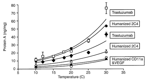
Figure 1 depicts protein A leaching as a function of temperature for various
antibody
products on PROSEP ATM. Leached protein A is shown in ng/mg (ng protein A per
mg antibody).
Temperature on the x-axis refers to the temperature of the water bath. The
column was
equilibrated and washed with 25mM Tris, 25mM NaCl, 5mM EDTA, pH 7.1, washed
with 25mM
Tris, 25 mM NaCI, 0.5 M TMAC, 5 mM EDTA pH 5.0 or 7.1, eluted with either 25
mM citrate pH
2.8, or 0.1 M acetic acid pH 2.9, regenerated with 0.1 M phosphoric acid, and
stored in 0.2 M
sodium acetate, 2 % benzyl alcohol pH 5Ø Trastuzumab was run on a bed height
of 20 cm,
loaded to 20 g Trastuzumab/ L resin, washed with TMAC pH 5.0, eluted with 25
mM citrate pH
2.8, and pooled from 0.1 AU to 2 CV's. Humanized 2C4 was run on a 20 cm bed
height column,
loaded to 15 g humanized 2C4 per liter resin, washed with TMAC pH 7.1, eluted
with 25 mM
citrate pH 2.8, and pooled from 0.1 AU to 2 CV's pool volume. Humanized VEGF
antibody was
run on 14cm bed height, loaded to 20 g humanized VEGF antibody per liter of
resin, washed with
TMAC pH 5.0, eluted with 0.1M acetic acid pH 2.9, and pooled from 0.2 AU to 2
CV's pool
volume. Humanized CD11a antibody was run on a 14 cm bed height, loaded to 20 g
humanized
CD 11a antibody per liter of resin, washed with TMAC pH 7.1, eluted with 0.1M
acetic acid pH 2.9,
and pooled from 0.2 AU to 2CV's.
Figure 2 depicts a comparison of temperature dependent protein A leaching from
PROSEP ATM and PROSEP vAim with Trastuzumab, humanized 2C4, and humanized
CD11a
antibody. Leached protein A is shown in ng/mg (ng protein A per mg antibody).
Temperature on
the x-axis refers to the temperature of the water bath. All columns were
0.66cm in diameter and
either 14 cm or 20 cm in height One lot of harvested cell culture fluid (HCCF)
was used for each
pair of runs. The column was equilibrated and washed with 25 mM Tris, 25mM
NaCI, 5mM
-3a-
CA 02531595 2006-01-05
WO 2005/016968
PCT/US2004/020480
EDTA, pH 7.1, washed with 25 mM Tris, 25 mM NaCI, 0.5 M TMAC, 5 mM EDTA pH 5.0
or 7.1,
eluted with either 25 mM Citrate pH 2.8, or 0.1 M acetic acid pH 2.9,
regenerated with 0.1 M
phosphoric acid, and stored in 0.2 M sodium acetate, 2 % benzyl alcohol pH 5.0
at 40 CV/hr.
Humanized CD11a antibody was run on a 14 cm bed height, loaded to 20 g
humanized CD11a
antibody per liter of resin, washed with TMAC pH 7.1, eluted with 0.1M acetic
acid pH 2.9, and
pooled from 0.2 AU to 2CV's. Humanized 2C4 was run on a 20 cm bed height
column, loaded
to 15 g humanized 204 per liter resin, washed with TMAC pH 7.1, eluted with 25
mM citrate pH
2.8, and pooled from 0.1 AU to 2 CV's pool volume. Trastuzumab (from pilot
plant at 400L scale
at concentration of 0.57 mg/ml) was run on a bed height of 20 cm, loaded to 20
g Trastuzumab
L resin, washed with TMAC pH 5.0, eluted with 25 mM citrate pH 2.8, and pooled
from 0.1 AU
to 2 CV's.
Figure 3 depicts protein A leaching at pilot scale versus temperature. Leached
protein
A is shown in ng/mg (ng protein A per mg antibody). Temperature on the x-axis
refers to the set
temperature of the HCCF tank. The column was packed with 1.26 L PROSEP vATM, 9
cm in
diameter by 20 cm in height. Trastuzumab HCCF was at 0.59 mg/ml, and the
temperature of the
HCCF in the tank was maintained at 10, 15, 20, 25, or 30 C. The column was
loaded to 20g
Trastuzumab per liter of resin. Temperature was measured in the HCCF tank,
between the
pump and the column, and at the outlet to the column. The column was
equilibrated and washed
with 25 mM Tris, 25mM NaCI, 5mM EDTA, pH 7.1, washed with 25 mM Tris, 25 mM
NaCI, 0.5
M TMAC, 5 mM EDTA pH 5.0, eluted with either 25 mM citrate pH 2.8, regenerated
with 0.1 M
phosphoric acid, and stored in 0.2 M sodium acetate, 2 % benzyl alcohol pH
5Ø A sample of
each HCCF was taken and run at lab scale on a 0.66 cm diameter by 20 cm high
column packed
with PROSEP vATM using the same buffers as at pilot scale, represented on the
graph by the
circles.
Figures 4A-B show the light chain amino acid sequence (SEQ ID NO:1) and heavy
chain
amino acid sequence (SEQ ID NO:2), respectively, of Trastuzumab (HERCEPTIN ).
Figures 5A-B depict the amino acid sequences of the variable light (SEQ ID
NO:3) and
variable heavy (SEQ ID NO:4) domains, respectively, of a humanized 204.
Figures 6A-B depict the amino acid sequences of the variable light (SEQ ID
NO:5) and
variable heavy (SEQ ID NO:6) domains, respectively, of a humanized CD11a
antibody
RAPT I VATM .
Figures 7A-B depict the amino acid sequences of the variable light (SEQ ID
NO:7) and
variable heavy (SEQ ID NO:8) domains, respectively, of a humanized VEGF
antibody
AVAST I N TM .
Figure 8 depicts the effect of EDTA and temperature on Protein A leaching.
-4-
CA 02531595 2006-01-05
WO 2005/016968
PCT/US2004/020480
Figure 9 depicts the effect of 4-(2-aminoethyI)-benzenesulfonyl-fluoride,
hydrochloride
(AEBSF) (PEFABL000), a serine protease inhibitor, on Protein A leaching
DETAILED DESCRIPTION OF THE PREFERRED EMBODIMENTS
Definitions:
When used herein, the term "protein A" encompasses protein A recovered from a
native
source thereof, protein A produced synthetically (e.g. by peptide synthesis or
by recombinant
techniques), including variants or derivatives thereof which retain the
ability to bind proteins which
have a CH2/CH3 region. Protein A can be purchased commercially from Repligen,
Pharmacia
and Fermatech.
"Protein A affinity chromatography" refers to the separation or purification
of substances
and/or particles using protein A, where the protein A is generally immobilized
on a solid phase.
A protein comprising a CH2/CH3 region may be reversibly bound to, or adsorbed
by, the protein
A. Examples of protein A affinity chromatography columns for use in protein A
affinity
chromatography herein include protein A immobilized onto a controlled pore
glass backbone,
including the PROSEP ATM and PROSEP vATM columns (Millipore Inc.); protein A
immobilized
on a polystyrene solid phase, e.g. the POROS 5OATM column (Applied BioSystems
Inc.); or
protein A immobilized on an agarose solid phase, for instance the rPROTEIN A
SEPHAROSE
FAST FLOWTM or MABSELECTTm columns (Amersham Biosciences Inc.).
By "solid phase" is meant a non-aqueous matrix to which the protein A can
adhere or be
covalently bound. The solid phase may comprise a glass, silica, polystyrene,
or agarose surface
for immobilizing the protein A, for instance. The solid phase may be a
purification column,
discontinuous phase of discrete particles, packed bed column, expanded bed
column,
membrane, etc.
Herein, "leaching" refers to the detachment or washing of protein A (including
fragments
thereof) from a solid phase to which it is bound. Leaching may result from
various mechanisms
such as mechanical shearing, low pH exposure, proteolytic activity etc.
An "impurity" is a material that is different from the desired protein
product. The impurity
may be a viral impurity, a variant of the desired protein or another protein,
nucleic acid, endotoxin
etc. Specific examples of impurities herein include proteins from the host
cell producing the
desired protein (e.g. Chinese Hamster Ovary proteins, CHOP, where the host
cell is a CHO cell),
protease(s), leached protein A etc.
"Proteases" are proteolytic enzymes including, but not limited to, serine,
cysteine, metallo-
and aspartic proteases. Proteases present in a composition comprising a
protein of interest may
be derived from a recombinant host producing the protein, or from a natural
source of the protein.
Examples of proteases include thermolysin, trypsin, chymotrypsin, plasmin,
kallikrein, thrombin,
papain, plasmin, cathepsin B, renin, chymosin etc.
-5-
CA 02531595 2006-01-05
WO 2005/016968
PCT/US2004/020480
"Protease activity" refers to the enzymatic activity of one or more proteases.
Such activity
may be measured indirectly by measuring leaching of protein A, for instance.
The activity may
be reduced by reducing temperature of a composition comprising the
protease(s), and/or by
adding one or more protease inhibitors to the composition etc.
A "protease inhibitor" is a compound or composition which reduces, to some
extent, the
enzymatic activity of protease(s). Examples of protease inhibitors include
phenylmethylsulfonyi
fluoride (PMSF), 4-(2-aminoethyl)-benzenesulfonyl-fluoride, hydrochloride
(AEBSF)
(PEFABLOCO SC), leupeptin, pepstatin, benzamidine, a metal ion chelator such
as EDTA or
imidazole for inhibiting metalloprotease activity etc. The preferred protease
inhibitors inhibit
metalloprotease activity (e.g. EDTA) and/or inhibit certain serine protease
activities.
The protein of interest herein is one which comprises a CH2/CH3 region and
therefore is
amenable to purification by protein A affinity chromatography. The term
"CH2/CH3 region" when
used herein refers to those amino acid residues in the Fc region of an
immunoglobulin molecule
which interact with protein A. In preferred embodiments, the CH2/CH3 region
comprises an intact
CH2 region followed by an intact CH3 region, and most preferably comprises a
Fc region of an
immunoglobulin.
Examples of CH2/CH3 region-containing proteins include antibodies,
immunoadhesins and fusion proteins comprising a protein of interest fused to,
or conjugated
with, a CH2/0H3 region.
The term "antibody" is used in the broadest sense and specifically covers
monoclonal
antibodies (including full length monoclonal antibodies), polyclonal
antibodies, multispecific
antibodies (e.g., bispecific antibodies), and antibody fragments so long as
they retain, or are
modified to comprise, a CH2/CH3 region as herein defined.
"Antibody fragments" comprise a portion of a full length antibody, generally
the antigen
binding or variable region thereof. Examples of antibody fragments include
Fab, Fab', F(ab')2,
and Fv fragments; single-chain antibody molecules; diabodies; linear
antibodies; and
multispecific antibodies formed from antibody fragments.
The term "monoclonal antibody" as used herein refers to an antibody obtained
from a
population of substantially homogeneous antibodies, i.e., the individual
antibodies comprising the
population are identical except for possible naturally occurring mutations
that may be present in
minor amounts. Monoclonal antibodies are highly specific, being directed
against a single
antigenic site. Furthermore, in contrast to conventional (polyclonal) antibody
preparations which
typically include different antibodies directed against different determinants
(epitopes), each
monoclonal antibody is directed against a single determinant on the antigen.
The modifier
"monoclonal" indicates the character of the antibody as being obtained from a
substantially
homogeneous population of antibodies, and is not to be construed as requiring
production of the
antibody by any particular method. For example, the monoclonal antibodies to
be used in
-6-
CA 02531595 2006-01-05
WO 2005/016968
PCT/US2004/020480
accordance with the present invention may be made by the hybridoma method
first described by
Kohler etal., Nature 256:495 (1975), or may be made by recombinant DNA methods
(see, e.g.,
U.S. Patent No. 4,816,567). The "monoclonal antibodies" may also be isolated
from phage
antibody libraries using the techniques described in Clackson etal., Nature
352:624-628 (1991)
and Marks etal., J. Mol. Biol. 222:581-597 (1991), for example.
The monoclonal antibodies herein specifically include "chimeric" antibodies
(immunoglobulins) in which a portion of the heavy and/or light chain is
identical with or
homologous to corresponding sequences in antibodies derived from a particular
species or
belonging to a particular antibody class or subclass, while the remainder of
the chain(s) is
identical with or homologous to corresponding sequences in antibodies derived
from another
species or belonging to another antibody class or subclass, as well as
fragments of such
antibodies, so long as they exhibit the desired biological activity (U.S.
Patent No. 4,816,567; and
Morrison etal., Proc. Natl. Acad. Sc!. USA 81:6851-6855 (1984)).
The term "hypervariable region" when used herein refers to the amino acid
residues of
an antibody which are responsible for antigen-binding. The hypervariable
region comprises
amino acid residues from a "complementarity determining region" or "CDR" (i.e.
residues 24-34
(L1), 50-56 (L2) and 89-97 (L3) in the light chain variable domain and 31-35
(H1), 50-65 (H2) and
95-102 (H3) in the heavy chain variable domain; Kabat et al., Sequences of
Proteins of
Immunological Interest, 5th Ed. Public Health Service, National Institutes of
Health, Bethesda,
MD. (1991)) and/or those residues from a "hypervariable loop" (i.e. residues
26-32 (L1), 50-52
(L2) and 91-96 (L3) in the light chain variable domain and 26-32 (H1), 53-55
(H2) and 96-101
(H3) in the heavy chain variable domain; Chothia and Lesk J. MoL BioL 196:901-
917 (1987)).
"Framework" or "FR" residues are those variable domain residues other than the
hypervariable
region residues as herein defined.
"Humanized" forms of non-human (e.g., murine) antibodies are chimeric
antibodies which
contain minimal sequence derived from non-human immunoglobulin. For the most
part,
humanized antibodies are human immunoglobulins (recipient antibody) in which
hypervariable
region residues of the recipient are replaced by hypervariable region residues
from a non-human
species (donor antibody) such as mouse, rat, rabbit or nonhuman primate having
the desired
specificity, affinity, and capacity. In some instances, Fv framework region
(FR) residues of the
human immunoglobulin are replaced by corresponding non-human residues.
Furthermore,
humanized antibodies may comprise residues which are not found in the
recipient antibody or
in the donor antibody. These modifications are made to further refine antibody
performance.
In general, the humanized antibody will comprise substantially all of at least
one, and typically
two, variable domains, in which all or substantially all of the hypervariable
loops correspond to
those of a non-human immunoglobulin and all or substantially all of the FR
regions are those of
-7-
CA 02531595 2006-01-05
WO 2005/016968
PCT/US2004/020480
a human immunoglobulin sequence. The humanized antibody optionally also will
comprise at
least a portion of an immunoglobulin constant region (Fc), typically that of a
human
immunoglobulin. For further details, see Jones et al., Nature 321:522-525
(1986); Riechmann
etal., Nature 332:323-329 (1988); and Presta, Curr. Op. Struct. Biol. 2:593-
596 (1992).
As used herein, the term "immunoadhesin" designates antibody-like molecules
which
combine the "binding domain" of a heterologous "adhesin" protein (e.g. a
receptor, ligand or
enzyme) with the effector functions of an immunoglobulin constant domain.
Structurally, the
immunoadhesins comprise a fusion of the adhesin amino acid sequence with the
desired binding
specificity which is other than the antigen recognition and binding site
(antigen combining site)
of an antibody (i.e. is "heterologous") and an immunoglobulin constant domain
sequence. The
immunoglobulin constant domain sequence in the immunoadhesin is preferably
derived from y1,
y2, or y4 heavy chains since immunoadhesins comprising these regions can be
purified by
protein A affinity chromatography (Lindmark etal., J. ImmunoL Meth. 62:1-13
(1983)).
The term "ligand binding domain" as used herein refers to any native cell-
surface receptor
or any region or derivative thereof retaining at least a qualitative ligand
binding of a
corresponding native receptor. In a specific embodiment, the receptor is from
a cell-surface
polypeptide having an extracellular domain which is homologous to a member of
the
immunoglobulin supergenefamily.
Other receptors, which are not members of the
immunoglobulin supergenefamily but are nonetheless specifically covered by
this definition, are
receptors for cytokines, and in particular receptors with tyrosine kinase
activity (receptor tyrosine
kinases), members of the hematopoietin and nerve growth factor receptor
superfamilies, and cell
adhesion molecules, e.g. (E-, L- and P-) selectins.
The term "receptor binding domain" is used to designate any native ligand for
a receptor,
including cell adhesion molecules, or any region or derivative of such native
ligand retaining at
least a qualitative receptor binding ability of a corresponding native ligand.
This definition,
among others, specifically includes binding sequences from ligands for the
above-mentioned
receptors.
An "antibody-immunoadhesin chimera" comprises a molecule which combines
at
least one binding domain of an antibody (as herein defined) with at least one
immunoadhesin (as
defined in this application). Exemplary antibody-immunoadhesin chimeras are
the bispecific
CD4-IgG chimeras described in Berg etal., PNAS (USA) 88:4723-4727 (1991) and
Chamow et
al., J. ImmunoL 153:4268 (1994).
The expression "HER2" refers to human HER2 protein described, for example, in
Semba
et al., PNAS (USA) 82:6497-6501 (1985) and Yamamoto et al. Nature 319:230-234
(1986)
(Genebank accession number X03363).
"Trastuzumab" or "HERCEPTINO" is a humanized HER2 antibody comprising the
light
chain amino acid sequence of SEQ ID NO:1 and the heavy chain amino acid
sequence of SEQ
-8-
CA 02531595 2011-09-16
ID NO:2, or amino acid sequence variants thereof which retain the ability to
bind HER2 and
inhibit growth of tumor cells which overexpress HER2 (see US Patent
5,677,171).
"Humanized 2C4" is a humanized HER2 antibody comprising the variable light
amino acid
sequence of SEQ ID NO:3 and the variable heavy amino acid sequence of SEQ ID
NO:4, or
amino acid sequence variants thereof which retain the ability to bind HER2 and
block ligand
activation of HER2 (see W001/00245 ).
Modes for Carrying Out the Invention
The process herein involves purifying a C142/CH3 region-containing protein
from impurities
by protein A affinity chromatography. In preferred embodiments, the protein is
an antibody,
immunoadhesin or a protein fused to, or conjugated with, a CH2/CH3 region.
Techniques for
generating such molecules will be discussed below.
1. Antibodies
The preferred protein according to the present invention is an antibody.
Antibodies within
the scope of the present invention include, but are not limited to: anti-HER2
antibodies including
Trastuzumab (HERCEPTINC)) (Carter at al., Proc. NatL Acad. ScL USA, 89:4285-
4289 (1992),
U.S. Patent No. 5,725,856) and humanized 2C4 (W001/00245, Adams at al.); anti-
CD20
antibodies such as chimeric anti-CD20 "C2B8" as in US Patent No. 5,736,137
(RITUXAN0), a
chimeric or humanized variant of the 2H7 antibody as in US Patent No.
5,721,108B1, or
Tositumomab (BBO<ARC)); anti-1L-8 antibodies (St John et al., Chest, 103:932
(1993), and
International Publication No. WO 95/23865); anti-VEGF antibodies, including
humanized and/or
affinity matured anti-VEGF antibodies such as the humanized anti-VEGF antibody
huA4.6.1
AVASTIN (Kim et al., Growth Factors, 7:53-64 (1992), International
Publication No. WO
96/30046, and WO 98/45331, published October 15, 1998); anti-prostate stem
cell antigen
(PSCA) antibodies (W001/40309); anti-CD40 antibodies, including S2C6 and
humanized
variants thereof (W000/75348); anti-CD11a antibodies (US Patent No. 5,622,700,
WO 98/23761,
Steppe at aL, Transplant Intl. 4:3-7 (1991), and Hourmant at aL,
Transplantation 58:377-380
(1994)); anti-CD18 (US Patent No. 5,622,700, issued April 22, 1997, or as in
WO 97/26912,
published July 31, 1997); anti-IgE antibodies (including E25, E26 and E27; US
Patent No.
5,714,338, issued February 3, 1998 or US Patent No. 5,091,313, issued February
25, 1992, WO
93/04173 published March 4, 1993, or International Application No.
PCT/US98/13410 filed June
30, 1998, US Patent No. 5,714,338, Presta et al., J. Immunot 151:2623-2632
(1993), and
International Publication No. WO 95/19181); anti-Apo-2 receptor antibodies (WO
98/51793
published November 19, 1998); anti-TNF-a antibodies, including cA2
(REMICADE0), CDP571
-9-
CA 02531595 2006-01-05
WO 2005/016968 PCT/US2004/020480
and MAK-195 (See, US Patent No. 5,672,347 issued September 30, 1997, Lorenz et
al. J.
lmmunol. 156(4):1646-1653 (1996), and Dhainaut etal. Crit. Care Med.
23(9):1461-1469 (1995));
anti-Tissue Factor (TF) antibodies (European Patent No. 0 420 937 B1 granted
November 9,
1994); anti-human c(4137 integrin antibodies (WO 98/06248 published February
19, 1998); anti-
epidermal growth factor receptor (EGFR) antibodies (e.g. chimerized or
humanized 225 antibody
as in WO 96/40210 published December 19, 1996); anti-CD3 antibodies such as
OKT3 (US
Patent No. 4,515,893 issued May 7, 1985); anti-CD25 or anti-Tac antibodies
such as CHI-621
(S I MU LECTO) and ZENAPAXO (See US Patent No. 5,693,762 issued December 2,
1997); anti-
CD4 antibodies such as the cM-7412 antibody (Choy etal. Arthritis Rheum
39(1):52-56 (1996));
anti-CD52 antibodies such as CAMPATH-1H (Riechmann et al. Nature 332:323-337
(1988));
anti-Fc receptor antibodies such as the M22 antibody directed against FcyRI as
in Graziano et
al. J. lmmunol. 155(10):4996-5002 (1995); anti-carcinoembryonic antigen (CEA)
antibodies such
as hMN-14 (Sharkey et al. Cancer Res. 55(23Suppl): 5935s-5945s (1995);
antibodies directed
against breast epithelial cells including huBrE-3, hu-Mc 3 and CHL6 (Ceriani
et al. Cancer Res.
55(23): 5852s-5856s (1995); and Richman et al. Cancer Res. 55(23 Supp): 5916s-
5920s (1995));
antibodies that bind to colon carcinoma cells such as C242 (Litton etal. Eur
J. lmmunol. 26(1):1-
9 (1996)); anti-CD38 antibodies, e.g. AT 13/5 (Ellis etal. J. lmmunol.
155(2):925-937 (1995));
anti-CD33 antibodies such as Hu M195 (Jurcic et al. Cancer Res 55(23
Suppl):5908s-5910s
(1995) and CMA-676 or CDP771; anti-CD22 antibodies such as LL2 or LymphoCide
(Juweid et
al. Cancer Res 55(23 Suppl):5899s-5907s (1995)); anti-EpCAM antibodies such as
17-1A
(PANOREXO); anti-Gpl lb/Illa antibodies such as abciximab or c7E3 Fab
(REOPROO); anti-RSV
antibodies such as MEDI-493 (SYNAGISO); anti-CMV antibodies such as PROTOVIRO;
anti-HIV
antibodies such as PR0542; anti-hepatitis antibodies such as the anti-Hep B
antibody
OSTAVIRQ anti-CA 125 antibodies, such as OvaRex; anti-idiotypic GD3 epitope
antibody BEC2;
anti-av133 antibodies, including VITAXINQ anti-human renal cell carcinoma
antibody such as ch-
G250; ING-1; anti-human 17-1A antibody (3622W94); anti-human colorectal tumor
antibody
(A33); anti-human melanoma antibody R24 directed against GD3 ganglioside; anti-
human
squamous-cell carcinoma (SF-25); and anti-human leukocyte antigen (HLA)
antibodies such as
Smart ID10 and the anti-HLA DR antibody Oncolym (Lym-1). The preferred target
antigens for
the antibody herein are: HER2 receptor, VEGF, IgE, CD20, CD11a, and CD40.
Aside from the antibodies specifically identified above, the skilled
practitioner could
generate antibodies directed against an antigen of interest, e.g., using the
techniques described
below.
(i) Antigen selection and preparation
The antibody herein is directed against an antigen of interest. Preferably,
the antigen is
a biologically important polypeptide and administration of the antibody to a
mammal suffering
-10-
CA 02531595 2006-01-05
WO 2005/016968
PCT/US2004/020480
from a disease or disorder can result in a therapeutic benefit in that mammal.
However,
antibodies directed against nonpolypeptide antigens (such as tumor-associated
glycolipid
antigens; see US Patent 5,091,178) are also contemplated. Where the antigen is
a polypeptide,
it may be a transmembrane molecule (e.g. receptor) or ligand such as a growth
factor. Exemplary
antigens include those proteins described in section (3) below. Exemplary
molecular targets for
antibodies encompassed by the present invention include CD proteins such as
CD3, CD4, CD8,
CD19, CD20, CD22 and CD34; members of the ErbB receptor family such as the
EGFR, HER2,
HER3 or HER4 receptor; cell adhesion molecules such as LFA-1, Mac1, p150,95,
VLA-4, ICAM-
1, VCAM and av/I33 integrin including either a or 13 subunits thereof (e.g.
anti-CD11a, anti-CD18
or anti-CD11b antibodies); growth factors such as VEGF; IgE; blood group
antigens; flk2/flt3
receptor; obesity (OB) receptor; mpl receptor; CTLA-4; protein C, or any of
the other antigens
mentioned herein.
Soluble antigens or fragments thereof, optionally conjugated to other
molecules, can be
used as immunogens for generating antibodies. For transmembrane molecules,
such as
receptors, fragments of these (e.g. the extracellular domain of a receptor)
can be used as the
immunogen. Alternatively, cells expressing the transmembrane molecule can be
used as the
immunogen. Such cells can be derived from a natural source (e.g. cancer cell
lines) or may be
cells which have been transformed by recombinant techniques to express the
transmembrane
molecule.
Other antigens and forms thereof useful for preparing antibodies will be
apparent to those
in the art.
(ii) Polyclonal antibodies
Polyclonal antibodies are preferably raised in animals by multiple
subcutaneous (sc) or
intraperitoneal (ip) injections of the relevant antigen and an adjuvant. It
may be useful to
conjugate the antigen to a protein that is immunogenic in the species to be
immunized, e.g.,
keyhole limpet hemocyanin, serum albumin, bovine thyroglobulin, or soybean
trypsin inhibitor
using a bifunctional or derivatizing agent, for example, maleimidobenzoyl
sulfosuccinimide ester
(conjugation through cysteine residues), N-hydroxysuccinimide (through lysine
residues),
glutaraldehyde, succinic anhydride, SOCl2, or R1N=C=NR, where R and R1 are
different alkyl
groups.
Animals are immunized against the antigen, immunogenic conjugates, or
derivatives by
combining, e.g., 100 pg or 5 pg of the protein or conjugate (for rabbits or
mice, respectively) with
3 volumes of Freund's complete adjuvant and injecting the solution
intradermally at multiple sites.
One month later the animals are boosted with 1/5 to 1/10 the original amount
of antigen or
conjugate in Freund's complete adjuvant by subcutaneous injection at multiple
sites. Seven to
14 days later the animals are bled and the serum is assayed for antibody
titer. Animals are
-11-
CA 02531595 2006-01-05
WO 2005/016968 PCT/US2004/020480
boosted until the titer plateaus. Preferably, the animal is boosted with the
conjugate of the same
antigen, but conjugated to a different protein and/or through a different
cross-linking reagent.
Conjugates also can be made in recombinant cell culture as protein fusions.
Also, aggregating
agents such as alum are suitably used to enhance the immune response.
(iii) Monoclonal antibodies
Monoclonal antibodies may be made using the hybridoma method first described
by
Kohler et al., Nature, 256:495 (1975), or may be made by recombinant DNA
methods (U.S.
Patent No. 4,816,567).
In the hybridoma method, a mouse or other appropriate host animal, such as a
hamster
or macaque monkey, is immunized as hereinabove described to elicit lymphocytes
that produce
or are capable of producing antibodies that will specifically bind to the
protein used for
immunization. Alternatively, lymphocytes may be immunized in vitro.
Lymphocytes then are
fused with myeloma cells using a suitable fusing agent, such as polyethylene
glycol, to form a
hybridoma cell (Goding, Monoclonal Antibodies: Principles and Practice, pp.59-
103 (Academic
Press, 1986)).
The hybridoma cells thus prepared are seeded and grown in a suitable culture
medium
that preferably contains one or more substances that inhibit the growth or
survival of the unfused,
parental myeloma cells. For example, if the parental myeloma cells lack the
enzyme
hypoxanthine guanine phosphoribosyl transferase (HGPRT or HPRT), the culture
medium for
the hybridomas typically will include hypoxanthine, aminopterin, and thymidine
(HAT medium),
which substances prevent the growth of HGPRT-deficient cells.
Preferred myeloma cells are those that fuse efficiently, support stable high-
level
production of antibody by the selected antibody-producing cells, and are
sensitive to a medium
such as HAT medium. Among these, preferred myeloma cell lines are murine
myeloma lines,
such as those derived from MOPC-21 and MPC-11 mouse tumors available from the
Salk
Institute Cell Distribution Center, San Diego, California USA, and SP-2 or X63-
Ag8-653 cells
available from the American Type Culture Collection, Rockville, Maryland USA.
Human myeloma
and mouse-human heteromyeloma cell lines also have been described for the
production of
human monoclonal antibodies (Kozbor, J. Immunol., 133:3001(1984); Brodeur et
al., Monoclonal
Antibody Production Techniques and Applications, pp. 51-63 (Marcel Dekker,
Inc., New York,
1987)).
Culture medium in which hybridoma cells are growing is assayed for production
of
monoclonal antibodies directed against the antigen. Preferably, the binding
specificity of
monoclonal antibodies produced by hybridoma cells is determined by
immunoprecipitation or by
an in vitro binding assay, such as radioimmunoassay (RIA) onenzyme-linked
immunoabsorbent
assay (ELISA).
-12-
CA 02531595 2006-01-05
WO 2005/016968
PCT/US2004/020480
After hybridoma cells are identified that produce antibodies of the desired
specificity,
affinity, and/or activity, the clones may be subcloned by limiting dilution
procedures and grown
by standard methods (Goding, Monoclonal Antibodies: Principles and Practice,
pp.59-103
(Academic Press, 1986)). Suitable culture media for this purpose include, for
example, D-MEM
or RPMI-1640 medium. In addition, the hybridoma cells may be grown in vivo as
ascites tumors
in an animal.
The monoclonal antibodies secreted by the subclones are suitably separated
from the
culture medium, ascites fluid, or serum by conventional immunoglobulin
purification procedures
such as, for example, protein A-Sepharose, hydroxylapatite chromatography, gel
electrophoresis,
dialysis, or affinity chromatography. Preferably the protein A affinity
chromatography procedure
described herein is used.
DNA encoding the monoclonal antibodies is readily isolated and sequenced using
conventional procedures (e.g., by using oligonucleotide probes that are
capable of binding
specifically to genes encoding the heavy and light chains of the monoclonal
antibodies). The
hybridoma cells serve as a preferred source of such DNA. Once isolated, the
DNA may be
placed into expression vectors, which are then transfected into host cells
such as E. coli cells,
simian COS cells, Chinese hamster ovary (CHO) cells, or myeloma cells that do
not otherwise
produce immunoglobulin protein, to obtain the synthesis of monoclonal
antibodies in the
recombinant host cells.
The DNA also may be modified, for example, by substituting the coding sequence
for
human heavy- and light-chain constant domains in place of the homologous
murine sequences
(U.S. Patent No. 4,816,567; Morrison, etal., Proc. Nat/Acad. ScL USA, 81:6851
(1984)), or by
covalently joining to the immunoglobulin coding sequence all or part of the
coding sequence for
a non-immunoglobulin polypeptide.
Typically such non-immunoglobulin polypeptides are substituted for the
constant domains
of an antibody, or they are substituted for the variable domains of one
antigen-combining site of
an antibody to create a chimeric bivalent antibody comprising one antigen-
combining site having
specificity for an antigen and another antigen-combining site having
specificity for a different
antigen.
In a further embodiment, monoclonal antibodies can be isolated from antibody
phage
libraries generated using the techniques described in McCafferty et al.,
Nature, 348:552-554
(1990). Clackson etal., Nature, 352:624-628 (1991) and Marks et al., J. MoL
Biol., 222:581-597
(1991) describe the isolation of murine and human antibodies, respectively,
using phage libraries.
Subsequent publications describe the production of high affinity (nM range)
human antibodies
by chain shuffling (Marks etal., Bio/Technology, 10:779-783 (1992)), as well
as combinatorial
infection and in vivo recombination as a strategy for constructing very large
phage libraries
-13-
CA 02531595 2006-01-05
WO 2005/016968 PCT/US2004/020480
(Waterhouse etal., Nuc. Acids. Res., 21:2265-2266(1993)). Thus, these
techniques are viable
alternatives to traditional hybridoma techniques for isolation of monoclonal
antibodies.
(iv) Humanized and human antibodies
A humanized antibody has one or more amino acid residues introduced into it
from a
source which is non-human. These non-human amino acid residues are often
referred to as
"import" residues, which are typically taken from an "import" variable domain.
Humanization can
be essentially performed following the method of Winter and co-workers (Jones
et al., Nature,
321:522-525(1986); Riechmann etal., Nature, 332:323-327 (1988); Verhoeyen
etal., Science,
239:1534-1536 (1988)), by substituting rodent CDRs or CDR sequences for the
corresponding
sequences of a human antibody. Accordingly, such "humanized" antibodies are
chimeric
antibodies (U.S. Patent No. 4,816,567) wherein substantially less than an
intact human variable
domain has been substituted by the corresponding sequence from a non-human
species. In
practice, humanized antibodies are typically human antibodies in which some
CDR residues and
possibly some FR residues are substituted by residues from analogous sites in
rodent antibodies.
The choice of human variable domains, both light and heavy, to be used in
making the
humanized antibodies is very important to reduce antigenicity. According to
the so-called "best-
fit" method, the sequence of the variable domain of a rodent antibody is
screened against the
entire library of known human variable-domain sequences. The human sequence
which is
closest to that of the rodent is then accepted as the human FR for the
humanized antibody (Sims
et al., J. Immunol., 151:2296 (1993)). Another method uses a particular
framework derived from
the consensus sequence of all human antibodies of a particular subgroup of
light or heavy
chains. The same framework may be used for several different humanized
antibodies (Carter
etal., Proc. Natl. Acad. ScL USA, 89:4285 (1992); Presta etal., J. Immnol.,
151:2623 (1993)).
It is further important that antibodies be humanized with retention of high
affinity for the
antigen and other favorable biological properties. To achieve this goal,
according to a preferred
method, humanized antibodies are prepared by a process of analysis of the
parental sequences
and various conceptual humanized products using three-dimensional models of
the parental and
humanized sequences. Three-dimensional immunoglobulin models are commonly
available and
are familiar to those skilled in the art. Computer programs are available
which illustrate and
display probable three-dimensional conformational structures of selected
candidate
immunoglobulin sequences. Inspection of these displays permits analysis of the
likely role of the
residues in the functioning of the candidate immunoglobulin sequence, i.e.,
the analysis of
residues that influence the ability of the candidate immunoglobulin to bind
its antigen. In this
way, FR residues can be selected and combined from the recipient and import
sequences so that
the desired antibody characteristic, such as increased affinity for the target
antigen(s), is
-14-
CA 02531595 2006-01-05
WO 2005/016968 PCT/US2004/020480
achieved. In general, the CDR residues are directly and most substantially
involved in influencing
antigen binding.
Alternatively, it is now possible to produce transgenic animals (e.g., mice)
that are
capable, upon immunization, of producing a full repertoire of human antibodies
in the absence
of endogenous immunoglobulin production. For example, it has been described
that the
homozygous deletion of the antibody heavy-chain joining region (JH) gene in
chimeric and germ-
line mutant mice results in complete inhibition of endogenous antibody
production. Transfer of
the human germ-line immunoglobulin gene array in such germ-line mutant mice
will result in the
production of human antibodies upon antigen challenge. See, e.g., Jakobovits
et aL, Proc. Natl.
Acad. Sci. USA, 90:2551 (1993); Jakobovits etal., Nature, 362:255-258 (1993);
Bruggermann
et aL, Year in lmmuno., 7:33 (1993); and Duchosal et al. Nature 355:258
(1992). Human
antibodies can also be derived from phage-display libraries (Hoogenboom et
al., J. MoL BioL,
227:381 (1991); Marks etal., J. MoL Biol., 222:581-597 (1991); Vaughan etal.
Nature Biotech
14:309 (1996)).
(v) Antibody fragments
Various techniques have been developed for the production of antibody
fragments.
Traditionally, these fragments were derived via proteolytic digestion of
intact antibodies (see,
e.g., Morimoto et al. , Journal of Biochemical and Biophysical Methods 24:107-
117 (1992) and
Brennan et al., Science, 229:81 (1985)). However, these fragments can now be
produced
directly by recombinant host cells. For example, the antibody fragments can be
isolated from the
antibody phage libraries discussed above. Alternatively, Fab'-SH fragments can
be directly
recovered from E. coli and chemically coupled to form F(ab')2 fragments
(Carter et al.,
Bio/Technology 10:163-167(1992)). According to another approach, F(ab')2
fragments can be
isolated directly from recombinant host cell culture. Other techniques for the
production of
antibody fragments will be apparent to the skilled practitioner. In other
embodiments, the
antibody of choice is a single chain Fv fragment (scFv). See WO 93/16185.
(vi) Multispecific antibodies
Multispecific antibodies have binding specificities for at least two different
antigens. While
such molecules normally will only bind two antigens (i.e. bispecific
antibodies, BsAbs), antibodies
with additional specificities such as trispecific antibodies are encompassed
by this expression
when used herein.
Methods for making bispecific antibodies are known in the art. Traditional
production of
full length bispecific antibodies is based on the coexpression of two
immunoglobulin heavy chain-
light chain pairs, where the two chains have different specificities
(Millstein et al., Nature,
- 305:537-539 (1983)). Because of the random assortment of immunoglobulin
heavy and light
chains, these hybridomas (quadromas) produce a potential mixture of 10
different antibody
-15-
CA 02531595 2006-01-05
WO 2005/016968
PCT/US2004/020480
molecules, of which only one has the correct bispecific structure.
Purification of the correct
molecule, which is usually done by affinity chromatography steps, is rather
cumbersome, and the
product yields are low. Similar procedures are disclosed in WO 93/08829, and
in Traunecker et
al., EMBO J., 10:3655-3659 (1991).
According to another approach described in W096/27011, the interface between a
pair
of antibody molecules can be engineered to maximize the percentage of
heterodimers which are
recovered from recombinant cell culture. The preferred interface comprises at
least a part of the
CH3 domain of an antibody constant domain. In this method, one or more small
amino acid side
chains from the interface of the first antibody molecule are replaced with
larger side chains (e.g.
tyrosine or tryptophan). Compensatory "cavities" of identical or similar size
to the large side
chain(s) are created on the interface of the second antibody molecule by
replacing large amino
acid side chains with smaller ones (e.g. alanine or threonine). This provides
a mechanism for
increasing the yield of the heterodimer over other unwanted end-products such
as homodimers.
Bispecific antibodies include cross-linked or "heteroconjugate" antibodies.
For example,
one of the antibodies in the heteroconjugate can be coupled to avidin, the
other to biotin. Such
antibodies have, for example, been proposed to targot immune system cells to
unwanted cells
(US Patent No. 4,676,980), and for treatment of HIV infection (WO 91/00360, WO
92/200373,
and EP 03089). Heteroconjugate antibodies may be made using any convenient
cross-linking
methods. Suitable cross-linking agents are well known in the art, and are
disclosed in US Patent
No. 4,676,980, along with a number of cross-linking techniques.
Techniques for generating bispecific antibodies from antibody fragments have
also been
described in the literature. For example, bispecific antibodies can be
prepared using chemical
linkage. Brennan etal., Science, 229: 81(1985) describe a procedure wherein
intact antibodies
are proteolytically cleaved to generate F(ab')2 fragments. These fragments are
reduced in the
presence of the dithiol complexing agent sodium arsenite to stabilize vicinal
dithiols and prevent
intermolecular disulfide formation. The Fab' fragments generated are then
converted to
thionitrobenzoate (TNB) derivatives. One of the Fab'-TNB derivatives is then
reconverted to the
Fab'-thiol by reduction with mercaptoethylamine and is mixed with an equimolar
amount of the
other Fab'-TNB derivative to form the bispecific antibody. The bispecific
antibodies produced can
be used as agents for the selective immobilization of enzymes.
Recent progress has facilitated the direct recovery of Fab'-SH fragments from
E. coli,
which can be chemically coupled to form bispecific antibodies. Shalaby etal.,
J. Exp. Med., 175:
217-225(1992) describe the production of a fully humanized bispecific antibody
F(ab1)2molecule.
Each Fab' fragment was separately secreted from E. coli and subjected to
directed chemical
coupling in vitro to form the bispecific antibody. The bispecific antibody
thus formed was able
-16-
CA 02531595 2006-01-05
WO 2005/016968
PCT/US2004/020480
to bind to cells overexpressing the ErbB2 receptor and normal human T cells,
as well as trigger
the lytic activity of human cytotoxic lymphocytes against human breast tumor
targets.
Various techniques for making and isolating bispecific antibody fragments
directly from
recombinant cell culture have also been described. For example, bispecific
antibodies have been
produced using leucine zippers. Kostelny et al., J. Immunol., 148(5):1547-1553
(1992). The
leucine zipper peptides from the Fos and Jun proteins were linked to the Fab'
portions of two
different antibodies by gene fusion. The antibody homodimers were reduced at
the hinge region
to form monomers and then re-oxidized to form the antibody heterodimers. This
method can also
be utilized for the production of antibody homodimers. The "diabody"
technology described by
Hollinger et al., Proc. Natl. Acad. Sci. USA, 90:6444-6448 (1993) has provided
an alternative
mechanism for making bispecific antibody fragments. The fragments comprise a
heavy-chain
variable domain (VH) connected to a light-chain variable domain (VL) by a
linker which is too short
to allow pairing between the two domains on the same chain. Accordingly, the
VH and VL
domains of one fragment are forced to pair with the complementary VL and VH
domains of
another fragment, thereby forming two antigen-binding sites. Another strategy
for making
bispecific antibody fragments by the use of single-chain Fv (sFv) dimers has
also been reported. -
See Gruber et al., J. Immunol., 152:5368 (1994). Alternatively, the antibodies
can be "linear
antibodies" as described in Zapata et aL Protein Eng. 8(10):1057-1062 (1995).
Briefly, these
antibodies comprise a pair of tandem Fd segments (VH-CH1-VH-CH1) which form a
pair of antigen
binding regions. Linear antibodies can be bispecific or monospecific.
Antibodies with more than two valencies are contemplated. For example,
trispecific
antibodies can be prepared. Tutt etal. J. Immunol. 147: 60 (1991).
2. lmmunoadhesins
The simplest and most straightforward immunoadhesin design combines the
binding
domain(s) of the adhesin (e.g. the extracellular domain (ECD) of a receptor)
with the hinge and
Fc regions of an immunoglobulin heavy chain. Ordinarily, when preparing the
immunoadhesins
of the present invention, nucleic acid encoding the binding domain of the
adhesin will be fused
C-terminally to nucleic acid encoding the N-terminus of an immunoglobulin
constant domain
sequence, however N-terminal fusions are also possible.
Typically, in such fusions the encoded chimeric polypeptide will retain at
least functionally
active hinge, CH2 and CH3 domains of the constant region of an immunoglobulin
heavy chain.
Fusions are also made to the C-terminus of the Fc portion of a constant
domain, or immediately
-17-
CA 02531595 2006-01-05
WO 2005/016968
PCT/US2004/020480
N-terminal to the CH1 of the heavy chain or the corresponding region of the
light chain. The
precise site at which the fusion is made is not critical; particular sites are
well known and may be
selected in order to optimize the biological activity, secretion, or binding
characteristics of the
immunoadhesin.
In a preferred embodiment, the adhesin sequence is fused to the N-terminus of
the Fc
domain of immunoglobulin G1(IgG1). It is possible to fuse the entire heavy
chain constant region
to the adhesin sequence. However, more preferably, a sequence beginning in the
hinge region
just upstream of the papain cleavage site which defines IgG Fc chemically
(i.e. residue 216,
taking the first residue of heavy chain constant region to be 114), or
analogous sites of other
immunoglobulins is used in the fusion. In a particularly preferred embodiment,
the adhesin
amino acid sequence is fused to (a) the hinge region and CH2 and CH3 or (b)
the CH1, hinge, CH2
and CH3 domains, of an IgG heavy chain.
For bispecific immunoadhesins, the immunoadhesins are assembled as multimers,
and
particularly as heterodimers or heterotetramers. Generally, these assembled
immunoglobulins
will have known unit structures. A basic four chain structural unit is the
form in which IgG, IgD,
and IgE exist. A four chain unit is repeated in the higher molecular weight
immunoglobulins; IgM =
generally exists as a pentamer of four basic units held together by disulfide
bonds. IgA globulin,
and occasionally IgG globulin, may also exist in multimeric form in serum. In
the case of
multimer, each of the four units may be the same or different.
Various exemplary assembled immunoadhesins within the scope herein are
schematically
diagrammed below:
(a) ACL-ACL;
(b) ACH-(ACH, ACL-ACH, ACL-VHCH, or VLCL-ACH);
(c) ACL-ACH-(ACL-ACH, ACL-VHCH, VLCL-ACH, or VLCL-VHCH)
(d) ACL-VHCH-(ACH, or ACL-VHCH, or VLCL-ACH);
(e) VLCL-ACH-(ACL-VHCH, or VLCL-ACH); and
(f) (A-Y),,-(VLCL-VHCH)2,
wherein each A represents identical or different adhesin amino acid sequences;
VL is an immunoglobulin light chain variable domain;
VH is an immunoglobulin heavy chain variable domain;
CL is an immunoglobulin light chain constant domain;
CH is an immunoglobulin heavy chain constant domain;
n is an integer greater than 1;
Y designates the residue of a covalent cross-linking agent.
In the interests of brevity, the foregoing structures only show key features;
they do not
indicate joining (J) or other domains of the immunoglobulins, nor are
disulfide bonds shown.
-18-
CA 02531595 2006-01-05
WO 2005/016968
PCT/US2004/020480
However, where such domains are required for binding activity, they shall be
constructed to be
present in the ordinary locations which they occupy in the immunoglobulin
molecules.
Alternatively, the adhesin sequences can be inserted between immunoglobulin
heavy
chain and light chain sequences, such that an immunoglobulin comprising a
chimeric heavy chain
is obtained. In this embodiment, the adhesin sequences are fused to the 3' end
of an
immunoglobulin heavy chain in each arm of an immunoglobulin, either between
the hinge and
the CH2 domain, or between the CH2 and CH3 domains. Similar constructs have
been reported
by Hoogenboom, etal., Mol. lmmunol. 28:1027-1037 (1991).
Although the presence of an immunoglobulin light chain is not required in the
immunoadhesins of the present invention, an immunoglobulin light chain might
be present either
covalently associated to an adhesin-immunoglobulin heavy chain fusion
polypeptide, or directly
fused to the adhesin. In the former case, DNA encoding an immunoglobulin light
chain is
typically coexpressed with the DNA encoding the adhesin-immunoglobulin heavy
chain fusion
protein. Upon secretion, the hybrid heavy chain and the light chain will be
covalently associated
to provide an immunoglobulin-like structure comprising two disulfide-linked
immunoglobulin
heavy chain-light chain pairs. Methods suitable for the preparation of such
structures are, for =
example, disclosed in U.S. Patent No. 4,816,567, issued 28 March 1989.
lmmunoadhesins are most conveniently constructed by fusing the cDNA sequence
encoding the adhesin portion in-frame to an immunoglobulin cDNA sequence.
However, fusion
to genomic immunoglobulin fragments can also be used (see, e.g. Aruffo etal.,
Cell 61:1303-
1313 (1990); and Stamenkovic etal., Cell 66:1133-1144 (1991)). The latter type
of fusion
requires the presence of Ig regulatory sequences for expression. cDNAs
encoding IgG heavy-
chain constant regions can be isolated based on published sequences from cDNA
libraries
derived from spleen or peripheral blood lymphocytes, by hybridization or by
polymerase chain
reaction (PCR) techniques. The cDNAs encoding the "adhesin" and the
immunoglobulin parts
of the immunoadhesin are inserted in tandem into a plasmid vector that directs
efficient
expression in the chosen host cells.
3. Other CH2/CH3 region-containing proteins
In other embodiments, the protein to be purified is one which is fused to, or
conjugated
with, a CH2/CH3 region. Such fusion proteins may be produced so as to increase
the serum half-
life of the protein and/or to facilitate purification of the protein by
protein A affinity
chromatography. Examples of biologically important proteins which can
be conjugated this
way include renin; a growth hormone, including human growth hormone and bovine
growth
hormone; growth hormone releasing factor; parathyroid hormone; thyroid
stimulating hormone;
lipoproteins; alpha-1-antitrypsin; insulin A-chain; insulin B-chain;
proinsulin; follicle stimulating
-19-
CA 02531595 2011-09-16
hormone; calcitonin; luteinizing hormone; glucagon; clotting factors such as
factor VIIIC, factor
IX, tissue factor, and von Willebrands factor; anti-clotting factors such as
Protein C; atrial
natriuretic factor; lung surfactant; a plasminogen activator, such as
urokinase or human urine or
tissue-type plasminogen activator (t-PA); bombesin; thrombin; hemopoietic
growth factor; tumor
necrosis factor-alpha and -beta; enkephalinase; RANTES (regulated on
activation normally 1-cell
expressed and secreted); human macrophage inflammatory protein (MIP-1-alpha);
a serum
albumin such as human serum albumin; Muellerian-inhibiting substance; relaxin
A-chain; relaxin
B-chain; prorelaxin; mouse gonadotropin-associated peptide; a microbial
protein, such as beta-
lactamase; DNase; IgE; a cytotoxic T-lymphocyte associated antigen (CTLA),
suchis CTLA-4;
inhibin; activin; vascular endothelial growth factor (VEGF); receptors for
hormones or growth
factors; Protein A or D; rheumatoid factors; a neurotrophic factor such as
bone-derived
neurotrophic factor (BDNF), neurotrophin-3, -4, -5, or -6 (NT-3, NT-4, NT-5,
or NT-6), or a nerve
growth factor such as NGF-13; platelet-derived growth factor (PDGF);
fibroblast growth factor
such as a FGF and bFGF; epidermal growth factor (EGF); transforming growth
factor (TGF) such
as TGF-alpha and TGF-beta, including TGF-131, TGF-132, TGF-03, TGF-134, or TGF-
135; insulin-
like growth factor-I and -11 (1GF-1 and IGF-11); des(1-3)-1GF-1 (brain IGF-I),
insulin-like growth =
factor binding proteins; CD proteins such as CD3, CD4, C08, CD19 and CD20;
erythropoietin;
osteoinductive factors; immunotoxins; a bone morphogenetic protein (BMP); an
interferon such
as interferon-alpha, -beta, and -gamma; colony stimulating factors (CSFs),
e.g., M-CSF, GM-
CSF, and G-CSF; interleukins (Is), e.g., 1L-1 to IL-10; superoxide dismutase;
T-cell receptors;
surface membrane proteins; decay accelerating factor; viral antigen such as,
for example, a
portion of the AIDS envelope; transport proteins; homing receptors;
addressins; regulatory
proteins; integrins such as CD11a, CD11b, CD11c, CD18, an ICAM, VLA-4 and
VCAM; a tumor
associated antigen such as EGFR, HER2, HER3 or HER4 receptor; and fragments of
any of the
above-listed polypeptides.
4. Protein A Affinity Chromatography
The protein to be purified using the method described herein is generally
produced using
recombinant techniques or isolated from a native source thereof. Methods for
producing
recombinant proteins are described, e.g., in US Pat No's 5,534,615 and
4,816,567.
Preferably the CH2/CH3 region-containing protein or product of interest is an
antibody, e.g.
one which binds an antigen selected from the group consisting of HER2,
vascular endothelial
growth factor (VEGF), IgE, CD20, CD40, CD11a, tissue factor (TF), prostate
stem cell antigen
(PSCA), interleukin-8 (1L-8), epidermal growth factor receptor (EGFR), HER3,
HER4,= a4[37 or
a533. For instance, the antibody may bind the HER2 antigen as leaching of
protein A during
-20-
CA 02531595 2006-01-05
WO 2005/016968
PCT/US2004/020480
protein A affinity chromatography of such antibodies, was found to be
particularly problematic.
More specific examples of antibodies herein include Trastuzumab, humanized
204, humanized
CD11a antibody, or humanized VEGF antibody. Other CH2/CH3 region-containing
proteins of
particular interest herein are immunoadhesins, e.g. TNF receptor immunoadhesin
(e.g.
etanercept, ENBRELO).
When using recombinant techniques, the protein may be produced
intracellularly, in the
periplasmic space, or directly secreted into the medium. If the protein is
produced intracellularly,
as a first step, the particulate debris, either host cells or lysed fragments,
is removed, for
example, by centrifugation or ultrafiltration. Where the protein is secreted
into the medium, the
recombinant host cells may also be separated from the cell culture medium by
centrifugation or
tangential flow filtration, for example.
The method herein reduces leaching of protein A which may occur during protein
A
affinity chromatography of a composition comprising a CH2/CH3 region-
containing protein and
one or more impurities.
In one embodiment, the susceptibility of the protein to be associated with
protein A
leaching during protein A affinity chromatography is first assessed. Thus, the
protein is =
subjected to protein A affinity chromatography and protein A leaching in the
recovered
composition is determined. For instance, where the recovered composition
comprises greater
than about 20 ng protein A per mg protein of interest (ng/mg), e.g. from about
20 ng/mg to about
500 ng/mg protein A, this may be considered unacceptable levels of leached
protein A, in which
case subsequent protein A chromatographic purification of the protein will
include step(s) which
reduce the amount of protein A in the recovered composition. Preferably, the
amount of protein
A in the recovered protein composition following the implementation of these
step(s) is in the
range from about 0 ng protein A per mg protein of interest (ng/mg) to about 15
ng/mg.
Protein A leaching can be measured using various techniques including enzyme
linked
immunosorbent assay (ELISA), SDS PAGE, Western blot, high pressure liquid
chromatography
(HPLC), mass spectrometry, etc.
The preferred assay for measuring leached protein A is ELISA. For example, a
sandwich
ELISA may be used. In this assay format, anti-protein A antibody may be coated
onto a 96 well
microtiter plate. Samples may be diluted to 0.2 mg/mL product antibody and
applied onto the
wells. The protein A in the samples binds to the coat antibody and the amount
of bound protein
A can be detected with anti-protein A coupled to Horseradish Peroxidase (HRP).
To prevent
product antibody inhibiting binding of protein A to the coat antibody and the
HRP-conjugated
antibody, one may match the inhibition exerted by product antibody in diluted
samples using
individual protein A standard curves that are spiked with 0.2 mg/mL homologous
product
antibody. Although this method is more time-consuming and costly, it provides
a more accurate
-21-
CA 02531595 2006-01-05
WO 2005/016968
PCT/US2004/020480
and precise determination of protein A levels. An exemplary protein A sandwich
ELISA is
described in more detail in the Example below.
Preferably, the method comprises reducing the temperature of the composition
subjected
to the protein A affinity chromatography, e.g. where the temperature of the
composition is
reduced below room temperature, for instance in the range from about 3 C to
about 20 C, e.g.
from about 10 C to about 18 C. The temperature of the composition may be
reduced prior to
and/or during protein A affinity chromatography thereof. However, according to
the preferred
embodiment of the invention, the method comprises lowering the temperature of
the composition
prior to subjecting the composition to protein A affinity chromatography, e.g.
by lowering the
temperature of harvested cell culture fluid (HCCF) which is subjected to
chromatography.
In one embodiment, temperature reduction as disclosed above is combined with
one or
more other methods for reducing protein A leaching, e.g. by adding protease
inhibitor(s) and/or
lowering the pH of the composition that is subjected to protein A affinity
chromatography.
Protease inhibitors (such as phenylmethylsulfonyl fluoride (PMSF),
4-(2-aminoethyl)-benzenesulfonyl-fluoride, hydrochloride (AEBSF) (PEFABLOC
SC), pepstatin,
benzamidine, and/or a metal ion chelator such as EDTA or imidazole for
inhibiting.
metalloprotease activity) may be added to the composition that is subjected to
protein A affinity
chromatography. The preferred protease inhibitors inhibit metalloprotease
activity (e.g. EDTA)
and/or inhibit certain serine protease activities. For instance, one may add
the protease
inhibitor(s) to the composition subjected to protein A affinity chromatography
in an amount from
about 0.001pM to about 100mM. The protease inhibitor(s) may be added to the
composition
before and/or during protein A affinity chromatography.
The present invention also contemplates lowering the pH of the composition
prior to
subjecting it to protein A affinity chromatography, e.g. to a pH in the range
from about 2.5 to
about 3.5, in order to reduce protein A leaching.
Various exemplary equilibration, loading, washing, and elution buffers and
methods will
now be described.
As an optional preliminary step, the solid phase for the protein A affinity
chromatography
may be equilibrated with a suitable buffer before chromatography of the
protein of interest. For
example, the equilibration buffer may be 25mM Tris, 25mM NaCI, 5mM EDTA, pH
7.1.
The preparation comprising the protein of interest may then be loaded on the
equilibrated
solid phase using a loading buffer which may be the same as the equilibration
buffer. As the
contaminated preparation flows through the solid phase, the protein is
adsorbed to the
immobilized protein A.
Sometimes, certain impurities (such as Chinese Hamster Ovary Proteins, CHOP,
where
the protein is produced in a CHO cell) may bind nonspecifically to the solid
phase, protein or
-22-
CA 02531595 2006-01-05
WO 2005/016968
PCT/US2004/020480
protein A. If this occurs, an "intermediate wash step" may be used to remove
such impurities
prior to elution of the protein of interest. The solid phase may be
equilibrated with equilibration
buffer before beginning the intermediate wash step.
In one embodiment, the intermediate wash step is performed using a hydrophobic
electrolyte solvent, e.g. where the hydrophobic electrolyte in the wash
solvent is TMAC and/or
TEAC. See US Patent Nos. 6,127,526 and 6,333,398 (Blank, G.). While a single
hydrophobic
electrolyte may be present in the wash solvent, in certain embodiments, two or
more such
electrolytes may be used. The hydrophobic electrolyte is preferably added to a
pH buffered
solution having a pH in the range from about 4 to about 8, and preferably in
the range from about
5 to about 7. Suitable buffers for this purpose include Tris, phosphate, MES,
and MOPSO
buffers. The preferred final concentration for the hydrophobic electrolyte in
the wash solvent is
in the range from about 0.1 to about 1.0M, and preferably in the range from
about 0.25 to about
0.5M.
In an alternative embodiment, the intermediate wash buffer may comprise salt
and a
further compound, where the further compound is (a) detergent (preferably
polysorbate, e.g.
polysorbate 20 or polysorbate 80); (b) solvent (preferably hexylene glycol);
and (c) polymer
(preferably PEG).
The salt employed may be selected based on the protein of interest, but
preferably is
acetate (e.g. sodium acetate), especially where the antibody is an anti-HER2
antibody such as
Trastuzumab; or citrate (e.g. sodium citrate), particularly where the antibody
is an anti-IgE
antibody such as E26.
The amounts of the salt and further compound in the composition are such that
the
combined amount elutes the impurity or impurities, without substantially
removing the protein of
interest. Preferred salt concentrations in such wash buffers are from about
0.1 to about 2M, and
more preferably from about 0.2M to about 0.6M. Useful detergent concentrations
are from about
0.01 to about 5%, more preferably from about 0.1 to 1%, and most preferably
about 0.5%, e.g.
where the detergent is polysorbate. Exemplary solvent concentrations are from
about 1% to
40%, preferably from about 5 to about 25%. The preferred concentration of the
solvent
(hexylene glycol) for E26 is about 20%, whereas for Trastuzumab the preferred
concentration
of the solvent (again hexylene glycol) is about 10%. Where the further
compound is a polymer
(e.g. PEG 400 or PEG 8000), the concentration thereof may, for example, be
from about 1% to
about 20%, preferably from about 5% to about 15%.
In another embodiment, the intermediate wash step involves the use of a highly
concentrated buffer solution, e.g. a buffer at a concentration of greater than
about 0.8M, e.g. up
to about 2M, and preferably in the range from about 0.8M to about 1.5M, most
preferably about
1M. In this embodiment, the buffer is preferably a Tris buffer, such as Tris
acetate.
-23-
CA 02531595 2011-09-16
The pH of the intermediate wash buffer is preferably from about 4 to about 8,
more
preferably from about 4.5 to about 5.5, and most preferably about 5Ø In
another preferred
embodiment, the pH is about 7Ø
The protein of interest may be recovered from the column, using a suitable
elution buffer.
The protein may, for example, be eluted from the column using an elution
buffer having a low pH,
e.g. in the range from about 2 to about 5, and preferably in the range from
about 2.5 to about 3.5.
Examples of elution buffers for this purpose include citrate or acetate
buffers. The eluted
protein preparation may be subjected to additional purification steps either
prior to, or after, the
protein A affinity chromatography step. Exemplary further purification steps
include, but are not
limited to, filtration, hydroxylapatite chromatography; dialysis; affinity
chromatography using an
antibody to capture the protein; hydrophobic interaction chromatography (HIC);
ammonium
sulphate precipitation; anion or cation exchange chromatography; ethanol
precipitation; reverse
phase HPLC; chromatography on silica; chromatofocusing; gel filtration, etc.
The protein thus recovered may be formulated in a pharmaceutically acceptable
carrier
and is used for various diagnostic, therapeutic or other uses known for such
molecules.
The following examples are offered by way of illustration and not by way of
limitation.
EXAMPLE 1
Temperature Reduction for Reducing Protein A Leaching
During Protein A Affinity Chromatography
Protein A affinity chromatography is a powerful and widely-used tool for
purifying
antibodies. It efficiently removes host cell proteins, DNA, and small
molecules from the product.
Harvested cell culture fluid (HCCF) can be loaded directly onto the resin and
the antibody binds
to the protein A. Low pH elutes the bound antibody, but may carry leached
protein A into the
product pool. Since protein A ligand is immunogenic, derived from
Staphylococcus aureus, it
must be cleared from the product pool by downstream processing.
To characterize the temperature dependence of protein A leaching, the effect
of
temperature on protein A leaching was evaluated with respect to the following
proteins:
1. Recombinant humanized HER2 antibody Trastuzumab (HERCEPTINO); Carter et
al., Proc.
Natl. Acad. ScL USA, 89:4285-4289 (1992), U.S. Patent No. 5,725,856, US Pat
No. 5,821,337,
and Figs. 4A-B herein.
2. Humanized CD11 a antibody MHM24, RAPT1VATm; Werther etal. J. Immunology
157: 4986-
4995 (1996), US Patent No. 5,622,700, WO 98/23761, and Figs. 6A-B herein.
-24-
CA 02531595 2006-01-05
WO 2005/016968
PCT/US2004/020480
3. Humanized VEGF antibody A4.6.1, F(ab)-12, AVASTINC); Kim etal., Growth
Factors, 7:53-64
(1992), Presta et al. Cancer Research 57: 4593-4599 (1997), International
Publication No. WO
96/30046, WO 98/45331, published October 15, 1998, and Figs. 7A-B herein.
4. Humanized 204; W001/00245, and Figs. 5A-B herein.
MATERIALS AND METHODS
Small-Scale: All small-scale experiments were performed using an AKTA EXPLORER
100TM. The temperature was controlled by immersing the column and the 5 ml
stainless-steel
upstream line in a water bath controlled to the desired temperature of the
run. The inlet line
acted as a heat exchanger cooling or heating the HCCF prior to entering the
protein A column,
similar to the effect of chilling the HCCF in a tank at manufacturing scale.
The outlet temperature
was measured to be sure the desired temperature was achieved.
Several sets of protein A runs were performed to determine the temperature
dependence
of protein A leaching from PROSEP ATM and PROSEP vATM for various antibodies.
Various lots
of each type of resin were tested. Each condition was tested in triplicate.
The column was pre-
cycled with 3 column volumes (CV's) of elution buffer and 3 CV's of
regeneration buffer prior to
each use, and stored in 0.1 M sodium acetate, 2% benzyl alcohol pH 5.0 after
each use.
Trastuzumab was run at 7 temperature settings (10, 12, 15, 18, 20, 25, and 30
C). The other
antibodies were run at 3 temperature settings (10, 20, and 30 C). The
temperatures were run
out of order to reduce systematic error. Trastuzumab HCCF from six 400L runs
were compared.
Using one lot of Trastuzumab HCCF on one lot of resin at 20 C, the effect of
bed height on
protein A leaching was explored.
Pilot Scale: The pilot scale experiments were run with Trastuzumab HCCF. The
HCCF
was stored and chilled in a 400L-jacketed tank. The temperature of the HCCF
was controlled
to within 1 C of the desired temperature. The temperature was measured in the
tank, after the
pump but prior to the column, and at the outlet of the column. The column was
pre-cycled with
3 CV's elution buffer and 3 CV's of regeneration buffer prior to each use, and
stored in 0.1 M
sodium acetate, 2% benzyl alcohol pH 5.0 after each use. Trastuzumab was run
at 7
temperature settings (10, 12, 15, 18, 20, 25, and 30 C). The temperatures
were run out of order
to reduce systematic error.
Full Scale (12,000 L cell culture): The column was 80cm in diameter by 20cm
high for
a total volume of 100.5 L PROSEP vATM. Five harvests were recovered through
the protein A
step. The HCCF was collected and held at 15+/- 3 C for the duration of
loading.
Analysis: Each protein A pool was analyzed by OD at A280-A320/extinction
coefficient
for concentration. The extinction coefficient were 1.5 (mg/m1)-1cm-1 for
Trastuzumab and
humanized 204, 1.46 (mg/m1)-1cm-1 for humanized CD11a antibody, 1.7 (mg/m1)-
1cm-1 for
-25-
CA 02531595 2006-01-05
WO 2005/016968
PCT/US2004/020480
humanized VEGF antibody. The yield of each run was calculated. If the yield
was less than
85%, the run was repeated. Protein A leaching in each pool was measured using
ELISA. Each
sample was assayed in triplicate on separate plates to encompass as much of
the assay and
dilution variability as possible.
ELISA: Chicken anti-protein A is coated on a 96-well, polystyrene, microtiter
plate and
incubated at 2-8 C for 12-72 hours. The plate is washed with a PBS/TWEEN 2OTM
Wash Buffer
and Assay Diluent containing NaCl/NaPO4/Fish Gelatin/TWEEN 20TM is added to
the plate wells
to block any unbound coat antibody. The plate is incubated at room temperature
for 1-2 hours.
During the plate incubation, protein A standard curve is prepared at a range
of 0.39-50 ng/ml
using Assay Diluent spiked with 0.2 mg/ml of product antibody homologous to
the product
antibody contained in the samples. Samples are diluted with unspiked Assay
Diluent to 0.2
mg/ml of product antibody. An assay control prepared from the same product
antibody is used.
After the 1-2 hour incubation, the plate is washed with Wash Buffer to remove
the Assay Diluent.
The standard curve, assay control and samples are then applied onto the plate
wells, and
incubated at room temperature for 2 hours where the protein A in the
standards, control and
samples will bind to the coat antibody. After the 2 hour incubation, the plate
is then washed with
Wash Buffer to remove any unbound antibodies as well as the sample matrix. HRP-
conjugated
Chicken anti-protein A is then applied onto the wells and incubated at room
temperature for 1
hour. The HRP-conjugated Chicken anti-protein A will bind to any bound protein
A. After the 1
hour incubation, the plate is washed again with Wash Buffer to remove any
unbound antibodies.
The substrate solution, consisting of o-phenylenediamine tablet dissolved in
H202 in phosphate
buffered saline (PBS), is then added onto the plate wells and is processed by
the HRP enzyme,
causing the substrate solution to change color. Once the substrate color has
reached a desired
OD range, the enzyme reaction is stopped by the addition of sulfuric acid. The
amount of bound
protein A is determined by measuring the Optical Density at 490 nm using a
microtiter plate
reader.
RESULTS AND DISCUSSION
Several antibodies were purified from HCCF by protein A affinity
chromatography on
PROSEP ATM or PROSEP vATM at up to 7 temperatures at small scale to
characterize the effect
of temperature on protein A leaching. Protein A leaching is affected by
temperature to varying
degrees for the antibodies tested (Figure 1). Protein A leaching during
elution of HER2
antibodies, Trastuzumab and humanized 2C4, is most significantly affected,
while humanized
VEGF and humanized CD11a antibodies were only slightly affected by
temperature. The small
error bars in conjunction with randomized run order ensure the effect of
temperature on protein
A leaching is real. The trend-lines on the graph represent an exponential fit
for each set of data.
-26-
CA 02531595 2006-01-05
WO 2005/016968
PCT/US2004/020480
This type of non-linear correlation would be consistent with temperature-
activated proteolytic
cleavage.
Several lots of Trastuzmab HCCF from 400L pilot plant runs were run on PROSEP
ATM
at room temperature, to investigate the effect of HCCF lot-to-lot variability
on Protein A leaching
The results are shown in Table 1 below. Each lot of HCCF was run on PROSEP ATM
in triplicate.
The lots showed a range of protein A leaching from 4 to 13 ng/mg with a small
standard deviation
of 0.2 to 1.1 ng/mg. These numbers are low in comparison to previous protein A
ELISA results
using Trastuzumab. The positive control run in the ELISA on that day also ran
low. Compared
only with each other and not with samples assayed at other times, the results
show some
variability in leaching between lots of Trastusumab HCCF.
Table 1: Lot-to-Lot Variability
Runs were performed in triplicate on PROSEP ATM resin packed in a 0.66 cm
diameter
by 20 cm high column. The column was equilibrated and washed with 25 mM Tris,
25mM NaCI,
5mM EDTA, pH 7.1, washed with 25 mM Tris, 25 mM NaCI, 0.5 M TMAC, 5 mM EDTA pH
5.0,
eluted with 25 mM citrate pH 2.8, regenerated with 0.1 M phosphoric acid, and
stored in 0.2 M
sodium acetate, 2 % benzyl alcohol pH 5.0 at 40 CV/hr. Trastuzumab from the
400L pilot plant
runs was run on a bed height of 20 cm, loaded to 20 g Trastuzumab/ L resin,
eluted with 25 mM
citrate pH 2.8, and pooled from 0.1 AU to 2 CV's.
Lot of Trastuzumab HCCF Protein A (ng/mg)
1 7 +/- 0.3
2 4 +/- 0.4
3 5 +/- 0.2
4 7 +/- 0.8
5 13 +/- 1.1
6 7 +/- 0.7
Figure 2 compares the effect of temperature on protein A leaching between
PROSEP ATM and PROSEP vATM for 3 antibodies. For humanized CD1 1a antibody,
the
PROSEP ATM and PROSEP vATM results overlay exactly. In the cases of humanized
2C4 and
Trastuzumab, the results do not overlay, but they are within the expected
range for lot-to-lot
variability of the resins (Table 1), and the results are probably not due to
differences between
PROSEP ATM and PROSEP vATM. The effect of temperature on protein A leaching
from
PROSEP ATM is equivalent to that from PROSEP vATM.
The product sequence of increasing leaching shown in Figure 1 may have been
related
to inconsistencies in running each antibody, since we ran each at its pre-
determined
manufacturing conditions. Since the resin bed heights and elution buffers were
not the same for
each antibody tested initially, the possible dependence on bed height and
elution buffer was also
explored. Humanized 2C4 was tested previously using the acetate elution
buffer, and the results
-27-
CA 02531595 2006-01-05
WO 2005/016968
PCT/US2004/020480
are shown in Table 2. Humanized 204 was run at lab scale at room temperature
and at pilot
. scale at 15 C. Within the variability between the runs and error in
the assay, all the conditions
produced similar leached protein A results. Citrate and acetate have
approximately equivalent
effects on protein A leaching. Bed height was the other potential contributor
to the higher levels
of protein A leaching seen with humanized 204 and Trastuzumab in comparison
with the other
antibodies tested. When one lot of Trastuzumab HOOF was run on three bed
heights in triplicate,
the leached protein A results were nearly identical as shown in Table 2. Bed
height does not
appear to affect the level of protein A leaching.
Table 2: Effect of Bed Height on Protein A Leaching
Runs were performed at 20 C using Trastuzumab HOOF on PROSEP vATM resin packed
in a 0.66cm diameter by 20 cm high column. The column was equilibrated and
washed with 25
mM Tris, 25mM NaCI, 5mM EDTA, pH 7.1, washed with 25 mM Tris, 25 mM NaCI, 0.5
M TMAC,
5 mM EDTA pH 5.0 or 7.1, eluted with either 25 mM citrate pH 2.8, or 0.1 M
acetic acid pH 2.9,
regenerated with 0.1 M phosphoric acid, and stored in 0.2 M sodium acetate, 2%
benzyl alcohol
pH 5.0 at 40 CV/hr. The titer of Trastuzumab pilot plant 400L HOOF was 0.7
mg/ml, and the
column was loaded to 20 g Trastuzumab per liter of resin. The elution pool was
collected from
0.2 AU to 2 CV's.
Bed Height Protein A
cm ng/mg
10 55 +/- 6
14 50 +/- 2
20 55 +/- 0
The effect of elution buffer on protein A leaching was also assessed. Citrate
and acetate
have approximately equivalent effects on protein A leaching as shown in Table
3 below.
Table 3: Effect of Elution Buffer on Leached Protein A
Leached protein A is shown in parts per million humanized 204 antibody was run
on a
20 cm bed height column, loaded to 14 g humanized 204 per liter resin
antibody. The column
was equilibrated and washed with 25 mM Tris, 25mM NaCI, 5mM EDTA, pH 7.1,
washed with
25 mM Tris, 25 mM NaCI, 0.5 M TMAC, 5 mM EDTA pH 7.1, eluted with 0.1 M acetic
acid pH 2.9,
regenerated with 0.1 M phosphoric acid, and stored in 0.2 M sodium acetate, 2
% benzyl alcohol
pH 5.0 at 40 CV/hr. Some runs were eluted with 25 mM citrate pH 2.8. The pool
was collected
from 0.5 AU to 2 CV's pool volume. The lab scale runs were performed on a 0.66
cm diameter
column and the pilot scale runs were performed using a 10 cm diameter column
containing
-28-
'
CA 02531595 2006-01-05
WO 2005/016968
PCT/US2004/020480
PROSEP ATM. Two humanized 2C4 antibody runs were eluted with citrate and three
humanized
2C4 antibody runs were eluted with acetate at pilot scale. Three humanized 2C4
antibody runs
were performed with each elution buffer at lab scale.
MAb Scale Temperature Protein A from
Protein A from
oc Acetate
Citrate
(ng/mg)
(ng/mg)
Humanized 2C4 Lab room temp.
18 +/- 1 22 +/- 5
Humanized 204 Pilot 15 10 +/- 2 15 +/-
6
Protein A leaching with respect to temperature for 2 lots of Trastuzumab HCCF
at pilot
scale (1.26 L column) is shown in Figure 3. The same exponential trend at
pilot scale observed
at small scale was reproduced. Small-scale duplicate runs were performed using
the lots of
HCCF, which were used in the pilot plant. The pilot plant results line up
exactly with lab scale
results from runs performed with the same HCCF on the same lot of PROSEP vATM.
Trastuzumab at full scale. The HCCF was chilled to 15 +1-3 C and run on
PROSEP vATTM resin.
Table 4 shows the level of protein A in the protein A pools for 5 runs. In all
runs the leached
protein A level was 10 ng/mg or less demonstrating that controlling the
temperature of the HCCF
controls protein A leaching.
Table 4: Leached Protein A In Protein A Pools 12,000L Process
HCCF was chilled to 15 +/- 3 C, column was 100.5L, 80cm in diameter by 20 cm
in
height, and eluted with citrate. Temperature was measured in the HCCF tank,
between the
pump and the column, and at the outlet to the column. The column was
equilibrated and washed
with 25 mM Tris, 25mM NaCl, 5mM EDTA, pH 7.1, washed with 25 mM Tris, 25 mM
NaCl, 0.5
M TMAC, 5 mM EDTA pH 5.0, eluted with either 25 mM citrate pH 2.8, regenerated
with 0.1 M
phosphoric acid, and stored in 0.2 M sodium acetate, 2 % benzyl alcohol pH
5Ø
Trastuzumab Protein A in Pool
concentration (ng/mg)
(mg/mL)
0.69 8
0.69 7
0.67 10
0.72 8
0.68 7
-29-
CA 02531595 2006-01-05
WO 2005/016968
PCT/US2004/020480
CONCLUSIONS
Temperature affects protein A leaching during protein A affinity
chromatography of
antibodies to varying degrees. Some antibodies are more affected than others;
HER2 antibodies
Trastuzumab and humanized 204 were greatly affected. The lower leaching
antibodies are all
run on 14cm bed height columns and are eluted with 0.1M acetic acid, while the
higher-leaching
ones are run on 20cm bed height columns and eluted using 25mM citric acid. The
bed height
correlation was investigated and found to have no influence on protein A
leaching. Citrate or
acetate elution had essentially equivalent effects on protein A leaching.
By controlling the HCCF temperature, the level of protein A in the protein A
pool can be
controlled, or reduced. A similar test was performed at pilot scale. Two lots
of Trastuzumab
HCCF were run on a 1.26L PROSEP vATM column at 5 temperatures and the level of
protein A
in the elution pools was measured. Protein A leaching depended on temperature
identically to
the same HCCF run at small scale, and to other lots of HCCF run at small
scale. At large scale,
Trastuzumab HCCF was chilled to 15 +/- 3 C and protein A leaching was
controlled to less than
or equal to 10 ng/mg. All antibodies are affected by temperature, but to
varying degrees. At all
scales, controlling the temperature of the HCCF during loading could control
protein A leaching.
Increasing HCCF temperature has an exponentially increasing effect on Protein
A leaching.
EXAMPLE 2
Protease Inhibitors for Reducing Protein A Leaching
- During Protein A Affinity Chromatography
Protein A chromatography may be used as an initial capture step in a recovery
process
for an antibody, such as an antibody recombinantly produced by a Chinese
Hamster Ovary
(CHO) cell. This step achieves a high degree of purity while maintaining a
high yield. Leaching
of the Protein A ligand into the elution pool is a disadvantage of this step,
which may require
subsequent chromatography steps to remove the leached Protein A. PROSEP ATM
and PROSEP
vATM resins which can be used for Protein A chromatography, comprise the
Protein A ligand
immobilized onto a controlled pore glass (CPG) backbone.
Protein A can leach from the CPG backbone through several mechanisms,
including, but
not limited to, mechanical shearing, low pH exposure during the elution phase,
and/or proteolytic
activity. As shown in Example 1 above, Protein A leaching was shown to be
dependent on
temperature during loading.
Protein A leaching was also shown to be partially inhibited by pH treatment of
the
harvested cell culture fluid (HCCF). In particular, a 2 hour incubation of
HCCF at pH 3 reduced
leaching from approximately 30 ppm to 4 ppm.
Proteases can be organized into four major classes based on their mode of
action.
-30-
CA 02531595 2006-01-05
WO 2005/016968
PCT/US2004/020480
These are serine, cysteine, metallo- and aspartic proteases. Inhibitors that
selectively inhibit
these classes were tested over a range of concentrations (Table 5). These
inhibitors were
individually added to Trastuzumab HCCF, and the conditioned HCCF was purified
across
PROSEP vATM resin at a fixed temperature of 25 C. If a reduction in leached
Protein A was
observed with a specific inhibitor, its effect was re-examined at 15 C, a
temperature known to
reduce leaching. This allowed an examination of the combined effect of
temperature and
inhibitor concentration on Protein A leaching. The inhibitors listed in Table
5 below have been
tested, with the exception of Pepstatin.
Table 5: Inhibitors of the Four Major Classes of Proteases
Inhibitor Class of Inhibits Does not Recommended
Protease inhibit Starting
concentration
EDTA Metallo- thermolysin etc. N/A
PEFABLOC Serine trypsin,
0.4 ¨ 4.0 mM
SC chymotrypsin,
plasmin, plasma
kallikrein, and
thrombin.
Aprotinin Serine plasmin, kallikrein, thrombin
or 0.01 - 0.3 mM
trypsin, and Factor X
chymotrypsin
Leupeptin Cysteine and trypsin, papain, 1 mM
serine with plasmin, and
trypsin-like cathepsin B.
activity
Pepstatin* Aspartic pepsin, renin, 1 mM
cathepsin D,
chymosin, and many
microbial acid
proteases.
*Pepstatin is not soluble in aqueous solutions; a water soluble aspartic
protease inhibitor may
be used instead.
RESULTS AND DISCUSSION
With increasing EDTA concentration, there was a decrease in Protein A leaching
(Figure
8). There was further a combined effect of EDTA and temperature on the
inhibition of Protein A
leaching.
With increasing PEFABLOC concentration, there was a decrease in Protein A
leaching
(Figure 9). This experiment shall be repeated at 15 C.
Aprotinin, another serine protease, did not have an effect on Protein A
leaching (Table
6). Leupeptin, a protease inhibitor that can inhibit both serine and cysteine
proteases, did not
have an effect on Protein A leaching (Table 7).
-31-
CA 02531595 2006-01-05
WO 2005/016968
PCT/US2004/020480
Table 6: Effect of Aprotinin, a Serine Protease Inhibitor, on Protein A
Leaching
1 Standard
Aprotinin (mg) Aprotinin (uM) Protein A (ppm) Deviation
0 0 37.4 5.2
14 12 35.6 0.1
28 25 31.1 0.4
54 47 34.53 0.0
Table 7: Effect of Leupeptin, a Serine and Cysteine Protease Inhibitor,
on Protein A Leaching
Leupeptin (mM) Protein A (ppm) 1
Standard Deviation
0 37.4 5.2
0.15 32.9 0.7
0.3 32.4 1.5
0.6 34.4 1.1
-32-
CA 02531595 2006-01-05
Sequence Listing
<110> GENENTECH, INC.
<120> REDUCING PROTEIN A LEACHING DURING PROTEIN A AFFINITY CHROMATOGRAPHY
<130> 81014-153
<140> PCT/US2004/020480
<141> 2004-06-24
<150> US 60/490,500
<151> 2003-07-28
<160> 8
<210> 1
<211> 214
<212> PRT
<213> Artificial sequence
<220>
<223> sequence is synthesized
<400> 1
Asp Ile Gin Met Thr Gin Ser Pro Ser Ser Leu Ser Ala Ser Val
1 5 10 15
Gly Asp Arg Val Thr Ile Thr Cys Arg Ala Ser Gin Asp Val Asn
20 25 30
Thr Ala Val Ala Trp Tyr Gin Gin Lys Pro Gly Lys Ala Pro Lys
35 40 45
Leu Leu Ile Tyr Ser Ala Ser Phe Leu Tyr Ser Gly Val Pro Ser
50 55 60
Arg Phe Ser Gly Ser Arg Ser Gly Thr Asp Phe Thr Leu Thr Ile
65 70 75
Ser Ser Leu Gin Pro Glu Asp Phe Ala Thr Tyr Tyr Cys Gin Gin
80 85 90
His Tyr Thr Thr Pro Pro Thr Phe Gly Gin Gly Thr Lys Val Glu
95 100 105
Ile Lys Arg Thr Val Ala Ala Pro Ser Val Phe Ile Phe Pro Pro
110 115 120
Ser Asp Glu Gin Leu Lys Ser Gly Thr Ala Ser Val Val Cys Leu
125 130 135
Leu Asn Asn Phe Tyr Pro Arg Glu Ala Lys Val Gln Trp Lys Val
140 145 150
Asp Asn Ala Leu Gin Ser Gly Asn Ser Gin Glu Ser Val Thr Glu
155 160 165
Gin Asp Ser Lys Asp Ser Thr Tyr Ser Leu Ser Ser Thr Leu Thr
170 175 180
-32a-
CA 02531595 2006-01-05
Leu Ser Lys Ala Asp Tyr Glu Lys His Lys Val Tyr Ala Cys Glu
185 190 195
Val Thr His Gin Gly Leu Ser Ser Pro Val Thr Lys Ser Phe Asn
200 205 210
Arg Gly Glu Cys
<210> 2
<211> 449
<212> PRT
<213> Artificial sequence
<220>
<223> sequence is synthesized
<400> 2
Glu Val Gin Leu Val Glu Ser Gly Gly Gly Leu Val Gin Pro Gly
5 10 15
Gly Ser Leu Arg Leu Ser Cys Ala Ala Ser Gly Phe Asn Ile Lys
20 25 30
Asp Thr Tyr Ile His Trp Val Arg Gin Ala Pro Gly Lys Gly Leu
35 40 45
Glu Trp Val Ala Arg Ile Tyr Pro Thr Asn Gly Tyr Thr Arg Tyr
50 55 60
Ala Asp Ser Val Lys Gly Arg Phe Thr Ile Ser Ala Asp Thr Ser
65 70 75
Lys Asn Thr Ala Tyr Leu Gin Met Asn Ser Leu Arg Ala Glu Asp
80 85 90
Thr Ala Val Tyr Tyr Cys Ser Arg Trp Gly Gly Asp Gly Phe Tyr
95 100 105
Ala Met Asp Tyr Trp Gly Gin Gly Thr Leu Val Thr Val Ser Ser
110 115 120
Ala Ser Thr Lys Gly Pro Ser Val Phe Pro Leu Ala Pro Ser Ser
125 130 135
Lys Ser Thr Ser Gly Gly Thr Ala Ala Leu Gly Cys Leu Val Lys
140 145 150
Asp Tyr Phe Pro Glu Pro Val Thr Val Ser Trp Asn Ser Gly Ala
155 160 165
Leu Thr Ser Gly Val His Thr Phe Pro Ala Val Leu Gin Ser Ser
170 175 180
Gly Leu Tyr Ser Leu Ser Ser Val Val Thr Val Pro Ser Ser Ser
185 190 195
Leu Gly Thr Gin Thr Tyr Ile Cys Asn Val Asn His Lys Pro Ser
200 205 210
-32b-
CA 02531595 2006-01-05
Asn Thr Lys Val Asp Lys Lys Val Glu Pro Lys Ser Cys Asp Lys
215 220 225
The His Thr Cys Pro Pro Cys Pro Ala Pro Glu Leu Leu Gly Gly
230 235 240
Pro Ser Val Phe Leu Phe Pro Pro Lys Pro Lys Asp The Leu Met
245 250 255
Ile Ser Arg The Pro Glu Val The Cys Val Val Val Asp Val Ser
260 265 270
His Glu Asp Pro Glu Val Lys Phe Asn Trp Tyr Val Asp Gly Val
275 280 285
Glu Val His Asn Ala Lys Thr Lys Pro Arg Glu Glu Gln Tyr Asn
290 295 300
Ser Thr Tyr Arg Val Val Ser Val Leu The Val Leu His Gln Asp
305 310 315
Trp Leu Asn Gly Lys Glu Tyr Lys Cys Lys Val Ser Asn Lys Ala
320 325 330
Leu Pro Ala Pro Ile Glu Lys The Ile Ser Lys Ala Lys Gly Gin
335 340 345
Pro Arg Clu Pro Gln Val Tyr The Leu Pro Pro Ser Arg Glu Glu
350 355 360
Met Thr Lys Asn Gln Val Ser Leu Thr Cys Leu Val Lys Gly Phe
365 370 375
Tyr Pro Ser Asp Ile Ala Val Glu Trp Glu Ser Asn Gly Gln Pro
380 385 390
Glu Asn Asn Tyr Lys The The Pro Pro Val Leu Asp Ser Asp Gly
395 400 405
Ser Phe Phe Leu Tyr Ser Lys Leu Thr Val Asp Lys Ser Arg Trp
410 415 420
Gln Gln Gly Asn Val Phe Ser Cys Ser Val Met His Glu Ala Leu
425 430 435
His Asn His Tyr Thr Gln Lys Ser Leu Ser Leu Ser Pro Gly
440 445
<210> 3
<211> 107
<212> PRT
<213> Artificial sequence
<220>
<223> sequence is synthesized
<400> 3
Asp Ile Gln Met The Gln Ser Pro Ser Ser Leu Ser Ala Ser Val
1 5 10 15
-32c-
CA 02531595 2006-01-05 .
Gly Asp Arg Val Thr Ile Thr Cys Lys Ala Ser Gin Asp Val Ser
20 25 30
Ile Gly Val Ala Trp Tyr Gin Gln Lys Pro Gly Lys Ala Pro Lys
35 40 45
Leu Leu Ile Tyr Ser Ala Ser Tyr Arg Tyr Thr Gly Val Pro Ser
50 55 60
Arg Phe Ser Gly Ser Gly Ser Gly Thr Asp Phe Thr Leu Thr Ile
65 70 75
Ser Ser Leu Gin Pro Glu Asp Phe Ala Thr Tyr Tyr Cys Gin Gin
80 85 90
Tyr Tyr Ile Tyr Pro Tyr Thr Phe Gly Gin Gly Thr Lys Val Glu
95 100 105
Ile Lys
<210> 4
<211> 119
<212> PRT
<213> Artificial sequence
<220>
<223> sequence is synthesized
<400> 4
Glu Val Gin Leu Val Glu Ser Gly Gly Gly Leu Val Gin Pro Gly
1 5 10 15
Gly Ser Leu Arg Leu Ser Cys Ala Ala Ser Gly Phe Thr Phe Thr
20 25 30
Asp Tyr Thr Met Asp Trp Val Arg Gin Ala Pro Gly Lys Gly Leu
35 40 45
Glu Trp Val Ala Asp Val Asn Pro Asn Ser Gly Gly Ser Ile Tyr
50 55 60
Asn Gin Arg Phe Lys Gly Arg Phe Thr Leu Ser Val Asp Arg Ser
65 70 75
Lys Asn Thr Leu Tyr Leu Gin Met Asn Ser Leu Arg Ala Glu Asp
80 85 90
Thr Ala Val Tyr Tyr Cys Ala Arg Asn Leu Gly Pro Ser Phe Tyr
95 100 105
Phe Asp Tyr Trp Gly Gin Gly Thr Leu Val Thr Val Ser Ser
110 115
<210> 5
<211> 108
<212> PRT
<213> Artificial sequence
-32d-
CA 02531595 2006-01-05
<220>
<223> sequence is synthesized
<400> 5
Asp Ile Gin Met Thr Gin Ser Pro Ser Ser Leu Ser Ala Ser Val
1 5 10 15
Gly Asp Arg Val Thr Ile Thr Cys Arg Ala Ser Lys Thr Ile Ser
20 25 30
Lys Tyr Leu Ala Trp Tyr Gin Gin Lys Pro Gly Lys Ala Pro Lys
35 40 45
Leu Leu Ile Tyr Ser Gly Ser Thr Leu Gin Ser Gly Vol Pro Ser
50 55 60
Arg Phe Ser Gly Ser Gly Ser Gly Thr Asp Phe Thr Leu Thr Ile
65 70 75
Ser Ser Leu Gin Pro Glu Asp Phe Ala Thr Tyr Tyr Cys Gin Gin
80 85 90
His Asn Glu Tyr Pro Leu Thr Phe Gly Gin Gly Thr Lys Val Glu
95 100 105
Ile Lys Arg
<210> 6
<211> 121
<212> PRT
<213> Artificial sequence
<220>
<223> sequence is synthesized
<400> 6
Glu Val Gin Leu Val Glu Ser Gly Gly Gly Leu Val Gin Pro Gly
1 5 10 15
Gly Ser Leu Arg Leu Ser Cys Ala Ala Ser Gly Tyr Ser Phe Thr
20 25 30
Gly His Trp Met Asn Trp Vol Arg Gin Ala Pro Gly Lys Gly Leu
35 40 45
Glu Trp Vol Gly Met Ile His Pro Ser Asp Ser Glu Thr Arg Tyr
50 55 60
Asn Gin Lys Phe Lys Asp Arg Phe Thr Ile Ser Val Asp Lys Ser
65 70 75
Lys Asn Thr Leu Tyr Leu Gin Met Asn Ser Leu Arg Ala Glu Asp
80 85 90
Thr Ala Val Tyr Tyr Cys Ala Arg Gly Ile Tyr Phe Tyr Gly Thr
95 100 105
Thr Tyr Phe Asp Tyr Trp Gly Gin Gly Thr Leu Vol Thr Val Ser
110 115 120
-32e-
CA 02531595 2006-01-05
Ser
<210> 7
<211> 108
<212> PRT
<213> Artificial sequence
<220>
<223> sequence is synthesized
<400> 7
Asp Ile Gin Met Thr Gin Her Pro Ser Ser Leu Ser Ala Ser Val
1 5 10 15
Gly Asp Arg Val Thr Ile Thr Cys Ser Ala Ser Gin Asp Ile Ser
20 25 30
Asn Tyr Leu Asn Trp Tyr Gin Gin Lys Pro Gly Lys Ala Pro Lys
35 40 45
Val Leu Ile Tyr Phe Thr Ser Ser Leu His Ser Giy Val Pro Ser
50 55 60
Arg Phe Ser Gly Ser Gly Ser Gly Thr Asp Phe Thr Leu Thr Ile
65 70 75
Ser Ser Leu Gin Pro Glu Asp Phe Ala Thr Tyr Tyr Cys Gin Gin
80 85 90
Tyr Ser Thr Val Pro Trp Thr Phe Gly Gin Gly Thr Lys Val Glu
95 100 105
Ile Lys Arg
<210> 8
<211> 123
<212> PRT
<213> Artificial sequence
<220>
<223> sequence is synthesized
<400> 8
Glu Val Gin Leu Val Glu Ser Gly Gly Gly Leu Val Gin Pro Gly
1 5 10 15
Gly Ser Leu Arg Leu Ser Cys Ala Ala Ser Gly Tyr Thr Phe Thr
20 25 30
Asn Tyr Gly Met Asn Trp Val Arg Gin Ala Pro Gly Lys Gly Leu
35 40 45
Glu Trp Val Gly Trp Ile Asn Thr Tyr Thr Gly Glu Pro Thr Tyr
50 55 60
Ala Ala Asp Phe Lys Arg Arg Phe Thr Phe Ser Leu Asp Thr Ser
65 70 75
-32f-
CA 02531595 2006-01-05
Lys Ser Thr Ala Tyr Leu Gin Met Asn Ser Leu Arg Ala Glu Asp
80 85 90
Thr Ala Val Tyr Tyr Cys Ala Lys Tyr Pro His Tyr Tyr Gly Ser
95 100 105
Ser His Trp Tyr Phe Asp Val Trp Gly Gin Gly Thr Leu Val Thr
110 115 120
Val Ser Ser
-32g-