Note: Descriptions are shown in the official language in which they were submitted.
CA 02536094 2006-02-16
WO 2005/018491
PCT/US2004/026824
ACELLULAR MATRIX IMPLANTS FOR TREATMENT OF ARTICULAR
CARTILAGE, BONE OR OSTEOCHONDRAL DEFECTS AND INJURIES AND
A METHOD FOR USE THEREOF
Field of Invention
The current invention concerns acellular matrix
implants and compositions for treatment of articular
cartilage, bone or osteochondral defects and injuries and
a method for treatment of such osteochondral defects
and/or injured, damaged, diseased or aged articular
cartilage or bone using an acellular matrix implant
implanted into a joint cartilage lesion and/or into the
osteochondral defect in situ wherein the osteochondral or
bone defect is further implanted with a bone inducing
composition or a carrier comprising said composition.
The acellular matrix implant of the invention comprises
a two or three dimensional biodegradable scaffold.
structure implanted into the joint cartilage lepion
typically below or over one, two or several layers, or
between two layers of biologically acceptable sealants.
The implant and the method are particularly useful for
repair and restoration of function of the injured or
traumatized articular cartilage, bone or osteochondral
defects of younger individuals. In
particular, the
invention concerns a method where the implantation of the
acellular matrix implant of the invention initiates and
achieves natural healing of the cartilage by activation
and migration of chondrocytes from a native, surrounding
cartilage into the cartilage defect and/or by inducing
bone formation by depositing a bone inducing composition
into the osteochondral and/or bone defect in conjunction
with the acellular matrix implant or without the implant.
The method further concerns a formation of a new
superficial cartilage layer overgrowing and sealing the
lesion in the joint cartilage by applying a top sealant
over the cartilage lesion as well as insulation of the
lesion from the cell and blood debris, by applying a
bottom sealant. Such formation of the superficial
CA 02536094 2006-02-16
WO 2005/018491
PCT/US2004/026824
2
cartilage layer is also applicable to osteochondral
cartilage and bone lesions where the bottom sealant is
used for sealing and separating the cartilage and bone
lesions and the top sealant is used to form the
superficial cartilage layer.
The method for treatment of articular cartilage
comprises preparation of the acellular implant,
preparation of the lesion for implantation of said
implant including a step of depositing a bottom sealant
at the bottom of the cartilage lesion for sealing the
joint cartilage lesion and protecting the implant from
effects of blood-borne agents, implanting the implant of
the invention into the lesion and depositing the top
sealant over the implant. The method for treatment of
osteochondral defects additionally typically comprises
depositing a bone inducing composition or a carrier
comprising said composition into the bone lesion wherein
said bone lesion is covered by the bottom sealant thereby
separating said bone and cartilage lesions. The method
for treatment of bone defects comprises depositing the
bone inducing composition or a carrier comprising said
composition in a bone lesion which may optionally be
lined with or covered with a bottom or top sealant.
The invention further concerns a method for repair
and restoration of the injured, damaged, diseased or aged
cartilage or bone into its full functionality and for
treatment of injured cartilage by implanting the
acellular matrix implant into the cartilage lesion
between two or more layers of biologically acceptable
sealants and/or depositing the bone inducing composition
or a carrier comprising said composition into the bone
lesion, covering said bone inducing composition or a
carrier comprising said composition with the bottom
sealant, depositing the acellular matrix implant into the
cartilage lesion and covering said implant with the top
sealant.
Additionally, the invention concerns a method for
= CA 02536094 2010-02-22
3
fabrication of an acellular implant of the invention for
use in treatment of cartilage defects and for preparation
of a bone inducing composition or a carrier comprising
said composition for use in treatment of bone or
osteochondral defects.
BACKGROUND AND RELATED DISCLOSURES
Damage to the articular cartilage which occurs in
active individuals and older generation adults as a
result of either acute or repetitive traumatic injury or
aging is quite common. Such damaged cartilage leads to
pain, affects mobility and results in debilitating
disability.
Typical treatment choices, depending on lesion and
symptom severity, are the rest and other conservative
treatments, minor arthroscopic surgery to clean up and
smooth the surface of the damaged cartilage area, and
other surgical procedures such as microfracture,
drilling, and abrasion.
All of these may provide
symptomatic relief, but the benefit is usually only
temporary, especially if the person's pre-injury activity
level is maintained. For example, severe and chronic
forms of knee joint cartilage damage can lead to greater
deterioration of the joint cartilage and may eventually
lead to a total knee joint replacement.
Nowadays,
approximately 200,000 total knee replacement operations
are performed annually. The artificial joint generally
lasts only 10 to 15 years and the operation is,
therefore, typically not recommended for people under the
age of fifty.
Osteochondral diseases or injuries, which are a
combination lesions of bone and cartilage, present yet
another challenge for a treatment of which need is not
being met by the currently available procedures and
methods. For example, treatment of osteochondritis
dissecans with autologous chondrocvte transplantation,
described in Talheden et al., J. Bone and Joint Surgery, 85A-
Supplement 2: 17-24 (2003), requires multiple surgeries and
at least
CA 02536094 2006-02-16
WO 2005/018491
PCT/US2004/026824
4
three weeks for cell cultivation and growth.
It would, therefore, be extremely advantageous to
have available a method for in situ treatment of these
injuries which would effectively restore the cartilage or
bone to its pre-injury state during one surgery and with
minimal time needed for recovery, which treatment would
be especially suitable for younger individuals who are
more active and have better recovery capabilities.
Attempts to provide means and methods for repair of
articular cartilage are disclosed, for example, in U.S.
patents 5,723,331; 5,786,217; 6,150,163; 6,294,202;
6,322,563 and in the U.S. patent application Ser. No.
09/896,912, filed on June 29, 2001.
U.S. patent 5,723,331 describes methods and
compositions for preparation of synthetic cartilage for
the repair of articular cartilage using ex vivo
proliferated denuded chondrogenic cells seeded ex vivo,
in the wells containing adhesive surface. These cells
redifferentiate and begin to secrete cartilage-specific
extracellular matrix thereby providing an unlimited
amount of synthetic cartilage for surgical delivery to a
site of the articular defect.
U.S. patent 5,786,217 describes methods for
preparing a multi-cell layered synthetic cartilage patch
prepared essentially by the same method as described in
'331 patent except that the denuded cells are non-
differentiated, and culturing these cells for a time
necessary for these cells to differentiate and form a
multicell layered synthetic cartilage.
U.S. application Ser. No. 09/896,912, filed on June
29, 2001 concerns a method for repairing cartilage,
meniscus, ligament, tendon, bone, skin, cornea,
periodontal tissues, abscesses, resected tumors and
ulcers by introducing into tissue a temperature dependent
polymer gel in conjunction with at least one blood
component which adheres to the tissue and promotes
support for cell proliferation for repairing the tissue.
CA 02536094 2010-02-22
U.S. patent applications Ser. Nos: 10/104,677;
10/625,822; 10/625,245 and 10/626,459 filed on July 22,
2003, by inventors,
disclose neo-cartilage constructs subjected to an
5 algorithm of certain specific conditions suitable for
repair of injured or damaged articular cartilage.
None of the above cited references, however, results
in repair and regeneration of cartilage or bone in situ
without a need for several surgeries.
It is thus a primary objective of this invention to
provide a method and a means for treatment of injured or
traumatized cartilage, bone or cartilage-bone defects by
depositing at least two separate layers of biologically
acceptable adhesive sealants thereby forming a cavity in
the injured lesion of the cartilage and implanting an
acellular implant into said cavity between these two
layers and, additionally, by providing a bone inducing
composition or a carrier comprising said composition
containing bone inducing agents and implanting said
composition into the bone lesion of the osteochondral
defects followed by the implantation of the acellular
matrix implant into the cartilage defect. The method
according to the invention results in induction of
chondrocyte activation and migration from the surrounding
native cartilage into the acellular implant's matrix and
in the growth of the superficial cartilage layer over the
implant thereby sealing the lesion and, when used for
treatment of osteochondral defects, in migration of
osteoblast into the bone lesion and in healing of the
bone defect as well as defect of the articular cartilage.
All patents, patent applications and publications
cited herein are hereby incorporated by reference.
SUMMARY
One aspect of the current invention is an acellular
matrix implant for treatment of defects and injuries of
articular cartilage.
Another aspect of the current invention is an
CA 02536094 2006-02-16
WO 2005/018491
PCT/US2004/026824
6
acellular matrix implant in combination with a bone
inducing composition or a carrier comprising said
composition for treatment of osteochondral defects and
injuries.
Still another aspect of the current invention is an
acellular bone implant comprising a bone inducing
composition or a carrier comprising said composition for
implantation into a bone lesion for treatment of bone
defects and injuries.
Yet another aspect of the current invention is a
method for fabrication of an acellular matrix implant of
the invention.
Still another aspect of the current invention is a
method for preparation of an acellular matrix implant
wherein said matrix is a sponge, honeycomb, scaffold,
thermo-reversible gelation hydrogel (TRGH), caprolactone
polymer or a polymer of an aromatic organic acid.
Yet another aspect of the current invention is a
method for treatment of injured, damaged, diseased or
aged articular cartilage using the acellular matrix
implant implanted into a joint cartilage lesion in situ.
Still yet another aspect of the current invention is
an acellular matrix implant used in a method where the
implantation of the acellular matrix implant of the
invention initiates and achieves activation and induction
of migration of chondrocytes from a native surrounding
cartilage into the acellular matrix implant deposited
within a cartilage defect.
Still yet another aspect of the current invention is
a method for treatment of osteochondral defects by
implanting an acellular matrix implant into the cartilage
lesion in conjunction with depositing a bone inducing
composition or a carrier comprising said composition into
an osteochondral lesion in situ.
Still another aspect of the current invention is a
bone inducing composition or a carrier comprising said
composition containing bone inducing agents such as a
CA 02536094 2006-02-16
WO 2005/018491
PCT/US2004/026824
7
demineralized bone powder, calcium phosphate,
hydroxyapatite, organoapatite, titanium oxide, poly-L-
lactic or polyglycolic acid or a copolymer thereof or a
bone morphogenic protein used in a method where the
deposition of said composition into the bone lesion
initiates migration of osteoblast and achieves natural
healing of the underlying bone.
Still yet another aspect of the current invention is
a bone inducing composition or a carrier comprising said
composition deposited into a bone lesion of the
osteochondral defect in conjunction with implantation of
an acellular matrix implant into the cartilage lesion
useful for treatment of osteochondral defects.
Still yet another aspect of the current invention is
a method for treatment of bone lesions caused by bone
injuries or defects said treatment accomplished by
implanting a bone inducing composition or a carrier
comprising said composition into the bone lesion in situ.
Still another aspect of the current invention is a
bone inducing composition or a carrier comprising said
composition containing bone inducing agents such as a
demineralized bone powder, calcium phosphate,
hydroxyapatite, organoapatite, titanium oxide, poly-L-
lactic or polyglycolic acid or a copolymer thereof or a
bone morphogenic protein alone, in combination, or
incorporated into a carrier, such as a matrix, hydrogel,
sponge, honeycomb, scaffold, caprolactone polymer or a
polymer of an aromatic organic acid, used in a method
where the deposition of said composition into the bone
lesion initiates migration of osteoblast and achieves
natural healing of the underlying bone.
Still yet another aspect of the current invention is
a bone inducing composition or a carrier comprising said
composition deposited into a bone lesion for treatment of
a bone defect alone or, where appropriate, in conjunction
with implantation of an acellular matrix implant into the
CA 02536094 2006-02-16
WO 2005/018491 PCT/US2004/026824
8
cartilage lesion or osteochondral implant useful for
treatment of osteochondral defects.
Yet another aspect of the current invention is a
method for treatment of injured, damaged, diseased or
aged articular cartilage using an acellular matrix
implant implanted into a joint cartilage lesion in situ,
said method further comprising a formation of a new
superficial cartilage layer overgrowing and sealing the
lesion in the joint articular cartilage by applying a top
sealant over the lesion and further applying a bottom
sealant over the bottom of the lesion, said bottom
sealant providing protection of the lesion against a cell
and blood debris migration.
Another aspect of the current invention is a method
for treatment of osteochondral defects by depositing a
bone inducing composition or a carrier comprising said
composition comprising bone inducing agents into a bone
lesion, depositing a bottom sealant over the bone
inducing composition or a carrier comprising said
composition, implanting an acellular matrix implant into
the articular lesion and depositing a top sealant over
the acellular matrix implant.
Still another aspect of the current invention is an
acellular matrix implant for use in treatments of the
cartilage or bone lesions comprising a two or three
dimensional biodegradable sponge, honeycomb, hydrogel,
scaffold, caprolactone polymer or a polymer of an
aromatic organic acid matrix implanted into the joint
cartilage lesion between two layers, top and bottom, of
biologically acceptable sealants.
Still yet another aspect of the current invention is
a method for treatment of articular cartilage injury
comprising steps:
a) preparation of an acellular matrix implant;
b) preparation of a
cartilage lesion for
implantation of said implant, including a step of
depositing a bottom sealant at the bottom of the
CA 02536094 2006-02-16
WO 2005/018491
PCT/US2004/026824
9
cartilage lesion for sealing of said lesion and
protecting the implant from migration of blood-borne
agents;
c) implanting the implant into the lesion; and
d) depositing a top
sealant over the acellular
matrix implant.
Still yet another aspect of the current invention is
a method for repair and restoration of damaged, injured,
diseased or aged cartilage to a functional cartilage,
said method comprising steps:
a) preparing an acellular matrix implant as a
collagenous sponge, collagenous porous scaffold or
honeycomb, thermo-reversible gelation hydrogel (TRGH),
caprolactone polymer or a polymer of an aromatic organic
acid matrix, wherein said sponge, scaffold, caprolactone
polymer, polymer of the aromatic organic acid or TRGH are
biodegradable, will disintegrate with time and be
metabolically removed from the healed lesion and replaced
with a hyaline cartilage, said matrix optionally
comprising matrix remodeling enzymes, such as matrix
metalloproteinases, aggrecanases,
cathepsins and/or
other biologically active components;
b) introducing a layer of a biologically
acceptable bottom sealant into a cartilage lesion;
c) implanting said
implant into said lesion into
a cavity formed by the bottom layer of said bottom
sealant; and
d)
introducing a top layer of a second
biologically acceptable top sealant over said implant
wherein said top sealant may or may not be the same as
the bottom sealant and wherein a combination of said
implant and said top sealant results in formation and
growth of a superficial cartilage layer sealing the
cartilage lesion in situ.
Still another aspect of the current invention is an
acellular matrix implant comprising a thermo-reversible
gelation hydrogel (TRGH) deposited into a lesion cavity
CA 02536094 2006-02-16
WO 2005/018491
PCT/US2004/026824
formed above the bottom sealant layer, or into the cavity
between the top and bottom sealant, said TRGH deposited
into said cavity either incorporated into a collagenous
sponge or scaffold or as a sal at temperatures between
5 about 5 to about 30 C, wherein within said cavity and at
the body temperature said TRGH converts from the fluidic
sol into a solid gel and in this form, its presence
provides a structural support for migration of
chondrocytes from a surrounding native cartilage and
10 formation of extracellular matrix, wherein said TRGH is
biodegradable, will disintegrate with time and be
metabolically removed from the lesion and replaced with
a hyaline cartilage.
Still yet another aspect of the current invention is
a method for treatment of osteochondral defects, said
method comprising steps:
a) preparing a bone inducing composition or a
carrier comprising said composition comprising one or
several bone inducing agents for implantation into a bone
lesion;
b) preparing an acellular matrix implant for
implantation into a cartilage lesion as a collagenous
sponge, collagenous porous scaffold or honeycomb or
thermo-reversible gelation hydrogel (TRGH) matrix support
wherein said sponge, scaffold or TRGH are biodegradable,
will disintegrate with time and be metabolically removed
from the lesion and replaced with a hyaline cartilage,
said matrix optionally comprising matrix remodeling
enzymes, matrix metalloproteinases, aggrecanases and
cathepsins;
c) introducing said bone inducing composition or a
carrier comprising said composition into a bone lesion;
d) covering said bone inducing composition or a
carrier comprising said composition with a bottom
sealant;
e) implanting said acellular matrix implant into
said cartilage lesion over the bottom sealant; and
CA 02536094 2006-02-16
WO 2005/018491 PCT/US2004/026824
11
f) introducing a layer of a top sealant over said
implant wherein said top and bottom sealants may or may
not be the same and wherein a combination of said
acellular matrix implant and said top sealant results in
formation and growth of a superficial cartilage layer
sealing the cartilage lesion in situ.
Still yet another aspect of the current invention is
a bone inducing composition or a carrier comprising said
composition comprising bone inducing agents for treatment
of osteochondral defects further in combination with an
acellular matrix implant comprising a thermo-reversible
gelation hydrogel (TRGH) each deposited separately into
a bone or cartilage lesion, wherein said composition
provides a means for rebuilding the bone and migration of
osteoblast into the bone lesion and wherein said implant
provides a structural support for migration of
chondrocytes from a surrounding native cartilage and
formation of extracellular matrix.
BRIEF DESCRIPTION OF DRAWINGS
Figure 1A is an enlarged schematic representation of
the cartilage lesion within the host cartilage with
underlaying uninjured bone, showing a bottom sealant
deposited at the bottom of the lesion, an acellular
matrix implant deposited over the bottom sealant and
covered with a top sealant. Figure 1B is an enlarged
schematic representation of the osteochondral defect
showing the articular lesion, bone lesion, emplacement of
the bone inducing composition (bone material) or a .
carrier comprising said composition into the bone lesion,
emplacement of top and bottom sealants and emplacement of
the acellular matrix implant. Figure 1C is an enlarged
schematic representation of the bone defect showing the
articular lesion, and combined osteochondral and skeletal
bone lesion, emplacement of the bone inducing composition
or a carrier comprising said composition into the bone
and osteochondral lesion, emplacement of top and bottom
sealants and emplacement of the acellular matrix implant.
CA 02536094 2006-02-16
WO 2005/018491
PCT/US2004/026824
12
Figure 1D is a schematic depiction of creation of defects
A and B at weight bearing site for implantation of an
acellular matrix implant or serving as an empty control
defect.
Figure 2A is an image of an acellular matrix implant
held in the forceps. The actual size of the sponge is 5
mm in diameter and 1.5 mm of thickness. Figure 2B is a
longitudinal scheme of a honeycomb structure of an
acellular matrix implant showing a relative localization
of collagen sponge and porous collagen gel wherein the
pore size is between 200 and 400 Am.
Figure 3 shows a micrograph of the two control empty
defect sites A and B (4 mm in diameter and 1-1.5 mm in
depth) created on the weight-bearing site of the swine
medial femoral condyle.
Figure 4 is a micrograph of the two defect sites A
and B generated on the weight-bearing site of the swine
medial femoral condyle, implanted with acellular matrix
implants. The defect has 4 mm in diameter and 1-1.5 mm in
depth. The
implants have 5 mm diameter and 1.5 mm
thickness. Each implant is sutured using 4 absorbable
sutures and two non-absorbable sutures. The defect was
lined up with the bottom sealant and the implant was
covered with the top sealant.
Figure 5 shows arthroscopic evaluation of a
magnified empty defect 2 weeks after defect creation
showing the defect to be fully exposed and empty.
Figure 6 shows arthroscopic evaluation of a
magnified defect treated with the acellular matrix
implant 2 weeks after the defect creation. The
superficial cartilage layer overgrowing the implant site
forms a smooth flat surface over the defect.
Figure 7 is a graph illustrating a histological
grading of the repair tissue.
Figure 8A shows a histological evaluation (29x
magnification) of the empty defect (D) at a control site
(A). Figure 8B shows a higher (72x) magnification of the
CA 02536094 2006-02-16
WO 2005/018491
PCT/US2004/026824
13
defect site (D). The defect is surrounded by the host
cartilage (H) with underlying subchondral bone (SB) area.
Fibrous tissue (F) formation is seen in both figures at
the empty defect site.
Fibrovascular pannus (F) is
formed at empty defect site as indicated by the absence
of the S-GAG accumulation.
Figure 9A shows a histological evaluation (29x
magnification) of the empty defect (D) at a control site
(B). Figure 95 shows a higher (72x) magnification of the
defect site (D). The defect is surrounded by the host
cartilage (H) with underlying subchondral bone (SB) area.
Fibrous tissue (F) formation is seen in both figures 9A
and 9B at the empty defect site with slight accumulation
of S-GAG accumulation.
Figure 10A shows a histological evaluation (29x
magnification) of the acellular implantation (I) at the
implant site (A). Figure 10B shows acellular
implantation at higher (72x) magnification of the implant
site (I). The implant site is surrounded by the host
cartilage (H) with underlying subchondral bone (SB) area.
Superficial cartilage layer is shown to cover the implant
site. In
both Figure 10A and 10B normal S-GAG
accumulation and formation of hyaline-like cartilage was
observed at the implant site.
Figure 11A shows a histological evaluation (29x
magnification) of the acellular implantation (I) at the
implant site (B). Figure
115 shows acellular
implantation at higher (72x) magnification of the implant
site (I). The implant site is surrounded by the host
cartilage (H) with underlying subchondral bone (SB) area.
Superficial cartilage layer is shown to cover the implant
site. In both figures 11A and 115 normal S-GAG (*)
accumulation and formation of hyaline-like cartilage was
observed at the implant site.
Figure 12 illustrates a degradation pattern in vivo
of the top sealant 3 months after the acellular matrix
implantation. The formed superficial cartilage layer was
= CA 02536094 2010-02-22
14
formed over the implant and the sealant was partially
degraded at three months after the implantation. Figure
12A shows a surface view of the Safranin-O stained
implantation site. Figure 123 shows a side view of the
Safranin-0*stained implantation site. Figure 12C shows
the bottom view of the Safranin-O stained implantation
site.
Safranin-O staining, seen as reddish color,
indicates S-GAG accumulation.
Figure 13 shows an example image of a full thickness
defect (D) after harvest created at femoral condyle of
mini-pig at 72x magnification.
Surrounding host
cartilage (H), subchondral bone area (SB) and remaining
calcified cartilage area are also indicated.
DEFINITIONS
As used herein:
"Acellular" means an implant lacking any
biologically active cells.
"Acellular matrix implant" or "acellular implant"
means a biologically acceptable collagenous implant
whether in the form of collagenous sponge, collagenous
honeycomb, collagenous scaffold or thermo-reversible
gelation hydrogel without any biologically active cells,
forming a matrix into which the chondrocytes may migrate.
"Articular cartilage" means a hyaline cartilage of
the joints, such as the knee joint.
"Subchondral" means a structure underlying a joint
cartilage.
"Subchondral bone" means a bone of specific
composition, typically very dense, but thin layer of bone
just below the zone of calcified cartilage and above the
cancellous or trabecular bone that forms the bulk of the
bone structure of the limb.
"Osteochondral" means combined area of the cartilage
and bone where a lesion or lesions occur.
"Osteochondral defect" means a lesion which is a
composite lesion of cartilage and underlying bone.
"Bone defect" or "bone lesion" means the defect
* Trade-mark
CA 02536094 2006-02-16
WO 2005/018491
PCT/US2004/026824
which is localized under the subchondral bone region and
is thus a defect/lesion in a skeletal bone.
"Osteoblast" means a bone forming cell.
"Chondrocyte" means a nondividing cartilage cell
5 which occupies a lacuna within the cartilage matrix.
"Support matrix" means biologically acceptable sol-
gel or collagenous sponge, scaffold, honeycomb, hydrogel,
caprolactone polymer or a polymer of an aromatic organic
acid suitable for receiving activated migrating
10 chondrocytes or osteocytes that provides a structural
support for growth and three-dimensional propagation of
chondrocytes and for formulating of new hyaline cartilage
or for migration of osteochondrocytes into the bone
lesions. The
support matrix is prepared from such
15 materials as Type I collagen, Type II collagen, Type IV
collagen, gelatin, agarose, cell-contracted collagen
containing proteoglycans, glycosaminoglycans or
glycoproteins, polymers of aromatic organic acids,
fibronectin, laminin, bioactive peptide growth factors,
cytokines, elastin, fibrin, synthetic polymeric fibers
made of poly-acids such as polylactic, polyglycolic or
polyamino acids, polycaprolactones, polyamino acids,
polypeptide gel, copolymers thereof and combinations
thereof. The gel solution matrix may be a polymeric
thermo-reversible gelling hydrogel. The support matrix
is preferably biocompatible, biodegradable, hydrophilic,
non-reactive, has a neutral charge and is able to have or
has a defined structure.
"Mature hyaline cartilage"
means cartilage consisting of groups of isogenous
chondrocytes located within lacunae cavities which are
scattered throughout an extracellular collagen matrix.
"Sealant" means a biologically acceptable typically
rapidly gelling formulation having a specified range of
adhesive and cohesive properties.
Sealant is thus a
biologically acceptable gelling synthetic compound having
adhesive and/or gluing properties, and is typically a
hydrogel, such as derivatized polyethylene glycol (PEG),
CA 02536094 2006-02-16
WO 2005/018491
PCT/US2004/026824
16
or a protein, such as albumin, which is preferably cross-
linked with a derivatized polyethylene glycol or collagen
compound, as described in the U.S. patent 5,583,114. The
sealant of the invention typically gels and/or bonds upon
contact with tissue, particularly with tissue containing
collagen.
"Modified sealant" means any suitable sealant for
use in the invention which has a polymerization time
longer than at least 2 minutes.
"Bone-inducing composition" or "a carrier comprising
said composition" means a composition comprising at least
one bone-inducing agent or, preferably, a combination of
several agents, typically dissolved in a carrier or
incorporated into a matrix similar to the acellular
matrix implant.
"Bone-inducing carrier", "carrier comprising bone-
inducing composition" or " bone acellular implant" means
any carrier which contains bone-inducing agents and which
by itself promotes bone formation or is suitable for
depositing said bone-inducing composition comprising at
least one bone-inducing agent or, preferably, a
combination of several agents. Typically, the carrier
will be an acellular biodegradable porous matrix,
hydrogel, sponge, honeycomb, scaffold thermo-reversible
gelation hydrogel, caprolactone polymer or a polymer of
an aromatic organic acid structure having large pores
from about 50 to about 150 pm, which pores encourage
migration of osteoblast and interconnecting small pores
of about 0.1 to about 10 um which promote support and
encourage formation of bone. The surface of such carrier
might be negatively charged encouraging pseudopod
attachment of osteoblasts and subsequent bone formation.
One example of the suitable carrier promoting bone
formation is a polymer of an aromatic organic acid with
controllable degree of degradation which is sufficiently
hard but has a spongiform structure or an absorbable
epsilon-caprolactone polymer.
CA 02536094 2006-02-16
WO 2005/018491 PCT/US2004/026824
17
"Bone-inducing agents" means agents which induce,
support or promote bone growth and repair of bone
defects. Exemplary bone-
inducing agents are calcium
phosphate, hydroxyapatite, organoapatite, titanium oxide,
demineralized bone powder, poly-L-lactic and polyglycolic
acid or a copolymer thereof or a bone morphogenic
protein, among others.
"Bottom sealant" or "first sealant" means a
biologically acceptable tissue sealant which is deposited
at the bottom of the lesion. In case of the osteochondral
defect, the first sealant is deposited over the bone-
inducing composition or a carrier comprising said
composition deposited into the bone lesion effectively
sealing, separating and protecting the bone lesion from
chondrocyte migration as well as protecting the cartilage
lesion from migration of osteocytes.
"Top sealant" or "second sealant" means a
biologically acceptable sealant which is deposited above
and over the acellular matrix implant implanted into a
lesion and may promote formation of the superficial
cartilage layer. The second (top) sealant may or may not
be the same as the first (bottom) sealant and is
preferably a cross-linked polyethylene glycol hydrogel
with methyl-collagen.
"De novo" or "de novo formation" means the new
production of cells, such as chondrocytes, fibroblasts,
fibrochondrocytes, tenocytes, osteoblasts and stem cells
capable of differentiation, or tissues such as cartilage
connective tissue, hyaline cartilage, fibrocartilage,
tendon, and bone within a support structure, such as
multi-layered system, scaffold or collagen matrix or
formation of superficial cartilage layer.
"Superficial cartilage layer" means an outermost
layer of cartilage that forms the layer of squamous-like
flattened superficial zone chondrocytes covering the
layer of the second sealant and overgrowing the lesion.
"Thermo-reversible" means a compound or composition
CA 02536094 2006-02-16
WO 2005/018491
PCT/US2004/026824
18
changing its physical properties such as viscosity and
consistency, from sol to gel, depending on the
temperature. The thermo-reversible composition is
typically completely in a sol (liquid) state at between
about 5 and 15 C and in a gel (solid) state at about 25-
30 C and above. The gel/sol state in between shows a
lesser or higher degree of viscosity and depends on the
temperature. When the temperature is higher than 15 C,
the sol begins to change into gel and with the
temperature closer to 30-37 the sol becomes more and
more solidified as gel. At lower temperatures, typically
lower than 15 C, the sol has more liquid consistency.
"TRGH" means thermo-reversible gelation hydrogel
material in which the sol-gel transition occurs on the
opposite temperature cycle of agar and gelatin gels.
Consequently, the viscous fluidic phase is in a sol stage
and the solid phase is in a gel stage. TRGH has very
quick sol-gel transformation which requires no cure time
and occurs simply as a function of temperature without
hysteresis. The sol-gel transition temperature can be set
at any temperature in the range from 5 C to 70 C by
molecular design of thermo-reversible gelation hydrogel
TRGH), a high molecular weight polymer of which less than
5 wt% is enough for hydrogel formation.
"Sol-gel solution" means a colloidal suspension
which, under certain conditions, transitions from a
liquid (sol) to a solid material (gel). The "sol" is a
suspension of aqueous collagen that is transitioned, by
heat treatment, into a gel.
"GAG" means glycosaminoglycan.
"S-GAG" means sulfated glycosaminoglycan.
"Aggrecanase" means aggrecanase enzyme.
"Cathepsin" means a proteinase or peptidase enzyme.
"MMP" means matrix metalloproteinase, an enzyme
associated with cartilage degeneration in an injured or
diseased joint.
"DME" means dimethylene blue used for staining of
CA 02536094 2010-02-22
19
chondrocytes.
"Superficial zone cartilage" means the flattened
outermost layer of chondrocytes covering the
extracellular matrix intermediate zone and deeper zone of
mature articular cartilage in which non-dividing cells
are dispersed.
"Connective tissue" means tissue that protect and
support the body organs, and also tissues that hold
organs together. Examples of such tissues include
mesenchyme, mucous, connective, reticular, elastic,
collagenous, bone, blood, or cartilage tissue such as
hyaline cartilage, fibrocartilage, and elastic cartilage.
"Adhesive strength" means a peel bond strength
measurement, which can be accomplished by bonding two
plastic tabs with an adhesive formulation. The tabs can
be formed by cutting 1 x 5 cm strips from polystyrene
weighing boats. To the surface of the boat are bonded
(using commercial cyanoacrylate Superglu4), sheets of
sausage casing (collagen sheeting, available from butcher
supply houses). The sausage casing is hydrated in water
or physiological saline for 20 min to one hour and the
adhesive is applied to a 1 x 1 cm area at one end of the
tab; the adhesive is cured. Then, the free ends of the
tab are each bent and attached to the upper and lower
grips, respectively, of a tensile testing apparatus and
pulled at 10 mm/min strain rate, recording the force in
Newtons to peel. A constant force trace allows estimation
of N/m, or force per width of the strip. A minimum force
per width of 10 N/m is desired; 100N /m or higher is more
desirable. Alternatively, the same tab can be bonded (a
single tab) over a 1 x 1 cm area to tissue, either
dissected or exposed tissue in a living animal, during
surgery. The free end of the tab is then gripped or
attached through a perforation to a hook affixed to a
hand-held tensile test device (Omega* DFG51-2 digital
force gauge; Omega Engineering, Stamford, CT) and pulled
upward at approximately 1 cm/sec. The maximum force
* Trade-mark
CA 02536094 2006-02-16
WO 2005/018491
PCT/US2004/026824
required to detach the tab from the tissue is recorded.
The minimum force desired in such measurements would be
0.1 N to detach the tab. Forces or 0.2 to 1 N are more
desirable.
5 "Cohesive
strength" means the force required to
achieve tensile failure and is measured using a tensile
test apparatus. The glue or adhesive can be cured in a
"dog-bone"-shaped mold. The wide ends of the formed solid
adhesive can then be affixed, using cyanoacrylate
10
(Superglue) to plastic tabs, and gripped in the test
apparatus. Force at extensional failure should be at
least 0.2 MPa (2 N/cm2) but preferably 0.8 to 1 MPa or
higher.
"Lap shear measurements" means a test of bonding
15 strength,
in which the sealant formulation is applied to
overlapping tabs of tissue, cured, and then the force to
pull the tabs apart is measured. The test reflects
adhesive and cohesive bonding; strong adhesives will
exhibit values of 0.5 up to 4-6 N/cm2 of overlap area.
20 DETAILED DESCRIPTION OF THE INVENTION
This invention is based on findings that when a
biodegradable acellular matrix implant, such as a
collagenous sponge matrix, collagenous scaffold matrix or
thermo-reversible gelation hydrogel matrix implant, is
deposited into a lesion of injured, traumatized, aged or
diseased cartilage or, in conjunction with a bone-
inducing composition or a carrier comprising said
composition comprising bone activating agents, into an
osteochondral or bone defect, within time, this acellular
matrix implant activates mature but non-dividing
chondrocytes present in the surrounding native cartilage,
induces them to migrate to a site of the articular
cartilage defect and generates a new extracellular matrix
ultimately resulting in formation of a healthy hyaline
cartilage and/or, in case of the bone or osteochondral
defect, it induces migration of osteoblast cells from
surrounding healthy bone or subchondral bone. Under these
CA 02536094 2006-02-16
WO 2005/018491
PCT/US2004/026824
21
circumstances, the second, top sealant deposited over the
acellular matrix implant will promote in situ formation
of superficial cartilage layer over the cartilage lesion
containing the implant. Such superficial cartilage layer
will be also generated when the top sealant is deposited
over the osteochondral defect, which, additionally, will
comprise depositing of the bone-inducing composition or
a carrier comprising said composition into the bone
lesion and covering said composition with a first, bottom
sealant.
The invention thus, in its broadest scope, concerns
a method for repair and restoration of damaged, injured,
traumatized or aged cartilage or for repair of bone or
osteochondral defects and restoration of both the
cartilage and/or bone into their full functionality by
implanting, during arthroscopic surgery, an acellular
matrix implant and/or depositing a bone-inducing
composition or a carrier comprising said composition into
the bone lesion before implanting the acellular matrix
implant into the cartilage lesion. The invention further
includes a method for fabrication of said acellular
matrix implant, preparation of said bone-inducing
composition or a carrier comprising said composition and
a method for de nova formation of a superficial cartilage
layer in situ.
Briefly, for treatment of the articular lesions, the
invention comprises preparation of the acellular matrix
implant for implanting into a joint cartilage lesion,
said implant comprising a collagenous, thermo-reversible
gelation hydrogel, caprolactone polymer or an aromatic
organic acid polymer support matrix in two or three-
dimensions. The acellular matrix implant may contain
various supplements, such as matrix remodeling enzymes,
metalloproteinases (MMP-9, MMP-2, MMP-3), aggrecanases,
cathepsins, growth factors, donor's serum, ascorbic acid,
insulin-transferrin-selenium (ITS), etc., in
concentrations which are known in the art to induce
CA 02536094 2006-02-16
WO 2005/018491
PCT/US2004/026824
22
growth, differentiation and phenotype stability.
For treatment of osteochondral defects, the
invention comprises preparation of a bone-inducing
composition or a carrier comprising said composition
comprised of bone-inducing agents, such as demineralized
bone powder, calcium phosphate, hydroxyapatite,
organoapatite, titanium oxide, poly-L-lactic and
polyglycolic acid or a copolymer thereof, alone or in
combination, or a bone morphogenic protein, depositing
said composition into the bone lesion and covering said
bone-inducing composition or a carrier comprising said
composition with the first bottom sealant followed by
depositing said acellular matrix implant into the
cartilage lesion and covering said implant with the
second, top sealant.
For treatment of bone defects, the invention
comprises preparation of a bone-inducing composition or
a carrier comprising said composition comprised of bone-
inducing agents, such as demineralized bone powder,
calcium phosphate, hydroxyapatite, organoapatite,
titanium oxide, poly-L-lactic and polyglycolic acid or a
copolymer thereof, alone or in combination, or a bone
morphogenic protein in amounts needed to fill the bone
lesion, and depositing said composition into the bone
lesion. Said lesion may optionally be covered with the
bottom or top sealant. Typically, the bottom sealant is
not deposited at the bottom of the bone lesion but if
needed, it can be.
The acellular matrix implant is implanted into a
cartilage lesion cavity formed by at least two layers of
adhesive sealants. However, in certain circumstances,
the acellular matrix implant may be also deposited into
the cartilage lesion without either the bottom or top
sealant or without both sealants.
When the sealants are used in the method for repair
of cartilage, the first (bottom) layer of the sealant is
deposited at and covers the bottom of the cartilage
CA 02536094 2006-02-16
WO 2005/018491
PCT/US2004/026824
23
lesion. Its function is to protect the integrity of said
lesion from cell migration and from effects of various
blood and tissue debris and metabolites and also to form
a bottom of the cavity into which the acellular matrix
implant is deposited. The first layer of the sealant may
also become a covering layer deposited over the bone-
inducing composition or a carrier comprising said
composition placed into the bone lesion within the
subchondral bone or bone area.
Studies of induced defects of the pig's femoral
condyle confirmed that implantation of a biodegradable
acellular matrix implant combined with a implantation
procedure disclosed herein and performed under defined
conditions induces activation and promotes chondrocyte
migration from surrounding native host cartilage
resulting in formation of extracellular matrix (ECM) of
a regenerated hyaline-like cartilage within the lesion at
the injured site. Similarly, a deposition of a bone-
inducing composition or a carrier comprising said
composition comprising bone-inducing agents into the bone
defect promotes natural healing of bone by inducing
migration of osteoblast into said bone lesion and,
combined with the acellular matrix implant as described
above, leads to healing and reconstruction of both the
bone and cartilage.
The method for using the acellular matrix implant
for generation of the hyaline cartilage is particularly
suitable for treatment of lesions in younger patients
with focused lesions where the cartilage has not
developed an incipient osteoarthritic conditions, that is
in patients who would typically be treated with
microfracture or with cleaning the articular cartilage in
the joint, such as in, for example, arthroscopic surgery
following a sports injury. Such patients stand a high
probability of restoring a fully functional hyaline
cartilage, or in case of osteochondral defects, a fully
functional cartilage and bone, without need of and
CA 02536094 2006-02-16
WO 2005/018491
PCT/US2004/026824
24
aggravation associated with undergoing additional one or
multiple surgeries.
One advantage of using the above-described method is
that the acellular matrix implant and/or the bone-
inducing
composition or a carrier comprising such
composition is non-immunogenic, can be pre-manufactured
well before the operation and can be introduced during
the first arthroscopy, when the diagnosis, cleaning and
debridement of the lesion takes place without a need for
further biopsy, cell culturing, additional surgeries or
treatments to prevent immune reactions.
I. Cartilage, Bone and Properties Thereof
Cartilage and bone, both, are connective tissues
providing support in the body for other soft tissues.
Bone is a hard connective tissue forming a skeleton,
consisting of osteoblast cells embedded in a matrix of
mineralized ground substance and collagen fibers. The
collagen fibers are impregnated with a form of calcium
phosphate similar to hydroxyapatite as well as with
substantial quantities of carbonate, citrate, sodium and
magnesium. Bone is composed of approximately 75% of
inorganic material and 25% of organic material. Bone
consists of a dense outer layer of compact substance
covered by periosteum and an inner, loose spongy
substance, i.e. bone marrow. Bone emplaced immediately
below the cartilage is called subchondral bone and it is
a bone of specific composition and structure that is
itself underlain by the cancellous bone of the limb.
Cartilage is a mature connective tissue covering
joints and bones which is comprised of metabolically
active but non-dividing chondrocytes. This results in
essential non-existence of spontaneous ability of the
cartilage to self-repair following the injury or damage
caused by age or disease.
Cartilage is characterized by its poor vascularity
and a firm consistency, and consists of mature non-
dividing chondrocytes (cells), collagen (interstitial
CA 02536094 2006-02-16
WO 2005/018491 PCT/US2004/026824
matrix of fibers) and a ground proteoglycan substance
(glycoaminoglycans or mucopolysaccharides). Later two
are cumulatively known as extracellular matrix.
There are three kinds of cartilage, namely hyaline
5
cartilage, elastic cartilage and fibrocartilage. Hyaline
cartilage, found primarily in joints, has a frosted glass
appearance with interstitial substance containing fine
type II collagen fibers obscured by proteoglycan.
Elastic cartilage is a cartilage in which, in addition to
10 the collagen fibers and proteoglycan, the cells are
surrounded by a capsular matrix further surrounded by an
interstitial matrix containing elastic fiber network.
The elastic cartilage is found, for example, in the
central portion of the epiglottis.
Fibrocartilage
15
contains Type I collagen fibers and is typically found in
transitional tissues between tendons, ligaments or bones
and also as a low quality replacement of injured hyaline
cartilage. This invention utilizes properties of
acellular matrix implant combined with certain conditions
20 existing naturally in the surrounding native cartilage
further combined with certain steps according to the
method of the invention, to achieve the healing and
replacement of injured cartilage with the healthy and
functional hyaline cartilage.
25 A.
Articular Cartilage and Articular Cartilage
Defects
The articular cartilage of the joints, such as the
knee cartilage, is hyaline cartilage which consists of
approximately 5% of chondrocytes (total volume) seeded
in approximately 95% extracellular matrix (total volume).
The extracellular matrix contains a variety of
macromolecules, including collagen and glycosaminoglycan
(GAG). The structure of the hyaline cartilage matrix
allows it to reasonably well absorb shock and withstand
shearing and compression forces. Normal
hyaline
cartilage has also an extremely low coefficient of
friction at the articular surface.
CA 02536094 2006-02-16
WO 2005/018491
PCT/US2004/026824
26
Healthy hyaline cartilage has a contiguous
consistency without any lesions, tears, cracks, ruptures,
holes or shredded surface. Due to
trauma, injury,
disease such as osteoarthritis, or aging, however, the
contiguous surface of the cartilage is disturbed and the
cartilage surface shows cracks, tears, ruptures, holes or
shredded surface resulting in cartilage lesions.
The articular cartilage is an unique tissue with no
vascular, nerve, or lymphatic supply. The
lack of
vascular and lymphatic circulation may be one of the
reasons why articular cartilage has such a poor, almost
non-existent intrinsic capacity to heal. The mature
metabolically active but non-dividing chondrocytes in
their lacunae surrounded by extracellular matrix do not
respond to damage signals by generating high-quality
hyaline cartilage. After a significant injury, unique
mechanical functions of articular cartilage are never
reestablished spontaneously and never completely because
the water-absorption capacity of the type II
collagen/proteoglycan network is disturbed. The usual
replacement material for hyaline cartilage, which might
develop spontaneously in response to the injury of
hyaline cartilage and which replaces the injured
cartilage, is the much weaker and functionally inferior
fibrocartilage.
Defects occurring due to cartilage trauma, injury,
disease or aging are tears, cracks, ruptures or holes
which are solely located in the joint cartilage.
According to the method of the invention, when such
defect is treated, the implant is deposited within the
lesion, as illustrated in Figure LA.
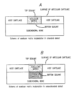
Figure 1A is a schematic representation of an
acellular matrix implant implantation into the cartilage
defect. The scheme shows the lesion implantation site
with acellular matrix implanted therein surrounded by
host cartilage with underlaying undisturbed subchondral
bone. Emplacement of the top and bottom sealants are
CA 02536094 2006-02-16
WO 2005/018491
PCT/US2004/026824
27
also illustrated.
B. Currently Available Procedures for Repair of
Cartilage
A variety of surgical procedures have been developed
and used in attempts to repair damaged cartilage. These
procedures are performed with the intent of allowing bone
marrow cells to infiltrate the defect and promote its
healing. Generally, these procedures are only partly, if
at all, successful. More
often than not, these
procedures result in formation of a fibrous cartilage
tissue (fibrocartilage) which does fill and repair the
cartilage lesion but, because it is qualitatively
different being made of Type I collagen fibers, it is
less durable, less resilient and generally inferior than
the normal articular hyaline cartilage and thus has only
a limited ability to withstand shock and shearing forces
than does healthy hyaline cartilage. Since
all
diarthroid joints, particularly knees joints, are
constantly subjected to relatively large loads and
shearing forces, replacement of the healthy hyaline
cartilage with fibrocartilage does not result in complete
tissue repair and functional recovery.
Among the currently available procedures for repair
of the articular cartilage injuries are the microfracture
technique, the mosaicplasty technique and autologous
chondrocyte implantation (ACI). However, in one way or
another, all these techniques are problematic. The
mosaicplasty technique and ACI, for example, need a
biopsy of cartilage from a non-damaged articular
cartilage area and subsequent cell culture to grow the
number of cells. As a
consequence, these techniques
require at least two separate surgeries. One system, the
Carticel system additionally requires a second surgery
site to harvest portion of and, therefore, disrupt the
tibial periosteum. While the microfracture technique
does not require a biopsy of articular cartilage, the
resulting tissue which develops is always
CA 02536094 2006-02-16
WO 2005/018491
PCT/US2004/026824
28
fibrocartilage.
The method for treatment of injured, traumatized,
diseased or aged cartilage according to the current
invention obviates the above problems as it comprises
treating the injured, traumatized, diseased or aged
cartilage with an acellular matrix implant without need
to remove tissue or cells for culturing, said implant
prepared by methods described below and implanted into
the cartilage lesion during the debriding surgery, as
described below.
C. Osteochondral Area and Osteochondral Defects
Osteochondral area, in this context, means an area
where the bone and cartilage connect to each other and
where the osteochondral defects often develop following
the injury.
Figure IB is a schematic representation of
implantation of an acellular matrix implant in the
osteochondral defect. The scheme shows the cartilage
lesion implantation site with the acellular matrix
implanted therein surrounded by host cartilage with
underlaying bone lesion in the subchondral bone. A bone-
inducing composition or an acellular implant carrier
comprising said composition is deposited into the bone
lesion separated from the cartilage lesion by the bottom
sealant. Emplacement of the top and bottom sealants
illustrates separation of the bone lesion from the
cartilage lesion by the bottom sealant such that each the
cartilage lesion and the bone lesion are treated
separately using different means, namely the acellular
matrix implant for treatment of the cartilage lesion and
the bone-inducing composition or the acellular carrier
comprising said composition for treatment of the bone
defect.
Osteochondral defects are thus defects that are
composites of cartilage and underlying bone. Up-to-date,
commonly used treatments for osteochondral defects are
surgical excisions, mosaicplasty,
osteochondral
CA 02536094 2006-02-16
WO 2005/018491
PCT/US2004/026824
29
autogenous grafting, allogenic grafting, bone cementing,
deposition of metal or ceramic solid composite materials,
porous biomaterials and, lately, a transplantation of
autologous chondrocytes.
Regretfully, none of these
procedures was found to be successful in treating these
defects and safe or comfortable for a patient.
Typically, these procedures involve two or more surgical
procedures and long period, generally at least two to
three weeks, of time to culture the transplantable cells.
For example, mosaicplasty requires removal of circular
pieces of healthy subchondral bone and cartilage to be
used as transplantable plugs at a defect site. One
obvious problem with mosaicplasty is that the surgeon, in
an open surgery, is disrupting healthy tissue in order
to repair the subchondral defect. Clearly, the multiple
surgeries and long period of time between them
necessarily extend a time of recovery to fully functional
joint and often result only in partial functional
restoration as both the bone and cartilage defects are
filled with the fibrocartilage instead of the bone and
hyaline cartilage.
One example of the osteochondral defect which is
common and very difficult to treat is osteochondritis
dissecans. Osteochondritis dissecans is a focal bone-
cartilage lesion characterized by separation of an
osteochondral fragment from the articular surface.
Attempts to treat this injury with allograph transplants
faces the same problem of second surgery and disruption
of the healthy tissue, as described above. Thus it would
be advantageous to have available a method which would
remove a need for second surgery and yet provide a means
for a cartilage and bone repair.
The current method provides a solution to the above-
outlined problems by implanting, during the first
arthroscopic surgery, a bone-inducing composition or a
carrier comprising said composition comprising a bone-
inducing agents into the bone lesion and an acellular
CA 02536094 2006-02-16
WO 2005/018491
PCT/US2004/026824
matrix implant into the cartilage lesion thereby
providing, in one surgery, treatments for both the bone
and cartilage defects.
D. Bone and Bone Defects
5 The restorative method according to this invention
is additionally also suitable for repair of the skeletal
bone lesions.
The skeletal bone lesions are lesions which are
either solely or at least partially located in the
10 skeletal part of the bone, that is the bone placed
immediately below the subchondral bone region, as seen in
Figure 1C.
Figure 1C is a schematic representation of the deep
osteochondro-skeletal bone injury extending into the
15 skeletal bone. The figure shows the positioning of the
host cartilage, subchondral bone and the skeletal bone as
well as emplacement of the acellular matrix implant into
the osteochondral defect and the bone-inducing
composition into the subchondral and skeletal bone
20 defect. The
scheme shows the cartilage lesion
implantation site with the acellular matrix implanted
therein surrounded by host cartilage with underlaying
bone lesion in the subchondral bone. The bone-inducing
composition or a carrier comprising said composition is
25 deposited into the bone lesion. The carrier for this
purpose may be any matrix described above but is
preferably collagenous, hydrogel or a polymer of an
aromatic organic acid or caprolactone containing
structure. Emplacement of the top and bottom sealant are
30 also
shown wherein the bottom sealant separates the bone
portion of the defect from the cartilage lesion such that
each is treated separately using different means.
In an alternative, the hone-inducing composition
and/or the acellular implant carrier comprising such
composition can be used for treatment of simple skeletal
bone defects, lesions or fractures without a need for
cartilage implant.
CA 02536094 2006-02-16
WO 2005/018491
PCT/US2004/026824
31
If and when the method of the invention is used for
treatment of skeletal bone lesions, the bone-inducing
composition alone or incorporated into a carrier,
preferably dissolved in collagen or another binding
agent, is deposited directly into the skeletal bone
lesion. The bone-inducing agent is selected from the
group consisting of calcium phosphate, hydroxyapatite,
organoapatite, titanium oxide, demineralized bone powder,
poly-L-lactic, polyglycolic acid or a copolymer thereof
and a bone morphogenic protein.
A preferred bone-inducing agent is the demineralized
bone powder (DMB). DMB is derived from bone by, for
example, acid extraction of the calcium phosphate.
Following such extraction, the DMB retains, in addition
to the bone collagen other chemical elements found in the
bone, including the naturally present members of TGF-13
superfamily of bone development factors. These factors
may also be extracted by further treatment of bone with
such materials as guanidine hydrochloride. When these
naturally occurring TGF-Ds are present in the DMB, no
further bone-inducing agents are needed to be present
because DMB has a porous microstructure suitable for bone
formation.
It is to be understood that the DMB itself is very
light powder and therefore, it is preferably formulated
in an agent having a binding capabilities. The most
preferred binding agent is collagen or collagen-like
agents, hydrogels, alginates, etc.
II. An Acellular Matrix Implant for Treatment of
Cartilage Lesions
The current invention provides a method for
treatment of injured, damaged, diseased or aged
cartilage. To this end, the method involves implantation
of the acellular matrix implant into
the injured,
damaged, diseased or aged cartilage at a site of injury
or at a site of a defect caused by disease or age, in a
single surgery. The
acellular matrix implant is a
CA 02536094 2006-02-16
WO 2005/018491
PCT/US2004/026824
32
collagenous construct, gel, sol-gel, thermo-reversible
gelation hydrogel, caprolactone polymer or a polymer of
an aromatic organic acid comprising various components as
described below.
A. Preparation of an Acellular Matrix Implant
Preparation of the acellular matrix implant for
implanting into the cartilage lesion involves preparation
of acellular support matrix, typically a collagenous
scaffold or sponge, thermo-reversible gelation hydrogel,
caprolactone polymer or a polymer of an aromatic organic
acid and implanting said matrix into the cartilage defect
in situ.
The acellular matrix implant, such as the one seen
in Figure 2A, is prepared according to the method of the
invention and implanted into artificially generated
lesions in a swine's knee weight bearing region. Figure
2A is an image of an actual acellular matrix sponge
implant used for implantation, here held in the forceps.
The sponge has a size of 5 mm in diameter and 1.5 mm in
thickness and comprises a composition of collagen sponge
and collagen gel having pores of sizes from about 200-400
lam (Figure 2B). When the sponge is implanted into the
lesion, chondrocytes are activated and migrate into the
porous structure of the sponge where they begin to
secrete a new extracellular matrix ultimately replacing
the collagen sponge and gel with the new hyaline
cartilage. The sponge and gel naturally biodegrade and
are metabolically removed from the lesion.
Figure 2B is a cross-side view scheme of a honeycomb
structure of the acellular matrix sponge seen in Figure
2A illustrating a relative positioning of the collagen
sponge, collagen gel and pores within the acellular
matrix sponge.
The matrices of the acellular matrix implant
deposited into the lesion are comprised of biodegradable
materials which permit said implant to function for
certain period of time needed for formation of the
CA 02536094 2006-02-16
WO 2005/018491
PCT/US2004/026824
33
hyaline cartilage. Such
biodegradable materials are
subsequently biodegraded and metabolically removed from
the site of implantation leaving, if any, only non-toxic
residues. These materials were additionally found to
promote formation of the superficial cartilage layer
which covers the lesion containing the implant thereby
protecting a newly formed hyaline cartilage. The
biodegradable materials may additionally include enzymes,
such as metalloproteinases, paracrine or autocrine growth
hormones, GAG-lyases and such like enzymes, soluble
protein mediators and other supplements. Presence or
addition of these materials may enhance activation of
mature, metabolically active but non-dividing
chondrocytes present in the surrounding native host
cartilage and migration of these chondrocytes from the
native host cartilage surrounding the lesion cavity into
said acellular matrix implant emplaced within said
lesion.
The present invention thus concerns a discovery that
when the acellular matrix implant according to the
invention is implanted into a cartilage defect, under
conditions described below, the older inactive
chondrocytes residing within the surrounding native
cartilage are induced to migrate into the defect where
these chondrocytes are activated from static non-dividing
stage to an active stage where they divide, multiply,
promote growth of the extracellular matrix and generate
a new hyaline cartilage in situ.
Following the
implantation of the acellular matrix implant, the
cartilage defect is quickly repaired, particularly in the
young individuals, by chondrocyte migration and by
formation of the extracellular matrix supported by the
metalloproteinases naturally present in sufficient
amounts in tissues of the young individuals. For the
repair of lesions in older subjects, the GAG-lyases and
metalloproteinases, growth factors and other components
are added or incorporated into said matrix before
CA 02536094 2006-02-16
WO 2005/018491
PCT/US2004/026824
34
implantation or they may be conveniently used to coat
said matrix to promote degradation of the injured cell.
A process for activation of chondrocytes was found
to require certain period of time, typically from about
1 hour to about 3 weeks, typically only about 6 hours to
about 3 days. The process for complete replacement of
the implant matrix with the hyaline cartilage typically
takes from one week to several months provided that the
treated individual becomes normally physically active
subjecting said new cartilage to the intermittent
hydrostatic pressure by, for example, walking, running or
biking.
B. Induction of Chondrocyte Migration
Induction of chondrocyte migration from the
surrounding native cartilage involves biological actions
of various agents either naturally present within the
cartilage, cartilage surrounding tissue, blood or plasma
or they are added either before, during or after the
surgery to promote release, activation and migration of
chondrocytes from the native surrounding host cartilage
into the implant.
One of the steps in achieving the activation of the
chondrocytes is the use of sealants at the top and bottom
of the articular cartilage lesion. This step results in
creation of a cavity into which the acellular matrix
implant is deposited. A container-like porous property
of the acellular collagenous matrix implant permits
infusion and concentration of soluble protein mediators,
enzymes, growth or other factors, etc., naturally present
in the host's surrounding healthy cartilage.
Sealing of the top and bottom of the defect before
and after insertion of the acellular matrix implant
results in accumulation of autocrine and paracrine
growth factors that are released by chondrocytes in the
adjacent extracellular matrix, enabling these factors to
induce cell migration into the implant. Suitable growth
factors include, among others, certain transforming
CA 02536094 2006-02-16
WO 2005/018491
PCT/US2004/026824
growth factors, platelet-derived growth factors,
fibroblast growth factors and insulin-like growth factor-
I. Additionally, these and other supplements, such as
the GAG-lyases (matrix remodeling enzymes), may be used
5 to coat the implant before its insertion into the lesion
or the lesion itself may be coated.
The acellular matrix implant sequestered within the
lesion cavity by the top and bottom sealant, however,
remains in flowable communication with the adjacent
10 cartilage. This arrangement creates conditions resulting
in decrease of
levels of inhibitors of the matrix
remodeling enzymes, such as tissue inhibitors of
metalloproteinase-1 (TIMP-1), metalloproteinase-2 (TIMP-
2) and metalloproteinase-3 (TIMP-3), at the defect site.
15 As a consequence, the matrix metalloproteinases (MMP-1,
MMP-2, MMP-3) become accessible to enzymatic activation
and degrade the adjacent extracellular matrix thereby
releasing chondrocytes localized therein resulting in
chondrocytes migration from the surrounding host
20 cartilage into the acellular matrix implant or coat the
walls of the lesion itself with the sugar lyases.
The acellular matrix implant sealed within the
lesion also becomes a repository of exogenous growth
factors that pass through the bottom sealant layer in
25 response to joint loading and hydrostatic pressure to
which the joint is subjected when undergoing a normal
physical activity such as walking, running or biking.
Consequently, in response to the hydrostatic pressure
load, these factors become more concentrated within the
30 defect site and chondrocytes released from adjacent areas
of the surrounding extracellular matrix migrate into the
lesion with ensuing chondrocyte proliferation and
initiation of the de novo extracellular matrix synthesis
within the lesion.
35 Moreover,
the acellular matrix of the implant fills
the defect with a material that has a reduced stiffness
relative to normal articular cartilage and permits
CA 02536094 2006-02-16
WO 2005/018491
PCT/US2004/026824
36
deformation of the adjacent native cartilage matrix edges
thereby increasing level of shear stress further
resulting in increased release of soluble mediators that
indicate matrix remodeling and chondrocyte migration into
the acellular matrix implant.
The presence of the acellular matrix implant sealed
to the adjacent cartilage boundaries thus creates
conditions by which matrix remodeling enzymes, namely
matrix metalloproteinases, aggrecanases and cathepsins,
become concentrated at the defect site and initiate
enzymatic opening of the adjacent extracellular matrix so
that chondrocytes may migrate into the acellular matrix
implant, be deposited within its matrix, begin to divide
and proliferate and secrete the new extracellular matrix,
ultimately leading to formation of normal healthy hyaline
cartilage.
C. Types of Acellular Matrix Implant
The acellular matrix implant provides a structural
support for migration, growth and two or three-
dimensional propagation of chondrocytes in situ.
Generally, the acellular matrix is biologically
biocompatible, biodegradable, hydrophilic and preferably
has a neutral charge.
Typically, the implant is a two or three-dimensional
structural composition, or a composition able to be
converted into such structure, containing a plurality of
pores dividing the space into a fluidically connected
interstitial network. In some embodiments the implant is
a sponge-like structure, honeycomb-like lattice, sol-gel,
gel or thermo-reversible gelation hydrogel.
Typically, the implant is prepared from a
collagenous gel or gel solution containing Type I
collagen, Type II collagen, Type IV collagen, gelatin,
agarose, hyaluronin, cell-contracted collagens containing
proteoglycans, polymers of organic aromatic acids,
glycosaminoglycans or glycoproteins, fibronectins,
laminins, bioactive peptide growth factors, cytokines,
CA 02536094 2006-02-16
WO 2005/018491
PCT/US2004/026824
37
elastins, fibrins, synthetic polymeric fibers made of
poly-acids such as polylactic, polyglycotic or polyamino
acids, polycaprolactones, polypeptide gels, copolymers
thereof and combinations thereof.
Preferably, the
implant matrix is a gel, sol-gel, caprolactone polymer or
a polymer of an aromatic organic acid or a polymeric
thermo-reversible hydrogel (TRGH). Most preferably the
implant matrix contains aqueous Type I collagen.
The acellular matrix implant may be of a type of
sponge, scaffold or honeycomb sponge, scaffold or
honeycomb-like lattice or it may be a gel, sol-gel or
thermo-reversible gel composition or it may be a
caprolactone polymer or a polymer of an aromatic organic
acid.
The acellular matrix implant may be produced as two
or three-dimensional entities having an approximate size
of the lesion into which they are deposited. Their size
and shape is determined by the shape and size of the
defect.
a. Acellular Sponges or Sponge-like Implants
In general, any polymeric material can serve as the
support matrix, provided it is biocompatible with tissue
and possesses the required geometry. Polymers, natural or
synthetic, which can be induced to undergo formation of
fibers or coacervates, can be freeze-dried as aqueous
dispersions to form sponges.
In addition to collagen, a wide range of polymers
may be suitable for the fabrication of sponges, including
agarose, hyaluronic acid, alginic acid, dextrans,
polyHEMA, and poly-vinyl alcohol alone or in combination.
Typically, such sponges must be stabilized by cross-
linking, such as, for example, ionizing radiation.
Practical example includes preparation of freeze-dried
sponges of poly-hydroxyethyl-methacrylate (pHEMA),
optionally containing additional molecules, such as
gelatin, advantageously entrapped within.
Incorporation of agarose, hyaluronic acid, or other bio-
CA 02536094 2006-02-16
WO 2005/018491
PCT/US2004/026824
38
active polymers can be used to modulate cellular
responses. All these types of sponges can function
advantageously as implant matrices for the purposes of
the present invention.
The gel or gel solution used for preparation of the
sponge or sponge-like implant is typically washed with
water and subsequently freeze-dried or lyophilized to
yield a sponge like matrix able to incorporate the
migrating chondrocytes within the matrix. The acellular
matrix implant of the current invention acts like a
porous sponge when infiltrated with the migrating
chondrocytes wherein the cells are distributed within the
sponge pores, providing a mesh-like support permitting
the chondrocytes to migrate and settle there, begin to
divide and proliferate and secrete materials for
generation of new extracellular matrix and eventually for
generation of hyaline cartilage contiguous with the
existing healthy surrounding cartilage.
One important aspect of the sponge implant is the
pore size of the sponge matrix. Sponges having different
pore sizes permit faster or slower infiltration of the
chondrocytes into said sponge, faster or slower growth
and propagation of the cells and, ultimately, the higher
or lower density of the cells in the implant. Such pore
size may be adjusted by varying the pH of the gel
solution, collagen concentration, lyophilization
conditions, etc., during implant fabrication. Typically,
the pore size of the sponge is from about 50 to about 500
um, preferably the pore size is between 100 and 300 um
and most preferably about 200 um.
The pore size of the acellular matrix implant will
be selected depending on the recipient. In the young
recipient where the metalloproteinases are present
naturally and active, the' pore size will be smaller as
the activated chondrocytes will rapidly proliferate
through the pores and secrete extracellular matrix. In
older recipients, the pores will be bigger as the
CA 02536094 2010-02-22
39
migrating chondrocytes will be sluggish and will need
more time to settle in the pores and proliferate.
An exemplary acellular matrix implant made of
collagen is seen in Figure 2. Figure 2A is an example
image of acellular collagenous matrix implant of size 4
mm in diameter and of 1.5 mm in thickness. The seeding
density of this implant is between 300,000-375,000
chondrocytes per 25 Al volume corresponding to about 12-
millions cells/ml. The cell density following the
10 implantation of the acellular matrix implant is, of
course, dependent on the rapidity of the migration of
chondrocytes from the surrounding native cartilage and on
their ability to divide and rapidity of their
multiplication, however, the collagenous matrix of the
15 implant has a capacity to accommodate this range of
migrating cells.
The acellular sponge may be prepared according to
procedures described in Example 1, or by any other
procedure, such as, for example, procedures described in
the U.S. Patent 6,022,744; 5,206,028; 5,656,492;
4,522,753; 6,080,194; 7,468,192; and 7,537,780; or in U.S.
application 2004/0151075.
b. Acellular Scaffold or Honeycomb Implants
One type of the implant of the invention is an
acellular scaffold, honeycomb scaffold, honeycomb sponge
or honeycomb-like lattice. All these implants contain a
honeycomb-like lattice matrix providing a support
structure for migrating and dividing chondrocytes. The
honeycomb-like matrix is similar to that of the sponge
described above but has that typical pattern of the
honeycomb. Such
honeycomb matrix provides a growth
platform for the migrating chondrocytes and permits
three-dimensional propagation of the migrated and divided
chondrocytes thereby providing a structural support for
formation of new hyaline cartilage.
Figure 2B is a side view scheme of honeycomb
CA 02536094 2006-02-16
WO 2005/018491 PCT/US2004/026824
structure of acellular matrix showing a collagen sponge
and collagen gel with pore (*) size of each column of
about 200-400 pm.
The honeycomb-like matrix is fabricated from a
5 polymerous compound, such as collagen, gelatin, Type I
collagen, Type II collagen or any other polymer, as
described above for the sponge, having a desirable
properties. In the preferred embodiment, the honeycomb-
like acellular matrix implant is prepared from a solution
10 comprising Type I collagen.
The pores of the honeycomb-like implant are evenly
distributed within said honeycomb matrix to form a
structure able of taking in and evenly distributing the
migrated chondrocytes.
15 One
preferred type of acellular matrix implant is
Type-I collagen support matrix fabricated into a
honeycomb-lattice, commercially available from Koken
Company, Ltd., Tokyo, Japan, under the trade name
Honeycomb Sponge.
20
Acellular matrix implant of the invention thus may
be any suitable biodegradable structure, gel or solution,
preferably containing collagen. For
the purposes of
convenience in implanting, such implant is typically a
gel, preferably sol-gel transitional solution which
25 changes the state of the solution from liquid sol to
solid gel above room temperature. The most
preferred such solution is the thermo-reversible gelation
hydrogel or a thermo-reversible polymer gel as described
below.
30 c. Sol-Gel Acellular Matrix Implant
Another type of acellular matrix implant is the
implant matrix fabricated from sol-gel materials wherein
said sol-gel materials can be converted from sol to gel
and vice versa by changing temperature. For
these
35
materials the sol-gel transition occurs on the opposite
temperature cycle of agar and gelatin gels. Thus, in
these materials the sol is converted to a solid gel at a
CA 02536094 2006-02-16
WO 2005/018491
PCT/US2004/026824
41
higher temperature.
Sol-gel material is a material which is a viscous
sol at temperatures below 15 C and a solid gel at
temperatures around and above 37 C. Typically, these
materials change their form from sol to gel by transition
at temperatures between about 15 C and 37 C and are in a
transitional state at temperatures between 15 C and 37 .
However, by changing the hydrogel composition, the
transition temperature of the sol-gel may be
predetermined to be higher or lower than those given
above. The most preferred materials are Type I collagen
containing gels and a thermo-reversible gelation hydrogel
(TRGH) which has a rapid gelation point.
In one embodiment, the sol-gel material is
substantially composed of Type I collagen and, in the
form of 99.9% pure pepsin-solubilized bovine dermal
collagen dissolved in 0.012 N HC1, commercially available
under the trade name VITROGEN from Cohesion Corporation,
Palo Alto, CA. One important characteristic of this sol-
gel is its ability to be cured by transition into a solid
gel form wherein said gel cannot be mixed or poured or
otherwise disturbed thereby forming a solid structure
optionally containing other components supporting the
chondrocytes activation and migration. Sterile collagen
for tissue culture may be additionally obtained from
other sources, such as, for example, Collaborative
Biomedical, Bedford, MA, and Gattefosse, SA, St. Priest,
France.
Type I collagen sol-gel is generally suitable and
preferred material for fabrication of an acellular sol-
gel implant.
d. Thermo-Reversible Gelation Hvdrogel Implants
Additionally, the acellular matrix implant may be
prepared from thermo-reversible materials similar to sol-
gel which materials, however, have much faster point of
transition, without hysteresis, from sol to gel and vice
versa.
CA 02536094 2006-02-16
WO 2005/018491
PCT/US2004/026824
42
The thermo-reversible property is important for
implantation of the acellular matrix implant into the
lesion cavity as it may be implanted into the lesion
cavity in its sol state whereby filling said cavity with
the sol wherein the sol forms itself according to the
exact shape of the cavity leaving no empty space or being
too large or too small, as the case may be, for a
prefabricated sponge or a honeycomb lattice. Following
the warming of the sol emplaced within the articular
lesion cavity to the natural body temperature, the sol
instantly transitions and becomes solid gel providing a
structural support for the migrating chondrocytes from
the surrounding native cartilage.
One characteristic of the sol-gel is its ability to
be cured or transitioned from a liquid into a solid form
and vice versa. This property may be advantageously used
for solidifying the liquid or liquefying the solid gel
acellular matrix implant within the cartilage lesion as
well as for delivery, storing or preservation purposes of
said acellular matrix implant.
Additionally, these
properties of sol-gel also permit its use as a support
matrix by changing its sol-gel transition by increasing
or decreasing temperature in the lesion, or exposing the
sol-gel to various chemical or physical conditions or
ultraviolet radiation.
In one embodiment, the acellular matrix implant is
a thermo-reversible gelation hydrogel or gel polymer kept
stored and implanted at temperatures between 5 C and
15 C. At that temperature, the hydrogel is at a liquid
sol stage and permits easy emplacement into the lesion as
the sol. Once the sol is emplaced within the lesion, the
sol is naturally or artificially subjected to higher
temperature of about 30 C and 37 C at which temperature
the liquid sol solidifies into solid gel. The gelling
time is from about several minutes to several hours,
typically about 1 hour. In such an instance, the
solidified gel may itself become and be used as an
CA 02536094 2006-02-16
WO 2005/018491
PCT/US2004/026824
43
implant or this sol may be loaded into a separate support
matrix, such as a sponge or scaffold honeycomb implant.
The primary characteristic of the thermo-reversible
gelation hydrogel (TRGH) is that upon its degradation
within the body it does not leave biologically
deleterious material and that it does not absorb water at
gel temperatures. TRGH
has a very quick sol-gel
transformation which requires no cure time and occurs
simply as a function of temperature without hysteresis.
The sol-gel transition temperature can be set at any
temperature in the range from 5 C to 70 C by the
molecular design of the thermo-reversible gelation
polymer (TRGH), a high molecular weight polymer, of which
less than 5 wt% is enough for hydrogel formation.
The thermo-reversible gelation hydrogel (TRGH),
should be compressively strong and stable at 37 C and
below till about 32 C, that is to about temperature of
the synovial capsule of the joint which is typically
below 37 C, but should easily solubilize below 30-31 C to
be able to be conveniently changed to the sol within the
lesion cavity. The compressive strength of the TRGH
must be able to resist compression by the normal activity
of the joint.
The typical TRGH is generally made of blocks of high
molecular weight polymer comprising numerous hydrophobic
domains cross-linked with hydrophilic polymer blocks.
TRGH has a low osmotic pressure and is very stable as it
is not dissolved in water when the temperature is
maintained above the sol-gel transition temperature.
Hydrophilic polymer blocks in the hydrogel prevent
macroscopic phase separation and separation of water from
hydrogel during gelation. These
properties make it
especially suitable for safe storing and extended shelf-
life.
In this regard, the thermo-reversible hydrogel is an
aqueous solution of thermo-reversible gelation polymer
(TGP) which turns into hydrogel upon heating and
CA 02536094 2010-02-22
44
liquefies upon cooling. TGP is
a block copolymer
composed of temperature responsive polymer (TRP) block,
such as poly(N-isopropylacrylamide) or polypropylene
oxide and of hydrophilic polymer blocks such as
polyethylene oxide.
Thermally reversible hydrogels consisting of co-
polymers of polyethylene oxide and polypropylene oxide
are available, for example, from BASF Wyandotte Chemical
Corporation under the trade name of Pluronics*.
In general, thermo-reversibility is due to the
presence of hydrophobic and hydrophilic groups on the
same polymer chain, such as in the case of collagen and
copolymers of polyethylene oxide and polypropylene oxide.
When the polymer solution is warmed, hydrophobic
interactions cause chain association and gelation; when
the polymer solution is cooled, the hydrophobic
interaction disappears and the polymer chains are dis-
associated, leading to dissolution of the gel. Any
suitably biocompatible polymer, natural or synthetic,
with such characteristics will exhibit the same
reversible gelling behavior.
e) Acellular Gel Implants
The acellular matrix implants of the invention may
alternatively be prepared from various gel materials,
such as suspending gels, not necessarily thermo-
reversible, which are commercially available and may be
suitable for use as acellular matrix implants as long as
they are biodegradable.
One example of such gel is polyethylene glycol (PEG)
and its derivatives, in which one PEG chain contains
vinyl sulfone or acrylate end groups and the other PEG
chain contains free thiol groups which covalently bond to
form thio-ether linkages. If one or both partner PEG
molecules are branched (three- or four-armed), the
coupling results in a gel network. If the molecular
weight of the PEG chains used for the implant preparation
is between 500 and 10,000 Daltons along any linear chain
* Trade-mark
CA 02536094 2006-02-16
WO 2005/018491
PCT/US2004/026824
segment, the network will be open and suitable for
receiving migrating chondrocytes, swellable by
interstitial water, and compatible with living
chondrocytes.
5 The
coupling reaction of PEG can be accomplished,
for example, by preparing 5 to 20% (w/v) solutions of
each PEG separately in aqueous buffers or cell culture
media. Just prior to implantation, thiol, PEG and the
acrylate or vinyl sulf one PEG are mixed and infused into
10 the
lesion. Gelation will begin spontaneously in 1 to 5
minutes. The rate of gelation can be modulated somewhat
by the concentration of PEG reagent and by pH. The rate
of coupling is faster at pH 7.8 than at pH 6.9. Thus, by
modifying the pH of the PEG containing mixture, the
15 gelation process may be controlled to be faster or
slower, as desired by the surgeon. Such gels are,
however, typically not degradable within the body unless
the additional ester or labile linkages are incorporated
into the
20 chain. PEG reagents may be purchased from Shearwater
Polymers, Huntsville, AL, USA; or from SunBio, Korea.
In a second alternative, the gelling material may be
alginate.
Alginate solutions are gellable in the
presence of calcium ions. This reaction has been employed
25 for many years to suspend cells in gels or micro-
capsules. A solution of alginate (1-2%;w/v) in culture
media devoid of calcium or other divalent ions is mixed
in a solution containing calcium chloride which will gel
the alginate. Analogous reactions can be accomplished
30 with other polymers which bear negatively charged
carboxyl groups, such as hyaluronic acid. Viscous
solutions of hyaluronic acid can be gelled by diffusion
of ferric ions.
f. A Polymer of an Aromatic Organic Acid Matrix
35 The
acellular implant may also conveniently be made
of a polymer of an aromatic organic acid. Polymers of
this type have typically a negative charge and are thus
CA 02536094 2010-02-22
=
46
preferred for use as a bone-inducing composition
carriers. However, these type of compounds may also be
used and are suitable for use as cartilage acellular
implants.
g. Absorbable Caprolactone Polymers
The acellular implant may also be conveniently made
of absorbable caprolactone polymers. These polymers are
typically crystalline, low melting, epsilon-caprolactone
polymers as described, for example, in US patents
6,197,320; 5,529,736; 6,485,749; 6,703,035 and 6,413,539.
The caprolactone polymers may be additionally
combined with comonomers, such as glycolide, glycolic
acid or lactones, or linked ionically or covalently to
amine or ester chains.
G. Biodegradable Implant
The acellular matrix implant of the invention is a
temporary structure intended to provide a temporary
supporting for the migrating, dividing, proliferating
=
and extracellular matrix secreting chondrocytes released
from the surrounding cartilage.
Consequently, the implant of the invention must be
fully biodegradable. Whether it is a sponge, honeycomb
lattice, sol-gel or TRGH, in time, the delivered implant
is disintegrated or incorporated into the existing
cartilage and the TRGH is subsequently degraded leaving
no undesirable debris behind.
Overall, any of the acellular matrix implants for
cartilage defects described above is suitable for
implantation into a cartilage lesion of any size and
shape and provides a support for a structural rebuilding
of the cartilage by migrating chondrocytes therein from
the surrounding healthy host cartilage. The implantation
of the implant of the invention results in the generation
of normal healthy hyaline cartilage and in complete
healing of the cartilage defect.
CA 02536094 2006-02-16
WO 2005/018491
PCT/US2004/026824
47
III. Osteochondral Defects and Treatment Thereof
Lesions of the articular cartilage are often
accompanied by lesions of the underlying bone. Such
defects are thus a composite of cartilage and underlying
bone. These
defects are herein cumulatively called
osteochondral defects.
A. Method for Treatment of Osteochondral Defects
The osteochondral defects are caused by injury of
the cartilage and bone. The
cartilage and bone are
histologically two different connective tissues, as
described above. Consequently, it is not possible to
effectively treat both using the same methods and means
and such treatment is thus complex and more difficult
than a treatment of the cartilage lesion or chipped bone
alone.
Furthermore, bone development can influence
cartilage development such that it acts as a barrier to
further cartilage development during its critical
developmental stages.
In one attempt to treat these complex injuries, a
mosaicplasty technique was developed. The mosaicplasty,
as already mentioned above, involves a removal of grafts
from the healthy tissue and plugging such grafts into the
both bone and cartilage lesions. An obvious defect of
this technique is that in order to treat the injured
site, surgeon has to remove, during the open surgical
procedure, a healthy tissue from another site thereby
disrupting the healthy tissue in the process.
When, however, the method of the current invention
is used to treat these complex osteochondral injuries,
it is possible to treat both the bone and cartilage
lesions during the same surgery without need to remove
and disturb the healthy tissue and/or undergo multiple
surgeries required, for example, for allograft
transplantation and other techniques.
The current method permits such dual treatment
simultaneously by implantation of, in combination, an
acellular matrix implant and a bone-inducing composition
CA 02536094 2006-02-16
WO 2005/018491
PCT/US2004/026824
48
or a carrier comprising said composition comprising a
bone-inducing agents further, preferably, in combination
with biologically acceptable sealants.
In practice, during the same surgery, the surgeon
first debrides both lesions and deposits the bone-
inducing composition or a carrier comprising said
composition into the bone lesion and covers said bone
lesions with one or several layers of a biologically
acceptable sealant, preferably a modified highly
polymerizable sealant selected from those described below
in section IV. After the sealant polymerizes, typically
within several minutes, preferably between 0.5 and 10
minutes, most preferably between 3 and 5 minutes, the
acellular matrix implant is deposited into the cartilage
lesion and covered with yet another layer of the sealant,
herein called the top sealant. In this way, the bone-
inducing composition or a carrier comprising said
composition is sequestered within the bone lesion and the
bone forming agents, such as, for example, demineralized
bone powder, calcium phosphates, calcium citrate,
hydroxyapatite, organoapatite, titanium oxide,
polyacrylate, alone or in combination, and a bone
morphogenic protein and/or other known bone-inducing
agents act as inducement for osteoblast migration from
the surrounding bone without interference from the
acellular matrix implant. As a
consequence of this
separation of the bone and cartilage lesion, there is no
invasion of the hyaline cartilage or formation of
fibrocartilage in the bone lesion.
Conversely, when the acellular implant is separated
from the bone-inducing composition or a carrier
comprising said composition, there is no interference
from any of the bone-inducing agents with the chondrocyte
migration, extracellular matrix formation and generation
of the hyaline cartilage. Each the
bone and the
cartilage are treated separately and yet simultaneously
during one arthroscopic surgery.
CA 02536094 2006-02-16
WO 2005/018491
PCT/US2004/026824
49
The sealant may be deposited, preferably, as is,
that is without any additional agents being added, or it
may be added to the bone-inducing composition or a
carrier comprising said composition, if desirable.
The bone-inducing composition or a carrier
comprising said composition deposited within the bone
defect covered with the sealant is left in the lesion in
order to achieve the bone reconstruction and growth.
Both the composition and the sealant are aiding in a bone
natural healing.
The acellular matrix implant implanted within the
cartilage defect separated from the bone lesion by a
layer of the sealant and covered with the top sealant is
left in the cartilage lesion until it biodegrades when
the hyaline cartilage replacement is formed in order to
achieve the chondrocyte migration and formation of
extracellular matrix. A
typical process for repair of
osteochondral defects is the cleaning and debridement the
osteochondral defect, depositing the bone-inducing
composition or a carrier comprising said composition
containing the bone-inducing agents, up to the upper
limit of the lesion in subchondral bone, applying a layer
of the sealant over the composition and letting the
sealant to polymerize. After the sealant polymerizes,
typically in from about 0.5 to about 10 minutes,
preferably about 3-5 minutes, the surgery proceeds with
implanting the acellular matrix implant into the
cartilage lesion, as described above. The cartilage
lesion containing the implant is then covered with yet
another layer of the sealant (top sealant) to seal and
protect the wound from the exterior.
The above described procedure is particularly
suitable for treatment of osteochondral injuries as it
permits dual treatment under different conditions being
implemented during the same surgery.
One specific case of osteochondral defects is
osteochondritis dissecans, where a focal lesion of the
CA 02536094 2006-02-16
WO 2005/018491
PCT/US2004/026824
bone and cartilage results in a loose or totally
dislocated osteochondral fragment. Currently the only
available treatment requires three independent surgeries
including biopsy harvesting of periosteum (first
5 surgery),
culturing cells, removal of the loose fragment
(second surgery), introduction of the cultured cells into
the lesion and bone-grafting (third surgery).
The current method, as described above, or modified
to include a step of the fragment removal, during a
10 single surgery, eliminates a need for two or three
surgeries, as all steps necessary for repair of the
osteochondritis dissecans are performed at the same time
during one surgery.
B. Bone-Inducing Agents
15 Bone-
inducing agents are compounds or proteins
having a definite ability to promote formation of the
bone.
The most suitable bone forming agents are
demineralized bone powder (DMP), calcium phosphate,
20 calcium
citrate, hydroxyapatite, organoapatite, titanium
oxide and growth factors, namely a group of growth
factors known as bone morphogenic proteins, fibroblast
growth factor (FGF), platelet derived growth factor
(PDFG), epithelial growth factor (EGF), glioma derived
25 factor
(GDF) and transforming growth factor beta-1 (TGF-
f3 ) = These
growth factors may be used individually
and/or in combination with each other or with other bone-
inducing factors.
The demineralized bone powder is particularly
30 suitable
to be used as a bone-inducing composition or as
a bone-inducing carrier and no other compounds are needed
to serve as bone inducer or supporting structure and
necessary because the demineralized bone powder mimics
microporous structure of the bone. Before depositing the
35 DMB into
the bone or subchondral bone lesion, the DMB may
be conveniently dissolved in collagen or some other
adhesive fluid or hydrogel which will permit its
CA 02536094 2006-02-16
WO 2005/018491
PCT/US2004/026824
51
deposition into the lesion but itself will have no bone-
inducing function. The used amount of DMB is such that is
makes a concentrated highly viscous paste. The used
amount depends on the structure and grind of the DMB.
Bone morphogenic proteins are typically identified
by the abbreviation BMP and are further distinguished
from each other by numbering, such as BMP-2, BMP-3, EMP-
4, BMP-5, BMP-6, BMP-7, BMP-8 and BMP-14. Some of them
are further identified by a generic name, such as, for
example BMP-3 is called osteogenin, BMP-3B is GDF-10,
etc. The bone morphogenic proteins are administered
generally in concentration (per carrier volume or weight)
of from about 0.01 to about 5 mg/cm', preferably from
about 0.1 to about 1.5 mg/cm' or from about 0.01 mg/g to
about 5 mg/g, preferably from about 0.1 mg to about 2.5
mg/g.
C. Bone-Inducing Composition
The bone-inducing composition or a carrier
. comprising said composition of the invention comprises
one or several bone-inducing agents as listed above, in
concentrations as disclosed. The bone-inducing
composition may be administered as a powder, solution,
gel, sol-gel, TRGH mixed in concentration given above or
incorporated into a structure similar to that of the
acellular implant, pre-prepared and implanted into the
bone lesion or fracture. The composition prepared as
TRGH, for example, is prepared as a sol solution and
administered as such. The sol subsequently changes its
state into the gel filling out the whole bone lesion.
The bone-inducing agents may also be dissolved in PEG,
collagen, alginate, etc., and deposited as such. It
could also be soaked up in a second sponge system like
the acellular matrix sponge described above.
The preferred mode for the deposition of the bone-
inducing agents into the osteochondral or bone lesion is
to dissolve the agent in a gel, such as diluted collagen,
alginate and such like gels.
CA 02536094 2006-02-16
WO 2005/018491 PCT/US2004/026824
52
D. Bone-Inducing Carrier
A bone-inducing carrier or a carrier comprising
bone-inducing composition is a carrier compound which is
suitable for depositing said bone-inducing composition
comprising at least one bone-inducing agent or,
preferably, a combination of several agents into a bone
lesion. Typically, the carrier will be a biodegradable
porous matrix, hydrogel, sponge, honeycomb, or scaffold
having large pores from about 50 to about 150 Am, which
pores encourage migration of osteoblast. The carrier will
also have an interconnecting small pores of about 0.1 to
about 10 um which connect the large pores, permit the
osteoblast to settle within the carrier and provide a
supporting matrix and connecting microstructure for
supply of nutrient and other factors thereby permitting
the bone formation. The surface of such carrier might be
negatively charged encouraging pseudopod attachment of
osteoblasts and their migration into the carrier
resulting in the bone formation.
IV. Biologically Acceptable Sealants
Generally, the implant is implanted into the
cartilage or bone lesion and between at least two layers,
of top and bottom of biologically acceptable adhesive
sealants.
In practice, the first (bottom) layer of the sealant
is introduced into the lesion and deposited at the bottom
of the lesion. The
first sealant's function is to
prevent entry and to block the migration of subchondral
and synovial cells of the extraneous components, such as
blood-borne agents, cell and cell debris, etc. Before
the implant is deposited, such debris could interfere
with the integration of the acellular matrix implant.
The second function of the first sealant is to contain
enzymes, hormones and other components which are
naturally present in the lesion and which are needed for
chondrocyte activation, migration, secretion of other
agents and proliferation of newly formed extracellular
CA 02536094 2006-02-16
WO 2005/018491
PCT/US2004/026824
53
matrix and hyaline cartilage. Then the acellular matrix
implant is implanted over the first sealant. The second
(top) sealant layer is placed over the acellular matrix
implant. The
presence of both these sealants in
combination with the acellular matrix implant results in
successful activation of chondrocytes, their migration
and integration into the implant matrix and ultimately in
new formation of joint hyaline cartilage.
A. A First - Bottom Sealant
In a method for treatment of cartilage lesions, the
first (bottom) sealant forms an interface between the
introduced
implant and the native tissue, such as
subchondral bone or cartilage. The
first sealant,
deposited at the bottom of the lesion, must be able to
contain migrating chondrocytes within the lesion, to
protect the implant from influx of undesirable agents and
to prevent chondrocyte migration into the sub-chondral
space. Additionally, the first sealant prevents the
infiltration of blood vessels and undesirable cells and
cell debris into the implant and it also prevents
formation of the fibrocartilage.
In a method for treatment of osteochondral defects,
the first (bottom) sealant forms a barrier between the
cartilage lesion and the bone lesion. Because these two
defects are in two qualitatively different tissues they
require different treatments. As described above, the
bone lesion is treated with the bone-inducing composition
or a carrier comprising said composition while the
cartilage lesion is treated with the acellular matrix
implant. Moreover, it is not desirable that the enzymes
present in the cartilage lesion activating chondrocyte
migration mix with the bone-inducing agents and growth
factors needed for bone lesion repair. When there is no
separation of one tissue from another, it can easily end
up with, for example, the fibrocartilage ingrowing into
the bone area and, in such an instance, instead of bone
being replaced with the bone, it is replaced with the
CA 02536094 2006-02-16
WO 2005/018491
PCT/US2004/026824
54
inferior fibrocartilage. Consequently, for treatment of
osteochondral defects, the bottom sealant is deposited
over the bone lesion filled with the bone-inducing
composition or a carrier comprising said composition
separating the bone lesion from the cartilage lesion
implanted with the acellular implant. In this way, each
the acellular implant and the bone-inducing composition
can work independently and without interference from the
other.
B. A Second - Top Sealant
The second (top) sealant acts as a protector of the
acellular matrix implant or the lesion cavity on the
surface and is typically deposited over the lesion after
the implant is deposited therein and in this way protects
the integrity of the lesion cavity from any undesirable
effects of the outside environment, such as invading
cells or degradative agents and seals the acellular
matrix implant gel in place after its deposition therein.
The second sealant also acts as a protector of the
acellular implant implanted within a cavity formed
between the two sealants. In
this way, the second
sealant is deposited after the implant is deposited over
the first sealant and seals the implant within the cavity
or it may be deposited over the space holding gel before
the implant deposition.
The third function of the second sealant is as an
initiator or substrate for the formation of a
superficial cartilage layer.
Performed studies described below confirmed that
when the second sealant was deposited over the cartilage
lesion, a growth of the superficial cartilage layer
occurred as an extension of the native superficial
cartilage layer. This superficial cartilage layer was
particularly well-developed when the lesion cavity was
filled with the thermo-reversible gel or sol gel thereby
leading to the conclusion that such gel might provide a
substrate for the formation of such superficial cartilage
CA 02536094 2006-02-16
WO 2005/018491
PCT/US2004/026824
layer.
C. Top and Bottom Sealant Properties
The first bottom or second top sealant used
according to an embodiment of the invention must possess
5 the following characteristics:
Sealant must be biologically acceptable, easy to use
and possess required adhesive and cohesive properties.
The sealant must be biologically compatible with
tissue, be non-toxic, not swell excessively, not be
10
extremely rigid or hard, as this could cause abrasion of
or extrusion of the sealant from the tissue site, must
not interfere with the formation of new cartilage, or
promote the formation of other interfering or undesired
tissue, such as bone or blood vessels and must be
15 bioresorbable and
biodegradable by any acceptable
metabolic pathway, or be incorporated into the newly
formed hyaline cartilage tissue.
The sealant must rapidly gel from a flowable liquid
or paste to a load-bearing gel within 2 to 10 minutes,
20
preferably within 3-5 minutes. However, the sealant must
not gel or polymerize too rapidly as it would cause
problems with its even distribution over the lesion.
Gelling faster than 2 minutes is undesirable. Longer
gelation times than 10 minutes are not compatible with
25
surgical time constraints. Additionally, the overall mode
of use should be relatively simple because complex and
lengthy procedures will not be accepted by surgeons.
Adhesive bonding is required to attach the sealant
formulation to tissue and to seal and support such
30 tissue.
Minimal possessing peel strengths of the
sealant should be at least 3N/m and preferably 10 to 30
N/m. Additionally, the sealant must itself be
sufficiently strong so that it does not break or tear
internally, i.e., it must possess sufficient cohesive
35
strength, measured as tensile strength in the range of
0.2 MPa, but preferably 0.8 to 1.0 MPa. Alternatively,
a lap shear measurement which define the bond strength of
CA 02536094 2006-02-16
WO 2005/018491
PCT/US2004/026824
56
the formulation should have values of at least 0.5 N/cm2
and preferably 1 to 6 N/cm2.
Sealants possessing the required characteristics are
typically polymeric. In the un-cured, or liquid state,
such sealant materials consist of freely flowable polymer
chains which are not cross-linked together, but are neat
liquids or are dissolved in physiologically compatible
aqueous buffers. The polymeric chains also possess side
chains or available groups which can, upon the
appropriate triggering step, react with each other to
couple or cross-link the polymer chains together. If the
polymer chains are branched, i.e., comprising three or
more arms on at least one partner, the coupling reaction
leads to the formation of a network which is infinite in
molecular weight, such as for example, a gel.
The formed gel has cohesive strength dependent on
the number of inter-chain linkages, the length expressed
as molecular weight of the chains between links, the
degree of inclusion of solvent in the gel, the presence
of reinforcing agents, and other factors. Typically,
networks in which the molecular weight of chain segments
between junction points (cross-link bonds) is between
100-500 Daltons are tough, strong, and do not swell
appreciably. Networks in which the chain segments are
between 500-2500 Daltons swell dramatically in aqueous
solvents and become mechanically weak. In some cases the
latter gels can be strengthened by specific reinforcer
molecules; for example, the methylated collagen
reinforces the gels formed from 4-armed PEGs of 10,000
Daltons (2500 Daltons per chain segment).
The gel's adhesive strength permits bonding to
adjacent biological tissue by one or more mechanisms,
including electrostatic, hydrophobic, or covalent
bonding. Adhesion can also occur through mechanical
inter-lock, in which the uncured liquid flows into tissue
irregularities and fissures, then, upon solidification,
the gel is mechanically attached to the tissue surface.
CA 02536094 2006-02-16
WO 2005/018491
PCT/US2004/026824
57
At the time of use, some type of triggering action
is required. For example, it can be the mixing of two
reactive partners, it can be the addition of a reagent to
raise the pH, or it can be the application of heat or
light energy.
Once the sealant is in place, it must be non-toxic
to adjacent tissue, and it must be incorporated into the
tissue and retained permanently, degraded in situ, or be
naturally removed, usually by hydrolytic or enzymatic
degradation. Degradation can occur internally in the
polymer chains, or by degradation of chain linkages,
followed by diffusion and removal of polymer fragments
dissolved in physiological fluids.
Another characteristic of the sealant is the degree
of swelling it undergoes in the tissue environment.
Excessive swelling is undesirable, both because it
creates pressure and stress locally, and because a
swollen sealant gel losses tensile strength, due to the
plasticizing effect of the imbibed solvent which, in this
case, is physiological fluid. Gel swelling is modulated
by the hydrophobicity of the polymer chains. In some
cases it may be desirable to derivatize the base polymer
of the sealant so that it is less hydrophilic. For
example, one function of methylated collagen containing
sealant is presumably to control swelling of the gel. In
another example, the sealant made from penta-erythritol
tetra-thiol and polyethylene glycol diacrylate can be
modified to include polypropylene glycol diacrylate,
which is less hydrophilic than polyethylene glycol. In a
third example, sealants containing gelatin and starch can
also be methylated both on the gelatin and on the starch,
again to decrease hydrophilicity.
D. Suitable Sealants
Sealants suitable for purposes of this invention
include the sealants prepared from gelatin and dialdehyde
starch triggered by mixing aqueous solutions of gelatin
and dialdehyde starch which spontaneously react and gel.
CA 02536094 2006-02-16
WO 2005/018491
PCT/US2004/026824
58
In general, a sealant useful for the purposes of
this application has adhesive, or peel strengths at least
N/m and preferably 100 N/cm; it needs to have tensile
strength in the range of 0.2 MPa to 3 MPa, but preferably
5 0.8 to 1.0 MPa. In so-called "lap shear" bonding tests,
values of 0.5 up to 4-6 N/cm2 are characteristic of strong
biological adhesives.
Such properties can be achieved by a variety of
materials, both natural and synthetic. Examples of
10 suitable sealant include gelatin and di-aldehyde starch
described in PCT WO 97/29715, 4-armed pentaerythritol
tetra-thiol and polyethylene glycol diacrylate described
in PCT WO 00/44808, photo-polymerizable polyethylene
glycol-co-poly(a-hydroxy acid) diacrylate macromers
described in US Pat No. 5,410,016, periodate-
oxidized
gelatin described in US Pat No. 5,618,551, serum albumin
and di-functional polyethylene glycol derivatized with
maleimidyl, succinimidyl, phthalimidyl and related active
groups described in PCT WO 96/03159.
Another acceptable sealant is made from a copolymer
of polyethylene glycol and polylactide, polyglycolide,
polyhydroxybutyrates, polycaprolactone, or polymers of
aromatic organic amino acids and sometimes further
containing acrylate side chains, gelled by light, in the
presence of some activating molecules.
The acceptable sealant made from periodate-oxidized
gelatin remains liquid at acid pH, because free aldehyde
and amino groups on the gelatin cannot react. To trigger
gelation, the oxidized gelatin is mixed with a buffer
that raises the pH to pH at which the solution gels.
Still another sealant made from a 4-armed
pentaerythritol thiol and a polyethylene glycol
diacrylate is formed when these two neat liquids (not
dissolvable in aqueous buffers) are mixed.
Another type of the suitable sealant is 4-armed
polyethylene glycol derivatized with succinimidyl ester
and thiol plus methylated collagen in two-part polymer
CA 02536094 2010-02-22
59
compositions that rapidly form a matrix where at least
one of the compounds is polymeric, such as polyamino
acid, polysaccharide, polyalkylene oxide or polyethylene
glycol and two parts are linked through a covalent bond,
for example a cross-linked PEG with methyl collagen, such
as a cross-linked polyethylene glycol hydrogel with
methyl-collagen, as described in US patents 6,312,725E1
and 6,624,245E2. One
drawback of the type of the bioadhesive described therein
is that it gels and/or bonds extremely fast upon contact
with tissue, particularly with tissue containing
collagen. Consequently, this type of bioadhesive, which
is designed for rapid gelling or bonding during vessel or
tissue injury typically needs to be modified in order to
prolong the gelling and/or bonding time to be suitable
for use as a sealant of the invention.
One group of suitable sealants comprises albumin.
Albumin containing sealants typically comprise at least
human or bovine serum albumin conjugated with a cross-
linking agent. The cross-linking agent may be selected
from the group consisting of glutaraldehyde, amino acids,
polypeptides and proteins.
Further modification may
include conjugation with a fibrous protein, such as
collagen or with a gel compound although this portion of
the sealant is, in the current invention, generally
provided by the support matrix of the invention.
Sealants and bioadhesives or portions thereof which fall
within a category of this type of suitable sealants are
disclosed in U.S. patents 5,583,114 reissued as RE38,158,
6,310,036; 6,217,894 and 6,685,726.
It is worth noting that it is not the presence or
absence of particular protein or polymer chains, such as
gelatin or polyethylene glycol, which necessarily governs
the mechanical strength and degradation pattern of the
sealant. The mechanical strength and degradation pattern
are controlled by the cross-link density of the final
CA 02536094 2006-02-16
WO 2005/018491
PCT/US2004/026824
cured gel, by the types of degradable linkages which are
present, and by the types of modifications and the
presence of reinforcing molecules, which may affect
swelling or internal gel bonding.
5 The first
and second sealant, or the sealant used
for separation of the bone and cartilage lesions, must be
a biologically acceptable, gelling and polymerizable
synthetic compound having adhesive, bonding and/or gluing
properties, and is typically a hydrogel, such as
10
derivatized polyethylene glycol (PEG) which is preferably
cross-linked with a collagen compound, typically
alkylated collagen. The sealant used for separation of
the bone and cartilage lesions should polymerize within
2 and 10 minutes, preferably within 3 and 5 minutes, in
15 order to
permit surgeon to perform the surgery without
premature polymerization but also wihout any delay. For
the purposes of this invention, the sealant should have
a tensile strength of at least 0.3 MPa.
Additionally, the sealant may be two or more polymer
20
compositions that rapidly form a matrix where at least
one of the compounds is polymer, such as, polyamino acid,
polysaccharide, polyalkylene oxide or polyethylene glycol
and two parts are linked through a covalent bond and
cross-linked PEG with collagen. The sealant of the
25 invention
typically gels and polymerizes within about 0.5
to about 5 minutes upon contact with tissue, particularly
with tissue containing collagen.
The second sealant or the sealant used for
separation of the bone and cartilage lesions may or may
30 not be the
same as the first sealant and the first and
second sealants may be utilized as a barrier between the
bone and cartilage lesions but the different sealant may
also be used for this purpose. For
the use in the
current invention, the sealant is slowly polymerized in
35 situ after
its deposition at the bottom of the lesion or
between the bone-inducing composition and acellular
implant. Such slow polymerization is necessary to avoid
CA 02536094 2006-02-16
WO 2005/018491 PCT/US2004/026824
61
uneven distribution of the sealant over the bottom of the
lesion and also to avoid the random and uneven
accumulation of the sealant on some parts of the surface
while leaving other parts of the bottom surface
uncovered. The primary function of the sealant is to
protect the acellular implant from undesirable effects of
migrating cells, tissue debris and various factors
present in the blood or serum, as already discussed
previously. Consequently, its even distribution over the
bottom of the lesion or over the bone-inducing
composition is of great importance and to achieve such
even distribution, the polymerization of the sealant must
not be too slow or too rapid in order to reach the bottom
of the lesion, cover it and then polymerize in situ and
still meet surgeon's time constraints. For arthroscopic
surgery and implantation of the acellular implant, the
sealant polymerization at the bottom of the implant site
needs to occur between 2 and 10 minutes, preferably
between 3 seconds and 5 minutes.
I. Method for Formation
of Superficial Cartilage
Layer Over the Acellular Matrix Implant
An accompanying aspect of this invention is a
finding that when the acellular matrix implant produced
according to procedures described above is implanted into
a cartilage lesion cavity and covered with a
biocompatible adhesive top sealant, the resulting
combination leads to a formation of a superficial
cartilage layer completely overgrowing said lesion.
In practice, the method for formation of the
superficial cartilage layer comprises several steps.
First, the bottom of the lesion is covered with a first,
bottom sealant deposited as polymerizable solution.
Following the sealant polymerization, the acellular
matrix implant is implanted into said lesion and a
second, top sealant is deposited over the implant. In
one embodiment, the implant may be a thermo-reversible
gel which easily changes from sol to gel at the body
CA 02536094 2006-02-16
WO 2005/018491
PCT/US2004/026824
62
temperature thereby permitting an external preparation
and delivery of the implant into the lesion. The gel is
then covered with the top sealant which promotes
formation of the superficial cartilage layer overgrowing
the cartilage lesion thereby sequestering the implant
within the lesion and protecting it from outside
environment.
The superficial cartilage layer begins to form very
quickly after the implant is implanted into the cartilage
lesion and covered with the top sealant layer. As shown
in Figure 6, two weeks after acellular matrix
implantation superficial cartilage layer was observed on
the surface of acellular matrix implanted site. Figure 6
shows arthroscopic evaluation two weeks after the defect
was made in the femoral condyle where the superficial
cartilage layer is clearly visible compared to untreated
empty defect made at the same time, seen in Figure 5.
The top sealant gives support and promotes formation
of the superficial cartilage layer in some instances
further assisted by the gel components of the matrix. At
the time when the implant matrix is completely degraded
and the new hyaline cartilage is formed in the defect,
the superficial cartilage layer completely covers and
insulates the newly formed cartilage similarly to a
synovial membrane naturally present and covering the
joints. The
second top sealant is eventually also
biodegraded and removed from the site, not however, until
the superficial cartilage layer, a synovial-like
membrane, has formed over.
VI. Method for Use of Acellular Matrix Implant
The method for repair and restoration of damaged,
injured, diseased or aged cartilage to a functional
cartilage is based on implantation of an acellular matrix
implant into a cartilage lesion.
The method for use of the acellular matrix implant
in these treatments comprises following steps:
CA 02536094 2006-02-16
WO 2005/018491
PCT/US2004/026824
63
a) Preparing an Acellular Matrix Implant
The first step involves preparation of the acellular
matrix implant for implanting into the cartilage lesion.
Preparation of acellular matrix implants is described in
greater detail in sections II.A.
a) Selecting and Depositing the First and Second
Sealant into the Cartilage Lesion
The second step is optional and involves selection
and depositing bottom and/or top sealant layers into a
cartilage lesion.
Specifically, this step involves deposition of the
first sealant at the bottom of the cartilage lesion and
the second sealant over the acellular matrix implant. The
first and the second sealants can be the same or
different, however, both the first and the second
sealants must have certain definite properties to fulfill
their functions.
The bottom sealant, deposited into the lesion before
the acellular matrix implant is introduced, acts as a
protector of the lesion cavity integrity. It protects
the lesion cavity from contamination by extraneous
substances such as blood and tissue debris. It protects
integrity of the naturally present enzymes and other
mediators needed for and involved in formation of
extracellular matrix and activation of chondrocytes and
their migration from a surrounding host cartilage into
the acellular implant implanted in the lesion. It also
protects the lesion cavity from formation of the
fibrocartilage.
The top sealant deposited over the implant and
effectively sealing the lesion from external environment
acts as a protector of the lesion cavity as well as a
protector of the
implant deposited within a lesion
cavity formed between the two sealants as well as an
initiator of the formation of the superficial cartilage
layer.
CA 02536094 2006-02-16
WO 2005/018491
PCT/US2004/026824
64
c) Implanting the Acellular Matrix Implant
Next step in the method of the invention comprises
implanting said acellular matrix implant into a lesion
cavity formed between two layers of sealants.
The implant is preferably deposited into said lesion
cavity after the bottom sealant is deposited but before
the top sealant is deposited over it or the implant may
be deposited into the lesion cavity without the bottom
sealant being deposited there and then covered with the
top sealant.
d) Generation of the Superficial Cartilage Layer
A deposition of the top sealant over the acellular
matrix implant leads to sealing of the lesion cavity and
overgrowth of said cavity with a superficial cartilage
layer.
Typically, a biologically acceptable top sealant is
deposited over the acellular matrix implant implanted
into the lesion cavity. The second sealant acts as an
initiator for formation of the superficial cartilage
layer which in time completely overgrows the lesion and
strongly resembles a healthy synovial membrane. In
several weeks or months, usually in about two weeks, the
superficial cartilage layer completely covers the lesion,
protects the implant, migrating, dividing and
proliferating chondrocytes and newly secreted
extracellular matrix.
Protecting the implant from
extraneous environment permits integration of the newly
formed cartilage tissue into the native surrounding
cartilage substantially without formation of
fibrocartilage.
Formation of the superficial cartilage layer is thus
a very important aspect of the healing of the cartilage
and its repair and regeneration.
VII. Method for Treatment of Cartilage Lesions
The method for treatment of damaged, injured,
diseased or aged cartilage according to the invention is
suitable for healing of cartilage lesions due to acute
CA 02536094 2006-02-16
WO 2005/018491
PCT/US2004/026824
injury by providing conditions for regeneration of the
healthy hyaline cartilage and for its integration into
the surrounding native cartilage.
The method generally encompasses several novel
5 features, namely, fabrication of a biologically
acceptable biodegradable acellular matrix implant,
selecting and depositing a top and bottom adhesive
sealants to the lesion and the implantation of the
acellular matrix implant within a cavity generated by
10 two sealants, a formation of the superficial cartilage
layer covering the lesion and protecting the integrity of
the acellular matrix implant deposited therein, and
providing conditions for activation, migration, dividing
and proliferation of chondrocytes and for secretion of
15 extracellular matrix ultimately leading to formation of
the new hyaline cartilage and its integration into the
native cartilage.
The method generally comprises steps:
a) fabrication of the acellular matrix implant
20 according to the above described procedures;
b) debridement an articular cartilage lesion in
surgical procedure;
c) during the debridement, preparing the lesion for
implantation of the acellular matrix implant by
25 depositing the bottom sealant at the bottom of the lesion
thereby insulating said cavity from the surrounding
tissue;
d) implanting the acellular matrix implant into said
cavity formed by the polymerized bottom sealant to allow
30 the activated and migrating chondrocytes to proliferate
within said implant;
e) depositing the top sealant over the lesion, and
thereby sealing said implant within the cavity formed
between the two sealant layers;
35 f) optionally introducing enzymes, hormones, growth
factors, proteins, peptides and other mediators into said
sealed cavity by incorporating them into the acellular
CA 02536094 2006-02-16
WO 2005/018491
PCT/US2004/026824
66
matrix, or coating said matrix with them, introducing
them separately or generating conditions for their
transport or transfer through the bottom sealant; and
g) following the surgery, subjecting an individual
undergoing a surgery for repair of said lesion to a
normal physical activity thereby naturally providing an
intermittent hydrostatic pressure which was shown to
promote formation of the healthy hyaline cartilage and
its integration into the surrounding native intact
cartilage.
There are several advantages of the current method.
The main advantage of this method is that the
acellular matrix implant is prepared beforehand and is
implanted during the first and only surgery where the
cleaning and debridement is immediately followed by
implantation of the acellular matrix implant.
Second, the acellular implant avoids immunological
reactions to develop as there is/are no foreign tissue or
cells involved because the implant is wholly synthetic
and acellular.
The method using the acellular matrix implant
permits a three-dimensional expansion of chondrocytes and
extracellular matrix.
The deposition of the top sealant layer resulting in
formation of superficial cartilage layer constitutes a
substitute for synovial membrane and provides the outer
surface of healthy articular cartilage overgrowing,
protecting, containing and providing critical metabolic
factors aiding in protecting the implant and activated
migrating chondrocytes in the lesion. The superficial
cartilage layer also prevent invasion of pannus as seen
in Figures 10A, 10B, 11A and 11B compared with Figures
8A, 8B, 9A and 9B, where the presence of the invading
pannus is clearly visible. In some instances, a selection
of the thermo-reversible gel may be crucial as certain
TRGH may function as a promoter for growth of the
superficial cartilage layer without a need to apply the
CA 02536094 2006-02-16
WO 2005/018491
PCT/US2004/026824
67
top sealant.
Deposition of the bottom sealant layer protects the
integrity of the lesion after cleaning during surgery and
prevents migration of subchondral and synovial cells and
cell products thereby creating milieu for formation of
healthy hyaline cartilage from the activated migrating
chondrocytes into the acellular matrix implant and also
preventing formation of the fibrocartilage.
The method further permits said acellular matrix
implant to be enhanced with hyaluronic acid or other
components or mediators named above, typically added in
about 5 to about 50%, preferably about 20% (v/v), wherein
such hyaluronic acid or such other components act as
enhancers of the matrix-forming characteristics of the
gel and also as a hydration factor in the synovial space
in general and within the lesion cavity in particular.
Further, the method is very versatile and any of the
implant type variations may be advantageously utilized
for treatment of a specific cartilage, osteochondral or
bone injury, damage, aging or disease.
For treatment of the cartilage, a subject is
treated, according to this invention, with a prepared
acellular matrix implant implanted into the lesion, the
implant is left in the lesion covered with the top
sealant for as long as needed. Usually, during the two-
three months following the surgery and implant
implantation the new hyaline cartilage is formed and
integrated into the native surrounding host cartilage.
Typically also, there is no need for any further surgical
or other intervention, as during these two-three months,
at a normal physical activity, such as walking, running
or biking, etc., a sufficient hydrostatic pressure is
applied to the lesion to initiate and promote formation
of the hyaline cartilage fully integrated into the native
cartilage. Such cartilage will then become a fully
functional cartilage covered with a superficial cartilage
layer which eventually grows into or provides the same
CA 02536094 2006-02-16
WO 2005/018491
PCT/US2004/026824
68
type of surface as a synovial membrane of the intact
joint.
Finally, the method also permits replacement of the
age worn-out or diseased osteoarthritic cartilage by the
regenerated hyaline-like cartilage when treated according
to this invention.
The implantation protocol may assume any variation
described above or possible within the realm of this
invention. It is thus intended
that every and all
variations in the treatment protocol, the types of the
implants, use of one or two sealants, implantation
process, selection of added mediators and not the least
the normal physical activity of the individual are within
the scope of the current invention.
VII. Method for Treatment of Bone or Osteochondral
Defects
The method for treatment of osteochondral defects is
typically practiced in conjunction with treatment of
cartilage. The method for treatment of bone defects and
lesions may be practiced in conjunction with
osteochondral defects or separately without steps
involving deposition of the acellular implant into the
cartilage.
VIII. Osteochondral Defects
Due to its anatomical arrangement where the
subchondral bone is localized directly beneath the
injured cartilage and the injury is both the injury to
the cartilage and to the subchondral bone or subchondral
skeletal bone, the method for treatment of osteochondral
defects is an extension of the method for treatment of
cartilage lesions described in section VII, with
exception that during the step c) of that method, the
surgeon, after debridement, deposits into the subchondral
bone lesion a bone-inducing composition or a carrier
comprising said composition typically comprising one or
several bone-inducing agent(s), as described above, then
covers said composition with a layer of the bottom
CA 02536094 2006-02-16
WO 2005/018491
PCT/US2004/026824
69
sealant, and after permitting the sealant or the
composition or both to polymerize, performs the steps a-
g. The nature of this type of defects is such that as a
consequence of the thinness of the subchondral bone layer
there is high probability that the lesion will extend
into the underlying cancellous bone. In such an instance,
the bone-inducing composition or bone acellular implant
is deposited into the skeletal bone and in flowable
continuation into the osteochondral bone which is then
covered with the bottom sealant layer and the acellular
implant is deposited as described above.
IX. Bone Defects
The true bone defects, lesions or fractures are
stand alone injuries in the skeletal bone. These types
of injuries may also be conveniently treated according to
the invention with the bone-inducing composition or with
a carrier comprising such bone-inducing composition.
The carrier, in this setting, corresponds to the
acellular implant utilized for treatment of bone. This
bone acellular implant comprises bone-inducing agents.
The treatment of the skeletal bone injuries
comprises depositing of the bone-inducing composition
into the lesion or fracture during the surgery.
Typically, the bone-inducing composition will be
administered directly into the lesion or fracture as a
powder or a solution, such as an adhesive or
polymerizable solution, or the composition will be
incorporated into a bone-inducing carrier or porous
matrix as described above. The bone lesion may or may
not then be covered with the top sealant or any other
surface to contain the composition within the lesion.
In the preferred embodiment, the demineralized bone
powder is used as a powder or in solution wherein said
powder is dissolved in the collagen, hydrogel or some
other adhesive solution which has no bone forming effect.
The bone-inducing composition is added in amount which
will completely fill the lesion or fracture.
CA 02536094 2006-02-16
WO 2005/018491
PCT/US2004/026824
X. Treatment of Human Osteoarthritic Cartilage
Articular cartilage is a unique tissue with no
vascular, nerve, or lymphatic supply. The
lack of
vascular and lymphatic circulation may be one of the
5
reasons why articular cartilage has such a poor intrinsic
capacity to heal, except for formation of fibrous or
fibrocartilaginous tissue. Unique mechanical functions
of articular cartilage are never reestablished
spontaneously after a significant injury, age wear or
10 disease, such as osteoarthritis (OA).
Currently, the only available treatment of severe
osteoarthritis of the knee is a total knee replacement in
elderly patients. In young and middle aged patients,
however, this is not an optimal treatment.
15
Although the current invention is more practicable
for treatment of injuries in young individuals who
naturally possess sufficient levels of extracellular
matrix building enzymes, growth factors, and other
mediators, the method may be advantageously modified to
20 also provide treatment for older population.
For treatment of elderly patients or for treatment
of larger lesions, the acellular matrix implant is
incorporated, before implantation, with one or more
metalloproteinases, mediators, enzymes and proteins
25 and/or
with drugs stimulating endogenous production of
these factors and mediators. These factors, as described
above, stimulate and promote chondrocytes activation,
migration and extracellular matrix secretion. The method
of the invention thus is also suitable for treatment of
30 the cartilage defects in older generation. It is
expected, however, that such treatment will require
longer period of treatment.
In osteoarthritis, or in age worn out cartilage,
disruption of the structural integrity of the matrix by
35 the degeneration of individual matrix proteins leads to
reduced mechanical properties and impaired function.
Consequently, the current invention reverses this process
CA 02536094 2006-02-16
WO 2005/018491
PCT/US2004/026824
71
by providing a means for rebuilding the diseased
osteoarthritic or worn cartilage with the new healthy
hyaline cartilage.
XI. In vivo Studies in Swine of the Weight-Bearing
Region of the Knee
The method according to the invention was tested and
confirmed in in vivo studies in swine.
The studies, described below, were designed to
evaluate feasibility of the porcine acellular matrix
implant by detecting chondrocyte activation and induction
of chondrocyte migration on the surrounding cartilage,
generation of newly synthesized hyaline cartilage within
the lesion and formation of superficial cartilage layer.
Studies involved the creation of defects in the
weight- bearing region of the femoral medial condyle of
the knee joint, implantation of the acellular matrix into
the defect, depositing bottom and top sealants, detection
of growth of a superficial cartilage layer after two
weeks following the defect creation, detection of
chondrocyte morphology, detection of pannus invasion and
presence of fibrocartilage, detection of presence or
absence of S-GAG secretion, histochemical evaluation of
presence or absence of sealants.
Gross anatomy of the empty defect creation and
acellular matrix implantation at day zero is shown in
Figures 3 and 4. Formation of the healthy hyaline
cartilage and generation of the superficial cartilage
layer in defects treated with the acellular matrix
implant and the fibrocartilage pannus invasion in control
defects at seven month following the defect creation are
seen in Figures 5-12.
Figure 3 shows two empty defects sites A and B at a
time of the defect creation (time zero). Figure 4 shows
two defects created at time zero implanted with the
acellular matrix implants at sites A and B.
Figures 5 and 6 show arthroscopic evaluation two
weeks after defect creation in the control group (Figure
CA 02536094 2006-02-16
WO 2005/018491 PCT/US2004/026824
72
5) and in the experimental group implanted with the
acellular matrix (Figure 6). Histological grading is
seen in Figure 7 and histological evaluation, in two
magnifications, is seen in Figures 8 and 9 for the
control animals and in Figures 10 and 11 for the
experimental group treated with the acellular implant.
Degradation of the sealant from the cartilage lesion is
seen in Figures 12A-12C. One example of full thickness
defect at femoral condyle of mini-pig is seen in Figure
13.
Schematic representation of the femoral articular
surface, defect creation and implant implantation sites
within said defect is shown in Figure 1D. Figure 1D
shows two defects A and B created in the femoral medial
condyle on the medial side of the femoral articular
surface. The defects have sizes of 4 mm in diameter and
1-1.5 mm in depth. The defects are created in the
weight-bearing region.
Table 1 is a tabulation of conditions of a study
design as schematically illustrated in Figure 1D.
Table 1
Study Design
Group Number of Number of Procedure Arthroscopy Necropsy
Number Animals Samples
1 8 16** Implantation 2 weeks
after 7 months after
Experimental of acellular implantation
implantation
biodegradable
matrix*
2 8 16** Empty defect 2 weeks
after 7 months after
Control control defect creation defect
creation
Matrix was secured with tissue adhesive and sutures.
** Each group has two samples at weight-bearing site (site A and B,
Fig. 1D).
Table 1 illustrates the study design for the seven
CA 02536094 2006-02-16
WO 2005/018491
PCT/US2004/026824
73
months study of feasability of the acellular matrix
implant for treatment of cartilage lesions. Study
involved 8 castrated male Yucatan micro-swine, 9-12
months old in each of the two groups. Two
defects (A.
and B) were created at time zero in the knee of each
animal, with a total number of 16 defects. The
experimental group was implanted with acellular matrix
implant at a time of defect creation. In the control
group, the defect was left empty without any treatment
and was used for visual, microscopical, histological and
histochemical comparisons. Arthroscopy was performed at
2 weeks after implantation and defect creation. Necropsy
was performed 7 months after implantation and defect
creation.
The acellular matrix implant was prepared from a
collagen solution VITROGEN (35 AL) obtained from
Cohesion, CA. The collagen gel solution was absorbed
into a collagen honeycomb sponge (5 mm in diameter and
1.5 mm in thickness) obtained from Koken Co., Japan. The
combined collagen gel/sponge constructs seen in Figure 2A
were pre-incubated for 1 hour at 37 C to gel the
collagen, followed by incubation in culture medium with
1% penicillin and streptomycin at 37 C at 5% CO2. After
about 24 hours of polymerization, the biodegradable
scaffolds were transferred to the tissue container with
pre-warmed culture medium (37 C) for the implantation.
Arthrotomy was performed under an inhalation
anesthesia. After opening knee joint capsule, two empty
full-thickness defects (4 mm in diameter and about 1.5 mm
in depth) were created in the femoral articular cartilage
on the weight-bearing site of the medial femoral condyle
of each animal. After creating defects, tissue sealant
was placed on the bottom of the defect. Then, the pre-
prepared acellular biodegradable matrix was placed over
the bottom sealant within the cartilage lesion. The
acellular matrix was secured with absorbable sutures
(usually 4 to 6 sutures) and with two non-absorbable
CA 02536094 2006-02-16
WO 2005/018491
PCT/US2004/026824
74
sutures. The non-absorbable sutures were used as a maker
for arthroscopic evaluation and are visible in Figure 6.
The implanted defect was then sealed with the top
sealant. For
the controls, two empty full-thickness
defects were created and left intact, that is empty,
without implants, or deposition of the bottom or top
sealants.
Figure 3 shows a photograph of the two empty full-
thickness defects A and B (4 mm in diameter and 1-1.5 mm
in depth) created in the articular cartilage on the
weight-bearing site of the medial femoral condyle. The
empty defects were left intact during the whole time of
the study and were used as controls for the experimental
group.
Figure 4 is a photograph of the two full-thickness
defects created in the same way as the empty defects seen
in Figure 3. These two defects were treated, according
to the method of the invention, with the bottom sealant
deposited on the bottom of the lesion. The acellular
matrix implant was implanted into the lesion cavity over
the bottom sealant and the top sealant deposited the over
the implanted acellular matrix implant. The implants were
collagenous sponges (Figure 2A) and had 5 mm in diameter
and 1.5 mm in thickness. Both sites A and B were
implanted. Each implant was secured with four absorbable
sutures and two non-absorbable sutures used as markers
for future arthroscopic evaluation.
Two weeks after defect creation and acellular matrix
implantation, the empty defects and implant sites were
evaluated with arthroscopy. Arthroscopic evaluation after
2 weeks is seen in Figures 5 and 6.
Figure 5 is an arthroscopic microphotograph of an
empty defect 2 weeks after defect creation. Arthroscopic
evaluation showed that in the control group, if left
untreated, the lesion was invaded with synovial pannus
and filled with fibrocartilage. The
arthroscopic
evaluation clearly shows the defect depression indicating
CA 02536094 2006-02-16
WO 2005/018491
PCT/US2004/026824
that the defect is fully exposed and empty although some
synovial invasion have already occurred. Such synovial
invasion is a first step toward formation of
fibrocartilage. Formation of fibrocartilage to replace
5 the hyaline cartilage is undesirable as the
fibrocartilage is qualitatively and functionally inferior
to hyaline cartilage.
Arthroscopic evaluation of implanted sites showed
that already at two weeks time the defects are covered
10 with the superficial cartilage layer. Figure 6 is an
arthroscopic microphotograph of the defect treated with
the acellular matrix implant 2 weeks after the defect was
created. The Figure 6 shows the superficial cartilage
layer overgrowing the implant site forming a smooth flat
15 surface. The borders of the implant site are already
undefined compared to the empty defect which has a
definite and visible border, said implanted site
indicating the beginning of chondrocyte migration into
the implant and secretion of extracellular matrix in
20 confluency with the host cartilage, all this covered with
the superficial cartilage layer. The arthroscopic
evaluation seen in Figure 6 revealed that the lesion
implanted with the acellular matrix is unexposed and
covered with the superficial cartilage layer completely
25 overgrowing the implant sites, seen as a smooth flat
surface when compared to the fully exposed and empty
defects of controls, seen in Figure 5.
At 7 months after creating the defects and
implanting the acellular matrix implants, the animals
30 were euthanized. The implant and defect sites on the
femoral articular condyle were harvested for histological
evaluation. The
tissues were fixed with 4%
formaldehyde/PBS for 7 days at 4 C. The tissues were
decalcified with 10% formic acid, processed, and embedded
35 in paraffin. Thin
sections (5 m) were stained with
Safranin-O (Saf-O) and hematoxylin eosin (H-E) for
histological evaluation.
CA 02536094 2010-02-22
76
The stained sections were evaluated blindly by means
of a histological grading scale seen in Figure 7,
modified from Sellers et al., J. Bone Joint Surg. Am.,
79: 1452-63 (1997).
Only sections from the center of the defect were graded
in order to ensure unbiased analysis and to allow
comparison among specimens studied at different time-
point. The area from the center of the defect was also
chosen because it provided the most stringent test of
healing capacity, since the least amount of cartilage
healing was found consistently in specimens taken from
the middle of the defect.
Histological scoring system used for cartilage
repair evaluation is seen in Table 2.
Table 2
Category
1. Filling of defect
Score Filling of Defect
None (or almost none)
1
2 >50%
3 All (or almost all)
2. Integration of repair tissue with surrounding articular cartilage
Score Integration
0 Gap or lack of continuity on two sides
1 Gap or lack of continuity on one sides
2 Non-continuous gap or lack
3 Normal continuity and integration
3. Matrix staining with Safranin 0-fast green (compared to host
cartilage)
Score Matrix staining
0 None (or almost none)
1 Slight
=
2 Moderate
3 All (or almost all)
4. Cellular morphology
CA 02536094 2006-02-16
WO 2005/018491
PCT/US2004/026824
77
Score Chondrocytes morphology
0 Mostly spindle-shape (fibrous-like) cells
1 < 50% of round cells with morphology of chondrocytes
2 > 50% of round cells with morphology of
chondrocytes
3 Normal (mostly round cells with morphology of
chondrocytes)
5. Architecture within entire defect (not including margins)
Score Architecture within entire defect
0 Clefts or fibrillations
1 < 3 large voids
2 > 3 large voids
3 Normal
6. Architecture of surface
Score Architecture of surface
0 Severe fibrillation or irregularity
1 Moderate fibrillation or irregularity
2 Slight fibrillation or irregularity
3 Normal (or nearly normal)
7. Penetration of tissue to subchondral bone area
Score Penetration
0 Severe penetration
1 Moderate penetration
2 Slight penetration
3 Normal (or nearly normal)
Cumulative results of the histological grading of
the repaired chondral cartilage is seen in Table 3.
Table 3
Histological Grading of the Repaired Cartilage
Category Acellular Matrix Group Empty Defect Group
Filling of defect 3.00 2.60
Integration 2.00 1.40
Matrix staining 2.33 2.10
Chondrocyte morphology 1.78 0.80
Architecture within
entire defect 2.33 0.30
CA 02536094 2006-02-16
WO 2005/018491
PCT/US2004/026824
78
Architecture of surface 2.33 1.90
Tissue penetration into
subchondral bone area 2.11 1.40
Average total score 15.88 10.50
SD+ 1.90 3.60
As seen in Table 3 the average total score for
histological grading at 7 months after the defect
creating and treatment with the acellular matrix implant
was much higher in the implant group, with the score for
all indicators in the implant group being higher then in
the empty defect group.
Histological grading of the repair tissue is shown
in Figure 7, which graphically illustrates results shown
in Table 5. The average total scores on the histological
grading scale were significantly better (p - 0.001) for
the defects treated with acellular matrix implants than
for the untreated defects.
At seven months following the defect creation,
animals were sacrificed, their joints were harvested and
evaluated by Safranin-O staining. Results are seen in
Figures 8-11.
The non-implanted, empty defects A and B at 7 months
after defect creating are shown in Figures 8A, 8B, 9A and
9B.
Figure 8A is a Safranin-O staining microphotograph
(29 x magnification) of the empty, non-implanted defect
(D) at a control site A seven months after defect
creation. In higher magnification (Figure 8B), the defect
clearly shows a fibrous tissue (F) filling the defect
surrounded by the host cartilage (H) with underlying
subchondral bone (SB) area (Figure 8A). None or a very
small amount of S-GAG accumulation, depicted by red
color, was observed at the defect site. S-GAG
accumulation is evidence of the extracellular matrix
formation. If there is a little or none S-GAG present,
there is no extracellular matrix generated, indicating
CA 02536094 2006-02-16
WO 2005/018491
PCT/US2004/026824
79
the absence of migrating chondrocytes and absence of
formation of hyaline cartilage. It also indicates the
presence and formation of fibrocartilage within the
lesion. Figure
83 shows a 72x magnification of the
defect site confirming a presence of fibroblasts, that is
fibrous cells, indicating invasion of a fibrovascular
pannus (F) from synovium. Chondrocyte morphology showed
presence of mostly spindle (fibrous) cells.
Figure 9A is a Safranin-O staining microphotograph
(29 x magnification) of the empty, non-implanted defect
(D) at a site B of the control defect seven months after
defect creation showing a formation of fibrous tissue
filling the defect surrounded by the host cartilage (H)
with underlying subchondral bone (SB) area. Severe
irregularity of the lesion surface was observed. Only
very slight S-GAG accumulation, depicted by red color,
was observed at the defect site. S-GAG accumulation is
evidence of the extracellular matrix formation.
Figure 9B shows a 72x magnification of the defect
site showing a presence of fibroblasts indicating a
fibrovascular pannus (F) invasion from synovium. Cell
morphology observed at this site shows mostly spindle
fibrous cells.
Figures 8A, 8B, 9A and 93 clearly show that non-
implanted control defects without treatment with the
acellular implant of the invention do not indicate a
formation of the healthy hyaline cartilage which would
show as S-GAG accumulation, in Safranin-O stained
microphotograps seen as a red color.
Rather, these
microphotographs show fibrovascular pannus synovial
invasion into the defect with an accumulation of spindly
fibrous cells present in the empty defect sites.
While the no-treatment of the lesion resulted in the
filing of the defect with the fibrocartilage, the
implantation of the acellular matrix implant into the
defect induced chondrocyte activation and migration from
the surrounding native cartilage and resulted in massive
CA 02536094 2006-02-16
WO 2005/018491
PCT/US2004/026824
formation of cartilage extracellular matrix (ECM
accumulation) with minimal fibrovascular pannus in the
implant sites. ECM accumulation was detected by the
strong red color present at the implanted sites of
5 experimental animals. Results are seen in Figures 10A,
10D, 11A and 11B.
Figure 10A is a micrograph of Safranin-O staining
histological evaluation (29x magnification) of the
acellular matrix implant (I) implanted withing the defect
10 site A, seven month after defect creation and
implantation of the acellular matrix implant. Figure 10A
clearly shows inducement of cell migration from the
surrounding native host cartilage (H) into the implant
(I) implanted within the defect site. After seven months
15 following the implantation, hyaline-like cartilage was
observed at the acellular implant site. The presence of
the hyaline cartilage is indicated by the normal S-GAG
accumulation, seen as a predominant red present in the
defect site A. Superficial cartilage layer formed over
20 the lesion is also seen. There was minimal fibrovascular
pannus in the implant sites. Implant is surrounded by
the host cartilage (H) with underlying subchondral bone
area (SB).
Figure 10B shows a higher magnification (72x) of
25 the implant area with red color indicative of S-GAG
accumulation and chondrocyte morphology showing primarily
normal mostly round cells as compared to spindly fibrous
cells observed in the non-treated control defects.
Figure 11A is a Safranin-O staining histological
30 evaluation (29x magnification) of the acellular matrix
implant (I) implanted withing the defect site B, seven
month after implantation. Figure 11A confirms results
seen in Figure 10A. It clearly shows inducement of cell
migration from the surrounding native host cartilage (H)
35 into the implant (I) implanted within the defect site.
At seven months after implantation, hyaline-like
cartilage was observed at the acellular implant site. The
CA 02536094 2006-02-16
WO 2005/018491
PCT/US2004/026824
81
presence of the hyaline cartilage was indicated by the
normal S-GAG accumulation, seen as a predominant red
color present in the defect site B. Superficial cartilage
layer formed over the lesion and traces of non-absorbable
suture are also seen. No fibrovascular pannus synovial
invasion was observed in the implant site. Implant is
surrounded by the host cartilage (H) with underlying
subchondral bone area (SB). The non-absorbable suture
indicates the original border between the host cartilage
and the implant, now almost completely obscured.
Figure 11B shows a higher magnification (72x) of
the implant area with high accumulation of red color
indicative of S-GAG presence. Chondrocyte morphology
again show primarily normal, mostly round cells
confirming results observed at site A.
As seen in Figures 10A and 10B, 11A and 11B, there
was clearly visible integration between the biodegradable
acellular matrix and the host cartilage. Such integration
is not observed in Figures 8A and aA where the defect is
surrounded by the normal hyaline cartilage. These
figures show different cell morphology at the defect
sites than those at the implantation sites seen in
Figures 10A and 10B. Cell morphology of the empty sites
shows the presence of spindly fibrous cells dissimilar to
those cells of the surrounding hyaline cartilage. Cell
morphology at the implanted sites, on the other hand,
show the presence of the normal (round) cells also
observed in the surrounding healthy hyaline cartilage.
The implanted site thus, after seven months does not show
difference between the previously uninjured cartilage and
the one formed within the defect following the
implantation.
Additionally, the use of a top sealant deposited
over the implant implanted at a defect site had resulted
in formation of the superficial cartilage layer and
minimizing synovial tissue invasion at the implant site.
A superficial cartilage layer is formed over the
CA 02536094 2006-02-16
WO 2005/018491
PCT/US2004/026824
82
cartilage lesion after the top sealant is deposited over
the lesion implanted with the acellular implant. As seen
in Figure 6, the presence of the superficial cartilage
layer was already observed in two weeks after the
implantation. The top sealant which causes the
superficial cartilage layer to be formed is biodegradable
and biodegrades within the time. At three months after
the sealant deposition, remaining sealant was still
observed at the surface area along with the superficial
cartilage layer. At seven months after implantation, the
top sealant was completely biodegraded and superficial
cartilage layer was formed in its place, as seen in
Figures 10A and 11A.
In order to determine the sealant (top and bottom)
degradation in vivo, articular cartilage samples
implanted with an autologous chondrocyte construct using
the scaffold matrix were stained with Safranin-O (Figures
12A-12C). Reddish color in Safranin-O stained figures
indicates S-GAG accumulation. Purple color indicates
remaining tissue adhesive with amorphous structure.
Figure 12 thus illustrates a degradation pattern, in
time, of the top and bottom sealants three months after
the acellular matrix implantation. At that time, the
superficial cartilage layer was formed over the implant
and the top sealant was partially degraded. The bottom
sealant was, at three months following its deposition at
the bottom of the lesion, completely degraded and removed
from the lesion site.
Figure 12A shows a surface view of the Safranin-O
stained implantation site with the superficial cartilage
layer clearly visible and the small amount of the top
sealant remaining under the superficial cartilage layer.
Figure 12B shows a side view of the Safranin-O stained
implantation site. Figure 12C shows the bottom view of
the Safranin-O stained implantation site where at time
zero the bottom sealant was deposited.
In this test, the remaining top sealant was observed
CA 02536094 2006-02-16
WO 2005/018491
PCT/US2004/026824
83
only at the surface between the top of the regenerated
hyaline like cartilage region and superficial cartilage
layer (Figure 12A). There was no indication in side view
of any remaining top or bottom sealant between the
interface of the implant site and the surrounding host
cartilage (Figure 12B). There was no remaining bottom
sealant at the bottom of the lesion interfacing with the
subchondral bone region where the bottom sealant was
deposited at time zero (Figure 12C).
These results indicate that the bottom sealant is
completely biodegraded and removed from the lesion site
in about three month after implantation. At that time,
there are still remnants of the top sealant visible on
the surface of the lesion where the sealant protects the
acellular implant from any migration or invasion of
synovium and at the same time supports the formation of
the superficial cartilage layer. With time even these
remnants of the top sealant are biodegraded and removed
from the healed lesion as evidenced by a complete absence
of any top or bottom sealant at the defect site.
A reason why the top sealant is still present at
three months time is that, compared to the surface area,
the side and bottom of the acellular implant site are
more active regions for cell migration which is important
for cell integration and formation of hyaline cartilage.
In these regions, the sealant was completely degraded
within 3 months. This
phenomenon occurred and was
observed in both the cellular and acellular matrix
implantation in vivo. Cellular implant is described in
copending application Serial No. 10/625,245 filed on July
22, 2003.
In order to confirm that the surgical technique used
for creation of the cartilage defects in control and
experimental animals is distinguished from the
microfracture technique which penetrates the subchondral
bone area, an image of full thickness defect at femoral
condyle of mini-pig was created and is shown, with 72x
CA 02536094 2006-02-16
WO 2005/018491
PCT/US2004/026824
84
magnification, in Figure 13. Figure 13 shows a paraffin
embedded and Safranin-0 stained reference tissue of the
created full thickness defect. The defect was created of
non-treated articular cartilage and bone from the femoral
condyle surrounded by the host cartilage and underlying
subchondral bone area. The remaining calcified cartilage
area is seen in the area above the subchondral bone.
This tissue was utilized in all studies as a reference
tissue used for histological evaluation.
The results described above show that implantation
of the biodegradable acellular matrix implant into the
cartilage lesion according to the invention induces
chondrocyte migration from surrounding native cartilage
and formation of an extracellular matrix and leads to
synthesis of a new hyaline cartilage with minimal
synovial invasion of fibrovascular pannus at the implant
sites.
Synthesis of the new hyaline cartilage was measured
by the extracellular matrix accumulation expressed as
accumulation of S-GAG. Also
observed was a cell
integration between the biodegradable acellular implant
and the host cartilage. The use of a bottom and top
sealants and sutures primarily to secure the implant
within the defect suggest that these could have a
secondary effect of minimizing synovial tissue invasion
at the implant site. On the other hand, the results
described above and illustrated by the figures clearly
show that the intact nontreated control defects result in
synovial invasion of the defect with fibrovascular
pannus.
The acellular matrix implant most suitable for
practicing the invention comprises a porous honeycomb
sponge of Type I atelocollagen filled with a
thermoreversing hydrogel of Type I collagen sandwiched
between a bottom layer and a top layer of the sealant.
The type I collagen cell walls of the porous honeycomb
add further strength to the sealing capacity of the
CA 02536094 2006-02-16
WO 2005/018491
PCT/US2004/026824
sealant by adding to the collagen-PEG chemical
interaction analogously to the reaction of metal
reinforcing bar to concrete.
The acellular implant itself is fully biodegradable
5 in time. During that time the following conditions are
observed in sexually mature but not fully epiphysealy-
fused mini-swine. It is observed that in a 2 mm lesion of
the femoral condyle covered with the top sealant, a
superficial cartilage layer extending from the edge of
10 the healthy cartilage region peripheral to the acellular
implant proceeds to overgrow the lesion and the sealant
layer. Additionally, chondrocyte migration into the
acellular implant and production of the new hyaline
cartilage matrix that eventually fills and replaces the
15 implant is observed. This new cartilage matrix is or
closely resembles hyaline cartilage as measured by
sulfated glycoaminoglycan content and histological
appearance. The source of these migrating chondrocytes
are likely to be both the peripheral deeper layers of
20 healthy chondrocytes peripheral to the acellular implant,
and also the overgrown superficial cartilage layer, since
it is shown that this layer is the source of
differentiated chondrocytes capable of producing hyaline
cartilage. Eventually hyaline-like cartilage is found to
25 fill the implant while at the same time the implanted
acellular matrix is gradually biodegraded.
In the current methodological arrangement, the top
and bottom sealants is intended to prevent debris from
subchondral space to enter the implant (bottom sealant)
30 and to sequester the implant within a lesion space (top
sealant). The
acellular matrix implant sequestered
within the lesion permits chondrocytes from the
surrounding healthy cartilage to migrate and enter the
matrix. Naturally applied hydrostatic pressure during a
35 normal physical activity promotes chondrogenesis leading
to a formation of true hyaline cartilage and to a healing
of the lesion.
CA 02536094 2006-02-16
WO 2005/018491
PCT/US2004/026824
86
Results of studies described above confirm that the
damaged, injured, diseased or aged cartilage may be
repaired by using acellular implants prepared according
to the invention and that the acellular matrix implant
of the invention induces cell migration from surrounding
healthy host cartilage and its implantation induces the
inward growth of the superficial cartilage membrane from
the healthy tissue on the periphery. This membrane,
superficial cartilage layer, protects the implant within
the lesion from any synovial invasion. Once the implant
is properly implanted within the lesion, the natural
physicochemical factors, such as intermittent hydrostatic
pressure, low oxygen tension and growth factors induce
the cartilage recovery.
The advantages of the acellular matrix implant
system are multiple. There is no need for biopsy and cell
harvesting, no need to cover periosteum over the lesion,
no damage to healthy tissue, the second and third surgery
is eliminated resulting in faster recovery and
elimination of waiting periods for the next surgery.
Advantages listed above are similarly attached to
treatments of subchondral or bone lesions.
EXAMPLE 1
Preparation of Acellular Collagenous Implants
This example illustrates preparation of the
acellular matrix implant.
300 grams of a 1% aqueous atelocollagen solution
(VITROGENfl, maintained at pH 3.0, is poured into a 10 x
20 cm tray. This
tray is then placed in a 5 liter
container. A 50 ml open container containing 30 ml of a
3% aqueous ammonia solution is then placed next to the
tray, in the 5 liter chamber, containing 300 grams of
said 1% aqueous solution of atelocollagen. The 5 liter
container containing the open trays of atelocollagen and
ammonia is then sealed and left to stand at room
temperature for 12 hours. During this period the ammonia
gas, released from the open container of aqueous ammonia
= CA 02536094 2010-02-22
87
and confined within the sealed 5 liter container, is
reacted with the aqueous atelocollagen resulting in
gelling said aqueous solution of atelocollagen.
The collagenous gel is then washed with water
overnight and, subsequently, freeze-dried to yield a
sponge like matrix. This freeze dried matrix is then cut
into squares, sterilized, and stored under a sterile
wrap.
Alternatively, the support matrix may be prepared as
follows.
A porous collagen matrix, having a thickness of
about 4 mm to 10 mm, is hydrated using a humidity-
controlled chamber, with a relative humidity of 80% at
25 C, for 60 minutes.
The collagen material is
compressed between two Teflon* sheets to a thickness of
less than 0.2 mm. The compressed material is then cross-
linked in a solution of 0.5% formaldehyde, 1% sodium
bicarbonate at pH 8 for 60 minutes. The cross-linked
membrane is then rinsed thoroughly with water, and
freeze-dried for about 48 hours.
The dense collagen
barrier has an inner implantation of densely packed
fibers that are intertwined into a multi-layer structure.
In alternative, the integration layer is prepared
from collagen-based dispersions or solutions that are air
dried into sheet form.
Drying is performed at
temperatures ranging from approximately 4 to 40 C for a
period of time of about 7 to 48 hours.
For histological evaluation, 4% paraformaldehyde-
fixed, paraffin sections were stained with Safranin-O
(Saf-O) and Type II collagen antibody.
For biochemical analysis, seeded sponges were
digested in papain at 60 C for 18 hours and DNA content
was measured using the Hoechst* 33258 dye method.
Sulfated glycosaminoglycan (S-GAG) accumulation was
measured using a modified dimethylmethylene blue (DMB)
microassay.
* Trade-mark
CA 02536094 2010-02-22
88
EXAMPLE 2
Biochemical and Histological Assays
This example describes assays used for biochemical
and histological studies.
For biochemical (DMB) assay, the implant taken from
the animal after certain time following the implantation,
transferred to microcentrifuge tubes and digested in 300
Al of papain (125 jig/ml in 0.1 M sodium phosphate, 5 mM
disodium EDTA, and 5 mM L-cysteine-HC1) for 18 hours at
60 C. S-GAG production in the implant is measured using
a modified dimethylene blue (DMB) microassay with shark
chondroitin sulfate as a control according toFarndale et al.,
Connective Tissue Research, 9: 247-248 (1982).
DNA content is determined by Hoechst 33258 dye
method according to Kim et al., Anal. Biochem., 174: 168-176
(1988).
For histological assay, the remaining implants from
each group were fixed in 4% paraformaldehyde. The
implants were processed and embedded in paraffin. 10 Am
sections were cut on a microtome and stained with
Safranin-O (Saf 0).
For immunohistochemistry, the samples are contacted
with diaminobenzidine (DAB). The DAB is a color substrate
showing brown color when the reaction is positive.
EXAMPLE 3
Evaluation of Integration of Acellular Matrix Implant
in a Swine Model
This example describe the procedure and results of
study performed for evaluation of integration of porcine
in a swine model.
An open arthrotomy of the right knee joint was
performed on all animals, and a biopsy of the cartilage
was obtained.
A defect was created in the medial femoral condyle
of the pig's right knee. This defect (control) was not
implanted with an acellular matrix implant but was left
intact. Following surgery, the joint was immobilized
with an external fixation implant for a period of about
CA 02536094 2006-02-16
WO 2005/018491
PCT/US2004/026824
89
two weeks. Two weeks after the arthrotomy on the right
knee was performed, an open arthrotomy was performed on
the left knee and defects were created in this medial
femoral condyle. The
acellular matrix implant was
implanted within the defect(s) in this knee which was
similarly immobilized. The
operated sites were
subsequently viewed via arthroscopy two weeks after
implantation or defect creation and thereafter at monthly
intervals.
Animals were euthanized and the joints harvested and
prepared for histological examination approximately 7
months after acellular matrix implant implantation. The
implanted sites were prepared and examined histological.