Note: Descriptions are shown in the official language in which they were submitted.
CA 02538038 2006-03-07
WO 2005/025413
PCT/US2004/029462
Medical Device for Analyte Monitoring
and Drug Delivery
BACKGROUND OF THE INVENTION
1. Field of the Invention
This invention relates to the fields of diagnosis and drug delivery. More
particularly it relates to medical devices and methods capable of monitoring
levels of
a bodily fluid analyte and optionally releasing of appropriate therapeutic
agents.
2. Background
"Point of care" devices that are capable of detecting biological
macromolecular activity or drug concentration levels are in high demand
because they
eliminate the need for patient lab visits, thus providing savings in both time
and
expense. One of the most valuable aspects of modern microarray technology is
the
ability to detect biological macromolecular dysfunction, malformation or
mutation
resulting in disease. However, this capability has not been fully exploited
because
such arrays have not been incorporated into ingestible, implantable or
wearable point
of care devices. Modern microarray technology is limited to characterization
of
biological macromolecules and their metabolites by analysis of immobilized
analytes
stabilized on slides to be inserted into a machine or analyzed manually
outside of
living organisms.
Because whole blood contains cells, platelets, a myriad of proteins and other
macromolecules, assays involving blood typically require pre-processing of the
sample to remove these components. Integrating pre-processing steps into a
point of
care device drives up the cost of the device itself, thus making use of the
device
financially unviable. For example, some devices currently on the market using
whole
blood in their assays; among them are Boeluinger Mannheim's ReflotronTM system
for measuring blood borne analytes I-most notably cholesterol) and the iStatTM
(iStat
CA 02538038 2013-02-01
Inc.), which performs a number of critical care assays, including
electrolytes, general
chemistries, blood gases and hematology. 'The Reflotronlm relies on dry
chemistry
technology in which enzymes or other reactive elements are immobilized on the
'
surface of a test strip. The assay is a calorimetric activity assay in which
the reaction
$ produces a color change and is thus indicative of the amount of analyte
present The
iStatim relies on electrochemical detection to produce a sigaal. In either
case, a blood
sample is taken separately (typically by a finger prick) and then placed on
the chip (or
cartridge in the case of the iStat), where the reaction occurs and is analyzed
by an
external detection unit. These existing monitoring systems are insufficient
and
inconvenient as they usually require the user to prick themselves and multiple
steps to
obtain a result As such, there is a need for a wearable device that can
repeatedly,
automatically and accurately monitor bodily fluids stieh as blood.
Point of Care devices are also usefulin certain situations when systemic
biological samples such as blood, urine or stool, cannot provide adequate
information
as to subtle molecular changes at the situs of disease. In such a case, even
if the
clinician could pinpoint the exact situs of an ailment, obtaining a biological
sample
for analysis comes only at great risk, pain and expense for the patient
Additionally, a
paint of care device would be desirable where the systemic administration of
drug
agents, such as by tnmsdennal or intavenous means, treats the body as a whole
even
though the disease to be treated may be localized. Here, systemic
administration may
not be desirable because the drug agents often have unwanted effects on parts
of the
body that arc not intended In be treated, or because treatment of the diseased
part of
the body requires a high concentration of drug agent that may not be
achievable by
systemic administration. For example, when administered to a patient
systemically,
some drugs (e.g., chemotherapeutic drugs such as those used to treat cancer
and other
proliferative disorders) may cause undesirable side effeets. It is therefore
often
desirable to detect disease and administer drug agents at a localized sites
within the
body.
As such there is a demand for point of core devices capable of detecting
biological macromolecular activity or drug concentration levels that may also
administer a specific therapeutic agent at a localized site within the body in
response
to changes in biological macromolecular activity or drug concentration levels.
2
CA 02538038 2013-02-01
=
SUMMARY OP THE INVENTION
One aspect of the invention relates to a medical device comprising a
microarray which comprises a bioactive agent cap le of interacting with a
disease
marker biological analyte; a reservoir which comprises at leaat one
therapeutic agent
and is capable of releasing the therapeutic agent(s) from the medical devise;
and a
plurality of microchips comprising a microarray scanning device capable of
obtaining
physical parameter data of an interaotion between the disease marker
biological
' analyte with the bioactive agent; a biometric recognition device capable
of comparing
the physical parameter data with an analyte interaction profile; a therapeutic
agent
releasing device capable of controlling release of the therapeutic agent from
the
reservoirs; an interface device capable of f).cilitating communications
between the
microarray scanning device, biometric recognition device and the therapeutic
agent
releasing device; and an energy source to power the medical device.
In one embodiment of this aspect of the invention the device is coated and the
coating is a biostable polymer which may have channels. In another embodiment
of
this aspect of the invention, the Polymer is porous.
In a different embodiment, bodily fluids are transported through microfluidic
lanes which move molecules by means of pressure diffininces over the
mionearray.
In one embodiment, an osmotic pump is used to propel the fluids through the
top
portion of the device. In another embodiment fluid transport is powered by
natural
electric currents.% the body conducted through Personal Area Network
technology.
.In yet another embodiment of this aspect of the invention, the microarray
comprises mi.embeads. In another embodiment, the bioaotive agent is a nucleic
add.
In yet another embodiment, the bioactive agent is a polypeptide. In yet
another
embodiment, the bioactive agent is an inummoglobulin.
In an additional embodiment of the medical devices of the invention, the
bioactive agent is fluorescent) labeled. In another embodiment, the bioactive
agent
is fluotescently labeled with i nanoorystal.
3
CA 02538038 2006-03-07
WO 2005/025413
PCT/US2004/029462
In yet another embodiment, the disease marker biological analyte is a nucleic
acid. In a further embodiment, the disease marker biological analyte is a
polypeptide.
In another embodiment, the disease marker biological analyte is an
immunoglobulin.
= In yet a further embodiment, the plurality of microchips comprise silicon
germanium.
hi another embodiment, the microarray scanning device comprises fiber optic
elements.
In an additional embodiment, the analyte interaction profile is stored in the
biometric recognition device. In an altematiye. embodiment, the analyte
interaction
profile is stored externally from the medical device.
In another embodiment, the medical device has a plurality of reservoirs.
In an additional embodiment, the interface device comprises a personal area
network.
In an additional embodiment, the energy source is a battery. In an alternate
embodiment, the energy source is provided by a personal area network.
Another aspect of the invention relates to a method of detecting and treating
a
disease in a patient comprising administering to the patient a coated medical
device
comprising a microarray comprising a bioactive agent capable of interacting
with a
disease marker biological analyte; at least one reservoir comprising at least
one
therapeutic agent and capable of releasing the at least one therapeutic agent
from the
medical device; a plurality of microchips comprising a microarray scanning
device
capable of obtaining physical parameter data of an interaction between the
disease
marker biological analyte with the bioactive agent; a biometric recognition
device
capable of comparing the physical parameter data with an analyte interaction
profile;
a therapeutic agent releasing device capable of controlling release of the
therapeutic
agent from the reservoir; and an interface device capable of facilitating
communications between the microarray scanning device, the biometric
recognition
device and the therapeutic agent releasing device; an energy source to power
the
medical deyice; and biocompatible coating enabling the medical device to be
swallowed, pass through the patient's intestinal tract and be naturally
excreted.
=
In one embodiment of the method the coating is a biostable polymer which
may have channels. In another embodiment, the polymer is porous.
In yet another embodiment of the method, the microarray comprises
microbeads. In another embodiment, the bioactive agent is a nucleic acid. In
yet
4
CA 02538038 2006-03-07
WO 2005/025413
PCT/US2004/029462
another embodiment, the bioactive agent is a polypeptide. In yet another
embodiment, the bioactive agent is an immunoglobulin.
In an additional embodiment of the method of the invention, the bioactive
agent is fluorescently labeled. In another embodiment, the bioactive agent is
a
fluorescently labeled with a nanocrystal.
In yet another embodiment of the method, the disease marker biological
analyte is a nucleic acid. In a further embodiment, the disease marker
biological
analyte is a polypeptide. In another embodiment, the disease marker biological
analyte is an innnunoglobulin.
In yet a further embodiment of the method, the plurality of microchips
Comprise silicon germanium.
In another embodiment of the method, the microarray scanning device
. comprises fiber optic elements.
In an additional embodiment of the method, the analyte interaction profile is
stored in the biometric recognition device. In an alternative embodiment, the
analyte
interaction profile is stored externally from the medical device.
In another embodiment of the method utilizes a plurality of reservoirs.
In an additional embodiment of the method, the interface device comprises a
personal
area network.
In an additional embodiment of the method, the energy source is a battery. In
an alternate embodiment, the energy source is provided by a personal area
network.
hi an additional embodiment of the method, the communications are
monitored by an external computer. In another embodiment, the external
computer
directs release of the therapeutic agent.
Another aspect of the invention relates to a medical device capable of
detecting an analyte in a bodily fluid comprising at least one microneedle
capable of
obtaining a sample of a bodily fluid, a first microchannel through which the
sample
flows and is in fluid communication with the at least one microneedle, a
second
microchannel in fluid communication with the first microchannel, through which
a
buffer flows, wherein the second channel comprises a microarray with a
bioactive
agent, a microarray scanning device to detect an interaction between the
bioactive
agent and the analyte in the bodily fluid; and an interface device capable of
facilitating communications between said microarray scanning device and a
biometric
recognition device.
5
CA 02538038 2006-03-07
WO 2005/025413
PCT/US2004/029462
In one embodiment, the bodily fluid is blood. In another embodiment, the at
least one microneedle is a plurality of microneedles. In yet another
embodiment the
microneedle is between about 10 and about 200 microns in diameter. In a
further
embodiment, the microneedle is capable of drawing about 100 microliters of
blood.
In another embodiment, the first microchannel is about 100 micrometers in
diameter.
In an additional embodiment, the second microchannel is about 100 micrometers
in
diameter.
In still a further embodiment, the analyte in the bodily fluid flowing through
the first microchannel diffuses into the second microchannel and interacts
with the
bioactive agent. In another embodiment, the analyte in the bodily fluid
flowing
through the first microchannel diffuses into the second microchannel and
competitively displaces labeled analyte from binding the bioactive agent. In a
further
embodiment, the labeled analyte is provided in a predetermined amount. In
another
embodiment, the labeled analyte is labeled with a fluorescent moiety. In yet
another
embodiment, the microarray is a portion of the second microchannel having a
coating
of an antibody specifically binding the analyte in the bodily fluid. In a
further
embodiment, the microarray scanning device comprises a total internal
reflection
fluorescence (TIRF) spectrometer.
In another embodiment of this aspect of the invention the biometric
recognition device is located outside of the device and the communication is
through
wireless transmission. In another embodiment, the analyte is insulin and the
bioactive
agent is an antibody specific for insulin. In yet a further embodiment, the
analyte is
glucose and the bioactive agent is an antibody specific for glucose. In still
another
embodiment, the device is a worn on the skin as a patch.
In a further embodiment of this aspect of the invention, the analyte is
indicative of disease.
In another embodiment of this aspect of the invention, the medical device
further comprises a reservoir having a therapeutic agent therein and a
therapeutic
agent releasing device, capable of controlling release of a therapeutic agent
from a
reservoir in response to an instruction from the biometric recognition device.
In
another embodiment, the analyte is glucose and the therapeutic agent is
insulin. In a
further embodiment, the analyte and the therapeutic agent are the same.
In another embodiment of this aspect of the invention, the medical device has
¨ at least one disposable assay device which comprises the at least one
microneedle, the
6
CA 02538038 2006-03-07
WO 2005/025413
PCT/US2004/029462
first microchannel and the second channel and has a non-disposable assay
reader
device compriseing the microarray scanning device the interface device. In a
further
embodiment, the assay device and assay reader device are in optical
communication
with one another. In yet a further embodiment there are a plurality of
disposable
assay devices fitted in a single assay reader device.
In another embodiment, the microarray comprises an uncladded portion of a
single glass optical fiber functionalized with the bioactive agent whererin
the
uncladded portion of single glass optical fiber is in fluid contact with the
second
microchannel. Alternatively, the microarray may comprise a plurality a
uncladded
portions of single glass optical fibers functionalized with the bioactive
agent whererin
the uncladded portions of single glass optical fibers are in fluid contact
with the
second microchannel.
Additional advantages of the present invention will become readily apparent to
those skilled in this art from the following detailed description, wherein
only the
preferred embodiment of the invention is shown and described, simply by way of
illustration of the best mode contemplated of carrying out the invention. As
will be
realized, the invention is capable of other and different embodiments, and its
several
details are capable of modifications in various obvious respects, all without
departing
from the invention. The present invention may be practiced without some or all
of
these specific details. In other instances, well known process operations have
not
been described in detail, in order not to unnecessarily obscure the present
invention.
Accordingly, the drawings and description are to be regarded as illustrative
in nature,
and not as restrictive.
BRIEF DESCRIPTION OF THE DRAWINGS
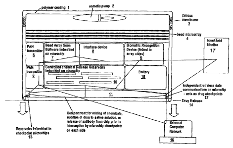
FIG. 1 is schematic drawing of an exemplary medical device of the invention.
The
device has a biostable polymer coating 1 as well as an osmotic pump in this
preferred
embodiment 2 to facilitate fluid movement through the device's porous coating
3.
The device comprises a microarray 4 comprising a bioactive agent capable of
interacting with a disease marker biological analyte; a reservoir 10
comprising a
therapeutic agent and capable of releasing therapeutic agent from the medical
device;
and a plurality of microchips 5,7, 8, 9, 6,10, 12, 13 & 14 comprising; a
microarray
scanning device 7 capable of obtaining physical parameter data of an
interaction
7
CA 02538038 2013-02-01
between the disease marker biological analyte with the bioactive agent(s); a
biometric
recognition device 9 capable of comparing the physical parameter data with an
analyte bateraction profile; a therapeutic agent releasing device 10 capable
of
controlling release of therapeutic agent(s) from a plurality of reservoirs and
checkpoints 13 & 14; and an interface device 8 capable of facilitating
communications between themiomarray scanning device 7, biornetric recognition
device 9 and the therapeutic agent releasing device 10; and an energy source
to power
the medical device 15. Additionally, the exemplary device contains
transmitters for a
personal area network 5 &6 and transmission pathways for communication between
the PAN and a hand-held computer monitor 17 or external computer network 16.
Additionally, the exemplary device contAieR a compartment 11 for the Mixing of
therapeutic agents prior to release.
FIG. 2. illustrates the inventive device in its external patch embodiment. It
is worn
on the skin and may be capable of releasing a therapeutic agent. Additionally,
it is
'capable of interfacing with an external network.
FIG. 3. illustrates a plurality of medical devices, here in the form of
patches, in
wireless communication with an external server. The ealetual server may
contain a
biometric recognition device and pharmacokinetic database of physical
parameters of
the interaction between a bioactive agent and an analyte.
FIG, 4. (a) 100 micrometer diameter microneedle is roughly the diameter of
human
hair. (b) An array of silicon microneedles.
FIG. 5. (a) Illustrates various views of the inventive device in its patch
embodiment
100. The exemplary patch is 2 cm in length and 0.5 cm in width. It is also has
a
thickness of about 1,5 mm. The patch contains a plurality of micro-needles 12
(h)
illustrates the internal fhanzes of the patch. device. The device has a
reservoir 13 into
which a blood is pumped from the microneedles 12, a second reservoir
containing a
buffer 14 and common microchannel for laminar flow 15 which is the confluence
of a
buffer 15a and a blood inlet 15b, as well 38 a receptacle for waste 16.
Additionally,
the figure shows that the device may be separable in two components: A
disposable
layer having microneedles, rnicrochannels and a mietoarray 100a and a non
8
CA 02538038 2006-03-07
WO 2005/025413
PCT/US2004/029462
disposable portion 100b in optical communication with the disposable portion
having
the microarray scanning device and other electronics.
FIG. 6. (a) Illustrates how the patch 100 may be packaged prior to application
to a
patient. The patch may be covered with a protective layer 17 and have a patch
base
18 through with the microneedles will penetrate upon application. The base 18
provides the added benefit of maintaining sterility of the microneedles prior
to
application. An adhesive 19 serves to fasten the patch to the skin of the
subject.
Additionally, a protective cover 20 is provided which is removed to expose the
adhesive layer 19.
FIG. 7. Illustrates how a plurality of patches 100 may simultaneously be
applied to a
patient. Such a plurality of patches may then be sequentially activated to
provide
analyte detection of an extended period of time.
FIG 8. (a) Side view of an exemplary laminar flow microchannel 15 in which
blood is
fed into one inlet 15b of a two inlet microchannel. The blood contains cells
21, a
variety of proteins 25, and the analytes to be measured 22. The fluids flow in
parallel
streams with molecules passing across the interface only by diffusion. As
shown in
(b), only the small molecule analytes 22 reach the opposite wall where an
equilibrium
exchange takes place with fluorescently labeled analyte molecules 24 pre-bound
to
bioactive agents 23 on the surface. In this example, the channel wall coated
with
bioactive agents 23 constitutes the microarray.
FIG. 9. Shows the concept of an evanescent field arising during total internal
relflection. The evanescent field extends no more than one wavelength beyond
the
medium in which the light beam is traveling.
FIG. 10. Illustrates how an optical fiber 26 utilizes total internal
reflection
fluorescence to detect changes in fluorescence indicative of an interaction
between a
bioactive agent and an analyte that occur at the microarray. The optical fiber
may
have multiple configurations. For example, it may run parallel along the
length of the
laminar flow channel 15. Alternatively, a plurality fibers may terminate in
the
channel and themselves be coated with bioactive agent. The first 15a and
second 15b
9
CA 02538038 2013-02-01
micrechannels are in fluid communication with one another. Only small
molecules
will diffuse across the diffusional interface to the mioroarray i.e.
functionalized sensor
surface. Fluorescent detection by a IMF spectrometer does not extend beyond
one
wavelength beyond the surface.
FIG. 11. Illustrates an optical fiber 26 that is part of an microarray. The
optical fiber
has a cladded 31 and an uncladded portion 27. The distal uncladded portion 27
is
Emotionalized with a bioactive agent that interacts with a target analyte in
the bodily
fluid being assayed. The proximal end of the fiber 26 is in optical
communication
with a portion of the microarray scanning device. This contact is facilitated
by a
connector 28. Beyond the connector an input 29 directs light to fiber splitter
31a which
directs light returning to. tbrongh the fiber to a detector such as a
photodiode detector
30. As discussed elsewhere, the functionalized imcladded portion of the fiber
27 may
constitute a. portion of the wall of the laminar flow micro-channel 15 or a
plurality of
fibers may protrude into the channel 15.
FIG, 12, Illustrates an exemplary portion of a microarray and microarray
scanning
device utilizing a MP sensor. Incoming laser light from a laser 33 is directed
through a multimode fiber 26 and the output leg of a 50:50 fiber optic
splitter 31b onto
the functionalized unload fiber 27. In the case alone assay the fluorophore-
labeled
analyte displaced from the bioactive agent by a competitive binding process
resulting
from the presence of analyte in the bodily fluid, and as a result the photonic
energy
coupled into the fiber at the evanescent wave is reduced. This reduction in
light
intensity is deWoted by the photo diode and associated amplifier. Emitted
fluorescence
characteristic of the interaction between an analyte 22 and a bioactive agent
23
couples back into the fiber and propagates towards the detector 30 with little
interference from the laser light. A laser coupled to a fiber provides light
at 660 ma.
In one example, the system works with either a 200 pm core fimotionalized Baer
and
splitter or a 62.5 pm core functionalized fiber and splitter. The fiber core
diameter is
the same for the entire system. In either a 62.5 or 200 pan core system,
higher order
modes of the fiber (the edges of the core) are excited to both maximize the
evanescent
wave energy and make the 1x2 coupler perform more uniformly. This is different
based on the fiber core diameter.
CA 02538038 2006-03-07
WO 2005/025413
PCT/US2004/029462
FIG. 13. Illustrates the fluorescence and absorbance of the Atto 655
fluorophore.
FIG. 14. An image of a model assay reader device worn on the human arm.
FIG. 15. Is an image of a two the convergence in a microchannel of a stream of
PBS
flowing at 0.1 aul/s and a stream of blood at 0.02 Al/min. Visually, there is
little
mixing between the streams at the diffusional interface. However, molecules
with
higher diffusional coefficients will traverse the diffusional interface.
FIG. 16. Is an image of the diffusional coefficients of cells, bovine serum
albumin
and vancornycin.
FIG. 17. Is an illustration of an exemplary device of the invention. A) The
figure
shows that the device may be separable in two components: A disposable layer
having microneedles, microcharmels and a microarray 100a and a non-disposable
portion 100b in optical communication with the disposable portion having the
microarray scanning device and other electronics. B) The disposable portion
100a of
the patch contains a reservoir 13 into which a blood is pumped from the
microneedles,
a second reservoir containing a buffer 14 and common microchannel for laminar
flow
15 which is the confluence of a buffer 15a and a blood inlet 15b, as well as a
receptacle for waste 16. Additionally, the uncladed portion of a fiber optic
comprising the microarray is shown 26. C) shows several disposable and non-
disposable portions together.
DETAILED DESCRIPTION OF THE INVENTION
In its most basic form, the invention relates to a medical device which acts
as a
sensor to qualitatively and/or quantitatively detect analytes in bodily
fluids. Such
analytes may potentially be indicative of disease or be drugs or drug
metabolites.
Additionally, the device may be capable of releasing therapeutic agent(s) in
response
to sensory inputs. As such, it may further provide continuous diagnosis and
medication. The inventive devices may be implantable, ingestible or worn on
the skin
as a patch.
11
CA 02538038 2006-03-07
WO 2005/025413
PCT/US2004/029462
The devices are capable of sampling analytes in biological fluids. Biological
fluids include but are not limited to blood, serum, urine, gastric and
digestive juices,
tears, saliva, stool, semen, and interstitial fluids derived from tumorous
tissues.
Bodily fluid drawn into the medical device is brought into contact with a
microarray which samples biological analytes in bodily fluids. Fluid may be
released
from the medical device and can contain therapeutic agerit(s) released in
response to
the presence or absence of a particular analyte. Most preferably, bodily fluid
movement into or out of the medical device is facilitated by a pump, such as a
microfluidic or osmotic pump. In another embodiment, molecular transport is
conducted through pressurized microfluidic lanes which cause fluids to flow
over a
microarray. In yet another embodiment molecules are transported by natural
electric
currents conducted by Personal Area Network (PAN) transmitters or
piezoelectric or
magnetic sensors.
With respect to implantable embodiments, the device may be sealed to the tip
of a catheter endoscope for realtime analysis and modeling of drug
concentrations
inside the body. For example the devices may associated with a vascular,
gastric or
biliary stent, for example. In another embodiment, the device is sealed to the
inside of
the stent. In another embodiment the devices are packaged in a polymer system
which allows it to be implanted into the body, lenses which could be placed in
the
back of the eye, external sensors of gases and air pollution, and other
objects in which
real time monitoring is called for.
In one embodiment, the device is in the form of a patch. FIG. 2. Preferably,
the device is an adhesive patch that is applied externally to the skin to be
used as a
monitor of whole blood analytes. More preferably, blood analytes are drugs
whose
levels are monitored by the patch. Such drugs have narrow therapeutic ranges
and are
present in micromolar concentrations in the blood. Most preferably, the
concentration
and/or identity of target analyte molecules in the blood is measured directly
on the
patch and such information can then be transmitted to internal or external
data storage
systems.
It is envisaged that the patch draws blood through the skin using at least
one, if
not a plurality, of microneedles. FIG. 4. Preferably, the microneedles are
about the
size of a human hair and have an integrated microreservoir or cuvefte. The
microneedle painlessly penetrates the skin and draws a tiny blood sample. More
preferably, the microneedles collect about 0.01 to about 1 microliter,
preferably, 0.05
12
CA 02538038 2013-02-01
to about 0.5 microliters and most preferably about 0.1-0.3 n:zioroliters of
capillary
blood and deliver them to a reservoir in the patch. Preferably, the
microneedles are
constructed out of silicon and are about 10 to about 200, preferably about 50
to 150
and most preferably 100 microns in diameter, making their application to the
skin
virtually painless. As the patch may most likely be placed on an area of the
body less
well perused than a fingertip, for example, capillary density is likely to be
fairly low,
In order to ensure that a capillary is actually struck by the needles, a
plurality will be
used for blood collection, as shown in FIG. 4. Preferably such mioroneedles
are of
the type marketed by Pelikan (Palo Alto, CA) and/or Kumetrix (Union City, CA)
see
also U.S. Patent No. 6,503,231.
In one embodiment envisages using polymer needles, some of which are
coated in porous gels and polymers which enable separation of targeted
molecules
based on size and or specificity. Gels include but are not limited to
polychlorimeride
and porous polycarbonate elastomers.
In general, microfabrication processes that may be used in making the
microneedles disclosed herein include lithography; etching teclmiques, such as
wet
chemical, dry, and photoresist removal; thermal oxidation of silicon;
electroplating
and electroless plating diffusion processes, such as boron, phosphorus,
arsenic, and
antimony diffusion.; ion implantation; film deposition, such as evaporation
(filament,
electron beam, flash, and shadowing and step coverage), sputtering, chemical
vapor
deposition ((VD), epitaxy (vapor phase, liquid phase, and molecular beam),
electroplating, screen printing, and lamination. See generally Saeger,
Introduction to
Microelectronic Fabrication (A.ddison-Wesley Publishing Co., Reading Mass.
1988);
Runyan, et at, Semiconductor Integrated Circuit Processing Technology (Addison-
Wesley Publishing Co., Reading Mass., 1990); Proceedings of the EE Micro Blear
Mechanical Systems Conference 1987-1998; Rai-Choudhury, ed., Handbook of
Ncrolitbography. Micromachining gYa Miontabrkation (SPIE Optical engineering
Press, Bellingham, Wash. 1999). Alternatively, needles can be molded in
silicon
= wafers and then plated using conventional wire cutting techniques with
nickel, gold,
titanium or various other biocompatible metals. In another embodiment, needles
can
be fashioned from biopolymers. Microneedles may be fabricated and employed for
the claimed devices according to the methods of Mukeijoe et al., Sensors and
Actuators A: Physical, Volume 114, Issues 2-3, 3. September 2004, Pages 267-
275.
13
CA 02538038 2006-03-07
WO 2005/025413
PCT/US2004/029462
It is also preferable that although the device is capable of taking multiple
measurements, a micro-needle is only to be used once. Preferably, multiple
blood
draws are carried out by a mechanical actuator that inserts and withdraws the
needle
and also disposes the used needle and reloads a new needle. The mechanical
technologies developed and manufactured in very high volumes for very small
disk
drives (e.g. IBM micro drive) have a similar set of motion and low cost
requirements.
Preferably, a micro actuator is a MEMS (micro machined electromechanical
system)
device fabricated using semiconductor-like batch processes. Such actuators
include
nickel titanium alloy, neumatic, or piezo electric devices. The smallest
needles are
about 1-10, preferably about 2-6 and most preferably about 4 microns in
thickness but
over about 10-100, preferably about 30-60, and most preferably about 40
microns in
height.
Alternatively, the needles are actuated by a spring-solenoid system in which a
pin triggers the release of a miniaturized spring coiled tightly enough to
generate
sufficient force and range of motion necessary for actuation.
In one embodiment, the inventive patch device has two separable components:
a disposable component having a plurality of microneedles, microcharmels and a
microarray (assay device); as well as a non-disposable component having a
microarray scanning device and the ability to transmit results of an analyte
interaction
with a bioactive agent on a microarray to a biorecognition device, preferably
by
wireless communications, e.g., by Bluetoothol) (assay reader device)(see FIG.
5). In
this embodiment, a used disposable component may be removed from the non-
disposable component while the non-disposable portion remains in place on the
subject's body. A fresh disposable component having fresh needles may then be
applied to the non-disposable portion already in place on a patient's body.
The fresh
disposable component may be capable to quantitatively or qualitatively
detecting the
same or a different analyte as the previously used disposable component. FIG.
7. In
this embodiment it is preferable to apply fresh disposable components once the
micro-
needles of the used disposable component become clogged with blood clots, for
example. The non-disposable component may also contain one or more disposable
components. In this set up, each of the disposable components is capable
simultaneously detecting a different analyte. Alternatively, the disposable
components each detect the same analyte yet are sequentially actuated in such
a
manner as to sample bodily fluid, e.g. blood, in discrete periods of time. In
this set
14
CA 02538038 2006-03-07
WO 2005/025413
PCT/US2004/029462
up, the device detects analyte over an extended period of time by deploying
one
disposable component after the other over a period of time. Preferably, the
device has
12 disposable components and can detect an analyte over a 24 hour period by
deploying a new disposable component every 2 hours.
In swallowable or implantable embodiments, it is preferable to coat the device
with a "biostable polymer," which refers to those materials that do not
undergo
significant degradation upon prolonged exposure (e.g., up to one week, six
months, a
year, or longer) to bodily fluids, tissues, and the like and thus enables the
device to
pass through the entirety of the intestinal tract. It is preferred that fluid
is drawn into
and released from the medical device either through pores or channels in the
polymer.
FIG. 1.
The biostable coating materials of certain embodiments of this aspect of the
invention are porous polymer materials that are characterized by
interconnected pores
of sufficient size to allow for the flow of bodily fluids into the medical
device and the
release therefrom, of therapeutic agents. The porous polymer materials are
preferably
characterized by an average pore diameter of at least about 5 microns, more
preferably at least about 8 microns, and more preferably at least about 10
microns.
Suitable polymers for use in embodiments wherein a porous structure is
obtained by
freeze-drying include any suitable biostable polymer, such as polyurethanes
(including polyurethane dispersions), ethylene vinylacetate polymers,
hydrogels such
as crosslinked gelatin, dextran, polycarboxylic acids, cellulosic polymers,
gelatin,
polyvinylpyrrolidone, maleic anhydride polymers, acrylic latex dispersions,
polyamides, polyvinyl alcohols, polyethylene oxides, glycosaminoglycans,
polysaccharides, polyesters, polyacrylamides, polyethers, and blends and
copolymers
thereof.
The term "analyte" as used herein refers to antibodies, serum proteins,
cholesterol, polysaccharides, nulceic acids, drugs and drug metabolites, etc.,
found in
bodily fluids and tissues of the body. In another embodiment, the analyte is
any
biological analyte, marker, gene, protein, metabolite, or hormone or
combination
therein indicative of a biological state desirable for analysis to determine a
physical
state or condition. It is the purpose of the inventive device to qualitatively
and/or
quantitatively "detect" analytes in the bodily fluids. Preferably, such
detection occurs
periodically. Most preferably, it occurs in real time. In one embodiment, the
analytes
are present in micromolar to nanomolar concentrations and are highly potent
CA 02538038 2006-03-07
WO 2005/025413
PCT/US2004/029462
chemotherapeutics, such as aminoglycocides or antibiotics, e.g., vancomycin,
for
which minute to minute monitoring is highly desirable because the analytes
have
narrow therapeutic ranges.
Through continuous monitoring of analyte levels in the body, the inventive
devices allow the investigator to optimize therapeutic and dosage regimens and
quickly develop pharmacokinetic models for experimental drugs. Target
validation,
lead optimization, and compound optimization (therapeutic range and toxicity
studies)
can now be done in a much faster and more accurate manner because monitoring
trough concentrations enables rapid target elimination or validation of dosing
schemes
in addition to development of target leads. Thus, the inventive devices are
useful in
reducing the uncertainty as to whether to enter Phase II and III clinical
trials thereby
decreasing the time to registration and the overall costs of drug development.
Moreover, the inventive devices provide a way of sensing drug concentrations
of
novel compounds in a fluorescent based assay, which remains the gold standard
of
sensitivity, and for the first time provides a targeted fluorescence based
solution for
monitoring of novel compounds.
The term "disease marker" as referred to herein is a detectable analyte, e.g.,
antibodies, serum proteins, cholesterol, polysaccharides, nulceic acids, drugs
and drug
metabolites, etc., found in bodily fluids and tissues which is present or
absent in the
body and known to be correlated with disease. Analytes, which allow for the
detection of certain physiological conditions, can also be indicative of
normal healthy
physiology. These are referred to herein as "normal" or "healthy" biological
analytes.
Preferably, the biorecognition device of the invention detects a disease
marker based
on physical parameter data discerning between the physical characteristics of
an
interaction between 1) a disease marker biological analyte and a bioactive
agent on
the microarray and 2) a normal biological analyte with a bioactive agent on
the
microarray. Disease marker biological analytes allow for the detection of
certain
physiological conditions, e.g., infection, inflammation, autoimmune disease,
cancer,
etc. Disease markers presently known to those of skill and disease markers
that will
be known in the future are encompassed by this invention. The presence of a
disease
marker indicates the presence of disease and warrants the release of a
therapeutic
agent.
The disease marker biological analytes may be genes or their products which
are over-expressed or over-active in cells undergoing unwanted proliferation.
For
16
CA 02538038 2006-03-07
WO 2005/025413
PCT/US2004/029462
example, the inventive device may be implanted into a tumor or a tissue
suspected of
containing a tumor such as a cavity or space left behind following a biopsy
procedure.
If the invention detects increased concentrations of such biological analytes
or
mutated over-active forms of such analytes, e.g., disease markers, a release
of
therapeutic agent(s) such as a cytotoxic agent is warranted. These disease
marker
biological analytes can be indicative of unwanted cellular proliferation such
as cancer,
neointimal proliferation resulting in arterial stenosis, psoriasis, etc.
Disease marker
biological analytes may be detected by analyzing gene expression in tissues
and
matching it to known tumor-gene expression patterns or comparing them to known
normal expression patterns. In a preferred embodiment, the microarrays are
used to
detect the presence of a disease marker biological analyte as defined by the
presence,
absence or over-abundance of a particular nucleotide sequence, including a
single
nucleotide polymorphism (SNP), mRNA or a particular protein, such as an
enzyme,
an antibody or an antigen.
In one embodiment, the disease marker biological analytes are tumor specific
antigens. For example, such antigens are expressed on the surface of or
released from
cancer cells, for example the tumor specific antigen MUC-1. Detection of MUC-1
expression through nucleic acid detection or by protein activity, can trigger
the release
of cytotoxic agents as therapeutic agents.
Another example relates to receptor tyrosine kinases (RTKs), which are
important in the transduction of mitogenic signals. RTKs are large membrane
spanning proteins which possess an extracellular ligand binding domain for
growth
factors such as epidermal growth factor (EGF), an intracellular portion which
functions as a kinase to phosphorylate tyrosine amino acid residues on cytosol
proteins thereby mediating cell proliferation. Various classes of receptor
tyrosine
kinases are known based on families of growth factors which bind to different
receptor tyrosine kinases. Class I kinases such as the EGF-R family of
receptor
tyrosine kinases include the EGF, HER2-neu, erbB, Xmrk, DER and 1et23
receptors.
These receptors are frequently present in common human cancers such as breast
cancer, squamous cell cancer of the lung, bladder cancer, oesophageal cancer,
gastrointestinal cancer such as colon, rectal or stomach cancer, leukaemia and
ovarian, bronchial or pancreatic cancer. As further human tumor tissues are
tested for
the EGF family of receptor tyrosine kinases it is expected that its widespread
prevalence will be established in other cancers such as thyroid and uterine
cancer.
17
CA 02538038 2006-03-07
WO 2005/025413
PCT/US2004/029462
Specifically, EGFR tyrosine lcinase activity is rarely detected in normal
cells whereas
it is more frequently detectable in malignant cells. It has been more recently
shown
that EGFR is overexpressed in many human cancers such as brain, lung squamous
cell, bladder, gastric, breast, head and neck, oesophageal, gynaecological and
thyroid
tumours. Receptor tyrosine kinases are also important in other cell-
proliferation
diseases such as psoriasis. EGFR disorders are those characterized by EGFR
expression by cells normally not expressing EGFR, or increased EGFR activation
leading to unwanted cell proliferation, and/or the existence of inappropriate
EGFR
levels. The EGFR is known to be activated by its ligand EGF as well as
transforming
growth factor-alpha (TGF-a). The Her2-neu protein is also a member of the
class I
receptor tyrosine kinase (RTK) family. Her2-neu protein is structurally
related to
EGFR. These receptors share a common molecular architecture and contain two
cysteine-rich regions within their cytoplasmic domains and structurally
related
enzymatic regions within their cytoplasmic domains. Accordingly, detection of
abnormally high levels of RTK expression or signaling activity through nucleic
acid
detection or by protein activity can constitute a disease marker and can
warrant the
release of RTK inhibitors or cytotoxic agents as therapeutic agents.
The relatively high expression of genes that directly or indirectly inhibit
chemotherapeutics constitute a disease marker for purposes of the invention.
For
example, high tumor expression of the DNA repair gene ERCC1 warrants release
of
genotoxic chemotherapeutic agents to a high local yet low systemic
concentration.
Thus, achieving concentrations that would not be safely sustained
systemically.
Additionally, high tumor levels of the gene DPD are known to inhibit 5-FU
based
chemotherapeutic regimen. Similarly, high tumor expression of the DPD warrants
release of 5-FU chemotherapeutic agents to a high local yet low systemic
concentration. Alternatively, the skilled artisan would also realize that high
levels of
ERCC1 or DPD may be indicative of chemotherapeutic resistance and that the use
of
genotoxic agents or 5-FU, respectively, may not be appropriate. In such a
case,
cytotoxic therapeutic agents other than genotoxic agents or 5-FU should be
released
from the device, respectively.
Alternatively, the device can be set up as to detect a panel of disease
markers
indicative of a disease such as cancer and release high local concentrations
of
Cytotoxic agents such as a therapeutic agent.
18
CA 02538038 2006-03-07
WO 2005/025413
PCT/US2004/029462
In a further embodiment, disease marker biological analytes can be indicative
of inflammation, which plays a crucial role in the etiology of inflammatory
bowel
disease, multiple sclerosis, childhood-onset diabetes, psoriasis, rheumatoid
arthritis,
etc. Such diseases previously required regular large systemic doses of
potentially
harmful steroids to address only localized inflammation. High localized
concentrations of biological analytes such as TNF'-alpha, IL-1, IL-8, IL-2, IL-
3, MIF
(IL-4), GM-CSF, INF-gamma, and TNF-beta are indicative of inflammation. The
detection of abnormally high concentration of such biological analytes
constitutes a
disease marker and warrants localized release of anti-inflammatory drugs or
antibodies as therapeutic agents.
In another embodiment, disease marker biological analytes can be indicative
of infection by a microorganism. As such, disease markers can include viral or
bacterial proteins or nucleic acids or fragments thereof. For example,
detection of
biological analytes such as bacterial toxins including exotoxins and
enterotoxins as
well as TSST-1, or other bacterial superantigen, or botulinum toxin,
diphtheria toxin,
anthrax protective antigen, anthrax edema factor, and anthrax lethal factor,
etc., as
well as viral proteins such as influenza hemagglutinin or neuraminidase, would
constitute a disease marker indicative of infection and warrant localized
release of
anti-microbial drugs or toxin-specific antibodies as therapeutic agents.
Another aspect of the invention relates to a microarray. The microarray is the
portion of the inventive devices that facilitates an interaction between an
analyte and a
bioactive agent. It its most basic embodiment, a "microarray" as defined
herein may
constitute any surface e.g. the wall of a microfluidic channel, covered or
fimctionalized by a bioactive agent such that a microarray scanning device can
detect
interactions between a bioactive agent and an analyte. FIGs. 8, 10, 11. In
another
embodiment, the microarray is a collection of miniaturized test sites arranged
on a
surface that permits many tests, or assays, to be performed in parallel. In
this context,
the microarray is directly exposed to bodily fluids and/or tissues and may be
able to
simultaneously process a plurality of different assays and provide for the
interaction
of one or more bioactive agents with one or more biological analytes.
For example, the ability of a fluorescence-based array biosensor to measure
and quantify the binding of an antigen to an immobilized antibody has been
demonstrated using the four different immunoassay formats: direct,
competitive,
displacement, and sandwich. Sapsford et al., Anal Chem. 2002 Mar 1;74(5):1061-
8
19
CA 02538038 2013-02-01
used a patterned array of antibodies specific for 2,4,6-trinitrotoluene (TNT)
immobilized onto the surface of a planar waveguide and measured signals
from different antigen concentrations simultaneously. For direct, competitive,
and displacement assays, which are one-step assays, measurements were
obtained in real time. Dose-response curves were calculated for all four assay
formats, demonstrating the array biosensor's ability to quantify the amount of
antigen
present in solution.
ht one embodiment of this aspect of the invention, the microarray is an area
on
a glass optical fiber that is funotionalized with a bioactive agent. FIG. 11.
In another
embodiment, the microarray can have a plmality of glass optical fibers each
functionalized with the same or different bioactive agents. In one particular
embodiment the bioactive agent of the microarray is a protein such as an
antibody
specific for an analyte. Two exemplary procedures may be employed for
attaching
protein bioactive agents to the glass optical fibers. The Srst is based on
that
13 developed by Bhatia etal. 1998, Analytical Biochemistry, 178 40843. This
involves
functionalizirig a surface with 3-mercaptopropyltrimethoxysilane. Pollowing
that, a
cross-linker of N-7-malemidobutylryloxyauccimide ester is used to attach the
protein
bioactive agent to the fimotionalized surface. The second procedure involves
using a
Dextan-based method described by Tedeschi et al. 2003, Biosensors and
Bioelectronics, 19 85-93. This method uses glycidyl 3-(trimetlioxysilyl)propyl
ether
to link ftte.free hydroxyl groups on clean glass to the Dextran polymer.
Protein
bioactly agents are bound to the Dextrcm matrix following acidification of
the
carboxylic acid groups therein. Optionally, the fiber may be coated with a
strip
membrane which separates targeted analytes.
Preferably, the fiber is directly inserted into the microneedle and. the walls
of
the microneedles ate coated with polymer gels for selectivity and specificity
based
binding events.
ln embodiments utilizing glass optical fibers, a light source is utilized to
excite
fluorescently labeled bioactive agents and/or aaalytes such that fluorescence
is
detectably altered upon interaction with target warns in bodily fluids. FIG.
11. A
light source for excitation may be a laser module, Light may be launched into
the
optical fiber that ()ordains a functionalized region, i.e. a region stripped
of fiber
cladding and chemically prepared for hioactive agent coating. Fies. 9,11. Due
to the
lack of cladding, an evanescent wave emanates from the fiber at point and
incites
CA 02538038 2013-02-01
fluorescence from flyorescent tagged bioactive agents or fluorescent tagged
analytes
bound to bioactive agents meant to be competitively displaced analytes in the
bodily
fluid being sampled. FIGs. 8, 11. Emitted light reenters the through the same
fiber.
Light returning into the fiber is detected by the microarray scanning device
which
may have a fiber optic splitter, bandpass filters capable of removing ambient
background light, and a photodiode detector, A schematic of the described
setup can
be seen in Figure 11.
Preferably, the bioactive agent is an antibody that is capable of specifically
binding an analyte drug. Alternatively, the bioactive agent is an antigen that
is
capable of specifically binding serum antibodies. In this latter embodiment,
the
inventive devices can detect the production of specific types of antibodies
produced in
response to certain immunological stimuli, for example HIV or tuberculosis
infection.
In another embodiment, the microarray facilitates interaction between 1) a
disease marker biological analyte and a bioactive agent on the microarray and
2) a
normal biological analyte with a bioactivc agent on the microarray. In this
context the
bioactive agent differentially interacts with normal biological analyte and a
disease
marker biological analyte,
in another embodiment of the microarray, inicrobead arrays are used. By
"microspber. es" or "beads" or "particles" or grammatical equivalents herein
is meant
small discrete particles. The composition of the beads will vary, depending
ort the
class of bioactive agent and the method of synthesis. Suitable bead
compositions
include those used in peptide, nucleic acid and organic moiety synthesis,
including,
but not limited to, plastics, ceramics, glass, polystyrene, methylstyrene,
acrylic
polymers, paramagnetic materials, thoria sol, carbon graphitcd, titanium
dioxide, latex
or cross-linked dextrans such as Sepharose, cellulose, nylon, cross-linked
micelles
and teflon may all be used. "Microsphere Detection Guide" from Bangs
Laboratories,
Fishers hid. Is a helpful guide. The beads need not be spherical; irregular
particles
may be used. In addition, the beads may be porous, thus increasing the surface
area
of the bead available for either bioactive agent attachment or tag attachment.
The
bead sizes range from nanometers, e.g. 100 nm, to millimeters, e.g., 1 mm,
with beads
from about 0.2 micron to about 200 microns being preferred, and from about 0.5
to
about 5 microns being particularly preferred, although in some embodiments
smaller
or larger beads may be used. Preferably, each microsphere comprises a
bioactive
agent.
21
CA 02538038 2013-02-01
Another aspect of the invention relates to a "bioactive agent". As used
herein,
it describes any molecule, e.g., protein, oligopeptide, small organic
molecule,
polysaccharide, polynucleotdde, etc. which is used in the raioroarray and can
interact
with an analyte or differentially interact with normal and disease marker
biological
analytes present in bodily fluids or tissues. Bicective agents may be labeled
in such a
way as to allow the microarray scanning device to ascertain certain physical
parameters specific to the bioactive agent that are altered upon interaction
with -
biological analytes.
In one embodiment, bioactive agents are fluorescently labeled and their
fluorescence is detectably altered upon interaction with target analytes in
bodily
fluids. Alternatively, bioactive agents are pre-associated with labeled
analytes such
that the labeled analytes are competitively displaced by analytes in bodily
fluids. In
either ease, the fluorescent characteristics of the microarray are altered
upon
microarray interaction with analytes in bodily fluids in such a rammer that
can be
detected by a mioroarray scanning device.
Mostpreferably, either analytes or the bioactive agents are labeled with
fluorescent nanocrystals. In comparison to organic dyes such as thodamine,
nanocrystals are approximately at least 20 times as bright, approximately at
least 100
times as stable against photahleaching, and are approximately one-third as
wide in the
emission spectral linewidth. See, for example, Bmchez, et al., Science,
281:2013-
2016 (1998): Chan and Nie, Science, 281:20164018 (1998); Bawen.di et at, Annu.
Rev. Phys. Chem. 41:477-496(1990), and references cited therein.
The brightness, stability and narrowness of
emission bandwidth all contribute to the ability to use a relatively large
manlier of
23 different colors as further described below (i.e. different size
namocrystals) while
preserving the ability to resolve them from each other, and to resolve
different
quantities of each nanociyatal. In addition, the broad excitation spectrum
allows =
many different nanocrystala to be excited by a common light source.
Bioactive, agents may comprise functional groups necessary for snuctural
=
interaction with proteins, particularly hydrogen bonding, and typically
include at least
an amine, carbonyl, hydroxyl or carboxyl group, and preferably at least two of
the
functional chemical groups. The bioactive agents often comprise cyclical
carbon or
heterocyclic structures and/or aromatic or polyaromatie structures substituted
with
one or more of the above functional groups. Bioactive agents are also found
among
22
CA 02538038 2006-03-07
WO 2005/025413
PCT/US2004/029462
biomolecules including peptides, nucleic acids, saccharides, fatty acids,
steroids,
purines, pyrimidines, derivatives, structural analogs or combinations thereof.
Particularly preferred are nucleic acids and proteins.
"Interact with," as used herein refers to the ionic, covalent or hydrogen
bonding, protein binding, nucleic acid hybridization, magnetic or hydrophobic
attraction or other detectable and/or quantifiable association of an analyte
and a
bioactive agent on the microarray. "Differentially interact with," refers to
the fact that
a disease marker biological analyte will interact with a bioactive agent
differently than
a biological analyte indicative of normal physiology.
For example, the physical differences in interaction between 1) a disease
marker biological analyte and a bioactive agent and 2) a normal biological
analyte
with a bioactive agent, are detectable by comparing the physical
characteristics of the
bioactive agent before, during or after interaction with the biological
analyte. The
detectable and/or quantifiable changes in bioactive agents upon interaction
with a
biological analyte are measurable through a series of physical parameters that
depend
on the nature of the bioactive agent employed. For example a detectable and/or
quantifiable association may be evidenced by a shift in fluorescence intensity
or
wavelength due to binding or hybridization of the bioactive agent with a
biological
analyte.
In another embodiment, the binding (interaction), of a fluorescence-associated
antibody on a microarray (bioactive agent), specific for a particular tumor-
specific
protein (disease marker biological analyte), results in a detectable shift in
the intensity
of the fluorescence of the bioactive agent. This stereotyped shift is
indicative of the
presence of a particular disease marker has previously been empirically
determined
while selecting the appropriate bioactive agent and target disease marker.
Whereas
non-specific binding may alter the fluorescence of the bioactive agent, it
will not do
so in a predicable and stereotyped way consistent with empirically determined
results,
and as such, will not be indicative of the presence of a disease marker
biological
analyte.
One feature of the invention relates to a "microarray scanning device". The
physical parameter data of an interaction between analytes and the bioactive
agents of
the microarray are preferably "read" by a microarray scanning device and
transmitted
to a biorecognition device to determine the presence, absence, or quantity of
analytes
in bodily fluids. Preferably, a change in the physical characteristics of the
microarray
23
CA 02538038 2006-03-07
WO 2005/025413
PCT/US2004/029462
is detected upon interaction between the analyte and the bioactive agent.
Alternatively, the scanning device is able to discern between the physical
characteristics of an interaction between 1) a disease marker biological
analyte and a
bioactive agent on the microarray and 2) a normal biological analyte with a
bioactive
agent on the microarray.
"Physical parameter data" as referred to herein include information relating
to
interaction between analytes with bioactive agents on the microarray gathered
by the
microarray scanning device. Physical parameter data are transmitted to the
biometric
recognition device for analysis. The scanning device measures the physical,
e.g., bio-
electric, bio-magnetic, or biochemical, characteristics of interactions
between
biological analytes and the bioactive agent of the microarray by collecting
data on one
OT more physical parameters relating to the interaction. Such parameters can
include
but are not limited to: fluorescence, binding strength, binding specificity,
charge, etc.
Preferably, physical parameter data is stored in or compared to store profiles
of physical parameter data in a bioinformtatics system incorporating
pharmacogenomic and pharmacokinetic data into its models for the determination
of
toxicity and dosing. Not only does this enable generation of data for clinical
trials
years prior to current processes but also enables the elminination of current
disparaties
between apparent efficacy and actual toxicity of drugs through realtime
continuous
monitoring. For use in clinical trials during the go/no go decision process
large scale
comparative population studies can be conducted with the data stored on the
data base
through the information stored on the sever. This allows more patients to
enter clinical
trials in a safe fashion earlier. In another embodiment biomarkers discovered
in
human tissue studies can be targeted by the device for improved accuracy in
determining drug pathways and efficacy in cancer studies.
In one embodiment of this feature, the microarrays are designed such that
fiber
optical elements are capable of emitting and receiving light at a particular
wavelength
to enable physical parameter data acquisition relating to interaction between
the
bioactive agent and analyte. In one example, the bioactive agents in the
microarray
are substantially saturated with a predetermined amount of fluorescently
labeled
analyte such that when they interact with unlabeled target analyte from a
bodily fluid,
the unlabeled analyte competitively displaces labeled analyte on the
microarray to an
extent commensurate with its concentration within the bodily fluid. As such,
the
24
CA 02538038 2013-02-01
microarray scanning device will detect and transmit a corresponding decrease
in
fluorescence On the microarray.
In another example, once the light has been absorbed by a dye on the
bioactive agent,. some light of vaiying wavelength and intensity returns, and
is
conveyed through either the same fiber or collection fiber(s) to the
microarray
scanning device for quantification. The interactions between the light
conveyed by
the optical fiber and the properties of a light absorbing dye provide an
optical basis
for both qualitative and. quantitative determinations of changes in physical
characteristics evidenced by the interaction between analytes and bioactive
agents.
See 'U.S. Patent No. 6,482,593 and 6,544,732. The biometric recognition device
receives optical and fluorescence reception signal data, i.e. physical
parameter data,
and may instruct the therapeutic agent release device which dispenses
specified
therapeutic agents. An example of a suitable microarray scanning device is
available
commercially from several sources such as Illumina, Inc. San Diego, CA.
One possibility for detecting differences in fluorescence resulting from
interactions between analytes and bioactive agents, is by detecting emissions
with a
detector in the vicinity of the emitting molecules. Another possibility is
coupling
emissions into a fiber to be detected at the distal end by a detector. The
fiber
detecting the fluorescence may be the same fiber that delivers incoming light
or a
separate fiber exclusively for fluorescence detection. In the case of the
latter, the
detection fiber of the microarray must be stripped of cladding and treated for
optimal
coupling. Coupling back into a fiber may be more efficient using lenses
adjacent to
the fiber to focus emitted light more accurately. Detectors, as previously
described,
can include CCDs, PMTs, and most preferably photodiodes. The detectors will
most
likely be selective to the wavelength of emission by use of a bandpass filter.
This
detector may be located at the distal end of the delivery fiber
An exemplary tnicroarray optical glass fiber connected to a portion of a
microarray scanning device is shown in FIG. 11. The figure depicts a
functionalized
unoladded fiber that extends into the micro-channels of the device and
constitutes a
portion of the microarray. The microarray of the inventive deviceimay include
at
least one or a plurality of optical fibers which can be in a bifm-cated fiber
optic
system.
CA 02538038 2006-03-07
WO 2005/025413
PCT/US2004/029462
In the figure, the optic fiber is functionalized with an antibody bioactive
agent
and is set up to function as displacement assay similar to that of a
fluorescence
polarization immunoassay. Since fibers propagate light using the principles of
total
internal reflection(TIR), evanescent waves are emitted perpendicular to the
fiber at
bare regions (i.e. the functionalized region). An evanescent wave will be
absorbed by
any molecules present on the surface of the fiber, and a Stokes-shifted
spectra is
emitted by fluorophores (if present). The fiber is in optic communication with
a fiber
splitter which allows for light to pass into the functionalized uncladded
fiber and re-
routes light returning from the functionalized uncladded fiber to a photodiode
detector.
In the patch embodiment of the inventive device having a disposable and a
non-disposable component, the disposable component has micro-needles, micro-
channels and a microarray. When inserted, the optic fibers of the microarray
of the
disposable component are in optical communication with a corresponding fiber
splitter and photodiode detector, constituting a portion of the microarray
scanning
device of non-disposable component of the patch.
In another embodiment of the microarray scanning device, a change in the
fluorescence of the microarray is detected upon its interaction with an
analyte using a
total internal reflection fluorescence (TIRF) spectrometer. The principle of
TIRE is
depicted schematically in FIG. 9, 10. Total internal reflection is an optical
phenomenon which occurs when light propagating in a dense medium (such as
glass)
meets an interface with a less dense medium, such as the buffer depicted in
FIG. 9. If
the light meets the interface at a small angle, some of the light passes
through the
interface (is refracted) and some is reflected back into the dense medium. At
a certain
angle, all of the light is reflected. This angle is known as the critical
angle, and its
value depends on the refractive indices of the media. However, some of the
energy of
the beam propagates a short distance (preferably a few hundred nanometers)
into the
buffer, generating an evanescent wave. If this energy is not absorbed, it
passes back
into the glass where it can be detected. However, if a fluorophore molecule
associated with a bioactive agent or labeled analyte, is within the evanescent
wave it
can absorb photons and be excited. In this way, it is possible to get
fluorescence with
a very low background of excitation light.
The levels of fluorescence from a single fluorophore are extremely low
(hundreds to
thousands of photons per second). However, it is preferably detected in two
ways.
26
CA 02538038 2006-03-07
WO 2005/025413
PCT/US2004/029462
The first is to use an intensified CCD camera which can produce an image, in
which
bound fluorophores will appear as bright spots. Alternatively, it is possible
to image
the fluorophore through a pinhole onto a photomultiplier tube (PMT), with
which one
can count the number of photons detected. Preferably, such a microarray
scanning
device utilizes an integrated optics system is employed such as the Texas
Instruments
SpreetaTM sensor. More preferably, the microarray scanning device makes use of
surface plasmon resonance, a similar evanescent wave based technique to TIRF.
In
such a sensor, a polarized LED light source is used along with a photodetector
array
for measuring the position of reflected light.
Another feature of this aspect of the invention relates to a biometric
recognition device which through analysis of the physical parameter data e.g.
for
example fluorophore image or photon counts, collected by the microarray
scanning
device determines the absence, presence or quantity of an analyte. When an
analyte
interacts with a bioactive agent on the microarray, the microarray scanning
device
conveys data on the physical parameters of the interaction to the
biorecognition
device which in turn, matches that data with a known analyte interaction
profile to
determine the presence, absence and/or quantity of an analyte.
In one embodiment, disease marker biological analytes interact with a
bioactive agent on a microarray in stereotyped and predicable fashion and the
interaction is. evidenced by reproducible and predictable physical parameter
data.
Known data are referred to herein as an "analyte interaction profile." Such
profiles
will have been empirically established in vitro and the biometric recognition
device
may have access to both analyte interaction profiles of disease markers and
normal
analytes. The biometric recognition device receives raw physical parameter
data from
the microarray scanning device and compares that information with stored
analyte
interaction profiles. The biometric recognition device may have access to both
analyte interaction profiles of disease markers and normal analytes.
The biometric recognition device is either located in the inventive medical
device or it is located externally. Communication between the microarray
scanning
device and the biometric recognition device may be facilitated by a local area
network
(LAN) or a wireless local area network (WLAN), e.g. by Bluetooth technology.
Additionally, the biometric recognition device can also store analyte
interaction
profiles and build a pharrnacokinetic database of accessible information in
the form of
analyte interact-ion profiles.
27
CA 02538038 2006-03-07
WO 2005/025413
PCT/US2004/029462
In a particularly preferred configuration for detecting and quantifying the
presence of analytes, the device is a patch with microfluidic channels as
shown in
FIG. 5. The device has at least two inlets feeding into a main channel. Sample
blood
(containing the analyte) is fed into one inlet and the opposing inlet is fed
by a buffer
solution. At small dimensions, fluids flow in the absence of inertia and
turbulent
mixing; thus, the blood and buffer flow in parallel streams. The microchannels
are
preferably between about 50 and about 200 pm, more preferably about 75 and
about
150 ptm and most preferably about 100 i.tm in diameter.
Preferably, pumping the fluids through the channels in a controlled manner is
done by wicking or a vacuum in which a membrane is broken by activation of the
microneedles to create a pressurized pulling force which brings the fluid
through.
Channels may be produced by precision injection molding or laser etching.
Channel size as well as microarray surface chemistry may be adjusted to
account for the size of the analytes measures. The addition of a pneumatic
pumping
system and fluid valves or a micro-PCR system and novel chemistries may be
further
included for enhancement of sensitivity.
The microchatmel system enables a diffusion controlled binding event to occur
either on the surface of a functionalized channel or on a functionalized fiber
threaded
in the middle of the channel for optimization of optical surface area. This
allows an
evanescent wave based sensor to detect analyte from fluid such as whole blood,
by
penetrating only about 1000 angstroms into the surface. Alternatively, in the
case of
the fiber imbedded in the middle of the stream, diffusion and separation can
allow for
an even simpler system in which readings can be taken on either sides of the
fiber.
The fabrication of microfluidics in the inventive devices may be undertaken
using technology from Micronics, Inc of Redmond, WA. Specifically, thin film
plastic laminate technology allows the creation of three dimensional
microfluidic
devices by laser cutting. Features are cut in plastic films and then
subsequently
layered together in the proper orientation to form a microfluidic network.
Alternatively, the channels can be made in polydimethylsiloxane (PDMS), for
example, using soft lithography techniques (Duffy et al., Anal Chem., 1998).
Additionally, channels may directly be etched in silicon. Once the channels
are
fabricated, the bioactive agents may then be introduced to the device by
immobilizing
them to a glass surface. A glass surface may bonded to the channel forming the
"cap"
or top surface of the channel, such that the buffer stream comes in contact
with the
28
CA 02538038 2006-03-07
WO 2005/025413
PCT/US2004/029462
antibody laden surface. Alternatively, the glass surface is a glass optic
fiber. The
fiber optic may be either a single mode or preferably a multimode fiber. One
or more
fibers can be threaded through the center of the channel. In this case, the
channel can
be split into two blood streams surrounding a central buffer stream and
diffusion
would occur from both directions.
As opposed to cellular material and macromolecules, molecules such as the
target analytes may pass across the blood/buffer fluid/fluid interface by
diffusion.
Because diffusion rate is inversely proportional to molecular size, a small
molecule
drug will migrate much farther than either blood-borne proteins or cells. This
effectively creates a separation by size.
In one embodiment, the channel is designed such that only the drug molecules
diffuse as far as the opposite wall of the microchannel (adjacent to the
buffer stream).
This wall constitutes a microarray as defined herein, as it may be coated with
a
predetermined amount of anti-drug antibodies that are pre-bound with
fluorescently
labeled drug molecules. An equilibrium exchange arises such that some of the
labeled
drug molecules are competitively displaced by the unlabeled drugs that have
diffused
to the wall (FIGs. 8, 19). The rate of exchange is concentration dependent,
thus
giving a measure of the concentration of drug in the blood. It is important to
recognize that as an immunoassay, the forgoing may be adaptable to detect
virtually
any analyte for which an antibody can be generated.
In the foregoing embodiment, the interaction between the bioactive agent and
analyte being detected, takes place on the buffer side of the channel, a
fluorescence
measurement can be done per TIRF spectrometer using a whole blood sample. As
such, the fluorescence detection takes place on the buffer side of the channel
and is
not obscured by fluorescent moieties in the whole blood sample. Additionally,
since
the measurement is done in microchannels, only very small volumes of sample
are
needed.
In the preferred patch embodiment having a microarray of anti-glucose
antibodies, glucose 'concentration may be measured in a sample of about 0.01
to about
0.4 ,ttl, preferably, about 0.05 to about 0.3 d and most preferably 0.1 to 0.2
d of
blood. In another preferred patch embodiment having a microarray of anti-
vancomycin antibodies, vancomycin concentration may be measured in a sample of
about 0.01 to about 0.4 id, preferably, about 0.05 to about 0.3 ttl. and most
preferably
29
CA 02538038 2006-03-07
WO 2005/025413
PCT/US2004/029462
0.1 to 0.2 1 of blood. Additionally, in these embodiments, very rapid
measurement
of less than about a minute can be conducted.
In yet a further embodiment, the device monitors the concentration of an
analyte and releases therapeutic agent in response to the analyte's
concentration.
Preferably, the analyte and is a particular drug or a metabolite of that drug
and the
therapeutic agent is the same drug. This configuration is particularly
desirable when a
drug has a narrow therapeutic range and it is important to maintain a certain
concentration of the analyte/ drug in the blood stream or at a particular site
within the
body. Accordingly, when the device detects a drop in concentration of the drug
or
one of its metablites in the blood stream or at a particular site within the
body, the
device can release a certain amount of the same drug to adjust the systemic or
local
drug concentration back to the desired level. For example, insulin or
antibiotics such
as vancomycin, maybe both the target analyte and the therapeutic agent.
The invention also contemplates a medical device capable of the localized
delivery of one or more therapeutic agents upon detection of an analyte
indicative of
disease, i.e., a disease marker analyte. In another embodiment of this aspect
of the
invention, the device releases a single therapeutic agent in response to
detection of
several disease markers. Alternatively, the device may release different
therapeutic
agents appropriate for the detection of different disease markers. In another
embodiment, drug is released through microneedles. In another embodiment, a
therapeutic agent can be released into a saline solution compartment within
the device
which serves as a carrier fluid. In yet another embodiment of this aspect of
the
invention, liposomes are filled with a therapeutic agent and the liposomes are
coated
with antibodies specifically binding a specific cell-type. This method permits
delivery of large amounts of drug to the appropriate cell type upon detection
of a
disease marker.
The device may contain one or more reservoirs comprising therapeutic
agent(s). The reservoir holds therapeutic agent until it is directed by the
biorecognition device upon detection of a disease marker, to release
therapeutic agent
in a controlled fashion, e.g., receives instruction as to release rate and
quantity of
agent to be released. Alternatively, a single release rate or dose may be
programmed
into the device. The reservoir can contain a mixture of one or more
therapeutic
agents. Alternatively, the device can comprise several reservoirs of one or
more
therapeutic agents. Preferably there are a plurality of reservoirs.
CA 02538038 2006-03-07
WO 2005/025413
PCT/US2004/029462
A "therapeutic agent," as used herein refers to compounds that are useful in
or
appropriate for treating a disease associated with a particular biological
anomaly
indicative of disease, e.g., disease marker analyte. Therapeutic agents of the
invention are any therapeutic substance for the treatment of diseases
including for
example: pharmaceutical compounds that are preferably delivered locally such
as
chemotherapeutics, steroids, therapeutic nucleic acids including DNA, RNA,
double
stranded RNA (by means of RNA interface) and antisense RNA, or proteins such
as
immunoglobulins, growth factors, anti-inflammatory agents, or enzyme
inhibitors,
etc.
By release of therapeutic agent from the device, it may be preferable to
establish an effective local concentration of the drug. For example in
investigable and
implantable embodiments of the device, the local concentration may
substantially
exceed the safe systemic concentration for the same drug, thus sparing the
patient
substantial discomfort yet maximizing efficacy. The localized release of
corticosteroids appropriate for the treatment of localized inflammation is
encompassed herein. Additionally, the localized release of pathogen-specific
antibodies for the treatment of infection, is encompassed herein. The exact
formulation and dosage can be chosen by the individual clinician in view of
the
patient's condition. (See e.g. Fingl et al., in The Pharmacological Basis of
Therapeutics, 1975, Ch. 1 p. 1).
In another embodiment, a biological analyte indicative of unwanted cellular
proliferation is detected and it is preferable to locally release therapeutic
agent(s) that
have an anti-proliferative effect. For example, sirolimus (rapamycin) or
paclitaxel are
very effective in inhibiting smooth muscle cell proliferation during
neointimal
hyperplasia.
In another example for responding to the presence of biological analytes
indicative of unwanted proliferation, 5-FU-based chemotherapy comprises
administration of 5-FU, its derivatives, alone or with other
chemotherapeutics, such as
leucovoiin or with a DPD inhibitor such as uracil, 5-ethynyluracil,
bromovinyluracil,
thymine, benzyloxybenzyluracil (BBU) or 5-chloro-2,4-dihydroxypyridine, is
released from the medical device. Furthermore, it has been found that co-
administration of a 5'-deoxy-cytidine derivative of the formula (I) with 5-FU
or a
derivative thereof significantly improves delivery of a chemotherapeutic agent
31
CA 02538038 2006-03-07
WO 2005/025413
PCT/US2004/029462
selectively to tumor tissues as compared with the combination of 5-FU or a
derivative
thereof with a DPD inhibitor 5-eth3myluracil.
Alternatively, genotoxic agents are those that form persistent genomic lesions
and are preferred for use as chemotherapeutic agents in the clinical
management of
unwanted cellular proliferation. The rate of cellular repair of genotoxin-
induced DNA
damage, as well as the rate of cell growth via the cell division cycle,
affects the
outcome of genotoxin therapy. A general class of genotoxic compounds that are
used
for treating many cancers are DNA alkylating agents and DNA intercalating
agents.
Psoralens are genotoxic compounds known to be useful in the
photochemotherapeutic
treatment of cutaneous diseases such as psoriasis, vitiligo, fungal infections
and
cutaneous T cell lymphoma. Harrison's Principles of Internal Medicine, Part 2
Cardinal Manifestations of Disease, Ch. 60 (12th ed. 1991). Another general
class of
genotoxic compounds, members of which can alkylate or intercalate into DNA,
includes synthetically and naturally sourced antibiotics. Of particular
interest herein
are antineoplastic antibiotics, which include but are not limited to the
following
classes of compounds represented by: amsacrine; actinomycin A, C, D
(alternatively
known as dactinomycin) or F (alternatively KS4); azaserine; bleomycin;
carminomycin (carubicin), daunomycin (daunorubicin), or 14-hydroxydaunomycin
(adriamycin or doxorubicin); mitomycin A, B or C; mitoxantrone; plicamycin
(mithramycin); and the like. Still another general class of genotoxic agents
that are
commonly used and that alkylate DNA, are those that include the
haloethylnitrosoureas, especially the chloroethylnitrosoureas. Representative
members of this broad class include carmustine, chlorozotocin, fotemustine,
lomustine, nimustine, ranirnustine and streptozotocin. Haloethylnitrosourea
first
agents can be analogs or derivatives of any of the foregoing representative
compounds.
Tumors currently manageable by platinum coordination compounds such as
cisplatin or oxaliplatin include testicular, endometrial, cervical, gastric,
squamous
cell, adrenocortical and small cell lung carcinomas along with
medulloblastomas and
neuroblastomas. Other cytotoxic anti-cancer therapeutic agents include, for
example,
BEP (bleomycin, etoposide, cisplatin) for testicular cancer, MVAC
(methotrexate,
vinblastine, doxorubicin, cisplatin) for bladder cancer, MVP (mitomycin C,
vinblastine, cisplatin) for non-small cell lung cancer treatment.
32
CA 02538038 2006-03-07
WO 2005/025413
PCT/US2004/029462
Yet another general class of genotoxic agents, members of which alkylate
DNA, includes the sulfur and nitrogen mustards. These compounds damage DNA
primarily by forming covalent adducts at the N7 atom of guanine.
Representative
members of this broad class include chlorambucil, cyclophosphamide,
ifosfamide,
melphalan, mechloroethamine, novembicin, trofosfamide and the like.
Oligonucleotides or analogs thereof that interact covalently or noncovalently
with
specific sequences in the genome of selected cells can also be used as
genotoxic
agents, if it is desired to select one or more predefined genomic targets as
the locus of
a genomic lesion.
Another class of agents, members of which alkylate DNA, include the
ethylenimines and methylmelamines. These classes include altretamine
(hexamethylmelamine), triethylenephosphoramide (TEPA),
triethylenethiophosphoramide (ThioTEPA) and triethylenemelamine, for example.
Additional classes of DNA alkylating agents include the alkyl sulfonates,
represented by busulfan; the azinidines, represented by benzodepa; and others,
represented by, e.g., mitoguazone, mitoxantrone and procarbazine. Each of
these
classes includes analogs and derivatives of the respective representative
compounds. ,
Additional examples of cytotoxic therapeutic agents are antibodies
complexing with a cell-specific antibody activates serum complement and/or
mediate
antibody-dependent cellular cytotoxicity. The antibodies which bind the cell
can also
be conjugated to a toxin (immunotoxins). The cytotoxic moiety of the
inununotoxin
may be a cytotoxic drug or an enzymatically active toxin of bacterial or plant
origin,
= or an enzymatically active fragment of such a toxin. Enzymatically active
toxins and
fragments thereof used are diphtheria, nonbinding active fragments of
diphtheria
toxin, exotoxin (from Pseudomonas aeruginosa), ricin, abrin, modeccin, alpha-
sarcin,
Aleurites fordii proteins, dianthin proteins, Phytolacca americana proteins
(PAPI,
PAP% and PAP-S), momordica charantia inhibitor, curcin, crotin, sapaonaria
officinalis inhibitor, gelonin, mitogellin, restrictocin, phenomycin, and
enomycin. In
another embodiment, the antibodies are conjugated to small molecule anticancer
drugs. Conjugates of the monoclonal antibody and such cytotoxic moieties are
made
using 0. variety of bifunctional protein coupling agents. Examples of such
reagents are
SPDP, IT, bifunctional derivatives of imidoesters such a dimethyl adipimidate
HC1,
active esters such as disuccinimidyl suberate, aldehydes such as
glutaraldehyde, bis-
azido compounds such as bis (p-azidobenzoyl) hexanediamine, bis-diazonium
33
CA 02538038 2006-03-07
WO 2005/025413
PCT/US2004/029462
derivatives such as bis-(p-diazoniumbenzoy1)-ethylenediamine, diisocyanates
such as
tolylene 2,6-diisocyanate, and bis-active fluorine compounds such as 1,5-
difluoro-2,4-
dinitrobenzene. The lysing portion of a toxin may be joined to the Fab
fragment of the
antibodies. Cytotoxic radiopharmaceuticals for treating cancer may be made by
conjugating radioactive isotopes to the antibodies. The term "cytotoxic
moiety" as
used herein is intended to include such isotopes.
In one embodiment, therapeutic agents are inhibitors of receptor tyrosine
kinases such as EGFR and HER2-neu and are employed as selective inhibitors of
the
growth of proliferative cells such as mammalian cancer cells. For example,
erbstatin,
an EGF receptor tyrosine kinase inhibitor, reduces the growth of EGFR
expressing
human carcinoma cells. Various derivatives of styrene are also stated to
possess
tyrosine kinase inhibitory properties and to be of use as anti-tumour agents.
Two such
styrene derivatives are Class I RTK inhibitors whose effectiveness have been
demonstrated by attenuating the growth of human squamous cell carcinoma
injected
into nude mice. Certain 4-anilinoquinazoline derivatives are useful as
inhibitors of
receptor tyrosine kinases. The very tight structure-activity relationships
shown by
these compounds suggests a clearly-defined binding mode, where the quinazoline
ring
binds in the adenine pocket and the anilino ring binds in an adjacent, unique
lipophilic
pocket. Three 4-anilinoquinazoline analogues (two reversible and one
irreversible
inhibitor) have been evaluated clinically as anticancer drags. Additionally,
the
monoclonal antibody trastazumab (HerceptinTM) for the treatment of HER2-neu
overexpressing metastatic breast cancers. Scheurle, et al., Anticancer Res
20:2091-
2096, 2000.
In another embodiment, when a biological analyte indicative of a microbial
pathogen is detected, it is preferable to locally release therapeutic agent(s)
that have
an antimicrobial effect. For example, it is preferable to release an
antibiotic such as
beta-Lactam Antibiotics, Aminoglycosides, Macrolides, Lincomycin, and
Clindamycin
Tetracyclines, Quinolones, Sulfonamides, Trimethoprim-Sulfamethoxazole and
specifically: Amoxicillan, amoxicillian, Amoxicillin, ampicillin, Augmentin,
Bactrim,
BIAXIN, Ceclor, CEFTIN, Cipro, Clindamycin, Decadron, Diflucan, Doxycycline,
erythromyacin, erythromycin, Erythromycin, flagyl, Floxin, Keflex, levoxil,
macrobid, Metronizadole(Flagy1), Minocin, Minocyclin / Minocin, nizarol,
34
CA 02538038 2006-03-07
WO 2005/025413
PCT/US2004/029462
norfioxacin, Nystatin, Penicillin, Polarol, Rocefin, Sulfa, Septra,
Streptomycin,
Tequinn, Tetracycline, tinnidazole, Valtrex, vibramcin, Zithromax, or
zithromycin.
Upon detection of biological analytes indicative of viral infection, it is
preferable to release antiviral compounds including protease inhibitors such
as
Invirase, Norvir, Viracept, Crixivan, or Frotovase, Saquinavir or other
antivirals such
as amantadine, rimantadine, zanamivir, oseltamivir, ribavirin, AZT,
Didanosine,
Zalcitabine, Stavudine, Lamivudine, Nevirapine, Delavirdine, Idoxuridine,
Vidarabine, Trifluridine, Acyclovir, Famciclovir, Penciclovir, Valacyclovir,
Ganciclovir, Foscamet, Ribavirin, Amantadine and Rimantadine, Cidofovir,
Interferons.
In another embodiment, when a biological analyte indicative of inflammation
is detected, it is preferable to locally release therapeutic agent(s) that
have an anti-
inflammatory effect. Preferably such therapeutic agents are steroids such as
prednisone/prednisolone, or non-steroidal an anti-inflammatory drugs (NSAIDs)
such
as Aspirin, Ibuprofen, Naproxen, Nabumetone, Celecoxib, Rofecwdb, or
Valdecoxib.
Such agents are particularly appropriate for the treatment of inflammation
related
diseases as Inflammatory Bowel Disease, Rheumatoid Arthritis and the like.
In another embodiment, when a biological analyte indicative of hyperglycemia
is detected, it is preferable that the device release a therapeutic agent that
will reduce
serum glucose levels. For example, when excessively high levels of glucose are
detected by the device, the device will responded by releasing a sufficient
amount of
insulin to bring the excessively high levels of glucose in the blood back to
normal.
The invention envisages the medical device to have a plurality of microchips.
Preferably, the microchips have the greatest currently available processing
ability.
Preferably, the plurality of microchips are all in communication with one
another.
Most preferably, the microchips are made of silicon germanium. Even more
preferably, the microchips are International Business Machines (IBM)'s CMOS 9S
low-k dielectric insulation high-performance chips to further provide for the
highest
efficiency, speed and power available in operating the medical device. The
skilled
artisan can readily appreciate that the device can have varying number of
microchips
because of the fact the devices listed below are capable of being embedded on
a
variable numbers of microchips.
Furthermore, each technological component of the device is optimized by the
method in which it is uniquely integrated into this system. Recently, low-k
dielectric
CA 02538038 2013-02-01
insulation and silicon germanium technology has maximized microchip processing
capabilities and efficiency. These chips are ideal for optical communication
networks
and by combining theta with microarray bead technology, which conducts data by
means of photo-optic signaling, the power behind both systems is optimized.
=
Another feature of the invention relates to a therapeutic agentreleasing
device
capable of controlling release of therapeutic agent from a reservoir. For
example,
when the biomettio recognition device determines the presence of a disease
marker,
the therapeutic agent releasing device is signaled to release therapeutic
agent from a
reservoir in a controlled fashion, i.e., it receives instruction as to release
rate and/or
quantity of drug to be released. In one embodiment, the therapeutic agent
releasing
device is a microchip located below microchips containing the device listed
above and
includes reservoirs for the controlled release of therapeutic agents. The
substrate of
the microchip contains the etched, molded, or machined reservoirs and serves
as the
= support for the microchip. Any material that can serve as a support, is
suitable for
etching, molding, or machining, and is impermeable to the molecules to be
delivered
and to the surrounding fluids, for example, water, organic solvents, blood,
electrolytes
or other solutions, may be used as a substrate. Examples of substrate
materials
include ceramics, semiconductors, and degradable and non-degradable polymers.
It is
preferred that the substrate itself isnon-toxic, sterile, and biocompatible.
Nevertheless, toxic or otherwise non-biocompatible materials may be
encapsulated in
a biocompatible material, such as poly(ethylene glycol) or tztrafluoroethylene-
like
materials, before use. See U.S. Patent No. 6,491,666. A suitable therapeutic
agent
releasing device is available from MicroChips (Cambridge, MA). Preferably, the
therapeutic agent releasing device has a plurality of reservoirs. In another
embodiment of this aspect of the invention, the therapeutic agent releasing
device
signals the other devices or an external database as to the status of
appropriate
therapeutic agent release. In yet another embodiment therapeutic agent release
is in
small doses serving as preliminary treatment while the therapeutic agent
passes
through additional microchips with independent wireless signaling systems
which
serve as checkpoints to ensure correct dosage prior to delivery.
Another feature of the invention relates to an interface device capable of
facilitation communications between the microarray scanning device, the
biorecognition device, and optionally, the therapeutic agent releasing device.
36
CA 02538038 2013-02-01
Preferably, the interface device receives information regarding the presence;
absence
or quantity of an analyte from the biorecognition device and signals
therapeutic agent
releasing device to release a therapeutic agent or mixture of agents from one
or more
reservoirs. In one embodiment the interfitoe device has a wireless local area
network
(WLAN) transmitter and receiver. In particular see U.S. Patent No, 5,892,296
or
6,542,717, In another embodiment the invention contemplates the use of
a Personal Area Network (PAN) electrostatic communication to transmit
signals between microchips and utilizes a therapeutic agent releasing
device associated with reservoirs for therapeutic agent release in order
to deliver drugs into the body upon receiving respective signals from
the analysis in the biorecognition device. Preferably, in implantable and
ingestible
embodiments, two bordering PAN transmitters are located underneath the
microarray
¨ one bordering the microarray scanning device and the otherbordering the
therapeutic agent releasing device controlling the reservoir below. PAN
transmitters
signal for release of therapeutic agent as specified by array results.
Appropriate
hardware may be obtained from Interval Research Corp., Palo Alto, CA and PAN
transmitters frott International Business Machines Corp., Armonk, NY.
= In another embodiment of this aspect of the invention, the plurality of
microchips transmit their information to external sources such as a hand held
monitoring device or computers at network headquarters operated by wireless
data
communications systems. In a farther embodiment, where the device is a patch
for
treating diabetes, the patch measures insulin levels and communicates with a
second
device measuring carbohydrate levels or third device measuring sweat glands or
arithmie levels. A process control decision through a comparison of the
interactions
between analytes and the different devices and the database of physical
parameter
data will determine whether a release an amount of glucose or insulin is
appropriate,
forming a closed loop system which accounts for other factors imperative in
determining glucose/insulin release.
In one embodiment the invention has an energy source to power the medical
=
device. For example, the device is powered by a battery. In another
embodiment, the
power source is provided by a Personal Area Network.
Applications of this invention range from military to commercial use. For
instance, the device could be used by civilians in nations afflicted by
viruses such as
SAM where real-time diagnosis acquires a substantial importance. With the rise
of
37
CA 02538038 2006-03-07
WO 2005/025413
PCT/US2004/029462
bioterrorism methods of detecting pathogens are of increasing value to defense
departments worldwide. Likewise, the invention could be used to detect
bacterial
infections or other gut-related diseases and to deliver an immediate real time
diagnosis of protein activity as it travels through the intestinal system
seeing as the
gut is one of the largest centers for the growth of infectious diseases.
Likewise,
applications of protein microarray technology which are currently limited by
problems such as isolating high affinity and specificity protein ligands or
BSA
obscuring peptides of interest on aldehyde slides could be maximized by using
selective protein arrays in vivo and dispensing antibodies or drugs
corresponding to
targeted protein classes. Additionally, the inventive devices could be
particularly
useful for clinical trial research purposes for efficient monitoring the
levels and
effects of experimental drugs to develop pharmacokinetic models.
Indeed, there could be commercial, medical, research / educational, and
military and community service / humanitarian applications of this device.
EXAMPLES
Example 1: Fiber-Optic Total Internal Reflection Fluorescence Biosensor
Specifications
A fiber-optic total internal reflection fluorescence (TIRF) biosensor was
constructed and constitutes a microarray and microarray scanning device as
defined in
this specification. See Preininger et al. (Analytica Chimica Acta, 2000, 403,
67-76).
The laser light is directed from the laser light source to the flow cell to
the detector
all via a series of optical fibers. A schematic of this fiber optic based unit
is shown in
FIG. 12. In the sensor, incoming laser light is directed through the output
leg of a
50:50 fiber optic splitter onto the functionalized fiber. Emitted fluorescence
couples
back into the fiber and propagates towards the detector with little
interference from
the laser light. This design has several advantages: The start-to-finish use
of the
fibers eliminates losses due to free space coupling; the fibers are robust
transporters of
light and thus are insensitive to vibration and multiple fibers can readily be
joined
together by commercially available fiber optic connectors. Therefore, a
microarray
can be either the functionalized surface of one fiber or the functionalized
surfaces of a
plurality of fibers.
38
CA 02538038 2006-03-07
WO 2005/025413
PCT/US2004/029462
The expected output fluorescence intensity delivered to the photodiode as a
function of input laser power and fiber characteristics of the Atto 655
fluorophore (see
FIG. 13), using the methodology described in Celebre et al. (Measurement
Science
and Technology, 1992, 3, 1166-1173) are shown in Table 1 with the following
system
parameters:
= a surface concentration of ¨ 200 ng/cm2 [Tedeschi et al., Biosensors and
Bioelectronics, 2003, 19(2), 85-93]
= the fluorophore Atto 655 (Sigma Aldrich) with spectral characteristics QY
=
0.3 s = 110,000
Table 1: Fluorescence output as a function of laser power and fiber
characteristics.
Fiber Input Laser Fiber Output Power
Length Power (mW) Diameter (PW)
(cm) (1-1m)
1 0.5 62.5 82
1 0.5 200 163
1.0 62.5 163
1 1.0 200 327
1 3.0 62.5 489
1 3.0 62.5 489
1 3.0 200 980
1 3.0 200 980
1 5.0 62.5 815
1 5.0 200 1,630
3 0.5 62.5 244
3 0.5 200 490
3 3.0 62.5 1,470
3 3.0 200 2,940
5 0.5 62.5 407
5 0.5 200 817
5 3.0 62.5 2,440
5 3.0 200 4,900
A typical photodiode (e.g. Pacific Sensor part 1-6-T052S1) accurately
measures signals in the picowatt range. It is clear that even with a
conservative
estimate of 50% losses in the system, the parameters of the biosensor can be
adjusted
such that the output power is two orders of magnitude greater than the
sensitivity floor
of the detector.
39
CA 02538038 2006-03-07
WO 2005/025413 PCT/US2004/029462
Example 2: Integrated Patch System
The exemplary patch device represents a painless method of automatically
drawing and sampling 0.1m1 of blood for vancomycin. Each patch consists of two
parts, a disposable portion (Assay Device) that contains the single use
microneedles
and micro channel, and a reusable part that contains the remaining optics,
electronics
and mechanics (Assay Reader Device). FIGs. 5, 7
Micro-needles automatically draw small quantities of blood painlessly. A
mechanical actuator inserts and withdraws the needle. The inventive devices
makes
several measurements after the patch is applied. However, each micro-needle is
only
used once to avoid clotting. The requirement for multiple blood draws calls
for a
mechanical actuator that can not only insert and withdraw the needle but also
dispose
the used needle and reload a new needle. The micro needles are sharp, robust
and
minute enough to penetrate the outer layer of the skin in a completely
painless
manner. Their design contributes to the low-cost, disposable, self-employed,
biocompatible nature of the device.
Needles are produced photolithographically in molds at SNF. Microchannels
on the "top" assay device portion of the patch device contain laminar flow and
reservoir elements, along with the necessary structures to capture the fiber
sensor.
Two separate fluid flow elements operate of the patch ¨ blood flow through the
needle
into a reservoir and blood/buffer flow through the channel. FIG. 5. The
following
.table shows the design specifications for the channel.
Table 2
Blood Blood Length 1 cm
Cells Proteins Vancomycin Height 100 gm
Thickness 25 pm
=
Hydrodynamic Stripe Height 50 gm
- 5 gm -8 nm - 1 nm
Size Cross Sec. Area 2500 Am2
Stripe Cross Sec. Area 1250 gm2
Channel Volume 0.125 I
Diffusion
Flow Rate 0.15 gl/min
Coefficient -1x10⁹ -1x10 1 XI
(cm2/s) Total Sample Size 0.1 pl
Flow Velocity 0.1 crnts
Diffusion Time 10 s
Diffusion -1
Distance (gm) -32 -100 Viscosity of Buffer 0.01
cm2/s
Reynolds Number 0.11
CA 02538038 2006-03-07
WO 2005/025413
PCT/US2004/029462
The non-disposable component (Assay Reader Device) of the patch contains
12 single use disposable components (Assay Devices) which will be mounted on
it.
FIG. 7. Custom microfluidics fabrication is obtained from Micronics.
Specifications
are as follows:
Specifications
Item Specifications
1 Sample Loading = Whole blood, 100 n1
2 Functionalized = Glass surface immobilized with fluorescent
sensor
surface molecules
(microarray) = Prepared after card fabrication by Client
3 Reagents = Buffer (Phosphate buffer saline), 1-2 gl
4 Fluid actuation = Active pumping
5 Sensing channel = Capped on one side by functionalized surface
= Channel is about 100 gm deep orthogonal from
funcfionalized surface
= Channel length is about 1 cm
6 Detection = Fluorescent measurements (photomultiplier or
equivalent detector).
7 Time of Assay = Less than 2 minutes
8 User Interface to = WLAN
device
9 Card Materials = low auto-fluorescent 1
The optical sensor microarray scanning device provides an electronic signal to
a biorecognition device based on the fluorescence of the interaction between
bioactive
agent and analyte excited by an evanescent wave produced by the laser. The
optical
sensor frequency is determined based on a cost tradeoff between laser, PIN
diode, and
fluorescent molecule costs.
The non-disposable evanescent sensor fiber (microarray scanning device
within the Assay Reader Device) is attached to the disposable blood draw
fluidics
subsystem (containing the microarray in the Assay Device) to create a complete
single
use assay device. The assay device is packaged in groups of 6 and 12 per assay
reader
device.
For testing the maximum size of an integrated system is similar to the body
media device which is show in FIG. 14.
41
CA 02538038 2006-03-07
WO 2005/025413
PCT/US2004/029462
Blood flows through the micro needles into the blood reservoir. The buffer
and blood form a laminar flow through the channel (FIG. 5; shown in black). A
660
nm laser excites fluorophore, which are bound to the surface of the fiber (in
gray).
Drugs in blood displace the labeled drugs on the fiber, and the intensity of
the
fluorescence is decreased. A sensor on the end of the fiber in the Reader
detects a
reduction in signal level. This reduction is reported to the biometric
recognition
device's associated database.
The devices are formed into a comb like structure; the 12-unit assay model is
shown in FIG. 7. In the figures the control electronics are mounted in the top
portion
of the device (assay reader device). The actuation mechanisms are in the
bottom of
the device (assay device).
The end view of the reader shows the cavity for the assay device in the bottom
of the reader. An optical and mechanical interface exists between the two
components.
Along the top of the cavity are 12 springs which are used to force the micro
needles
into the skin. Also there is a solenoid that releases the spring. Each spring
presses on
the top of one of the 12 disposable components.
One end of each of the assay device fingers forms a hinge within the assay
device, so the spring forces the assay device down through a layer of film,
which
covers the bottom of the assay device.
The optical fiber passes over the hinge and terminates at an optical splitter,
which is mounted on the bottom of the electronics printed circuit board. The
interface
between the assay device and the assay reader device is a small air gap.
This end view of one of the 12 assay device fingers shows the package. The
assay device is inside a sterile patch package. Under the micro needles there
is a
portion of the patch that is designed to allow the needles to penetrate and
enter skin.
The patch is held in place with an adhesive as shown in FIG. 6. Finally there
is a
protective cover. The top of the patch is designed to allow insertion into the
reader.
The optical signal passes through a portion of this seal between the end of
the fiber
and the splitter.
In this disclosure there is described only the preferred embodiments of the
invention and but a few examples of its versatility. It is to be understood
that the
invention is capable of use in various other combinations and environments and
is
42
CA 02538038 2006-03-07
WO 2005/025413
PCT/US2004/029462
capable of changes or modifications within the scope of the inventive concept
as
expressed herein. Thus, for example, those skilled in the art will recognize,
or be able
to ascertain, using no more than routine experimentation, numerous equivalents
to the
specific substances and procedures described herein. Such equivalents are
considered
to be within the scope of this invention, and are covered by the following
claims.
43