Note: Descriptions are shown in the official language in which they were submitted.
CA 02538953 1993-03-24
WO 93/20440 p.CI'/US93/02Ci44
1
AUTOMATED CONTINUOUS AND RANDOM ACCESS
ANALYTICAL SYSTEM AND COMPONENTS TABRBOF
This application is a divisional application of
Canadian patent application No. 2,132,960, filed
March 24, 1993.
-je.td of ft Inventinr+
The presat invention relat,u bo an automated amlytiM system aad mathods for
the snalysis of liquW ft sdmptes. la anothec espect, ft inrcotion is te1ftd to
a
eondn.uoos and random accxss system which is capable of sim~ol~t$aeousty
performing a
plmalityof assa)'s, partiarlarly beterogaaeoas and/or bomog~ Imnam~oe.4s~tys.
In yet
another atpect, the p¾vsat invemiun rcbm to ft varioas components io=posatnd
into
and ntilized by snch system.
Bac gmnd of the ventinn
Altbaigh various lrnom clinical analyrers fcx ehemmiW, immm.rocbemMal and
biological testing of samples are available, elinical techaobgy is rapidly
changing due to
incxeasing deunnds in ttK climcal laboratory to provide aew levels of snvice.
These new
levels of seavice must be mono cost r.ffective tn decxease the opanft
expendituares suc8
as labor cost and the hke, and mnst provide Shwrtec tuinsrovnd time of tcst
remults to
reduoe ft p&tieat's len,gth of stay iD the hosQital as vvrdl as improvo
efficieacy of
ouVadW tt=tn t. Modaoi0tian of aaalytical apporatas aad pcwedores demsmds.
~on of work statito~s to meet tbe growing cWengo placed on clinical
laboratmius.
(ienarally, aWdysis pf atest sample ivnlves.tbe roaction of test smples With
one
or mm reagepU with rapM to one or maro. analytes wherda it Is firoqamtly
desired timt
the analysis bRe , pe~faMeat on a wlootive bads wob napecc lo each 6est
sampde. However,
doe tD the 6igh demaads placeo on CERM bbouaarees ngarding not cmly volnme
throagbput bat also the nambar and freqnency of various analyses, thpt is a
need to
provide sn saounuad analpsis cystem which is cspable of oombiaiAg acxanft
on,atytical
resolts, high tbrougkx" multiple test meau versadiity as well as low reagm
comamption.
Typically, analysis of a test sample iiaEvohres forming a reaction mAwe
c~vmpaising
the test sample aad oae a mome reagao, aad &e reaction miftm is thm :aalyzed
by an
apperatus ft one or mare c4uacberis-ft- of ft ta smk. RQliana an 40u0amated
CA 02538953 1993-03-24
WO 93/20440 PCT/US93/02644
2
clinical analyzers improves the efficiency of the laboratory procedures
inasmuch as the
technician has fewer tasks to performed. Automated clinical analyzers provide
results
much more rapidly while frequently avoiding operator or technician error, thus
placing
emphasis on accuracy and repeatability of a variety of tests. Automated
clinical analyzers
presently available for routine laboratory tests include a transport or
conveyor system
designed to transport containers of sample liquids between various operating
stations. For
example, a reaction tube or cuvette containing a test sample may pass through
a reagent
filling station, mixing station, reaction forming station, detection stations,
analysis
stations, and the like. However, such transport systems are not flexible in
that transport is
in one direction and the reaction tubes or cuvettes, once inserted into the
apparatus, must
pass through without access before analysis occurs.
Automated immunoassay analyzers have been provided such as the Abbott IMx
analyzer and the Abbott TDxm analyzer (Abbott Laboratories, Abbott Park,
lllinois, USA)
which utilize procedures involving a variety of different assay steps but
typically rely on
detection and measurement of optical changes in a reaction mixture during the
assay
process. For example, a number of well-known techniques using single or multi-
wavelength fluorescence include fluorescent polarization immunoassays (FPIA)
employing
homogeneous immunoassay techniques, microparticle enzyme immunoassays (MEIA)
employing heterogeneous immunoassay techniques, and the like. The MEIA
technology,
such as that used on the Abbott IMx analyzer, is used for high and low
molecular weight
analytes requiring greater sensitivity, and FPIA technology, such as that used
on the
Abbott TDxm analyzer, is used primarily for lower molecular weight analytes. A
front
surface fluorometer is used to quantify a fluorescent product generated in the
MEIA
assays, while a fluorescence polarization optical system is used to quantify
the degree of
tracer binding to antibody in the FPIA assays. The test samples are
automatically
processed in the Abbott IMxm analyzer and Abbott TDxm analyzer by a robofic
arm with a
pipetting probe and a rotating carousel which positions the samples for
processing. These
instruments are compact table-top analyzers which offer fully automated, walk-
away
immunoassay testing capabilities for both routine and specialized
immunoassays. These
nonisotopic methods eliminate radioactivity disposal problems and increase
reagent shelf
life while meeting the diverse requirements of a multitude of different
assays.
Instead of loading the test sample into a container and obtaining sequential
testing,
such as one direction only systems as described above, the Abbott IMxm
analyzer and the
Abbott TDxm analyzer, often referred to as batch analyzers, permit the
analysis of
multiple samples and provide for access to the test samples for the formation
of
subsequent reaction mixtures. However, such batch analyzers permit only one
type of
analysis at a time. In a random access analyzer, not only can multiple test
samples be
analyzed, but multiple analytes may be analyzed from each test sample. Another
common
CA 02538953 1993-03-24
WO 93/20440 PCT/US93/02644
3
feature of presently available sequential and random access analyzers is the
inclusion of
various reagents within the apparatus itself or placed near the apparatus for
pipetting
purposes. Liquid reagents, in bulk form, are selected for the various types of
tests which
;, are to be performed on the test sample, and are stored in or near the
apparatus. The
reagent delivery units, such as pumps and the like, along with valves, control
and pipette
mechanisms, are included in these automated analyzers so that different
reagents can be
mixed according to the type of test to be performed. The Abbott IMx analyzer
automatically performs all the steps required for analysis of test samples and
includes
numerous checks of the subsystems to insure that the assay can be run to
completion and
that results are valid. Quantification of the fluorescence intensity in the
MEIA method
and polarization in the FPIA method, as well as the final data reduction, are
also fully
automated on the analyzer. Results are printed by the analyzer and can be
accessed
through suitable means for automatic data collection by a laboratory computer.
Automated analytical apparatus for performing homogeneous assays, the
detection
of precipitate formed by reaction between antigens and antibodies in a test
sample-cell to
form light scattering centers, and methods and apparatus for detecting
immunological
agglutination reactions are also known in the art. Such apparatus and methods
include, for
example, the steps of measuring light absorption of the liquid medium with
antibody
before and after the antigen-antibody reaction by using light which is
absorbable by the
antibody, and calculating the difference of the absorptions. In this way, the
presence or
absence of agglutination can be detected based on the fact that the
agglutination reaction
reduces the concentration of antibody, which affects the light absorption of
the liquid
medium. As is typical of methods and apparatus for performing homogeneous
assays,
these procedures do not require separation of a solid phase from the reaction
mixture for
further analysis.
Heterogeneous assays are also known through the use of a sample analyzer for
quantitating relatively small amounts of clinically significant compounds in a
liquid test
sample by focusing a light source onto the sample so that, for example,
fluorescent
particles in the sample cause fluorescent conditions, the intensity of which
is the function
of the intensity of the light beam and the concentration of fluorescent
particles in the
sample. A detector senses photons forming the fluorescent emissions of the
particles when
excited by the light beam. The introduction of a solid phase material into the
sample
requires subsequent separation of the solid phase from the reaction mixture
for further
analysis and before the fluorescent emissions can be detected and measured.
Recently, apparatas and methods have been proposed for performing, selectively
on the same sample, various homogeneous and heterogeneous assays concurrently
in a
random access fashion. Such apparatus and methods provide for the analysis of
a plurality
of liquid samples wherein each sample is analyzed with respect to at least one
analyte
CA 02538953 1993-03-24
WO 93/20440 PCT/US93/02644
4
utilizing both homogeneous and heterogeneous assay techniques.
Various assay protocols and formats often have specific temperature
requirements
for incubation and various analytical reactions during the course of an assay.
Therefore,
liquids such as, for example, the test sample, buffers, wash liquids, liquid
reagents and
the like, are generally maintained within such temperature requirements, and
in many
cases, require precision temperature control upon addition to an assay
reaction. These
temperature dependent assays require, in addition to general ambient air
temperature
controlõ specific Iiquid temperature control which cannot be provided by
general ambient
air temperature flux.
When multiple assays are run simultaneously in a random fornnat, the control
of
carryover or contamination of test samples or reagents involved in different
assays is an
issue to consider with respect to assay performance and reliability. Although
not all
assays are at risk for such carryover, every assay requires handling of test
samples and
reagents, which can be a source of caaryover for any assay susceptible to
carryover
contamination. Accordingly, all assays either perform a wash step after each
pipetting
step where a test sample is pipetted, or these assays susceptible to
carnryover contamination
must perform a wash step every time such pipetting steps are performed. Such
approach,
however, requires the system to operate on a cautious basis, requiring
excessive amounts
of wash fluids. In addition, if a pipette probe performs a number of time
consuming amd
unnecessary washing steps, the efliciency of performing assays is hindered to
result in low
throughput.
Previous attempts to identify interactive steps within and between multiple
assays
have given rise to cumbersome wash tables having rows and columns marked by
every
pipettmg sequence performed by the instrument. For example, every combination
of
sequences is empirically tested and the volume of wash is determined that
eliminates
carryover between the two steps, wherein the wash volume then goes into the
table.
However, this type of approach is laborious to implement and difficult to
control when
new assays are introduced. Another approach is utilized by the Abbott IMxm
Select
Analyzer (Abbott Laboratories, Abbott Park, IL) to reduce wash volume. In such
analyzer, a sensitive step is identified (step B) that requires extra wash
when it follows
step A, but not when it follows another sbep B. A flag is used to identify
when a ti
pipetting step occurs after step A and a larger wash is used. However,
application of this
approach is limited because only a limited number of assays are run
simultaneously on
such analyzer, and does not address the problems of possible carryover and
contamination
which may be encountered in a random aceess, continuous access analyzer.
Generally, automated analytical systems which perform different types of
assays on
a plurality of test samples require appropriate test sample handling and
loading means.
However, the operadon of such automated analytical systems can be limited by
test
CA 02538953 1993-03-24
WO 93/20440 PCT/US93/02644
sample container handling and loading due to the varying dimensions of test
sample
containers which may be received by a laboratory. Such varying dimensions
often require
that a test sample be transferred from one container to a container which is
adapted to a
particular analytical instrument. Thus, a need exists in these systems to
provide means for
5 adapting an analytical instrument to receive such test sample container in
order to provide
flexibility, precision and throughput.
Automated pipetting systems that contact a liquid test sample or liquid
reagent with
an electrode or level sensing device have been described. For example, a
conducting
pipette tip or an electrode adjacent to the pipette tip generates an
electrical signal when the
conducting pipette tip or the elecbrode touches the surface of an electrically
conducting
fluid, such as a buffer solution, a liquid test sample, and liquid reagents.
Detecting the
surface of a fluid is very important for the precise pipetting of that fluid.
Locating the
fluid or liquid surface permits the controlled immersion of the pipette tip
into the liquid.
By controlling the depth of immersion of the pipette tip in the liquid, a
consistent amount
of liquid will adhere to the outside of the tip to result in greater
consistency of the total
volume dispensed. Non-invasion liquid surface sensing systems have also been
described,
including methods involving blowing or forcing air utilizing stepper-motor
controls to
detect a liquid surface level, as well as utilization of a bridged circuit
which generates a
signal corresponding to a vertical displacement of a pipette for extracting a
liquid sample
when such vertical displacement movement is caused.
However, problems exist with such liquid level sensing systems previously
described, particularly with respect to ambient air pressure sensing and
electronic circuitry
sensing. In the case of the ambient air sensing, blowing of air can cause
bubbles and
generate aerosols in and around the liquid samples. Moreover, the prior art
liquid level
sensing apparatus previously described utilizing, for example, electrostatic
capacitance
between the pipette and liquid level is instable and varies due to variations
in humidity,
device parameter drift with time and temperature, and variations in the
electrical
grounding, which must be as close as possible to the vessel in which fluid is
to be sensed
resalts in false readings because of background signal noise and the like.
Automated chemiluminescent instruments utilizing photographic means and a
densitometer for recording signals have been described. Automated
chemiluminescent
instruments which process assays in a lock-step mode have also been described.
However, the architecture of such systems are inflexible and accordingly, a
need for
chemiluminescent detection methods within an automated, continuous and random
access
analytical system exists since certain immunoassay processing requires greater
sensitivity
than can be provided. using, for example MEIA and FPIA technologies. These
fluorescence-based tecbnologies, while robust, offer limited sensitivity with
some assays as
compared to chemiluminescent detection methods.
CA 02538953 1993-03-24
WO 93/20440 PC'I'/US93/02644
6
Accordingly, since such previously described automated analyzers do not
contemplate an automated analytical system for simultaneously performing both
homogeneous and heterogeneous assays in a continuous and random access fashion
utilizing a commonality of various process work stations and transfer means,
there is a
need to provide an automated analytical system having these features and
sufficient
flexibility to meet the growing needs of the modern clinical laboratory.
Swu= of the Invention
The automated analytical system of the present invention is capable of
simultaneously performing two or more assays on a phnility of test samples in
a
continuous and random access fashion. In particular, the automated immunoassay
analytical system apparatus of the invention can be viewed as a microprocessor
based
system of integrated subassemblies with different groups of assays being run
through
is separate and changeable software modules. The microprocessor based system
uses robotic
arm pipetiers with two degrees of freedom and bidirectional rotating carousels
to process
samples. Critical assay steps such as incubations, washes and specimen
dilution are
performed autiomatically by the instrument as scheduled.
According to the invention, automated, continuous and random access analytical
system capable of simultaneously effecting multiple assays of a plurality of
liquid samples
is provided, and enables performing a method wherein various assays are
scheduled for a
plurality of liquid samples. Through kitting means the present system is
capable of
creating a unit dose disposable by separately transferring liquid sample and
reagents to a
reaction vessel without initiation of an assay reaction sequence. From the
kitting means
multiple, kitted unit dose disposables are transferred to a process area,
wherein an aliquot
is mixed for each independent sample with one or more liquid reagents at
different times
in a reaction vessel to form independent reaction mixtures. Independent
scheduling of such
kitting and miaing is achieved during incubation of the multiple reaction
mixtnres,
simultaneously and independently.
The system of the present invention is capable of performing more than one
scheduled assay in any order in which plurality of scheduled assays are
presented. The
incubated reaction mixpmes are analyzed independently and individually by at
least two
assay procedures which are previously scheduled.
The automated, continuous and random access analytical system apparatus of
this
invention is comprised of a front end carousel assembly inclusive of a sample
cup
carousel, a reagent pack carousel and a reaction vessel carousel mounted
concentrically
and serviced by a transfer pipetting means suitable for kitting and/or mixing.
reagents with
a sample. The kitted and pipetted reaction vessels are trausfeffed through a
transfer scation
CA 02538953 2008-11-21
7
which provides means for transferring the kitted and pipetted reaction vessels
to a processing work station 4 which includes a controlled environment for
maintaining temperature and provides timing for mixing of reagents and
incubation. At least two assay procedural apparatus are provided which are
scheduled for the various samples and kitted reagents in a unit dose
disposable
means for analyzing the incubated reaction mixtures. The unit dose disposable
reaction vessels are removed from the process carousel by operation of the
transfer station, which includes means for removing the disposable reaction
vessel from the system.
In a particular aspect of the invention, there is provided a liquid level
sensing device for sensing the level of a liquid in a container, said device
comprising:
(a) means for producing an electrical sinusoidal signal;
(b) means for evaluating the amount of signal as it propagates from a
probe to a sense antenna;
(c) circuit means for creating an electrical signal wherein said
electrical signal changes when a probe contacts liquid;
(d) signal processing means for enhancing the electrical signal and
for degrading and suppressing signals not associated with the probe contacting
said liquid;
(e) decision circuit means for causing a basic digital output
indicating either the presence or absence of liquid; and
(f) means for utilizing the digital signal for controlling motion, and
wherein said sensing device detects signal change and rate of signal change.
In another particular aspect of the invention, there is provided an
automated, continuous and random access analytical system apparatus capable
of simultaneously effecting multiple assays of a plurality of liquid samples,
comprising:
(a) a front end carousel assembly inclusive of a sample cup carousel,
a reagent pack carousel and a reaction vessel carousel mounted concentrically
and serviced by a transfer pipetting means suitable for kitting a reaction
vessel;
(b) sensing liquid levels in said containers by inserting the containers
between a probe and a sensing antenna, propagating an electrical signal with
the probe and transmitting the electrical signal to the sensing antenna, which
signal changes when the probe contacts liquid in the containers, processing
the
transmitted electrical signal by enhancing the signal with suppression of
signals
CA 02538953 2008-11-21
7a
not associated with the probe contacting the liquid, detecting signal change
and
rate of change and evaluating the resulting signal to determine when liquid
has
been contacted to thereby cause a digital output signal indicating presence or
absence of liquid;; (c) transfer station providing means for transferring a
kitted reaction vessel to a process carousel, the process carousel maintained
within a controlled environment;
(d) a process carousel transfer pipetting means suitable for mixing
reagents with the sample in a reaction well of the reaction vessel;
(e) means for transferring the resulting reaction mixture to one of at
least two assay reader means;
(f) means for transferring a reaction vessel from the assay reader to a
transfer station; and
(g) means associated with said transfer station for removing the
disposable reaction vessel from the system.
In yet another particular aspect of the invention, there is provided a
method of sensing liquid level of a liquid in a container, said method
comprising the steps of:
(a) generating an electrical sinusoidal signal;
(b) evaluating the amount of signal as the signal propagates from a
probe to a sense antenna;
(c) positioning the sense antenna at a predetermined distance from
the probe;
(d) inserting a liquid container between said probe and said sensing
antenna;
(e) filtering and processing the electrical signal by enhancing the
signal sought while degrading and suppressing electrical signals not
associated
with the probe when contacting liquid;
(f) detecting signal change and rate of signal change;
(g) electronically evaluating the process signal to determine when
liquid has been contacted to thereby cause a basic digital output indicating
either the presence or the absence of liquid; and
(h) utilizing the digital signal for controlling motion of the probe in
relationship to the liquid container.
In still another particular aspect of the invention, there is provided a
method of operating an automated, continuous and random access analytical
CA 02538953 2008-11-21
7b
system capable of simultaneously effecting multiple assays of a plurality of
liquid samples, comprising:
(a) introducing containers for liquids for performing said assays onto
concentric carousels of a front end carousel, said containers comprising
sample
cups, reagent packs, and reaction vessels, the reaction vessels being
introduced
to an outer carousel;
(b) sensing liquid levels in said containers by inserting the containers
between a probe and a sensing antenna, propagating an electrical signal with
the probe and transmitting the electrical signal to the sensing antenna, which
signal changes when the probe contacts liquid in the containers, processing
the
transmitted electrical" signal by enhancing the signal with suppression of
signals
not associated with the probe contacting the liquid, detecting signal change
and
rate of change and evaluating the resulting signal to determine when liquid
has
been contacted to thereby cause a digital output signal indicating presence or
absence of liquid;
(c) identifying the reagent packs and sample cups;
(d) scheduling the assays;
(e) . aligning the sample cups and reagent packs with a reaction vessel
at a kitting station by rotating the respective carousels;
(f) kitting a unit dose disposable in a reaction vessel having multiple
independent open chambers in accordance with the scheduled assay by transfer
of the sample from the sample cup to a reaction vessel chamber and transfer of
specific reagents to separate reaction vessel chambers from the reagent pack;
(g) transferring the kitted reaction vessel to a process carousel which
is maintained under controlled environment conditions;
(h) pipetting the sample and various reagents into a reaction chamber
of the reaction vessel, the amounts of reagent, sequencing of transfer and
time
spacing therebetween being predetermined by assay scheduling;
(i) incubating the pipetted sample and reagent mix;
(j) identifying and transferring the incubated mixture in the reaction
chamber to one of at least two assay analytical stations;
(k) performing an analysis by reading the prepared reaction mixture
and calibrating the reading; and
(1) recording the resulting assay reading analysis.
CA 02538953 2007-11-08
7c
Additional advantages and novel features of the invention will be set forth in
part
in the description which follows, and will become apparent to those skilled in
the art upon
examination of the following or may be Iearned by practice of the invention.
The objects
and advantages of the invention may be obtained by means of the exemplary
combinations
more pardcularly pointed out, in the following specification and appended
claims,
including all equivalents thereof.
Brief Desctiedon of the Drawings
: ~
FIGURE 1 is an isometric view of the automated analytical system illustrating
the
system cabinetiy, exposed front end carousel, computer screen and keyboard.
FIGURE 2 is an isometric view of the automated analytical system apparatus
frame
and cabinet.
FIGURE 3 is a top plan view of the automated analytical system in section with
component covers removed to show the automated analytical system apparatus in
detail
and relative position.
FIGURE- 3A is a top plan view of the automated analytical system in section
with
component covers removed to show the automated analytical apparatus in detail
and
relative position inclusive of a chemilumine.scent reader for a magne.tic
particle capture
technology and a chemituminescent reader for membrane particle capture
technology.
FIGURE 3B is a cross sectional view of a detection head of the detection
device
for chemtiuminescent detection.
FIGURE 3C is a cross sectional view taken along the perpendicular axis of the
detection head shown in FIGURE 3B.
FIGURE 3D is a cross sectional view in section of the detection device light
pipe
positioned over a chemiluminescent particle capture container with light
shield in place.
FIGURE 4 is a front elevational view of the automated analytical system in
isolation and partial section of elements of the front end cuousel.
FIGURES 4A and 4B represent a perspective side elevational view and partial
end
view of a reagent pack and reagent pack cover means for use with the automated
CA 02538953 1993-03-24
WO 93/20440 PCF/US93/02644
8
analytical system.
FIGURE 4C is a perspective view of a test sample container segment assembly.
FIGURE 4D is a bottom view of the test sample container segment assembly of
FIGURE 4C.
-FIGURE 4E is a cross sectional view in isolation of the sample carousel with
a
mounted test sample container segment assembly also in cross section.
FIGURE 4F is a cross sectional view of a modified sample cup with skirts.
FIGURE 4G is a perspective view of a short test sample Vacutainerm tube
segment
assembly.
-FIGURE 4H is a top cross sectional view of the short test sample Vacutainer'
tube
segment assembly taken along the line A-A of FIGURE 4G.
-FIGURE 41 is a bottom view of the short test sample Vacutainerm tube segment
assembly of FIGURE 4G.
, FIGURE 4J is a cross sectional view of a long test sample cup adaptor with
tube in
place.
FIGURE 4K is a cross sectional view of a short test sample cup adaptor with a
tube in place.
-,FIGURE 5 is a top view in isolation and partial section of drive and guide
elements of the front end carousel of the automated analytical system being
removed.
'FIGURE 6 is a cross-sectional side view of a process carousel of the
automated
analytical system in isolation with two reaction vessels in place, one of
which is in
position for an FPIA read.
- FIGURE 7 is an isometric view of the probe, probe arm and pipettor of the
automated analytical system in isolation.
-FIGURE 8 is a schematic side view of the probe arm wiring and sensor means of
the automated analytical system.
FIGURE 8A is a simplified diagram showing one embodiment of the liguid level
sensing device of the present invention in connection with an automated
analytical system.
FIGURE 8B is a simplified diagram showing a current flow with the sense
amplifier measuring only current from the antenna, the diluent amount not
included.
FIGURE 8C is a graph showing system noise versus loop frequency, stressing the
importance of having a high center frequency along with a narrow filter band
width.
- FIGURES 8D and 8E are graphs showing conditions where threshold is crossed #
(fluid detection) even though the probe is not in contact with fluid or
liquid.
FIGURE 9 is a cross-sectional side elevational view of an automatic bubble
flushing syringe apparatus of the automated analytical system.
FIGURE 9A is a sectional side view in isolation of the syringe bore end
portion of
the automatic bubble flushing syringe with the reciprocating piston near the
end of travel
toward the bore end portion.
CA 02538953 1993-03-24
WO 93/20440 PCT/US93/02644
9
FIGURE 9B is a sectional end view in isolation of the piston and bore of the
automatic bubble flushing system syringe taken along line 9B-9D.
FIGURES 10 and 10A represent a top plan view of a reaction vessel and a side
view of the reaction vessel for use with the automated analytical system,
respectively,
with reaction vessel compartments labeled where appropriate for FPIA
processing.
FIGURE 11 is a sectional side view of the transfer element of the automated
analytical system engaging a reaction vessel for transfer from the main
carousel into the
transfer station.
, FIGURE 12 is a perspective side elevational view of the transfer station of
the
automated analytical system.
-' FIGURE 13 is a top plan view in section illustrating in isolation the
controlled
environment portion of the automated analytical system.
FIGURE 14 is a top plan view in section of the lower cabinet of FIGURES I and
2
illustrating water and/or buffer supply station as well as liquid and solid
waster containers
of the automated analytical system.
, FIGURE 15 is a schematic view illustrating the system control environment
airflow
and temperature control of the automated analytical system.
- FIGURE 15B is a perspective view of a beater assembly for liquid temperature
control.
FIGURE 15C is a cross-sectional view through the heater assembly of FIG. 15B
showing the heater element within the block.
.- FIGURE 15D is a partial cross-sectional view of the heater assembly of FIG.
15B
showing liquid tubing, for example, a tubing coil within the heater assembly.
FIGURE 16 is a side elevational view in partial section of a MEIA cartridge
for
use with the automated analytical system.
~, FIGURE 17 is a side elevational view in section of a MEIA cartridge feeder
of the
automated analytical system.
- FIGURE 18 is a side sectional view in isolation of the MEIA cartridge feeder-
cartridge orientation pin mechanism of the automated analytical system.
FIGURE 19 is a side sectional view in isolation of the MEIA cartridge ejector
of
the automated analytical system.
, FIGURE 20 is a box diagram of the optics signal processor of the automated
analytical system.
-- FIGURE 21 is a schematic of the FPIA optical system of the automated
analytical
system.
- FIGURE 22 is a schematic of the FPIA reader sequence of the automated
analytical system.
-FIGURE 23 is a side sectional view in isolation of a MEIA cartridge carousel
of
CA 02538953 1993-03-24
WO 93/20440 PGT/US93/02644
the automated analytical system, MEIA cartridge and MEIA reader.
-- FIGURE 24 is a schematic of the MEIA system optical assembly of the
automated
analytical system.
- FIGURE 25 is a schematic of the MEIA read sequence of the automated
analytical
5 system.
- FIGURE 26 is a schematic reaction sequence of a FPIA for T4 performed on the
automated analytical system.
- FIGURE 27 is a schematic reaction sequence of a one-step sandwich MEIA
performed on the automated analytical system.
10 FIGURE 28 is a schematic reaction sequence of a two-step sandwich MEIA
performed on the automated analytical system.
~i,ption of the Invention
Definitions
The following definitions are applicable to the present invention:
The term "test sample", as used herein, refers to a material suspected of
containing
the analyte. The test sample can be used directly as obtained from the source
or following
a pretreatment to modify the character of the sample. The test sample can be
derived from
any biological source, such as a physiological fluid, including, blood,
saliva, ocular lens
fluid, cerebral spinal fluid, sweat, urine, milk, ascites fluid, raucous,
synovial fluid,
peritoneal fluid, amniotic fluid or the like. The test sample can be
pretreated prior to use,
such as preparing plasma from blood, diluting viscous fluids, or the like;
methods of
treatment can involve filtration, distillation, concentcation, inactivation of
interfering
components, and the addition of reagents. Besides physiological fluids, other
liquid
samples can be used such as water, food products and the like for the
performance of
environmental or food production assays. In addition, a solid material
suspected of
containing the analyte can be used as the test sample. In some instances it
may be
beneficial to modify a solid test sample to form a liquid medium or to release
the analyte.
The term "analyte" or "analyte of interest", as used herein, refers to the
compound
or composition to be detected or measured and which has at least one epitope
or binding
site. The analyte can be any substance for which there exists a natwally
occurring binding
member or for which a binding member can be prepared. Analytes include, but
are not
limited to, toxins, organic compounds, proteins, peptides, microorganisms,
amino acids,
nucleic acids, hormones, steroids, vitamins, drugs (including those
administered for
therapeutic purposes as well as those admin.istered for illicit purposes),
virus particles and
metabolite,s of or antibodies to any of the above substances. The term
"analyte" also
CA 02538953 1993-03-24
WO 93/20440 PCT/US93/02644
11
includes any antigenic substances, haptens, antibodies, macromolecules and
combinations
thereof.
The term "analyte-analog", as used herein, refers to a substance which cross-
reacts
with an analyte-specific binding member, although it may do so to a greater or
lesser
extent than does the analyte itself. The analyte-analog can include a modified
analyte as
well as a fragmented or synthetic portion of the analyte molecule, so long as
the analyte-
analog has at least one epitopic site in common with the analyte of interest.
An example
of an analyte-analog is a synthetic peptide sequence which duplicates at least
one epitope
of the whole-molecule analyte so that the analyte-analog can bind to an
analyte-specific
binding member.
The term binding member", as used herein, refers to a member of a binding
pair,
i.e., two different molecules wherein one of the molecules specifically binds
to the second
molecule through chemical or physical means. In addition to antigen and
antibody binding
pair members, other binding pairs include, as examples without limitation,
biotin and
avidin, carbohydrates and lectins, complementary nucleotide sequences,
complementary
peptide sequences, effector and receptor molecules, enzyme cofactors and
enzymes,
enzyme inhibitors and enzymes, a peptide sequence and an antibody specific for
the
sequence or the entire protein, polymeric acids and bases, dyes and protein
binders,
peptides and specific protein binders (e.g., ribonuclease, S-peptide and
ribonuclease S-
protein), and the like. Furthermore, binding paius can include members that
are analogs of
the original binding member, for example, an analyte-analog or a binding
member made
by recombinant techniques or molecular engineering. If the binding member is
an
immunoreactant it can be, for example, a monoclonal or polyclonal antibody, a
recombinant protein or recombinant antibody, a chimeric antibody, a mixture(s)
or
fragment(s) of the foregoing, as well as a preparation of such antibodies,
peptides and
nucleotides for which suitability for use as binding members is well known to
those slcilled
in the art.
The term "detectable moiety", as used herein, refers to any compound or
conventional detectable chemical group having a detectable physical or
chemical property
and which can be used to label a binding member to form a conjugate therewith.
Such
detectable chemical group can be, but is not intended to be limited to,
enzymatically
active groups such as enzymes, enzyme substrates, prosthetic groups or
coenzymes; spin
labels; fluorescers and fluorogens; chromophores and chromogens; luminescers
such as
chemiluminescers and bioluminescers; specifically bindable ligands such as
biotin and
avidin; electroactive species; radioisotopes; toxins; drugs; haptens; DNA;
RNA;
polysaccharides; polypeptides; liposomes; colored particles and colored
microparticles;
and the like.
The ternm "continuous access", as used herein, refers to the ability to add
additional
CA 02538953 1993-03-24
WO 93/20440 PGT/US93/02644
12
test samples or reagents to the automated analytical system of the present
invention
without the intemiption of assays which are being performed by the automated
analytical
system of the present invention at the time of such addition.
The term "random access", as used herein, refers to the ability of the
automated
analytical system of the present invention to simultaneously perform more than
one
scheduled assay in any order in which such plurality of scheduled assays are
presented
into the automated analytical system of the present invention.
The term "simultaneous", as used herein, refers to the ability of the
automated
analytical system of the present invention to independently perform two or
more scheduled
assays at the same time.
The term "kitting", as used herein, refers to the ability of the automated
analytical
system of the present invention to create a unit dose disposable by separately
transferring
test samples and reagents to a reaction vessel of the present invention
without initiation of
an assay reaction sequence.
The term "quat" refers to a polycationic material solution for assays which
use
these materials which are not an antibody or antigen to capture the analyte
from the
sample on the matrix of, for example, MEIA cartridge. In the present inventive
system,
quat is dispensed to the matrix during test processing, prior to the transfer
of the reaction
- mixture from the reaction vessel.
The term "flexible protocols" refers to the variety of different assay
protocols
capable of being processed in accordance with the inventive system. Examples
include
MEIA formats configured in 1- and 2-step sandwich and competitive assay
formats; order
of activity processing, including the ability to initiate sample processing
for both MEIA
formats and FPIA formats on the front-end carousel prior to transfer onto the
process
carousel; variable incubation periods; optical read formats and wash
sequences. This
contrasts to some prior art, known random access systems which force all assay
protocols
to adhere to a strict "lock-step" format, in which assay configuration (i.e. 1-
versus 2-step
formats), activity order, incubation timing, and other similar protocols are
fixed by the
instrument.
Scheduler
According to the present invention, a system scheduler generates and optimizes
the
workload for the system's mechanical resources from all the tests ordered to
run on the
system. The main goal of the scheduler is to keep the system's resources from
sitting idle
while there are tests remaining to be processed by the system. Keeping each of
the
resources busy also minimizes the time required by the instrument to perform
the tests.
A high-level view of the scheduling process can be broken into two steps: (1)
CA 02538953 1993-03-24
WO 93/20440 PCT/US93/02644
13
proper scheduling of each of the activities in a test is ensured before the
test is kitted, and
(2) an attempt to perform each test activity prior to its original scheduled
execution time,
to minimize resource idle time and increase test throughput in the system.
To enable scheduling a test in advance of its performance in the system, each
test's
assay protocol contains several timing parameters used in the scheduling
process. Each
activity of the test contains time values which the scheduler uses to
determine which
resources the activity requires and the time period that these resources are
needed. Also,-
each activity in the test can be tied to other activities by incubation
periods. These
incubation periods, which are dictated by the chemistry of the assay, help the
scheduler
determine the amount of time that must elapse between the execution of two
activities.
Each incubation period in the assay protocol provides for the minimum and
maximum
time that may elapse between the execution of each activity. These limits are
referred to
in the scheduling process as the incubation window for the activities.
In the inventive system, the operator chooses the order that tests are
prepared to
run on the inshument by selecting the placement of samples on the instrument.
The
sample placed closest to the pipette station is the first sample prepared to
run on the
instrument. To guard against evaporation, a test will not be prepared until
the scheduler
ensures that all resources used by the test's activities will be available at
the required
times set forth in the test's assay protocol. Preparation of a particular test
will be
postponed whenever an activity of another test already in the instrument has a
resource
scheduled at the time it is needed by an activity on that test. The sample
preparation area
of the instrument will remain idle until the test can be scheduled without
conflicting with
tests already in the instiument. When proper scheduling of the test can be
achieved, the
test will be prepared and transferred into the process area.
The second step in the scheduling process is to opfimize the workload for each
system resource to minimize both the resource's idle time and the time
required to
perform the resource's workload. once tests are transferred into the process
area, the
scheduler optimizes the existing schedule for each resource. At predetermined
intervals,
the scheduler examines the next interval of work for each resource. If there
is any idle
time in this interval, the scheduler attempts to minimize the idle time by
rearranging the
resource's workload to eliminate idle time, providing the activities remain
within their
allowed incubation windows. When optimization of this interval is complete,
this section
of the workload is performed by the resource at the designated times.
The scheduler continues to prepare samples as long as there are samples on the
instrument that have tests ordered to be run. optimization of the resources'
workloads will
continue until all tests transferred into the system have finished processing.
CA 02538953 1993-03-24
WO 93/20440 PCT/US93/02644
14
Stat Procedure
The inventive system allows special priority handling of specific samples
identified
by the user as being stat samples. A stat sample, as defined by the inventive
system, is a
sample that must be processed by the instniment in the shortest amount of time
possible.
Special handling of stat samples occurs both in the front sample entry area
and in the
processing area of the instcnment.
In the inventive system, the operator chooses the order that tests are
prepared to
run on the instrnment by selecting the placement of samples on the instrument.
The
sample placed closest to the pipette station is the first sample prepared to
run on the
instivment This pattern of sample preparation is inteirmpted whenever the user
places a
stat test on the instivment. Whenever a stat test is ordered, the system will
finish
preparing the test on the current sample, and then move directly to the stat
sample to
prepare all its tests. To guard against evaporation, sample preparation will
not begin for a
test before proper scheduling of the test's activities in the processing area
is ensured.
The system scheduling algorithm is also modified for stat processing. The
scheduling algorithm used for normal tests attempts to maximize the number of
tests
processed in the instcument each hour. This occurs by allowing sufficient time
between
test activities to enable other tests' activities to be performed in these
gaps. The
scheduling approach used for stat tests attempts to process this one test in
the shortest
amount of time possible. Each activity of a stat test is scheduled at the
earliest possible
time of eaecation as defined in the test's assay definition. When all
activities of a test are
guaranteed proper scheduling in the instivment, sample preparation of the test
will begin.
After all tests on the stat sample are prepared, the system will return to the
sample it was
working on before it serviced the stat.
Stat tests receive special consideration in the processing area when there is
idle
time in a resource's workload. At predetermined intervals, the scheduler
examines the
next interval of work alloca,ted to each resource in the processing area of
the system. If
there is any idle time during this interval, the scheduler attempts to
minimize it by
reamanging the resource's workload. Test activities scheduled for this
resource that can be
performed earlier than they are cutremly scheduled, as defined by their assay
protocols,
are moved forward to fill the idle time. Stat test activities are the first
candidates to be
pulled forward in the workload, thus further decreasing the amount of time
needed to
process the stat test in the instrument.
The system stat test handling algorlthms have been shown to allow stat tests
to be
processed in the minimum amounts of time possi'ble, without having a negative
effect on
the instrument's overall throughput of tests per hour.
The automated analytical system of the present invention is capable of
performing
--- ~
CA 02538953 1993-03-24
wb 93/20440 PCT/US93I02644
various assays employing various detection sysoams known in the art and
include, but are
not intendcd to be limitod to, spe~botometric absoabance assay such as es-d
point
reaction analysis and rato of reaction analysis, twrbidimetric assays,
nephelometric assays,
radiative energy attearaation assays (such as thaae described in U.S. Patent
No. 4,496,293
5 and U.S. Patept No. 4,743,561), ion capture assays,
colorlmetrlc assays, fluorometric assays, elear+odanical detection systems,
potentiometric
detecdon systems, amperomctric det,ecdon syatem asni immnnaas~ys. Immnnoassays
include, but are not intiended t4 be Ilmited to, Leterogeueoas immanoassays
snch as
cotnpetitive immaa~oassays, aaadwicb immunoassays, immutwmetric immtmoausays,
and
10 the like, where the amount of a deuctible moiety employed thereia can be
measarod and
ccmetated to the amount of analyoe pr+eseat tn a teat sample.
Generally, in a spoctrvpl~atometric assay, sacn as alrose performed on the
Abbott
Spactrum clinical analyr,er and tim Abbott Spectnim Series II clinical
analyzer (Abbott
T.abocatoms, Abbott Park, II., USA) the hmnetion In an assay solntioa betwecn
the
15 analyte to be determined and a reagent system specific for the analyte
produces a
detectable cbange in the trmm.smittive properties of the assay solutioa. Tlze
change in the
transmittive properties refers to the amount of light absorbed or scattcred by
an assay
solution witbin a pacticular wavelengtb band wlsea a beam of bight of known
inoensity is
passed through the assay sotution. The chaage in the ftnsmittive properties of
an assay
20 soiution is measured by passing nionochromic light having a known intensity
thoagb the
assay solation and debermining ft ratio of the intensity of the ttmmitted or
scatiered
ligbt to the intensity of the incident light. Nesdy all analytes eitbet absorb
energy of a
specific wavelength or inteiact in an assay solution with a patticular reagent
system to
prodace a detectable cbange in the baasmittive properties of the aM solution,
25 chara~eristics which have named im the developmeat of numerous apocl8c
spxiroghotoINtric assays.
SP~aPh~mdnic assays vhicb miy upon `the measanemeut of the cbaage in the
tranamiuive propmtks of an 4ssay solottion as a meabure of an aaslyte in ft
assay .
sQlatian include, for example, assays whmft tbere is a rbavge in the cobr of
the assay
when tbm b a chaup ln the tmbidiiy of the assay eolution, that is, troWdhnetdc
or
nephelometric assays.
In a co1orlmettdc assay, the change in the traasmittive properties of an assay
solutioa is geaecaffy refenmd to as the absorbance of the assay solation and
is dependent
upon the change in the oolw of tbe assay solation due to the interacfiw of the
anmlyte to
be dctemiined and reageat sltstom specific for the analyte. Thc absorbtm of
the assay
solution is xelated tu tbe eoncentration of the analybe in tho assay solution.
A colorimetric
assay utilizes a chromogenic reagent sysbem capable of inteacting in an assay
sohifion
with tbe paiticat}ar analytc of inba+es4 to p+ndace a detectable cbaage in Zhe
tranonfttive
*trade-mark
CA 02538953 1993-03-24
WO 93/20440 PCT/US93/02644
16
Pcvputiw, specificaIty the color, of dte assay soltition. Numcmas chuQmogenic
reagm
sysans uwbl in the demiminadon of aped8c aniybas have been developed and are
oommmciaIlp available.
T6e pmc* of aabidimetric asssys is to deeamiae the amoant of lfght scawrod
or bloclccd by pattiailate mattcr as li& passas tlmqgh an aasay solutLon. in a
4~rbidin~ic assay, the anAlyoe of inoonat interacts with a reagGnt sysoem
specific fnr ft
analyte to fom a suspension of particles in the asssy soltNon. As a beam of
lig6t Bavin,g
a loaown inmnsity is passed ttiroagh an sasay solu0am, t1EO sospmslon of
Particks fanmed
by the inkaction of the analyte reogeot sy,bem blooles or scerw tlke incident
ligK
therebp redaaing the imtcasitis of the ftht bmaniod dmirttgh the assay
solntean. I'he
change of the aansmiuive pnnpettias ia a tarbidimatdc atay refers to the
decreaw in the
imnsity of the ligbt traumitted through an aasay aoladoo, is rrlated to the
amnuat of
incidant light *at is acaamd or blocJoed by the suspenaion of particles, and
depends upon
tlm aumber of paYtLele,s pceam and ft crom-sectlooal area of snch particles.
A nephelomatrtc assay is sunrilar to a turbidimetric assay in that ttre
analyte of
iaieresE mteracu with a ragent system qecific foe the lipod to form a
suspension of
gmticles in the sssay solation. In a negbelame~c assay, the change in the
tcansmittive
~apcrcies of the assay solatioa is also relaDed ta the amount of incident Ugbt
amttenod or
blockcd by the sqpeadon of pattides, but unlilae awbiftiWk assay wherein the
lnteasity of the Hgbt ttrooemhtod tlnrough.t#ar assay soiutioa is mmmrod, ft
scalt;erod or
blocioCd light is measmred at an angle to ft light inddeat tQ the assay
solutian. Therefore,
in a nephelomaric assay ft cbange in the umsmiuive propaRies refers to the
d'ifferenoe
in intc~es of fight inddent tn the assay sohitiam and light scattered at an
angle to the
inaideat W Tbrbidimetrlc and nepLaomethc assays ac+e ntllimd in ft anatysis of
blood; wrine, spinal fluid, and ft L7as, for dio d- - - - - i adan of snatytes
such as pmtieins
wberein there is no oomparable oolodnnetria assay daa to ttte hk of an
affaxive
clu+omog+enic rnagaat sysbem. Yoe and %llmmaa, ' Yol.
U: Neplbdvm~ry, Wic.y & 5ons,lnc., New Yor1t, 1929, dpcetbe vuioms
nephelametrk
assays. vmm i+eageats and reagft `y*e= ahich co be employed for paforwing
spec:ttophammetric assays on ft ant+oamaaed atatytk:al ryatm of t6e proM
iaveation
inciwle, but am nat iaftndccl to be tlmtoed to, thoee for tha simaitanedas
deietmination of
glacQae and wea, such as damtod In U.S. Pdeat No. 5,037,738 . The
simultaneous determination of calcium and phosphorous; the
simultsnavus deftmmination of cholestanl and tdglyaaddes; deounkning
isoenzytnes;
deter.-.- g biood ammamia levels, aod ft 1I(oe, cmn be peiformed on ft
appma,as and
by ft metbotls of ft preseat Invention.
'l"icauy in a fluooametic May, an aaslyte In an ny sAdim is chpmicAy or
im~mw~ofogicatty ttanftmal io a fluocmat ooaVlac or oca*8ate f>*by pf+adodag a
CA 02538953 1993-03-24
WO 93/20440 PC.T/US93/02644
17
detectable change in the fluorescent properties of the assay solution. The
change in the
fluorescent properties of the assay solution is measured by exciting the
fluorescent
complex or conjugate properties produced with monochromatic light of a
wavelength
within the excitation wavelength band of the fluorescer, and measuring the
intensity of the
emitted light at a wavelength within the emission wavelength band of the
fluorescer. The
fluorescent intensity of the emitted light is related to the concentration of
the analyte.
However, the intensity of the fluorescence emitted by the assay solution may
be inhibited
when the ligand to be determined complexes with nonfluorescent interferences
such as
protein or phosphates present in the sample, or when the sample containing the
ligand to
be determined has sufficient color so as to act as a filter and thereby reduce
the intensity
of the emitted fluorescence. It is well recognized that in order to maximize
the sensitivity
and specificity of a fluorometric assay, these inhibiting factors, if present,
must be
overcome either by removal of the nonfluorescent interferences or color
producing
material prior to the analysis, or by compensating for the presence of such
factors using
an internal standard added to a second aliquot of sample and carrying out the
entire assay
procedure using the aliquot containing the internal standard.
Generally, homogeneous and heterogeneous immunoassays depend upon the ability
of a first binding member of a binding member pair to specifically bind to a
second
binding member of a binding member pair wherein a conjugate, comprising one of
such
binding members labeled with a detectable moiety, is employed to determine the
extent of
such binding. For example, where such binding pair members are an analyte and
an
antibody to such analyte, the extent of binding is determined by the amount of
the
detectable moiety present in the conjugate, which either has or has not
participated in a
binding reaction with the analyte, wherein the amount of the detectable moiety
detected
and measured can be correlated to the amount of analyte present in the test
sample.
Homogeneous immunoassays typically are performed in a competitive
immunoassay format involving a competition between an analyte from a test
sample and a
tracer for a limited number of receptor binding sites on an antibody to the
analyte. The
tracer comprises the analyte or analog thereof labeled with a detectable
moiety wherein the
concentration of analyte in the test sample determines the amount of the
tracer that will
~ specifically bind to the antibody. The amount of the tracer-antibody
conjugate produced
by such binding may be quantitatively measured and is inversely proportional
to the
amount of analyte present in the test sample. For example, fluorescent
polarization
techniques for making such determination, such as in fluorescent polarization
immunoassays as described berein, are based on the principle that a
fluorescently labeled
compound when excited by linearly polarized light will emit fluorescence
having a degree
of polarization inversely related to its rate of rotation. When a molecule
such as a tracer-
antibody conjugate having a fluorescent label is excited with a linearly
polarized
CA 02538953 1993-03-24
WO 93/20440 PCT/OS93/02644
18
flaorw,eat mwleaile it is constrained from rofatiAg betwoan the tiinme light
is absocbed and
emitted. Wfiea a Yree' tracer moleaale (i.e., unbound to an urtibody) is
amcibod by
lianljr lwladud lig#et, ita rotatina is mamh fasti:r thaa tho ootrapoading
aaoer-aua-body
comjugate and the molecuks a+e mare candomly orientatod, thesr.fa+e, the
emitted light is
polarized. Accardingty, when plane polarixed light is passed through a
aolutioa wftining
the aforemeationed ieagents, a$noresew polarizadwa respoa9e ia detecW nd
evcrelatod
to ft amatmt of anatyoe presmt in tlx test sample.
Vamus snomescent compounds which wn be cmplayed for petfarmiog 8aarescent
poladmatka assays on the automated aanlytica[ system of the pe+eseof invention
include,
bnt are not inY,ended tio be limited to, aminafluorescxins, svdt as descrbod
in U.S. Patat
No. 4,510,251 and U.S. Pateat. No. 4+,614,823,
triazinylaminoflaa~soeias, such as descrftW in U.S. Pateat No. 4,420,568 and
U.S.
PBteAt No. 4,593,089, carboxyf luoresceins, such as
desasbed in U.S. Patmt No. 4,668,640, and the l ike .
Hetet+ogeaous amm~ys typicxlly involve a labeled reageat or aaoer
comprising an snalyte, an analog of the analyte, or an andbody tl-erotio,
labeted with a
detectabk moiety, to fonn a free apecies and a bowd species. In order to
cocrelate the
amotmt of tracer in ono of such spe¾ies to the amoaat of analyto preseat in
the test
sample, the free spoc,ies must fuul be sepaVed from the bound species, whicb
caa be
accomplisbed according ta mmOods known in the art employing solid phase
matetlals for
the direct immobflizuion of one of the binding particip" in the binding
reaction, such
as the antibody, analyto or aaalog of the anatyte, wherein one of the binding
paztictpaats
is ianmobffized oa a solid phase mate,rial, socli as a test tabe, beads,
particles,
micropaFcicTes or the matrix of a fibrous mataYal, aad @ie lilm, aecocdiag to
methods
koown in ft ut
Hcterogenoas immanoassa3-s wn be performod In a eompetitit-e immnaoa,ssay
format as desaibed above wherein, for exampLe, thc antibody can be immobiliwd
to a
aa13d pham material whereby apon aquatiaat, the amomt of the tcwer wh'rch-is
bound to
such aalid phase matmial can be detected and coutbaDod to the amouat of
srnlyte pm.9ent
in the best sample. Another form of a hcterogeaoous imiaaomosssay emptoyleg a
soiid
plaase mateaal is rafcn-ed to as a sandwich immoaoassay, Nhich invotves
oantacting a test
sample coamhg, for mwmple, an 4nftea with a protela snch as an antibodp or
another
substanoe capabie .of bdnding ft aadgen, and which is immobilized on a mM
phase
maeerial. TU solid phese mmtcriat typunlly is tcubed with a aecood amtigea or
aatibody
which has been labeled with a dewxiable maiety. The swond antigea a acd'body
tbea
beoomes boimd to thc oanresponcW~g antigen or andbody an the solid phase
material and,
following one or more waSbing steps tio remave aay uaboW material, an
iadicabat
snaberial swh as a c}a+omogetic svbstanpe wbich reacts ar#th the dtaec,table
moiety (e.g.,
CA 02538953 1993-03-24
WO 93/20440 PCT/US93/02644
19
where the detectable moiety is an enzyme, a substrate for such enzyme is
added) to
produce a color change. The color change is then detected and correlated to
the amount of
antigen or antibody present in the test sample.
For example, a heterogeneous immunoassay which can be performed by the
automated analytical system of the present invention, in either a competitive
or sandwich
immunoassay format, is a microparticle capture enzyme immunoassay, such as
that
described in Clinical Chemist.rv, Volume 34, No. 9, pages 1726-1732 (1988),
employing
microparticles as the solid phase material.
In addition, the use of sucrose in microparticle diluent has been found to
achieve
neutral density of the microparticles. The methodology entails the
determination of the
optimum sucrose concentration which will eliminate the settling of
microparticles. The
sucrose concentration required to achieve neutral density is assay specific
and
microparticle lot specific. The principal involves dissolving sucrose in
solution to increase
the density of the diluent. When the density of the diluent and microparticles
are
equivalent, the microparticles will be in a suspended state. Density
neutralization can also
be achieved by using other materials such as met.rizamide and/or met.rizoic
acid.
Separation of the bound and free species is accomplished by capture of the
microparticles on a glass fiber matrix of an MEIA cartridge, a process that
relies on the
high affinity of glass fibers for the microparticles, wherein the
microparticles adhere to
the surface of the fibers irreversibly, and.nonspecifically bound material can
be effectively
removed by washing the matrix. The matrix also provides a precisely located
mechanical
support for the microparticles during the optical quantification phase of the
assay protocol
as described herein.
When performing a sandwich immunoassay, microparticles coated with antibody to
the analyte in the test sample are incubated with the test sample containing
the analyte of
interest to form a capture complex with the analyte from the test sample. A
conjugate
comprising antibody to the analyte labeled with a detectable moiety,
preferably an
enzyme, is then incubated with the capture complex to form the second of a
sandwich
complex. When performing a competitive immunoassay, microparticles coated with
antibody to the analyte in the test sample are incubated with the test sample
containing the
analyte of interest and a conjugate comprising the analyte or analog thereof
labeled with a
detectable moiety, preferably an enzyme. Removal of unbound conjugate is
accomplished
with the glass fiber matrix of the MEIA cartridge and, where the detectable
moiety is an
enzyme, a substrate for the enzyme capable of providing a detectable signal is
added and
the signal provided thereby is measured and correlated to the amount of
analyte present in
the test sample. Preferably, the enzyme-substrate system employed by the
competitive
and sandwich MEIA formats is allcaline phosphatase and 4-methylumbelliferyl
phosphate
(MUP), although other enzyme-substrate systems known in the art can be
employed as
CA 02538953 1993-03-24
WO 93/20440 PCT/US93/02644
well.
The MEIA cartridge which is employed by the automated analytical system of the
present invention comprises a reaction well for retaining and immobilizing
microparticle-
analyte complexes. The reaction well has an entrance port and means for
holding a
5 quantity of sample and assay reaction mixtures positioned over a fibrous
matrix which
retains and immobilizes microparticle-analyte complexes as dese.ribed above.
The fibrous
matrix is composed of fibers having an average spatial separation greater than
the average
diameter of the microparticles. Preferably, the average fiber spatial
separation is greater
than 10 microns.
10 The reaction well further comprises an absorbent material positioned below
the
fibrous matrix to enhance the flow of sample and assay reaction mixtures
through the
fibrous matrix. Preferably, the absorbent material is a fibrous material whose
fibers
predominantly fie in a plane perpendicular to the lower surface of the fibrous
matrix. The
absorbent material is in fluid communication with the fibrous matcix.
Generally, the
15 absorbent material is in physical contact with the lower surface of the
fibrous matrix. The
interior of the reaction well, therefore, is generally sized or contains
positioning means to
maintain the fluid communication between the absorbent material and the
fibrous matrix.
Preferably, a spike located at the bottom of the reaction well can be used to
force the
absorbent material into contact with the lower surface of the fibrous matrix.
Additionally,
20 it is preferable to vent to the atmosphere ihe gases displaced in the
absorbent material by
the liquids absorbed therein during the performance of an immunoassay.
According to the immunoassay methodologies described above, standard solutions
of the analyte of known concentrations covering the clinical concentration
range are
typically prepared and assayed as is the test sample to be assayed. This blank
assay
provides a series of signal measurements corresponding to the known
concentrations from
which a standard curve is drawn. The optical signal corresponding to the
unknown sample
is correlated in a concentration value through interpretation from the blank
or standard
curve.
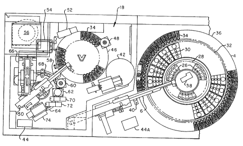
Automated analytical methodology for effecting analysis of a plurality of test
samples according to the present invention is achieved by introducing reagent
packs, test
sample container and reaction vessels onto concentric carousels of a main
carousel. The
test sample container can be a test tube, cuvette, vacutainer tube, and the
like, for holding
a test sample. The reagent packs and test sample containers are identified and
aligned
respectively with a reaction vessel for transfer and kitting of the reaction
vessel by transfer
of test sample and specific reagents from the reagent pack for preparation of
a
predetermined test. The reaction vessel containing the test sample and one or
more
reagents is tsansferred to a process carousel wherein controlled environment
conditions
exist for incubation once the sample has been appropriately mixed with various
-reagents
CA 02538953 1993-03-24
WO 93/20440 PCT/US93/02644
21
to form a reaction mixture. When all assay processing steps have been
completed, the
reaction mixtare is identified and transferred to at least, for example, one
of a fluorescent
polarization immunoassay reader or a microparticle enzyme immunoassay
cartridge
positioned on a separate cartridge wheel or carousel for further preparation
before reading.
The processed test samples are read and the readings are calculated with the
resulting data
being recorded and/or printed.
The methodology of the automated immunoassay analytical system is achieved
through the use of a self-contained, fully automated, continuous and random
access
instrument comprising a main carousel assembly consisting of the reagent pack
carousel, a
reaction vessel carousel and a test sample container carousel concentrically
and
independently rotatable. The main carousel assembly is provided with a
transfer pipette
operated by a boom arm for transferring and kitting test sample and reagents
into the
reaction vessel automatically following a predetermined test schedule. The
main carousel
assembly is provided with bar code readers for reagent packs and test sample
containers
and has the capability of aligning the reagent pack carousel and test sample
container
carousel and a reaction vessel for pipette transfer operations. Once the assay
to be
performed is scheduled, the reaction vessel carousel, the reagent pack
carousel and the test
sample container carousel are rotated until the reaction vessel, a reagent
pack and a test
sample container, respectively, are determined to be in the transfer pipette
access position.
The transfer pipette then transfers the test sample from the test sample
container and,
depending upon the assay to be performed, the reagents from the reagent pack
are
transferred to the reaction vessel. The reaction vessel carousel is then
rotated to a transfer
station position which contacts the reaction vessel with a transfer mechanism
and pulls the
reaction vessel into the transfer station. The reaction vessel is then loaded
onto the process
carousel by the transfer mechanism.
When performing a fluorescent polar'szation immunoassay (FPIA) with the
automated analytical system of the present invention, various pipetting
activities are
performed by a second transfer pipette apparatus which is in service for the
process
carousel, and the process carousel is rotated so that the reaction vessel,
when properly
pipetted with, for example, FPIA reagents, is at the read station of the FPIA
processing
stations and the FPIA determination on reading, is made on the reaction
vessel. The
process carousel is then rotated so that the read reaction vessel is at the
transfer station.
The reaction vessel is again contacted and transferred by the transfer
station. The transfer
station is rotated and pushes the reaction vessel into a release container
opening.
For a nricroparticle enzyme immunoassay (MEIA) performed with the automated
analytical system of the present invention, after the various pipetting
activities for the
MEIA, which can be completed at the main carousel assembly, the reaction
vessel is
transferred to the process carousel as described in the FPIA process.
Pipetting can also be
CA 02538953 1993-03-24
WO 93120440 PCT/US93/02644
22
acaomplished in the process cuousel or joindy bet.wmea the two carousels. To
cumpkbe
the MEIA, the reacdan mahme is tiaasfwed fram the reaction vessel to a matiix
of an
MffiA cattridge on a c:utridge carousei with tbe second iransfer pipette. The
mabrix is
washed with a buffer and a substrate, aoch as MUP (defined earlier), or other
surtabie
snbsbrats known in the art. The cafiridge cu+ousel is thea rotated so that the
lulElA
carlridge is positioned at aa MBIA processing assembly and the MELA
determination is
made. T6e MEIA reaction vesset is ejectied inba tbc waste oontainer as
desuibed for the
FPTA reactioa vessei. Tbe MBIA cattidge is ~y ajmed from the cartridge
vrhoei by an ejectoar at an eppropciabe ejeaw stsbivat inW a waste cootainer.
Preferably, two distinct analytical technologies as descn'bed above, FPYA and
MBIA, are incorriorabed into the ammated analytical spsbem of the preseat
invmtion;
however, mae+e than two distinct amalytical teclmologies can be bmporaed into
the
inventive system These meftds are cvmplimentary and shsre a commonality of
apparatus
and Ixvoedmal seeps, with the FPIA geoerally being the method of choice for
analytos of
low malecaW weight and 1VIBIA for molecules such as pt+obein hormones,
antbodies or
analyte.s of low molocalar we,ot requiring Ing>xx seasitivity. The two
technologies share
system comp~m kdwft Ow operaWr control paneL pipet#ing boom assemblies,
flaidic systems,'air and liqaid reageat heaters, printers, bar code reader and
stepper
motors. Such commooality of use of system components atlows for a oo.mpact
iastrameat
despite the dual kRA and MEIA capabIity.
The FPIA. optic systems (such as described in U.S. Patcnt No. 4,269,,511)
can utilize a polarizing filter which is an electrically
switched liquid crystaL, maintaining a compact size and avoiding oomplex aad
potmtialty
unreliable moviiag paits. When pe<forming FPIA, assays otilizing the amwated
anWytical
system of the preseat inveoltm the FPIA reageat packs will typiCally iaclnde a
ftacer
oo~n~g the aAalyte or aaalog theceaf, ooapled to a deboctsble moiety, an
anti'bod.y
spodfic to that saalybe, and agocimm pWenwift reaSad. In a preferrod FPIA
ftmat,
the amlyte being deommined oampetea wb du traoet for a limibed namber
of=binding
siles on the andbodies spodffc to the pmtion or poctioos of the aoalyee and
tracer. Thc
detecUble moieay oampooeat of tLe tracer is, prefeaabty a flaaaescmt moiet,g
selecbed fnam
tbe gtmaap consiffiing of fluaaaccins, aminofho+oseains, cuboxyftorseeins,
fluoresceinamines, and ft liloe, more pceferably carboxymethyl-amioomethyl-
fluoresvein,
catboaYtxhylatniaomeihYl-cacboa~y8uot~n, G-catbaaYfluomsce3it, S-
mboaygaaoescein,
succinylanimome&yl-flaorescein, thioaree-amiao8nc+resoGm,
35- MethoaytriamoiylaminoSuaresvein, amino8uoresceia, aad the liiw.
In anoger embodimcat, the FPIA formst ebii~oes a unigue, moad, plastic,
reaction
cavette sastable for flaarescence pdarizuion aad abanimcx assay t,ochnologie,s
whlch
raquit+e no orienatioo othw thaa top4oboRtom. Ttds plastic r8actioa cuvetle
has physicai
CA 02538953 1993-03-24
w0 93/20440 PCF/US93/02644
23
characteristics of low birefringence throughout the optical read region as
well as sdringcnt
climeosionat tolerances which allow reproducible absorbance readin.gs.
Bifriag+wce is
defrned as the degree of ratardadon of the extraoidinary ray as it passes
thrtwgh a
material. The grleater the degm of retardation, the greater will be the level
of
birefringence. Retardstion of the Gxtraa-ordinary ray is dependent on the
maguitade and
direcdan of the indneed sbress. Therefore, passing a ray of linearly polarized
light through
a matcrial with induced smess will rmlt in depolarization of the ray. In order
for a
cuvette to be ntilized foor fluoreseme polariztion mu- , 1 - ts, it is
important that the
cuvette be prepared ander conditions which yield mmiamm stress. no geomctry of
the
cuvette has been designed to utilize the inhemt fluidica of aut4mmed medical
diagostic
instrumentation to minimize the hydrog bobic effect of pfastic.
MBIA results can be deterimned by qaantify3ng the rate of tluorescence
developed
when fluorogenic substrate is converted by the actiou of an enzyme labeled
conjugate. For
example, when performing dther a compditive MEIA or sandwich MEIA, the
specific,ally
bound allcaline phosphatase on the mic,ropardcl.es is detected by addition of
the fluorogenic
substrate MUP to the matrix. T6c alltsline phosphatase catalyzes hydiulysis of
the MUP to
inorganic phosphate and fluotrscent 4methylumbelli.feroae (4-MU). Tbe
libernted 4-mu is
detected by the MEXA optics assembly froa satface flaot+ometrr which is
designed to
detect flaoceseeace of low coacmtrations of 4-MU without intGrferenco by
fluoascece of
4-MIJP at a wavelength of 367nm. A system of lenses and optical filters focus
filtered light
(wavelength = 365nm) from a mercury arc lamp onto the surface of the matrix
and focus
emitted fluorescene from 4-MU (wavelength = 448nm) onto a photo multiplier
tube. Like
the FPIA opdcs assanbly, the MEIA optics system is compaat and has no mooving
parts.
Abwt Sve pereent of the eadtatiaa light is detected by a photudiode, ultowing
narmatiuition of de fluorescence data and generatia of a oonlroi sigasl nsod
by the ]anrp
power sap* to maintain.the intaasitj+ of the ocitation light within five
paceat over the
usefnl Nfe of the lamp. The MEiA post Processor uses linear regresdon analpsis
to
eonvert Ibe data from mattiple socessive det~aadons of 4-MU tlnaresoeace to a
rate
which is propoitioaat IIn aha ooncenristion of alhgline phowhatase owjugate
speclHcaUy
bound to 6e mictn~ardcles.
MBIA formo can be ntn with a multl-position MEIA mxu'liary ccarmsel and
pcocess carnwwi as well as a MEIA reagent pack oontaining micropardcle
reagetrt, an
alkaline phosphatase conjngaae and, in some cLses, a dilute buffer specific
for the assay
being pafarmed. Becavw the mi.croparticles tead not to sct#ie out of
suspen4ion during the
caarae of the assay, they can reacgly be pipetted. T6e efl'ective surfaoe area
of polystyme
latex micmparticles is- sevecal fold greater then dmt of a 1age diameter
polystyrene bead
(e.g., one quarter incb beads) commonly used jn commu+cid immunotssays.
Because of
t6is large surfaae area and the vety small diffusion distntoe between analyte
and the
CA 02538953 1993-03-24
24
capture molecules on the surface of the microparbic.les, the capture phase
employed in
many of the MEIA methods being performed reaches equilibrium within several
minutes,
allowing for a fiill carousel of test samples to be completed in a very short
time frame.
Unb'b an FPIA, the heterogeneous immunoassays, such as a MEIA, require a
separation step as described above. In particular, after incubation of the
microparticles
with a test sample, the microparticles are separated from the reaction mixhue
by tcansfer
to the matrix contained in the MEIA cartridge as described above. The matrix
provides a
precisely located mechanical support for
the microparticles during the subsequent optical read phase-of the assay. This
precisely
located mechamtcat support, i.e. the cartridge, is fit into the auxiliary
carousel at a
predetermined spacing from the reader apparatus by camming means.
Detailed Description of the Drawings
Preferred embodiments of the automated immunoassay analytical system according
to the present invention are presented only with those components of primary
interest with
respect to the inventive system apparatus and processes of the present
invention. The
drawings do not illuslrate all of the mechanical and electrical elements for
driving and
controlling the various components of the system, wherein an of such omitted
elements
may have various known forms whhich can be readily realizQd by one of ordinary
skill in
the art having knowledge of the information provided herein with regard to the
mode of
operation of the system and the various components and related processes
utilized for
treating samples and determining analytical results.
Referring to the drawings, FIGURES I and 2 present isometric views of the
?5 automatic immumassay analytical system apparatus of the present invention.
The system
apparatus as it appears in FIGURE 1 presents the system apparam as used by the
technician, with FIGURE 2 illustratmg an isometric view of the frame and
cabinetry with
component parts removed. The system apparatus of the present invention is
identified
generally by the reference numeral 2 in FIGURE 1. The system apparatus 2 has
an
exposed front end carousel 4 which is serviced by a first transfer pipette
mechanism 6 for
kitting scheduled te.sts along with samples into a reaction vessel. The system
provides a
computer screen 8 and computer keyboard 10 along with access panels 12 for
accessing
storage and waste compariments. The system apparatus 2 is provided with
rollers 114 for
movement of the system apparatus within a laboratory complex as required. The
freedom
of movement of the system apparatus through rollers 114 is allowed since the
system is
fully self-contained but for power requirements.
In FIGURE 2, the system apparatus 2 cabinet frame 16 is illustrated with
substantially all functioning components of the system apparatus removed. A
controlled
CA 02538953 1993-03-24
WO 93/20440 PCT/US93/02644
environment zone 18 is a closed unit during operation with light shielding and
rigid
control of airflow as well as temperature as opposed to the open front end
carousel 4. The
front end carousel 4 communicates with the controlled environment zone 18
through a
transfer port 20. The front end carousel 4 is mounted to an aluminum base
plate which
5 rests on a support platform 22 and the fust transfer pipette mechanism is
mounted on
means 24.
The top plan view in section of FIGURE 3 presents the functioning component
system apparatus in some detail with relative positioning of the system
apparatus to further
illustrate the process flow of the system apparatus. For example, sample cups
26 are
10 mounted on a sample cup carousel 28 which is concentrically fitted within
the front end
carousel 4 along with reagent pack carousel 32 and reaction vessel carousel
36. The
reagent pack carousel 32 is concentrically fitted between the sample cup
carouse128 and
the reaction vessel carousel 36. The reagent pack carousel carries reagent
packs 30 and the
reaction vessel carousel 36 carries reaction vessels 34. The front end
carousel 4 has an
15 operable bar code reader 38 for automatically identifying reagent pack
carousel 32 and
sample carousel 28. A wash cup 40 is provided for the first transfer pipette
mechanism 6
for washing as required between transfer of various sample and reagents. The
first transfer
pipette mechanism 6 is utilized in kitting the various reagent pack liquid
materials and
sample into a reaction vessel 34. The reagents and the sample are properly
icitted through
20 means of the first transfer pipette mechanism 6 inclusive of pump means.
The various
carousels are rotated and aligned for kitting at the pipetting station. The
lcitted reaction
vessel 34 is positioned by reaction vessel carousel 36 into the proper
position for transfer
to the transfer station 42. The reaction vessel 34 is transferred to the
transfer station 42
through transfer means wherein the tcansfer station 42 is then rotated to move
the reaction
25 vessel onto process carousel 46. As shown, the process carousel is driven
by a stepper
motor 48 and is serviced by a second transfer pipette mechanism 50. Both the
FPIA and
MEIA procedures utilize the system apparatus commonly up through and including
the
process carousel 46. The process carousel 46 includes FPIA processing 52 and
FPIA
processing lamp 54 for direct reading of FPIA analysis of kitted, pipetted and
properly
reacted reagents sample from the reaction vessel 34. The controlled
environmental zone
18, which includes the transfer station 42 and process carousel 46, provides
FPIA
processing with air circulation under temperature control by cabinet air
circulation fan 56.
A wash cup 58 for the second transfer pipette mechanism 50 is provided. The
second
transfer pipette 50 is utilized for adding reagents (pipetting) under
conditions of incubation
and timing to the sample in the FPIA test schedule reaction vessel 34 for FPIA
processing. MEIA processing can also utilize the second transfer pipette 50
for adding
reagents to the sample before the reaction mix is added to MEIA cartridges 68
which are
mounted on the cartridge wheel carousel 64. The transfer of the MEIA reagent
mixed
CA 02538953 1993-03-24
WO 93/20440 PCF/US93/02644
26
sample to the MEIA cartridge 68 is by the function of the second transfer
pipette 50. A
motor 60 drives the cartridge wheel 64. The cartridge wheel 64 is provided
with MEIA
cartridges 68 through the operation of a cartridge hopper 66 which
automatically feeds and
positions the MEIA carttidges 68 onto the cartridge wheel 64. The process area
includes
the second t.ransfer pipette mechanism 50 and heater/pump 44. The cartridge
wheel
carousel 64 is further serviced by a MEIA buffer heater and dispenser 70, MUP
heater
and dispenser probe 72, and MEIA reader 74. The MEIA cartridges 68 are removed
from
the cartridge whee164 by a cartridge ejector 62 after the MEIA read has been
completed.
According to another embodiment, methods and apparatus for measuring a
chemiluminescent signal produced by an immune complex formed with analyte from
a test
sample such as chemiluminescent homogeneous immunoassays and chemiluminescent
heterogeneous immunoassays are provided. According to one embodiment, a
chemiluminescent detection signal is produced by an immobilized immune complex
comprising antibody coated magnetic particles which are separated by a
magnetic field.
According to such method, an optical cuvette containing the immune complex
bound to
magnetic particles suspended in solution is utilized wherein a magnetic field
is imposed
along the wall of the cuvette to perform the separation. The particles which
contain the
immune complex are washed, a trigger reagent is added, and the resulting -
chemiluminescence from the labeled particles is detected and measured in the
cuvette
using a chemiluminescent detection system. According to another method,
analyte is
captured in a liquid phase employing, for example, microparticles, polyionic
capture
agents and the like, having a binding affmity for the analyte wherein the
capture analyte is
subsequentially immobilized by a porous element and a chemiluminescent signal
is then
chemically excited and detected. Accordingly, such method within a continuous
and
random access analytical system advantageously employs fast fusion rates in
solution to
provide highly sensitive assays for a wide range of analytes. Such methods are
particularly useful with an automated, continuous and random access analytical
system
apparatus as described herein, and which can further include fluorescent and
chemiluminescent assay processing on the same platform_ Such automated
analytical
system of the present invention is capable of simultaneously performing two or
more
assays on a plurality of test samples in a continuous and random access
fashion. In
particular, the automated immunoassay analytical system apparatus of the
invention can be
viewed as a microprocessor based system of integrated subassemblies with
different groups
of assays being run through separate and changeable software modules. The
microprocessor based system uses robotic arm pipetters with two degrees of
freedom and
bidirectional rotating carousels to process samples. Critical assay steps such
as
incubations, washes and specimen dilution are performed automatically by the
instrnment
as scheduled.
CA 02538953 1993-03-24
WO 93/20440 PCTIUS93102644
27
In particular, the automated., cantinms and random access aaalytical system
apparatus is mpable of performing chemilaminescent assays such as de,scribed
in
commonly owacd U.S. Pstent No. 5,089,424.
In particular, the apparatus includes a container having an aperture and a
solid, porous
ekxneot, preferably in the form of a fibrnus matmc, cagable of immobilizing a
chemiluminescent genateting reaction product complex, while at the same
tiuoQe, pettuitting
the passage of other reaction componenta Which are not immobiliz,ed by the
porous matrix.
The reaction product is immobilited by the pacous element through particulate
reactants or
as the result of an intwaetive pmpecty betwea the poraus element and the
resction
product snch as hydc+ophilio-]rydrophobic bindiog intmactions, ionic binding
interactions,
and the like. A detectioa device is siiusbed adjacent to the oonteiner wbich
moves t4
create a light-tight seal with the ovntaiw to allow low ligbt level
chemituminescent
measarements. The debxbion device includes means for evenly distributing a
ct~emilammesceat acdvating soludon tn the pmous element. The apatiu+e may be
funnel-
shaped and the meaas for applying the activating solutlon may include ports
disposed
toward an interior surface of the funneL
The solid, porous elemeat is preferably in the foam of a fibrous matrix and is
used
to immobilize the immobilizable reaction complex as a result of the
iutetaction
tfierrebetwem, from which an assay signal can be geaerated_ The porous element
can be
selectied from woven fibrons materials such as glass, cellulose, nylon or
other natincal or
synthetic matGrial known tio those stilled in the art. Mat,etiai choice,
dimensions and pore
size of these porous elements can be easily selected by those skilled in the
art, to provide
an effective pore size and adequate void ucas to germit p"er flow of owvacted
mageats
and sample tiirongh the porous element. It is to be appreca$ted that the
preseat invention
is not intcudeA to be limibed to chemilnmmesrT!nt assays In a heterogeneous
immunoassay
format as described haeun, and that chewiluminascent assays can be performed
according
to otbzr immunoassay fasiats ]mown in the at, sach as bomogeneaas immanoassay
formats and the like.
FIGURE 3A is a top plan view of the automatod aaalytical system in section
with
componmt cavers removed to show the anrtomatied analytical appustu.s in detail
and
relative position inda4ive of a cheamilu~m~ines~xat readrr for membnme
particle caipture
technulogy. The process carowel has two magnetic separation stations 67 and a
chemih,mineaaent reader 69 for magnetic particle capture inccx~xted titereon
for
providing chemitaminisceat magnatic pnrticle capbure assays. The ca=nridga
wheel
35- camusel has mounted doem a chGmiluminiseent reader 71 for providing
micropatticle
meaobrane capwre assays.
Across-s,xtional view of the sigaal detoctim module is shown in PlG. 3D and
comprises a light gaida 602, ligbt-pipe protecting slexve 604, injectars 606,
608 that are
CA 02538953 1993-03-24
WO 93/20440 PCT/US93/02644
28
connected to two black Teflon'' fluid lines, photomultiplier tube (PMT) 610, a
photomultiplier tube socket 612 and a collar for holding the PMT socket 614.
The PMT
is springloaded by stainless steel spring wire 616 to ensure proper spacing
from the light
pipe and is protected from moisture by two 0-ring seals 618 and 620. 0-rings
622 and
624 fix the light pipe 602 in space and protect the face of the PMT from
moisture. O-
ring 622 is preferably made of a material with high dielectric constant to
prevent Ohmic
leakage at the photocathode. A mumetal shield 626 surrounds the PMT, with a
nylon
spacer 628 inside 626 which protects the PMT during assembly. The mumetal
shield 626
is kept electricaliy isolated by the use of two 0-rings 630, 632 and a Teflon"
sleeve 634
to separate it from the outer housing. An electrically conducting outer
housing 636
protects the detection module. The bottom of shroud 638 has a groove 640 that
locates
the surface feature on the disposable device and a light sealing gasket 642 of
FIG. 3D.
The light sealing gasket is made of black inert compressible polymer. A
preferred
material is a nylon nap on a rayon backing COE7-1673 (Schlegal Corporation,
Rochester,
NY). Two low wattage anti-fog heaters 644, 646, are used to create a
temperature
gradient in the vicinity of the light pipe to prevent any condensation on the
light pipe
during measurement. A top view of the analyzer system containing the alternate
chemiluminescent detection is shown. Magnetic separation stations 67 are
positioned
adjacent to the optical cuvette on the processing carousel. Trigger reagent is
added by the
pipettor 50 and the resulting chemiluminescent signal is read by the detector
69.
FIG. 3C is a cross-sectional view taken along the side of the detector device
of
the present invention and shows a detector "assembly, shroud 648, groove 650
and light
sealing gasket 642, the anti-fog heating elements 644, 646 and the end of an
injector tip
607.
FIG. 3D is an partial cross sectional view illustrating the light pipe 650.
The light
pipe 650 is shown positioned above the fiber cartridge 654 with fluid
injection conduits
652 on either side of the light pipe 650. The fiber cartridge 654 has a
funne1656 and a
depth filter 658. A light shield 660 provides light shielding in order to
avoid
environmental light from interfering with the reading of chemiluminescent
released
photons which are bransmitted through the light pipe 650, generally a quartz
light pipe to
a photomultiplier tube not shown. The photomultiplier tube assembly inclusive
of the
light pipe measures the chemiluminescent signal produced on the reaction
matrix surfaces
of the cartridge 654. During sample processing, an alkaline urea peroxide
solution added
to the immune complex triggers production of photons of light. The photons are
channeled through the quartz light pipe and counted by a photomultiplier tube.
The
amount of light present (photons) is either directly or inversely proportional
to the amount
of in light present in the sample.
It is to be understood that the utilization of the first transfer pipette
mechanism 6
CA 02538953 1993-03-24
WO 93/20440 PCT/US93/02644
29
and the second transfer pipette mechanism 50 as described herein provide a
safety
mechanism to ensure that test samples and reagents are pipetted to thereby
prevent false
negative results in the event there are incorrect amounts of the respective
sample and
reagents for a particular assay.
Approaching the operable elements of the system apparatus in greater detail,
FIGURE 4 provides a front elevational view in isolation and partial section of
elements of
the front end carousel 4. FIGURES 4A and 4B illustrate a reagent pack with a
cover
means 31 which is opened and closed pivoting along axis 37. A return notched
drive arm
35 is utilized to open and close the cover means 31 by contact with the cover
contact
surface 33.
According to another embodiment, a test sample container segment assembly
adapted to receive a plurality of test sample containers of varying dimensions
for use with
an automated analytical instrument is provided. The segment assembly
cooperates with a
test sample carousel of the automated analytical instrument to provide
unlimited variability
in test sample containers which can be processed by such automated analytical
instrument.
Accordingly, the test sample container segment assembly obviates the need to
transfer test
samples from an otherwise incompatible test sample container to a test sample
container
which is required for use with an analytical instrument.
In particular, the test sample carousel comprises a plurality of positions for
receiving the test sample segments of the present invention. For example, the
carousel is
preferably adapted to receive six test sample segments, with each segment
containing
receiving positions for either ten or twelve test sample containers. The
segment can
accommodate, for example, one or more sample cups and primary tubes, wherein
the
number of primary tube sizes and dimensions will vary. The primary tubes and
sample
cups may be mixed within the same sample segment. The different sample
segments will
support the primary tubes and sample cups so that the aspiration of samples
will be
accomplished at substantially the same height to provide the automated
analytical system
with a common level for a probe, which is utilized in combination with
pipetting means,
to provide sample transfer and kitting of reagents into a reaction vessel on a
reaction
vessel carousel. Accordingly, since probe pipetting means are in demand in
relationship
to time and use, the test sample segment assembly enables, for example, less
travel and
level sense time when such primary tubes and sample cups are utilized to
thereby increase
throughput of the system. Preferably, each sample segment includes an
identifying
number and code which is read by an instrument and associated with sample
segments for
kitting and processing then the automated, random access analytical system
instivment.
Accordingly, the test sample carousel, in combination with the test sample
segment
assemblies, provide a maximum amount of accessibility for loading and
unloading of
sample containers, such as the primary tubes, sample cups, and the like
containers as
CA 02538953 1993-03-24
described herein.
The test sample container segment assembly 600 is shown in FIGURE 4C in a
perspectEve view. It is to be undeistood that test sample containers
contemplated
according to the present invention include, but are not intended to be limited
to,
5 Vacutainerm tubes, test tubes, cuvettes, vials, sample cups,
and the like, all of which can be of varying
sizes and dimensions. The assembly has a frame 601 and a handling means 602.
The
assembly 600 has a test sample container mounting shelf 604 into which test
sample
10 containers can be inserted through container insertion openings 606. Once
inserted into
the container insertion opening 606, the containers are received and
surrounded by liquid
level sense sleeves 608. However, insertion openings 606 of the assembly can
adequately
support and fix said test sample containers without use of sleeves 608. The
test sample
container segment assembly 600 has two curved dimensions which are in. a
parallel
15 relationship and are equal to the radius of curvature of the test sample
container carousel.
The outer curved portion 610 of the test sample container segment assembly 600
and the
inner curved 612, both being vertical and in relationship to the container
mounting shelf
604 present an assembly which is mateable with the test sample container
carousel. The
test sample container carouse128 has positioning and mounting pins which are
receivable
20 within pin receivers 614 and 616 of the test sample container segment
assembly 600.
These pin receiving elements allow an operator to properly position and mount
the test
sample container segment assembly 600 into the test sample container
carouse128. The
test sample container carouse128 has mounting pins 618 as shown in FIGURE 4E
which
are received by the test sample container of assembly 600 receiving segments
614 and
25 616. These receiving segments 614 and 616 are shown in FIGURE 4D, which is
a
bottom view of the test sample container segment assembly of FTGURE 4C.
A cross sectional view in isolation of the test sample container carousel with
a
mounted test sample contaiaer segment assembly 600 mounted therein is shown in
FIGURE 4E. The view ia FIGURE 4B clearly illusbrates the adaptation of the
test sample
30 container segment assembly 600 to the test sample container carouse128
providing an
operator with handling means 602 and alignment pins 618 for fitting into test
sample
container segment assembly 600 receiving portions 610 and 612.
A cross sectional view of a modified test sample cup 620
is shown with upper and lower skirt
portions 624 and 622, respectively, in FIGURE 4A. Such modified test sample
cup 620
can be ntiliud within the test sample container segment assembly for
presenting tio the
assembly uniform outer dimension which fits routinely inta the test sample
container
segment assembly 600, even though the interior of the test sample cup 620 is
buried for
CA 02538953 1993-03-24
WO 93/20440 PCT/US93/02644
31
various purposes.
A short test sample Vacutainer tube sample assembly 626 is shown in
perspective
view in FIGURE 4G. The short test sample Vacutainer tube segment assembly 626
has
a frame 628 and a handling means 630. The assembly has Vacutainer tube
mounting
shelf 632 in which Vacutainer's tube insertion opening 634 is provided for
mounting the
short test sample Vacutainer tubes into the short test sample Vacutainer
tube segment
assembly 626.
A top cross sectional view of the short test sample Vacutainer tube segment
assembly is shown in FIGURE 4H, the view is taken along line A-A of FIGURE 4G.
Vacutainer tube mounting spring means 636 provide a holding means for the
insertable
Vacutainer tube elements which are of a tubular or test tube configuration.
In addition
to the Vacvtaine& tube mounting spring means 636, Vacutainer tube holding
arms 638
are presented which further stabilize and maintain the Vacutainer tubes in a
specified
position in relationship to the short test sample Vacutainer tube segment
assembly 626 so
that when the assembly is inserted into the test sample carouse128, the test
sample
Vacutainer tubes will not only be positioned as to a uniform height, but will
also be
positioned at a specific location within the carousel and mounted short test
sample
Vacutainer tube segment assembly 626.
A bottom view of the short test sample Vacutainer4l tube segment assembly of
FIGURE 4G is shown in FIGURE 41. Test sample carousel 28 mounting pin
receiving
elements 642 and 644 provide assembly mounting positioning guides as well as
holding
means for holding an exact positioned short test sample Vacutainer tube
segment
assembly 626 within the test sample carousel 28. In FIGURES 4J and 4K various
length
test sample cup adaptor sleeves are presented. In FIGURE 4J, a cross sectional
view of a
long test sample cup adaptor sieeve 650 is shown. In FIGURE 4K, a cross
sectional view
of a short test sample cup is shown. FIGURES 41 and 4K allow the sample cups
of
FIGURE 4F to be used in Vacutainer tube segments.
The sample acquisition carousel utilized in the present instrument preferably
has,
for example, six positions or sections for mounting test sample container
segment
assemblies as illustrated in FIGURES 4C and 4G. The test sample segment
assemblies
are interchangeable, as the operator desires, with positioning pins either on
the segments
or on the carousel with appropriate receiving means ensuring proper
positioning of the
~ segments to the carousel. Accordingly, such interchangeable segments allow
the
acquisition of a variety of test sample containers which can reside on the
carousel at any
given point in time. It is to be understood, of course, that the number of
sample
acquisition carousel positions or sections may vary, and such number will
depend upon the
size of each portion or section and dimensions of the carousel.
According to the present invention, different types of se$ment assemblies can
be
CA 02538953 1993-03-24
WO 93/20440 PCf1US93/02644
32
placed in the test sample carousel such as, for example, (1) test sample cup
segments
which can be used in conjunction with the inslrument sample cup 620, wherein
the
segment can hold about twelve or more of such test sample cups; (2) large
Vacutainer
tube segments, which can be used with Vacutainer* tubes from about 0.400 to
about
0.650 inches in diameter and about 3.000 to 4.500 inches in length. Such large
Vacutainerm tubes can be positioned in the segments to accommodate about ten
or more
VacutainerO tubes; and (3) small Vacutainerm tube segments, which can be
utilized with
the short test sample Vacutainer tube segment assembly of FIGURE 4G, can
accommodate Vacutainer'O tubes of about 0.400 to about 0.650 inches in
diameter and
about 2.000 to 3.000 inches in length, with about ten positions to accommodate
about ten
or more Vacatainerm tubes.
Sample container adapters are also available which allow sample cup containers
to
be placed in a Vacutainer tube segment, particularly for sample cups 620.
Such adapters
allow sample containers to be used when a minimum number of sample containers
are
needed and space for a sample container is not available.
Level sensing of fluid in any of the test sample container in the sample
segments
can be accomplished, for example, by capacitive level sensing. Conductive
material is
assembled into the segment assemblies and into adaptor sleeves to create a
capacitive path.
In addition, bar code identification is provided on the segment assemblies and
adaptor
sleeves. Such bar code identifications are used to identify the segment type
and the
substitution of, for example, an adaptor sleeve, as well as, of course,
identifying the test
sample, itself.
In one embodiment, an operator can load empty test sample cups 620 into a test
sample segment and pipette test samples into the sample cups 620. The operator
may
choose to configure the test samples according to a predetermined load list
generated via a
data entry process. Vacutainerm tube adaptor sleeves and other tubes can, of
course, be
used in place of a sample cup 620 in the appropriate segment. The operator
will then
place the test sample segment on the test sample carousel and indicate to the
self-contained
scheduling and computer system that fittther test samples are to be processed.
The test
sample carousel will scan all segments at the appropriate time in order to
track all onboard
test samples. The instrument will continue, in sequence, to process test
samples unless a
"stat" test is ordered. When a "stat" test is ordered, the instrument will
scan all segments
until it finds the one which contains the "stat" test sample. When the stat
kitting cycle has
completed, the front end carousel will return to a previous sequence. The
operator may
choose to brace the instrument into a hold phase during loading and unloading
of test
sample segment assemblies. The ki.tting process will be suspended after the
next kitting
cycle. Once loading and unloading is complete, a second instruction will cause
the kitting
process to resume. The status of onboard test samples will be subject to audit
through a
CA 02538953 1993-03-24
WO 93/20440 PCT/US93/02644
33
data entry screen of the instrument. With such audit, the operator will know
which
segment assemblies are completed and may be removed. Single test samples may
be
placed into a segment assembly which is residing on the test sample carousel.
FIGURE 5 provides a top view in isolation and partial section of elements of
the
drive and guide systems of the main carousel 4 with the various carousels
removed. In
FIGURE 5 a sample cup carousel stepper motor 76 is shown mounted with mounting
spring 78. The reagent pack carousel motor 80 is also shown with a mounting
spring 82.
The reaction vessel carousel motor 84 and mounting spring 86 are positioned to
the
exterior of the two inner carousels, i.e. the sample cups carousel 28 and the
reagent pack
carousel 32. Roller guides 88 are provided for the sample cup carousel 28 and
a
tensioning spring 90. The reagent pack carousel is provided with roller guides
92 and
tensioning means 94. The reaction vessel roller guides 96 are also provided
with spring
elements 98, the purposes of the guide and these various spring elements being
to
maintain very finite tracking of the concentric carousels when motivated by
the individual
stepper motors.
The front end carousel 4 inclusive of the three front end carousels, the
sample cup
carousel 28, reagent pack carousel 32 and reaction vessel carouse136 can by
example
contain the following capacities. The sample cup carousel 28 can hold 60 blood
collection
tubes, such as Vacutainer blood collecteon tubes, or 90 sample cups which are
injection
molded as one piece and can be provided with standalone base mounts.
Standalone base
mounts are suitable for technician handling and pipetting of samples into the
sample cups.
The reagent pack carousel 32 provides for 20 different reagent packs 30. The
reaction
vessel carousel 36 provides 90 reaction vessels 34.
The process carousel 46 as shown in FIGURE 6 is an isolational cross-sectional
side view. One reaction vessel 34 is at rest or nonoperative position and a
second reaction
vessel 34 is in position for FPIA read. The process carousel 46 is capable of
bidirectional
motion for timely movement of the various reaction vessels 34 to pipettor
action, read, or
transfer to and from the carousel. Up to about 36 or more reaction vessels 34
can be
processed at one time on the process carousel 46 depending on diameter and
sizing of the
reaction vessels 34.
The first transfer pipette mechanism 6 of FIGURE 7 includes a tcansfer pipette
Z
axis motor 102 which moves the probe arm 104, probe 106 and probe tip 108 in a
vertical
direction while transfer pipette R axis motor 100 drives the probe arm 104,
probe
adjustment means 106 and probe tip 108 in a horizontal motion. The first
transfer pipette
mechanism 6, sometimes labeled "Sample Probe Arm Mechanism", moves the probe
between the sample cup 26, the reagent pack 30, the reaction vessel 34 and the
wash cup
40. The wash cup 40 is used to wash the interior and exterior surfaces of the
fust transfer
pipettor mechanism 6 probe. The drive of the first transfer pipette mechanism
is a rack-
CA 02538953 1993-03-24
WO 93/20440 PCF/US93/02644
34
and pinion drive means along the Z and R axis by two- stepper motor drivers. A
brake is
provided to hold the Z axis position when power is lost, thus avoiding damage
to the
system apparatus. For example, the first transfer pipette mechanism can be
designed to
have a Z axis travel of about 3 inches and an R axis travel of about 11-1/2
inches.
The first transfer pipette mechanism 6 and the second transfer pipette
mechanism
50 are closely related in general system apparatus finnction and design, with
variation on
travel and size being the only substantial differences. Both units have a
probe arm circuit
110 as illustrated by the schematic side view of FIGURE 8. The schematic
illustrates the
R axis motor 100 and the Z axis motor 102 in relationship to an upper PCB 112
and a R
axis home sensor 114. A lower PCB 116 is illustrated in relationship to the Z
axis home
sensor 118 with a coil cable 120 connecting the various elements.
The present invention also presents two unique approaches to the very
necessary
and complicated fluidics systems within an automated analytical system. Level
sensing by
robotic automatic pipetting are presented in two different methodologies, one
being by
detecting signal amplitude changes associated with a pipettor when it touches
a liquid.
The significant feature of this liquid level sense is that the signal
detection process is
based on signal change and rate of signal change, where previous systems have
been based
on actnal signal amplitudes, therefore, being affected by changes induced by
temperature,
humidity, aging of parts, parts variation, and most significantly, pipettor
position. This
complementation may be used with conductive diluent in the fluid line to the
pipettor.
Pipetting is limited to being above a level sense antenna.
A second liquid level sensing methodology for automated analytical systems is
presented which does not require an antenna below the pipettor. It requires
only a
grounded plate, but it cannot be used with conductive fluid in the pipettor
line. It can be
used with deionized water. It functions by detecting the capacitance change
and rate of
change when the probe contacts the liquid. The capacitance from probe to
ground is
measured in the form of an electrical phase shift of a sinusoidal signal
present on the
liquid probe. The design continually tracks the capacitance of the probe in
air and focuses
on a quick change in capacitance in response to the probe contacting liquid.
A capacitance liquid level sense 800 is shown in FIGURE 8A as one of the
liquid
level sense system embodiments according to the present invention. A liquid
level sense
board is utilized to monitor a probe (pipette, or the lke) when enabled by the
automated
analytical system computer and to stop probe movement when the probe has
contacted
liquid. As shown in the FIGURE 8A, the board contains process liquid level
sense
circuits 803 and kitting liquid level sense circuit 805, each completely
independent of the
other. The liquid level circuit 803 is dedicated to the process center probes
and the liquid
level circuit 805 is dedicated to the lcitting center probe. Process liquid
level sense 802
includes a pipette 806 and kitting liquid level sense 804 includes a similar
pipette 807.
CA 02538953 1993-03-24
WO 93/2044(1 PC'T/US93/02644
Sach of the two circuits are oontrolled by a calibrated signal and provides
two outpart
sigflals- ready md detect. In operadw, 'CALIBRATB' is set except when level
smsing
is desired. The probe is placxd over the liqM ta sease, preferably immediabety
over the
flnid, and the desired gain bits am set and ready as checked by the
controlling oomputer.
5 When "READY" is asserbed, the probe is thm moved toward the liquid until
liquid is
encountered, at which time "DBTBCr" is set. "DLTBGT" is routed ta the motor
controller to stop vertical prrobe moveinent, if software has enabled the
proper moCor
conÃrol functiam. The liquid- kvd sdm openbes by npplying a signal to a
pipette, probe
or the lilm, made from metal or and othwaviee snitable elecoicaliy aon&Kdve
miterial,
10 and doteoing signal changes wbicb ocm whan the pipette contscts: a liquid.
An
importaut Ãeattmc of the liqoid level sense is that the signal debxtion
pcocess is based on
signai chaoge and rate of signal rbanp eoaditioms and is thprefo~e
sabstandatly anaifeebed
by clamgas caused by benpeMure, humidity and the h7re. The liquid kwel sease
system
comprises one VME type PC board whieb conrains two sMarate, mdepeadeat level
sense
15 pzncessing cincants for each 1eve1 sease circuit, and a probe arm assembly
which moves
the metal pipxuor over the sense positioa. A coax cable from the system board
caeries the
transmit sigual to the p4xaoc 807 or 806. A receive antenna assembly 806
electrode lies
beneath the sreas where liqaid sensing is deshe<i. The antmna as9embly 808 is
connected
to the boand with a triaac cable assembly. FKiM 8A stwws the intmoamection of
the
20 liqaid level senae vvithin the enviromment of an automatod, cofltiauvas and
rnndom acoess
analydcat system described herdn. it is ta be understood tbat the liquid level
sense
system of the pre,seat invention can be utilized in any automated iasttvment
where liquid
le.vel sewing is desirecl.
Thc capacitancx liqmd sense 800 pravides a krtting center reactioa vessel
etectnode
25 810 and a lcittioag coter sample electrode 812, as we11 ss a process area
electrode 813.
T6e capacitaoce liqaid level suw 800 fiacther oamprlm cicruibryr flow from a
VMB styk
PC board badk piane 814; motor eoatrol via'back plane 816; aad frnm V11+iE
back plane
818 and motor eonVOl via baclc plaae 820. In opaatim when the pipM 806 m' 807
iaa~eases
cmtacts liqaid, the signal frnm the pipettrJliquid oombinatioa to the sensor
30 above that i+ocxived befora titio pipetbe tiouches liqaid. 7fie
tnunsatitlrxdve aperAtion can
be modeled using cdretut deamts. The tcaosmission media appears as a small
c~padtaaoe
from the pipette to the sensor. The lfne diluent appears as a large
capacitance compared
to that from piperbe to seosar, whea the diloent is conductive or "ionized".
Under such
condWms, it Is diffiiatlt and tmrelliable to detect the signal cuuyr,at 830 as
part of the total
35 cmrent 826. Pbicmg the obenoa allows only the signal of intS+est to.
bedeteftd,
Tbe liquid level mm aymem operates by detecting signet changos asodat,ed with
a pipettor when it camtacb a liquid A sigoal of about 125 BF;z !s applied to
the pipetbor,
prnbe or the iioe, tlmoogh a low impedwe driver. The signW couples ac= the
spaca
*Versa Nbdule Eurocard
CA 02538953 1993-03-24
WO 93/20440 PCF/US93/02644
36
between the pipettor and a receive antenna located below the point where
liquid sense is
desired. As the pipette contacts liquid, the signal transmitted to the antenna
increases
slightly because the liquid surface, in effect, becomes part of the
transmitter, increasing
the amount of signal transmitted. The coupling mechanism is primarily by
electrical field,
which can be mathematically modeled by capacitance. For this reason, the
generic type
of sensing is referred to as capacitance level sense. Since the electrical
field is acmally
part of an electromagnetic field radiating from the pipette, such sensing
devices have also
been referred to as "RF" (radio frequency), although the actual frequencies
normally
employed are about several octaves below standard radio frequencies.
The transmit and receive capacitative liquid level sense of the analytical
system
described herein employs an electrical sinusoidal signal of about 125 KHz and
evaluates
the amount of signal as it propagates from probe to a sense antenna. As the
probe
distance to the antenna changes, and as the probe contacts materials with
dielectric
constants higher than the surrounding air, the level of the signal reaching
the antenna will
change. The wavelength of the signal is low compared to the geometries
involved, so
almost all of the coupling from the probe to antenna is by the electric field.
Since
capacitance is a theoretical circuit element which models the electrical
field, the technique
is referred to as "capacitance" level sensing. Circuit analysis tools are
refined and easy to
use compared to solving electrical field theory problems, so it is common- to
model
complex electrical and magnetic systems as circuit elements or combinations of
elements.
By applying a low impedance transmit signal to the pipette and receiving the
signal with a
separate antenna, shunting effects of conductive diluent in the pipettor
plumbing can be
avoided. FIGURE 8B presents a simplified diagram showing current flow with the
sense
amplifier measuring only cucrent from the antenna, the diluent current not
included. The
ctnrent pass associated with the transmit and receive capacitance liquid level
sense system
is shown in FIGURE 8B. As can be seen, current from the transmit source flows
into
paths. Current leaves the probe, pipette or the like, and flows through
diluent to ground
and through coupling capacitance to ground, retarning to the signal source.
Separately, a
much smaller current enters the pipette, probe or the like and couples through
space to the
receive antenna. When the probe, pipette or the like contacts fluid,
additional current
flows through the additional capacitance added by the increased surface area
of the liquid.
Since the antenna is positioned to receive mostly signal from the pipette and
pipette liquid
combination, the signals received can be effectively analyzed to determine if
the pipette is
contacting liqaid. Although the information is pre,sent in the enrrent flowing
to the
pipette, the signal of interest would be very difficult to hold as reliably
and repeatedly
detect because of the large cuaent flow in the diluent. The simplified current
flow 822 of
FIGURE 8B has a signal source 824 and a total current 826. Pipette 828
capacitance is
shown in the simplified current flow as well as signal current 830 and fluid
to antenna
CA 02538953 1993-03-24
WO 93/20440 PCT/US93/02644
37
capacitance 832. Current in diluent 836 is illuslrated in combination with
diluents
resistance 834 and diluent capacitance 838. The circuitry also illustrates a
sense amplifier
840 and sense amplifier input impedance 842.
The system liquid level sense employs a synchronous receiver to provide
exceptionally narrow band acceptance of the electrical signals detected by the
antennas.
The synchronous amplitude detector is one which multiplies the incoming signal
by a
reference signal to extract amplitude information from the signal. The
transmitted signal
is generated from the reference signal so that both the transmitted and
received signals of
interest are of substantially the same frequency. The incoming signal must
substantially
be in phase with the reference signal. Although the resultant output is
complex, the
multiplier is followed by a low pass filter of a few KHz to extract the
information desired.
The filter employed is a Bessel linear phase filter, whereby there is minimal
or no
overshoot and minimal or no ringing. Synchronous receivers are also referred
to as
heterodyne receivers and correlation detectors.
FIGURE 8C illustrates the purpose of having a high center frequency and narrow
band width. Since the system noise peak is at lower frequencies and reduces as
frequency
increases, it is advantageous to operate at higher frequencies with narrow
band width to
reduce noise.
A minimum amount of increase in the received signal enables the system liquid
level sense to determine whether fluid has been found. Preferably, such
increase will
occur rapidly compared to the change that occurs when moving the probe toward
the
antenna. When the probe, pipettor or the like contacts fluid, the signal
increase is
sudden. This is accomplished using an autozero loop followed by a simple fixed
threshold. The autozero loop timing is such that the output of the Bessel
filters is
maintained at about zero if the probe is stationary or moving vertically. When
a sudden
signal change increase occurs due to fluid contact, the autozero circuitry
does not prevent
the Bessel filter output from increasing. If that increase is sufficient, then
a digital bit is
output indicating "DETECT", that is, liquid found, at which time the autozero
circuit is
disabled.
The board mounxs in a standard VME card cage, receiving only about +5V and
ground from the VME bus. DC/DC converters on the board generate the local
operating
voltages, isolating the board from the VME bus. There are two liquid level
sense
circuits, each completely independent. Control signals to and from the board
are routed
to the system I/O boards. Additionally, the "DETECT" (indicating fluid found)
signals are
routed to the system motor control boards, so that when fluid is detected, the
motor
,control board can immediately stop the pipettor movement. Each circuit must
be
referenced to the system ground. The connection for each circuit is done in
the immediate
vicinity of the sensing area. One circuit operates in the system process area
and one
CA 02538953 1993-03-24
WO 93/20440 . PCT/US93/02644
38
circuit operates in the kitting center. Each circuit applies a transmit signal
to a pipette
through a coax cable. Also, each circuit connects to a sensing "antenna" by a
triax cable,
with the circuit ground on the outermost conductor. That outermost connector,
in
addition to connecting to the antenna, connects to the system baseplate
adjacent the
antenna, providing the ground reference for each circuit. The inner shield of
the triax
cable is a "driven shield", wherein the signal on the inner conductor is
applied to a buffer
which, in tarn, drives the middle shield. This reduces the effect of the
capacitance of the
cable and antenna on the sense signal by a factor of about ten or more.
Each circuit is controlled by a "CALIBRATE" signal and three gain bits, all
optically isolated. Each liquid level sense circuit outputs two optically
isolated signals,
"READY" and "DETECT". In operation, "CALIBRATE" is set except when level
sensing is required. The "CALIBRATE" control forces the liquid level sense
board into
an autozero mode, where the analog output of the liquid level sense is forced
to zero.
This is done so that, after the "CALIBRATE" is removed, the analog output will
be
simply the change in the signal, not the absolute value. The pipettor is
placed preferably
immediately over the fluid to sense, the desired gain bits are set, and
"READY" is
checked by the controlling computer. When "READY" is asserted by the liquid
level
sense, indicating the analog output has been forced to zero, the system
microprocessor
removes the "CALIBRATE" command and moves the pipettor downward until fluid is
contacted, at which time "DETECT" is set. "DETECT" will remain set as long as
the
pipette is in fluid. After removal from fluid, "DETECT" resets, but will set
again if fluid
is contacted again. When the pipette is withdrawn from the fluid and fluid
sense is no
longer required, "CALIBRATE" is again asserted. In "CALIBRATE" mode "DETECTS"
do not occur, being disabled logically, regardless of the activity of the
analog signal
received.
The liquid level sense board will only pass rapid signal changes, and if the
amount
of signal change is sufficient to cross a present value, then a"DETECT" signal
is output
from the board. An autozero circuit nulLs the analog output to zero before
each level
sense operation, as long as "CALIBRATE" is set. After "CALIBRATE" is removed,
the
autozero circuit operates slowly during sensing operation, causing slowly
changing signals,
such as that caused by pipette movement, to be nulled to zero. Rapid signals,
such as
those caused by fluid contact, are not immediately affected by autozero. If
the fast signal
is larger than the fixed threshold, then "DETECT" is triggered and autozero is
disabled
until "CALIBRATE" is reasserted.
Bessel filters are used in the transmit circuits for repeatability and in the
receive
circuit for minimum ringing due to noise spikes, meaning minimum noise levels.
Sharper
filters have potentially high overshoots and actually result in a higher noise
level.
According to another embodiment, the invention presents a liquid sensing
device
CA 02538953 1993-03-24
WO 93/20440 PCT/US93/02644
39
which detects the capacitance change when a probe contacts a liquid.
Capacitance is
sensed between the probe and system ground. This type requires no antenna, but
must
use only deionized diluent or no diluent. This type is more tolerant of
spacing of liquid to
ground. The capacitance is measured in the form of electrical phase shift of a
sinusoidal
signal present on a test sample probe. This liquid level sense continually
tracks the
capacitance of the probe in air and looks for a rapid change in capacitance in
response to
contacting liquid. The design uses a phase synchronous det,ector, followed by
a phase
locked loop (for background tracking) coupled with detection logic for liquid
sensing.
The center frequency of the synchronous detection is from between about 27.000
KHz and
about 30.000 KHz, preferably about 28.799 KHz. The rejection filter band width
for
tracking is preferably about 1.78 KHz around the center frequency. The
rejection filter
band width for detection is preferably about + 85 Hz around the center
frequency.
FIGURE 8C illustrates the importance of having a high center frequency along
with a
narrow filter band width pin. Since the system noise (dominated by motor
noise) peak is
at the lower frequencies and reduces as the frequency increases, it is
preferred to operate
at higher frequencies and with a narrow band width to eliminate noise
problems.
The present liquid level sense device has a tracking function that dynamically
subtracts out the background signal and looks for a rapid change in
capacitance (contact
with liquid) to trigger a detection. ExisGa-g fluid sense systems report a
fluid detection
when the impedance present at the probe exceeds a threshold set by adjusting a
sensitivity
potential. It is to be understood that since a background signal is not
constant, varying
due to temperature, humidity, physical proximity to surrounding apparatus and
the like
surrounding environmental conditions, it has been the cause of a number of
problems
being experienced with various equipment. According to the present invention,
such a
fixed threshold does not vary due to temperature, humidity and age.
The result of threshold and background variations can result in non-optimal
operation of existing liquid level sense devices. In FIGURES 8D and 8E,
additions are
illustrated wherein the threshold is crossed (fluid detection) even though the
probe is not
in contact with the liquid. FIGURE 8D depicts the rise in "background" signal
as the
sample probe drives into a test sample_cup or tube. If the threshold is set
too low, a false
liquid detection can occur. FIGURE 8E illustrates the background signal as a
sample
probe drives in and out of an empty test sample cup. On the second drive into
the test
sample cup, the threshold has varied enough to allow a false detection.
The preferred 28.799 KHz sine wave is one of the inputs (Y) to the phase
detector
and should be considered the reference for phase detection. The tracker and
variable
phase control circuitry maintain the output of the phase detector/filterl at
theta volts.
Since the transfer function of the phase detector/filterl is XYcos(0), where
theta equals
phase angle between the reference signal (Y) and the probe signal (X), the
signal from the
CA 02538953 1993-03-24
WO 93/20440 PCT/US93/02644
probe is preferably about 90 out of phase with respect to the reference
signal, which is
maintained by a variable phase control circuit. The variable phase control
circuit is
controlled by a traclang circuit that senses the phase detector/filterl
output, compares it to
the desired output (about zero volts), and sends a control voltage to the
variable phase
5 control circuit. The rate at which the tracker responds depends upon the
sample clock
speed. The sample clock has two speeds, one for fast tracking of the
background during
non-fluid sensing movements (sample arm above Z axis home flag movements and
horizontal axis movements) and one for slower tracking of the background for
fluid
sensing movements (sample arm below Z axis home flag movements). The slower
10 tracking is required so that the fluid detection occurs prior to the signal
being nulled by
the tracking function. When the fluid is detected, the tracking function is
halted by a
latch until the below home sensor clears the latch. Fluid detection occurs
when the phase
detector output, after the low pass filters, exceeds the threshold voltage
(about 3V) for a
time longer than the set delay period of about 1.1 ms (milliseconds). If at
any time the
15 signal falls below the threshold prior to the delay period being completed,
the delay
counter resets to zero and waits for another detection. When a true detection
occurs
(signal above threshold for longer than the delay period), the fluid detection
triggers a
user selectable delay. The delay circuit can be set for a delay from between
about zero
and about fifteen steps into fluid. Upon completion of the user selectable
delay, the fluid
20 sense flag is activated, informing the system that fluid was detected.
Various elements of syringe 122 which provides automatic bubble flushing and
fluids to the various pipetting mechanisms is provided in various views in
FIGURES 9,
9A and 9B. The ability of diagnostic instrumentation to accurately perform an
assay is
critically dependent on the precision and accuracy with which syringes, i.e.
pipetting, can
25 aspirate and dispense reagents and samples. The precision and accuracy of a
syringe is
severely degraded by the presence of small air bubbles inside a syringe.
Bubbles,
unfortunately, are all too common and are difficult to remove or avoid.
Syringe 122
avoids these problems by automatically flushing bubbles completely out of the
fluidics
system. The syringe 122 is configured such that a piston 124 reciprocates
through a seal
30 126 and into a close-fitting bore 128. The end of the bore 130 is closed.
The piston 124
has a piston end 132 which approximates the geometry of the closed bore end
130. Two
ports to the bore are 1800 apart and are located near the seal and are
comprised of a fluid
entry port 134 and a fluid exit port 136. An annulus 138 exists between the
piston 124
and bore 128. Pressurized line diluent is introduced to the fluid entry port
134. The fluid
35 flows out into the annulus 138 around both sides of the piston 124 and then
into the fluid
exit port 136. This crossflow flushes bubbles from the area near the seal.
While the
crossflow is occurring, the piston 124 is reciprocated inside the bore 128.
This
reciprocation causes high fluid flow velocities in the annulus 138 between the
piston 124
CA 02538953 1993-03-24
WO 93/20440 PC.T/US93/02644
41
and the bore 128. The high flow velocity dislodges any bubbles that may be
adhering to
the piston 124 or bore wall. The inward stroke of the piston 124 pushes these
dislodged
bubbles across the crossflow area where they are swept out of the syringe. The
piston end
132 and the bore end 130 have similar spherical shapes. When the piston 124
strokes to
its full inward extension, it comes very close to the bore end 130. Any bubble
that may
be stuck on the bore end 130 is disrupted and dislodged. Likewise, when the
piston
strokes to its full outward extension, its end is flush with the seal 126. The
sequence of
reciprocating the piston while crossflowing can be automatically executed any
time by the
system apparatus.
Once the fluid leaves the fluid exit port 136 of the syringe 122, it must
travel
through a tube fitting, through a length of tubing, through another tube
fitting, into a
probe 106 and out the probe tip 108. It is at the probe tip 108 that the
aspirating and
dispensing of reagents actually occurs. Any bubbles trapped between the
syringe and the
probe tip will also degrade performance, so there must be no place for the
bubbles flushed
out of the syringe to lodge. It is therefore necessary to use zero dead volume
tubing
fittings on the tubing between the syringe and the probe.
The reaction vesse134 is discussed in detail relative to either the MEIA
scheduling
or the FPIA scheduling in FIGURES 10, 10A, 10B and 10C. FIGURES 10 and 10A
present the FPIA kitting utilization wherein cuvette 140 is illustrated in
both the top plan
view, FIGURE 10, and the side view, FIGURE 10A. S reagent antiserum is
deposited in
well 142 while T reagent tracer is deposited in well 144 with P reagent popper
being
deposited in well 146. Wells 150 and 152 can serve for providing a variety of
reagents,
buffers and/or dilution liquids to the apparatus. The sample is deposited in
well 148 and
predilution liquid in well 154. The utilization of the transfer pipettor in
depositing the
required reagents into a reaction vessel along with the sample is called
latting. The
depositing of the various required reagents and the like into a single
reaction vessel along
with a sample to be analyzed is called pipetting.
The MEIA reaction vessel as shown in top and side views of FIGURES lOB and
lOC, respectively, contains prediluent in well 156; microparticle materials
being deposited
in well 158; conjugate directly in the reaction well 166; assay diluent in
well 162; and the
sample in well 164. The buffer well is 168 and predilution well is 170. Once
lcitting is
complete, many of the subsequent FPIA and MEIA pipetting steps can be
performed
either in the main carousel or in the process carousel utilizing the pipetting
mechanisms of
both carousels. This is possible because the kitted reaction vessel, once
kitted, is
transferred immediately into the transfer station and thus into the process
carousel which
exists in a controlled temperature environment.
The transfer station 42 plays a key role in apparatus and process function. In
FIGURE 11, a sectional side view of the transfer element of the transfer
station 42 is
CA 02538953 1993-03-24
WO 93/20440 PGT/US93/02644
42
shown engaging reaction vesse134 by means of a reaction vessel transfer
projection 172.
The transfer arm 173 is projected out between reaction vessel elements of the
reaction
vessel carousel 36 and, by rotation of the transfer station 42, engages the
reaction vessel
transfer projection 172. By means of a transfer arm drive gear 174, the
transfer arm 173
rack gear 176 moves the transfer arm 173 out and in relationship to the
transfer station
42. The transfer station 42 has a rotation axis 178. In FIGURE 11A, a reaction
vessel is
shown in phantom as would be mounted on the front end carousel 4, reaction
vessel
carousel 36 engaged by the transfer arm 173 by means of reaction vessel
transfer
projection 172. The reaction vessel 34 in FIGURE 11 is illustrated onboard the
transfer
station by reaction transfer station 42 moves the reaction vessel 34 between
the front end
carousel 4 and the process carousel 46. The transfer station 42 moves the
discarded
reaction vessel 34 from the process carouse146 to the waste ejection station
(not shown).
The transfer station 42 is driven by a stepper motor drive and is supported by
precision
linear ball bearings and axis of rotation ball bearings.
The process carousel 46 holds, for example, 36 reaction vessels 34 and has a
carousel diameter of about 12.5 inches. The process carouse146 moves the
reaction vessel
34 between the transfer station 42, the second transfer pipettor mechanism 50,
the point of
pipetting, and the FPIA reader processing 52. The process carousel 46 is
driven by a
stepper motor and supported by three wheels for height control and control of
any radial
movement caused by irregularly shaped carousel elements.
The second transfer pipette mechanism 50 moves the pipette probe between the
wells in the reaction vessel 34 on the process carouse146 to the MEIA
cartridge 68 on the
auxiliary carousel 64 and to the wash cup 58. A rack-and-pinion drive through
two axis
stepper motor drives achieves precision drive on both the R and Z axis.
Travel, for
example, on the Z axis can be about 3 inches and on the R axis about 4.5 to
5.0 inches.
The auxiliary carousel 64 holds, for example, 32 MEIA cartridges 68 and has a
diameter of about 9.5 inches. The auxiliary carouser 64 moves the MEIA
cartridges 68
between various stations including the second transfer pipettor mechanism
pipette point,
the MUP dispense station 72, the MEIA wash station 70 and the MEIA reader 74
and the
MEIA carbridge ejection point 62. The auxiliary carousel 64 is stepper motor
driven and is
carried by three wheels with one wheel located at the Z axis height control at
the cartridge insertion point, the second wheel at the pipette point, and the
third wheel at the MEIA
reader in order to maintain the auxiliary carousel 64 within desired geometric
relationships
to these various functions.
MEIA cartridges 68 are loaded into a cartridge hopper 66 which feeds the MEIA
cartridges 68 into the auxiliary carousel 64. The automatic feeding of the
MEIA cartridges
68 is provided with a proper height adjustment of the cartridge 68 into the
auxiliary
carousel 64 as required by MEIA reading. The cartridge hopper 66 feeds
individual
CA 02538953 1993-03-24
WO 93/20440 PCT/US93/02644
43
cartridges 68 to the auxiliary carousel 64 and changes the axis of orientation
of the
cartridge 68 from horizontal to vertical by automatic means. Removal of the
MEIA
cartridges 68 is achieved through the use of an ejector 62 which operates
through an
; ejection rod and forces the MEIA cartridge 68 from the auxiliary carousel 64
which is
dropped into a solid waste container.
Buffer supply stations are presented in FIGURE 14 which is a top plan view in
section of the apparatus showing the cabinet frame 16, front end carousel 4 in
partial
phantom and a power supply element 192 along with diluent system or buffer
pressurization means 194. A supply bottle 196 is also mounted in the lower
cabinet of
frame 16 as well as solid waste 198 and liquid waste 200 containers for
receiving
processed liquids and solid waste.
A schematic view illustrating the environmental airflow and temperature
control
system is shown in FIGURE 15 wherein make up air 204 enters and hot air exits
at
exhaust 206. Airflow 202 is indicated by arrows and the controlled
environmental airflow
schematic 214 is provided with at least one heater element 208 and fan element
210. At
least one temperature sensor 212 is provided for control of the air
temperature and can be
correlated with the airflow 202 control.
According to another embodiment of the present invention, a heater assembly
for
the delivery and precise temperature control of liquids in an automated
analytical
instrument is provided. The heater assembly provides ambient temperature
control of, for
example, liquid reagents, liquid buffers, wash liquids, test samples, and the
like, in order
to maintain high throughput and assay accuracy in an automated analytical
instrument. In
addition, the heater assembly is capable of providing substantially
instantaneous delivery,
by force or by gravity, of various liquids into various reaction vessels,
cuvettes and the
like, within a precisely controlled temperature range. The heater assembly
comprises a
metal body or block having controllable heating means and internal liquid
transfer means
to provide heat exchange capabilities for maintaining the temperature of a
particular liquid
within about 1 C of the required temperature of such liquid.
The heater assembly of the present invention is particularly useful with an
automated analytical system as described herein which is capable of
simultaneously
performing two or more assays on a plurality of test samples in a continuous
and random
access fashion. In particular, the automated immunoassay analytical system
apparatus of
the invention can be viewed as a microprocessor based system of integrated
subassemblies
with different groups of assays being run through separate and changeable
software
modules. The microprocessor based system uses robotic arm pipetters with two
degrees of
freedom and bidirectional rotating carousels to process samples. Assay steps
such as
incubations, washes and specimen dilution are performed automatically by the
instrument
as scheduled, utilizing multiple robotic fluid transfers, as well as reaction
vessel transfer
CA 02538953 1993-03-24
WO 93/20440 PCr/US93/02644
44
and the li7ce; to various workstations_ Multiple pipetting actions are
performed in order to
accomplish high throughput of assays which can be performed on at least two
assay
procedural systems provided which are scheduled for the various samples.
According to one embodiment of the present invention, a controlled temperature
zone or environment in the automated, continuous and random access analytical
system as
described herein are controlled by various means so that temperature of
reagents, wash
liquids, buffer solutions, tubing, pipetting and pumping means and the like,
within various
incubation and reaction zones can be maintained. Such controlled environment
zone
provides a controlled temperature for optimization of the appropriate
analytical reactions
being performed by the system. For example, temperature control can be
achieved
utilizing air flow and air temperatare as the thermodynamic working fluid.
Although air
or gases do nottransfer heat as rapidly as a liquid bath, air does provide
reasonable
ambient air temperature control for major areas of the analytical system
instruments.
However, since certain assays require precision temperature control, the
additional use of
a heater assembly of the present invention is particularly useful.
The perspective view of the heater assembly of FIG. 15B illustrates the heater
assembly which is constructed of, for example, a metal such as aluminum,
silver, copper,
or the like, or any other suitable thermoconductive material known in the art,
with the
various electrical terminals for an electrical resistive heater system, as
well as sensing and
control elements for controlling precision temperature of the heater assembly
for purposes
of precision temperature control heat exchange with liquids which flow through
the heater
assembly and maintained at specific temperatures. The heater assembly 500
comprises a
metal body or block 502 having resistance heater electrical post 504 and 506
for providing
controlled amounts of energy to the resistance heater element 505 which is
shown in FIG.
15C. A thermistor connection 508 provides an electrical resistor whose
resistance varies
sharply or in a precise manner with temperature and is part of the temperature
control
feature of the heater assembly 500. The thermistor connection 508 and the
thermistor
mounted inside the metal block 502 cooperates with thermostatic connections
510 and a
back-up thermostatic connection 512 for controlling precisely the temperature
of the heater
block as well as any liquids contained therein or passing therethrough.
The cross-sectional view of FIG. 15C presents a cross-section through the
heater
assembly 500 of FIG. 15B and clearly illustrates the resistance heater element
505, as well
as liquid inlet 514 and liquid outlet 515. A mounting means 516 is shown as
well as a
ground pin 520 imbedded in the metal block 502.
A partial cross-sectional view of the heater assembly of FIG. 15B is shown in
FIG.
15D, and presents a coiled liquid tubing arrangement within the heater
assembly as well as
the continuous tubing from the liquid inlet 514 to the liquid outlet 515. The
heater
assembly 500 can be sized to accommodate increased or decreased liquid volume
capacity,
CA 02538953 1993-03-24
WO 93/20440 PCT/US93/02644
as well as heating means for such increased or decreased liquid capacities.
The heater
assembly 500 is positioned within the automated, continuous and random access
analytical
systems for precision temperature control of liquids, whether on the process
carousel or
the MEIA cartridge carousel. The positioning of the heater assembly 500
immediately
5 above the use point avoids significant air gap transfer from the heater
assembly 500 to the
receiving materials. Temperature controls of the liquids within the heater
assembly are
controlled from between about f 1.0 C and about 0.5 C of the required liquid
temperature. Positioning of the heater assembly 500 in relationship to the
receiving
means, for example, an MEIA cartridge, allows for about 3/8 inch or less of an
air gap
10 transfer from the tip of the liquid outlet 515 to the point of deposition
of a liquid onto the
cartridge, whereby the liquid is deposited with little or no temperature
change thereof.
The MEIA cartridge 68 is shown in a side elevational view in FIGURE 16. The
MEIA cartridge 68 has a funnel throat 216 and a cartridge opening 218. The
MEIA
cartridge 68 contains support matrix materia1222.
15 A MEIA cartridge 68 and cartridge hopper 66 are shown in a side elevational
view
in FIGURE 17. The MEIA cartridges are positioned horizontally in the cartridge
hopper
66 and are manipulated from the bottom of the V-shaped cartridge hopper 66 one-
by-one
through a cartridge shuttle 222. The cartridge feeder has a cartridge cam
block 224 and a
cartridge orientation shoot 226 which functions through cartridge orientation
pin 228 and
20 cartridge orientation pin 230 for providing the MEIA cartridge 68 in
vertical alignment
for insertion into the auxiliary carousel 64. The orientation pins 228 and 230
are -
illustrated in FIGURE 18 which is a side sectional view in isolation of the
MEIA cartridge
feeder cartridge orientation mechanism. The MEIA cartridge 68 is shown in an
enlarged
view in FIGURE 18 as being engaged and disengaged by cartridge orientation pin
228 and
25 cartridge orientation pin 230. The cartridge orientation pin 230 is shown
in engagement
position at position 232 against the base 236 of the MEIA cartridge 68 while
cartridge
orientation pin 228 is shown in engagement position 234 of the cartridge
funnel throat
portion 216. Upon withdrawal of these pins from the engaging positions, the
MEIA
cartridge 68 is released from the bottom portion first, i.e. the withdrawal of
cartridge
30 orientation pin 230, thus allowing the bottom of a cartridge 68 to drop by
gravity before
the top of the cartridge is released which is engaged by cartridge orientation
pin 228 in
the cartridge funnel throat 216. The rounded or semicircular holding surfaces
of the
orientation pin allow the release of the bottom of the MEIA cartridge and the
rolloff of
the funnel throat portion 216 from the cartridge orientation pin 228. The
vertically aligned
35 MEIA cartridge 68 is then inserted into the auailiary carousel 64 to a
controlled height by
the action of an insertion cam means 227 as shown in FIGURE 17.
A side view of a MEIA cartridge ejector 62 is illus6rated in FIGURE 19. The
cartridge ejector 62 fnnctions through an ejector rod 240 and can be driven by
manual or
CA 02538953 1993-03-24
WO 93/20440 PCT/US93/02644
46
automatic drive means 242. The ejected MEIA cartridge is ejected through an
ejection
passage to the solid waste 198 container.
A box diagram of the optics signal processor of the apparatus is provided in
FIGURE 20 wherein the signal from the FPIA optics 248 is fed to a DSP AID 250
which
also sends serial bus signa1252 from an optic signal processor 8-bit
microcontroller 254.
The controller 254 is connected to computer elements through 256. Signal from
the MEIA
optics 258 are fed into a DSP A/D element 260 which also sends serial bus 262
from the
controller 254. Signal is fed to the FPIA optics through 264 from high voltage
power
supply 266 and serial bus 268 which is in communication between the
microcontroller 254
and the optics power supply board 270A. The FPIA tungsten lamp power supply
FPIA
270 is in electronic communication with the FPIA optics 272. Signal is sent to
the MEIA
optics through 274 from high voltage power supply 276 which is in
communication
through serial bus 268 to the microcontroller 254 and mercury lamp power
supply MEIA
280. The MEIA mercury lamp power supply 280 is also in electronic
communication with
MEIA optics through 282.
A schematic view of the FPIA optical system 284 is shown in FIGURE 21. The
FPIA optical system 284 has a tungsten halogen source lamp 286 which focuses
light
through a heat reflector 288, an aperture 290 and heat absorber 292 to a lens
293 for
introduction int+o an excitation filter 294. The light energy is then
contacted with a beam
splitter 296 which presents part of the beam to a polarizer 298 and liquid
crystal 300. The
light continues into another lens 301. before being focused on the cuvette 140
containing
the FPIA reaction mixture. Light is emitted from the cuvette ttnough lens
means 303
before entering an emission filter 302. The reflected light from the emission
filter 302
passes through a polarizer 304 before going to a focusing lens 306 and being
focused for
feed into photo multiplier tube 308. The beam splitter 296 splits out part of
the light from
the original source through lens 310 into a reference detector 312 which, in
turn, controls
the tungsten halogen source lamp.
A schematic view of the FPIA read sequence 314 is presented in FIGURE 22. The
FPIA read sequence 314 has a preread time 316 divided into carousel move time
318 and
carousel settle time 320. Sub-read inteiva1340 is divided into a horizontal
sub-read 342,
A/D converter settle time 344, and a liquid crystal activation time 346. A
vertical sub-
read interval is identified by 348 which is inclusive of A/D converter settle
time 350.
Liquid crystal relaxation time is indicated by 352. The liquid crystal
relaxation time 352 is
illustrated in a preread time sequence. High voltage settle time 324 is
further illustrated by
lamp settle time 326 that shows the lamps in a sinner 328 and full burn 330
activation.
Activities of the FPIA read sequence 314 provide for activities where
scheduling windows
332 as exemplified by read prep. 334, read parameter 336 during which the
lamps are at
full bnrn, and collection results 338 during the lamp settlement time and
liquid crystal
CA 02538953 1993-03-24
WO 93/20440 PGT/US93/02644
47
relaxation time 352.
FIGURE 24 is a schematic view of the MEIA system optical assembly 364. An
MEIA light source is provided by mercury source lamp 364 which passes light
through an
excitation filter 362 to a filter reflector 360 before being fed through lens
358 into MElA
cartridge 68. Reflected fluorescent light is fed back through the filter 360
to a
photomultiplier tube 374 after passing through a wide band-pass emission
filter 370 and
narrow band-pass emission filter 372. Part of the light energy from the
mercury source
lamp 364 passes directly through filter 360 to a bandpass filter 368 before
influencing the
photo diode 366.
An MEIA read sequence schematic is presented in FIGURE 25 wherein the MEIA
read sequence 376 has a preread time 378 inclusive of carousel move time 380
and
carousel settle time 382. High voltage settle time is indicated by graph 384
which is
coincident with the lamp settlement time 386 showing lamp simmer 388 and lamp
full
burn 390. MEIA read sequence 376 has activities with scheduling windows 392
inclusive
of read prep 394, read parameter 396 and collection results 398. The actual
MEIA read
sequence 376 is inclusive of sub-read interval 400 having a sub-read 402 and a
dwell time
404. Another segment of the MEIA read sequence 376 is indicated by sub-read
interval
406 inclusive of sub-read number to 408 and dwell time 410 with additional sub-
reads 412
as indicated by number 3 through (N-1) and partial sub-read interval 414
inclusive of sub-
read number N-416. The next possible preread time is indicated by 418.
Multiple automated assay analytical systems are feasible through use of the
apparatus, software, hardware and process technology of the present invention
and
include, but are not intended to be limited to, the fbllowing menus: ferritin,
creatinine
kinase MIB (CK-MB), digoxin, phenytoin, phenobarbitol, carbamazepine,
vancomycin,
valproic acid, quinidine, Ieutinizing hormone (LH), follicle stimulating
hormone (FSH),
estradiol, progesterone, IgE, vitamin B2 micro- globulin, glycated hemoglobin
(Gly. Hb),
cortisol, digitoxin, N-acetylprocainamide (NAPA), procainamide, rubella-IgG,
rabella-
IgM, toxoplasmosis IgG (Toxo-IgG), toxoplasmosis IgM (Toxo-IgM), testosterone,
salicylates, acetaminophen, hepatitis B surface antigen (HBsAg), anti-
hepatitis B core
antigen IgG IgM (Anti-HBC), human immune deficiency virus 1 and 2 (HIV 1 and
2),
human T-cell leukemia virus 1 and 2 (HTLV), hepatitis B envelope antigen
(HBeAg),
anti-hepatitis B envelope antigen (Anti-HBe), thyroid stimulating bormone
(TSH),
thyroxine (T4), total triiodothyronine (Total T3), free triiodothyronine (Free
T3),
carcinoembryoic antigen (CEA), and alpha feta protein (AFP).
A method is also provided for identifying analytical interactions between
various
steps in a random access analytical system is provided, particularly pipetting
sequences
whereby the interaction is test sample or reagent carryover or cross
contamination. The
method of the present invention not only enables the determination of when
such
CA 02538953 1993-03-24
WO 93/20440 PGT/US93/02644
48
interactions are possible, but it also allows for random access processing,
that is, allows
the software to randomly insert and remove pipetting events from the
processing timeline
while maintaining control over such carryover or cross-contamination. The
method of the
present invention allows for processing test sample and reagent with reduced
wash
S volumes since excessive washing only occurs in those instances when
carryover or
contamination is probable, but not every time such step is performed.
Accordingly, the
method of the present invention controls carryover or contamination with
minimum wash
volumes in a random access processor utilizing a simple matrix as described
below to
relate pipetting steps relative to potential for carryover and contamination
and modifies
wash volumes between pipetting steps accordingly, and is particularly useful
with the
automated analytical system described herein which is capable of
simultaneously
performing two or more assays on a plurality of test samples in a continuous
and random
access fashion.
In particular, such method is directed to reducing carryover or contamination
into
simple generic concepts based upon understanding the sources that create the
problem. In
particular, since each pipette step can result in carryover or contamination,
as well as
possibly being sensitive to carryover, providing simple categories for the
contaminating
potential of each pipette step and then identifying which of such categories
each assay step
is sensitive to, a simple matrix can identify when carryover or contamination
is possible.
Accordingly, the method of the present invention allows the analytical system
to be
cleaned to a nominal level, less than the extreme level of the cautious
approach previously
descn'bed. According to the present invention, extra washing can be performed
when the
software identifies a combination of a potentially contaminating step
occurring before a
sensitive step, and adds a predetermined super wash that is adequate for
controlling the
carryover. This approach reduces the amount of washing done in the system
because
sensitive steps do not necessarily always follow contaminating steps, but
because of the
nature of random access processing, there is no way to know a priori when
carryover is
or is not possible. The present invention also allows for pipette steps to be
removed or
inserted into the timeline as necessitated by random access, without danger of
creating a
contaminating situation. In addition, the present invention allows the
software to adjust
the required washing without having to manipulate other pipetting steps in the
timeline.
The method is designed to minimize wash fluid consumption on the instrument by
having the system software track some basic information relating to the
pipetting steps that immediately precede and follow any given step on the
timeline. Since it involves the
interaction of all assays with one another, it is preferred that all assays
use the same
approach to cleaning the pipette within their protocol. Unlike wash systems
and methods
previously described, the method according to the present invention (1)
reduces wash
volumes to help the management of onboard liquid and waste; and (2) reduces
was6ing
CA 02538953 1993-03-24
WO 93/20440 PCr/US93/02644
49
times to help improve throughput.
In particular, probe wash control in systems previously described was provided
by
recommendations for post washing after each pipetting block as follows:
Pipetting ~ Post ----- > Pipetting ~ Post
Sequence 1 Wash 1 Sequence 2 ~ Wash 2
According to the invention, the basic pipette cleaning is provided as before,
i.e.,
with a post wash which should be sufficient to control carryover for most of
assay steps
that might follow it. However, if the recommended post wash is inadequate for
controlling cross-contamination or carnyover to the following step, then a
prewash is
incorporated for that second step as follows:
Pipetting I Post ---- > Pre Pipetting Post
Sequence 11 Wash 1 Wash 2 Sequence 2 ~ Wash 2
The prewash is variable and has two levels, nominal and super. The nominal
prewash is the volume that should be used all the time. When carryover is
possible, the
super wash would then be used. Typically, the nominal wash volume would be
zero.
Since the methodology software feature identifies when carryover is possible,
the post
wash volumes used across the system can be reduced in value from what they
were prior
to the method, whereby each assay is no longer required to be cleaned well
enough to
control the worst case carryover situation. Additional wash needed to control
carryover
will be added through the super wash when the software identifies a carryover
potential.
Parameters- Tables and Terminology
The method preferably utilizes five parameters to describe each pipetting
step, two
index values and three wash parameters, wherein (i) the two index values are
sus
(susceptibility to contamination) and con (probability to contaminate); and
(ii) the three
wash parameters are nom (nominal prewash number), sup (super prewash number),
and
pw (post wash number). The wash parameters are not volumes. The wash
parameters are
numbers that identify washes in a wash library as described below.
CA 02538953 1993-03-24
WO 93/20440 PCT/US93/02644
C"-ent Wash Librarv
Wash Total
Number Volume Waste Washcup
5
0 0 ml - -
1 2 1 ml 1 ml
2 2.5 1 1.5
3 3 1 2
10 4 3.5 1.5 2
5 4 2 2
6 4.5 2 2.5
7 5 2 3
8 1 no yes
15 9 2 no yes
10 3 no yes
11 4 no yes
12 5 no yes
The sus and con parameters are used to flag the probability for carryover or
cross-
contamination to occur. They are related to each other through the matrix of
the present
method.
The matrix of the present method contains only 0's and 1's, corresponding to
off
and on, respectively; 0 no probability for carryover; I probability for
carryover does
exist.
Method Matrix
sus parameter
1 ~ 3
none 1 0 0 0
con
w/airgap 2 0 1 1
parameter
w/o airgap 3 0 0 1
CA 02538953 1993-03-24
WO 93/20440 PCf/US93/02644
51
cQn desc~ption
1 not contaminating (no sample)
2 aspiration of sample or sample mix with airgap
3 aspiration of sample or sample mix without an airgap
descrio..,`
1 not susceptible to contamination
2 sensitive to aspiration of sample or sample mix with an airgap
3 sensitive to aspiration of sample or sample mix without and with an
airgap
For example, a pipette block is susceptible to all sample pipetting (sus index
= 3).
For a preceding pipette step which has a con index of 1(matrix value = 0), no
super
wash is performed. For a preceding pipette step which has a con index of 2 or
3 (matrix
value = 1), the super wash is performed.
The matrix of the present method provides information to the software that the
probability for carryover or cross-contamination exists, but it does not
provide information
to the software as to what volumes to use for a wash step, which is instead
provided from
the nom, sup and pw parameters. The matrix of the present method may be
expanded
should other contaminating species in addition to sample be defined.
The con parameter and the pw numbers describe to the software what state the
probe is in prior to the next pipetting step. The rules established for
identifying these
parameters for pipetting steps are requirements for all assays to follow.
The con and pw parameters are defined as follows:
Description _ m v -0 nw number/vol
Not contaminating (no sample) 1 (2 ml)
Asp of sample/sample mix with airgap 2
* < = 50 ul aspirated 1 (2 ml)
* < = 100 ul aspirated 3 (3 ml)
* < = 150 ul aspirated 5 (4 ml)
i
Aspirating > 150 ul of sample or sample mix with an airgap is discouraged
because of
the necessity to use excessive washing.
Asp of sample/sample mix without an airgap 3, use the same pw values as above.
*Indicates the level of sample carryover present when the method of the
present invention
CA 02538953 1993-03-24
WO 93/20440 PCr/US93/02644
52
is not utilized (post wash only) is 10 ppm or less with the above
recommendations. In all
cases, the minimum allowable pw value is 2 mi wash.
The sus, nom and sup parameters are under the control of the assay protocol.
It is
to be understood that any criteria established for identifying these
parameters are #
recommendations, and that the assay protocol developer will best lmow which
pipetting
sequences are sensitive to carryover, which sequences create the problem and
what wash
volume is necessary to clean the probe.
Nominal and super washes are used for a snsceptible pipette block for control
of
carryover. Use 0 for Wash Library numbers 8 through 12, where only wash to
washcup
is needed: nom = 0- no nominal prewash is preformed; nom = 8 to 12 - use Wash
Library numbers 8 through 12 (1-5 ml wash-washcup); sup = 0; no super prewash
is
performed; sup = 8 to 12 - use Wash Iabrary numbers 8 through 12 (1-5 ml wash-
washcup).
Because of scheduling constraints, the super wash volume may not be greater
than
the minimum post wash (2 ml), plus the nominal wash; if it is necessary to use
more
super wash volume, the nominal wash should be increased as well. For example,
if the
nominal wash is 0 ml, super wash may only be 0, 1 or 2 mt. If the required
super wash
is 4 ml, nominal wash must be at least 2 ml.
. The Kitting Center is treated as one pipette block. Carryover experiments
have
shown that a post wash of at least about 2 ml is sufficient to clean the probe
to a
carryover level of 1 ppm or less when sample is kitted first followed by wash
and
pipetting of reagents. Total wash following sample should be about 4 ml total
wash
before next kitting activity. Contamination of the reagent bottle following
sample will
come from the outside of the probe. This is reduced to insignificant levels by
wash to
waste cup, e.g., 200 to 1,000 ul, foIIowed by from between about 1 ml to about
2 ml
wash to the wash cup.
In order to ensure consistent, rapid resuspension and continued mixing of
reagents
with minimal operator involvement, the reagents are mixed automatically each
time a new
reagent pack is added to the reagent carousel, and periodically during
instrument
operation. This automated mixing can be accomplished by a back and forth
motion of the
reagent carousel with asymmetric pauses and is complete within approximately 1-
2
minutes. The carousel acceleration, velocity, distance moved, and pause-
asymmetry are
optimi7.ed to yield the most rapid reagent resuspension without foaming or
bubble
formation for the range of fill volumes used on the instrument.
Automated reagent mixing provides the following benefits. The operator need
not
manually mix (e.g. by inversion or shaking) reagents which have been stored
prior to their
placement on the instrament. This allows the reagents to be loaded onto the
insbrument in
less time and with less involvement of the operator. There is less tendency
for reagents to
CA 02538953 1993-03-24
WO 93/20440 PCT/US93/02644
53
foam or form bubbles with automatic mixing than with manual mixing such as
inversion.
Foam and bubble formations are detrimental to instrument function and can
negatively
impact assay performance. Automated mixing insures that reagents are always
mixed
sufficiently and that they are mixed consistently. Occasional automatic mixing
during
instrument operation keeps reagents in a consistent suspension, and makes it
unnecessary
for the operator to periodically remove reagent packs in order to mix the
reagents. In
some circumstances, automated mixing can dissipate bubbles present at the
start of
mixing. A detailed description of lritting and process activities according to
the invention
are presented in the following for FPIA procedures; system description of
process
activities for a phenobarbital assay; and MEIA procedures for a CEA assay.
It is to be appreciated that the following description comprises an outline of
the
various functions and steps involved in preferred methods of the automated
analytical
system of the invention, which functions and methods as also will be
appreciated by those
skilled in the art, are conducted under computer control using various types
of
mathematical algorithms and associated computer software, depending on the
particular
menu of assays being performed on the instrument.
DESCRIPTION OF KITfING AND PROCESS AREA ACT'.tV1T7ES FOR FPIA
SYSTEM DESCRIPTION OF IKITTING AREA FOR PHENOBARBITAL ASSAY
A. ASSUMPTIONS
1. Analyzer is in Standby/Ready mode when sample is loaded. System has
been previously initialized (All motors are homed, syringe and pumps are
purged, all
electronics and sensors are checked.)
2. Waste has been emptied, Diluent, MEIA buffer, MUP, and Quat bulk
liquid consumables have been checked for sufficient volume.
3. All Consumable inventory files have been updated.
B. PREPARATION STEPS
1. User loads empty Reaction Vessel (RV) into RV carousel.
2. To load a reagent pack(s), the user must first pause the front end
carousels.
The system will complete kitting of the current test and transfer the test to
the process
area.
3. User opens the reagent carousel cover, loads reagent pack(s) into reagent
carousel, closes the reagent carousel cover, then resumes the front-end.
4. Instrument automatically scans all reagent packs onboard to verify reagent
status.
(a) Each reagent pack is positioned in front of the reagent pack barcode
reader by rotation of the reagent carousel.
CA 02538953 1993-03-24
WO93/20440 PCT/US93/02644
54
(b) Reagent pack barcode reader reads barcode to identify assay type
and carousel location.
(c) If the barcode is unreadable, the system will request a barcode
override.
(d) If the barcode is good or override complete, the system will check
the system inventory. The user will be notified if the pack is found to be
empty, invalid
or outdated. Once the reagent pack is found to be good, it is ready to use.
C. REQUESTING A TEST
1. User has two options for requesting a test or group of tests on one or more
patient samples.
(a) User may download the test request loadlist from a host computer to
create an order list.
(b) User enters test request or creates an order list on the System
directly.
2. If sample cups (no barcode) are used, the following scenario occurs:
(a) User refers to order list for segment ID and position number to
place sample. -
(b) User loads a sample cup into referenced position in segment.
(c) User transfers patient sample from blood collection tube into sample
cup-
(d) Segment is placed into sample carousel.
(e) Indication is made to instrument that samples have been loaded.
(f) Instrument checks consumable inventories, waste status, cal status,
etc.
(g) Sample carousel rotates segment to segment identification reader.
(h) Instrument reads segment identification.
3. If primary tubes (with barcode) are used, the following scenario occurs
(two types of carriers are used for primary tubes: one for tubes with heights
of 75 mm
and a second for tubes with heights of 100 mm.):
(a) User loads primary tube into next available segment location on
sample carousel.
(b) Indication is made to instrument that samples are available to be run.
(c) Instrument checks consumable inventories, waste status, cal status,
etc.
D. SCHEDULING A TEST
1. When the sample is presented to the pipettor, the System attempts to
CA 02538953 1993-03-24
WO 93/20440 PCT/US93/02644
schedule the tests ordered on that sample for processing. Each test ordered
for the sample
will be scheduled separately.
(b) The System checks for adequate inventory (reagent packs,
certridges, buffer, MUP), system resources, sample time to complete the test.
5 (c) The System checks for valid calibration or orders for them on the
~ order list.
(d) , If all test requirements are met, the test is scheduled for processing.
(e) If all test requirements are not met, the test request is moved to the
exception list. Once the test requirements have been met, the test request is
moved back to
10 the order list by the user.
2. When a test has been scheduled, the System moves it to the processing list
and attempts to schedule other tests ordered for that sample.
3. When all tests for the current sample have been kitted, the System advances
to the next sample on the sample carousel.
E. KITTING A TEST
1. Once a test is scheduled, it is immediately kitted. (No tests are kitted
until
the scheduler ensures that the test can be transferred onto the process
carousel immediately
and processed within the timing requirements of the assay.)
2. RV carousel is rotated clockwise until an RV is detected in pipette axis
position.
3. Reagent pack carousel is rotated until reagent pack for test ordered is at
the
actuator position. The actuator opens the reagent cartridge caps and the
reagent pack
carousel is then rotated until a reagent pack for test ordered is in the
pipette axis position.
After all pipetting steps have been completed, the reagent pack carousel is
rotated back to
the actuator position where the reagent cartridge caps are closed.
4. Sample carousel is rotated until sample cup (or primary tube) is in pipette
axis position.
5. Pipette is always at "HOME" position (Pipette R-axis is parked over wash
station and Pipette Z-axis is at the Z-clear position) when not in use.
6. Sample ]citting.
(a) Sample aspirate.
(i) Syringe aspirates "X" uL of air at a rate of "X" ul/sec.
(ii) Pipette R-axis is moved over sample cup.
(iii) Pipette Z-axis is moved down to the Z-above position.
(iv) LLS is enabled to ensure that no liquid is currently detected.
(v) Pipette Z-axis is moved down at constant speed until fluid is
detected or until Z-Asp limit has been reached (It will be assumed that fluid
is detected)
CA 02538953 1993-03-24
WO 93/20440 PCr/US93/02644
56
(vi) Based on the Z-height position at which fluid is detected and
the Z-height/volume table, the System calculates the volume of fluid in the
well and
compares it to the volume specified in the pipetting description. If
sufficient volume is
present in the well, the aspiration sequence is initiated (If insufficient
volume is present,
the test is aborted and the test request moved to the exception list. The
exception list
provides notice to an operator of tests which cannot be completed).
(vii) The following occur simultaneously until the total volume of
sample required is aspirated:
(1) Pipette Z-axis motor is moved down at a rate of "X"
steps/sec.
(2) Syringe motor aspirates "X" uL at a rate of "X"
ul/sec.
(3) LLS is checked to ensure probe still in liquid Level
Sense (LLS) is disabled. Pipette Z-axis is moved up to Z-clear position.
(4) Pipette R-axis is moved over the RV sample well.
(5) Pipette Z-axis is moved down to the dispense position
within the RV sample well.
(6) Syringe dispenses "X" uL of sample at a rate of "X"
ullsec.
(7) Pipette Z-axis is moved up to Z-clear position.
(b) Probe Post-Wash
The probe is washed to ensure that it is free from contamination. It
is to be understood that all pipette activities (in both kitting and process
areas) are
followed with a probe post-wash to minimize carryover from one fluid aspirate
to another.
In some cases, pipette activities may be preceded with a probe prewash if
necessary to
guarantee the validity of the next fluid aspirate. For this assay description,
it will be
assumed that only a post-wash is used.
(i) The inside of the probe is cleaned first.
(1) Pipette R-axis is moved over waste area.
(2) Pipette Z-axis is moved down to appropriate position
within the waste area.
(3) The wash valve is opened for the amount of time
specified in the assay protocol.
(4) Wash valve is closed.
(5) Pipette Z-axis is moved up to the Z-clear position.
(ii) The outside of the probe is cleaned next.
(1) Pipette R-axis is moved over wash cup.
(2) Pipette Z-axis is moved down to wash position within
CA 02538953 1993-03-24
WO 93/20440 PCT/US93/02644
57
the wash cup.
(3) The wash valve is opened for the amount of time
specified in the assay protocol.
(4) Wash valve is closed.
(iii) Pipette is returned to "HOME" position.
7. Popper kitting ("Popper" is defined as a substance which eliminates in
general interfering substances in assays such as, for example, those discussed
and claimed
in U.S. Patent 4,492,762 issued January 8, 1985 and hereby incorporated by
reference.)
(a) Popper aspirate.
(i) Syringe aspirates "X" uL of air at a rate of "X" ul/sec.
(ii) Pipette R-Axis is moved over the popper reagent bottle in the
Reagent Pack.
(iii) Pipette Z-axis is moved down to the Z-above position.
(iv) LLS is enabled to ensure no liquid currently detected.
(v) Pipette Z-axis is moved down at constant speed until fluid is
detected or until the Z-aspiration-lower (Z-Asp) limit is reached (it will be
assumed that
fluid is detected).
(vi) Based on the Z-height position at which fluid is detected and
the Z-heightlvolume table, the System calculates the volume of fluid in the
well and
compares it to the volume specified in the pipetting description. If
sufficient volume is
present in the well, the aspiration sequence is initiated (if sufficient
volume is not present,
the test is aborted and the test request moved to the exception list).
(vii) The following occur simultaneously until the total volume of
popper required is aspirated:
(1) Pipette Z-axis motor is moved down at a rate of "X"
steps/sec.
(2) Syringe aspirates "X" uL at a rate of "X" ul/sec.
(3) LLS is checked to ensure probe still in liquid.
(4) LLS is disabled.
(5) Pipette Z-axis is moved up to Z-clear position.
(6) Pipette R-axis is moved over the RV reagent 1 well.
(7) Pipette Z-axis is moved down to the dispense position
within the RV reagent 1 well.
(8) Syringe dispenses "X" uL of popper at a rate of "X"
ul/sec.
(9) Pipette Z-axis is moved up to Z-clear position.
(b) Probe post-wash.
The probe is again washed to ensure that it is free from
CA 02538953 1993-03-24
WO 93/20440 PC.'I'/US93/02644
58
contamination as described in section 6 (Sample Kitting).
8. Antiserum kitting
(a) Antiseram aspirate.
(i) Syringe aspirates "X" uL of air at a rate of "X" ul/sec.
(ii) Pipette R-Axis is moved over the antiserum reagent bottle in
the Reagent Pack.
(iii) Pipette Z-axis is moved down to the Z-above position.
(iv) LLS is enabled to ensure no liquid currently detected.
(v) Pipette Z-axis is moved down at constant speed until fluid is
detected or until the Z-Asp limit is reached (it will be assumed that fluid is
detected).
(vi) Based on the Z-height position at which fluid is detected and
the Z-height/volume table, the System calculates the volume of fluid in the
well and
compares it to the volume specified in the pipetting description. If
sufficient volume is
present in the well, the aspiration sequence is initiated (if sufficient
volume is not present,
the test is aborted and the test request moved to the exception list).
(vii) The following occur simultaneously until the total volume of
antiserum required is aspirated:
(1) Pipette Z-axis motor is moved down at a rate of "X"
steps/sec.
(2) Syringe aspirates "X" micro liter (uL) at a rate of "X"
ul/sec. LLS is checked to ensure probe still in liquid.
(3) LLS is disabled.
(4) Pipette Z-axis is moved up to Z-clear position.
(5) Pipette R-axis is moved over the RV reagent 2 well.
(6) Pipette Z-axis is moved down to the dispense position
within the RV reagent 2 well.
(7) Syringe dispenses "X" uL of antiserum at a rate of
"X" uUsec.
(8) Pipette Z-axis is moved up to Z-clear position.
(b) Probe post-wash.
The probe is again washed to ensure that it is free from
contamination as described in section 6 (Sample Kitting).
9. Tracer kitting.
(a) Tracer aspirate.
(i) Syringe aspirates "X" uL of air at a rate of "X" ul/sec.
(ri) Pipette R-Axis is moved over the tracer reagent bottle in the
Reagent Pack.
(iii) Pipette Z-axis is moved down to the Z-above position.
CA 02538953 1993-03-24
WO 93/20440 PCf/US93/02644
59
(iv) LI.S is enabled to ensure no liquid currently detected.
(v) Pipette Z-axis is moved down at constant speed until fluid is
detected or until the Z-Asp limit is reached (it will be assumed that fluid is
detected).
(vi) Based on the Z-height position at which fluid is detected and
the Z-height/volume table, the System calculates the volume of fluid in the
well and
compares it to the volume specified in the pipetting description. If
sufficient volume is
present in the well, the aspiration sequence is initiated.(if sufficient
volume not is present,
the test is aborted and the test request moved to the exception list).
(vii) The following occur simultaneously until the total volume of
tracer required is aspirated:
(1) Pipette Z-axis motor is moved down at a rate of "X"
steps/sec.
(2) Syringe aspirates "X" uL at a rate of "X" ul/sec.
(3) LLS is checked to ensure probe still in liquid.
(4) LLS is disabled.
(5) Pipette Z-axis is moved up to Z-clear position.
(6) Pipette R-axis is moved over the RV reagent 3 well.
(7) Pipette Z-axis is moved down to the dispense position
within the RV reagent 2 well.
(8) Syringe dispenses "X" uL of tracer at a rate of "X"
ul/sec.
(9) Pipette Z-axis is moved up to Z-clear position.
(b) Probe post-wash.
The probe is again washed to ensure that it is free from
contamination as described in section 6 (Sample Kitting).
F. TRANSFER OF REACTION VESSEL (RV) INTO PROCESS AREA
1. RV carousel is rotated to transfer station.
2. Process carousel is rotated so that the empty position is aligned with the
transfer station.
= 3. Transfer mechanism 0-axis is rotated to sample entry area.
4. Transfer mechanism R-axis grabs the RV and pulls it into the transfer
mechanism.
5. Transfer mechanism 0-axis is rotated so that RV is aligned with the empty
position on the process carousel.
6. RV is loaded onto process carousel.
CA 02538953 1993-03-24
WO 93/20440 PC.T/US93/02644
SYSTEM DFSCRIPTION OF FPIAPROCESS AREA FOR PHENOBARBITAL
A. Wait for temperature equilibration time and evaporation window to expire.
B. FIRST PIPETTE ACTIVITY (preparation of sample blank comprising diluted
sample and popper). ii
5 1. Incubation timer is set according to assay file specifications.
2. Precision diluent aspirate. The following activities are performed
simultaneously:
(a) Syringe aspirates "X" uL at a rate of "X" ul/sec.
(b) Wash valve is opened.
10 (c) Wait "n" seconds.
(d) Wash valve is closed.
3. Sample aspirate.
(a) Pipette R-axis is moved over the RV sample well.
(b) LLS is enabled to ensure no liquid currently detected.
15 (c) Pipette Z-axis is moved down at constant speed until fluid is
detected OR until the Z-Asp limit is reached (it will be assumed that
fluid is detected).
(d) Based on the Z-height position at which fluid is detected and the Z-
heightlvolume table, the System calculates the volume of fluid in the well and
compares it
20 to the volume specified in the pipetting description. If sufficient volume
is present, the
aspiration sequence is initiated (if sufficient volume is not present, the
test is aborted and
the test request moved to the exception list).
(e) The following occur simultaneously until the total volume of sample
required is aspirated:
25 (i) Pipettor Z-axis motor is moved down at a rate of "X"
steps/sec.
(ii) Syringe aspirates "x" uL of sample at a rate of "X" ul/sec.
(iii) LLS is checked to ensure probe still in liquid.
(iv) LLS is disabled.
30 (v) - Pipette Z-axis is moved up to Z-above position.
4. Diluent/sample dispensed to the RV predilute well.
(a) Pipette R-axis is moved over the RV predilute well.
(b) Pipette Z-axis is moved down to the dispense position within the RV
predilute well.
35 (c) Syringe dispenses "X" uL of diluent/sample at a rate of "X" ul/sec.
(d) Pipette Z-axis is moved up to Z-clear position.
5. Probe post-wash.
The probe is again washed to ensure that it is free from contamination as
CA 02538953 1993-03-24
WO 93/20440 P(:'1'/US93/02644
61
described in section 6 (Sample kitting).
6. Precision diluent aspirate. The following activities are performed
simultaneously:
(a) Syringe aspirates "X" uL at a rate of "X" ul/sec.
(b) Wash valve is opened.
(c) Wait "n" seconds.
(d) Wash valve is closed.
7. Popper aspirate.
(a) Pipette R-axis is moved over the RV Reagent (popper) well.
(b) LLS is enabled to ensure no liquid curtently detected.
(c) Pipette Z-axis is moved down at constant speed until fluid is
detected or until the Z-Asp limit is reached (it will be assumed that fluid is
detected).
(d) Based on the Z-height position at which fluid is detected and the Z-
height/volume table, the System calculates the volume of fluid in the well and
compares it
to the volume specified in the pipetting description. If sufficient volume is
present, the
aspiration sequence is initiated (if sufficient volume is not present, the
test is aborted and
the test request moved to the exception list).
(e) The following occur simultaneously until the total volume of popper
required is aspirated:
(i) Pipette Z-axis motor is moved down at a rate of "X"
steps/sec.
(ii) Syringe aspirates "X" uL at a rate of "x" ul/sec.
(iii) LLS is checked to ensure probe still in liquid.
(iv) LLS is disabled.
(v) Pipette Z-axis is moved up to the Z-above position.
8. Diluted sample aspirate. ~
(a) Pipette R-axis is moved over the RV predilute well.
(b) LLS is enabled to ensure no liquid currently detected.
(c) Pipette Z-axis is moved down at constant speed until fluid is
detected or until the Z-Asp limit is reached (it will be assumed that fluid is
detected).
(d) Based on the Z-height position at which fluid is detected and the Z-
height/volume table, the System calculates the volume of fluid in the well and
compares it
to the volume specified in the pipetting description. If sufficient volume is
present, the
aspiration sequence is initiated (if sufficient volume is not present, the
test is aborted and
the test request moved to the exception list).
(e) The following occur simultaneously until the total volume of diluted
sample required is aspirated:
(i) Pipette Z-axis motor is moved down at a rate of "X"
CA 02538953 1993-03-24
WO 93/20440 PCT/US93/02644
62
steps/sec.
(ii) Syringe aspirates "X" uL at a rate of "x" ul/sec.
(iii) LLS is checked to ensure probe still in liquid.
(iv) LLS is disabled.
(v) Pipette Z-axis is moved up to the Z-above position.
11. Diluted sample/popper diluent dispensed to RV cavette.
(a) Pipette R axis is moved over to the RV cuvette position.
(b) Pipette Z-axis is moved down to the dispense position in the RV
cuvette.
(C) Syringe dispenses "X" uL of diluted sample/popper/diluent at a rate
of "X" uL/sec.
(d) Pipette Z-axis is moved up to the Z-above position.
12. Probe post-wash.
The probe is again washed to ensure that it is free from contamination as
descnbed in section 6 (sample kitting) to complete first pipette activity.
C. BLANK READ PREPARATION
When incubation timer expired, the following activities are started:
1. The FPIA reader is prepared to take a read; lamp intensity is brought from
simmer state to burn state.
2. Photomultiplier tube (PMT) gain is set.
D. BLANK READ (BACKGROUND)
1. Incubation timer is set according to assay file specifications.
2. Process carousel is rotated so that the RV is at the read station.
3. Horizontal intensity is read for "X.XX" seconds.
4. The crystal is flipped for the vertical read.
5. Wait "n" seconds until the crystal settles.
6. Vertical intensity is read for "X.XX" seconds.
7. The raw reads are converted to normalized reads (light intensity hitting
detector/lamp intensity) by the optics microprocessor.
8. Background reads are stored.
9. System caiculates BLANK I to complete blank read.
10. Next activity siarted when incubation timer expires.
E. SECOND PIPETTE AC."TIVTfY (for reaction between diluted sample, popper,
tsacer and antiserum).
1. Incubation timer is set according to assay file specifications.
CA 02538953 1993-03-24
WO 93/20440 PCT/ US93/02644
63
2. Precision diluent aspirate.
(a) The following activities are performed simultaneously:
(i) Syringe aspirates "X" uL at a rate of "X" ul/sec.
(ii) Wash valve is opened.
(iii) Wait "n" seconds.
(iv) Wash valve is closed.
3. Antiseram aspirate.
(i) Pipette R-axis is moved over the RV Reagent 2 (antiserum) well.
(ii) LS is enabled to ensure no liquid cucrently detected.
(iii) Pipette Z-axis is moved down at constant speed until fluid is
detected OR until the Z-Asp limit is reached (it will be assumed that fluid is
detected).
(iv) Based on the Z-height position at which fluid is detected and the Z-
height/volume table, the System calculates the volume of fluid in the well and
compares it
to the volume specified in the pipetting description. If sufficient volume is
present, the
aspiration sequence is initiated. (If sufficient volume is not present, the
test is aborted and
the test request moved to the exception list.)
(v) The following occur simultaneously until the total volume of
antiserum required is aspirated:
(1) Pipette Z-axis motor is moved down at a rate of "X"
steps/sec.
(2) Syringe aspirates "X" uL at a rate of "X" ul/sec.
(3) LLS is checked to ensure probe still in liquid.
(4) LLS is disabled.
(5) Pipette Z-axis is moved up to the Z-above position.
4. Tracer aspirate.
(a) Syringe aspirates "X" uL of air at a rate of "X" ul/sec.
(b) Pipette R-axis is moved over the RV Reagent 3 (tracer) well.
(c) LLS is enabled to ensure no liquid currently detected.
(d) Pipette Z-axis is moved down at constant speed until fluid is
detected OR until the Z-Asp limit is reached (it will be assamed that fluid is
detected).
(e) Based on the Z-height position at which fluid is detected and the Z-
height/volume table, the System calculates the volunie of fluid in the well
and compares it
to the volume specified in the pipetling description. If sufficient volume is
present, the
aspiration sequence is initiated (if sufficient volume is not present, the
test is aborted and
the test request moved to the exception list).
(f) The following occur simultaneously until the total volume of tracer
required is aspirated:
(i) Pipette Z-axis motor is moved down at a rate of "X"
CA 02538953 1993-03-24
WO 93/20440 PC'r/US93/02644
64
steps/sec.
(ii) Syringe aspirates "X" uL at a rate of "X" ul/sec.
(iii) LLS is checked to ensure probe still in liquid.
(v) LLS is disabled.
(vi) Pipette Z-axis is moved up to the Z-above position.
5. Diluted sample aspirate.
(a) Pipette R-axis is moved over the RV predilute well.
(b) LLS is enabled to ensure no liquid currently detected.
(c) Pipette Z-axis is moved down at constant speed until fluid is
detected OR until the Z-Asp limit is reached (it will be assumed that fluid is
detected).
(d) Based on the Z-height position at which fluid is detected and the Z-
height/volume table, the System calculates the volume of fluid in the well and
compares it
to the volume specified in the pipetting description. If sufficient volume is
present, the
aspiration sequence is initiated (if sufficient volume is not present, the
test is aborted and
the test request moved to the exception list.)
(e) The following occur simultaneously until the total volume of diluted
sample required is aspirated:
(1) Pipette Z-axis motor is moved down at a rate of "X"
steps/sec.
(2) Syringe aspirates "X" uL at a rate of "X" ul/sec.
(3) LLS is checked to ensure probe still in liquid.
(4) LLS is disabled.
(5) Pipette Z-axis is moved up to the Z-above position.
6. Diluted sample/tracer/aspirate/antisenim/diluent dispensed to RV cuvette.
(a) Pipette R axis is moved over to the RV cuvette position.
(b) Pipette Z-axis is moved down to the dispense position in the RV
cuvette.
(c) Syringe dispenses "X" uL of diluted sample/tracer/air/
antiserum/diluent at a rate of "X" ul/sec.
(d) Pipette Z-axis is moved up to the Z-above position.
7. Probe post-wash. The probe is again washed to ensure that it is free from
contamination as
described in section 6 (Sample kitting) to complete the second pipette
activity.
8. Next activity started when incubation timer expires.
E. FINAL READ PREPARATION
l. The FPIA reader is prepared to take a read; lamp intensity is brought from
simmer state to bum state.
CA 02538953 1993-03-24
WO 93/20440 PCT/US93/02644
2. PMT gain is set.
F. FINAL READ
ro 1. Process carousel is rotated so that the RV is at the read station.
5 2. Horizontal intensity is read for "X.XX" seconds.
3. The crystal is flipped for the vertical read.
4. The System delays "n" seconds until the crystal settles.
5. Vertical intensity is read for "X.XX" seconds.
6. The raw reads are converted to normalized reads (light intensity hitting
10 detector/lamp intensity) by the optics microprocessor.
7. Reads are stored.
8. System calculates NET intensity (I) and milipolarization (mP).
9. mP value is fitted to calibration curve to yield a concentration result.
15 G. RV UNLOAD (this activity occurs when resources are not in use. The
following
are performed simultaneously:
1. Process carousel is rotated so that the empty position is at the transfer
station. Transfer mechanism 0-axis is moved to process carousel.
2. RV is grabbed with the transfer mechanism R-axis and pulled into- the
20 transfer mechanism.
3. Transfer mechanism 0-axis is rotated so that RV is aligned with the waste
container.
4. RV is pushed into the waste container.
25 DESCRIPTION OF KI7TING AND PROCESS AREA ACllV1T7ES FOR MEIA
SYSTEM DESCRIPTION OF KITfING AREA FOR CEA ASSAY
A. ASSUMPTIONS
1. Analyzer is in Standby/Ready mode when sample is loaded. System has
been previously initialized (All motors are homed, syringe and pumps are
purged, all
30 electronics and sensors are checked).
2. Waste has been emptied, dilution, 1VIEIA buffer, MUP, and Quat bulk
liquid consumables have been checked for sufficient volume.
3. Cartridges have been placed into hopper and are available for loading onto
auxiliary carousel when needed (for MEIA assays only).
35 4. All Consumable inventory files have been updated.
B. PREPARATION STEPS
1. User loads empty RVs into RV carousel.
CA 02538953 1993-03-24
WO 93/20440 PCr/US93/02644
66
2 To load a reagent pack(s), the user must first pause the front end
carousels.
The system will complete kitting of the current test and transfer the test to
the process
area.
3. User opens the reagent carousel, loads reagent pack(s) into reagent
carousel, closes the reagent carousel cover, then resumes the front-end.
4. Instrument automatically scans all reagent packs onboard to verify reagent
status.
5. Each reagent pack is positioned in front of the reagent pack barcode reader
by rotation of the reagent carousel.
6. Reagent pack barcode reader reads barcode to identify assay type and
carousel location. If the barcode is unreadable, the system will request a
barcode override.
7. If the barcode is good or override complete, the system will check the
system inventory. The user will be notified if the pack is found to be empty,
invalid or
outdated. Once the reagent pack is found to be good, it is ready to use.
C. REQUESTING A TEST
1. User has two options for requesting a test or group of tests on one or more
patient samples.
(a) User may download the test request loadlist from a host computer to
create an order list.
(b) User enters test request or creates an order list on the System
directly.
2. If sample cups (no barcode) are used, the following scenario occurs:
(a) User refers to order list for segment ID and position number to
place sample.
(b) User loads a sample cup into referenced position in segment.
(c) User tcansfers patient sample from blood collection tube into sample
cup.
(d) Segment is placed into sample carousel.
(e) Indication is made to instrument that samples have been loaded.
(t) Instrument checks consumable inventories, waste status, assay
calibration, etc.
(g) Sample carousel rotates segment to segment identification reader.
(h) Insdrament reads segment identification.
3. If primary tubes (with barcode) are used, the following scenario occurs:
(a) User loads primary tube into next available segment location on
sample carousel (two types of carriers are used for primary tubes: one for
tubes with
heights of 75 mm and a second for tubes with heights of 100 mm.).
CA 02538953 1993-03-24
WO 93/20440 PCT/US93/02644
67
(b) Indication is made to instrument that samples are available to be ran.
(c) Sample carousel rotates segment to segment identification reader.
D. SCHEDULING A TEST
1. When the sample is presented to the pipettor, the System attempts to
schedule the tests ordered on that sample for processing. Each test ordered
for the sample
will be scheduled separately.
(a) The System checks for adequate inventory (reagent packs,
cartridges, buffer, MUP), system resources, sample time to complete the test.
(b) The System checks for valid calibration or orders for them on the
order list.
(c) If all test requirements are met, the test is scheduled for processing.
(d) If all test requirements are not met, the test request is moved to the
exception list. Once the test requirements have been met, the test request is
moved back to
the order list by the user.
2. When a test has been scheduled, the system moves it to the processing list
and attempts to schedule other tests ordered for that sample.
3. When all tests for the current sample have been kitted, the System
advances to the next sample on the sample carousel.
E. KITFING A TEST
1. Once a test is scheduled, it is immediately kitted (no tests are kitted
until
the scheduler ensures that the test can be transferred onto the process
carousel immediately
and processed within the timing requirements of the assay).
2. RV carousel is rotated clockwise until an RV is detected in pipette axis
position.
3. Reagent pack carousel is rotated until reagent pack for test ordered is at
the
actuator position. The actuator opens the reagent cartridge caps and the
reagent pack
carousel is then rotated until reagent pack for test ordered is in the pipette
axis position.
After all pipetting steps have been completed, the reagent pack carousel is
rotated back to
the actuator position where the reagent cartridge caps are closed.
4. Sample carousel is rotated until sample cup (or primary tube) is in pipette
axis position.
5. Pipette is always at HOME position (Pipette R-axis is parked over wash
station and Pipette Z-axis is at the Z-clear position) when not in use.
6. Sample kitting.
(a) Sample aspirate.
(i) Syringe aspirates "X" uL of air at a rate of "X" ul/sec.
CA 02538953 1993-03-24
WO 93/20440 PGT/US93/02644
68
(ii) Pipette R-axis is moved over sample cup.
(iii) Pipette Z-axis is moved down to the Z-above position.
(iv) Pipette Z-axis is moved down to the Z-LLS position.
(v) LLS is enabled to ensure that no liquid is currently detected.
(vi) Pipette Z-axis is moved down at constant speed until fluid is
detected or until Z-Asp limit has been reached (it will be assumed that fluid
is detected).
(vii) Based on the Z-height position at which fluid is detected and
the Z-height/volume table, the System calculates the volume of fluid in the
well and
compares it to the volume specified in the pipetting description. If
sufficient volume is
present in the well, the aspiration sequence is initiated (if sufficient
volume is not present,
the test is aborted and the test request moved to the exception list).
(viii) The following occur simultaneously until the total volume of
sample required is aspirated:
(1) Pipette Z-axis motor is moved down at a rate of "X"
steps/sec.
(2) Syringe aspirates "X" uL at a rate of "X" ul/sec.
(3) LLS is checked to ensure probe still in liquid.
(4) LLS is disabled.
(5) Pipette Z-axis is moved up to Z-clear position.
(6) Pipette R-axis is moved over the RV sample well.
(7) Pipette Z-axis is moved down to the dispense position
within the RV sample well.
(8) Syringe dispenses "X" uL of sample at a rate of "X"
ul/sec.
(9) Pipette Z-axis is moved up to Z-clear position.
(b) Probe post-wash.
The probe is washed to ensure that it is free from contamination. It
is to be understood that pipette activities in both kitting and process areas
are generally
followed with a probe post-wash to *ninimm carryover from one fluid aspirate
to another.
In some cases, pipette activities may be preceded with a probe prewash if
necessary to
guarantee the validity of the next fluid aspirate. For this assay description,
it will be
assumed that only a post-wash is used .
(i) The inside of the probe is cleaned first.
(1) Pipette R-axis is moved over waste area.
(2) Pipette Z-axis is moved down to appropriate position
within the waste area.
(3) The wash valve is opened for the amount of time
specified in the assay protocol.
CA 02538953 1993-03-24
WO 93/20440 PCT/US93/02644
69
(4) Wash valve is dosed.
(ii) Pipette Z-axis is moved up to the Z-clear position.
(iii) The outside of the probe is cleaned next.
(1) Pipette R-axis is moved over wash cup.
(2) Pipette Z-axis is moved down to wash position within
the wash cup.
(3) The wash valve is opened for the amount of time
specified in the assay protocol.
(4) Wash valve is closed.
(5) Pipette is returned to "HOME" position.
7. Microparticle kitting. -
(a) Microparticle aspirate (microparticles are pipetted directly into the
RV incubation well to save on volume, as this is the most costly MEIA
reagent).
(i) Syringe aspirates "X" uL of air at a rate of "X" ul/sec.
(ii) Pipette R-Axis is moved over the microparticle reagent bottle
in the Reagent Pack.
(iii) Pipette Z-axis is moved down to the Z-above position.
(iv) Pipette Z-axis is moved down to the Z-LLS position.
(v) LLS is enabled to ensure no-liquid currently detected.
(vi) Pipette Z-axis is moved down at constant speed until fluid is
detected or until the Z-Asp limit is reached (it will be assumed that fluid is
detected)
(vii) Based on the Z-height position at which fluid is detected and
the Z-height/volume table, the System calculates the volume of fluid in the
well and
compares it to the volume specified in the pipetting description. If
sufficient volume is
present in the well, the aspiration sequence is initiated (if sufficient
volume is not present,
the test is aborted and the test request moved to the exception list).
(viii) The following occur simultaneously until the total volume of
microparticles required is aspirated:
(1) Pipette Z-axis motor is moved down at a rate of "X"
steps/sec.
(2) Syringe aspirates "X" uL at a rate of "X" uUsec.
(3) LLS is checked to ensure probe still in liquid.
(ix) LLS is disabled.
(x) Pipette Z-axis is moved up to Z-clear
position.
(xi) Pipette R-axis is moved over the RV incubation well.
(xii) Pipette Z-axis is moved down to the dispense position within
the RV incubation well.
CA 02538953 1993-03-24
WO 93/20440 PCr/US93/02644
(xiii) Syringe dispenses "X" uL of microparticles at a rate of "X"
ul/sec. Pipette Z-axis is moved up to Z-clear position.
(b) Probe post-wash.
The probe is again washed to ensure that it is free from
5 contamination as described in section 6 (Sample kitting).
8. Conjugate kitting_
(a) Conjugate aspirate (conjugate, special wash fluid, and/or specimen
diluent are pipetted into either RV reagent wells or RV predilution well,
depending on
volume requirements).
10 (i) Syringe aspirates "X": uL of air at a rate of "X" ullsec.
(ii) Pipette R-Axis is moved over the conjugate reagent bottle in
the Reagent Pack.
(iii) Pipette Z-axis is moved down to the Z-above position.
('iv)- Pipette Z-axis is moved down to the Z-LLS position.
15 (v) LLS is enabled to ensue no liquid currently detected.
(vi) Pipette Z-axis is moved down at constant speed until fluid is
detected or until the Z-Asp limit is reached (it will be assumed that fluid is
detected.
(vii) Based on the Z-height position at which fluid is detected and
the Z-height/volume table; the System calculates the volume of fluid in the
well and
20 compares it to the volume specified in the pipetting description. If
sufficient volume is
present in the well, the aspiration sequence is initiated (if sufficient
volume is not present,
the test is aborted and the test request moved to the exception list).
(viii) The following occur simultaneously until the total volume of
conjugate required is aspirated:
25 (1) Pipette Z-axis motor is moved down at a rate of "x"
steps/sec.
(2) Syringe aspirates "X" uL at a rate of 'X" ul/sec.
(3) LLS is checked to ensure probe still in liquid.
(ix) LIS is disabled.
30 (x) Pipette Z-axis is moved up to Z-clear position.
(m) Pipette R-axis is moved over the RV reagent well.
(xii) Pipette Z-axis is moved down to the dispense position within
the RV r reagent well.
(xiii) Syringe dispenses "X" uL of conjugate at a rate of "X" uUsec.
35 (xiv) Pipette Z-axis is moved up to Z-clear position.
(b) Probe post wash.
The probe is again washed to ensure that it is free from
contamination as desanled in section 6 (Sample kitting).
CA 02538953 1993-03-24
WO 93/20440 PGT/US93/02644
71
9. MEIA Buffer Kitting.
(a) RV Carousel is rotated until RV buffer well is under the MEIA
buffer dispenser at buffer kitting station.
(b) "X" uL of MEIA buffer is dispensed into the buffer well at a rate of
"X" ul/sec
.
F. TRANSFERRING RV INTO PROCESS AREA
1. RV carousel is rotated to transfer station.
2. Process carousel is rotated so that the empty position is aligned with the
transfer station.
3. Transfer mechanism 0-axis is rotated to sample entry area.
4. Transfer mechanism R-axis grabs the RV and pulls it into the transfer
mechanism.
5. Transfer mechanism 0-axis is rotated so that RV is aligned with the empty
position on the process carousel.
6. RV is loaded onto process carousel.
SYSTEM DESCRIPTION OF MEIA PROCESS AREA FOR CEA
A. System waits for temperature equilibration time and evaporation window to
expire.
B. FIRST PIPETTE ACTIVITY (microparticle/sample reaction)
1. Incubation timer is set according to assay file specifications.
2. MEIA buffer aspirate.
(a) The process carousel is moved so that the RV is at the pipetting
station.
(b) Syringe aspirates "X" uL of air at a rate of "X" ul/sec.
(c) Pipette R-axis is moved over the RV buffer well.
(d) Pipette Z-axis is moved down to the Z-above position over the RV
buffer well.
(e) Pipette Z-axis is moved down to the Z-LLS position.
(t) LLS is enabled to ensure no liquid currently detected.
(g) Pipette Z-axis is moved down at constant speed until fluid is
detected or until the Z-Asp limit is reached (it will be assumed that fluid is
detected).
(h) Based on the Z-height position at which fluid is detected and the Z-
height/volume table, the System calculates the volume of fluid in the well and
compares it
to the volume specified in the pipetting description. If sufficient volume is
present, the
aspiration sequence is initiated (if sufficient volume is not present, the
test is aborted and
the test request moved to the exception list).
(i) Tbe following occur simultaneously until the total volume of MEIA
CA 02538953 1993-03-24
WO 93/20440 PCT/US93/02644
72
buf fer required is aspirated:
(1) Pipette Z-axis motor is moved down at a rate of "X"
steps/sec.
(2) Syringe aspirates "X" uL. at a rate of "X" ul/sec.
(j) LIS is checked to ensure probe still in liquid.
(k) LLS is disabled.
(1) Pipette Z-axis is moved up to Z-above position.
3. Sample aspirate
(a) Pipette R-axis is moved over the RV sample well.
(b) Pipette Z-axis is moved down to the Z-LLS position.
(c) LLS is enabled to ensure no liquid currently detected.
(d) Pipette Z-axis is moved down at constant speed until fluid is
detected or until the Z-Asp limit is reached (it will be assumed that fluid is
detected).
(e) Based on the Z-height position at which fluid is detected and the Z-
height/volume table, the system calculates the volume of fluid in the well and
compares it
to the volume specified in the pipetting description. If sufficient volume is
present, the
aspiration sequence is initiated (if sufficient volume is not present, the
test is aborted and
the test request moved to the exception list). -
(f) The following occur simultaneously until the total volume of sample
required is aspirated:
(1) Pipettor Z-axis motor is moved down at a rate of "X"
steps/sec.
(2) Syringe aspirates "X" uL at a rate of "X" ul/sec.
(g) LLS is checked to ensure probe still in liquid.
(h) LLS is disabled.
(i) Pipette Z-axis is moved_up to Z-above position.
4. MEIA buffer and sample are added to microparticles in incubation well.
(a) Pipette Z-axis is moved down to the dispense position within the RV
incubation well.
(b) Syringe dispenses "X" uL of MEIA buffer and sample at a rate of
"X" ul/sec.
(c) Pipette Z-axis is moved up to Z-clear position.
5. Probe post-wash.
The probe is again washed to ensure that it is free from contamination as
described in section 6 (Sample kitting).
C. CARTRIDGE LOAD (This activity occurs when resources are not in use)
1. Move the auxiliary carousel so that reserved position is under feeder.
CA 02538953 1993-03-24
WO 93/20440 P(.T/US93/02644
73
2. Cycle trap-door mechanism to load flashlight into carousel.
3. Cycle shuttle mechanism to place another MEIA cartridge on trap door
(for next tab load).
4. Check incubation timer. When expires start next pipetting.
D. SECOND PIPETTE ACTIVITY (transfer of reaction mixture to matrix)
1. Incubation timer is set according to assay file specifications.
2. Buffer aspirate.
(a) The process carousel is moved so that the RV is at the pipetting
station.
(b) Syringe aspirates "X" uL of air at a rate of "X" ul/sec.
(c) Pipette R-axis is moved over the RV buffer well.
(d) Pipette Z-axis is moved down to the Z-above position.
(e) Pipette Z-axis is moved down to the Z-LIS position.
(f) LIS is enabled to ensure no liquid currently detected.
(g) Pipette Z-axis is moved down at constant speed until fluid is
detected or until the Z-Asp limit is reached (it will be assumed that fluid is
detected).
(h) Based on the Z-height position at which fluid is detected and the Z-
height/volume table, the system calculates the volume of fluid in the well and
compares it
to the volume specified in the pipetting description. If sufficient volume is
present, the
aspiration sequence is initiated (if sufficient volume is not present, the
test is aborted and
the test request moved to the exception list).
(i) The following occur simultaneously until the total volume of buffer
required is aspirated:
(1) Pipette Z-axis motor is moved down at a rate of "X"
steps/sec.
(2) Syringe aspirates "X" uL at a rate of "X" ul/sec.
(i) LLS is checked to ensure probe still in liquid.
(k) LLS is disabled.
(1) Pipette Z-axis is moved up to the Z-above position.
3. Reaction mixture aspirate.
(a) Pipette R-axis is moved over the RV incubation well.
(b) Pipette Z-axis is moved down to the Z-LLS position.
(c) LLS is enabled to ensure no liquid currently detected.
(d) Pipette Z-axis is moved down at constant speed until fluid is
detected or until the Z-Asp limit is reached (it will be assumed that fluid is
detected).
(e) Based on the Z-height position at which fluid is detected and the Z-
height/volume table, the system calculates the volume of fluid in the well and
compares it
CA 02538953 1993-03-24
WO 93/20440 PCT/US93/02644
74
to the volume specified in the pipetting description. If sufficient volume is
present, the
aspiration sequence is initiated (if sufficient volume is not present, the
test is aborted and
the test request moved to the exception list).
(f) The following occur simultaneously until the total volume of
reaction mixture required is aspirated:
(1) Pipette Z-axis motor is moved down at a rate of "X"
steps/sec.
(2) Syringe aspirates "X" uL at a rate of "X" ul/sec.
(g) LLS is checked to ensure probe still in liquid.
(h) LLS is disabled.
(i) Pipette Z-axis is moved up to the Z-clear position.
4. Reaction mixture dispense onto matrix.
(a) The following are performed simultaneously and concurrently with
the reaction mixture aspirate (above):
(i) The auxiliary carousel is moved so that the cartridge is at the
pipetting station.
(ii) Pipette R-axis is moved over the MEIA cartridge (matrix)
surface.
(iii) Pipette Z-axis is moved down to the matrix dispense
position.
(iv) Syringe dispenses "X" uL of reaction mixture at a rate of
"X" ul/sec.
(v) System delays "X" seconds until reaction mixture has been
absorbed by matrix.
5. Buffer wash of matrix.
(a) Syringe dispenses "X" uL of buffer at a rate of "X" ul/sec.
(b) Pipette Z-axis is moved up to the Z-clear position.
6. Probe post-wash.
The probe is again washed to ensure that it is free from contamination as
described in section 6 (Sample kitting).
7. When incubation timer expires, next pipette activity begins.
E. TBM PIPETTE ACTIVITY (conjugate addition)
1. Incubation timer is set according to assay file specifications.
2. Conjugate aspirate.
(a) The process carousel is moved so that the RV is at the pipetting
station.
(b) Syringe aspirates "X" uL of air at a rate of "X ul/sec.
CA 02538953 1993-03-24
WO 03/20440 PGT/US93/02644
(c) Pipette R-axis is moved over the RV reagent 1(conjugate) well.
(d) Pipette Z-axis is moved down to the Z-above position.
(e) LLS is enabled to ensure no liquid currently detected.
(f) Pipette Z-axis is moved down at constant speed until fluid is
5 detected or until the Z-Asp limit is reached (it will be assumed that fluid
is detected).
(g) Based on the Z-height position at which fluid is detected and the Z-
height/volume table, the System calculates the volume of fluid in the well and
compares it
to the volume specified in the pipetting description. If sufficient volume is
present, the
aspiration sequence is initiated (if sufficient volume is not present, the
test is aborted and
10 the test request moved to the exception list).
(h) The following occur simultaneously until the total volume of
conjugate required is aspirated:
(i) Pipette Z-axis motor is moved down at a rate of "X"
steps/sec.
15 (ii) Syringe aspirates "X" uL at a rate of "X" ul/sec.
(i) LLS is checked to ensure probe still in liquid.
(j) LLS is disabled.
(k) Pipette Z-axis is moved up to the Z-clear position.
3. Conjugate dispense (performed simultaneously).
20 (a) The auxiliary carousel is moved so that the cartridge is at the
pipetting station.
(b) Pipette R-axis is moved over the cartridge (matrix) surface.
(c) Pipette Z-axis is moved down to the matrix dispense position.
(d) Syringe dispenses "X" uL of conjugate at a rate of "X" ul/sec.
25 (e) Pipette Z-axis is moved up to the Z-clear position.
(f) Wait "X" seconds until reaction mixture has been absorbed by
matrix.
4. Probe post-wash.
The probe is again washed to ensure that it is free from contamination as
30 described in section 6 (Sample ]citting).
F. RV UNLOAD (This activity occurs when resources are not in use)
1. The following are performed simultaneously:
(a) Process carousel is rotated so that the empty position is at the
35 transfer station.
(b) Transfer mechanism 0-axis is moved to process carousel.
2. RV is grabbed with the transfer mechanism R-axis and pulled into the
transfer mechanism.
CA 02538953 1993-03-24
WO 93/20440 PCT/US93/02644
76
3. Transfer mechanism 0-axis is rotated so that RV is aligned with the waste
container.
4. RV is pushed into the waste container.
5. Check incubation timer. When expires start next activity.
G. MEIA READ PREPARATION
1. Lamp intensity is brought from simmer state to burn state.
2. PMT gain is set.
H. MATRIX WASH
1. Auxiliary carousel is rotated so that the cartridge is at the matrix wash
station.
2. The following steps are repeated until all the buffer specified in the
assay
file for cartridge wash has been dispensed.
(a) "X" uL of heated MEIA buffer are dispensed in 5OuL cycles at a
rate of "X" ul/sec onto the matrix.
(b) Wait "n" seconds.
I. MUP DISPENSE
1. Auxiliary carousel is rotated so that the cartridge is at the MUP station.
2. 5OuL of heated MUP are dispensed at a rate of "X" uL/sec onto the
matcix.
3. Wait "n" seconds.
J. MEIA READ
1. Auxiliary carousel is rotated so that the cartridge is at the read station.
2. The following steps are repeated until the number of micro-reads specified
in the assay file have been taken (usaally 8)
(a) Read for "X.XX" seconds.
(b) Wait "X.XX" seconds.
3. The reader is returned to its idle state.
(a) Lamp intensity is turned to simmer state.
(b) PMT gain is set.
4. The raw reads are converted to normalized reads (light intensity hitting
detector/lamp intensity) by the optics microprocessor.
5. A rate is calculated by the System from the normalized reads vs time.
6. For quantitative assays, the rate is fitted to a calibration curve to yield
a
concentration result.
CA 02538953 1993-03-24
WO 93/20440 PCr/US93/02644
77
7. For qualitative assays, the sample rate is compared to an index or cutoff
rate to determine if the sample is positive or negative (or reactive or
nonreactive).
K. CARTRIDGE UNLOAD (This activity occurs when resources are not in use)
1. Auxiliary carousel is rotated so that cartridge is at the ejector station.
2. Ejector is cycled to place cartridge into waste container.
Schematic reaction sequences are presented in FIGURES-26, 27 and 28 which are
typical of assays that can be handled by the automated immunoassay analytical
system of
the invention. In FIGURE 26, a T4 assay, FPIA sequence 420, is presented
wherein Step
1, T4 bound by thyroxine binding protein (TBP) 424, is reacted with T4
displacing agent
426 to yield TBP 428 plus unbound T4 (430). In step 2, the T4 (430) is added
to T4
antibody 432 which yields a reaction product 434 (T4 antibody-T4 complex). In
Step 3,
the T4 antibody-T4 complex 434 is treated with T4 tracer (fluorescent) 436
which yields a
fluorescent polarization measurable reaction product 438.
In FIGURE 27, a schematic reaction sequence 440 for a 1-step sandwich MEIA
determination (ferritin) is presented. In Steps 1 and 2 an anti-ferritin
alkaline phosphatase
conjugate is mixed with ferritin sample 444 and anti-ferritin microparticles
446 to yield a
ferritin antibody-antigen-antibody complex 448. In step 3, the antibody-
antigen-antibody
complex 448 is reacted with 4methylumbelliferyl phosphate (MUP) 450 which
yields
methylumbelliferone (MU) which is fluorescent. The rate of MU production is
measured.
In FIGURE 28, the scbematic reaction sequence 456 for a 2-step sandwich MEIA
is provided for HTSH assay. Anti-HTSH specific microparticles 458 are added to
the
HTSH sample 460 which provides a reaction product HTSH antibody-antigen
complex
462. In Steps 2 through 4, the complex 462 is combined with an anti-hTSH
alkaline
phosphatase 464 yielding hTSH antibody-antigen-antibody complex 466. In step
5, the
complex 466 is reacted with MUP 450 to yield MU which is fluorescent. The rate
of MU
production is measured.
In accordance with the embodiments of the present invention, the automated
immunoassay analytical system provides apparatus, software, hardware and
process
technology for performing a multitude of assays continuously and with random
access
being available to the operator. The utilization of carousel pipettor
technology for kitting
and pipetting operations at either the main carousel or the process carousel,
depending on
the scheduled test, provides scheduling flexibilities heretofore unachievable.
The inventive
system allows for a commonality of kitting and pipetting for either immuno
precipitation
or competitive immunoassay technologies utilizing a common main carousel,
transfer
station, first kitting and pipetting probe and process carousel as well as a
second pipetting
probe before separating into respective apparatus and process requirements.
Also shared is
CA 02538953 1993-03-24
WO 93/20440 PCr/US93/02644
78
the commonality of cabinetry disposal and supply materials as well as a common
computer
network for scheduling, testing, kitting and pipetting.
E~.MPLE 1 w
Seta-hCG Assay Protocol
The beta-hCG protocol (TABLE 1) is an assay sensitive to carryover which can
be
performed on the automated continuous and random access analytical system
described
herein. It is an example of precision pipetting of sample to the incubation
well followed
by a wash. The reagents are sequentially aspirated and dispensing to the
sample well.
This sequential pipetting saves time as no washing occurs between reagent
applications.
The wash requirements in the Kitting Center for this assay include the method
parameters
shown (NOTE 1). Since the wash recommendations in the Kitting Center result in
carryove,r of 1 ppm or less to the next kitting activity, so that even beta-
hCG is not
susceptible or contaminating (these indices are each = 1), no prewashes are
needed; the
post wash is number 3. This wash occurs at the end of the block shown in (NOTE
3).
The contamination of the reagent pack bottle following sample is reduced to
insignificant
levels by wash to waste cup of 200 ul, followed by a 2 mi wash to the wash cup
as shown
in (NOTE 2).
The parameters of the present method for the processing area are identified in
(NOTE 4). The first pipette block iransfers only reagents, so that it is
susceptible (calls
for 1 ml super wash), but not contaminating, and requires minimal post wash of
about 2
ml. The second pipette block transfers about 93 ul of sample mixture to the
matrix. This
step is susceptible to carryover (sus index = 2) and is contaminating (no
airgap, con index
= 3) and requires about a 4 ml post wash to keep carryover to about 10 ppm or
less.
HAVABM assay is an example of separate pipetting of sample followed by
reagents
(TABLE 2). After each aspiration and dispensation of sample and reagent, a
WASH2
WASTE CUP of 500 ul and WASH2 WASH CUP of 1,000 ul keeps each reagent from
being contaminated. The post wash of 2 ml is called (NOTE 5) and occurs after
kitting
is complete (NOTE 6). This minimal post wash results in a total wash volume
after
sample of about 6.5 ml, more than enough to prevent a sample carryover.
CA 02538953 1993-03-24
WO 93/20440 PC.'T/US93/02644
79
TABLE 1
Be1a-hCG Assav Protocol
The data reflected in Table 1 (beta-hCG Assay Protocol) was generated by
AxSym System Software Version: V-2.1.1; Module: Ace Version: V-2.1.0 (Abbott
Laboratories, Abbott Park, IL):
Method parameters are highlighted by underlining.
Platform of the assay
2
Generic Assay Name:
BHCG
Assay Comment:
partkit5 gain = 16
Assay Type:
MEIA
Priority/Duration
310
Equilibration Time:
120
Evaporation Time:
600
Sample Dilution Ratios:
10.00 200.00 100.00
Sample Volumes (NeaVol Di11Vol Dil2Vol Dil3Vol):
50 25 25 25 -
Reagent Volumes (RgntPackl RgntPack2 RgntPack3 RgntPackBuf):
75 75 495 0
Order Description:
P1 P2 R1
= Incubation Description (Start Stop IncPer PosWin NegWin Scale):
Pl P2300.00.00.0 1
P2 Rl 10.00.00.0 1
Kitting Description: (, usc t Contam Nom Wash Sup Wash Post Wach)
@K.0 1 1 0 0 3 NOTE 1
Kitting Times (cup best cup_worst 7ml best 7ml_worst lOml_best lOml_worst)
22.223.223.224.224.225.2
AIRSIP SAMPLE_CUP10
CA 02538953 1993-03-24
WO 93/20440 PCT/US93/02644
ASPIRATE SAMPLE_CUP 50-1 0 0
SAMPLE CRSL AVAILABLE
DISPENSE INCUBATION 0 0 50 0 10 20
WASH2 WASTE CUP 200 0 .0 1 0.0 0 0 NOTE 2
5 WASH2 WASH CUP 2000 0 .0 1 0.0 0 0
AIRSIP RGNT PACK 110
ASPIRATE RGNT PACK 1 75 -1 0 0
ASPIRATE RGNT PACK 3 15 -1 0 0
ASPIRATE RGNT PACK 2 75 -1 0 0
10 RGNT CRSL AVAII..ABLE
DISPENSE SAMPLE 0 0 165 0 10 40
@@ NOTE 3
Read Descriptions (Type...):
TAB MUP MEIA 20 8 8 64.8
15 8 50 1.0 150000 1500001 24009.9
1 50 0.0 150000 1500001 14000.3
0.5 3.0 18.5
Pipette Protocols:(Dil_id PipeT IncT AuxT ProcT Sus Con Nom SuP PW)
P01.05.64.2-1.04.2 3 10 8 1 NOTE 4
20 AIRSIP SAMPLEIO
ASPIRATE SAMPLE 110 -1 0 0
DISPENSE INCUBATION 25 0 110 1 10 40
PROC CRSL DONE
INC TRIGGER
25 @P02.26.35.83.53.5 2 3 0 0 5 NOTE 4
ASP LINE DILUENT WASH CUP 40 500
ASPIRATE INCUBATION 93 -10 0
PROC CRSL DONE
AUX CIiSIlTRIGGER
30 DISPENSE TAB 0 0 113 0 5 0
INC TRIGGER
CA 02538953 1993-03-24
WO 93/20440 PCT/US93/02644
81
EXAMPLE 2
TABLE 2
(HAVABM Assav Protocol)
The data shown in TABLE 2 (HAVABM (Hepatitis A Virus Antibody) Assay
Protocol) was generated for the automated continuous and random access
analytical
system. Method parameters are highlighted by underlining.
Platform of the assay
2
Generic Assay Name:
HAVABM
Assay Comment:
add start hght. 5,dec wsh to 500ul-dly,conj 0 10 0
Assay Type:
MEIA
Priority/Duration
460
Equilibration Time:
300
Evaporation Time:
450
Sample Dilution Ratios:
0.00 0.00 0.00
Sample Volumes (NeaVol DiliVol Dil2Vol Di13Vol):
31 0 0 0
Reagent Volumes (RgntPackl RgntPack2 RgntPack3 RgntPackBuf):
56 120 213 260
Order Description:
P1 Tl P2 Rl
Incubation Description (Start Stop IncPer PosWin NegWin Scale):
P1T1 10.00.00.0 0
T1P2150.0 150.000.0 0
P2R1300.00.00.0 0
Kitting Description: lSusc pt Contam Nom Wash Sup Wash Post Wash)
@K.0 1 1 0 0 1
Kitting Times (Cup_best cup_worst 7m1_best 7ml worst lOml_best lOml worst)
CA 02538953 1993-03-24
WO 93/20440 PCF/US93/02644
82
36.637.637.638.638.639.6
AIRSIP SAIvIPLE10
ASPIRATE SAMPLE CUP 31 -1 0 0
SAMPLE CRSL AVAILABLE NOTE 5
DISPENSE INCUBATION 15 0 21 0 5 0
WASH2 WASTE CUP 500 0.01 0.2 0 0
WASH2 WASH CUP 1000 0.01 0.2 0 0
AIRSIP RGNT PACK 210
ASPIRATE RGNT PACK 2 120 -1 0 0
DISPENSE REAGENT2 0 0 110 0 10 0
WASH2 WASTE CUP 500 0.0 1 0.2 0 0
WASH2 WASH_CUP 1000 0.0 1 0.2 0 0
AIRSIP RGNT PACK BUFFER 10
ASPIRATE RGNT_PACg BUFFER 260 -1 0 0
DISPENSE REAGENTI 0 0 250 0 10 50
WASH2 WASTE CUP 500 0.0 10.2 0 0
WASH2 WASH CUP 1000 0.0 10.2 0 0
AIRSIP RGNT PACK 3 10
ASPIRATE RGNT PACg 3 213 -1 0 0
ASPIRATE RGNT PACK 1 56 -1 0 0
RGNT CRSL AVAILABLE
DISPENSE INCUBATION 15 0 259 0 5 44
@@ NOTE 6
Tab Wash Descriptions (Pulses Volume Delay Accel Decel Base Final Time):
11501.5150000150000124002.2
Read Descriptions(Type ...): -
TAB_MUP MEIA 14 8 8 64.8
6501.01500015000124007.4
1500.01500015000124000.3
0.5 2.0 15.0
Pipette Protocols:(Dil_id PipeT IncT AuxT ProcT Sus Con Nom Sun PW)
@P01.08.40.03.03.0 2 2 89 3 NOTE 7
INC TRIGGER
AIRSIP INCUBATION 10
ASPIRATE INCUBATION 150 -1 20 0
PROC CRSL DONE
AUXD CRSL TRIGGER
DISPENSE TAB 0 0 150 0 10 0
CA 02538953 1993-03-24
WO 93/20440 PCF/US93/02644
83
DELAY 3.0
@P02.05.00.02.52.5 11 0 0 1 NOTE 7
INC_TRIGGER
AIRSIP REAGENT2 10
ASPIRATE REAGENT2 60 -1 0 0
PROC CRSL DONE
AUX_CRSL TRIGGER
DISPENSE TAB 0 0 50 0 10 0
DELAY 0.5
@@
It will be seen that multiple assays can be performed with a minimum of
operator
input or handling on the system and the system can be utilized for other
processes and
assays which have not been directly discussed but will be readily apparent to
one practiced
in the art in view of the above invention disclosure and the claims. It will
also be
appreciated that although particular embodiments of the present invention have
been
disclosed, various changes and adaptations to the apparatus and methods can be
made
without departing from the teachings of the specification and scope of the
invention as set
out in the following claims.