Note: Descriptions are shown in the official language in which they were submitted.
CA 02544041 2011-06-27
WO 2005/042763 PCT/US2004/035426
1
OPTIMIZATION OF GENE EXPRESSION ANALYSIS
USING IMMOBILIZED CAPTURE PROBES
Government Inte. rest
9 Agencies of the United States government may have certain rights in this
application, as certain work was performed under a DARPA contract.
BACKGROUND OF THE INVENTION
Gene Expression Analysis - Fundamental biological processes such as cell cycle
13 progression, cell differentiation and cell death are associated with
variations in gene
expression patterns which therefore provide a means of monitoring these
processes on a
molecular level. Gene expression patterns can be affected by exposure to
therapeutic
agents, and they are thus useful molecular indicators of efficacy of new drugs
and
17 validation of drag targets. At present, gene expression analysis plays an
increasingly
important role in connection with target discovery.
Gene expression analysis also offers a systematic molecular approach to the
analysis of multigenic traits. In the context of plant molecular biology and
molecular
21 agriculture, expression patterns of designated genes and their temporal
evolution are
finding increasing application to guide "breeding" of desirable properties
such as the rate
of growth or ripening of fruits or vegetables.
Changes in expression levels also are indicators of the status and progression
of
25 pathogenesis. Thus, the under-expression of functional tumor suppressor
genes and/or
over-expression of oncogenes or protooncogenes is known to be associated with
the
presence and progression of various cancers. Specific genes have been
identified whose
expression patterns undergo characteristic variations in the early stages of
immune
1
CA 02544041 2006-04-27
WO 2005/042763
PCT/US2004/035426
1
response to inflammation or exposure to pathogenic agents including common
viruses
such as HSV or CMV as well as biochemical warfare agents such as anthrax.
Contrary
to the expression of protein markers such as antibodies, gene expression
occurs at the
earliest stages of immune response, thereby offering the possibility of early
and specific
therapeutic intervention.
Accordingly, the rapid quantitative analysis of expression levels of specific
genes
("messages") and their evolution in time following exposure to infectious
agents - or
following treatment - holds significant promise as a tool to advance the
molecular
9 diagnosis of disease. However, as elaborated in the present
invention, standard methods
of quantitative gene expression analysis produce data of uncertain quality.
Further, as a
reliable and practical tool of molecular diagnostics, gene expression
analysis, and
specifically multiplexed expression monitoring (herein also referred to in
abbreviation
13 as "mEM"), must be simple in protocol, quick to complete, flexible
in accommodating
selected sets of genes, reliable in controlling cross-reactivity and ensuring
specificity,
capable of attaining requisite levels of sensitivity while performing
quantitative
determinations of message abundance over a dynamic range of three to four
orders of
17 magnitude and convenient to use.
These attributes generally do not apply to current methods. That is, while
gene
expression analysis has become a standard methodology of target discovery, its
use as
a diagnostic methodology, particularly in expression monitoring, requiring the
21 quantitative determination of cDNA levels in the target mixture as a
measure of the
levels of expression of the corresponding mRNAs, has been limited by the lack
of
flexible and reliable assay designs ensuring rapid, reliable and quantitative
multiplexed
molecular diagnosis.
25 Spatially Encoded Arrays: In-situ Synthesis and "Spotting" - The
practical utility of
gene expression analysis is greatly enhanced when it is implemented using
parallel assay
formats that permit the concurrent ("multiplexed") analysis of multiple
analytes in a
2
CA 02544041 2006-04-27
WO 2005/042763
PCT/US2004/035426
1 single
reaction. In a commonly practiced format (see, e.g., U. Maskos, E. M.
Southern,
Nucleic Acids Res. 20, 1679-1684 (1992); S. P. A. Fodor, et al., Science 251,
767-773
(1991)), the determination of gene expression levels is performed by providing
an array
of oligonucleotide capture probes - or, in some cases, cDNA molecules -
disposed on a
planar substrate, and contacting the array ¨ under specific conditions
permitting
formation of probe-target complexes - with a solution containing nucleic acid
samples
of interest; these can include mRNAs extracted from a particular tissue, or
cDNAs
produced from the mRNAs by reverse transcription (RT). Following completion of
the
9 step of
complex formation ("hybridization"), unbound target molecules are removed, and
intensities are recorded from each position within the array, these
intensities reflecting
the amount of captured target. The intensity pattern is analyzed to obtain
information
regarding the abundance of mRNAs expressed in the sample. This "multiplexed"
assay
13 format is
gaining increasing acceptance in the analysis of nucleic acids as well as
proteins in molecular medicine and biomedical research.
Lack of Flexibility, Reproducibility and Reliability - However, spatially
encoded probe
arrays generally are not well suited to quantitative expression analysis of
designated sets
17 of genes. Thus, in-situ photochemical oligonucleotide synthesis does not
provide a
flexible, open design format given the time and cost involved in customizing
arrays. As
a result, "spotted", or printed arrays, which provide flexibility in the
selection of probes,
have been preferred in applications requiring the use of only a limited gene
set. However,
21 "spotting"
continues to face substantial technical challenges akin to those encountered
by the standard "strip" assay format of clinical diagnostics, which generally
is unsuitable
for quantitative analysis. Poor reproducibility, relating to the non-
uniformity of coverage,
and uncertain configuration and accessibility of immobilized probes within
individual
25 spots, remains a significant concern. In addition, these arrays
require expensive confocal
laser scanning instrumentation to suppress substantial "background"
intensities, and
further require statistical analysis even at the early stages of subsequent
data processing
3
CA 02544041 2011-06-27
WO 2005/042763
PCT/US2004/035426
1 to account for non-uniform probe coverage and heterogeneity. Another concern
is the
comparatively large footprint of spotted arrays and the correspondingly large
quantities
of reagent consumed. Finally, scale-up of production to levels required for
large-scale
diagnostic use will be complex and economically unfavorable compared to batch
processes such as those available for the preferred embodiment of the present
invention
in the form of planar arrays of encoded microparticles.
In addition to limited sensitivity, other problems with array-based
diagnostics
include limited ability to detect genes expressed in widely varying copy
number (from
9 1 or 2 copies
per cell to ¨104 copies per cell). Thus, what is needed is an assay method
which avoids these problems by maximizing detection sensitivity, minimizing
cross-
reactivity and permitting detection over a wide dynamic range of transcript
copies.
Lack of Specificity - The most prevalent methods of the prior art rely on
multiplexed
13 probe-target hybridization as the single step of quantitative determination
of, and
discrimination between multiple target sequences. Hybridization is sometimes
lacking
in specificity in a multiplexed format of analysis (see discussion in US
Publication Serial
No. US20040002073, entitled: "Multiplexed Analysis of Polymeric Loci by
Concurrent
17 Interrogation and Enzyme-Mediated Detection," filed 10/15/2002). To enhance
specificity, some formats of multiplexed hybridization employ long probes in
spotted
arrays, e.g. Agilent EP 1207209 discloses probes of preferred length 10 to 30,
and
preferably about 25. These may help to offset the random obstruction and
limited
21 accessibility of capture sequences in spotted probes. That is, probe-target
complex
formation in spotted arrays generally will not involve the full length, but
rather randomly
accessible subsequences of the probe. However, as disclosed herein, the use of
long
probes in a solid phase format generally will be counterproductive.
Furthermore, the lack
25 of specificity
remains a source of concern: as shown herein, cross-hybridization generally
will distort intensity patterns, thereby precluding quantitative analysis
unless careful
primer and probe designs are employed, using, for example the methods of a co-
pending
4
CA 02544041 2011-06-27
WO 2005/042763 PC
T/US2004/035426
application (US Publication Serial No. US20060127916, "Concurrent Optimization
in
Selection of Primer and Capture Probe Sets for Nucleic Acid Analysis," filed
7/15/2004)
and performing careful analysis taking into account the molecular interactions
between
non-cognate probes and targets.
Differential Gene Expression ("Transcript Profiling")- Given these
difficulties of
standard methods of the art, and the potential for serious uncertainty and
error in the
quantitative determination of absolute expression levels, the format usually
preferred in
practice is differential expression analysis. This format characterizes
differences in
9 expression patterns between normal tissue or cells vs diseased or
otherwise altered
tissue or cells, or differences between normal ("wild-type") vs transgenic
plants. In
accordance with a commonly practiced approach, a set of cDNA clones is
"spotted"
onto a planar substrate to form the probe array which is then contacted with
DNA from
13 normal and altered sources. DNA from the two sources is differentially
labeled to
permit the recording of patterns formed by probe-target hybridization in two
color
channels and thus permitting the determination of expression ratios in normal
and
altered samples (see, e.g., U.S. Patent No. 6,110,426 (Stanford University)).
The
17 system of two-color fluorescent detection is cumbersome, requiring
careful calibration
of the laser scanning instrumentation generally required to read spotted or
other
spatially encoded probe arrays - and as well as separate scans for each of the
two color
channels. These disadvantages are overcome by the subtractive method of
differential
21 gene expression disclosed herein which requires only a single detection
color.
Complex Protocols - In a commonly practiced approach to multiplexed expression
profiling, mRNA molecules in a sample of interest are first reverse
transcribed to
produce corresponding cDNAs and are then placed in contact with an array of
25 oligonucleotide capture probes formed by spotting or by in-situ
synthesis. Lockhart et
al. (US Patent No. 6,410,229) invoke a complex protocol to produce cRNA
wherein
mRNA is reverse transcribed to cDNA, which is in turn transcribed to cRNA
under
5
CA 02544041 2006-04-27
WO 2005/042763
PCT/US2004/035426
1 heavy labeling - of one in eight dNTPs on average - and detected on an
array of
synthesized oligonucleotide probes using a secondary "decoration" step. Such a
laborious, error-prone and expensive process not only greatly increases the
complexity
of the method but greatly contributes to the uncertainty of final
determinations of
message abundance, for example by producing non-linear amplification.
A preferred method of the prior art for multiplexed expression analysis is the
use either of randomly placed short reverse transcription (RT) primers to
convert a set
of RNAs into a heterogeneous population of cDNAs or the use of a universal RT
9 primer directed against the polyA tail of the mRNA to produce full-length
cDNAs.
While these methods obviate the need for design of sequence-specific RT
primers, both
have significant disadvantages in quantitative expression monitoring.
Randomly placed RT primers will produce a representative population of
13 cDNAs, that is, one in which each cDNA is represented with equal
frequency, only in
the limit of infinitely long mRNA molecules. The analysis of a designated set
of short
mRNAs by random priming generally will produce cDNAs of widely varying lengths
for each type of mRNA in the mixture, and this in turn will introduce
potentially
17 significant bias in the quantitative determination of cDNA
concentration, given that
short cDNAs will more readily anneal to immobilized capture probes than will
long
cDNAs, as elaborated in the present invention. Further, the production of full-
length
cDNAs, if in fact full-length RT is successful, provides a large sequence
space for
21 potential cross-reactivity between probes and primers, making the
results inherently
difficult to interpret and hence unreliable.
The Role of Target and Probe configurations - DNA in solution has been shown
to
display the characteristics of polymers governed by chain entropy (see Larson
et al.,
25 "Hydrodynamics of a DNA molecule in a flow field," Physical Review E
55:1794-97
(1997)). Especially single-stranded (ss) DNA is quite flexible, a fact which
manifests
itself in a short persistence length of the order of only a few nucleotides
(nt) under most
6
CA 02544041 2011-06-27
WO 2005/042763
PCT/US2004/035426
I experimentally relevant conditions, considerably smaller than that of
double stranded
DNA (Marko JF, Siggia ED, "Fluctuations and supercoiling of DNA," 22:265, 506-
508 (1994)). Capture of ssDNA to immobilized probes thus involves considerable
restriction of the molecules' conformational freedom. At the same time if
duplex
formation is to occur, immobilized probes used in solid phase formats of
nucleic acid
analysis must accommodate invading target strands by elastic deformation.
Conformational adjustments in target and probe molecules, considered as
polymers,
heretofore have not been appreciated in designing assays for nucleic acid
analysis.
9 In view of the foregoing considerations, it will be desirable to have
flexible,
rapid, sensitive and specific methods, compositions and assay protocols
particularly for
diagnostic applications of gene expression analysis ¨ herein also referred to
as
multiplexed expression monitoring (mEM). The present invention discloses such
13 methods and compositions, specifically methods and compositions for
rapid,
customizable, multiplexed assay designs and protocols for multiplexed
expression
monitoring, preferably implemented in the format of random encoded array
detection
for multianalyte molecular analysis. A co-pending application discloses
methods by
17 which to select optimized sets of desirable conversion probes (e.g. RT
primers) and
detection probes (e.g., probes for hybridization-mediated target capture) to
further
enhance the level of reliability (see US Publication Serial No. US 20060127916
"Concurrent Optimization in Selection of Primer and Capture Probe Sets for
Nucleic
21 Acid Analysis,"filed 7/15/2004).
SUMMARY OF THE INVENTION
Described herein are methods of multiplexed analysis of oligonucleotides in a
sample, including: methods of probe and target "engineering", as well as
methods of
25 assay signal analysis relating to the modulation of the probe-target
affinity constant, K
by a variety of factors including the elastic properties of target strands and
layers of
immobilized ("grafted") probes; and assay methodologies relating to: the
tuning of
7
CA 02544041 2006-04-27
WO 2005/042763
PCT/US2004/035426
1 assay signal
intensities including dynamic range compression and on-chip signal
amplification; the combination of hybridization-mediated and elongation-
mediated
detection for the quantitative determination of abundance of messages
displaying a high
degree of sequence similarity, including, for example, the simultaneous
determination
of the relative expression levels, and identification of the specific class
of, untranslated
AU-rich subsequences located near the 3' terminus of mRNA; and a new method of
subtractive differential gene expression analysis which, requires only a
single color
label.
9 Specifically, disclosed are methods, designs and compositions relating
to:
( i) modulating the probe-target affinity constant, K, (and the corresponding
"denaturing" temperatures for probes and targets) for optimizing the
sensitivity of detection by exploiting entropic effects relating to probe
13 layer elastic properties and target confinement, specifically:
- controlling target ("transcript") length and configuration;
- controlling the selection of capture subsequences within the
transcript, i.e., the preferred placement of the capture
17
subsequence in proximity to the transcript's 5' terminus;
- controlling concentration of target in solution;
- configuring of the grafted probe layer;
- controlling ionic strength and pH to confine duplex formation
21 to the probe-
target region, and to minimize target
reannealing in solution;
( ii) systematically constructing optimal compositions of, and analyzing
intensity patterns recorded from, assays probing multiplexed gene
25 expression analysis;
( iii) implementing assay methodologies of
- tuning the dynamic range of assay signal intensity in order to
8
CA 02544041 2011-06-27
WO 2005/042763
PCT/US2004/035426
1 accommodate a wide dynamic range of message abundance
(from approximately 1 fmole per 10111 of total reaction volume
to 10,000 finoles per 10 p1 of total reaction volume), by way of:
- controlling probe density in conjunction with probe
length and target interaction so as to control
"packing" constraints affecting target capture;
- adjusting array composition, i.e., the numbers of
binding sites;
9 - adjusting transcript length, transcript abundance and
labeling density;
- enhancing sensitivity by elongation-mediated sequence-specific signal
amplification;
13 - enhancing specificity by combining hybridization-mediated analysis
and elongation -mediated analysis to detect highly homologous
sequences;
- performing differential expression analysis by a subtractive method
17 requiring only a single color for detection of differences in
the
expression levels of specific genes in "altered" and "normal"
samples;
For optimizing the specificity of detection, the sequence specificity in
21 multiplexed reverse transcription and detection is optimized by
appropriate selection of
primers and corresponding probes, as described in co-pending United States
Publication Serial No. US20060127916, entitled "Concurrent Optimization
in Selection of Primer and Capture Probe Sets for Nucleic Acid Analysis,"
25 and also referred to herein for convenience as "Publication
US20060127916."
Use of these methods of optimizing sensitivity and specificity permits the
9
CA 02544041 2011-06-27
WO 2005/042763
PCT/US2004/035426
1 rapid, quantitative concurrent analysis of a designated set of genes by
way of a reverse
transcription of the given set of mRNAs to cDNAs and detection of these eDNAs
by
capture to a set of matching oligonueleotide probes, preferably on the basis
of a simple
protocol as disclosed herein, preferably obviating the need for a separate
target
amplification step, thereby simplifying the protocol and reducing the time to
completion of the assay. The methods, protocols and designs described herein
are
particularly useful for a parallel format of multiplexed nucleic acid
analysis,
specifically quantitative analysis of expression patterns of a designated set
of genes, the
9 set of designated genes typically comprising between 2 and 100 different
mRNAs
("messages"), and more typically between 10 and 30 messages, the process
herein
referred to as multiplexed expression monitoring (mEM). The methods, protocols
and
designs herein can be used advantageously in conjunction with the READ format
of
13 multiplexed expression monitoring, as described in US Publication Serial
No.
US20040132122, entitled: Multianalyte molecular analysis using application-
specific
random particle arrays.
CA 02544041 2006-04-27
WO 2005/042763
PCT/US2004/035426
1 The utility and advantages of the various methods, designs and
compositions
are set forth in detail below. A description of the drawings follows, which
aid in
understanding the inventions set forth herein.
BRIEF DESCRIPTION OF THE DRAWINGS
Fig. I shows the steps in the process of performing multiplexed expression
monitoring;
Fig. 2 shows a typical workflow relating to the process of Fig. 1;
Fig. 3A shows titration ("binding") curves for model probes and targets listed
in Table
I-1;
9 Fig. 3B shows the affinity constants ("K") and number of probe sites (Po)
per
microparticle for the curves in Fig. 3A extracted from the regression analysis
of the
curves in terms of the law of mass action;
Fig. 4 shows a calibration curve for conversion between intensity and
concentration of
13 fluorophores displayed on microparticle surfaces;
Fig. 5 shows the target length dependence of the degree of complex formation
between
probes and targets listed in Table I-I along with exponents extracted from the
regression analysis of the data in terms of a power law;
17 Fig. 6A shows adsorption isotherms relating to complex formation between
the 175nt
model target listed in Table I-1 and probes of various lengths;
Fig. 6B shows the affinity constants ("K") and number of probe sites (130) per
microp article for the curves in Fig. 6A extracted from the regression
analysis of the
21 curves in terms of the law of mass action;
Figs. 7A, 7B, 7C, show the probe length dependence of the degree of complex
formation between targets of length, respectively, 175nt, 9Ont and 25nt probes
and
probes of various lengths as listed in Table I-I;
25 Fig. 8A shows a multiple primer - multiple probe (mpmp) design,
illustrated for the
case of producing a 150nt cDNA;
11
CA 02544041 2006-04-27
WO 2005/042763
PCT/US2004/035426
1 Fig. 8B shows titration curves for a 150nt cDNA and for a 1,000nt cDNA
produced by
application of such mpinp designs from a 1,200 nt Kanamycin mRNA;.
Fig. 9 shows a schematic illustration of the steps involved in hybridization-
mediated
expression monitoring in accordance with Random Encoded Array Detection
(READTn;
Fig. 10A shows linearized titration curves ("isotherms") obtained by
transformation of
the titration curves shown in Fig. 8 for cDNAs of three different lengths,
each
produced by reverse transcription from Kanamycin mRNA; "breaks" in the
isotherms
9 indicate the existence of a "dilute" and a "concentrated" regime of
adsorption;
Fig. 10B shows a schematic illustration of the "footprint" of target strands
captured to
immobilized probes in the concentrated regime;
Fig. 10C shows a schematic illustration of the "footprint" of target strands
captured to
13 immobilized probes in the dilute regime;
Fig. 11 shows the target length dependence of the value c* characterizing the
cross-
over from dilute to concentrated regimes in the isotherms of Fig. 10;
Fig. 12A shows a multiple primer - multiple probe (mpmp) design, illustrated
for the
17 case of producing a 500nt cDNA;
Fig. 12B shows a comparison of titration curves for the 500nt cDNA, one of
these
obtained by capture to a probe matching a subsequence in the interior of the
cDNA, the
other obtained by capture to a probe matching a subsequence near the cDNA's 5'
21 terminus;
Fig. 13 shows adsorption isotherms, in a linearized representation obtained by
transformation of the titration curves for the 500nt cDNA depicted in Fig. 12;
Fig. 14 shows a schematic illustration of different configurations adopted by
end-
25 grafted polymer chains as a function of grafting density;
Fig. 15 shows a schematic illustration of target strand confinement in the
course of
capture to end-grafted probes;
12
CA 02544041 2006-04-27
WO 2005/042763
PCT/US2004/035426
1 Fig. 16A shows a schematic illustration of the method of controlling the
grafting
density of probes displayed on the surface of a rnicroparticle by way of
introducing a
bifunctional polymeric modifier;
Fig. 16B shows a larger view of a probe interacting with a polymer;
Fig. 17 shows the variation of (normalized) fractional occupancy, shown on the
ordinate, with the quantity, shown on the abscissa, which is directly
proportional to the
number of microparticles ("beads") included in an array and to the
(dimensionless)
target concentration;
9 Fig. 18 shows the effect of dynamic range compression produced by
optimization of
microparticle redundancy, producing, for a 5Ont Kanamycin cDNA and for a 7Ont
IL8
cDNA present at concentrations differing in range by a factor of 5,000, a
difference in
corresponding signal intensities of only a factor of approximately 20;
13 Fig. 19A shows the location of probe and primer in relation to the mRNA
target;
Fig. 19B shows a table of a dilution series for a short cDNA obtained by
reverse
transcription of an IL-8 mRNA indicating a lower limit of detection of lfmole
of
mRNA;
17 Fig. 19C shows a curve plotted from the table of Fig. 19B.
Fig. 20A shows the location of probe and primer in relation to the mRNA
target;
Fig. 20.8 shows a dilution series for a 5Ont cDNA, obtained by reverse
transcription of
Kanamycin mRNA by several protocols specified herein, including dilution
series
21 illustrating the "spiking" of the cDNA into a mixture ("background") of
8 cytokine
mRNAs and into a mixture of human placental RNAs;
Fig. 21 shows adsorption isotherms in a linearized representation obtained by
transformation of dilution series depicted in Fig. 19;
25 Fig. 22 shows a schematic illustration of a method of signal
amplification by enzyme-
catalyzed probe elongation and subsequent decoration;
13
CA 02544041 2006-04-27
WO 2005/042763 PCT/US2004/035426
1 Fig. 23 shows an illustration of the degree of improvement in sensitivity
attained by
application of the signal amplification method depicted in Fig. 19; the lower
plot show
signals recorded - in a first color channel - from a labeled Kanamycin cDNA
while the
upper plot shows signals recorded - in a second color channel - from the same
Kanamycin following probe elongation and subsequent decoration.
Fig. 24A shows a table representing results from multiplexed expression
analysis
performed on a panel of seven cytokine and two "housekeeping" genes;
Fig. 24B shows a histogram showing the results in Fig. 24A;
9 Fig. 25A shows an illustration of locations of targets and probes in a
design permitting
discrimination of closely homologous sequences by application of a two-step
process of
polymorphism analysis;
Fig. 25B shows four encoded beads with different probes attached;
13 Fig. 25C shows the results of the assay with the probes in Fig. 25A and
Fig. 25B;
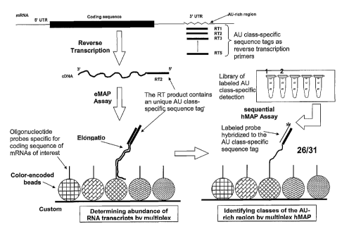
Fig. 26 shows a procedure for the combined quantitative determination of the
concentration, and the identification of the specific class of, AU-rich mRNA
sequences;
17 Fig. 27 shows the sequence alignment of seven maize genes from the zein
gene family
(azs 22) of maize;
Fig. 28 shows a design combining hybridization and elongation permitting the
detection of closely homologous sequences within the zein gene family (az2 22)
of
21 maize;
Fig. 29 shows a design combining hybridization and elongation permitting
detection
of closely homologous genes 16 and 31 identified in Fig. 28; and
Fig. 30 shows a procedure of subtractive differential gene expression analysis
25 employing one detection color.
DETAILED DESCRIPTION
14
CA 02544041 2006-04-27
WO 2005/042763
PCT/US2004/035426
1 Disclosed
are methods, protocols and designs, including systematic procedures
for enhancing the reliability of the process of determining levels of
concentration
("abundance") of multiple nucleic acid analytes by capture to anchored
oligonucleotide
probes, specifically including the concurrent ("multiplexed") analysis of the
expression
levels of a designated set of genes. More specifically, disclosed are methods
for the
optimization of sensitivity, specificity and dynamic range of multiplexed gene
expression analysis, and further, assay protocols including a subtractive
format of
performing differential expression analysis using only a single detection
color. Also
9 introduced is an explicit phenomenological description of the interaction
of targets with
anchored probes in order to evaluate the actual affinity constant governing
this process.
A preferred embodiment of forming planar arrays of capture probes displayed on
color-
encoded microp articles, without recourse to target amplification as in the
case of a
13 cytokine reference panel described herein, may permit completion of
quantitative
multiplexed expression monitoring in as little as three hours or less, from
sample
collection to data analysis (Figs. 1 and 2). These methods and designs are
herein
illustrated by application to a variety of problems involving the capture of
target
17 nucleic acid strands to a layer of immobilized oligonucleotide probes.
I Optimizing Sensitivity and Dynamic Range: Modulation of Probe-Target
Affinity
1.1 Sequence-specific Affinity Governing Hybridization Complex ("Duplex"
Formation - The standard analysis of the hybridization-mediated formation of a
21 complex ("annealing") of two oligonucleotides invokes the law of mass
action to relate
the concentration of complexed ("bound") probes and targets, c = [TP], to the
concentration of uncomplexed ("unbound", "free") probes, herein preferably
displayed
on encoded beads, p = [P], and the concentration of uncomplexed targets, t =
[T], as
25 follows:
[TP] = K [T] [P]
Or
CA 02544041 2006-04-27
WO 2005/042763 PCT/US2004/035426
1 c=Kpt
In analogy to the common practice of computing "melting temperatures", the
(sequence-dependent) affinity constant is computed using a phenomenological
"nearest-neighbor" (NN) model to represent the interaction between adjacent
base pairs
formed within the probe-target complex for given experimental conditions
including
salt concentration and temperature. The free energy of duplex formation, also
referred
to herein as "binding energy" or "condensation energy", is computed in the
form:
AGc= AGNucleation NN-Pairs {AH; + TAS; )
9 where AH; and AS; represent enthalpy and entropy, respectively. The
condition AGc=
0 defines the "melting temperature", TM, widely used in the field to estimate
the
stability of a duplex.
In accordance with standard thermodynamics, the (sequence-specific) affmity
13 constant, Kss, is computed from the expression
Kss = Koexp( -AGc/kT)
wherein K. represents a constant and k denotes the Boltzmann constant.
Given an affinity constant, and given initial concentrations of probe, [P10,
and
17 target, [T]o, the equilibrium concentration of probe-target complex,
[TP], is obtained as
a function of initial target concentration [T]o.
Using this standard model, melting temperatures and affinity constants were
calculated for complexes formed by a 175nt DNA target and seven different DNA
21 oligonucleotide probes varying in length from 15nt to 35nt at a
temperature of 55 C
and a salt concentrations of 2M. Target and probe sequences are shown below in
Table
I-1.
Table I-1
25 Seq ID Sectuence
Target 175- AG GGT AAA ATT AAG CAC AGT GGA AGA ATT TCA TTC
mer TGT TCT CAG TTT TCC TGG ATT ATG CCT GGC ACC
ATT AAA GAA AAT ATC ATC TTT GGT GTT TCC TAT
16
CA 02544041 2006-04-27
WO 2005/042763
PCT/US2004/035426
1 SEQ ID NO. GAT GAA TAT AGA AGC GTC ATC ATC AAA GCA TGC
1 CAA CTA GAA GAG GTA AGA AAC TAT GTG AAA ACT
TTT TG
Target 90- T CAG TTT TCC TGG ATT ATG CCT GGC ACC ATT AAA
mer GAA AAT ATC ATC TTT GGT GTT TCC TAT GAT GAA
TAT AGA AGC GTC ATC ATC AA
SEQ ID NO.
2
Target 40- C ACC ATT AAA GAA AAT ATC ATC TTT GGT GTT TCC
9 mer TAT GAT
SEQ ID NO.
3
Target 25- GAA AAT ATC ATC TTT GGT GTT TCC T
13 mer
SEQ ID NO.
4
Probe 15- CTT TTA TAG TAG AAA
17 mer
SEQ ID NO.
5
Probe 17- CTT TTA TAG TAG AAA CC
21 mer
SEQ ID NO.
6
Probe 19- CTT TTA TAG TAG AAA CCA C
25 mer
SEQ ID NO.
7
Probe 21- CTT TTA TAG TAG AAA CCA CAA
29 mer
SEQ ID NO.
8
Probe 25- CTT TTA TAG TAG AAA CCA CAA AGG A
33 mer
SEQ ID NO.
9
Probe 30- CTT TTA TAG TAG AAA CCA CAA AGG ATA CTA
37 mer
SEQ ID NO.
17
CA 02544041 2006-04-27
WO 2005/042763 PCT/US2004/035426
1 10
Probe 35- CTT TTA TAG TAG AAA CCA CAA AGG ATA CTA CTT AT
mer
SEQ ID NO.
11
Calculated melting temperatures and affinity constants are summarized in Table
1-2.
The very high affinity constants predicted for the longer probes would imply a
9 favorable sensitivity for detection of target. For example, using planar
arrays of color-
encoded microparticles ("beads") of 3.2 L.Lm diameter to display probes in
accordance
with the Random Encoded Array Detection format of multianalyte molecular
analysis,
and setting the number of probes per bead to [P]c, = 105, the law of mass
action provides
13 the following estimate for the lower limit of target detection with the
21-mer probe:
[T] min [PT] min / K [P]c, = [PT] min /1.7 x 101 /M x 105;
here, [PT] min represents the minimum number of probe-target complexes per
bead
17 required to ensure detection, and with [PT] nth, = 103, [T]min 0.6 x 10-
12 pM, a value
corresponding to a message abundance of single copies per cell.
Table 1-2
21 Probe Length T- Melting Temperature, C Affinity Constant (/M)
48.4 5.382x10⁵
17 56.1 3.536x10⁷
19 61.3 1.129x10⁹
21 64.9 1.712x101
29 25 71.1 1.116x10¹³
74.0 2.717x10¹⁵
_
76.2 7.823x10¹⁷
33
1.2 TheRole of Target and Probe Configurations: Implications for Assay Design
As described below, the size and configuration of the target as well as the
size,
37 configuration and arrangement of substrate-anchored probes have a
substantial effect
18
CA 02544041 2006-04-27
WO 2005/042763 PCT/US2004/035426
1 on probe-target interaction which leads to substantial deviations of
actual probe-target
affinities from those predicted by the NN model.
The adverse role of steric effects ("hindrance") in the capture of target
analytes
to immobilized probes, and specifically the importance of probe accessibility,
have
been known in the art; see e.g., Guisan, J.M. in "Immobilization of Enzymes
and
Cells,"Gordon F. Bickerstaff, Humana Press, Totowa, NJ, pp. 261-275 (1997).
Thus,
empirical strategies of enhancing capture efficiency by introducing spacers of
preferred
length in order to alleviate constraints related to probe "packing" have been
described;
9 see e.g., Southern E. et al., Nat. Genet.( suppl.) 21, 5-9 (1999).
However, in contrast to
the known methods, the methods disclosed herein establish the fundamental
interconnection between certain properties of target and probe layer as the
foundation
of a systematic design process guiding the optimization of probe-target
interaction.
13 Probe layer compressibility is identified as a property to be maximized
in order to
facilitate penetration of the target, or portions of the target, into the
layer in the course
of duplex formation. More generally, the design criteria herein reflect the
nature and
magnitude of effects of length, grafting density and electrostatic charge of
substrate-
17 anchored probes, length and configuration of target, and selection of
the location of the
capture subsequence relative to the target's 5' terminus on capture efficiency
and hence
assay signal. Conversely, to pet nit the correct determination of target
abundances,
methods are disclosed to determine the re-normalized constants governing probe-
target
21 interaction.
Disclosed are methods, designs and design rules relating to the selection of
sizes, configurations and arrangements of anchored capture probes, sizes and
configurations of target including the selection of capture subsequences and
the
25 selection of array compositions and protocols, in order to modulate
probe-target capture
efficiencies and to optimize assay sensitivity, specificity and dynamic range.
In order to establish design criteria, the nature and magnitude of effects of
19
CA 02544041 2011-06-27
WO 2005/042763
PCT/US2004/035426
length, grafting density and charge of substrate-anchored probes as well as
size and
configuration of target, or designated subsequences of target, on capture
efficiency and
hence assay signal, are disclosed. Relevant experiments were performed in
accordance
with the Random Encoded Array Detection (READ) format of multianalyte
molecular analysis in which probes are displayed on color-coded polymer
microparticles ("beads"), and beads are arranged in a planar array on a
silicon chip. See
US Publication Serial No. US20040132122, entitled: "Multianalyte
molecular analysis using application-specific random particle arrays."
9 ' Probes preferably are "end-grafted" to beads by way of
a
covalent linkage at the 5' terminus. The analysis of experiments performed on
synthetic model DNA targets as well as model cDNAs generated by reverse
transcription from a 1,200nt Kanamycin mRNA (Promega), establishes a critical
role of
13 target and probe configurations in the interaction of targets with an
immobilized set of
probes, even when the target strands of interest are of such relatively modest
size.
1.2.1 Synthetic Model Targets - Binding isotherms were recorded over a wide
range of
concentration of labeled synthetic DNA targets varying from 25nt to 175nt in
length,
17 and over a range of capture probe lengths varying from 15nt to 35nt (see
Table I-1 and
Example 1).
Target Length Dependence - To investigate the dependence of probe-target
capture
efficiency on the length of the target strand, four fluorescently end-labeled
synthetic
21 DNA targets, 25nt, 4Ont, 9Ont and 175nt in length (see Table 1-1), all
containing a
common subsequence, were permitted to hybridize to a 19nt capture probe
displayed on
color-coded beads of 3.2 [..tm diameter and arranged in a planar array in
accordance
with the READ format. Representative binding curves, reveal a significant
dependence
25 on target length, L. As illustrated in Fig. 3A, the longer the target,
the lower the
signal intensity attained at any given target concentration below saturation;
here, the
intensity is normalized, for each curve, to that attained at saturation.
CA 02544041 2006-04-27
WO 2005/042763
PCT/US2004/035426
1
Estimates of the experimental affinity constants, K*, and the number densities
of available capture probes, [P]0 = p0, were obtained by fitting each profile
to the law
of mass action; results are summarized in Fig. 3B. To compute affinities, the
signal
intensity, I, is herein taken to be proportional to the product of the number
of captured
targets per bead, c, and the number of fluorophores per target, nF, that is, I
- nF c;
interconversion between I and c is facilitated by reference to a calibration
curve,
described in Example II in conjunction with Table 1-3 and Fig. 4. Typical
observed
affinity constants are of the order of K* = 108/M where target length is about
equal to
9 probe length, an order of magnitude lower than those predicted by the NN
model
(Table 1-2). Typical values of p0, the number of occupied sites at saturation,
are of the
order of 105 per bead.
Under typical experimental conditions of interest in the context of gene
13 expression analysis, the size of the target will exceed that of the
probe, and each
captured target will thus occlude more than a single probe; accordingly,
saturation will
reflect the capture of a limiting number, NT Sat, of targets to a bead of
finite area, A0 . A
lower limit of NTsat is obtained by assuming that the bead surface is
decorated with
17 captured targets assuming a "relaxed" configuration in which a target's
characteristic
size is set by its radius of gyration, RG,T^' a II, v denoting a
characteristic exponent
with numerical value v = Y2 for an ideal chain and v = 3/5 for a self-
excluding chain in
a good solvent in 3 dimensions (deGennes, "Scaling Concepts in Polymer
Physics",
21 Cornell University Press, 1979). Accordingly, for the smallest target,
NTSat"-Ao/RG,T2, or
NTSat 1/L. Identifying p0 with the number, NT', of targets captured per bead
at
saturation yields, for example for the smallest target (L = 25nt), an average
molecular
area of AT =-=-= 47c(1.6 m)2 /8*105 4*103k, a value comparable to that
obtained for
25 ATRetaxed õ, TaG;r2 6.5*103A2 when using an (experimental) estimate of
RG,T 9 L112
-45 A (Tinland et al, Macromolecules 30, 5763 (1997)). For the 175nt target,
comparison of the corresponding two values yields AT 1.6*104A2 AT Relaxed
21
CA 02544041 2006-04-27
WO 2005/042763 PCT/US2004/035426
1 4.5*104A.2
.These comparisons suggest that, at saturation, either the larger target
molecules are not in their relaxed, but in a more compact configuration, or
that they are
no longer isolated but are substantially "overlapping," that is,
interpenetrating.
When plotted at a fixed target concentration as a function of target length,
L, the
signal intensity displays a 1/Lx dependence (Fig. 5), with 3/2 sx s 2, as
target length is
varied from L=25nt to L=175nt, and target concentration, at each length, is
varied over
three orders of magnitude from 0.1nM to 100nM. Notwithstanding the fact that
all
targets hybridize to the 19nt probe via the same 19nt subsequence (Table I-1),
implying
9 identical "condensation" energies of duplex formation, the increase in
target length is
seen to result in a substantial reduction in signal intensity. Thus, for given
length of
capture probe, the longer the target, the less favorable the formation of the
duplex and
the lower the effective affinity.
13 The power-law
dependence of the effective affinity governing probe-target
hybridization provides a means of tuning the capture efficiency in accordance
with the
length of specific target strands. This is a particularly useful design
criterion in
applications such as expression monitoring permitting the control of cDNA
lengths by
17 placement of sequence-specific reverse transcription (RT) primers. As
discussed herein
in greater detail, rare messages preferably are converted to short cDNAs to
maximize
capture efficiency.
Probe Length Dependence - A complete set of binding curves such as those shown
for
21 the 19nt probe in Fig. 3 was generated using a set of capture probes
varying in length
from 15nt to 35nt. The binding curves for the 175nt target are shown in Figs.
6A, 6B
along with fits to the law of mass action, assuming, as stated above, I ¨ nF
c, nF
representing the (average) number of fluorescent labels per molecule. For this
set, fits
25 yield values of the affinity constant of the order of K* -5*107/M,
approximately a
factor of 20 lower than those predicted by the NN model (see Table 1-2). The
dependence of signal intensity, at a fixed concentration of targets of length
25nt, 9Ont
22
CA 02544041 2006-04-27
WO 2005/042763 PCT/US2004/035426
1 and 175nt, is shown as a function of increasing probe length in Figs. 7A
to 7C. The
intensity profiles for short probe lengths display the expected increase,
although smaller
than that predicted by the NN model; however, for all four target lengths, the
profiles
peak or level off at a probe length of approximately 3Ont. This is entirely
unexpected
from the point of view of the NN model. Instead, as discussed herein below,
these
results suggest that the capture of target to immobilized probes requires
elastic
deformation of not only the incoming target strands but also of the layer of
capture
probes.
9 I.2.2 Kanamycin mRNA: Selection of Transcript Length and Placement of
Capture
Sequence
It is further shown that, as with synthetic targets, the reduction in length,
L, of
cDNAs, herein also referred to as "transcripts," obtained by reverse
transcription,
13 produces a systematic and significant enhancement in the assay signal of
the shorter
transcript over that attained from the longer transcript given the same mRNA
concentration. As illustrated herein for a 1,200 nt Kanamycin mRNA (Promega),
cDNA products varying in length from ¨1,000 nt to ¨50nt were produced by
selecting
17 suitable RT primers (Example III). Placement of the capture subsequence
near the 5'
end of the cDNA is shown to produce an additional enhancement. Accordingly,
capture
probes preferably were designed to match subsequences located in close
proximity to
the transcript's 5' end (see Fig. 8A). Both enhancements reflect the
importance of
21 configurational contributions to the free energy governing the
interaction of targets
with anchored probes. As a result of these effects, the sequence-dependent
affinity,
Kss, is reduced to an effective affinity, K*(L) <K5, with significant
implications for
the design of anchored capttu-e probes as well as transcripts, particularly
when the
25 fraction of available substrate surface covered by adsorbed target
exceeds a
characteristic value, y* = c*/cmax.
Multiple Primer Multiple Probe (npmp)-RT Protocol - In some cases, multiple
23
CA 02544041 2006-04-27
WO 2005/042763 PCT/US2004/035426
1 reverse transcription (RT) primers were employed (Fig. 8A) so as to allow
for the
possibility of producing multiple cDNA transcripts from a single mRNA template
by
way of displacing a shorter cDNA incorporating a first RT primer placed in
close
proximity to the mRNA's 3 end, by a longer cDNA transcript incorporating a
second
RT primer placed farther from the mRNA's 3'end. For each cDNA, one or more
capture probes - here of length 19nt - were provided (Example In. An
embodiment
for multiplexed expression monitoring invokes the READ format, for example in
the
version illustrated in Fig. 9.
9 L2.2A Effect of Reduction in Transcript Length - Guided by the results of
titrations
on model compounds, as described in Sect. 1.2.1, it was established that a
reduction in
transcript length does indeed yield a substantial improvement in assay signal.
A series of RT reactions, performed on Kanamycin mRNA over a range of
13 initial concentrations in accordance with an mpmp-RT design and assay
protocol
(Example IV), produced the titration curves shown in Fig. 8B. At each mRNA
concentration, ranging from 3611M to 560 pM, the signal recorded for the 150nt
transcript exceeds that recorded for the 1,000nt transcript, notwithstanding
the fact that
17 the number, 1-1F, of fluorophores for the 1000nt transcript exceeds that
for the 150 nt
transcript.
For example, Iisont /I1000nt 3, at the target concentration corresponding to
1.13
nM. The experimental observation of an enhancement of ¨3, for example near the
21 cross-over concentration (see "break points" indicated in Fig. 10A) is
in accordance
with the enhancement anticipated from the reduction in transcript length, L.
That is, the
expected enhancement arising from the reduction in L from 1,000nt to 150nt
would be
given by ¨(1000/150)' (3/15), the first factor relating to length reduction,
as discussed
25 in Sect. 1.2.1 for the model targets (with 3/2 x and
the second factor reflecting
the fact that the 150-mer, at the chosen linear labeling densities, nF (150
nt) 3 and nr (1000
no ¨15. Setting x=3/2, this estimate yields an enhancement of-3.5, comparable
to the
24
CA 02544041 2006-04-27
WO 2005/042763 PCT/US2004/035426
1 experimental observation.
Similarly, a reduction of transcript length from 1,000nt to 5Ont results in an
enhancement of ¨(1000/50)3/2 (1/15) ¨ 6, the first factor relating to length
reduction
(with x=3/2) and the second factor reflecting the fact that the 50-mer, at the
chosen
labeling densities, would contain, on average, only a single label.
Linearized Adsorption Isotherm Representation - Further insight is gained by
representing the titration curves in the form of a linearized adsorption
isotherm
representation which directly follows from the law of mass action. For the
reaction P
9 (probe) + T (target) <¨> C (probe-target complex), mass action implies
the relation c =
Kpt, where c, p and t denote the respective concentrations and K denotes the
affinity
constant. Setting p = c-p0, t = c-to, where Po and to respectively represent
initial probe
and target concentrations, yields c = K(c-p0)(c-t0) and, provided that c
to, as in the
13 experiments reported here, c = K(po - c)to or c = po - c/K to.
Displaying titration results
in the latter form - assuming, as before, that the signal, I, is proportional
to c, I ¨flFC,nF
denoting the number of fluorophores per transcript - highlights the linear
dependence of
c on (c/Kto) and permits the determination of po, from the intercept, and K,
from the
17 slope.. Specifically, abrupt changes in slope signal a cross-over
between regimes, as
discussed in the text.
Fig. 10A displays the titration results for the 1,000nt and 150nt transcripts
in
this format, along with an isotheini obtained in the same manner for a 5Ont
transcript.
21 All three plots indicate a cross-over from a "dilute" regime
characterized by a
shallower slope and hence a higher affinity constant, to a "concentrated"
regime of
steeper slope and hence lower affinity constant. Slopes in the dilute regime
are
comparable for all three transcripts, indicating similar values for the
corresponding
25 affinity constants. In contrast, slopes, and hence effective affinity
constants, in the
concentrated regime are seen to be transcript-length dependent (see Table 1-
4).
As summarized in Table 1-4, at the cross-over - observed for all transcripts
at a
CA 02544041 2006-04-27
WO 2005/042763 PCT/US2004/035426
cDNA Length K [M-1] K [M-1] Crossover Fractional
(nt) (Dilute regime) (Concentrated regime) Conc. [nM]
Coverage at
Crossover [0]
1000 2 x 10⁸ 1 x 10⁷ 0.2
150 2 x 10⁸ 1 x 10⁸ 0.2
50 5 x 10⁸ 2 x 10⁸ 0.5
1
TABLE 1-4 (above)
concentration of approximately to mM - the affinity constant for the 1,000nt
transcript
drops by a factor of ¨20, and that for the 150nt and 5Ont transcripts by a
factor of-'2.
That is, the reduction in the effective affinity is increasingly less
pronounced as
transcript length decreases. In the dilute regime, the slope for adsorption
isotherm of
the 5Ont transcript displays a slope that is smaller by a factor of ¨2.5 than
that for the
9 isotherm of the 150M transcript, indicating a correspondingly higher
value for the
corresponding affinity constant of the former.
The cross-over to this regime occurs at low values of coverage, 0, as may be
seen from the following argument. Transformation of the linearized adsorption
13 isotherm representation to the standard form of the Langmuir isotherm,
1/{1+ 1/K to }
= c/po, displays the fraction of occupied probes, c/po = 0; as discussed
below, is more
precisely viewed as the ratio of the number of probes occupied at to relative
to the
number occupied at saturation. Specifically, extrapolating from the
concentrated regime
17 into the cross-over regime shows that, for the examples in Fig. 10A, K
t0<< 1 and
hence 1/K to = p0/c. Using the estimates obtained above for the effective
affinity
26
CA 02544041 2006-04-27
WO 2005/042763 PCT/US2004/035426
1 constants in the concentrated regime, the estimated fraction of occupied
sites, 0* =
c*/p0,, at the cross-over is ¨0.2 for the 150nt and the 1,000nt transcripts.
That is, the
larger transcripts start to interact at a fractional occupancy of available
bead-displayed
probes of 20%.
Fig. 11 shows the dependence of c* on transcript length, c 1/LY; the limited
available data suggest y a'3/2. This curve delineates the boundary between
dilute
(below the line) and concentrated (above the line) regimes. Generally, to
optimize
capture efficiency and hence sensitivity of detection of rare messages, it
will be
9 advantageous to operate in the dilute regime in order to benefit from a
high effective
affinity constant. This advantage is particularly significant for long
targets. Preferably,
to facilitate detection, targets will be labeled in multiple positions, for
example by
incorporation of labeled dNTPs during reverse transcription, as described
herein.
13 Conversely, the analysis of experimentally recorded signal intensities
must reflect the
fact that cDNAs of different lengths, even when they are present at equal
abundance,
generally will produce substantially different signal intensities. That is,
solution
concentrations must be evaluated using the effective affinity constants if
message
17 abundances are to be reliably determined.
L2.2B Effect of Capture Probe Placement: Terminal Capture Sequences - It is
also
disclosed herein that the effective affinity governing capture efficiency and
hence assay
signal and sensitivity is enhanced by locating capture subsequences near the
5' end of
21 long transcripts, as illustrated in Fig. 12A, depicting the relative
alignment of RT
primers as well as internal and terminal probes relative to the 1,200nt
Kanamycin
mRNA. Fig. 12B displays the comparison of titration results obtained for the
capture of
a 500nt transcript to two different (sets of) 19-mer probes, one (set)
directed to a
25 subsequence located near the 5'- end of the transcript, the other
directed to a
subsequence located in the interior of the transcript. The use of the
"terminal" capture
probe leads to an enhancement by a factor of ¨1.5 in assay signal over that
recorded
27
CA 02544041 2011-06-27
WO 2005/042763 PCT/US2004/035426
1 with "internal" probe. Transforming these results in accordance with the
adsorption
isotherm. format (Fig. 13) indicates the effect of placing the capture
subsequence near
the transcript's 5' terminus to have an effect on the isotherms analogous to
that
produced by length reduction. This is consistent with the view that capture of
the
terminal subsequence is equivalent to capture of a shorter target, requiring
less
configurational adjustment in probe layer as well as incoming target, and
thereby
reducing chain entropy-mediated repulsive effects, as elaborated below.
The results disclosed so far imply that the quantitative determination of
message
9 abundance requires a careful analysis of the effective affinities
governing the interaction
between targets and anchored probes.
L3 Empirical Design Rules - A priori knowledge of the sequence of transcripts
to be
detected. in "diagnostic" expression profiling permits the design of capture
probes directed
13 against specific target subsequences in order to enhance sensitivity,
preferably selecting
terminal capture probes, modulate the dynamic range by selecting the operating
regime to
be above or below c*, and to optimize specificity, methods and designs for
which are
described in greater detail in Publication US20060127916.
17 The following empirical design rules are useful in guiding the
optimization of
probe-target interaction. These rules also indicate the need for corresponding
corrections
in the analysis of signal intensity patterns, as further discussed in Sect.
II.
1 - Minimizing Target Length
21 Minimize the target length, L, in order to maximize the effective
affinity
constant, K* = K*(L), governing target hybridization to an intntobilized
probe;
25 2 - Placing Capture Subsequence near 5' Terminal
For given target length, place the designated capture subsequence as
close as practical to the target's 5' terminus;
29 3 - Selecting Dilute or Concentrated Regime of Operation
Control the effective affinity constant, K*, governing interaction of a
28
CA 02544041 2006-04-27
WO 2005/042763 PCT/US2004/035426
1 specific target with immobilized probe by working in the dilute
regime to
realize a high value of K*, or in the concentrated regime, to realize a
low(er) value of K*;
Corollary: Compressing Signal Dynamic Range
For high abundance messages, produce long transcripts so as to reduce
K*; for low abundance messages, produce short transcripts so as to
increase K*, thereby compressing a given range of message abundance
9 into a smaller range of signal intensity;
4 - Adjusting Grafting Density for Quantitative Analysis
To perform a quantitative determination of target concentration, limit
13 the capture probe length to a maximum for given probe grafting
density
or limit the grafting density for desired probe length so as to avoid
"saturation";
17 5 - Adjusting Layer Configuration for Maximal Sensitivity
Set the grafting density, a, to the maximal possible value without
substantially reducing the rate of target penetration; limit a to a preset
small multiple of probes per target at saturation;
21
6- Confining Duplex Formation (see below)
Select the bulk ionic strength (and, where practical, pH) so as to
minimize the rate oftarget-target duplexformation without substantially
25 reducing it in the probe layer;
These empirical rules will be made more precise on the basis of a
phenomenological
model developed in the following section.
29 H. Model of Target Capture to a Layer of Immobilized Probes
ILL General Description
To account for the observations presented in Sect. I, and to provide a basis
for the
refinement of design rules into a systematic design process guiding the
selection of optimal
33 probe layer and target configurations, the present invention discloses a
phenomenological
model for the capture of single-stranded (ss) DNA or RNA targets to a layer of
end-grafted
probes, each such probe designed to be complementary to a designated "capture"
subsequence within the cognate target. Specifically, this model views the
formation of a
29
CA 02544041 2011-06-27
WO 2005/042763 PCT/US2004/035426
1 duplex between a capture probe and a designated target subsequence as an
adsorption
process which requires the penetration of a portion of the target into the
probe layer. This
involves an elastic deformation of the layer as well as the confinement of (a
portion of) the
target which will be accompanied by a loss of configurational entropy. The
formation of
anchored probe-target complexes is thus viewed herein as a gating process
which
mediates the transformation of the end-grafted probe "monolayer" into a probe-
target
"bilayer".
Polyelectrolyte Brush - In one way, the model presented herein is thus
informed by the
9 process of polyelectrolyte adsorption to a deformable substrate, this
substrate displaying
the characteristics of a polyelectrolyte "brush", or, under certain
conditions, that of a
polymer "brush, "composed of end-grafted probes (Fig. 14; Pincus,
Macromolecules 24,
2912-29'19 (1991) see also:
Fleer et al, Sect.4 in: "Polymers
13 at Interfaces", Chapman Hal], 1993). In a layer of end-grafted probes at
lateral density a,
the characteristic separation, d, between adjacent probes, a ¨ d2, and the
characteristic
size, , of each
probe in a relaxed or expanded ("mushroom") configuration, are
interrelated: as long as Z< d, individual "mushroom"configurations are
unconstrained
17 by their neighbors; however, when probe chains start to overlap, "mushroom"
configurations become constrained, and probes will adopt increasingly
"stretched"
configurations, thereby transforming the probe layer into a "brush" in which
chain ends
tend to be displaced toward the free surface (Fleer et al, op.cit.; Milner,
Witten & Cates,
21 Macromolecules 21, 2610 - 2619 (1988)).
As described herein, the high charge density realized within a layer of
anchored
oligon u cleotide probes permits operation under a variety of external
conditions, with the
possibility of realizing a variety of probe layer configurations. These are
determined
25 prirnarily by the probe grafting density, a, and by the effective linear
charge density, f, 0
<f < 1, reflecting the degree of dissociation, a, of probes within the layer
in response to
solution conditions, especially pH, temperature and salt concentration, Cs.
CA 02544041 2006-04-27
WO 2005/042763 PCT/US2004/035426
1 For
example, denoting by k the dissociation constant for the solution reaction AH
'4A-+ H+, a Bulk [A]/[All] is given in terms of k and [HI in the form aBk =
1/{1 +
[H1/k} ; generally [H+] > [g]Buik and aa
<
Bulk, and f = f(a) or, more precisely, f = f(k,
CBuiks)= When the salt concentration, CBInkS in the bulk solution is low,
counterions are
retained in order to maintain electroneutrality in the interior of the brush
at the expense of
a loss of entropy ofmixing. Under the action ofthe corresponding osmotic
pressure, chains
are expected to be fully elongated, regardless of grafting density.
Conversely, at
sufficiently high bulk salt concentration, excess mobile co-ions and
counterions can
9 penetrate into the brush and screen electrostatic interactions within the
brush; as the
osmotic pressure associated with the trapped counterions is diminished, the
appearance
of relaxed chain configurations - and a corresponding reduction in layer
thickness - are
expected. Under the high salt concentrations, in the range of-100mM to ¨2M,
frequently
13 realized
in conventional hybridization experiments, a collapsed state can result in
which
counterions are no longer distributed throughout the layer but are associated
with anchored
probe chains (or probe-target duplexes).
Interfacial Film ofS hort Amphiphiles - In another way, the model herein is
informed by
17 the
process of adsorption of solutes, say proteins, to monomolecular ("Langmuir")
films
composed of amphiphilic molecules such as phospholipids, surfactants or
certain peptides
adsorbed at an air-water or oil-water interface. Insertion of solutes into
such a film requires
local film compression, mediated by changes in chain packing and
configuration, in a
21 manner analogous to that produced by lateral compression. As a function of
grafting
density, the interplay of orientational and configurational degrees of freedom
can produce
a variety of phases; for present purposes, phases, or coexistence regions of
high lateral
compressibility are of principal interest. While the following discussion
employs the
25 language of polymer theory, it is understood that any extensions or
refinements likely
possible for layers of short probe chains by reference to the known phase
behavior of
interface-adsorbed amphiphilic ("Langmuir") films also are included herein.
31
CA 02544041 2006-04-27
WO 2005/042763 PCT/US2004/035426
The phenomenological model is to elucidate the critical role played by elastic
effects arising from distortions in target and probe layer configurations
required for duplex
formation between targets and probes, particularly when either targets or
probes are
immobilized. Further, it is to provide a basis for the refinement of the
empirical design
rules delineating optimal "operating regimes" for target capture to
immobilized probe
layers and for the completion of assay protocols. For example, such protocols
may call for
target-mediated, polymerase-catalyzed probe elongation, as illustrated below
in
connection with a method of signal amplification which will require
penetration into the
9 probe layer of additional assay constituents including enzymes.
IL 1.1 Probe Layer Deformation and Target confinement: Renormalization of
Affinity
Constant
A (portion of a) target penetrating into a layer of end-grafted probes will
increase
13 the local segment concentration and will generate a corresponding osmotic
pressure; in
addition, the incoming target also will induce an elastic deformation of the
layer which is
mediated by chain elongation ("stretching"), as illustrated in Fig. 14. The
osmotic pressure
and elastic energy of chain elongation act to repel the incoming target, and
thus provide
17 a repulsive contribution, Gp, to the free energy of duplex formation. It
is this repulsive free
energy which contributes to the entropic stabilization of colloidal
suspensions; however,
while in that instance, optimal grafting layer configurations are those which
minimize
interpenetration of chains on colloidal particles coming into contact, the
present objective
21 in optimizing capture probe layer configurations is to facilitate target
strand penetration
into the layer.
At very low grafting density, for example, in the limit d e/2 >>RG, T,
isolated
probes assume a relaxed ("mushroom") configuration of size RG, p aPV, v = 3/5,
and
25 target capture will proceed in the absence of the constraints imposed by
local chain
"pact: itg"; however, the maximal number of targets captured will be small and
the
cones; o nding assay signal low. Conversely, at high grafting density, for
example such that
32
CA 02544041 2011-06-27
WO 2005/042763 PCT/US2004/035426
1 d s <<RG, T, particularly under conditions producing full chain
elongation, the
number of available capture probes will be high, but the lateral
compressibility of the layer
will be low and target capture will be inefficient and the assay signal low;
here, T denotes
a characteristic target "blob"size in a partially elongated target.
Accordingly, to optimize
target capture to a layer of immobilized probes, the grafting density is
optimized so as to
provide the highest possible number of probes per unit area without
substantially reducing
compressibility. For example, given an actual target of which a portion of
size T is to
participate in duplex formation, the optimal grafting density can be found by
providing a
9 synthetic target of size T and determining - under fixed external
conditions - the assay
signal reflecting fraction of captured target as a function of increasing
grafting density
until a plateau or peak in the resulting profile is obtained. "Indirect" probe
anchoring, for
example to a flexible "backbone" which is in turn attached to the solid phase,
also can
13 alleviate constraints. See US Publication Serial No. US20050260611,
entitled:
"Surface Immobilized Polyelectrolyte with Multiple Functional Groups Capable
of
Covalent Bonding to Biomolecules."
Targets, or portions of targets, in order to make contact with the capture
sequence,
17 must adjust to the local configuration of the probe layer or the already
formed composite
probe-target layer (see Fig. 10,10C, Fig. 15). The resulting confinement
oftarget strands
and corresponding loss of configurational entropy - even in the dilute regime -
represents
a repulsive contribution, GT, to the free energy of duplex formation. The
degree of
21 confinement imposed on ss DNA or RNA, will depend on the specific
unconstrained
("relaxed") configuration assumed by these polyelectrolytes under conditions
prevailing
in solution - even without the considering the possibility of sequence-
specific interactions
("folding"), a complex phase behavior is expected (see e.g., Schiessel &
Pincus,
25 Macromolecules 31, 7953 -7959 (1998)). For purposes of illustration:
penetration of a
portion of target of length T and, assuming a Gaussian coil configuration, of
size RG,
v = 3/5, into a probe layer of local grafting density, a, will require an
elastic energy
33
CA 02544041 2006-04-27
WO 2005/042763 PCT/US2004/035426
1 of target deformation GT (RG, T/CY)2
a i /o.
That is, the larger the portion of target
penetrating into the layer relative to the characteristic distance between
adjacent probes,
d cc', the more difficult the requisite deformation of the target.
The sequence-dependent "condensation" energy, Gc, which favors the formation
of probe-target pairs must be balanced against these repulsive contributions
to the free
energy, Get = Gp + GT; accordingly, the free energy governing probe-target
complex
formation has the form G- Get- G An immediate consequence of this form of the
free
e.
energy is a "renonnalization" of the sequence-dependent affinity constant,
Ics, to an
9 effective affinity constant, K* <K. As long as Gel < Gc, condensation
will still occur, but
with a smaller net gain in free energy, -AG*c = -AGc + Gei, > -AGc, and a
correspondingly
smaller effective condensation energy implies a smaller effective affinity
constant,
K* exp(-AG*c/RT) <K55 exp(-AGe/RT);
13 as well as a lower "melting temperature", T*m < TM, wherein T*m is
determined from the
condition AG(T*m) AG*c(T*m) =0 and TM is determined from the condition AG(TM)
= 0. Substantial corrections to the sequence-specific values must be
anticipated, in fact,
elastic effects can suppress duplex formation altogether.
17 One
method of assessing effective affinity constants is the empirical method,
described herein, of performing isotherm measurements using probe payers of
defined
configuration and synthetic targets comprised of one target containing only
the
subsequence of interest of length T, and additional targets containing the
subsequence of
21 length T embedded in a total sequence of length L> T. Ignoring excluded
volume effects,
the probe layer configuration is determined, for given probe length, P, by
grafting density,
cF, and effective linear charge density, f, 0 <f< 1, the latter in turn
reflecting experimental
conditions, especially salt, pH and temperature, realized in bulk solution.
From these
25 isotherm measurements, values for the effective affinity constant in
various regimes of
target concentration are readily extracted.
34
CA 02544041 2006-04-27
WO 2005/042763 PCT/US2004/035426
1 Another
method of assessing effective affinity constants, complementary to the
empirical method, is that of invoking a phenomenological model of probe-target
capture
to account for the effects of elastic and electrostatic interactions.
11.1.2 Design Considerations
Probe Layer Configuration: Preferred Grating Density - For given grafting
density, a,
overlap between adjacent chains in a "mushroom" configuration begins to occur
when the
transverse displacement of probe chains, s1, is comparable to d, that is, s,
aPv d, P
denoting probe length and a denoting a monomer or segment size. With v = 1/2,
the
9 condition becomes a213 ¨ (12 ¨ 1/ a and hence P 1/ aa2. Given a preferred
length, P, for
the capture probe of interest, the grafting density therefore preferably is
adjusted such that
a < 1/a2P.
Considering target penetration to increase segment density in a manner
equivalent
13 to that
of an increase in probe grafting density, suggests a modification of this
rule. Given
a preferred length, P, for the capture probe of interest, and anticipating
penetration of a
portion of target occupying at least the same footprint as the probe, select a
preferred
grafting density such that oaf = go < g/a2P, 1/2 < g < 1; for example, with
g=1/2, that is,
17 T = P (a
situation realized to good approximation in the case of terminal capture,
Figs.
12A, 12B, 13), select oeff < 1/2a2P in order to accommodate the anticipated
insertion of
target.
Free Energy of Probe Layer: Osmotic Pressure and Elastic Deformation - The
21
penetration of a target strand, or a portion thereof, into a brush of end-
grafted probes leads
to an increase in local segment density, ck. For a brush of area Ao and
thickness D = D(a)
containing np chains, cl) S/A0D(a) (np/A0)13/D(a), P representing the number
of
segments per chain; hence, cl) aP/D(a). An increase in leads to an increase in
the
25 osmotic
pressure, II ¨ (1)w, w denoting a characteristic exponent, and to a decrease
in the
layer compressibility, x := (14)avail. Introduction of each additional segment
also leads
to elastic deformation. For example, in a brush composed of strings of "blobs"
(Fig. 14),
CA 02544041 2006-04-27
WO 2005/042763 PCT/US2004/035426
1 elastic deformation reduces the characteristic "blob" size, with a
corresponding cost in
free energy arising from the requisite stretching of chain segments and the
concomitant
increase in brush thickness, D = D(o). Assuming each blob to contain Pg
segments,
-aPBv, yields PB
liv/a; if each probe chain of length P contains P/PB blobs spanning the
thickness of the brush, D -(P/P5)Cp aP 14/V and, with CI, G-1/2, D ap -1/3.
u That
is, an
increase in grafting density leads to an increase in layer thickness as a
result of chain
elongation. This type of scaling relation arises very generally from the
balance of a
repulsive contribution (e.g. excluded volume, electrostatic interactions) and
the attractive
9 contribution of chain elasticity.
Control of Grafting Density - Unless limited by the lateral density of
adsorption sites
provided on solid phase carrier surfaces, the grafting density realized in the
formation of
the probe layer by covalent end-grafting reflects the balance between a
characteristic
13 adsorption ("binding") energy (per probe) and repulsive interactions
such as the elastic
deformation of the growing probe layer required to accommodate an additional
probe. That
is, the grafting density defines a characteristic area per chain, AB d2 1/0.
hi this case,
grafting density reflects the conditions pertinent to the covalent
functionalization of solid
17 phase carriers, notably the concentration of probe and the conditions of
incubation.
The experimental observation of a maximal capture efficiency at typical values
of
P -30 suggests a characteristic "footprint", per
chain. Using pc, -6* 105 (Fig. 6B) as an
estimate of the maximal number of targets (of size L=25nt) accommodated per
bead (of
21 3.2 p,m diameter), and assuming each of these targets to be hybridized
to one probe equal
in size to the captured target, the average molecular area is estimated to be
AB Tc(1.6p,m)2
/2*6*105 0.65*103k following target capture, or twice that value prior to
target capture,
the latter corresponding to a probe grafting density o = 1/Ap -
7.5*1012/cm2.This suggests
25 a pictu re of a "self-limiting" grafting process producing - at least
under conditions applied
in the production of solid phase carriers used in the experiments cited here -
a layer in
which end-grafted probes are no longer in their relaxed configuration but
assume a
36
CA 02544041 2006-04-27
WO 2005/042763 PCT/US2004/035426
1 partially elongated configuration; partial elongation would be consistent
with a
configuration in the form of an elongated string of "blobs" of characteristic
radius 4 ¨
(1.25*103A2/m)1/2 ¨ 20A < RG,p =9 L1/2 -50A (Tinland et al, op.cit.), Re, p
denoting the
radius of gyration_ of an unconstrained probe chain in solution. That is, in a
brush produced
by a "self-limiting" grafting process, o =
As discussed herein, high grafting densities, particularly those realized in
typical
conditions of in-situ synthesis of oligonucleotide probes (Lipshutz, R.J. et
al., Nat. Genet.
(suppl.), 21,20-24 (1999); Shchepinov, M.S. et al., Nucleic Acids Research 25,
1155-1161
9 (1997)) generally maybe unfavorable. Spotting of probes generally will
not produce end-
grafted layers but rather more complex "crumpled" layers (Netz & Joanny,
Macromolecules 32, 9013-9025 (1999)) in which molecules may be attached to the
solid
phase at multiple (random) sites , leaving only a small portion of probe
sequences -
13 unknown a priori and highly variable from spot to spot - accessible to
the target. Control
of gra fling densities may be difficult to achieve in this situation.
Preset values of o lower than that attained in the "self-limiting" case are
realized,
for example, by introducing an intermediate step into the process of
microparticle
17 functionalization. Specifically, introduction of a bifunctional modifier
in the forn of a
functionalized polymer such as bifunctional polyethyleneglycol ("PEG")
molecules of
adjustable molecular weight, biotin-binding proteins like NeutrAvidin,
Streptavidin or
Avidin, and any other heterofunctional polymeric linkers of known molecular
size sets an
21 upper limit on the probe grafting density, which is now determined by
the size of the
modifier and its lateral "packing" at the bead surface (Figs. 16A, 16B). In
the embodiment
using the READ format, in a first step, the modifier is covalently attached to
a color-
encoded microp article ("bead"), and, in a second step, the modifier is
functionalized by
25 covalent attachment of the capture probe, preferably way of a 5'
modification introducing
a functional group such as amine or biotin using standard conjugation
chemistry.
37
CA 02544041 2006-04-27
WO 2005/042763 PCT/US2004/035426
1 Target Strand Confinement: Dilute and Concentrated Regimes of Adsorption -
The
discussion of the elastic response of the probe layer to target insertion
suggests that elastic
deformations of the composite probe-target layer give rise to the observed
cross-over
between dilute and concentrated regimes in the adsorption isotherms (Fig.
10A),
delineated by the locus c*(L) for which the limited available data suggest c*
1/L3'2 (Fig.
11).
In the limit of small targets, the principal effect of capture will be that of
increasing
the segment density within the probe layer, as discussed above, suggesting the
cross-over
9 to reflect the transition of the probe layer, or more generally, the
layer foimed by capture
probes of characteristic size p < and already captured targets of
characteristic size VT
<CT, into a regime of lower compressibility. That is, the cross-over occurs
when nT*VT2
np4v12 ,.._,T1*A0, hence 11* (np:IvA0)Ap2 +(nT*haio)FAT2,_ po Vp2 +c*CAT2 and
c* (11* _
13 po Ap2)IAT2.. In the special case CAp2 /\ T2 r A2,
C* Po¨
r1*/V2, or, assuming V2¨ LY, 0
1, c* + po¨ ri*/LY; in he special case np* = nT* = n, .q* (n*/A0)VpT2 or c* =
(n*/A0)
ri*g Ayr 2, where '`,,T2 represents the footprint of the probe target duplex;
here, as before,
0 .<1-1* 1. This limit may be realized either by providing a short target, not
a generally
17 available design in practice, or by placing the designated target
sequence in proximity to
the target's 5' end. The latter possibility is illustrated herein in
connection with Fig. 15.
In contrast, in the limit of large targets, in exact analogy to the "self-
limiting"
21 grafting process of producing the grafted probe layer, the cross-over
reflects the incipient
overlap ("crowding") of target strands in the growing layer of captured
targets of (overall)
size L and characteristic "footprint" CT2; target overlap occurs when nT*V
¨11*A , 0 µ1-1*
implying c* nr*/.A0 ri*V 1/L where ri*A0 represents the fraction of the
25 available area covered by captured target.
38
CA 02544041 2006-04-27
WO 2005/042763 PCT/US2004/035426
1 Adjusting Grafting Density to Allow for Target Penetration, Refilled
The expression derived for the second case represents a design rule which
maybe applied
to optimize the grafting density of the probe layer so as to ensure
realization of the dilute
regime in accordance with the boundary delineated in Fig. 11:
Adjust grafting density so as to maximize c* 77*/L + p 0 (or analogous
condition
for the more general case, T for example, in the preferred embodiment,
select
specific target lengths, L, for example, as described for the case of cDNA
targets
by placement of RT primers, then adjust a
9 The two limits represent special cases of the more general case in which
the cross-over
reflects a transition in the elastic response of the hybrid probe-target
layer. The elastic
deformation of the probe-target hybrid, in conjunction with the elastic
deformation of the
target assuming the confined configuration required for duplex formation, also
is invoked
13 herein to account for the observed dependence of target capture
efficiency on 1/Lx, 3/2
x s 2, in the adsorption isotherms recorded for model targets containing the
same capture
subsequence, T, embedded within a sequence of increasing overall length, L.
Thus, the
probability of "locating" a finite subsequence occupying a finite volume
within a "coil"
17 of volume RG, 3 129, will scale as ¨ 1/L3v, v = 3/5.
Target Capture under Conditions ofLow (Bulk) Ionic Strength: Polyelectrolyte
Brush -
Typical values of grafting densities described herein in relation to the
preferred
embodiment of the invention, namely ¨106per bead of 3.2 p.m diameter (or
¨3*1012/cm2)
21 correspond to high intralayer volume charge densities, zCP. For example,
for an
oligonucleotide of length P = 20, assuming a corresponding probe layer
thickness D
50A, CP =106/(7(3.2)2D) ¨ 10mM for the concentration of probe chains, and thus
yielding
a corresponding value of fCP = 200/7/M, f = 20, for the local concentration of
charges
25 associated with (fully dissociated) backbone phosphate groups.
In electrochemical equilibrium, the concentrations of cations and (poly)anions
present in the interior of the probe layer and in bulk solution are
interrelated in accordance
39
CA 02544041 2006-04-27
WO 2005/042763 PCT/US2004/035426
1 with the condition C+ C-= C Bulk+ CBulk- = Electroneutrality requires,
within the probe layer,
C- + fCP = C+, and in bulk solution, CBuik+ = C
- Bulk- = CBõik . Accordingly, the concentration
of cations within the layer, for given negative charge fCP, can substantially
exceed the
concentration of cations in bulk solutions:
C+'1/2 fCP (1 + {1+ (4CB,Lik2/fCp2)}14
For example, in the limit CBuik/fCP << 1, C+ fCP CBulk. That is, counterions
are
retained within the brush even in the presence of a large gradient in ion
concentration; in
fact, they are distributed throughout an effective volume,Veff which is
smaller than the
9 volume, V, of the brush by the finite volume occupied by the probe
chains, Veff V(14).
The corresponding Debye screening length, 1/x, associated with the
backbone
charge, fCP , per chain, is obtained from the expression Ic2= 47c1BfCP, 113 =
e2/ET denoting
the Bjerrum length, and CP = P/d2D. Balancing the repulsive contribution
arising from the
13 osmotic pressure II = fCPT generated by counterions trapped within the
brush with chain
elasticity, fCPT = l(D/d2, with an elastic constant k = T/a2P, yields D fl4aP,
independent
of grafting density, so that E d(a /4Tclf 1/4)'/2 . This scale is set by the
mean separation, d,
between chains, and hence the grafting density. In the limit E D, chains are
elongated
17 for any degree of charging, f> 0, producing the maximal brush thickness
independent of
grafting density. Provided that the grafting density is sufficiently low so as
to
accommodate penetration of incoming target, capture to such a layer in the
configuration
of a "bed of nails" can proceed without significant elastic distortion of the
probe layer. The
21 return to partial chain elongation in accordance with the "blob"
configuration is achieved
by addition of free co- and counterions at sufficient concentration so as to
ensure that the
Debye screening length 1cace-1 associated with these free ions is comparable
to E so that
E.KFree a I.For such a screened brush, the internal configuration, while
qualitatively
25 resembling that of the semidilute polymer brush composed of a string of
"blobs", will
respond to conditions maintained in bulk solution in order to maintain
electrochemical
equilib r: urn.
CA 02544041 2006-04-27
WO 2005/042763 PCT/US2004/035426
1
Confining Duplex Formation to Interior of Charged Probe Layer - In this case,
while
exposed to a salt concentration of only linM in solution, generally considered
to preclude
duplex forrnation (Primrose, "Principles of Genome Analysis", Blackwell
Science, 1995),
the target, once it has penetrated into the probe layer, actually encounters a
far higher local
salt concentration and conditions of electrostatic screening that are
favorable to duplex
formation. That is, the probe layer provides a local chemical environment
permitting
probe-target hybridization under nominal conditions of extreme stringency in
the bulk
solution which counteract the formation of secondary structures in ssDNA or
RNA and
9 prevent
rearmealing of dsDNA in bulk while permitting (local) duplex formation within
the probe layer. This scenario preferably is realized in accordance with the
rule:
Adjust grafting density so as to ensure a condition of high brush interior
charge
and eletroneutrality to realize conditions permitting duplex formation while
13
selecting conditions of high stringency in external solution so as to prevent
duplex
formation.
41
CA 02544041 2011-06-27
WO 2005/042763
PCT/US2004/035426
11.2. Procedures
11.2.1 Assay Design Optimization
Given a sequence, or sequences, of interest, specifically a set of mRNA
messages,
proceed as follows, applying design rules as appropriate:
= Target Subsequence of Interest
= Target Length (number of nucleotides);
Cr Target Abundance;
9 ampC Target Abundance following Amplification
Sp Primer Sequence
Sc Capture Sequence (i.e., target subsequence to be analyzed
by
capture to probe)
13 A Linear Labeling Density
= Probe Length (number of nucleotides);
Probe Grafting Density
Cs Salt Concentration
17 C* Target Concentration at Cross-over
L* L( C*);
SelectTargetLerigth(C, C*, Sp); 1* By placing primer, select
21 target Length
in accordance with given or
anticipated target abundance
IF(C LO W) RETURN( L < L* ); /* ensure operation in dilute
regime *1
IF(C HIGH) RETURN( L > L* ); 1* ensure operation in cone
29 regime *1
33 SelectCaptureSequence (ProbeSeq); /* The optimization of
primer and probe
sequences preferably is
performed concurrently (see
37 US Patent No. 7,574,305)
RETURN(Sc= TerminalCapture Sequence( ) );
41
42
CA 02544041 2006-04-27
WO 2005/042763
PCT/US2004/035426
1
SelectFinalTargetAbundance(L, L*, C); /* For given initial message
abundance, select target
amplification conditions to
establish operating regime */
IF( L >L*)
9 IF(C LOW) RETURN( ampC s C*); /*
dilute
regime
13 IF(C HIGH)
RETURN( ampC > C*); /* colic
regime
17 ELSE IF( L < L* )
IF ( C LOW) RETURN( ampC > C*); /*
best to operate in comic
21 regime */
IF( C HIGH)
IF( C < C*) RETURN( ampC s C*);
25 ELSE RETURN( ampC = C);
29
SelectLabelingDensity(L, ampC); /* NOTE: if m'plex RT or
m'plex amp,
33 A will be identical for all
targets */
37 /* for long targets:
operate
in dilute regime, select high
labeling density */
41 /* for long targets at high
abundance: select low
labeling density */
R_E TURN(2);
43
CA 02544041 2006-04-27
WO 2005/042763
PCT/US2004/035426
1
OptintizeTargetConfiguration(L, A, C, S0 S, S)
IF( C Fixed) L = SelectTargetLength(C, C*, Sp);
ELSE IF( L Fixed) anzpC =
SelectFinalTargetAbundance(L, L*,
C);
9
A= SelectLabelingDensity( );
Sc= SelectCaptureSequence (ProbeSeq);
13
OptintizeProbeLayerConfiguration()
P = SelectProbeLength( ); 1* maximize Kss while
17 minimizing cross-
hybridization */
a= AdjustGrafting Density(P, L); 1* the longer the probe, the
21 lower a; allowing for
insertion of target of known
length *1
Optimizellepresentation( )
SelectTypeRedundancy( );
29
OptimizeReactionConditions( )
33 SelectionicStrength();
main
37
44
CA 02544041 2006-04-27
WO 2005/042763
PCT/US2004/035426
1 FOR( each Target in Designated Set)
OptitnizeTargetConfiguration();
OptimizeProbeLayerConfiguration( );
OptimizeRepresentation( );
OptimizeReactionConditions( );
9 11.2.2 Evaluation of Effective Affinity Constant
Sc Capture Sequence (i.e., target subsequence to be analyzed by
capture to probe)
13 P Probe Length (number of nucleotides);
Cs Salt Concentration
EvalEffectiveFreeEttergy(S P, Cs, pH);
17
AGT= EvalTargetElasticFreeEnergy(TargetConfig,
ProbeLuyerConfig);
21 AGp = EvalProbeLayerElasticFreeEnergy(TargetConfig,
ProbeLayerConfig);
Return(dG AGT + ZiGp - tiGc);
EvalCondensationFreeEnergy(S0 P, Cs, pH, T);
29
Return( AG c= SuntNNBasePairInteractions(So P, Cs, pH, T) );
33
main( )
FOR( each Target in Designated Set)
37
4G c= EvalCondensationFreeEnergy(Sc, P, Cs, pH, T);
ZIG = EvalEffectiveFreeEnergy(dGc , TargetConfig,
ProbeLayerConfig);
41
K = K0exp(-11G/kT)
CA 02544041 2006-04-27
WO 2005/042763
PCT/US2004/035426
1 11.2.3 Assay Signal Analysis
al: Array of Assay Signal Intensities
aK: Array of Affinity Constants
aSc: Array of Designated Target Subsequences
aCT: Array of Target Concentrations
aP: Array of Probes
9
EvalEffectiveAffinityConstant(aK, aSc, aP) /*
See IL 2.2 */
13 FOR( j=0; j ..Nuntber of Targets in Designated Set; j++)
AGc EvalCondensationFreeEnergy(aSc(j), aP0), Cs,
Pil; IT);
17 LI G = EvalEffectiveFreeEnergy(AGc, Target Config,
ProbeLayerConfig);
aK(j) = Koexp(-AG/kT)
21
25 7* NOTE: evaluation of effective affinities generally will have to
include
coaffinities */
main()
29
RecordAssaySignal(1V, al);
EvalEffectiveAffinityConstant(aK, aS c, aP, Cs, pH, T);
CorrectAssaySignal(aI, aK);
33 EvalTargetConcentration(aI, aCT);
37
46
CA 02544041 2006-04-27
WO 2005/042763
PCT/US2004/035426
1 III. Assay Methodologies
This section discloses several methodologies relating to optimization of
sensitivity, dynamic range and assay specificity, particularly pertaining to
the
multiplexed analysis of abundances of highly homologous messages, and farther
discloses a design strategy for subtractive differential gene expression
analysis using
only a single detection color.
III.1 Tuning of Signal Intensities
In nucleic acid analysis, target analyte concentration can vary over a wide
9 range. Thus, multiplexed expression monitoring generally will encounter a
range of
message abundance from low, corresponding to one or two mR_NA copies per cell,
to high, corresponding to 104 copies per cell or more. The requisite dynamic
range of
4 decades for the simultaneous detection of signals from the weakest and the
13 strongest transcripts will exceed the capabilities of many cameras and
recording
devices. The modulation of probe-target affinities as well as certain methods
of array
composition provide the means to tune the signal intensity in accordance with
known
or anticipated message abundance.
17 III.1.1 Optimization of Array Composition: Operation in Dilute vs
Concentrated
Regime
The selection ofRT primers for producing cDNA transcripts of desired length
from an mRNA subsequence of interest, and the selection of 5'-terminal target
21 subsequences for capture, in accordance with the considerations
elaborated herein,
permit the modulation of probe-target affinity and thus the control of the
dynamic
range of assay signals indicating target capture.
Selection of Transcript Lengtlz - In the simplest case of an assay design
calling only
25 for reverse transcription, but not amplification, the concentration of
cDNAs reflects
the abundance of mRNAs in the original sample; that is, the target abundance
is
given. Then, a judicious choice of transcript length, and/or the placement of
capture
subsequences, permit the maximization of detection sensitivity and the
simultaneous
29 "compression" of signal dynamic range by way of tuning the effective
affinity
47
CA 02544041 2006-04-27
WO 2005/042763 PCT/US2004/035426
1 constant.
To compensate for the low abundance of transcripts representing rare
messages, a short transcript length is preferably selected in order to realize
the highest
possible effective affinity constant and to maximize the assay signal produced
by
hybridization of these transcripts to anchored probes. This will ensure
maximization
of the detection sensitivity. Conversely, to compensate for the high abundance
of
transcripts representing common messages, a long transcript length is
preferably
selected in order to realize the lowest possible effective affinity constant
and to
9 minimize the assay signal produced by hybridization of common
transcripts to
anchored probes. This will ensure the (approximate) "equalization" of assay
signals
from rare and abundant messages.
Tuning of Transcript Abundance - More generally, a situation may arise in
which
13 the selection of the optimal transcript length is subject to additional
constraints. For
example, as herein discussed, in the case of analyzing closely homologous
sequences,
the subsequences near the 5' termini of many or all targets in a given sample
may be
identical, and identification of a specific target may require preparation of
a longer
17 than otherwise desirable cDNA. Then, for given length, L, the target
abundance, to,
preferably will be selected (for example by one or more rounds of differential
amplification, see below) so as to ensure, for rare message, operation below
c*
and/or, for abundant message, operation above c*.
21 Placement of Capture Subsequence - Another method of enhancing the
sensitivity
of detection of transcripts present in low copy number is to provide capture
probes
directed to a target subsequence located near the 5' end of transcripts,
rather than to
subsequences located in the central portion of transcripts. As discussed in
Section
25 I, the central portions of the target tend to be less accessible, and
require a greater
degree of probe layer distortion, than do the terminal portions of the target,
with a
correspondingly lower effective affinity constant in the former situation.
By any available method, the preferred design aims to realize one of the
29 following configurations.
48
CA 02544041 2006-04-27
WO 2005/042763
PCT/US2004/035426
1 Short Transcript (L < L*) Long Transcript (L L*)
Rare Message high K* high K*
Abundant Message low or high K* low K*
With reference to Fig. 11, c* denotes the concentration indicating the cross-
over
from dilute to concentrated regime, and L* denotes the corresponding
transcript
length, L* := L( c*).
The corresponding design procedure is summarized in Section 112 as part of
9 the Assay
Design Optimization procedure within the functions:
SelectFinalTargetAbundance(L, L*, C), SelectTargetLength(C, C*, Sp) and
SelectCaptureSequence(ProbeSeq).
111.1.2 Control of Array Composition: Carrier Redundancy
13 Dynamic
range and detection sensitivity can be further optimized by matching
the number of probes of a given type to the anticipated concentration of the
specific
targets. Specifically, in the preferred READ format of the invention, the
number of
probes is readily adjusted by simply adjusting the number of microarticles
("beads")
17 of
particular type, a quantity also referred to herein as redundancy. A design
rule for
specifying the selection of optimal relative abundances of beads of different
types is
provided.
Ekins (US 5,807,755) discusses a related method of designing spotted arrays
21 of
receptors to perform receptor-ligand binding assays. This method of the art
requires that the concentration of receptors be significantly smaller than the
concentration of ligand. As discussed below, this situation corresponds to a
limiting
case of the theoretical description presented below in which both [11,3 and
the
25 number,
NB, of beads are small. However, Ekins neither contemplates the regime of
high receptor concentration nor the related methods for dynamic range
compression
disclosed herein. Furthatiaore, Ekins does not contemplate the use of random
encoded arrays of particles for receptor-ligand interaction analysis, nor does
he
29
contemplate the variation of the relative abundances of beads/probes of
different type
49
CA 02544041 2006-04-27
WO 2005/042763
PCT/US2004/035426
1 as a means to establish desirable assay conditions.
The reaction of interest is the complexation in solution of target molecules
(which
include, for example, ligands T) with receptor molecules P (which can be
probes) displayed
on solid phase carriers, such as color encoded beads, to formreversible
complexes P.T. This
reaction is governed by the law of mass action and has an affinity constant, K
Thus, for the
case of a single receptor binding a single ligand:
P + T ---> PT
The law of mass action in its basic form. delineates the relationship between
the number of
9 complexed
molecules on a bead, [PT], the number of uncomplexed receptor sites on a bead,
[P] and the total number of free ligand molecules available for reaction, [T].
Mathematically,
[PT]
K=
[P][T]
13
The bead displayed receptor molecules, P, are immobilized on the beads at the
concentration of [P]o (pa) molecules per bead. In the analyte, the initial
concentration of
ligand molecules, T, is [T]o (to) moles/1 (or M).
17 At any
instant, the concentration of complexed molecules on the surface is [PT] (c)
molecules/bead. The number of uncomplexed receptor sites, [T](t), is given by
(pa ¨ c). The
number of ligand molecules available for reaction at any time is the
difference between the
initial number of ligands and the number of molecules of ligand already
complexed. In an
21 array of
NB beads, all having receptor molecules of type P, the total number of
complexes
formed is equal to c1\TB . Thus, in an analyte solution of volume V, the
number of available
ligand molecules is given by VNAto ¨NB c; where NA denotes Avogadro's number.
The law
of mass action can be rewritten to include known variables in the form:
K _________________________________________
(pc, -c) to NBC
25 V NA
The number of complexes c is directly proportional to the fluorescent signal
obtained for
each bead.
CA 02544041 2006-04-27
WO 2005/042763
PCT/US2004/035426
1 In this scenario, two extreme cases can be identified:to >> NBpdvõA. The
total number of
ligand molecules in the analyte is far in excess of the number of total
receptor sites.
Addition of a few more beads into an equilibrated system does not affect the
number
of complexes on each bead appreciably. The number of complexes, and thus, the
intensity of beads displaying such complexes, is independent of the number of
beads.
to << NsPoNNA.
The number of receptor sites available for reaction far exceeds the number of
ligand molecules available. Under these circumstances, if a few more beads
were
9 added to an equilibrated system, some of the complexed ligand molecules
would have
to dissociate and redistribute themselves onto the newly-added beads to
reattain
equilibrium. In effect, the limiting situation is c = to VNA/ NB. Thus, for a
given
concentration of ligand molecules, the number of complexes displayed per bead,
and
13 thus the corresponding fluorescence intensity, is inversely proportional
to the number
of beads, c cc
Introducing dimensionless variables, Y = c/po, X = Kto, and C = KpoNB/NA/V ,
the equation for K can be rewritten in the form Y/(1-Y) = (X - CY). Fig. 17
shows the
17 variation of fractional occupancy, Y, with C, which is directly
proportional to the number
of beads and X, the nondimensionalized ligand concentration. For lower number
of beads,
Y is independent of C. This situation is equivalent to situation (a) above.
Nondimensionally,
when X>> C, Y---->X/(1+X) and is independent of C. Further, for X>> 1, Y--> 1,
which
21 indicates that high ligand concentration and large values of the
affinity constant
ensure that the beads reach full occupancy. For larger values of C, Y
decreases
monotonically with C. With respect to situation (b) above, the limiting case
is Y =
X/C.
25 Sensitivity of Detection - Control of the number of beads of a given
type within a random
encoded array provides a preferred means for producing signal intensities
within desired
limits. In the simplest case of single ligands binding to single receptors,
maximum
occupancy is obtained by reducing the number of beads below the knee of the
curves in Fig.
29 17, given by Cknee = 1+X.
51
CA 02544041 2006-04-27
WO 2005/042763
PCT/US2004/035426
Dynamic Range Contpression - As discussed earlier, in a multiplexed assay,
often there is
a large disparity in the concentrations of individual ligands to be detected.
To
accommodate within the dynamic range of a given detector the wide range of
signals
corresponding to this range in analyte concentration, it generally will be
desirable that
the number of beads of each type in a multiplexed reaction be adjusted
according to
the respective expected analyte concentrations. Specifically, it will be
desirable that
weak signals, produced by analytes present in low concentration, be enhanced
so as
to be detectable and that, at the same time, strong signals, produced by
analytes
9 present
in high concentration, be reduced so as not to exceed the saturation limit of
the detection system.
The equalization of specific signal intensities provided by dynamic range
compression is particularly desirable when:
13 a)
concentrations of ligands in an analyte solution are known (or anticipated) to
vary
widely.
b) binding affinitities of some ligands are known (or anticipated) to be very
weak.
c) receptor density for some bead types is known (or anticipated) to be low.
17 For
example, in a 2 ligand-2 receptor system, with ligand concentrations, tm >>
t0,2, it is
desirable that the corresponding relative abundances of beads displaying
cognate receptors
be adjusted in accordance with the condition NB,1 >> NB,2 = Such reasoning is
readily
extended to assays involving a multianalyte solution containing a large number
of
21 ligands
that is placed in contact with an array of beads containing corresponding
cognate receptors.
Therefore, an array design rule for purposes of compositional optimization
entails the following steps:
52
CA 02544041 2006-04-27
WO 2005/042763
PCT/US2004/035426
1 Select a desirable number of fluorophores or complexed molecules cfl on
beads of
each type of interest.
1. Set Yid for each receptor-ligand pair on the basis of known or
anticipated values of
2. Calculate Xi as a product of analyte concentrations and affinity
constants.
3. Calculate Cid = Xi/Yid ¨ 1/(1- yid) for each receptor-ligand pair.
4. Calculate the desired number of beads of each type from NAid = Cid VN
A/13011c
9 An Experimental Demonstration - As described herein, the effective
affinity
constants can display a substantial length-dependent variation: for example,
in the
case of Kanamycin, Keff (L=5011O/Keir (L-1000nt) ¨ 10 in the concentrated
regime.
An example of the dramatic effect of the combination of transcript length
selection
13 and bead redundancy on assay signal intensity is illustrated in Fig. 18,
produced in
accordance with the protocols of Example Vbut using ¨3,000 beads for detection
of
the Kanamycin cDNA, present at 10,000 femtomoles in a reaction volume of 20
ul,
and using ¨100 beads for detection of the IL-8 cDNA, present at 2 femtomoles
in a
17 reaction volume of 20 ul.
As depicted in Fig. 18, notwithstanding the fact that, in the fifth and
seventh
pairs of ratios shown in that figure (counting from the left), the 5Ont and
the 1,000
nt Kanamycin transcripts are present at an identical abundance of 1,000
femtomole,
21 the respective signal intensities recorded are seen to differ by more
than an order of
magnitude. Further, as depicted in Fig. 18, the Kanamycin cDNA, present at
approximately 5,000-fold excess over the IL -8 cDNA, produces only an
approximately 20-fold higher signal intensity, directly demonstrating dynamic
range
25 compression.
Without correction for the substantially differing effective affinity
constants
of the two transcripts, the analysis of the experimental data would lead to a
substantial error in message abundance.
29 Entanglement- This particular example illustrates a further effect on
signal intensity
of captured target which arises from entanglement of target strands in
solution. That
is, target strands in solution begin to overlap at a certain threshold, t*, in
target
concentration. For a target containing L nucleotides and assuming a Gaussian
coil
53
CA 02544041 2006-04-27
WO 2005/042763
PCT/US2004/035426
1
configuration, the corresponding target concentration is simply t* -uR3 a.-
3L13\, or,
with v = 3/5, t* L-4'5, implying, for the target volume fraction, 0* ¨ L-4/5.
For targets
of appreciable length, CI can be quite small: 0*(L = 1,000) -0.004. In the
example,
with a -5A, L= 1,000, yields a radius of gyration, RG,T 91,14 -9*33A -300A and
a molecular volume, V = (4/3)TERG,T 3 300* 1 06A3; with 103throles = 1012
molecules,
the volume occupied by target is VT 0.3 and hence = 0.3/20 -0.015 > (13*. That
is, in the example, the capture efficiency of the 1,000nt Kanamycin transcript
would
be expected to be further diminished by target entanglement.
9 As
necessary, an additional measure would be to perform multiple concurrent
multiple probe, multiple primer-RT reactions to permit different degrees of
initial
mRNA dilution. Products would be pooled to perform detection in a single
multiplexed reaction.
13 111.1.3
Differential Amplification - Because it is governed by an affinity constant
that approaches the sequence-dependent affinity constant, Kss, the dilute
regime of
operation generally will be the preferred regime of operation for detection of
low-
abundance messages. This is so particularly when the design of short cDNAs is
17 difficult
or impossible, as discussed herein in connection with the analysis of sets of
closely homologous sequences. RT-PCR protocols may devised which limit PCR
cycles to a small number, say 3-4, in order to bring the concentration of the
lowest-
abundance transcripts to the detectable range corresponding to the dilute
regime.
21 Given the
reduction in affinity constants in the concentrated regime, transcript
amplification to concentrations exceeding the cross-over concentration will
yield
diminishing returns. That is, for a target of any given length, target
amplification may
produce a relatively smaller increase in signal in accordance with the length-
25 dependent
effective affinities governing transcript capture, particularly in the
concentrated regime. Specifically, if high abundance transcripts are amplified
into the
regime of saturation, additional amplification will not translate into any
additional
gain in capture and hence detected signal. 'Unless taken into account in the
assay
29
design and the analysis of assay signals, this "saturation" effect can
seriously distort
54
CA 02544041 2006-04-27
WO 2005/042763
PCT/US2004/035426
1 the quantitative determination of target concentration.
However, if properly taken into account on the basis of the methods of the
present invention, this scenario therefore lends itself to dynamic range
compression
by differential amplification in which the signal of low abundance messages is
enhanced relative to that of high abundance messages undergoing the same
number
of amplification cycles and in the same multiplexed target amplification
reaction.
Pools - More generally, it may be desirable to equalize the concentrations of
transcripts from high and low abundance messages - regardless of target length
-
9 within a preset narrow range of concentration. In this instance, it will
be useful to
split targets into two or more sets undergoing separate multiplexed target
amplification reactions in order to be able to subject high abundance messages
to a
small number of amplification cycles while and to subject low abundance
messages
13 to a higher number of amplification cycles.
111.1.4 Labeling Density - Operation in the dilute regime requires detection
of a
small number of captured transcripts, and this is facilitated by a high rate
of
incorporation of.labeled dNTPs. in Examples described herein, a typical
labeling
17 density of 1:64 is achieved by a molar ratio of one labeled dCTP per
eight unlabeled
dCTPs. For a 150nt transcript, this ratio implies nE (150nt) 3, and
correspondingly
lower numbers for the shorter transcripts present in the mixture. In addition,
more
label can be added per unit length by adding more than one type of labeled
dNTP
21 during reverse transcription. For example, one can use biotin-dATP and
biotin-dCTP
both in a particular reaction mixture, which generates more label per unit
length than
either one alone. In an experiment (not shown) labeled biotin-dATP at a ratio
of
1:6.25 relative to unlabeled dATP was added as a reagent in a reverse
transcription
25 reaction. Comparing to end-labeled cDNA controls, there were about 20
labeled
nucleotides present on a 1,000 nucleotide ("nt") Kanamycin cDNA.
More generally, differential labeling also provides a further method of
equalizing the signal intensities produced by capture of transcripts differing
in
29 concentration. Preferably, this is accomplished by adjusting the number
of labels
CA 02544041 2006-04-27
WO 2005/042763
PCT/US2004/035426
1
incorporated into sets of transcripts in accordance with the respective known
or
anticipated levels of abundance as well as length. Preferably, a higher
density of
labeled dNTPs will be ensured in transcripts exceeding the length limit
associated
with the cross-o ver into the concentrated regime. In this instance, a higher
labeling
density will increase detection sensitivity by compensating for the lower
effective
= affinities of such longer transcripts of which fewer will be captured to
anchored
probes as discussed herein. The calculation must of course take into account
the fact
that the average total number of labels per target is proportional to target
length.
9 To
accomplish differential labeling of transcripts, RT reactions can be carried
out by separating the mRNA sample into two or more aliquots in different tubes
(reaction chambers) such that, for example, in one reaction, only short
transcripts are
generated and in another, only long transcripts are generated and adjusting in
each RT
13
reaction the ratio of the labeled dNTPs to unlabeled dNTPs i.e., the higher
the ratio,
the more label included in the transcript.
III.2 Elongation-mediated Sequence Specific Signal Amplification -
Sensitivity and Specificity - Results obtained to date using these assay
designs to
17
produce short, labeled cDNAs demonstrate sensitivity sufficient to detect -
without
recourse to mRNA or cDNA amplification but taking advantage of a novel signal
amplification method - labeled Kanamycin cDNA fragments, 5Ont - 7Ont in
length,
at the level of one femtomole of material in a total reaction volume of 10 1
(Fig.
21 19).
As set forth in Example VI and Figs. 20, 21, "spiking" experiments can be
performed to further evaluate the level of specificity attainable in detecting
a specific
mRNA in the complex environment typical of a clinical human sample.
25 Novel
Signal Amplcation Method - To attain higher sensitivity, a method of (post-
assay) signal amplification is disclosed which invokes sequence-specific probe
elongation and subsequent decoration with a fluorescent probe to produce an
enhancement in signal by an order of magnitude subsequent to cDNA capture.
This
29
elongation-mediated process (Fig. 22) takes only a few minutes and can be
employed
56
CA 02544041 2011-06-27
WO 2005/042763
PCT/US2004/035426
1 selectively, for example for low abundance messages, in conjunction RT
labeling of
cDNAs or exclusively, for all messages.
In elongation, the 5' end of the transcript hybridized to the probe is
elongated
only if there is a perfect match to the probe in. this region. See United
States
Publication Serial No. US20040002073, entitled "Multiplexed
Analysis of Polymorphic Loci by Concurrent Interrogation and Enzyme-Mediated
Detection,"
First, Kanamycin mRNA (here, in a range of concentrations from 1 to 32
9 finoles per 20 ul) is labeled, for example by incorporating Cy3-labeled
dCTPs into
the cDNA during the RT reaction. The labeled cDNA is captured to immobilized
capture probes as described in connection with Examples III, IV and V and Fig.
9.
To enhance the signal produced by the captured target, a probe elongation
reaction
13 is performed in-situ ("on chip") using biotinylated dCTPs ("Bio-14-
dCTP"). The
resulting biotinylated elongation product is then "decorated" by exposure to a
Streptavidin-Phycoerythrin conjugate, producing substantially enhanced
fluorescence
from the Phycoerythrin tags (see Example II).
17 In fact, as
shown in Fig. 23, the reaction is quantitative, producing a 10-fold
enhancement over a wide range of concentrations, and thus permitting
quantitative
determination of message abundance at increased sensitivity, readily
permitting the
resolution of two-fold changes in intensity over the entire dynamic range in
signal
21 of ¨3 decades.
Under assay protocols described herein in. various Examples, and using an
embodiment in accordance with the READ format, the signal produced by capture
of 5Ont 7Ont transcripts was readily detected without target amplification
(but with
25 signal amplification, as described herein) - at a level of signal to
(uncorrected)
background of 2:1 - at a cDNA concentration of approximately 0.1finole per
10111 of
sample. This is sufficient for the detection of mRNA present at a frequency of
10-30
copies per cell, assuming the collection of mRNA from 107 Peripheral Blood
29 Mononucleocytes per ml, as assumed in standard protocols (Lockhart,
D.J., Dong,
57
CA 02544041 2011-06-27
WO 2005/042763
PCT/US2004/035426
1 Byrne, M.C.,
Follettie, M.T., Gallo, M.V., Chee, M.S., et al., Nature
Biotechnology 14: 16754680 (1996)).
L11.3 Optimizing Specificity of Detection
The interaction of multiple transcripts with a set of immobilized
sequence-specific detection probes is governed by a multiplicity of competing
reaction equilibria and a corresponding set of co-affinities. These measure
the
strength of the interaction between a given probe in the set with all
available
target subsequences, and between any target subsequence and the set of
detection
9 probes.
Interactions of a given target with any but its "cognate" capture probe
has the potential to generate unwanted interference in the multiconstituent
probe-
target reaction kinetics and equilibria.
111.3.1. Optimizing Primer and Probe Selection
13 The risk of cross-
reaction increases with transcript length and also
increases with the number of transcripts in the reaction because the
conditional
probability of encountering a second subsequence which approximates a given
first ("cognate") subsequence increases with the total length of available
target
17 sequence. To
enhance specificity of capture, several references of the prior art
describe a strategy of "multi-dentate" capture using two or more probes
directed
to each anticipated target. However, in a multiplexed format of quantitative
analysis, this strategy generally is not advisable, given that it not only
increases
21 the complexity of
the probe array design but also increases the risk of cross-
reactivity with each added probe.
In order to minimize cross-reactivity, it is therefore preferable to produce
short transcripts by judicious placement of sequence-specific RT primers close
25 to the 3' end of
the mRNA. Other aspects of assay design relating to certain
entropic effects described herein likewise lead to this preference.
Accordingly,
the assay design techniques described herein are practiced by optimizing the
selection of sequence specific RT primers as well as sequence-specific
detection
29 probes, preferably in accordance with the methods of the co-pending
Publication
58
CA 02544041 2011-06-27
WO 2005/042763
PCT/US2004/035426
1 Serial No. US20060127916, supra.
The methods of the present invention take advantage of the a priori knowledge
of the sequences and anticipated levels of abun.dance of the designated mRNAs
of interest to select and place RT primers in specific regions of each mRNA in
order to control the length and degree of labeling of the cDNA produced in the
RT reaction. In some cases, it will be advantageous to place multiple RT
primers
on one or several of the mRNAs in the designated set and to analyze the
corresponding cDNAs using multiple probes directed against different
9 subsequences of these cDNAs. This is referred to herein as "Multiple
Primer
Multiple Probe" (mpmp) design, as described in the co-pending Publication
US20060127916, supra. In some situations, it will be advantageous to perform
the
further step of amplifying the reverse transcripts prior to detection.
13 These methods of
the invention relating to optimization of specificity are
useful in numerous applications, exemplified by those in Example ViT. They
also were applied to the multiplexed analysis of a set of cytokine genes,
described in detail in Example VIII and related Figs. 244, 24B.
17 111.3.2. Enhancing Specificity by MultiProbe Detection
Combining It MAP and eMAP - Another assay format of the invention is useful
to detect members of gene families where the members of the families have
subsequences, in relatively close proximity, of both: (i) significant
differences
21 in sequence, such as an insert of 3-or more nucleotides in some members,
and
(ii) substantial sequence homology, but with minor differences such as single
nucleotide polymorphisms (SNPs). Because of the substantial sequence
similarity, such sequences can be difficult to distinguish with a conventional
25 hybridization assay given the substantial cross-hybridization.
To solve the problems posed by cross-hybridization, and reduce the cost,
the members of the family can be discriminated, and respective abundances
determined, by performing a combination of elongation and hybridization in a
29 dual assay format, in which some probes hybridize to the transcripts
representing
59
CA 02544041 2006-04-27
WO 2005/042763
PCT/US2004/035426
regions with large differences, and other probes hybridize to the transcripts
representing regions with small differences, -wherein only the latter
transcripts
are detected using an elongation reaction. By a particular analysis of the
results,
the family members can be detected. That is, small differences between
otherwise homologous sequences preferably are detected by performing a
sequence-specific elongation reaction, thereby ensuring identification of
members of a gene family while simultaneously using either the elongation
reaction itself for the quantitative determination of message abundances (see
9 III.2) or
combining elongation with hybridization to ensure discrimination and
quantitation.
In the simplest example, one has a family of members having one region
of significant sequence differences (a section of 3 added bases) and one
region
13 with one
SNP. Using the format described above, one would use four beads and
two different transcript labels. As illustrated in Fig. 25B, one bead has
probe
hPi attached (hybridizing to region P1, which contains the added three bases),
another coded bead has hP2 probe attached (hybridizing to corresponding region
17 P2, which
does not contain the 3 added bases). A third bead has probe el3/
attached (hybridizing to region ePi, which has normal allele, and the fourth
bead
has probe eP2 attached (hybridizing to corresponding region eP2, which has a
variant allele). The 5' terminal end of each transcript is labeled with a
first color
21 ('red") by
using an appropriately labeled primer during reverse transcription. If
a transcript hybridized by the eP1 or eP2 probes is elongated following
hybridization, the elongation product is labeled by using extending
nucleotides
(dNTP or ddNTP) labeled with a second color ("green").
25 Following
hybridization of a sample, one can analyze the array. Where
red appears on beads hi'', or hP2, this indicates that the presence of to
region P1
or P2, respectively, in the transcript. Where the transcript on the ePi bead
is
elongated, as detected from the green label, this indicates capture of the ePi
29 normal
("wild type") allele, and where the eP2bead displays green, this indicates
CA 02544041 2006-04-27
WO 2005/042763
PCT/US2004/035426
1 capture of the eP2variant allele. Accordingly, one can readily detect the
presence
of transcripts with both regions, using only one elongation reaction, by
analyzing
patterns of hybridization and elongation. Families of mRNAs with more
complex patterns of differences could be analyzed in the same manner, using
the
appropriate numbers of encoded beads and hybridization and elongation
reactions.
I11 3.2A. Concurrently Determining Expression Levels and Class ofAU-Rich
mRNAs
9 Messenger
RNA (mRNA) turnover is involved in the transient response
to infection and stress. In mammalian cells, most mR_NAs undergo poly(A)
shortening as the initial step in their decay. Adenylate uridylate (AU)-rich
elements in 3' -untranslated regions (UTR) of mRNA is involved in effectively
13
destabilizing mRNA molecules. Many mRNAs containing an AU-rich element
(ARE) are highly expressed in disease states, and may function in selectively
boosting or inhibiting gene expression during disease response. The core
pentameric sequence of the ARE motif is AUUUA. AREs may contain several
17 copies of
dispersed ALTUUA motifs, often coupled with nearby U-rich sequences
or U stretches. A number of classes of AREs are currently known.
The method herein permits discriminating among the classes of AREs
associated with particular unique mRNA subsequences, using probes which can
21 detect the
different unique subsequences but which can be labeled with a dye of
one color (as opposed to needing multiple colors), and also of determining
relative expression levels ofunique mRNA subsequences associated with AREs.
In this method, one first attaches several of types of probes to encoded
beads,
25 where each beads' encoding correlates with the probe-type attached.
The probes
are selected to hybridize to cDNA regions which are complementary to unique
mRNA subsequences upstream of AREs and poly A tails. Samples of mRNA
are reverse transcribed to cDNA using primers selected so as to reverse
29
transcribe the ARE as well as the unique mRNA susequence upstream, and the
61
CA 02544041 2006-04-27
WO 2005/042763
PCT/US2004/035426
1 transcripts
are labeled and contacted with the probes on the beads under
hybridizing conditions.
Following hybridization, as a step in quantitating the relative gene
expression, one takes an assay image to show the labeled transcript associated
with each encoded bead, and provide an overall image of the labeled transcript
in the array. As a step in discriminating among ARE classes, the probes on the
beads which have hybridized with a cDNA are elongated under conditions
whereby the newly elongated product (which is attached to an encoded bead)
9 will include
a portion corresponding to the ARE. This is done by adding all
four types of cINTPs in large excess, so that a relatively long probe
elongation
can take place. An assay image is then recorded for identification of the
probe/transcript type on different beads.
13 The
transcript is then denatured from the elongated probe, for example
by heating, and the bead/probe is contacted, in sequence, with labeled probes
of
one sequence, from a library of probes complementary to various classes of
AREs. These "ARE probes" can all be labeled with the same dye, because they
17 are used in
succession, rather than being added to the same assay mixture. Upon
decoding, following hybridizing the ARE probes, the ARE class which is
associated with each bead, and therefore each unique gene sequence, can be
determined. The process is shown schematically in Fig. 26.
21 The relative
expression level of the unique gene sequences in vivo can be
determined at various points in time, based on the relative signal from the
labeled transcripts as determined at such points in time. Such a determination
can be useful in monitoring whether certain gene sequences associated with
25 AREs, and
thus often with disease conditions, are up or down regulated over
time.
III.3.2B. Discrimination of Closely Homologous Sequences: Inbred Strains
of Maize
29 Certain
applications such as those discussed herein in greater detail call
62
CA 02544041 2012-05-04
1 for the detection of specific targets within an ensemble ofhundreds or
thousands
of targets displaying substantial sequence homology with the target(s) of
interest.
These circumstances generally will require a degree of sequence-specificity
beyond that afforded by hybridintion. Certain aspects relating to the
selection
of suitable primer and probe sets are discussed in detail in Publication
Serial
No. US20060127916, supra. Here we disclose several specific array designs
and assay protocols which invoke combinations of sequence-specific
conversion by reverse transcription and/or amplification as well as
multiplexed
9 detection by hybridization (hMAP) and/or elongation (eMAP). Several
specific
instances are now described to illustrate these assay designs and
methodologies
of the present invention.
Interrogation of Elongation Products using Hybridization Probes - Another
13 assay format of the invention is useful to detect closely homologous
members
of gene families by a sequence of elongation-mediated detection to
discriminate
a first subset of genes from a second subset of genes, only the first subset
being
capable of forming an elongation product which may be detected by
17 incorporating therein a detection label of a first color. Members within
the first
set may then be further discriminated by the identification of a specific
subsequence in the elongation product, this identification involving a
hybridization probe modified with a detection label of a second color. Details
of
21 this method, previously disclosed in connection with "phasing"of
polymorphisms are described in pending US Publication Serial No.
US20040002073,
_
entitled: "Multiplexed Analysis of Polymorphic Loci by
Concurrent Interrogation and Enzyme-Mediated Detection," and are further
25 described in Example IX with reference to Figs. 27-29 (the DNA sequence
in
Fig. 27 is SEQ ID NO. 12; the he DNA sequence in Fig. 28 is SEQ ID NO. 13).
111.4 Subtractive Differential Analysis Using Single Color Detection
29 In one particular assay format of the invention, subtractive
hybridization
63
CA 02544041 2006-04-27
WO 2005/042763
PCT/US2004/035426
1 is used to determine differential expression of different mRNAs (Fig.
30). This
is useful, for example, in diagnosis of certain diseases and conditions, where
corresponding mRNA levels that differ between diseased and healthy subjects.
In this assay format, designated mRNAs are extracted from healthy ("normal",
N) and diseased ("variant", V) subjects and are equalized to ensure equal mRNA
concentrations in both samples. This is accomplished, for example, by
inclusion
of common reference mRNAs in both samples.
In both samples, mRNAs are first reverse transcribed to produce sense
9 cDNAs, respectively denoted cDNAN and cDNA.v . The RT primer used for
reverse transcription of one, but not the other sample, is modified with a tag
permitting subsequent strand selection. Following reverse transcription, the
sample containing the tagged primer, say the normal sample, is transcribed to
13 produce ccDNAN, that is, a strand of DNA that is complementary to cDNAN;
the
latter is enzymatically digested.
Next, cDNAv and ccDNAN are combined under conditions permitting
the annealing of these mutually complementary single strands to form a duplex.
17 This step removes ("subtracts") that amount of DNA that is equal in both
samples. Underexpression of one or more designated genes in the V-sample
leaves the corresponding excess in the N-sample, and conversely,
overexpression
of one or more designated genes in the V-sample leaves the corresponding
21 excess in the V-sample. The excess of single stranded DNA is detected
using
pairs of encoded "sense" and "antisense" probes, one matching cDNAv the other
matching ccDNAN. Preferably, sets of sense and anti-sense probes are displayed
on encoded microparticles ("beads") forming a random encoded array.
25 The
combined sample is placed in contact with the set of sense and
antisense probes and hybridized transcripts are detected, for example, by
recording from the set of beads fluorescence signals produced by captured
transcripts which may be fluorescently labeled by incorporation of fluorescent
29 RT primers
or by incorporation of labeled dNTPs. For each pair of sense and
64
CA 02544041 2006-04-27
WO 2005/042763
PCT/US2004/035426
1
antisense probes, the difference in the intensities indicates the sign and
amount
of the excess in the corresponding transcript. Significantly, in contrast to
standard methods of ratio analysis, only a single color is required here.
CA 02544041 2011-06-27
WO 2005/042763
PCT/US2004/035426
1 IV. GENERIC DISCLOSURE
Random Encoded Array Detection (READ) - The method of multiplexed quantitative
detection preferably employs an array of oligonucleotide probes displayed on
encoded
microparticles ("beads") which, upon decoding, identify the particular probe
displayed
on each type of encoded bead. Preferably, sets of encoded beads are arranged
in the form
of a random planar array of encoded microparticles on a planar substrate
permitting
examination and analysis by microscopy. Intensity is monitored to indicate the
quantity
of target bound per bead. The labels associated with encoded beads and the
labels
9 associated with the transcripts bound to the probes in the array are
preferably
fluorescent, and can be distinguished using filters which permit
discrimination among
different hues. This assay format is explained in further detail in United
States
Publication Serial No. US20040132122, entitles: "Multianalyte molecular
mlar
13 analysis using application-specific random particle arrays,"
Libraries ofProbe-Functionalized Encoded Ilficropartides ("Beads") - The
particles
to which the probes are attached may be composed of, for example, plastics,
ceramics,
17 glass, polystyrene, methylstyrene, acrylic polymers, paramagnetic
materials, thoria sol,
carbon graphite, titanium dioxide, latex or cross-linked dextrans such as
sepharose,
cellulose, nylon, cross-linked micelles and Teflon. (See, e.g., "Microsphere
Detection
Guide" from Bangs Laboratories, Fishers, IN).The particles need not be
spherical and
21 may be porous. The particle sizes may range from nanometers (e.g., 100
urn) to
millimeters (e.g., 1 mm), with particles from about 0.2 micron to about 200
microns
being preferred, with particles from about 0.5 to about 5 microns being more
preferred.
Particles are encoded so as to be correlated with the sequence-specific bead-
25 displayed probes that are placed on the surface of the particles by a
chemically or
physically distinguishable characteristic, for example fluorescence, uniquely
identifying
the particle. Chemical, optical, or physical characteristics may be provided,
for
example, by staining beads with sets of optically distinguishable tags, such
as those
29 containing one or more fluorophore or chromophore dyes spectrally
distinguishable by
66
CA 02544041 2011-06-27
WO 2005/042763 PCT/US2004/035426
1 excitation wavelength, emission wavelength, excited-state lifetime or
emission intensity.
The optically distinguishable tags may be used to stain beads in specified
ratios, as
disclosed, for example, in Fulwyler, U.S. Patent No. 4,717,655. Staining may
also be
accomplished by swelling particles in accordance with methods known to those
skilled
in the art, (See, e.g., Molday, Dreyer, Rembaura & Yen, J. Mol Biol 64, 75-88
(1975);
L. Bangs, "Uniform latex Particles, Seragen Diagnostics, 1984). Using these
techniques,
up to twelve types of beads were encoded by swelling and bulk staining with
two colors,
each individually in four intensity levels, and mixed in four nominal molar
ratios.
9 Alternatively, the methods of combinatorial color encoding described in
International
Publication No. W01998/054237 may be used to
endow the bead arrays with optically distinguishable tags.
Probes - A set of sequence-specific probes, known as a "capture probe set", is
13 used in the assay. Each member of a capture probe set is designed -
preferably using
methods of the co-pending provisional application entitled "Hybridization-
Mediated
Analysis of Polyrnorphisms (hMAP)," filed 5/17/2004, Publication No.
US20040229269 - to have
a unique complementary region with one "cognate" cDNA target molecule. As
17 explained above, the length of the complementary region of each member
of a capture
probe set may be different in order to tailor the binding affinity.
These oligonucleotide probes may be synthesized to include, at the 5' end, a
biotinylated TEG spacer for attachment to microp articles functionalized by
attachment
21 of Neutravidin, or an aminated TEG spacer (Synthegea TX) for covalent
attachment to
the functionalized surface of particles, using carboxylated beads and an EDAC
reaction.
Reverse Transcription - The total RNA used for these assays is isolated and
reverse
transcribed to cDNA, and the cDNA molecules are added in the presence of a
solution
25 containing dN'TPs, or ddNTPS, and DNA polymerase to elongate the cDNA on
those
probes on which the 5' end of the target and the complementary sequence on the
probe
are perfectly matched. The dNTP/ddNTP mixture contains at least one labeled
dNTP
or ddNTP, in order to incorporate fluorescent label in the elongated target.
The cDNA
29 target molecules of the assay are fluorescently labeled as described
herein, and the
67
CA 02544041 2011-06-27
WO 2005/042763
PCT/US2004/035426
1 density of the fluorescently labeling (e.g., the degree of incorporation
of fluorescently
labeled dNITs) of the cDNA target molecules may vary, depending on whether the
expression level of the corresponding mRNA is expected to be high or low. In
addition,
the region the probe binds to on the transcript affects the hybridization
pattern; i.e., it is
easier for probes to bind to the ends. Details are described in several
Examples below.
Methods of Array Assembly - To produce a custom array containing a specific
probe
combination, the encoded, probe-decorated beads are pooled together and
assembled
into arrays. Many different methods of assembling arrays are possible,
including a
9 technique known as LEAPS (Light-Controlled Electroldnetic Assembly of
Particles
Near Surfaces, described in U.S. Patent No. 6,251,691.
In LEAPSTM, the bead arrays are prepared by first providing a planar
electrode that is substantially parallel to a second planar electrode (in a
"sandwich"
13 configuration), with the two electrodes being separated by a gap, where
in the gap is a
polarizable liquid medium, such as an electrolyte solution. The surface or the
interior
of the second planar electrode is patterned to create areas of lowered
impedance. The
beads are then introduced into the gap. When an AC voltage is applied to the
gap, the
17 beads form a random encoded array on the second electrode, in accordance
with the
patterning, or, in the alternative, in accordance with an illumination pattern
on the
second electrode. The resulting arrays can exhibit a very high feature
density.
Alternative methods of assembly of particle arrays are described in US
Publication
21 Serial No. US20030082587, entitled: "Arrays of Microparticles and
Methods of
Preparation Thereof."
Decoding Image - In an assay of the invention, the population of particles is
encoded
with a distinct chemical or physical characteristic that allows the type of
particle to be
25 determined before and after the assay. For decoding, a decoding image of
the assembled
array is taken, prior to the assay or subsequent to the assay, to record the
spatial
distribution of encoded particles in the array and hence the spatial
distribution of the
members of the capture probe set.
68
CA 02544041 2006-04-27
WO 2005/042763
PCT/US2004/035426
1 Optical Signatures and Assay Images - To facilitate detection of captured
targets,
cDNA molecules are fluorescently labeled by incorporation, during reverse
transcription, of labeled dNTPs at a preset molar ratio, the total amount of
incorporated
dNTP varying with the length of the (reverse) transcript. Instead of, or in
addition to,
hybridization-mediated capture, the assays of the invention also include
elongation-
mediated detection; cDNA molecules are added in the presence of a solution
containing
dNTPs, or ddNTPS, and DNA polymerase to elongate the cDNA on those probes
whose
3' end is complementary to the captured target. The d.NTP/ddNTP mixture
contains at
9 least one labeled dNTP or ddNTP, in order to incorporate fluorescent
label in the
elongated probe.
The labels associated with the encoded beads and the labels associated with
the
transcripts bound to the probes in the array are preferably fluorescent, and
can be
13 distinguished using filter combinations which permit discrimination
among different
excitation and emission wavelengths and hence combinations of base colors that
are
combined in multiple combinations. In accordance with the preferred embodiment
of
READ, beads are assembled into planar arrays that can be readily examined and
17 analyzed using, for example, a microscope. The intensity of an optical
signature
produced in the course of caturing and analyzing targets is monitored to
indicate the
quantity of captured target.
Recording of Decoding and Assay Images - A fluorescence microscope is used to
21 decode particles in the array and to detect assay signals from the array
of probe-captured
cDNA molecules. The fluorescence filter sets in the decoder are designed to
distinguish
fluorescence produced by encoding dyes used to stain particles, whereas other
filter sets
are designed to distinguish assay signals produced by the dyes associated with
the
25 transcripts/amplicons. A CCD camera may be incorporated into the system for
recording of decoding and assay images. The assay image is analyzed to
determine the
identity of each of the captured targets by correlating the spatial
distribution of signals
in the assay image with the spatial distribution of the corresponding encoded
particles
29 in the array.
69
CA 02544041 2006-04-27
WO 2005/042763
PCT/US2004/035426
1 Assay - Either prior to, or subsequent to decoding, the array of encoded
particles is
exposed to the cDNA target molecules under conditions permitting capture to
particle-
displayed probes. After a reaction time, the array of encoded particles is
washed with
lx TMAC to remove remaining free and weakly annealed cDNA target molecules.
Instead of or in addition to hybridization assays, the assays of the invention
include
elongation-based detection.
An assay image of the array is then taken to record the optical signal of the
probe-cDNA complexes of the array. Because each type of particle is uniquely
9 associated with a sequence-specific probe, combination of the assay image
with the
decoding image, recorded, for example, prior to performing the assay, permits
the
identification of annealed cDNA molecules whose respective abundances -
relating
directly to the abundances of the corresponding original mRNA messages - are
13 determined from the fluorescence intensities of each type of particle.
The examples below provide further details regarding the making and using of
the invention.
EXAMPLE I: Effect of Probe and Transcript Length on Capture Efficiency
17 Synthetic
DNA polynucleotide targets varying in length from 25-mers to 175-
mers, were synthesized (by lDT, Madison, WI), and each of the larger targets
contained
the smaller target as an interior subsequence. All the targets were labeled
with Cy5
fluorescent label at the 5' end. Amine-modified (5' end) oligonucleotide
probes, varying
21 in length from 15nt to 35nt, were also synthesized (IDT, Madison, WI) .
The detailed
sequence information is shown in Table 1-1.
The probes were covalently linked to encoded tosylated microparticles using an
EDAC reaction, as is well known in the art. A precalculated amount of each of
the
25 synthetic targets was taken from a 10 ?AM stock solution of the target
in de-ionized
water, and was diluted with lx TMAC (4.5 M tetramethyl ammonium chloride, 75
mM
Tris pH 8.0, 3 mM EDTA, 0.15% SDS) to a desired final concentration. One or
more
of the probe types listed in Table I-1 were functionalized with fluorescent
microparticles
29 and were
then assembled into planar arrays on silicon substrates. Twenty microliters of
CA 02544041 2006-04-27
WO 2005/042763
PCT/US2004/035426
1 the
synthetic target was added to the substrate surface and the substrate was
placed in
a 55 C heater for 20 minutes. The slide was then removed from the heater and
the target
solution was aspirated. The substrate was washed thrice with lx TMAC at room
temperature. Following this, 10111 of lx TMAC was placed on the substrate
surface,
covered with a glass cover-slip and the fluorescence intensity of the array
was recorded.
Figs. 3, 5, 6 and 7 show the results obtained from these hybridization
experiments.
EXAMPLE II: Determination of the Absolute Number of Fluorophzores Present per
Particle
9
Experiments were performed with commercially available QuantiBRITE" PE
Phycoerythrin Fluorescence Quantitation kit from Becton-Dickinson, Franklin
Lakes,
NJ. The kit consists of 6.6 lam polymer beads, conjugated with known number of
Phycoerythrin (PE) molecules on the surface. For quantitative analysis of the
fluorescent
13 intensity
associated with the beads, random planar arrays of the beads were assembled
on the surface of a silicon wafer. The fluorescent intensity from the PE
fluorophores on
the particle surface was then monitored as a function of varying number of
surface
conjugated PE fluorophores (data supplied by manufacturer) using a standard
17
fluorescent microscope fitted with an appropriate fluorescence filter and a
CCD camera.
In this study, a Nikon Eclipse E-600FN epifluorescence microscope equipped
with 150
W xenon-arc lamp was used for measurements. A Nikon 20x 0.75 NA air objective,
and
a R&B PE Filter cube (Chroma Technology Corp., Battleboro, VT) was used for
the
21
measurements. Images were recorded with a cooled 16 bit CCD camera (Apogee
Instruments Inc.). The exposure/integration time for the experiment was 500ms.
User
interfaced programs for collection and analysis of images were developed using
MATLAB' which was run on a PC. The results are shown in Fig 4, from which it
can
25 be
seen that ¨ 100 PE molecules /particle (i.e. 1PE molecules/p.m') can be
detected
using this system.
The fluorescent properties of R-phycoerythrin. and 2 common CY dyes are
compared in the following Table 1-3.
29 Table 1-3
71
CA 02544041 2006-04-27
WO 2005/042763 PCT/US2004/035426
1
Name Abs. Max. Em. Max. Ext. Coeff.
QY for Mol. Wt.
(rim) (rim) protein (dye)
conjugates
R-phycoerythrin 480 578 1,960,000 0.82 240,000
546
565
Cy3 550 570 150,000 0.16 766
Cy5 649 670 250,000 0.28 792
Hence one PE molecule is equivalent to ¨ 60 Cy3 molecules or ¨ 20 Cy5
molecules. Accordingly, the anticipated detection threshold for the Cy3 is ¨
60
9
molecules/um2 and for Cy5 ¨ 20 molecules/um2. A 2 urn particle has a surface
area of
¨12.5 um2 and would hence need 750 molecules of Cy3 /particle for detection
and 250
molecules of Cy5/particle for detection. The corresponding numbers for a 3
micron
particle are 1700 for Cy3 and 600 for Cy5. Hence, a conservative estimate of
the
13 detection sensitivity using Cy dyes (for 2-3 micron particles) is ¨ 1000
fluorophores/particle.
In the same way as discussed above the slope of the curve can also be used as
an
approximate conversion factor (when using dyes other than PE) for converting
recorded
17 raw
intensities back to number of molecules/um2 and with the knowledge of the bead
size, then to the number of fluorophores/bead.
EXAMPLE III: Generic Protocol for Rapid Expression Monitoring
A typical experimental protocol for multiplexed expression monitoring is as
21 follows.
A protocol establishing optimized conditions in accordance with the methods
of the present invention is described below. The entire protocol including
signal
amplification in accordance with the methods of the present invention is
completed in
less than three hours (see Figs. 1 and 2).
25 Step 1 -
Total RNA is isolated from a blood or tissue sample using Qiagen silica-gel-
membrane technology. DNA oligonucleotides with a sequence complementary to
that
72
CA 02544041 2011-06-27
WO 2005/042763
PCT/US2004/035426
1 of mRNAs of interest are added to the preparation to prime the reverse
transcription of
the targeted mRNAs into cDNAs.
Step 2 - The solution containing mRNAs is heated to 65 C, typically for a
period of 5
minutes, to facilitate annealing of primers to denatured mRNAs, following
which the
solution is gradually cooled to room temperature at a typical rate of 2 C/min.
Reverse
transcriptase (for example Superscript DI, Contech) along with fluorescently
labeled
dNTPs (at a typical molar ratio of 1:8, labeled to unlabeled dCTP) are added
to initiate
the RT reaction. After synthesis of labeled cDNAs, RNA templates are digested
using
9 RNase.
Step 3 -Fluorescently labeled cDNAs are permitted to anneal, in lx TMAC buffer
at
50 C for 30 minutes, to arrays of color-encoded microparticles displaying DNA
oligonucleotide capture probes on silicon chips (Fig. 9) in accordance with
the READ
13 format. Hybridization was followed by three consecutive steps of washing
in 1X TMAC
buffer, each step requiring only the exchange of buffer.
As necessary, signal amplification in accordance with the methods of the
present
invention may be performed as described herein.
17 Capture probe
sequences are designed to be complementary to the 3' regions of
individual cDNAs in the mixture. The optimization of capture probe sequences
for use
in the multiplexed analysis of cDNAs is described in greater detail in the co-
pending
Publication Serial No. US20060127916 entitled: "Concurrent Optimization in
Selection of
21 Primer and Capture Probe Sets for Nucleic Acid Analysis," filed
7/15/2003. Arrays are
prepared as described herein. Step 4- The resulting pattern of fluorescence is
recorded
in the form of a fluorescence image by instant imaging (typically using
integration times
less than 1 second) on an automated Array Imaging System as described in
greater detail
25 in US Provisional Publications Serial No. US20040101191 entitled:
"Analysis, Secure Access
to, and Transmission of Array Images"
filed 11/14/2003. Manually operated fluorescence microscopy also may be used.
From
the assay image quantitative intensities are determined by analysis of the
assay image
29 as described herein and described in greater detail in the Serial No.
10/714,203.
73
CA 02544041 2006-04-27
WO 2005/042763
PCT/US2004/035426
1 EXAMPLE IV: Analysis of Kanamycin mRNA (Using Protocol of Example III)
Example IVA: mpmp- RT Design and Transcript Labeling - An nipmp-RT design
comprising six Cy3-modified RT primers and multiple microp article¨displayed
capture
probes was used, in a single reaction for each of a series of solutions of
successively
lower Kanamycin concentrations, in accordance with a 1:2 serial dilution. A
mixture
of fragments varying from 79nt to 150nt in size, incorporating into each
fragment Cy-3
modified dCTP at an average molar ratio of 1:16 of labeled to unlabeled dCTP
and
hence at an average labeling density of 1:64, was produced.
9 Example
IVB: Transcript Length and Improved RT Design - Using an mpmp-RT
design comprising either one or two Cy3-modified RT primers and microparticle-
displayed capture probes, RT reactions were performed on each of a series of
Kanamycin mRNA solutions of successively lower concentrations, spanning a
range
13 from 25
nM to ¨50 pM. Specifically, three combinations of RT primers and capture
probes were tested to produce and analyze cDNA fragments of 70 nt and/or 5Ont
in size.
The Cy3 labeling density of the transcripts was also doubled -- from 1:64 to
1:32 -- by
incorporating into each fragment Cy-3 modified dCTP at an average molar ratio
of 1:8
17 of
labeled to unlabeled dCTP. Using Cy3-labeled RT primers, each 5Ont transcript
will
on average contain 2-3 Cy3 labels.
Example IVC - Optimization of Assay in Titration of Model mRNA
Having established target configurational entropy as a critical factor
affecting the
21
sensitivity of cDNA detection, it was then confirmed in several assay designs
that a
further reduction in transcript length from 150nt to ¨50nt, along with a
doubling of the
Cy3 labeling density of transcripts obtained from a 1,200nt Kanam_ycin model
mRNA,
produced a further enhancement in assay signal by the anticipated factor of
¨5,
25 corresponding to a detection limit of ¨50pM.
Significantly, closely comparable results --including the critical role of
target
entropy -- were obtained with a mixture of 8 unknown mRNAs into which the
Kanamycin mRNA was "spiked" at molar ratios varying from --1:12 to ¨1:6,200,
29
respectively, corresponding to Kanamycin concentrations of 25pM and 50pM and
an
74
CA 02544041 2006-04-27
WO 2005/042763
PCT/US2004/035426
1 mRNA "background" of 300nM. The results of these model assays indicate
sufficient
sensitivity and specificity to detect a specific message in the presence of
other mRNA
molecules at an abundance as low as ¨3-5 copies per cell.
To test the predictions in Example III, namely that a further reduction in
transcript length from ¨150nt to ¨50nt would produce a further enhancement in
assay
signal, mpmp-RT reactions were designed to generate 5Ont and/or 70 nt
transcripts.
Having demonstrated the enhancement in assay signal arising from the use of
"5'-end-
directed" capture probes (see Example III), capture probes were designed so as
to target
9 a subsequence near the transcript's 5' terminus.
Optimization of Assay Protocol - In order to further improve assay sensitivity
and
dynamic range further, assay conditions were optimized. Specifically, RT
primer
concentrations in the Kanamycin mRNA titrations were reduced 25-fold (from 50
,M
13 to 2 M) and hybridization time was reduced by half (from 30 min to 15
min at 50 C).
This protocol modification not only avoids saturation of the detector at the
highest
target concentration of ¨500 pM (Fig. 10) but also reduces the background
signal
contributed by non-specific adsorption of fluorescently labeled RT primers and
dCTPs
17 remaining in the solution, thereby contributing to an extension in the
dynamic range of
the assay. A two-fold improvement was observed in assay sensitivity.
Example V - Optimization of Reverse Transcription of Model mRNA
To further improve upon assay performance of the mpmp-RT design reported in
21 Example DI, the Reverse Transcription (RT) protocol was optimized for 50 nt
kanamycin transcripts -- the best performer -- by performing RT reactions
under
stringent temperature control. Using a programmable temperature profile in a
thermocycler, the improved protocol for RT reactions in conjunction with
stringent RT
25 primer annealing and transcription conditions, an enhancement of
fluorescence signal
intensities by a factor of 2-3 was obtained (Fig. 19).
Specifically, RT reactions, configured as described in Example III, were
performed in
a thermocycler (Perkin-Elmer) ti implementing the following temperature
profile:
29 RNA denaturation: 5 min at 65 C;
CA 02544041 2006-04-27
WO 2005/042763
PCT/US2004/035426
1 Annealing: 30 min at 45 C;
Annealing: 20 mm at 38 C;
SuperScript 111 heat inactivation: 5 min at 85 C; and
Hold at 4 C.
Hybridization conditions were: incubation for 15 minutes at 50 C in 1xTMAC,
followed by 3 subsequent wash steps with the same buffer, each simply
involving
exchange of the 20111 volume in contact with BeadChips by fresh buffer.
This 2-step protocol enforcing stringent RT conditions produced an enhancement
9 in the specific fluorescence signal while leaving non-specific background
signal
comparable to that obtained earlier ("Protocol 2"), thus improving the signal
to noise
ratio of the assay about 2-fold.
Example 1/7 - Spiking Experiments in Total Human RNA Background: Specificity
13 To
further evaluate the level of specificity attainable in detecting a specific
mRNA in the complex environment typical of a clinical human sample enriched
with
multiple RNA messages, an additional series of "spiking" experiments were
performed
by replacing the background of unknown total RNA of bacterial origin by total
RNA
17 from Human Placenta (Ambion). Total Human Placental RNA more realistically
simulates conditions typically encountered in the determination of expression
patterns
of particular RNA species such as human interleukins and other cytokines in
clinical
samples.
21
Aliquots of Kanamycin mRNA, ranging in concentration from ¨12.5 nM to ¨50
pM, were spiked into solutions of total Human Placental RNA diluted to100
ng/ul,
corresponding to a concentration of'300nM. That is, the molar ratios of
specific to non-
specific mRNA ranged from 1:24 to 1:6,200. At each of eight ratios --
including a no-
25
target control -- an RT reaction was performed separately under optimized
assay
conditions.
The results (Fig. 20B) follow the trend previously observed in the absence of
total RNA. Thus, for a transcript of length 5Ont, spiked into a total RNA of
human
29
origin, the non-specific signal arising from the capture of fluorescently
labeled cDNAs
76
CA 02544041 2006-04-27
WO 2005/042763
PCT/US2004/035426
1 produced
by randomly primed reverse transcription was insignificant compared to the
specific signal generated by the capture of the entropically favored 5Ont
Kanamycin
cDNA. The lowest detected target level, at a molar ratio of -4:6,200,
corresponds to a
concentration of'-50 pM of the specific mRNA, equivalent to approximately
hundreds
of copies per cell. Thus, this assay design attains a sensitivity and
specificity comparable
to that of commercially available expression profiling protocols (Lockhart et
al, (1996))
not only in a mixture of eight unknown RNA in-vitro transcripts, but also in a
complex
environment using a real processed human sample.
9 Given the
critical importance of specificity in multiplexed gene expression
profiling, the previously reported Kanamycin "spiking" experiments to a pool
of human
placental RNAs was extended in order to simulate conditions relevant to
clinical
samples. The results are essentially identical in terms of specificity and
sensitivity to
13 those
previously reported for spiking of in-vitro transcribed RNAs of bacterial
origin,
suggesting that the combination of producing short RT transcripts, directing
capture
probes to regions near the transcript's 5'-end and performing RT and
hybridization under
stringent conditions enhances specificity. Randomly primed RT transcripts
generally
17 will
exceed the length of specific RT transcripts, providing the latter with a
significantly
entropic advantage in capture to immobilized probes.
The critical role of target entropy was again apparent under the optimized RT
conditions. Thus, the biphasic plots in Fig. 21 again indicate a cross-over
from a dilute
21 regime
characterized by a higher affinity constant to a "concentrated" regime -with
lower
affinity constant. As previously discussed, effective affinity constants in
the
concentrated regime, reflecting the "crowding" of targets, are strongly
transcript-length
dependent. Indeed, slopes of the adsorption isotherms in the concentrated
regime are
25
substantially identical for the 50 nt transcripts produced under two different
RT reaction
protocols (Fig. 19C). In contrast, in the dilute regime, the isotherm of the
5Ont unspiked
transcript prepared by the stringent Protocol 3 displays a slope that is
smaller by a factor
of-'2.5 than that of the isotherm of the 5Ont unspiked transcript prepared
under the less-
77
CA 02544041 2006-04-27
WO 2005/042763
PCT/US2004/035426
1 stringent Protocol 2, indicating a correspondingly higher value for the
affinity constant
under improved RT conditions.
EXAMPLE VII - Illustrative Applications
The assay formats described herein can be used for diagnosis and can, in
certain
cases, be used in connection with providing treatment.
Leukemia - For example, International Application No. WO 03/008552 describes
diagnosis of mixed lineage leukemia (MLL), acute lymphoblastic leukemia (ALL),
and
acute myellgenous leukemia (AML) according to the gene expression profile.
These
9 assay formats can also be used to analyze expression profiles of other
genes, such as for
Her-2, which is analyzed prior to administration of HerceptinTM. The gene
expression
profile could also be useful in deciding on organ transplantation, or in
diagnosing an
infectious agent. The effect of a drug on a target could also be analyzed
based on the
13 expression profile. The presence of certain polymorphisms in cytokines,
which can
indicate susceptibility to disease or the likelihood of graft rejection, also
can be analyzed
with the format described herein. Other examples for the application of the
methods of
the invention include such the analysis of the host response to exposure to
infectious
17 and/or pathogenic agents, manifesting itself in a change of expression
patterns of a set
of designated genes
ADME Panel - Adverse drug reactions have been cited as being responsible for
over
100,000 deaths and 2 million hospitalizations in one year in the USA.
Individual genetic
21 variation is responsible for a significant proportion of this. However,
the indirect method
of detecting genetic variation as a result of drug therapies is to monitor
gene expression
levels of the specific biomarkers.
The described methodology in Example 1 can be expanded to drug metabolism-
25 associated genetic markers with approximately 200 genes that regulate
drug metabolism.
These important markers are available in flexible, customizable ADME
(absorption-
distribution-metabolism-excretion/elimination) panels. The first ADME panel is
based
on cytochrome P450, a super-family of 60 genes that govern many drug-
metabolizing
29 enzymes.
78
CA 02544041 2006-04-27
WO 2005/042763
PCT/US2004/035426
1 The new
standard in multiplexed gene expression monitoring using BeadChips
offers unprecedented accuracy, sensitivity and specificity. For instance, hMAP
method
followed by eMAP (elongation reaction) was applied to discrimination of
closely related
sequences of cytochrome P 450 gene family, namely, CYP 450 2B1 and 2B2. The
established methodology on BeadChips allows to specifically measure 2-fold
changes
in gene expression levels of 96% homologous sequences in a highly multiplexed
assay
format.
EXAMPLE VIII: Multiplexed Expression Monitoring: Cytokine inRNA Panel
9
Preparation of nine (9) Human Cytokine In-Vitro Transcripts - To initiate the
development of a custom BeadChip for multiplexed gene expression profiling of
a.
clinically relevant panel of markers, we have designed a control system of
nine (9)
human cytokine mRNA targets, listed in Table 111-1.
13 Full-
length cDNA clones of seven cytokines (IL-2, -4, -6, -8, -10, TNF-a and.
IFN -y) and two endogenous controls (GAPDH, Ubiquitin) were characterized by
sequencing and recovered in the form of plasmid DNAs containing specific
cytokine
cDNA inserts in pCMV6 vector (OriGene Technologies, MD). PCR primers to the
17 cloning
vector sequence were designed to amplify all cDNAs with a standard primer
pair, thus eliminating the substantial cost of target-specific PCR
amplification.
Positioning of the Forward PCR primer upstream of the T7 promoter sequence --
located next to the cloning site of every cytokine insert (cDNA) -- enables T7
in-vitro
21
transcription of only the specific cDNA sequence located at the 5'-end of the
target of
interest. Following in-vitro transcription (MegaScript, Ambion), templates
were
characterized for purity in agarose gel using SybrGreen staining; DNA
concentrations
were determined by optical absorption following 200-fold dilution.
25 Next, a
multiplexed RT reaction was performed using a set of nine gene-specific
RT primers to produce a pool of nine Cy3-labeled cDNAs, according to the
optimized
protocol we developed for Kanamycin. Specifically, we applied our empirical
design
rules (see below) to select RT primers so as to produce cDNAs 5Ont to 70 nt in
length
29 while
minimizing cross-hybridization. This pool of cDNAs was placed, without any
79
CA 02544041 2006-04-27
WO 2005/042763
PCT/US2004/035426
1 purification, onto a BeadChip containing eleven types of encoded beads
displaying
specific capture probes designed for the set of seven cytokine cDNAs as well
as two
endogenous positive controls and two negative controls, namely a oligo-Cl 8
and
Kanamycin.
First results based on the empirical design rules for primer/probe selection
demonstrated the ability of Random Encoded Array Detection (READ) format of
multiplexed analysis to determine expression levels of multiple designated
cytokine
genes. However, two mRNA targets in 9-plex assay were detected with the signal
9 intensity
close to the marginal threshold of unspecific background signal, as a result
of
cross-reactive binding of the corresponding RT primers to other mRNA targets
in a
complex sample pool. These results indicated an urgent need in the further
optimization
of primer/probe design rules involving user-friendly computational tools based
on the
13 mathematical algorithms which we disclosed above.
Using the second version of our design rules for RT primer and capture probe
selection, we have re-designed 11 sets of capture probes with the
corresponding reverse
17
transcription primers specific for each mRNA of interest (Table III-1). To
increase
specificity of hybridization reactions between RT primers and targets, we also
extended
length of primer sequences to ¨20 nucleotides in length. Based on calculated
melting
temperatures for the re-designed RT primers and capture probes, we performed.
the RT
21 reaction
with a higher stringency than earlier, using a 2-step profile, starting with
RNA
denaturation at 70 C for 5 min, followed by primer annealing and extension at
52 C for
60 min. On chip hybridization was performed at 57 C - an average Tm of the
nine re-
designed probes.
25 Next, a multiplexed RT reaction was performed on 9 in vitro
transcribed RNAs,
containing 32 femtomoles of each message, using a set of nine gene-specific RT
primers to produce a pool of nine Cy3-labeled cDNAs in accordance with the 2-
step
temperature incubation protocol we optimized as discussed above. Specifically,
we
29
applied our computational design rules (see Report IV) to select RT primers so
as to
CA 02544041 2006-04-27
WO 2005/042763
PCT/US2004/035426
1 produce cDNAs from 6Ont to 200 nt in length while minimizing cross-
hybridization (see
above).
This pool of directly labeled Cy3-cDNAs, containing 16 femtomoles of each
added
mRNA, was placed ,without any purification, onto a BeadChip containing eleven
types
of encoded beads displaying specific capture probes designed for the set of
seven
cytokine cDNAs as well as two endogenous positive controls and two negative
controls,
namely a oligo-C 1 8 and Kanamycin. The results presented in Fig. 26
demonstrate
multiplexed reproducible detection of six cytokine cDNAs, IL-6 having been
omitted
9 from the RT reaction to provide an indication of the low level of non-
specific
hybridization. The signal to noise ratios were reproducible within the range
from 3.5 to
6 (see Table 111-2, included in Fig. 24A) , that confirms statistical
significance of signal
output for every message detected. BeadChips included ¨300 beads for each of
the
13 cDNAs - this redundancy provides an added level of reliability.
81
CA 02544041 2006-04-27
WO 2005/042763
PCT/US2004/035426
1 Table III-1 - Set of 9 human Cytokine cDNA Clones for Multiplexed
Analysis:
Designs of Reverse Transcription Primers and Capture Probes of the Analytes.
No Access- Sample RT primer Capture Probe
Bead
ion No. Description Code
1 NM_00Homo sapiens ATTGGGCGTC ATGTTGAAGCCAT G5B
0206 interleukin 2 AGAATTGTCG CATTACCATTC
receptor, 20-mer, 62.0C 25-mer, 62.6C
gamma SEQ ID NO. 54 SEQ ID NO. 55
(1L2RG),
mRNA
2 NM_15 Homo sapiens GGACGAGGAC TGTCCTGCTGTCAC G5C
2899 interleukin 4 GAGGAGGT CAAGAG
induced 1 18-mer, 20-mer, Tm=62.7C
(IL4I1), Tm=63.6C SEQ ID NO. 57
transcript SEQ ID NO. 56
variant 1,
mRNA
3 NIVI_00 Homo sapiens GCTAATGGGA CAGTGTGTGTAGA G5D
0565 interleukin 6 ACCGGGC GAGCCGG
receptor 17-mer, 20-mer, Tm=63.1C
(IL6R), Tm=61.5C SEQ ID NO. 59
mRNA SEQ ID NO. 58
9 4 NM_00Homo
sapiens TCTTTAGCACT GTGTAGGCACTGA G5E
0584 interleukin 8 CCTTGGCAAA GGACGG
(IL8), mRNA 21-mer, 60.8C 22-mer, 64.3
SEQ ID NO. 60 SEQ ID NO. 61
5 NM_00 Homo sapiens ATGAGCGTCT -ATGCTGCCGTGCC G5F
1558 interleukin 10 GAGCCAAGA TCGTAG
receptor, alpha 19-mer, 19-mer, Tm=66.1C
(ILlORA), Ttn=62.0C SEQ ID NO. 63
mRNA SEQ ID NO. 62
82
CA 02544041 2006-04-27
WO 2005/042763
PCT/US2004/035426
1 6 NM_00 Homo sapiens TCATAGTATTC CAGGTGGCATTTA G3B
1066 tumor necrosis TCTGAGCCGG CACCCTACG 22-
factor receptor 19-mer, 59.4C mer, 64.3C
superfamily,
member 1B.
TNFRSF1B, SEQ ID NO. 64 SEQ ID NO. 65
mRNA
7 NM_01 Homo sapiens GTCTTGCCGGT GCAGGATCCTGGT G3C
8955, ubiquitin B AAGGGTT ATCCGCTA
Internal (LTBB), 18-mer, 21-mer, Tm=64.4C
control mRNA Tm=60.4C SEQ ID NO. 67
SEQ ID NO. 66
8 NM_00 Homo sapiens ACGGTGCCAT GGAGTCAACGGAT G3D
2046, glycer- GGAATTTGC TTGGTCGT
Internal aldehyde-3- 19-mer, 21-mer, Tm=63.6C
control phosphate Tm=62.8C SEQ ID NO. 69
dehydrogenase SEQ ID NO. 68
(GAPD),
mRNA
9 NM_00 Homo sapiens GTGTAGGCAC GCATGGCTCTCCT G3E
0416 interferon TGAGGACGG CTTTCTCC
gamma 19-mer, Tm=63C 21-
mer, Tm=63.5C
receptor 1
(IFNGR1), SEQ ID NO. 70 SEQ ID NO. 71
9 mRNA
Neg Control for none Oligo-C18 G2A
control unspecific
binding of
nucleic acids
11 Neg Kanamycin none
TACAAGCTTGGGC G2B
control, mRNA GTGTCTC
Non- not present in 20-mer, Tm=63.4C
human a multiplexed SEQ ID NO. 72
mix
13 EXAMPLE
IX: Analysis of Highly Homologous mRNA Sequences in Maize Zein
Gene Family
In the two inbred maize lines B73 and BSSS53, certain mRNA sequences of the
17 zein gene
display a degree of 95% to 99% homology over the entire 945 nt of the
sequence. Figs. 27 and 28 illustrates the placement of capture and elongation
probes to
83
CA 02544041 2006-04-27
WO 2005/042763
PCT/US2004/035426
1 target
specific mutations (highlighted in red) for detection of seven highly
expressed
mR_NA sequences in the inbred maize line BSSS53.
The task of detecting these sequences and estimating their respective
expression
levels with current methods is a very laborious process, requiring of
sequencing large
sets of clones. A combination of elongation-mediated and hybridization-
mediated
detection methodologies is useful in discriminating between highly homologous
sequences of mRNAs, while simultaneously determining respective abundances of
these
messages in a highly parallel format of analysis. The detection assay was
performed as
9 follows.
First, the RT reaction was performed on the processed total RNA samples using
specific RT primer (highlighted in yellow) to convert mRNAs of interest into
Cy3-
labeled cDNAs. Seven cDNA targets were hybridized on a BeadChip to a perfectly
13 matched
capture/elongation probe. The probes are designed such that the 3 '-end of
each
probe aligns with each unique polymorphic position in the targets. The matched
hybridized probes were elongated using TAMRA-labeled dCTP. Therefore,
elongated
probes would emit a fluorescent signal.
17 A more
complicated case of sequence discrimination, involving two sequences
having a common mutation, but only one having a second specific mutation is
illustrated
in Fig. 29. Specifically, genes 16 and 31 have the same mutation T (replacing
C), that
discriminates them from all the other sequences in multiple sequence alignment
(not
21 shown).
Gene 31 is detected using a second specific capture/elongation probe to
discriminate a unique mutation C (replacing G). However, gene 16 does have
another
specific mutation which permits its identification in a pool of 7 closely
homologous
sequences by a "phasing" design. As depicted in detail in Fig. 29, in order to
ensure
25 discrimination, this design calls for three steps; steps 1 and 2 occur
simultaneously.
Step 1: Probe 16, with T at the 3'-end, was immobilized on bead type 1 and
placed
under annealing conditions in contact with a pool of 7 amplified gene
transcripts.
Elongation following hybridization discriminated two genes, 16 and 31, from
the other
29
sequences in the pool, as detected by the TMRA fluorescence from beads
carrying the
84
CA 02544041 2012-05-04
1 probes. Simultaneously, probe 31, with C at the 3'-end, was immobilized
on another
bead type and placed in hybridizing conditions with a pool of 7 amplified gene
transcripts. An elongation reaction followed hybridization, and gene 31 was
detected
by TMRA fluorescence from a particular encoded bead type.
Step 2: The next stage of the assay is removal of the target 16 from the
elongated probe
16, by a denaturation reaction at 95* C.
Step 3: The single-stranded elongated probe 16 is then hybridized with a short
Cy5-
labeled detection probe 16 at the melting temperature of the duplex formation
(Tm=49
9 ''C) using a matched probe with C in the middle of the sequence. If
hybridization at the
indicated melting temperature (Tm) occurs, and therefore Cy5 fluorescence is
detected
on beads of type 1, this indicates that gene 16 is present in the pool. Thus,
in this design,
a TMRA signal recorded from the bead type carrying probe 31 confirms the
presence of
13 gene31 and a TMRA signal recorded with subsequent Cy5 signal from the
bead type
carrying probe 16 confirms the presence of gene 16.
It should be understood that terms, expressions and examples used
herein are exemplary only. All steps in method claims can be performed in any
17 order, including that set forth in the claims, unless otherwise stated
in the claims.