Note: Descriptions are shown in the official language in which they were submitted.
CA 02544118 2013-11-08
DISPLAY OF TWO-DIMENSIONAL ULTRASOUND FAN
FIELD OF THE INVENTION
The present invention relates generally to medical imaging systems, and
particularly to methods and systems for constructing three-dimensional organ
models
from multiple ultrasonic images.
BACKGROUND OF THE INVENTION
Methods for three-dimensional (3-D) mapping of the endocardium (i.e., the
inner surfaces of the heart) are known in the art. For example, U.S. Patent
5,738,096
describes a method for constructing a map of the heart. An invasive probe is
brought
into contact with multiple locations on the wall of the heart. The position of
the
invasive probe is determined for each location, and the positions are combined
to form
a structural map of at least a portion of the heart.
In some systems, such as the one described by U.S. Patent 5,738,096 cited
above, additional physiological properties, as well as local electrical
activity on the
surface of the heart, are also acquired by the catheter. A corresponding map
incorporates the acquired local information.
Some systems use hybrid catheters that incorporate position sensing. For
example, U.S. Patent 6,690,963 describes a locating system for determining the
location and orientation of an invasive medical instrument.
A catheter with acoustic transducers may be used for non-contact imaging of
the endocardium. For example, U.S. Patents 6,716,166 and 6,773,402, describe a
system for 3-D mapping and geometrical reconstruction of body cavities,
particularly
of the heart. The system uses a cardiac catheter comprising a plurality of
acoustic
transducers. The transducers emit ultrasonic waves that are reflected from the
surface
of the cavity and are received again by the transducers. The distance from
each of the
transducers to a point or area on the surface opposite the transducer is
determined, and
the distance measurements are combined to reconstruct the 3-D shape of the
surface.
The catheter also comprises position sensors, which are used to determine
position
3 0 and orientation coordinates of the catheter within the heart.
1
CA 02544118 2013-11-08
U.S. Patent 5,846,205, whose disclosure is incorporated herein by reference,
describes a phased-array ultrasonic transducer assembly that includes a
catheter. An
end portion is mounted to the catheter around a transducer array, and the end
portion
defines an acoustic window, which is essentially non-focusing to ultrasonic
energy
passing therethrough. Because the acoustic window is non-focusing, the
inventors
claim that a relatively small radius of curvature can be used on the radial
outer surface
of this window.
U.S. Patent No. 6,066,096 describes an imaging probe for volumetric
intraluminal ultrasound imaging. The probe, configured to be placed inside a
patient
body, includes an elongated body having proximal and distal ends. An
ultrasonic
transducer phased array is connected to and positioned on the distal end of
the
elongated body. The ultrasonic transducer phased array is positioned to emit
and
receive ultrasonic energy for volumetric forward scanning from the distal end
of the
elongated body. The ultrasonic transducer phased array includes a plurality of
sites
occupied by ultrasonic transducer elements. At least one ultrasonic transducer
element
is absent from at least one of the sites, thereby defining an interstitial
site. A tool is
positioned at the interstitial site. In particular, the tool can be a fiber
optic lead, a
suction tool, a guide wire, an electrophysiological electrode, or an ablation
electrode.
U.S. Patent 6,059,731 describes a simultaneous side-and-end viewing
ultrasound imaging catheter system. The system includes at least one side
array and at
least one end array. Each of the arrays has at least one row of ultrasonic
transducer
elements. The elements are operable as a single ultrasound transducer and are
phased
to produce different views.
U.S. Patent 5,904,651 describes a catheter tube that carries an imaging
element
for visualizing tissue. The catheter tube also carries a support structure,
which extends
beyond the imaging element, for contacting surrounding tissue away from the
imaging
element. The support element stabilizes the imaging element, while the imaging
element visualizes tissue in the interior body region. The support structure
also carries
a diagnostic or therapeutic component to contact surrounding tissue.
U.S. Patent 5,876,345 describes an ultrasonic catheter for two-dimensional (2-
D) imaging or 3-D reconstruction. The ultrasonic catheter includes at least
two
ultrasonic arrays having good near and far field resolutions. The catheter
provides an
2
CA 02544118 2013-11-08
outline of a heart chamber, in order to assist in interpreting images obtained
by the
catheter.
U.S. Patent 6,228,032 describes a steering mechanism and steering line for a
catheter-mounted phased linear array of ultrasonic transducer elements.
U.S. Patent 6,226,546 describes a catheter location system for generating a 3-
D map of a part of a human body, from which a position of the catheter may be
determined. A plurality of acoustic transducers is disposed about the catheter
head at
predetermined locations. Acoustic signals are generated by the acoustic
transducers
acting as sources. A signal processing unit generates the 3-D map responsive
to
signals received by the acoustic transducers acting as acoustic receivers.
U.S. Patent 6,171,248 describes an ultrasonic probe for 2-D imaging or 3-D
reconstruction. The patent describes an ultrasonic probe that includes at
least two
ultrasonic arrays. The probe allows 3-D images to be constructed and examined.
Several methods are known in the art for non-contact reconstruction of the
endocardial surface using intracardial ultrasonic imaging. For example, PCT
Patent
Publication WO 00/19908 describes a steerable transducer array for
intracardial
ultrasonic imaging. The array forms an ultrasonic beam, which is steered in a
desired
direction by an active aperture. U.S. Patent 6,004,269 describes an acoustic
imaging
system based on an ultrasound device that is incorporated into a catheter. The
ultrasound device directs ultrasonic signals toward an internal structure in
the heart to
create an ultrasonic image. PCT Patent Publications WO 99/05971 and WO
00/07501
describe the use of ultrasound transducers on a reference catheter to locate
ultrasound
transducers on other catheters (e.g., mapping or ablation catheters) which are
brought
into contact with the endocardium.
Further examples of intracardial ultrasonic imaging are presented in U.S.
Patent 5,848,969. This publication describes systems and methods for
visualizing
interior tissue regions using expandable imaging structures.
PCT Patent Publication WO 99/55233 describes a method for delineating a 3-
D surface of a patient's heart. A 3-D mesh model is developed using training
data, to
serve as an archetypal shape for a population of patient hearts. Multiple
ultrasound
images of the patient's heart are taken in different image planes. Anatomical
locations
3
CA 02544118 2013-11-08
are manually identified in each of the images. The mesh model is rigidly
aligned with
the images, in respect to the predefined anatomical locations.
Other methods of contour extraction and 3-D modeling using ultrasonic
images are described in European Patent Application EP 0961135, whose
disclosure is
incorporated herein by reference. As another example, PCT Patent Publication
WO
98/46139 describes a method for combining Doppler and B-mode ultrasonic image
signals into a single image using a modulated nonlinear mapping function.
U.S. Patent 5,797,849 describes a method for carrying out a medical procedure
using a 3-D tracking and imaging system. A surgical instrument is inserted
into a
patient body. The position of the surgical instrument is tracked as it moves
through a
bodily structure. The location of the surgical instrument relative to its
immediate
surroundings is displayed to improve a physician's ability to precisely
position the
surgical instrument.
U.S. Patent 5,391,199 describes a method for ablating a portion of an organ or
bodily structure of a patient. The method includes obtaining a perspective
image of an
organ or structure to be mapped, and advancing one or more catheters to sites
adjacent
to or within the organ or structure. The location of each catheter distal tip
is sensed
using a non-ionizing field. At the distal tip of one or more catheters, local
information
of the organ or structure is sensed, and the sensed information is processed
to create
one or more data points. The data points are superimposed on a perspective
image of
the organ or structure, to facilitate the ablating of a portion of the organ
or structure.
Some medical imaging systems apply methods for reconstructing 3-D models,
based on acquired imaging information. For example, U.S. Patent 5,568,384
describes
a method for synthesizing 3-D multimodality image sets into a single composite
image. Surfaces are extracted from two or more different images and matched
using
semi-automatic segmentation techniques.
U.S. Patent 6,226,542, whose disclosure is incorporated herein by reference,
describes a method for 3-D reconstruction of intrabody organs. A processor
reconstructs a 3-D map of a volume or cavity in a patient's body from a
plurality of
sampled points on the volume whose position coordinates have been determined.
Reconstruction of a surface is based on a limited number of sampled points.
4
CA 02544118 2013-11-08
=
U.S. Patents 4,751,643 and 4,791,567 describe a method for determining
connected substructures within a body. 3-D regions exhibiting the same tissue
type are
similarly labeled. Using the label information, all similarly labeled
connected data
points are determined.
Some systems use image processing methods for analyzing and modeling body
tissues and organs based on information acquired by imaging. One such
technique is
described by McInerney and Terzopoulos in "Deformable Models in Medical Image
Analysis: A Survey," Medical Image Analysis, (1:2), June 1996, pages 91-108.
The
authors describe a computer-assisted medical image analysis technique for
segmenting, matching, and tracking anatomic structures by exploiting (bottom-
up)
constraints derived from the image data together with (top-down) a priori
knowledge
about the location, size, and shape of these structures.
Another analysis technique is described by Neubauer and Wegenkittl in
"Analysis of Four-Dimensional Cardiac Data Sets Using Skeleton-Based
Segmentation," the 11th International Conference in Central Europe on Computer
Graphics, Visualization and Computer Vision, University of West Bohemia,
Plzen,
Czech Republic, February 2003. The authors describe a computer-aided method
for
segmenting parts of the heart from a
5
CA 02544118 2006-04-19
sequence of cardiac CT (Computerized Tomography) images, taken at a number of
time points over the cardiac cycle.
SUMMARY OF THE INVENTION
Three-dimensional images of the heart are useful in many catheter-based
diagnostic and therapeutic applications. Real-time imaging improves physician
performance and enables even relatively inexperienced physicians to perform
complex
surgical procedures more easily. 3-D imaging also helps to reduce the time
needed to
perform some surgical procedures. Additionally, 3-D ultrasonic images can be
used in
planning complex procedures and catheter maneuvers.
Embodiments of the present invention provide improved methods and systems
for performing 3-D cardiac imaging. A probe that comprises an array of
ultrasound
transducers and a position sensor is used to image a target organ or structure
in the
patient's body. In one embodiment, the probe comprises a catheter, which is
inserted
into the patient's heart. The probe acquires multiple 2-D ultrasound images of
the
target organ and sends them to an image processor. For each image, location
and
orientation coordinates of the probe are measured using the position sensor.
A user of the system, typically a physician, examines the images on an
interactive display. The user employs the display to manually mark (also
referred to as
"tagging") contours of interest that identify features of the organ, on one or
more of
the images. Additionally or alternatively, the contours are tagged
automatically using a
contour detection software. An image processor automatically identifies and
reconstructs the corresponding contours in at least some of the remaining,
untagged
images. The image processor then constructs a 3-D structural model based on
the
multiple ultrasound images and the corresponding probe coordinates at which
each of
the images was captured, using the contours to segment the 3-D structures in
the
model.
In some embodiments, the contours comprise discrete points. The 3-D
coordinate of each point is calculated using the position sensor information
and the 2-
D ultrasound image properties. The calculated positions are used to construct
the 3-D
3 0 model. The
contours tagged by the physician may be projected and displayed on top of
the 3-D model.
6
CA 02544118 2006-04-19
The disclosed methods thus provide an interactive tool for user-aided
reconstruction of 3-D images of an internal body organ. These methods also
provide a
convenient, accurate way to define the anatomical surface onto which an
electrical
activity map (particularly in cardiac imaging applications) or a map or image
of
another kind is to be projected.
There is therefore provided, in accordance with an embodiment of the present
invention, a method for modeling of an anatomical structure, including:
acquiring a plurality of ultrasonic images of the anatomical structure using
an
ultrasonic sensor, at a respective plurality of spatial positions of the
ultrasonic sensor;
measuring location and orientation coordinates of the ultrasonic sensor at
each
of the plurality of spatial positions;
marking contours-of-interest that refer to features of the anatomical
structure
in one or more of the ultrasonic images; and
constructing a three-dimensional (3-D) model of the anatomical structure
based on the contours-of-interest and on the measured location and orientation
coordinates.
In a disclosed embodiment, constructing the 3-D model includes automatically
reconstructing the features in at least some of the ultrasonic images that
were not
marked, based on the marked contours-of-interest.
In another embodiment, the anatomical structure includes a heart, and
acquiring the plurality of ultrasonic images includes inserting a catheter
including the
ultrasonic sensor into a first cardiac chamber and moving the catheter between
the
respective plurality of spatial positions within the chamber. Additionally or
alternatively, constructing the 3-D model includes constructing the 3-D model
of a
target structure located outside the first cardiac chamber.
In yet another embodiment, acquiring the ultrasonic images and measuring the
location and orientation coordinates includes synchronizing a timing of
acquisition of
the ultrasonic images and measurement of the location and orientation
coordinates
relative to a synchronizing signal including one of an electrocardiogram (ECG)
signal,
an internally-generated synchronization signal and an externally-supplied
synchronization signal. Additionally or alternatively, synchronizing the
timing and
7
CA 02544118 2006-04-19
measurement includes synchronizing the measurement of at least one of a tissue
characteristic, a temperature and a blood flow relative to the synchronization
signal.
In still another embodiment, measuring the location and orientation
coordinates includes generating fields in a vicinity of a position sensor
associated with
the ultrasonic sensor, sensing the fields at the position sensor, and
calculating the
location and orientation coordinates of the ultrasonic sensor responsively to
the sensed
fields. In some embodiments, generating the fields includes generating
magnetic
fields, and sensing the fields includes sensing the generated magnetic fields
at the
position sensor.
In another embodiment, measuring the location and orientation coordinates
includes generating a field using a field generator associated with the
ultrasonic
sensor, sensing the field using one or more receiving sensors, and calculating
the
location and orientation coordinates of the ultrasonic sensor responsively to
the sensed
field. In some embodiments, generating the field includes generating a
magnetic field,
and sensing the field includes sensing the generated magnetic field at the one
or more
receiving sensors.
In an embodiment, automatically reconstructing the features includes accepting
manual input including at least one of an approval, a deletion, a correction
and a
modification of at least part of the automatically reconstructed features.
In another embodiment, constructing the 3-D model includes generating at
least one of a skeleton model and a surface model of a target structure of the
anatomical structure and displaying the 3-D model to a user. Additionally or
alternatively, generating the surface model includes overlaying at least one
of an
electrical activity map and a parametric map on the surface model.
In yet another embodiment, constructing the 3-D model includes overlaying
information imported from one or more of a Magnetic Resonance Imaging (MRI)
system, a Computerized Tomography (CT) system and an x-ray imaging system on
the
3-D model. Additionally or alternatively, overlaying the information includes
registering the imported information with a coordinate system of the 3-D
model.
In still another embodiment, constructing the 3-D model includes defining one
or more regions of interest in the 3-D model and projecting parts of the
ultrasonic
images that correspond to the one or more regions of interest on the 3-D
model.
8
CA 02544118 2006-04-19
In an embodiment, acquiring the plurality of ultrasonic images includes
scanning the anatomical structure using an extracorporeal ultrasonic probe
including
the ultrasonic sensor and moving the probe between the respective plurality of
spatial
positions.
There is additionally provided, in accordance with an embodiment of the
present invention, a method for modeling of an anatomical structure,
including:
acquiring an ultrasonic image of the anatomical structure using an ultrasonic
sensor, at a spatial position of the ultrasonic sensor;
measuring location and orientation coordinates of the ultrasonic sensor at the
spatial position;
marking contours-of-interest that refer to features of the anatomical
structure
in the ultrasonic image; and
displaying at least part of the ultrasonic image and the contours-of-interest
in a
3-D space based on the measured location and orientation coordinates.
There is also provided, in accordance with an embodiment of the present
invention, a system for modeling of an anatomical structure, including:
a probe, including:
an ultrasonic sensor, which is configured to acquire a plurality of
ultrasonic images of the anatomical structure at a respective plurality of
spatial
positions of the probe; and
a position sensor, which is configured to determine location and
orientation coordinates of the ultrasonic sensor at each of the plurality of
spatial positions;
an interactive display, which is coupled to display the ultrasonic images and
to
receive a manual input marking contours-of-interest that refer to features of
the
anatomical structure in one or more of the ultrasonic images; and
a processor, which is coupled to receive the ultrasonic images and the
measured location and orientation coordinates, to accept the manually-marked
contours-of-interest and to construct a 3-D model of the anatomical structure
based on
the contours-of-interest and on the measured spatial positions.
There is further provided, in accordance with an embodiment of the present
invention, a system for modeling of an anatomical structure, including:
9
CA 02544118 2006-04-19
a probe, including:
an ultrasonic sensor, which is configured to acquire an image of the
anatomical structure at a respective spatial position of the probe; and
a position sensor, which is configured to determine location and
orientation coordinates of the ultrasonic sensor at the spatial position;
a processor, which is coupled to receive the ultrasonic image and the measured
location and orientation coordinates and to calculate a 3-D position of the
ultrasonic
image based on the measured location and orientation coordinates; and
an interactive display, which is coupled to receive a manual input marking
contours-of-interest that refer to features of the anatomical structure in the
ultrasonic
image and to display at least part of the ultrasonic image and the contours-of-
interest
in a 3-D space based on the calculated 3-D position of the ultrasonic image.
There is additionally provided, in accordance with an embodiment of the
present invention, a computer software product for modeling of an anatomical
structure, the product including a computer-readable medium, in which program
instructions are stored, which instructions, when read by the computer, cause
the
computer to acquire a plurality of ultrasonic images of the anatomical
structure using
an ultrasonic sensor, at a respective plurality of spatial positions of the
ultrasonic
sensor, to measure location and orientation coordinates of the ultrasonic
sensor at each
of the plurality of spatial positions, to receive a manual input marking
contours-of-
interest that refer to features of the anatomical structure in one or more of
the
ultrasonic images and to construct a 3-D model of the anatomical structure
based on
the contours-of-interest and on the measured location and orientation
coordinates.
There is also provided, in accordance with an embodiment of the present
invention, a computer software product for modeling of an anatomical
structure, the
product including a computer-readable medium, in which program instructions
are
stored, which instructions, when read by the computer, cause the computer to
acquire
an ultrasonic image of the anatomical structure using an ultrasonic sensor, at
a
respective spatial position of the ultrasonic sensor, to measure location and
orientation
=30 coordinates of the ultrasonic sensor at the spatial position, to mark
contours-of-interest
that refer to features of the anatomical structure in the ultrasonic image,
and to display
CA 02544118 2006-04-19
at least part of the ultrasonic image and the contours-of-interest in a 3-D
space based
on the measured location and orientation coordinates.
The present invention also is directed to a system for imaging a target in a
patient's body wherein the system comprises:
a pre-acquired image;
a catheter comprising a position sensor and an ultrasonic imaging sensor, the
position sensor transmitting electrical signals indicative of positional
information
of a portion of the catheter in the patient's body, and the ultrasonic imaging
sensor
transmitting ultrasonic energy at the target in the patient's body, receiving
ultrasonic echoes reflected from the target in the patient's body and
transmitting
signals relating to the ultrasonic echoes reflected from the target in the
patient's
body;
a positioning processor operatively connected to the catheter for determining
positional information of the portion of the catheter based on the electrical
signals
transmitted by the position sensor;
an image processor operatively connected to the catheter and the positioning
processor, the image processor generating an ultrasonic image of the target
based
on the signals transmitted by the ultrasonic sensor and determining positional
information for any pixel of the ultrasonic image of the target, the image
processor
registering the pre-acquired image with the ultrasonic image; and
a display for displaying the registered pre-acquired image and ultrasonic
image.
Another embodiment of the present invention is a method for imaging a target
in a patient's body wherein the method comprises the steps of:
providing a pre-acquired image of the target;
placing a catheter comprising a position sensor and an ultrasonic imaging
sensor in the patient's body and determining positional information of a
portion of
the catheter in the patient's body using the position sensor;
generating an ultrasonic image of the target using the ultrasonic imaging
sensor;
11
CA 02544118 2006-04-19
=
determining positional information for any pixel of the ultrasonic image of
the
target and registering the pre-acquired image with the ultrasonic image; and
displaying the registered pre-acquired image and ultrasonic image.
Another embodiment in accordance with the present invention is directed to a
system for imaging a target in a patient's body wherein the system comprises:
a pre-acquired image of the target;
an electrophysiological map of the target;
a catheter comprising a position sensor and an ultrasonic imaging sensor, the
position sensor transmitting electrical signals indicative of positional
information
of a portion of the catheter in the patient's body, and the ultrasonic imaging
sensor
transmitting ultrasonic energy at the target in the patient's body, receiving
ultrasonic echoes reflected from the target in the patient's body and
transmitting
signals relating to the ultrasonic echoes reflected from the target in the
patient's
body;
a positioning processor operatively connected to the catheter for determining
positional information of the portion of the catheter based on the electrical
signals
transmitted by the position sensor;
an image processor operatively connected to the catheter and the positioning
processor, the image processor generating an ultrasonic image of the target
based
on the signals transmitted by the ultrasonic sensor and determining positional
information for any pixel of the ultrasonic image of the target, the image
processor
registering the pre-acquired image and the electrophysiological map with the
ultrasonic image; and
a display for displaying the registered pre-acquired image,
electrophysiological
map and ultrasonic image.
And, a further embodiment in accordance with the present invention is a
system for imaging a target in a patient's body wherein the system comprises:
a pre-acquired image of the target;
a catheter comprising a position sensor, an ultrasonic imaging sensor and at
least one electrode, the position sensor transmitting electrical signals
indicative of
12
CA 02544118 2006-04-19
=
positional information of a portion of the catheter in the patient's body, the
ultrasonic imaging sensor transmitting ultrasonic energy at the target in the
patient's body, receiving ultrasonic echoes reflected from the target in the
patient's
body and transmitting signals relating to the ultrasonic echoes reflected from
the
target in the patient's body and the at least one electrode acquiring
electrical
activity data-points of a surface of the target;
a positioning processor operatively connected to the catheter for determining
positional information of the portion of the catheter based on the electrical
signals
transmitted by the position sensor;
an image processor operatively connected to the catheter and the positioning
processor, the image processor generating an ultrasonic image of the target
based
on the signals transmitted by the ultrasonic sensor and determining positional
information for any pixel of the ultrasonic image of the target and for the
electrical
activity data-points of the target, the image processor creating an
electrophysiological map of the target based on the electrical activity data-
points
of the target and the positional information for the electrical activity data-
points
and registering the pre-acquired image and the electrophysiological map with
the
ultrasonic image; and
a display for displaying the registered pre-acquired image,
electrophysiological
map and ultrasonic image.
Additionally, the present invention is also directed to a method for imaging a
target in a patient's body, wherein the method comprises the steps of:
providing a pre-acquired image of the target;
providing an electrophysiological map of the target;
placing a catheter comprising a position sensor and an ultrasonic imaging
sensor in the patient's body and determining positional information of a
portion of
the catheter in the patient's body using the position sensor;
generating an ultrasonic image of the target using the ultrasonic imaging
sensor;
13
CA 02544118 2006-04-19
determining positional information for any pixel of the ultrasonic image of
the
target and registering the pre-acquired image and the electrophysiological map
with the ultrasonic image; and
displaying the registered pre-acquired image, electrophysiological map and
ultrasonic image.
Another embodiment according to the present invention is a method for
imaging a target in a patient's body wherein the method comprises the steps
of:
providing a pre-acquired image of the target;
placing a catheter comprising a position sensor, an ultrasonic imaging sensor
and at least one electrode, in the patient's body and determining positional
information of a portion of the catheter in the patient's body using the
position
sensor;
acquiring electrical activity data-points of a surface of the target using the
at
least one electrode;
generating an ultrasonic image of the target using the ultrasonic imaging
sensor;
determining positional information for the electrical activity data-points of
the
surface of the target and generating an electrophysiological map of the target
based
on the electrical activity data-points and the positional information for the
electrical activity data-points;
determining positional information for any pixel of the ultrasonic image of
the
target and registering the pre-acquired image and the electrophysiological map
with the ultrasonic image; and
displaying the registered pre-acquired image, electrophysiological map and
ultrasonic image.
Furthermore, the present invention is also directed to a medical imaging
system for imaging a patient's body wherein the system comprises:
a catheter comprising a position sensor and an ultrasonic imaging sensor, the
position sensor transmitting electrical signals indicative of positional
information
of a portion of the catheter in a patient's body and the ultrasonic imaging
sensor
14
CA 02544118 2006-04-19
transmitting ultrasonic energy at a target in the patient's body, receiving
ultrasonic
echoes reflected from the target in the patient's body and transmitting
signals
relating to the ultrasonic echoes reflected from the target in the patient's
body;
a positioning processor operatively connected to the catheter for determining
positional information of the portion of the catheter based on the electrical
signals
transmitted by the position sensor;
a display; and
an image processor operatively connected to the catheter, the positioning
processor and the display, the image processor generating an ultrasonic image
of
the target based on the signals transmitted by the ultrasonic sensor and
depicting in
real-time the generated ultrasound image on a display in a same orientation as
an
orientation of the portion of the catheter in the patient's body based on
positional
information derived from the position sensor.
Moreover, the present invention is also directed to a medical imaging system
for imaging a target in a patient's body wherein the system comprises:
a catheter comprising a position sensor and an ultrasonic imaging sensor, the
position sensor transmitting electrical signals indicative of positional
information
of a portion of the catheter in a patient's body and the ultrasonic imaging
sensor
transmitting ultrasonic energy at a target in the patient's body, receiving
ultrasonic
echoes reflected from the target in the patient's body and transmitting
signals
relating to the ultrasonic echoes reflected from the target in the patient's
body;
a positioning processor operatively connected to the catheter for determining
positional information of the portion of the catheter based on the electrical
signals
transmitted by the position sensor;
a display; and
an image processor operatively connected to the catheter, the positioning
processor and the display, the image processor generating a plurality of two-
dimensional ultrasonic images of the target based on the signals transmitted
by the
3 0 ultrasonic sensor and reconstructing a three-dimensional model using
the plurality
of two-dimensional ultrasonic images and depicting a real-time two-dimensional
ultrasonic image on the three-dimensional model on the display in a same
CA 02544118 2006-04-19
orientation as an orientation of the portion of the catheter in the patient's
body
based on positional information derived from the position sensor.
Additionally, the present invention is also directed to a medical imaging
system for imaging a target in a patient's body, wherein the system comprises:
a pre-acquired image;
a catheter comprising a position sensor and an ultrasonic imaging sensor, the
position sensor transmitting electrical signals indicative of positional
information
of a portion of the catheter in a patient's body and the ultrasonic imaging
sensor
transmitting ultrasonic energy at a target in the patient's body, receiving
ultrasonic
echoes reflected from the target in the patient's body and transmitting
signals
relating to the ultrasonic echoes reflected from the target in the patient's
body;
a positioning processor operatively connected to the catheter for determining
positional information of the portion of the catheter based on the electrical
signals
transmitted by the position sensor;
a display; and
an image processor operatively connected to the catheter, the positioning
processor and the display, the image processor registering the pre-acquired
image
with the ultrasonic image transmitted by the ultrasonic sensor and depicting
the
ultrasonic image on the three-dimensional model on the display in real-time in
a
same orientation as an orientation of the portion of the catheter in the
patient's
body based on positional information derived from the position sensor.
An alternative embodiment of the present invention is a medical imaging
system for imaging a target in a patient's body wherein the system comprises:
a pre-acquired image;
a catheter comprising a position sensor and an ultrasonic imaging sensor, the
position sensor transmitting electrical signals indicative of positional
information
of a portion of the catheter in a patient's body and the ultrasonic imaging
sensor
transmitting ultrasonic energy at a target in the patient's body, receiving
ultrasonic
echoes reflected from the target in the patient's body and transmitting
signals
relating to the ultrasonic echoes reflected from the target in the patient's
body;
16
CA 02544118 2006-04-19
a positioning processor operatively connected to the catheter for determining
positional information of the portion of the catheter based on the electrical
signals
transmitted by the position sensor;
a display; and
an image processor operatively connected to the catheter, the positioning
processor and the display, the image processor generating at least one two-
dimensional ultrasonic image of the target based on the signals transmitted by
the
ultrasonic sensor and reconstructing a three-dimensional model using the at
least
one two-dimensional ultrasonic image and registering the pre-acquired image
with
the three-dimensional model and depicting a real-time two-dimensional
ultrasonic
image on the registered pre-acquired image and three-dimensional model on the
display in a same orientation as an orientation of the portion of the catheter
in the
patient's body based on positional information derived from the position
sensor.
Moreover, an alternative embodiment of the present invention is a medical
imaging system for imaging a patient's body, wherein the system comprises:
a catheter comprising a position sensor and an ultrasonic imaging sensor, the
position sensor transmitting electrical signals indicative of positional
information
of a portion of the catheter in a patient's body and the ultrasonic imaging
sensor
transmitting ultrasonic energy at a target in the patient's body, receiving
ultrasonic
echoes reflected from the target in the patient's body and transmitting
signals
relating to the ultrasonic echoes reflected from the target in the patient's
body;
a positioning processor operatively connected to the catheter for determining
positional information of the portion of the catheter based on the electrical
signals
transmitted by the position sensor;
a display; and
an image processor operatively connected to the catheter, the positioning
processor and the display, the image processor displaying on the display a
catheter
icon in a same orientation as an orientation of the portion of the catheter in
the
patient's body based on positional information derived from the position
sensor,
the image processor also generating an ultrasonic image of the target based on
the
signals transmitted by the ultrasonic sensor and depicting in real-time the
17
CA 02544118 2006-04-19
generated ultrasound image on a display in a same orientation as the
orientation of
the portion of the catheter in the patient's body based on positional
information
derived from the position sensor. The catheter icon is used for directing the
transmitted ultrasonic energy at a target in the patient's body from the
ultrasonic
sensor of the catheter in a particular direction.
The present invention will be more fully understood from the following
detailed description of the embodiments thereof, taken together with the
drawings in
which:
BRIEF DESCRIPTION OF THE DRAWINGS
Fig. 1 is a schematic, pictorial illustration of a system for cardiac mapping
and
imaging, in accordance with an embodiment of the present invention;
Fig. 2 is a schematic, pictorial illustration of a catheter, in accordance
with an
embodiment of the present invention;
Fig. 3 is a flow chart that schematically illustrates a method for cardiac
mapping and imaging, in accordance with an embodiment of the present
invention;
Figs. 4-8 are images that visually demonstrate a method for cardiac mapping
and imaging, in accordance with an embodiment of the present invention;
Figs. 9 and 10 are images that visually demonstrate a modeled cardiac
chamber, in accordance with an embodiment of the present invention; and
Fig. 11 is an image that visually demonstrates an ultrasound image registered
with a pre-acquired image, in accordance with an embodiment of the present
invention.
DETAILED DESCRIPTION OF EMBODIMENTS
SYSTEM DESCRIPTION
Fig. 1 is a schematic, pictorial illustration of a system 20 for imaging and
mapping a heart 24 of a patient, in accordance with an embodiment of the
present
invention. The system comprises a catheter 28, which is inserted by a
physician into a
chamber of the heart through a vein or artery. Catheter 28 typically comprises
a handle
29 for operation of the catheter by the physician. Suitable controls on the
handle
18
CA 02544118 2013-11-08
enable the physician to steer, position and orient the distal end of the
catheter as
desired.
System 20 comprises a positioning sub-system that measures location and
orientation coordinates of catheter 28. (Throughout this patent application,
the term
"location" refers to the spatial coordinates of the catheter, and the term
"orientation"
refers to its angular coordinates. The term "position" refers to the full
positional
information of the catheter, comprising both location and orientation
coordinates.)
In one embodiment, the positioning sub-system comprises a magnetic position
tracking system that determines the position and orientation of catheter 28.
The
positioning sub-system generates magnetic fields in a predefined working
volume its
vicinity and senses these fields at the catheter. The positioning sub-system
typically
comprises a set of external radiators, such as field generating coils 30,
which are
located in fixed, known positions external to the patient. Coils 30 generate
fields,
typically electromagnetic fields, in the vicinity of heart 24. The generated
fields are
sensed by a position sensor 32 inside catheter 28.
In an alternative embodiment, a radiator, such as a coil, in the catheter
generates electromagnetic fields, which are received by sensors outside the
patient's
body.
The position sensor transmits, in response to the sensed fields, position-
related
electrical signals over cables 33 running through the catheter to a console
34.
Alternatively, the position sensor may transmit signals to the console over a
wireless
link. The console comprises a positioning processor 36 that calculates the
location and
orientation of catheter 28 based on the signals sent by position sensor 32.
Positioning
processor 36 typically receives, amplifies, filters, digitizes, and otherwise
processes
signals from catheter 28.
Some position tracking systems that may be used for this purpose are
described, for example, in U.S. Patents 6,690,963, 6,618,612 and 6,332,089,
and U.S.
Patent Application Publications 2002/0065455 Al, 2004/0147920 Al and
2004/0068178 Al. Although the positioning sub-system shown in Fig. 1 uses
magnetic fields, the methods described below may be implemented using any
other
suitable positioning
19
CA 02544118 2006-04-19
sub-system, such as systems based on electromagnetic fields, acoustic or
ultrasonic
measurements.
As will be explained and demonstrated below, system 20 enables the physician
to perform a variety of mapping and imaging procedures. These procedures
comprise,
for example, the following:
= Display real-time or near real-time (NRT) 2-D ultrasound images (See
Figs. 4
and 6 below).
= Reconstruct 3-D models of a target structure in the patient's body, based
on 2-
D ultrasound images (See Figs. 4-10 below).
= Register, overlay and display a parametric map, such as an electro-
physiological information map or an electro-anatomical map on the
reconstructed 3-D model (See Fig. 8 below).
= Register, overlay and display a 3-D image acquired from an external
system on
the reconstructed 3-D model.
= Register and display 2-D ultrasound images on a 3-D image acquired from an
external system (See Fig. 11 below).
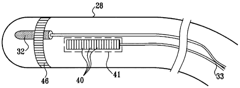
Fig. 2 is a schematic, pictorial illustration that shows the distal end of
catheter
28, in accordance with an embodiment of the present invention. The catheter
comprises an ultrasonic imaging sensor. The ultrasonic sensor typically
comprises an
array of ultrasonic transducers 40. In one embodiment, the transducers are
piezo-
electric transducers. The ultrasonic transducers are positioned in or adjacent
to a
window 41, which defines an opening within the body or wall of the catheter.
Transducers 40 operate as a phased array, jointly transmitting an ultrasound
beam from the array aperture through window 23. (Although the transducers are
shown arranged in a linear array configuration, other array configurations can
be used,
such as circular or convex configurations.) In one embodiment, the array
transmits a
short burst of ultrasound energy and then switches to a receiving mode for
receiving
the ultrasound signals reflected from the surrounding tissue. Typically,
transducers 40
are driven individually in a controlled manner in order to steer the
ultrasound beam in
a desired direction. By appropriate timing of the transducers, the produced
ultrasound
beam can be given a concentrically curved wave front, so as to focus the beam
at a
given distance from the transducer array. Thus, system 20 uses the transducer
array as
CA 02544118 2006-04-19
a phased array and implements a transmit/receive scanning mechanism that
enables
the steering and focusing of the ultrasound beam, so as to produce 2-D
ultrasound
images.
In one embodiment, the ultrasonic sensor comprises between sixteen and sixty-
four transducers 40, preferably between forty-eight and sixty-four
transducers.
Typically, the transducers generate the ultrasound energy at a center
frequency in the
range of 5-10 MHz, with a typical penetration depth of 14 cm. The penetration
depth
typically ranges from several millimeters to around 16 centimeters, and
depends upon
the ultrasonic sensor characteristics, the characteristics of the surrounding
tissue and
the operating frequency. In alternative embodiments, other suitable frequency
ranges
and penetration depths can be used.
After receiving the reflected ultrasound echoes, electric signals based on the
reflected echoes are sent by transducers 40 over cables 33 through catheter 28
to an
image processor 42 in console 34, which transforms them into 2-D, typically
sector-
shaped ultrasound images. Image processor 42 typically computes or determines
position and orientation information, displays real-time ultrasound images,
performs
3-D image or volume reconstructions and other functions which will all be
described
in greater detail below.
In some embodiments, the image processor uses the ultrasound images and the
positional information to produce a 3-D model of a target structure of the
patient's
heart. The 3-D model is presented to the physician as a 2-D projection on a
display 44.
In some embodiments, the distal end of the catheter also comprises at least
one
electrode 46 for performing diagnostic and/or therapeutic functions, such as
electro-
physiological mapping and/or radio frequency (RF) ablation. In one embodiment,
electrode 46 is used for sensing local electrical potentials. The electrical
potentials
measured by electrode 46 may be used in mapping the local electrical activity
on the
endocardial surface. When electrode 46 is brought into contact or proximity
with a
point on the inner surface of the heart, it measures the local electrical
potential at that
point. The measured potentials are converted into electrical signals and sent
through
the catheter to the image processor for display. In other embodiments, the
local
electrical potentials are obtained from another catheter comprising suitable
electrodes
and a position sensor, all connected to console 34.
21
CA 02544118 2006-04-19
. =
In alternative embodiments, electrode 46 can be used to measure different
parameters, such as various tissue characteristics, temperature and/or blood
flow.
Although electrode 46 is shown as being a single ring electrode, the catheter
may
comprise any number of electrodes 46 in any form. For example, the catheter
may
comprise two or more ring electrodes, a plurality or array of point
electrodes, a tip
electrode, or any combination of these types of electrodes for performing the
diagnostic and/or therapeutic functions outlined above.
Position sensor 32 is typically located within the distal end of catheter 28,
adjacent to electrode 46 and transducers 40. Typically, the mutual positional
and
orientational offsets between position sensor 32, electrode 46 and transducers
40 of
the ultrasonic sensor are constant. These offsets are typically used by
positioning
processor 36 to derive the coordinates of the ultrasonic sensor and of
electrode 46,
given the measured position of position sensor 32. In another embodiment,
catheter 28
comprises two or more position sensors 32, each having constant positional and
orientational offsets with respect to electrode 46 and transducers 40. In some
embodiments, the offsets (or equivalent calibration parameters) are pre-
calibrated and
stored in positioning processor 36. Alternatively, the offsets can be stored
in a
memory device (such as an electrically-programmable read-only memory, or
EPROM)
fitted into handle 29 of catheter 28.
Position sensor 32 typically comprises three non-concentric coils (not shown),
such as described in U.S. Patent 6,690,963 cited above. Alternatively, any
other
suitable position sensor arrangement can be used, such as sensors comprising
any
number of concentric or non-concentric coils, Hall-effect sensors and/or
magneto-
resistive sensors.
Typically, both the ultrasound images and the position measurements are
synchronized with the heart cycle, by gating signal and image capture relative
to a
body-surface electrocardiogram (ECG) signal or intra-cardiac
electrocardiogram. (In
one embodiment, the ECG signal can be produced by electrode 46.) Since
features of
the heart change their shape and position during the heart's periodic
contraction and
relaxation, the entire imaging process is typically performed at a particular
timing with
respect to this period. In some embodiments, additional measurements taken by
the
catheter, such as measurements of various tissue characteristics, temperature
and
22
CA 02544118 2006-04-19
blood flow measurements, are also synchronized to the electrocardiogram (ECG)
signal. These measurements are also associated with corresponding position
measurements taken by position sensor 32. The additional measurements are
typically
overlaid on the reconstructed 3-D model, as will be explained below.
In some embodiments, the position measurements and the acquisition of the
ultrasound images are synchronized to an internally-generated signal produced
by
system 20. For example, the synchronization mechanism can be used to avoid
interference in the ultrasound images caused by a certain signal. In this
example, the
timing of image acquisition and position measurement is set to a particular
offset with
respect to the interfering signal, so that images are acquired without
interference. The
offset can be adjusted occasionally to maintain interference-free image
acquisition.
Alternatively, the measurement and acquisition can be synchronized to an
externally-
supplied synchronization signal.
In one embodiment, system 20 comprises an ultrasound driver (not shown)
that drives the ultrasound transducers 40. One example of a suitable
ultrasound driver,
which can be used for this purpose is an AN2300TM ultrasound system produced
by
Analogic Corp. (Peabody, Massachusetts). In this embodiment, the ultrasound
driver
performs some of the functions of image processor 42, driving the ultrasonic
sensor
and producing the 2-D ultrasound images. The ultrasound driver may support
different
imaging modes such as B-mode, M-mode, CW Doppler and color flow Doppler, as
are known in the art.
Typically, the positioning and image processors are implemented using a
general-purpose computer, which is programmed in software to carry out the
functions
described herein. The software may be downloaded to the computer in electronic
form, over a network, for example, or it may alternatively be supplied to the
computer
on tangible media, such as CD-ROM. The positioning processor and image
processor
may be implemented using separate computers or using a single computer, or may
be
integrated with other computing functions of system 20. Additionally or
alternatively,
at least some of the positioning and image processing functions may be
performed
3 0 using dedicated hardware.
23
CA 02544118 2006-04-19
3-D IMAGING METHOD
Fig. 3 is a flow chart that schematically illustrates a method for cardiac
mapping and imaging, in accordance with an embodiment of the present
invention. In
principle, the disclosed method combines multiple 2-D ultrasound images,
acquired at
different positions of the catheter, into a single 3-D model of the target
structure. In
the context of the present patent application and in the claims, the term
"target
structure" or "target" may refer to a chamber of the heart, in whole or in
part, or to a
particular wall, surface, blood vessel or other anatomical feature. Although
the
embodiments described herein refer particularly to structures in and around
the heart,
the principles of the present invention may similarly be applied, mutatis
mutandis, in
imaging of bones, muscles and other organs and anatomical structures.
The method begins with acquisition of a sequence of 2-D ultrasound images of
the target structure, at an ultrasound scanning step 50. Typically, the
physician inserts
catheter 28 through a suitable blood vessel into a chamber of the heart, such
as the
right atrium, and then scans the target structure by moving the catheter
between
different positions inside the chamber. The target structure may comprise all
or a part
of the chamber in which the catheter is located or, additionally or
alternatively, a
different chamber, such as the left atrium, or vascular structures, such as
the aorta. In
each catheter position, the image processor acquires and produces a 2-D
ultrasound
image, such as the image shown in Fig. 4 below.
In parallel, the positioning sub-system measures and calculates the position
of
the catheter. The calculated position is stored together with the
corresponding
ultrasound image. Typically, each position of the catheter is represented in
coordinate
form, such as a six-dimensional coordinate (X, Y, Z axis positions and pitch,
yaw and
roll angular orientations).
In some embodiments, the catheter performs additional measurements using
electrode 46. The measured parameters, such as local electrical potentials,
are
optionally overlaid and displayed as an additional layer on the reconstructed
3-D
model of the target structure, as will be explained below.
3 0 After obtaining the set of ultrasound images, the image processor
displays one
or more of these images to the physician, at a manual tagging step 52.
Alternatively,
step 52 may be interleaved with step 50. The gray levels in the images enable
the
24
CA 02544118 2006-04-19
physician to identify structures, such as the walls of heart chambers, blood
vessels and
valves. The physician examines the ultrasound images and identifies contours-
of-
interest that represent walls or boundaries of the target structure. The
physician marks
the contours on display 44, typically by "tagging" them using a pointing
device 45,
such as a track-ball. (An exemplary tagged 2-D image is shown in Fig. 5
below.) The
pointing device may alternatively comprise a mouse, a touch-sensitive screen
or tablet
coupled to display 44, or any other suitable input device. The combination of
display
44 and pointing device 45 is an example of an interactive display, i.e., means
for
presenting an image and permitting the user to mark on the image in such a way
that a
computer is able to locate the marks in the image. Other types of interactive
displays
will be apparent to those skilled in the art.
The physician may tag the contours on one or several images out of the set in
this manner. The physician may also tag various anatomical landmarks or
artifacts, as
relevant to the medical procedure in question. The physician may similarly
identify
"keep away" areas that should not be touched or entered in a subsequent
therapeutic
procedure, such as ablation.
In some embodiments, the contours-of-interest are tagged in a semi-automatic
manner. For example, the image processor may run suitable contour detection
software. In this embodiment, the software automatically detects and marks
contours
in one or more of the 2-D images. The physician then reviews and edits the
automatically-detected contours using the interactive display.
The image processor may use the tagged contours to automatically reconstruct
the contours in the remaining, untagged ultrasound images, at an automatic
tagging
step 54. (In some embodiments, the physician may tag all 2-D ultrasound images
at
step 52. In this case, step 54 is omitted.) The image processor traces the
structures
tagged by the physician, and reconstructs them in the remaining ultrasound
images.
This identification and reconstruction process may use any suitable image
processing
method, including edge detection methods, correlation methods, motion
detection
methods and other methods known in the art. The position coordinates of the
catheter
that are associated with each of the images may also be used by the image
processor in
correlating the contour locations from image to image. Additionally or
alternatively,
step 54 may be implemented in a user-assisted manner, in which the physician
reviews
CA 02544118 2006-04-19
and corrects the automatic contour reconstruction carried out by the image
processor.
The output of step 54 is a set of 2-D ultrasound images, tagged with the
contours-of-
interest.
The image processor subsequently assigns 3-D coordinates to the contours-of-
interest identified in the set of images, at a 3-D coordinate assignment step
56.
Although in step 52 the physician marks the tags on 2-D images, the location
and
orientation of the planes of these images in 3-D space are known by virtue of
the
positional information, stored together with the images at step 50. Therefore,
the
image processor is able to determine the 3-D coordinates for each pixel or of
any pixel
in the 2-D images, and in particular those corresponding to the tagged
contours. When
assigning the coordinates, the image processor typically uses the stored
calibration
data comprising the position and orientation offsets between the position
sensor and
the ultrasonic sensor, as described above.
In some embodiments, the contours-of-interest comprise discrete points. In
these embodiments, the positioning processor assigns a 3-D coordinate to each
such
discrete point. Additionally, the positioning processor assigns a 3-D
coordinate to
discrete points of a surface or a volume (defined by surfaces) such as a
chamber of a
heart. Thus, registration of the pre-acquired image to the one or more 2-D
ultrasound
images or 3-D model of the ultrasound images can be performed using contours,
2 0 discrete points, surfaces or volumes.
In some embodiments, the image processor displays one or more of the 2-D
ultrasound images, appropriately oriented in 3-D space. (See, for example,
Fig. 6
below.) The contours-of-interest may optionally be marked on the oriented 2-D
image.
The image processor produces a 3-D skeleton model of the target structure, at
a 3-D reconstruction step 58. The image processor arranges the tagged contours
from
some or all of the 2-D images in 3-D space to form the skeleton model. (See an
exemplary skeleton model in Fig. 7 below.) In some embodiments, the image
processor uses a "wire-mesh" type process to generate surfaces over the
skeleton
model and produce a solid 3-D shape of the target structure. The image
processor
3 0 projects the contours-of-interest on the generated 3-D model. The model
is typically
presented to the physician on display 44. (See exemplary 3-D models in Figs. 8-
10
below.)
26
CA 02544118 2006-04-19
As described above, in some embodiments system 20 supports a measurement
of local electrical potentials on the surfaces of the target structure. In
this
measurement, each electrical activity data-point acquired by catheter 28
comprises an
electrical potential or activation time value measured by electrode 46 and the
Alternatively, a separate 3-D electrical activity map (often referred to as an
As noted above, information imported from other imaging applications may be
30 below.)
27
CA 02544118 2006-04-19
Additionally or alternatively, if additional parametric measurements were
taken at step 50 above, these measurements can be registered with the 3-D
model and
displayed as an additional layer (often referred to as a "parametric map.")
When implementing the disclosed method, the order of steps 50-60 may be
modified, and steps may be repeated in an interactive manner. For example, the
physician may acquire a first sequence 2-D images and tag them manually. Then,
the
physician may go back and acquire additional images and have the system tag
them
automatically, using the tagged contours in the first sequence of images. The
physician
may then generate the full 3-D model and examine it. If the model is not
accurate
enough in some areas, the physician may decide to acquire an additional set of
images
in order to refine the 3-D model. Additionally or alternatively, the physician
may
decide, after examining the images or the 3-D model, to change the manual
tagging of
one or more of the images, or to override the automatic tagging process. Other
sequences of applying steps 50-60, in order to reach a high quality 3-D model
of the
target structure, may also be followed by the physician. Additionally or
alternatively,
some of these steps may be carried out automatically, under robotic control,
for
example.
In some embodiments, features from the 2-D ultrasound images are selectively
displayed as part of the 3-D model. For example, features that are located
outside the
volume defined by the contours-of-interest may be discarded or hidden from the
displayed model. Alternatively or additionally, only the skeleton model or the
wire-
mesh model can be displayed. Other suitable criteria can be used for filtering
the
information to be displayed. For example, "keep away" areas marked in one or
more
of the 2-D images, as described above, may be suitably drawn and highlighted
in the
3-D model.
In some embodiments, system 20 can be used as a real-time or near real-time
imaging system. For example, the physician can reconstruct a 3-D model of the
target
structure using the methods described above, as a preparatory step before
beginning a
medical procedure. The physician can tag any desired anatomical landmarks or
features of interest, which are displayed on the 3-D model. During the
procedure,
system 20 can continuously track and display the 3-D position of the catheter
with
respect to the model and the tagged contours. The catheter used for performing
the
28
CA 02544118 2006-04-19
medical procedure may be the same catheter used for generating the 3-D model,
or a
different catheter fitted with a suitable position sensor.
CARDIAC IMAGING EXAMPLE
Figs. 4-8 are images that visually demonstrate the 3-D imaging method
described above, in accordance with an embodiment of the present invention.
The
figures were produced from ultrasound images generated by a cardiac imaging
system
implemented by the inventors. The images were produced during a real-life
experiment that imaged the heart of a pig using a catheter similar to the
catheter
shown in Fig. 2 above.
Fig. 4 shows a 2-D ultrasound image acquired by the ultrasonic transducers at
a particular position of catheter 28. The image shows two distinct features 80
and 82
of the heart. Multiple ultrasound images of this form were acquired at
different
positions of the catheter, in accordance with ultrasound scanning step 50 of
the
method of Fig. 3 above.
Fig. 5 shows the ultrasound image of Fig. 4, with features 80 and 82 marked
with contours 84 and 86, respectively. Fig 4 was taken with the catheter
positioned in
the right atrium. In this 2-D ultrasound image, feature 80 represents the
mitral valve
and feature 82 represent the aortic valve. The contours were manually tagged
by a
user, in accordance with manual tagging step 52 of the method of Fig. 3 above.
2 0 Contours 84
and 86 mark the anatomical structures in the 3-D working volume and
assist the physician to identify these structures during the procedure.
Fig. 6 shows a 2-D ultrasound image 85 oriented and projected in 3-D space.
The figure shows an exemplary split-screen display, as can be produced by
image
processor 42 and displayed on display 44 of system 20. The "raw" 2-D image is
displayed in a separate window on the right hand side of the figure.
An isometric display at the center of the figure shows a projected image 87,
produced by orienting and projecting the plane of image 85 in 3-D space, in
accordance with the position measurement of position sensor 32. An orientation
icon
81, typically having the shape of the imaged anatomical structure (a heart in
this
3 0 example), is
displayed with the same orientation as projected image 87 in real-time as
29
CA 02544118 2006-04-19
catheter 28 is moved within the patient's body. Icon 81 assists the physician
in
understanding the 3-D orientation of the projected image.
A beam icon 83 is used in association with projected 2-D image 87 to mark the
area scanned by the ultrasound beam. As such, icon 83 is oriented and
displayed in the
same plane (same orientation) as projected image 87 in real-time as catheter
28 is
moved within the patient's body. Icon 83 may comprise a web-like or fan-like
linear
depiction, preferably in color, such as red. Alternatively, icon 83 may
comprise a
colored line marking the perimeter of the area scanned by the beam to produce
image
87, or any other suitable means for visualizing the position and orientation
of the
ultrasound beam. In the example of Fig. 6, icon 83 comprises two straight
lines
indicating the angular sector defined by the ultrasound beam. In some
embodiments,
an additional icon 99 marking the location and position of the distal end of
catheter 28
is also displayed. For example, the distal end of catheter 28 is displayed as
a catheter
tip icon 99 that permits the physician or user of system 20 to understand the
location
and orientation of ultrasound images captured by the catheter 28,
independently of
whether any other image processing is used to orient the 2-D ultrasound image
or fan
87 or to superimpose the 2-D image on a 3-D image or frame. The physician or
user
of suystem 20 may also use the icon 99 for aiming or directing the ultrasound
beam in
a desired direction and/orientation. For example, the catheter tip icon 99 may
be used
in positioning the tip of catheter 28 adjacent to a known landmark in the
heart in order
to facilitate a more accurate estimation of the direction of the ultrasound
beam.
Projected image 87 is typically displayed inside a cube that marks the
boundaries of the working volume. The working volume is typically referenced
to the
coordinate system of field radiating coils 30 of the positioning sub-system
shown in
Fig. 1 above. In one embodiment, each side of the cube (i.e., the
characteristic
dimension of the working volume) measures approximately 12 cm. Alternatively,
any
other suitable size and shape can be chosen for the working volume, typically
depending upon the tissue penetration capability of the ultrasound beam.
A signal display 91 at the bottom of the figure shows the ECG signal, to which
the measurements are synchronized, as explained above.
When system 20 operates in real time, the position and orientation of the
projected image and of icon 83 change with the movements of catheter 28. In
some
CA 02544118 2006-04-19
embodiments, the physician can change the angle of observation, zoom in and
out and
otherwise manipulate the displayed images using the interactive display. The
user
interface features described herein are shown as an exemplary configuration.
Any
other suitable user interface can be used.
In some embodiments, system 20 and the associated user interface can be used
for 3-D display and projection of 2-D ultrasound images, without
reconstructing a 3-D
model. For example, the physician can acquire a single 2-D ultrasound image
and tag
contours-of-interest on this image. System 20 can then orient and project the
ultrasound image in 3-D space, in a manner similar to the presentation of
projected
image 87. If desired, during the medical procedure the system can continuously
track
and display the 3-D position of the catheter performing the procedure (which
may be
different from the catheter acquiring image 87) with respect to the projected
ultrasound image and the tagged contours.
Fig. 7 shows a skeleton model of the target structure, in this example
comprising the right ventricle, produced by the image processor in accordance
with 3-
D reconstruction step 58 of the method of Fig. 3 above. Prior to generating
the
skeleton model, the image processor traced and reconstructed contours 84 and
86 in
the untagged ultrasound images, in accordance with automatic tagging step 54.
Fig. 7
shows the original contours 84 and 86 projected onto 3-D space. Contours 88
were
automatically reconstructed by the image processor from other contours tagged
by the
physician.
Fig. 8 shows a solid 3-D model of the right ventricle, generated by the image
processor. Some of contours 88 are overlaid on the solid model. In addition,
contours
89 showing the left ventricle can also be seen in the figure. The surface of
the right
ventricle is overlaid with an electrical activity map 90, as measured by
electrode 46 in
accordance with overlaying step 60 of the method of Fig. 3 above. The map
presents
different electrical potential values using different colors (shown as
different shading
patterns in fig. 8).
Figs. 9 and 10 are images that visually demonstrate modeled left atria, in
3 0 accordance
with an embodiment of the present invention. In both figures, the atrium is
shown as a solid model 92. A contour 94 tagged by the physician marks the
location
of the fossa ovalis. Contours 96 mark additional contours of interest used to
construct
31
CA 02544118 2006-04-19
solid model 92. In Fig. 10, a 2-D ultrasound image 98 is registered with the
coordinate
system of model 92 and displayed together with the model.
Fig. 11 is an image that visually demonstrates an ultrasound image 102
registered with a pre-acquired image 100, in accordance with an embodiment of
the
present invention. In this example, a pre-acquired CT image is registered with
the
coordinate system of the 3-D model. The pre-acquired image and the 2-D
ultrasound
image are displayed together on display 44.
Although the embodiments described above relate specifically to ultrasound
imaging using an invasive probe, such as a cardiac catheter, the principles of
the
present invention may also be applied in reconstructing 3-D models of organs
using an
external or internal ultrasound probe (such as a trans-thoracic probe), fitted
with a
positioning sensor. Additionally or alternatively, as noted above, the
disclosed method
may be used for 3-D modeling of organs other than the heart. Further
additionally or
alternatively, other diagnostic or treatment information, such as tissue
thickness and
ablation temperature, may be overlaid on the 3-D model in the manner of the
electrical
activity overlay described above. The 3-D model may also be used in
conjunction with
other diagnostic or surgical procedures, such as ablation catheters. The 3-D
model
may also be used in conjunction with other procedures, such as an atrial
septal defect
closing procedure, spine surgery, and particularly minimally-invasive
procedures.
It will thus be appreciated that the embodiments described above are cited by
way of example, and that the present invention is not limited to what has been
particularly shown and described hereinabove. Rather, the scope of the present
invention includes both combinations and sub-combinations of the various
features
described hereinabove, as well as variations and modifications thereof which
would
occur to persons skilled in the art upon reading the foregoing description and
which
are not disclosed in the prior art.
= 32