Note: Descriptions are shown in the official language in which they were submitted.
CA 02545418 2006-05-10 1750P03CA01
1
DESCRIPTION
ENDOSCOPE DEVICE AND IMAGING METHOD USING THE SAME
Technical Field
[0001) The present invention relates to an endoscope device for medical or
industrial
use, and an imaging method using the same.
Background Art
[0002] Conventionally, a medical endoscope for capturing images inside a body
cavity by inserting an elongate inserting unit into the body cavity (lumen)
has been widely
used.
[0003] For example, as shown in FIG. 7, such an endoscope has an objective
lens 110
at the head of its inserting unit 100. The objective lens 110 has an optical
axis L defined
in the longer axis direction, and catches the testing target part (observation
part) ahead, in a
predetermined view angle (for example, about I40 degrees) fanning out about
the optical
axis L. An optical image of the testing target part caught by the objective
lens is formed
on an imaging element such as a CCD (Charge Coupled Device), etc. via an
unillustrated
internally disposed lens mechanism (relay optical system, etc.).
[0004] That is; when the inserting unit 100 as shown in FIG. 7 is inserted
into the
body cavity, the endoscope can capture the image of the testing target part
ahead of the
inserting unit 100.
The inserting unit 100 has a curving portion free to curve formed of jointed
curve pieces, and a limber flexible tubular portion adjoining the curving
portion and having
flexibility. With these, the inserting unit 100 is formed free to bend by a
predetermined
bending radius.
[0005] As disclosed in a Patent Literature I, by an operation of an observer
(for
example, an operation onto an unillustrated operation unit at hand), the
inserting unit 100
CA 02545418 2006-05-10
2
can be bent to an arbitrary direction and the direction of its viewing field
can be desirably
changed.
[0006] Such an endoscope is applied not only for medical use but only for
industrial
use. For example, since an endoscope can capture images inside the ducts of
plant
equipment or inside machines, etc., it is used for nondestructive test, etc.
[0007] As described above, an endoscope is formed free to bend. Hence, in a
case
where it is inserted into a lumen (for example, esophagus and stomach) having
a larger
inner diameter than the bending radius; an image of the lumen can be captured
within the
view angle of the objective lens, with the inserting unit 100 bent.
[0008] However, in a case where the inserting unit 100 is inserted into a
lumen having
a smaller inner diameter than its bending radius, the inserting unit 100 is
hard to bend.
Therefore, as shown in FIG. 8, the endoscope can only capture images of parts
ahead of the
objective lens within a view angle 0. That is, in a case where the inner wall
of the lumen
has folds, etc. thereon, there has been a problem that some parts (for
example, the depth
and back of a fold, etc.) cannot be captured because the view angle is narrow.
[0009] Even in the case where the inserting unit 100 can be bent, it has been
necessary
for the observer to adequately bend the inserting unit 100 in order to capture
images
around the inserting unit 100 over a wider range than the view angle 8 (for
example, over
the full range of 360 degrees). The observer has to appropriately operate the
operation
unit at hand (operation knob, etc.).
[0010] However, this operation not only is complicated, but also requires some
skill.
Further, even a skilled observer cannot simultaneously capture images of parts
separate from each other by a predetermined distance or larger, though he/she
can capture
images over a wide range. That is, in order to capture images of two parts
forming an
angle therebetween larger than the view angle of the objective lens 110, it is
necessary to
stagger the timings to capture images.
[0011] Thus, a conventional endoscope has a problem that it cannot allow two
parts
CA 02545418 2006-05-10
3
distanced by a predetermined angle or larger to be simultaneously captured so
that their
independent behaviors and influences of the behaviors of one part onto the
behaviors of the
other may be imaged. That is, the conventional endoscope cannot perform image
capturing that satisfies the observer's demands.
Patent Literature 1: Unexamined Japanese Patent Application KOKAI
Publication No. H5-15484
Disclosure of Invention
Problem to be Solved by the Invention
[0012] The present invention was made in view of the above-described
circumstance,
and an object of the present invention is to provide an endoscope device and
an imaging
method capable of appropriately performing image capturing over a wide range.
Means for Solving the Problem
[0013] To achieve the above object, an endoscope device according to a first
aspect of
the present invention is an endoscope device comprising an elongate inserting
unit,
wherein the inserting unit comprises:
plurality of objective optical means, having a predetermined view angle,
mounted so as to be oriented in different viewing field directions from one
another;
transmission optical means for transmitting a light flux entering each of the
objective optical means; and
imaging means for imaging respective optical images formed when the light
flux transmitted by the transmission optical means is converged.
[0014] According to this structure, the plurality of objective optical means
each have a
predetermined view angle A (for example, about 140 degrees), and are mounted
so as to be
oriented in different viewing field directions. The transmission optical means
is formed
of, for example, a prism, a relay lens, etc., and transmits a light flux
entering each objective
optical means. The imaging means is formed of, for example, a CCD, etc., and
images
each optical image formed when the light flux transmitted by the transmission
optical
CA 02545418 2006-05-10
4
means is converged.
As a result, image capturing ever a wide range can be performed.
[0015] The objective optical means may be disposed so as to have peripheral
portions
of their respective viewing fields overlap peripheral portions of other
viewing fields, and
the imaging means may image an optical image of a light flux captured within
a view angle ~ (for example, about 240 degrees) extending, with no
discontinuation, over a
wider range than the view angle of each objective optical means.
[0016] To achieve the above object, an endoscope device according to a second
aspect
of the present invention is an endoscope device comprising an elongate
inserting unit,
wherein the inserting unit comprises:
three or more objective lenses, having a predetermined view angle, mounted so
as to be oriented in different viewing field directions from one another;
a transmission optical system for transmitting light fluxes entering the
respective objective lenses; and
an imaging element for imaging respective optical images formed when the
light fluxes transmitted by the transmission optical system are converged on
three or more
regions.
[0017] According to this structure, the three or more objective lenses each
have a
predetermined view angle 0 (for example, about 140 degrees), and are mounted
to be
oriented in different viewing field directions. The transmission optical
system is formed
of, for example, a prism, a relay lens, etc., and transmits light fluxes
entering the respective
objective lenses. The imaging element is formed of, for example, a CCD, and
images
respective optical images formed when the light fluxes transmitted by the
transmission
optical system are converged on three or more regions.
As a result, image capturing over a wide range can be appropriately performed.
[0018] The objective lenses may be disposed so as to have peripheral portions
of their
respective viewing fields overlap peripheral portions of other viewing fields,
and
CA 02545418 2006-05-10
the imaging element may image, on three regions, optical images of light
fluxes captured within a view angle ~ (for example, about 240 degrees)
extending, with no
discontinuation, over a wider range than the view angle of each objective
lens.
[0019] To achieve the above object, an imaging method according to a third
aspect of
5 the present invention is an imaging method by an endoscope device comprising
an
elongate inserting unit,
wherein the inserting unit comprises a plurality of objective optical systems
having a predetermined view angle and mounted so as to be oriented in
different viewing
field directions from one another, a transmission optical system for
transmitting a light flux
entering each objective optical system, and an imaging element fox imaging
each optical
image formed when the light flux transmitted by the transmission optical
system is
converged;
wherein the imaging method comprises:
capturing testing target parts, by the respective objective optical systems,
within their respective viewing fields which partially overlap with viewing
fields of
adjoining other objective optical systems; and
simultaneously imaging, by the imaging element, optical images of the testing
target parts captured within a view angle extending, with no discontinuation,
over a wider
area than the view angle of each objective optical system.
[0020] According to this method, each objective optical system captures a
testing
target part within its own viewing field which partially overlaps with the
viewing fields of
adjoining other objective optical systems, and the imaging element
simultaneously images
respective optical images formed when light fluxes within the range of a view
angle ~
extending, with no discontinuation, over a wider range than the view angle 8
of each
objective optical system are converged.
As a result, image capturing over a wide range can be appropriately performed.
Effects of the Invention
CA 02545418 2006-05-10
6
[0021] According to the present invention, it is possible to provide an
endoscope
device and an imaging method capable of appropriately performing image
capturing over a
wide range.
Brief Description of Drawings
[0022] [FIG. 1] It is a perspective diagram showing one example of an
endoscope
device according to an embodiment of the present invention.
[FIGS. 2] (a) is a front elevation of a head portion of the endoscope device
of
FIG. 1. (b) is a side elevation of the head portion of the endoscope device of
FIG. 1. (c)
is a perspective diagram of the head portion of the endoscope device of FIG.
1.
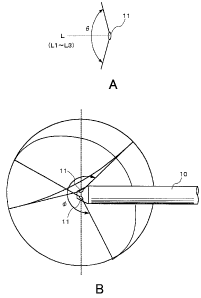
[FIGS. 3] (a) is an exemplary diagram for explaining a view angle 8 of an
objective lens. (b) is an exemplary diagram for explaining a view angle ~ of
the entire
head portion.
[FIG. 4] It is a partial cross-sectional diagram fox explaining an internal
structure of the head portion.
[FIG. 5] It is an exemplary diagram for explaining image capturing of a lumen
by the endoscope device.
[FIG. 6] It is a partial cross-sectional diagram for explaining an internal
structure of the head portion according to another embodiment of the present
invention.
[FIG. 7] It is an exemplary diagram for explaining a head portion of a
conventional endoscope.
[FIG. 8] It is an exemplary diagram for explaining image capturing by the
conventional endoscope.
Explanation of Reference Numerals
[0023] 1 inserting unit
2 operation unit
10 head portion
11 objective lens
~
CA 02545418 2006-05-10
7
12prism
13prism
14relay lens
15imaging element
20 curving portion
30 flexible tubular portion
40 operation knob
50 cable
Best Mode for Carrying Out the Invention
[0024] An endoscope device according to an embodiment of the present invention
will
be explained below with reference to the drawings.
[0025] FIG. 1 is a perspective diagram showing the appearance of an endoscope
device to be applied to the embodiment of the present invention. As shown, the
endoscope device comprises an inserting unit l, which is elongate and
flexible, and an
operation unit 2.
[0026] The inserting unit 1 comprises a head portion 10 in which a plurality
of optical
systems are disposed, a curving portion 20 free to curve formed of jointed
curve pieces,
and a limber flexible tubular portion 30 adjoining the curving portion 20 and
having
flexibility.
[0027] The operation unit 2 comprises an operation knob 40 and a cable 50.
[0028] The operation knob 40 is connected to the curving portion 20 by a known
drive
mechanism, and can curve the curving portion 20 to an arbitrary direction in
response to a
rotation (turn) of the operation knob 40.
[0029] The cable 50 is detachably connected to an unillustrated image
processing
apparatus, light source device, etc. The cable 50 supplies the image
processing apparatus
with video information originating from an optical image captured over the
full
circumference by the head portion 10 (on which an imaging element to be
described later
CA 02545418 2006-05-10
8
is disposed).
[0030] Next, the head portion 10 will be explained with reference to FIGS. 2,
etc.
FIG. 2A is a front elevation of the head portion 10, FIG. 2B is a side
elevation of the head
portion 10, and FIG. 2C is a perspective view of the head portion 10.
[0031] As shown in FIG. 2A to FIG. 2C, the head portion 10 has its head l0a
formed
generally like a truncated cone, and has three objective lenses 11 disposed
equidistantly on
the conic slope. The respective objective lenses are embedded in the slope of
the head
l0a and so disposed as to have their optical axes L1 to L3 in different
directions fram one
another.
[0032] As shown in FIG. 3A, the objective lens 11 has a predetermined view
angle 0
(for example, about 140 degrees) centered by the optical axis L (L1 to L3).
Here, the viewing field of each objective lens 11 is defined such that the
peripheral portions thereof overlap with the peripheral portions of the other
viewing fields,
as shown in FIG. 3B. With the viewing fields of the three objective lenses
combined, the
head portion 10 has a view angle ~ (an angle over 180 degrees, far example,
about 240
degrees) that extends over a wide range with no discontinuation as a whole.
[0033] The internal structure of the head portion 10 will be explained with
reference
to FIG. 4. FIG. 4 is a partial cross-sectional view of the head portion 10.
Though FIG. 4
shows the cross section of one objective lens 11 and an optical system
disposed with
respect to this, the remaining two objective lenses and optical systems
disposed with
respect to these have also the same cross-sectional structure.
As shown, the objective lens 11, prisms 12 and 13, a relay lens 14, and an
imaging element 15 are disposed in the head portion I0.
[0034] The objective lens 11 catches a testing target part (observation part)
ahead,
which is irradiated with illumination by an unillustrated illumination probe,
within a
predetermined view angle (for example, about 140 degrees) centered by the
optical axis L,
as described above. Then, each objective lens 11 supplies the light flux of
the incoming
~
CA 02545418 2006-05-10
9
reflection light from the testing target part to the prisms 12 and 13.
[0035] The prisms 12 and 13 refract the light flux entering the objective lens
11 at
their respective angles, in order to converge the light, via the relay lens 14
behind, on the
imaging element 15.
[0036] The relay lens 14 comprises a plurality of lenses and forms an optical
image of
the testing target part on the imaging surface of the imaging element 15 by
letting in the
light flux refracted by the prisms 12 and 13.
[0037] The imaging element 15 comprises a CCD (Charge Coupled Device), etc.
having a latticed color filter disposed on the front face thereof, and opto-
electrically
converts the optical image supplied via the relay lens 14 and formed on the
imaging
surface into an electric signal.
The imaging element 15 has its imaging surface divided into three regions, and
the light fluxes entering different objective lenses 11 (objective optical
systems) are
converged on these regions respectively.
[0038] The imaging element 15 supplies the video signal of each optical image
opto-electrically converted into the electric signal to the image processing
apparatus
connected to the cable 50 of the operation unit 2 through a unillustrated
signal line.
[0039] The operation of the endoscope device according to the embodiment of
the
present invention will be explained below.
Here, a case will be explained, where the inserting unit 1 (head portion 10)
of
the endoscope device is inserted into a relatively narrow lumen, as shown in
FIG. 5.
[0040] The head portion 10 inserted into a lumen is adequately turned in
response to
an operation of the operation knob 40. As a result, the head portion 10
catches the testing
target past (observation part) extending over a wider range than the view
angle 0 of the
objective lens, by the three objective lenses 11 within the view angle c~ (for
example, 240
degrees).
[0041] As described above, with reference to FIG. 4, the light fluxes entering
the
CA 02545418 2006-05-10
1~
respective objective lenses 11 are refracted by the prisms 12 and 13 and
converged on the
three regions on the imaging surface of the imaging element 15 via the relay
lens 14.
That is, the light fluxes representing the optical images of the testing
target
parts entering the different object lenses 11 are converged on the three
regions of the
imaging surface of the imaging element 15 respectively.
[0042] The imaging element 15 converts the converged optical images into
electric
signals, and supplies video signals of the respective converted optical images
to the image
processing apparatus via the unillustrated signal line and the cable 50.
[0043) Then, when the image processing apparatus acquires the video signals,
it
applies a predetermined image process thereto and simultaneously displays the
testing
target parts caught by the respective objective lenses 11 by the plurality of
videos
corresponding to the respective parts. That is, the image processing apparatus
displays
the videos of the testing target parts caught by the three objective lenses 11
respectively in
real time and simultaneously.
[0044] As described, even two testing target parts, which are distanced by
larger than
the view angle 8 of ane objective lens 1 l, can be image-captured
simultaneously.
Accordingly, even if the inner walls of the lumen are folded as shown in FIG.
5,
it is possible to capture even the depths and backs of the folds without
bending the head
portion 10.
Further, it becomes possible to simultaneously capture the independent
behaviors of the twa parts distanced by larger than the view angle 8 and
influences given
by the behaviors of one part onto the behaviors of the other, enabling an
image capturing
that satisfies demands of the observer.
[0045] In the above-described embodiment, the case has been explained where
the
light fluxes entering the three optical systems are converged on the imaging
surface made
of divided regions of one imaging element. The number of the imaging element
15 is not
limited to one.
CA 02545418 2006-05-10
11
[0046] For example, the same number of imaging elements 15 as the number of
objective lenses may be disposed. In this case, it is possible to omit the
prisms 12 and 13
by disposing each imaging element 15 in line with the direction of each
objective lens (in
line with orientation in which each is disposed).
That is, the imaging device 15 is disposed in line with the direction of the
objective lens 11 as shown in FIG. 6. That is, the imaging surface of the
imaging device
is disposed so as to be orthogonal to the optical axis L of the objective lens
11. FIG. 6
shows only the cross section of one objective lens 11, and the relay lens 14
and imaging
element 15 disposed with respect thereto. The other objective lenses also have
10 corresponding relay lenses and imaging elements disposed coaxially.
[0047] In case of such disposition, the light flux entering the objective lens
11 goes
straightforward and is converged on the imaging element 15 via the relay lens
14. That is,
the prisms 12 and 13 can be omitted because there is no need of refracting the
light flux.
[0048] In the above-described embodiment, a case has been explained where the
15 images of the three testing target parts captured by the endoscope device
are
simultaneously displayed by the image processing apparatus. However, image
selection
means may be disposed between the imaging element 15 and the image processing
apparatus, sa that the video signal of an optical image selected from the
optical images of
the plurality of testing target parts imaged by the imaging element 15 may be
output to the
image processing apparatus.
[0049] Further, the image processing device may perform some image process
based
on the video signals of the respective optical images obtained from the
imaging element 15
and generate a three-dimensional panoramic image.
[0050] That is, since the peripheries of the viewing field of each objective
lens 11
overlap with the peripheries of the other adjoining viewing fields as shown in
FIG. 3B, the
head portion 10 has got a view angle (for example, about 240 degrees)
extending over a
wide range with no discontinuation. This enables the image processing
apparatus to
CA 02545418 2006-05-10
12
generate a three-dimensional panoramic image on which the images of the
testing target
parts dispersed over a wide range are developed, based on the respective
obtained optical
images. Since this three-dimensional panoramic image has a relatively small
data size, it
is an easy option to use this image for electronic medical chart. Further, by
reproducing a
stored three-dimensional panoramic image, a detailed endoscopic video full of
live feeling
like, for example, a stereovision generated by stereoscopically converging
respective
optical images, will be reproduced.
The generation of a three-dimensional panoramic image may be realized not by
an image processing apparatus but by a circuit board possessed by the imaging
element 15.
[0051] In the above-described embodiment, a case has been explained where
three
optical systems (three objective lenses 11, and prisms 12 and 13 and relay
lenses 14
corresponding to the respective objective lenses 11) are disposed at the head
portion 10.
However, the number of these optical systems needs only to be equal to or
larger than two,
and is not limited to three.
[0052] In a case where objective lenses having a wide view angle are used,
even if the
number of such optical systems is, for example, two, the viewing fields of the
respective
objecfive lenses overlap each other partially, making it possible to secure a
viewing field
having a wide angle.
On the other hand, in a case where objective lenses having a narrow view angle
are used, the number of such optical systems may be increased to, for example,
four or
larger and disposed at the head portion 10, in order to secure overlapping
portions of the
viewing fields adjoining each other.
[0053] In the above-described embodiment, a case has been explained where the
imaging element 15 is disposed inside the head portion 10. However, the
imaging
element may be disposed in the operation unit 2. A light flux entering the
objective lens
11 is converted into an optical signal and transmitted through, for example, a
fiber optic
cable, and demodulated by an unillustrated demodulation unit. The demodulated
optical
CA 02545418 2006-05-10
13
signal is supplied to the imaging element in the operation unit 2 and
converged on the
imaging surface of the imaging element as the optical image of the testing
target part.
In this case, the head portion 10 can be downsized.
[0054] In the above-described embodiment, a case has been explained where the
least
components necessary for capturing images are disposed in the head portion 10,
in order to
facilitate understanding. However, for example, a component for irradiating an
illumination light, etc. may be disposed in the head portion 10. 1n this case,
an
illumination lens for irradiating the testing target part with an illumination
light is disposed
at the head 10 or therearound. An illumination light supplied from an
unillustrated Iight
source device connected to the cable 50 is transmitted to the illumination
lens through a
fiber optic, etc. A plurality of illumination lenses may be disposed
correspondingly to the
objective lenses 11.
In this case, an illumination probe, ete. becomes unnecessary at the time of
capturing images.
[0055] A person skilled in the art could add various modifications onto the
above-described embodiment, without departing from the sprit and scope of the
present
invention. The above-described embodiment is intended for illustration, not to
limit the
scope of the present invention. Accordingly, the scope of the present
invention should be
determined not by referring to the above description, but in accordance with
the entire
scope of equivalents to which the claims shown below are entitled.
[0056] The present invention based on Japanese Patent Application No. 2003-
385205
filed on November 14, 2003, and including specification, claims, drawings and
summary.
4
The disclosure of the above Japanese Patent Application is incorporated herein
by
reference in its entirety.
Industrial Applicability
[0057] As explained above, according to the present invention, it is possible
to
provide an endoscope device, etc. capable of appropriately performing image
capturing
Image