Note: Descriptions are shown in the official language in which they were submitted.
CA 02546545 2011-08-02
54346-4
- 1 -
NON-UNIFORM ELECTRIC FIELD CHAMBER FOR CELL FUSION .
Technical Field
The present invention relates generally to methods
and apparatus for fusing biological cells to one another.
More specifically, the present invention provides methods
and apparatus for treating biological cells with
electrical fields, such that the biological cells are
aligned and have increased cell membrane contact prior to
being subjected to cell fusing electric field pulses.
Background Art
If a neutrally charged particle, such as a
biological cell, is placed in a uniform electric field,
such as provided by a pair of same-size planar electrodes,
the biological cell does not move toward either one
electrode or the other because the attractive forces from
both electrodes are the same.
On the other hand, if a neutrally charged biological
cell is placed in a non-uniform electric field, such as
provided by two electrodes which are both not planar, as
shown in FIG. 1, the biological cell forms a
dipole, is attracted to one electrode with greater
attractive force than the other, and moves towards the
electrode having the greater attractive force.
Such a use of a non-uniform electric field is used
in dielectrophoresis, and the concept of using
dielectrophoresis to align living cells, followed by a
fusion/electroporation pulse, to fuse cells has been in
the literature since early 1970's_ This process is used
to produce hybrids of two different cell types for
therapeutic purposes, for hybridoma production for
producing monoclonal antibodies, for nuclear fusion, and
for producing other hybrid cells.
= Dielectrophoresis is the process of applying an
electrical force on neutrally charged particles such as
living cells. The electrical force causes adjacent living
cells to be compressed against one another, as shown in
FIG. 5. The force from dielectrophoresis
CA 02546545 2011-08-02
54346-4
-2 -
(dielectrophoretic force) results from applying a non-
uniform electric field, produced by an electrode pair to
which a voltage is applied. The non-uniform electric
field separates charges (ions) inside the cells forming a
dipole. After the dipole has been formed, the non-uniform
electric field then moves the cells towards the highest or
lowest electric field intensity. This movement is
dependent on the relative conductivities and
permittivities of the medium and the biological cells or
particles. The living cells are also aligned in the non-
uniform electric field, as shown in FIG. 2.
The dielectrophoretic force is a function of the
electric field squared, so electric field polarity is not
important. The dielectrophoretic force is also a function
of the relative conductivities and permittivities of the
medium and the particles or cells. The conductivities and
permittivities are a function of the frequency of the
applied electric field. Typically, an AC voltage wave,
such as a sine wave, is applied across electrodes to
produce an alternating electric field. The sine wave
voltage, frequency, and duration are optimized for
specific cell types.
After the AC wave is applied to align and compress
the cells, one or more fusion/electroporation pulses are
applied to permeabilize adjacent cell membranes (form
pathways between adjacent cell membranes) and to cause
cell membranes from both adjacent cells to fuse or
commingle. These pathways permit the contents of the
cells to mix forming a hybrid fused cell.
Permeabilization is conventionally done in electric
fields having uniform electric field intensity so that all
cells in the electric field are permeabilized in a uniform
manner. The uniform electric field is achieved by using
parallel flat plate electrodes.
On the other hand, it is known that permeabilization
of all cells in an electric field that has non-uniform
electric field intensity would result in the cells being
CA 02546545 2011-08-02
54346-4
- 3 -
permeabilized in a non-uniform manner. Such non-
uniformity in permeabilization is undesirable. Fewer
pathways form in the cell membranes resulting in fewer
cell fusions.
Following the fusion pulses, another AC field can be
applied to hold the cells together while the fused cells
stabilize (mature). In some cases, the AC voltage has
been linearly increased or decreased to prevent damage to
the cells due to a sudden application of a field.
The published PCT International Application No. NO
03/020915 A2 describes AC waveforms that can be applied at
a low level to align the cells without creating large
forces producing turbulence. After the cells are aligned,
the waveform then applied provides a large force which
compresses the cells creating a large mutual surface area
between the cells just before the permeabilization
electric field pulse is applied.
Examples of cell fusion applications include
hybridoma production and nuclear transfer. A recent
application for electrofusion is to produce therapeutic
hybrids for cancer immunotherapy. These hybrids are
produced from cancer tumor cells and immune system
dendritic cells in an ex vivo process. Each treatment
requires a large number of viable hybrids, which results
in a new requirement for high efficiency in the hybrid
production process. Commercial and clinical uses of these
techniques are now important requiring large numbers of
hybrid products to be produced in a single batch.
There are a number of techniques (electrical,
mechanical, and chemical) available to. perform cell
fusion. This invention relates to electrical means. The
current electric art uses a voltage waveform generator
connected to an electrode device or chamber. With respect
to known electrical, mechanical, and chemical techniques,
the following U.S. patents are of particular interest:
CA 02546545 2006-05-17
WO 2005/066342
PCT/US2003/035982
-4-
4,326,934 April 27, 1982 Pohl
4,441,972 April 10, 1982 Pohl
4,578,168 March 25, 1986 Hofmann
4,695,547 September 22, 1987 Hillard
4,699,881 October 13, 1987 Matschke et al
4,764,473 August 16, 1988 Matschke et al
4,784,954 November 15, 1988 Zimmermann
4,804,450 February 14, 1989 Mochizuki
5,007,995 April 16, 1991 Takahashi
5,304,486 April 19, 1994 Chang
From the above, it is known to use electrodes or
chambers that produce non-uniform electric fields. One
such example is two coaxial electrodes forming a chamber.
The coaxial chamber was described in detail by Pohl in a
book published in 1978. The coaxial chamber was discussed
in relation to theoretical dielectrophoresis
considerations.
Nevertheless, there has been no description of how
to effectively set the dimensions of the coaxial chamber
for any particular application. Cell fusion using
electrical means requires a non-uniform electric field to
align and compress the cells and a uniform electric field
to permeabilize the cells. To provide the highest
possible efficiency in producing the fused hybrid cells,
as required in commercial and clinical applications, the
geometric dimensions of the chamber must be carefully
selected.
Initially in any cell fusion process one must bring
the cells into alignment and contact. In any case,
sufficient force must be applied to each cell to overcome
the negative surface charge. As stated above, merely
applying a uniform electric field will not move a cell
because the net charge of the cell is zero. Thus, from
the definition of electric field, there is no force
applied, because the charge equals zero:
CA 02546545 2011-08-02
54346-4
- 5 -
Force (Electric Field) * (Charge)
However, a non-uniform field induces the positive
ions inside each cell to move to one side and the negative
ions to move to the opposite side producing a dipole, as
shown in FIG. 1. Once the dipole is induced,
because of the presence of a non-uniform electric field, a
net force is exerted on the cell because the intensity of
the field is greater on one side than the other. The
movement of cells in one direction causes the cells to
align. Since the cells are now dipoles, the negative side
of one cell will attract the positive side of another cell
overcoming the negative surface charge, as shown in
FIG. 2. The non-uniform electric field is produced by
the electrode device or chamber. The non-uniformity is a
function of the electrode configuration, examples of which
are shown in FIGs. 1 and 2.
Generally, the cell types to be fused are placed in
a low conductive medium (for example 100 microsemens/cm)
to minimize ohmic heating that may harm the cells and that-
causes turbulence in the medium, thus reducing the number
of fused hybrids. In this respect, it would be desirable
for biological cells being subjected to cell fusion to be
treated so as to reduce heating during cell alignment and
cell membrane contact.
The waveform generator has multiple functions. The
first function is to produce the AC voltage waveform that
is converted into an AC field by the electrode pair or
chamber. This AC field brings the cells into
alignment/contact. The second function is-to compress the
cells by briefly increasing the amplitude of the AC
waveform. The third function is to produce a pulse
voltage that produces an electric field that
electroporates the membranes of the cells in close
contact, fusing the cells. The fourth function is to
apply a low amplitude AC voltage to hold the cells in
alignment until the fusion products become viable or
CA 02546545 2006-05-17
WO 2005/066342
PCT/US2003/035982
- 6 -
stable (mature).
One of the factors for successful fusion is the
membrane contact between the adjacent cells. The closer
this contact before the fusion pulse is applied, the
higher the efficiency of fusion. In U. Zimmermann, et
al., "Electric Field-Induced Cell-to-Cell Fusion", J.
Membrane Biol. 67, 165-182 (1982), Zimmermann points out
that increasing the AC wave electric field strength just
before the fusion pulse may be the optimum approach.
Clearly, it would be desirable for biological cells that .
are to undergo cell fusion to be pretreated with pre-
fusion non-linear electric field waveforms to produce
sufficient force to bring about increased cell membrane
contact and then to immediately apply a uniform electric
field pulse(s) that permeabilizes the cell membranes in
contact, thereby leading to cell fusion.
It would be very desirable to have a chamber that
will produce a large number of fused products by applying
a large force (proportional to a non-uniform electric
field) on the adjacent cells to compress the cells to
create a larger surface area between them and then to
immediately apply a uniform electric field from one
electrode to the next that will permeabilize the largest
number of cell membranes in contact.
It is also desirable to have a chamber of sufficient
volume to produce a large number of hybrid products.
In view of the above, it would also be desirable to
produce a chamber with sufficient uniform and non-uniform
electric fields to provide the largest number of fused
hybrid cells.
Thus, while the foregoing body of prior art
indicates it to be well known to use coaxial chambers, the
prior art described above does not teach or suggest a
method to determine how to select the chamber geometry
which has the following combination of desirable features:
(1) provides sufficient force (non-uniform field
intensity) to compress the cells providing a large
CA 02546545 2006-05-17
WO 2005/066342
PCT/US2003/035982
-7 -
membrane contact area without excessive heating; (2)
provides sufficient uniform field intensity to
peLmeabilize the cells; and (3) produces a large number of
hybrid products. The foregoing desired characteristics
are provided by the unique coaxial cell fusion chamber of
the present invention as will be made apparent from the
following description thereof. Other advantages of the
present invention over the prior art also will be rendered
evident.
Additional U. S. patents and published U. S. patent
applications that are of interest include:
4,561,961 December 31, 1985 Hofmann
5,001,056 March 19, 1991 Snyder et al
5,589,047 December 31, 1996 Coster et al
5,650,305 July 22, 1997 Hui et al
US2003/0082163, May 1, 2003 Shu
Additional literature references include:
1. R. Bischoff, et al., "Human Hybridoma Cells Produced
by Electra-Fusion", Fed. Eur. Biochem. Soc. Lett.
147, 64-68 (1982).
2. L. Changben, et al., "Use of Human Erythrocyte Ghosts
for Transfer of 125.1-BSA and 125I-DNA into
Animal Cells from Cell Fusion", Scientia Sinica (Series B)
25, 680-865 (1982).
3. C. S. Chen, et al., "Biological Dielectrophoresis: The
Behavior of Lone Cells in a Non-unifolui Electric Field",
Ann. N.Y. Acad. Sci. 238, 176-185 (1974).
4. Coster, H. G. L. and Zimmermann, U. "Direct
Demonstration of Dielectric Breakdown in the Membranes of
Valonia utricularis. " Zeitschrift fur Naturforschung. 30
c, 77-79.1975.
5. Coster, H. G. L. and Zimmermann, U. "Dielectric
Breakdown in the Membranes of Valonia utricularis: the
role of energy dissipation". Biochimica et Biophysica
Acta. 382, 410-418,1975.
6. Coster, H. G. L. and Zimmermann, U. "The mechanisms of
CA 02546545 2006-05-17
WO 2005/066342
PCT/US2003/035982
- 8 -
Electrical Breakdown in the Membranes of Valonia
utricularis." Journal of Membrane Biology. 22, 73-90,1975.
7. K. Kaler, et al., "Dynamic Dielectrophoretic
Levitation of Living Individual Cells", J. Biol. Phys. 8,
18-31 (1980).
8. A. R. Murch, et al., "Direct Evidence that
Inflammatory Multi-Nucleate Giant Cells Form by Fusion",
Pathol. Soc. Gr. Brit. Ire. 137, 177-180 (1982).
9. Neumann, Bet al. "Cell Fusion Induced by High
Electrical Impulses Applied to Dictyostelium",
Naturwissenschaften 67, 414, 1980
10. Petrucci, General Chemistry: Principles and Modern
Applications, 4th ed., p. 621, 1985 (no month).
11. Zimmermann et al., Electric Field-Induced Cell-to-
Cell Fusion, The Journal of Membrane Biology, vol. 67, pp.
165-182 (1982) [no month).
12. Pohl, H. "Dielectrophoresis", Cambridge University
Press, 1978.
13. H. A. Pohl, "Biophysical Aspects of
Dielectrophoresis", J. Biol. Phys. 1, 1-16 (1973).
14. H. A. Pohl, et al., "Continuous Dielectrophoretic
Separation of Cell Mixtures", Cell Biophys. 1, 15-28
(1979).
15. H. A. Pohl, et al., "Dielectrophoretic Force", J.
Biol. Phys. 6, 133 (1978).
16. H. A. Pohl, et al., "The Continuous Positive and
Negative Dielectrophoresis of Microorganisms", J. Bio.
Phys. 9, 67-86 (1981).
17. Sale, J. H. and Hamilton, W. A. "Effects of High
Electric Fields on Micro-Organisms", Biochimica et
Biophysica Acta. 163, 37-43, 1968.
18. Sepersu, E. H., Kinosita, K. and Tsong, T. Y.
"Reversible and Irreversible Modification of Erythrocyte
Membrane Permeability by Electric Fields" Biochimica et
Biophysica Acta. 812, 779-785, 1985.
19. J. Vienken, et al., "Electric Field-Induced Fusion:
Electro-Hydraulic Procedure for Production of Heterokaryon
CA 02546545 2006-05-17
WO 2005/066342
PCT/US2003/035982
-9 -
Cells in High Yield", Fed. Eur. Biomed. Soc. Lett. 137,
11-13 (1982).
20. H. Weber, et al., "Enhancement of Yeast Protoplast
Fusion by Electric Field Effects", A Preprint for
Proceedings of the Fifth International Symposium on
Yeasts,London, Ontario, Canada, Jul. 80.
21. Zimmermann, U. "Electrical Field Mediated Fusion and
Related Electrical Phenomena", Biochimica et Biophysica
Acta. 694, 227-277. 1982.
22. Zimmermann, U. et al "Fusion of Avena Sativa Mesophyll
Proptoplasts by Electrical Breakdown", Biochimica et
Biophysica Acta. 641, 160-165, 1981. 1982.
23. U. Zimmermann, et al., "Electric Field-Induced
Release of Chloroplasts from Plant Protoplasts",
Naturwissen 69, 451 (1982).
24. U. Zimmermann, et al., "Electric Field-Mediated Cell
Fusion", J. Biol. Phys. 10, 43-50 (1982).
25. U. Zimmermann, "Cells with Manipulated Functions: New
Perspectives for Cell Biology, Medicine, and Technology",
Angew. Chem. Int. Ed. Engl. 20, 325-344 (1981).
26. Electromechanics of Particles, Thomas B. Jones, 1995,
Cambridge University Press.
27. Electroporation and Electrofusion in Cell Biology,
Eberhard Neumann, Arthur E. Sowers, and Carol A. Jordon,
Plenum Press, New York 1989.
As explained below with respect to the subject
invention, prior art ratios rl/r2 and gaps of known prior
art chambers are outside the respective ranges of the
subject invention. Such prior art are as follows:
1. Dielectricophoresis of cell size liposomes, 13
December 1993. r1/r2=0.25, gap=0.75 mm.
2. Hofmann 4,578,168, Mar 25, 1986, r1/r2=0.139,
gap = 0.155 mm.
3. Hillard 4,695,547, SEP 22, 1987, r1/r=0.162,
gap=13 mm
4. Matschke 4,699,881, Oct 13, 1987, r1/r2=0.98,
gap=0.4 mm.
CA 02546545 2011-08-02
= 54346-4
-10-
5. Zimmerman 4,764,473, Aug 16, 1988, no
dimensions.
6. Mochizuki, 4,804,450, Feb 14, 1989, rl/r2=0.962,
gap=2 rum.
7. Takahashi, 5,007,995, Apr 16, 1991, rl/r2=0.263,
gap= 2.8 mm
8. Chang, 5,304,486, Apr 19, 1994, rl/r2 not given,
gap=0.5 to 2.0 mm
9. Shu, US2003/0082163, May 1, 2003, r1/r2 not
given, gap 2 to 5 mm.
Disclosure of Invention
The present invention provides an apparatus for
carrying out fusion of biological cells and includes: an
inner electrode having a first electrode radius (rl) and
an electrode height and an outer electrode having a second
electrode radius (r2) and the same electrode height. The
inner electrode and the outer electrode are concentric. A
gap is provided between the inner electrode and the outer
electrode, and the size of the gap is the difference
between the second electrode radius and the first
electrode radius. A cell fusion volume is defined by the
electrode height, the gap, the first electrode radius, and
the second electrode radius. The first electrode radius,
the second electrode radius, and the gap are selected in
accordance with a predetermined range of selectable ratios
(r1/r2) of the first electrode radius to the second
electrode radius, wherein the range of selectable ratios
(rl/r2) is from 0.7 to 0.85,wherein a selected .gap limited
by the range of selectable ratios (r1/r2), and wherein a
determined ratio (rl/r2) of the selectable ratios is based
on the selected gap, such that compression between the
biological cells and permeability between cell membranes
are maximized and temperature rise is minimized for
providing cell fusion in the cell fusion volume.
CA 02546545 2006-05-17
WO 2005/066342
PCT/US2003/035982
- 11 -
I t is understood, that both the inner electrode and
the outer electrode are provided with means for connecting
with cables or other electrical conductors coming from a
an electrical waveform generator.
As discussed further below in greater detail, as the
ratio r1/r2 would be less than 0.7, the Percent Change in
Electric Field Intensity would be greater than 30%, which
would result in undesirably low cell permeabilization and
undesirably low cell fusion.
In addition, as the ratio r1/r2 would be greater
than 0.9, the electric field intensity would become very
uniform, which would result in a very low compressive
force for a fixed AC voltage. This would result in low
cell fusion. If to compensate, the AC voltage would be
increased to maintain a constant compressive force,
undesirable heating of the medium would occur which would
cause an undesirable temperature rise which would kill the
cells.
With the present invention the geometric parameters
of a coaxial chamber may be selected to produce a chamber
which will simultaneously provide cell compression and
permeabilization without excessive heating to produce
large numbers of fused hybrid cells.
All of the prior art that provided sufficient
information to determine the chamber parameters were
either well below or well above the preferred parameters
of this invention. All were very small volumes, less than
a few hundred microliters (in contrast with the subject
invention which is scalable up to many milliliters), and
none considered the trade-off between compressive force
and permeabilization (which the principles of the subject
invention teach).
In accordance with another aspect of the invention,
a method is provided for selecting an inner electrode, an
outer electrode, and a gap between the inner electrode and
the outer electrode for a cell fusion chamber for fusing
biological cells. The method includes the steps of:
CA 02546545 2013-11-14
54346-4
- 12 -
determining two of a first electrode radius of the inner electrode, a second
electrode radius of
the outer electrode, and the gap between the inner electrode and the outer
electrode;
setting the ratio of the first electrode radius to the second electrode radius
to a
value in a range between 0.7 to 0.85; and
calculating the third of the first electrode radius of the inner electrode,
the
second electrode radius of the outer electrode, and the gap between the inner
electrode and
the outer electrode, such that compression between the biological cells and
permeability
between cell membranes are maximized and temperature rise is minimized for
providing cell
fusion in the cell fusion chamber.
More specifically, with the method, the ratio of the first electrode radius to
the
second electrode radius is set to a value in a range between 0.80 to 0.85, and
the gap is in a
range of 2 to 10 millimeters for cell radius between 2 and 10 microns.
With further consideration of the concept of scalability, with the subject
invention, volume of the cell fusion chamber can be increased by simple
increasing the
electrode height and keeping the ratio rl/r2 and the gap constant. In
addition, by simply
increasing the electrode height and keeping rl/r2 constant, temperature in the
medium does
not change.
According to one aspect of the present invention, there is provided an
apparatus for carrying out fusion of biological cells, comprising: an inner
electrode having a
first electrode radius (r1) and an electrode height, an outer electrode having
a second
electrode radius (r2) and said electrode height, wherein said inner electrode
and said outer
electrode are concentric, a gap between said inner electrode and said outer
electrode,
wherein the size of said gap is the difference between said second electrode
radius and said
first electrode radius, and wherein a cell fusion volume is defined by said
electrode height,
said gap, said first electrode radius (r1)1 and said second electrode radius
(r2), wherein said
first electrode radius, said second
CA 02546545 2013-11-14
54346-4
- 12a -
electrode radius, and said gap are selected in accordance with a predetermined
range of
selectable ratios (r1/r2) of said first electrode radius to said second
electrode radius, wherein
said range of selectable ratios is from 0.7 to 0.85, a selected gap limited to
a range from 2
to 10 millimeters, and a determined ratio of said selectable ratios based on
said selected gap,
such that compression between the biological cells and permeability between
cell
membranes are maximized and temperature rise is minimized for providing cell
fusion in said
cell fusion volume.
According to another aspect of the present invention, there is provided a
method for selecting a radius, r1, of an inner electrode, a radius, r2, of an
outer electrode,
and a gap between the inner electrode and the outer electrode for a coaxial
cell fusion
chamber for fusing biological cells with a selected cell radius of greater
than 4 micrometers in
a buffer having a conductivity of 100 microseimens/centimeter or less,
comprising the steps
of selecting the first and second radius wherein r2-r1 is equal to or less
than ten millimeters
and r1/r2 is equal to 0.85 or less and the gap is limited to a range from 2 to
10 millimeters
and temperature increases of 40 C or less are induced when an AC voltage is
selected that
induces a dielectrophoretic force in a range of 0.1 to 1 nano-dyne for said
cell radius and the
selected r1/r2 and r2-r1 pair.
According to still another aspect of the present invention, there is provided
an
apparatus for carrying out fusion of biological cells, comprising: a non-
conductive base
member, a conductive outer electrode supported on said base member, wherein
said outer
electrode includes a concave outer electrode surface which has an outer
electrode radius (r2)
and has an electrode height, a conductive inner electrode supported on said
base member,
wherein said inner electrode includes a convex inner electrode surface which
has an inner
electrode radius (r1) and has the electrode height, wherein said outer
electrode surface and
said inner electrode surface are spaced apart from each other by a gap which
defines a
fusion chamber, wherein said gap is limited to a range from 2 to 10
millimeters, and wherein
a ratio of said inner electrode radius to said outer electrode radius (r1/r2)
is 0.7 to 0.85, a
non-conductive outer electrode cover member supported by said outer electrode,
and a non-
conductive inner electrode cover member supported by said inner electrode,
wherein said
CA 02546545 2013-11-14
=
54346-4
- 12b -
outer electrode cover member and said inner electrode cover member define an
access
channel, wherein said access channel is in communication with said fusion
chamber.
According to yet another aspect of the present invention, there is provided an
apparatus for carrying out fusion of biological cells, comprising: a non-
conductive support
member, a conductive outer electrode supported in a horizontal orientation by
said support
member, wherein said outer electrode includes a conductive concave outer
electrode surface
which has an outer electrode radius (r2) and has an electrode width, a
conductive inner
electrode supported in a horizontal orientation by said support member above
said outer
electrode, wherein said inner electrode includes a conductive convex inner
electrode surface
which has an inner electrode radius (r1) and has said electrode width, and non-
conductive
vertically oriented end walls located at ends of said outer electrode and said
inner electrode,
wherein said outer electrode surface and said inner electrode surface are
spaced apart from
each other by a gap, wherein said gap and said vertically oriented end walls
define a fusion
chamber, wherein said gap is limited to a range from 2 to 10 millimeters, and
wherein a ratio
of said inner electrode radius to said outer electrode radius (r1/r2) is 0.7
to 0.85.
According to a further aspect of the present invention, there is provided a
method for selecting an inner electrode, an outer electrode, and a gap between
the inner
electrode and the outer electrode for a coaxial cell fusion chamber for fusing
biological cells with
radius greater than 4 micrometers, comprising the steps of: selecting each set
of a radius, r1, of
the inner electrode and a radius, r2, of the outer electrode such that r2-r1
is equal to or less
than ten millimeters, the gap is limited to a range from 2 to 10 millimeters
and r1/r2 is equal to
0.85 or less; calculating an AC voltage for a dielectrophoetic force in a
range of 0.1 to 1 nano-
dyne for a cell radius at each r1/r2 and r2-r1 pair by using the following
formula:
V = {- ri3In(r1/r2)2 / ( 2a3[21-rclq ) x Fdep}112 volts rms
CA 02546545 2012-10-16
54346-4
- 12c -
where
V is the AC voltage
a is the cell radius
is the permittivity of medium external to the cell
K is the Clausius-Mossotti Function
Fdep is the force in nanodynes, range is 0.1 to 1.0
calculating a temperature increase from the voltage and voltage
duration between 5 and 20 seconds by using graphs in figures 11A and 12A, or
figures 11B and 12B, or figures 11C and 12C, selecting one r1/r2 and r2-r1
pair for
the temperature increase of 40 C or less, and producing the coaxial cell
fusion
chamber.
Brief Description of Drawings
The invention will be better understood and the above objects as well
as objects other than those set forth above will become more apparent after a
study
of the following detailed description thereof. Such description makes
reference to the
annexed drawing wherein:
FIG. 1 illustrates PRIOR ART dipole formation in biological cells under
the influence of a non-uniform
CA 02546545 2006-05-17
WO 2005/066342
PCT/US2003/035982
-13-
electric field created by non-symmetrical electrodes.
FIG. 2 illustrates a PRIOR ART path of movement of
biological cell in a non-unifoLm electric field created by
non-symmetrical electrodes and also illustrates pearl
chain alignment and formation of biological cells.
FIG. 3 shows independent biological cells 10 prior
to applying a relatively low amplitude, long duration pre-
fusion electric field waveform.
FIG. 4 shows tangentially contacting biological
cells 10 in pearl chain alignment during application of a
relatively low amplitude, long duration pre-fusion
electric field waveform.
FIG. 5 shows closely contacting and compressed
biological cells 10 during application of a relatively
high amplitude, short duration' pre-fusion electric field
waveform, following the application of the relatively low
amplitude, long duration pre-fusion electric field
waveform that was applied in FIG. 4.
FIG. 6 shows the equation for transmembrane voltage
(TMV) induced by the application of an electric field.
Also shown is the electric field equation of a coaxial
chamber. The critical point of onset of permeabilization
occurs with a TMV between approximately 0.5 and 1.5 volts.
The desirable electric field intensity for cell fusion is
larger than the electric field intensity required for
onset of permeabilization.
FIG. 7 relates to gene silencing using siRNA (small
interfering RNA) being delivered into biological cells,
wherein reduction on 96 expression of the gene is dependent
upon the efficiency of cell permeabilization, which is
also an essential step in cell fusion.
FIG. 8 shows the equation for dielectrophoretic
force applied to a neutral cell by a non-uniform electric
field. Also shown is the equation for the non-uniform
electric field intensity for a coaxial chamber.
FIG. 9A shows the Clausius-Mossotti Function for a
cell diameter of 1 micron. FIG. 9B shows the Clausius-
CA 02546545 2006-05-17
WO 2005/066342
PCT/US2003/035982
-14-
Mossotti Function for a cell diameter of 4 microns.
FIG. 10 shows the percent auto fusion of K562 cells
versus applied AC voltage and AC voltage duration.
FIGs. 11A, 11B, and 11C show Percent Electric Field
Change and Temperature Rise as a function of ratio rl/r2
for 0.1 nanodyne of compressive force between the
biological cells. More specifically, FIG. 11A is for a 2
micron cell radius; FIG. 11B is for a 6 micron cell
radius; and FIG. 11C is for a 10 micron cell radius.
FIGs. 12A, 12B, and 12C show Percent Electric Field
Change and Temperature Rise as a function of ratio rl/r2
for 1.0 nanodyne compressive force between biological
cells. More specifically, FIG. 12A is for a 2 micron cell
radius; FIG. 12B is for a 6 micron cell radius; and FIG.
12C is for a 10 micron cell radius.
FIG. 13A and FIG. 13B taken together show a second
embodiment of a coaxial electrode design with a horizontal
operating orientation. A portion of FIG. 13B, as
explained below, shows a first embodiment of a coaxial
electrode design with a horizontal operating orientation.
FIG. 14 shows a third embodiment of coaxial
electrode design, wherein the third embodiment has a
vertical operating orientation.
FIG. 15 shows an area of more intense electric
fields close to the inner electrode and an area of less
intense electric field close to the outer electrode.
Modes for Carrying Out the Invention
The cell membrane is permeabilized by the
application of an electric field. The equation is
presented and illustrated in PRIOR ART FIG. 6. The
permeabilization is directly proportional to the electric
field intensity.
CA 02546545 2006-05-17
WO 2005/066342
PCT/US2003/035982
-15-
Coaxial cell fusion chambers produce electric fields
having non-uniform electric field intensity, and as
mentioned above, electric fields having non-uniform
electric field intensity result in non-uniform
permeabilization. As shown in FIG. 15, the area 32 of
more intense electric field intensity with greater
permeabilization is closer to inner electrode 20, and the
area 30 of less intense electric field intensity with
lesser permeabilization is closer to outer electrode 18.
Referring back to the electric field formula for a coaxial
chamber in FIG. 6, as the ratio of rl/r2 decreases, the
intensity of the electric field becomes more non-uniform.
Referring again to FIG. 15, r1 is the radius of the inner
electrode 20; and r2 is the radius of the outer electrode
18. '
For purposes of the present invention, the percent
change of the electric field intensity (Percent Change)
from one electrode to the second coaxial electrode is
defined as:
Percent Change = 100* [E (at inner)-E (at outer)]/E (at
inner)
100* (1 - rl/r2) where (r2>r1)
where r1 is the radius of the inner electrode, and
r2 is the radius of the outer electrode.
The percent change in electric field intensity from
the inner to the outer electrode is only a function of the
ratio of r1/r2, and is independent of gap (G).
The electrode gap is defined as:
Gap = r2 -r1
The dimensions of the coaxial chamber are uniquely
defined by either rl/r2, gap, and electrode height or by
rl, r2, and electrode height. The electric field is most
intense at the inner electrode and least intense at the
outer electrode. As the gap decreases, the ratio r1/r2
CA 02546545 2006-05-17
WO 2005/066342
PCT/US2003/035982
-16-
approaches "one", and the more uniform (or less non-
uniform) the electric field changes from inner electrode
to outer electrode. Stated somewhat differently, the
herein-defined "Percent Change in Electric Field
Intensity" is a measure of the non-uniformity of the
electric field intensity.
A relevant question is just how non-uniform an
electric field from inner electrode to outer electrode
should the electric field be to permeabilize the largest
number of cells in the gap? An answer to this question
can be found in a study of FIG. 7 wherein an examination
of siRNA transfection data provides an example. Although
transfection does not involve cell fusion, transfection is
dependent upon cell membrane permeabilization as is cell
fusion. More specifically, siRNA transfection must
deliver only through the cell membrane and not into the
nucleus so this transfection data is representative of the
membrane permeabilization required in cell fusion.
FIG. 7 relates to gene silencing using siRNA (small
interfering RNA) being delivered into biological cells,
wherein reduction of percent expression of the native gene
is dependent upon the efficiency of cell membrane
permeability, which is also an essential step in cell
fusion. This is a model for cell permeabilization. The
siRNA works by causing destruction of targeted RNA thereby
silencing the effect of a gene expression. This produces
a reduction of native gene expression as more siRNA is
delivered. It is a good model for cell permeabilization
because its effects occur in the cytoplasm, and that is
where permeabilization using electric fields
(electroporation and electrofusion) delivers material. In
contrast, delivery of genes using plasmids (another
potential model) requires movement of DNA to the nucleus
and this becomes a second order effect not directly
related to the degree of permeabilization. It is
important to note that the electric field based
permeabilization used for electroporation is the same as
CA 02546545 2006-05-17
WO 2005/066342
PCT/US2003/035982
- 17 -
that used for electrofusion.
For example in FIG. 7 there is a 25% change
(increase) in electric field intensity from 1500 V/cm to
2000 V/cm (100 x (2000-1500)/2000). In addition, over the
same interval, there is an approximately 42% (95%-
53%)change (decrease) in percent expression (by
implication a 42% increase in cell membrane
permeabilization). While this example is a property of
the specific cell type and material, it is still quite
dramatic. By extrapolation, approximately a 10% increase
in electric field intensity resulted in approximately 15%
increase in delivery efficiency as shown by a reduction of
native gene expression (by implication an increase in cell
membrane permeabilization).
Clearly, from the above example, it can be concluded
that great care must be taken in selecting parameters
(r1/r2 and gap) to minimize the non-uniformity of the non-
uniform electric field intensity to achieve desirable cell
membrane permeability among the complete cell population
for cell fusion.
In contrast to permeabilization, the
dielectrophoretic force on a cell (important in cell
alignment and compression discussed above) is given by the
equation presented and illustrated in FIG. 8. This
equation from Pohl and Jones has four elements of
interest. The force is proportional to:
1. The cube of the cell radius
2. The permittivity of the medium external to the
cells.
3. K which is the Clausius-Mossotti function.
4. The del of the electric field squared.
The cube of the cell radius and the permittivity of
the medium external to the cells need no further
explanation.
CA 02546545 2006-05-17
WO 2005/066342
PCT/US2003/035982
-18-
The Clausius-Mossotti function is illustrated in
FIG. 9A and FIG. 9B. It is a function of the permittivity
of the medium and the conductivity inside and outside the
cell. The examples presented are for an external
conductivity of 10 microS/cm (in solid lines) and an
external conductivity of 100 microS/cm (in broken lines).
The Clausius-Mossotti function changes with frequency. At
DC and at lower AC frequencies, the function is negative;
this means the force on a cell is toward the outer
electrode. In the frequency range 0.2 to 2 MHz, the
function is positive, and the force on the cell is towards
the inner electrode. This is the preferred mode of
operation. K is approximately 0.95 at 100 microS/cm
external medium conductivity and cell radius greater than
4 micrometers. In view of the above, the Clausius-
Mossotti function is not factor in coaxial chamber
geometry for cell radius greater than 4 microns and
external medium with conductivity greater than 100
microS/cm.
The Electric Field Function delE2 is solely a
function of coaxial chamber geometry. The Electric Field
Function implies a differential (first derivative) of the
electric field squared. If the electric field is unifoLm,
the Electric Field Function is zero, and there is no force
on the cell.
As in the case of the electric field intensity, the
present invention also defines percent change in the
force as the Percent Change in the Electric Field
Function. This equation is:
Percent Change in delE2 = [1 - (rl/r2)2] * 100
As with the above-mentioned Percent Change in
Electric Field Intensity, the Percent Change in the
Electric Field Function is also related to just the ratio
rl/r2.
CA 02546545 2006-05-17
WO 2005/066342
PCT/US2003/035982
-19-
From the herein defined Percent Change in Electric
Field and the herein defined Percent Change in Electric
Field Function, it appears that the smaller ratio rl/r2,
the smaller the percent change in both. In addition, the
Electric Field Function (de1E21 has a second
characteristic, as the ratio r1/r2 approaches one, the
absolute magnitude of delE2 approaches zero.
In summary, here are two opposing considerations:
1. As the ratio r1/r2 approaches one, the electric
field intensity becomes more unifoLm, which is
desirable for cell permeabilization.
2. As the ratio r1/r2 approaches zero, the force on the
cell increases, which is desirable for cell
alignment and compression.
Following the principles of the present invention,
one can readily select a ratio of rl/r2 that is a best
compromise to select the geometric dimensions of the cell
fusion chamber for cell permeabilization and cell
alignment and compression.
In order to select coaxial electrode parameters
(r1/r2 and gap) to provide for an adequate compressive
force, the magnitude of an adequate compressive force
needs to be determined. To determine the magnitude of
that force, the Fõ,e, equation (in FIG. 8) was used with two
sets of empirical data. The results are in Table I below.
CA 02546545 2006-05-17
WO 2005/066342
PCT/US2003/035982
-20-
Table I
Cell Approx R1 r2 AC Amp AC Dur Force
Type Radius mm v-pk seconds nanodynes
1(562 7 19.5 23.5 70 15 0.240
micron
A549 7 19.5 23.5 75 10 0.270
micron
Both of these protocols resulted in a maximum number
of cell fusion hybrids for the cells and medium used. The
K562 self fusion experiments were done at Cyto Pulse,
Inc., Hanover, MD, USA, see FIG. 10. The A549 self fusion
experiments were done at the Arizona Cancer Center (AZCC)
and presented by poster at the American Association of
Cancer Researchers (A.AOR.) in April 2002. The AZCC/AACR
data used lower AC voltage to align and higher voltage AC
to compress. Only the compression data is included in the
table, above. The Cyto Pulse PA-4000/PA-101 cell fusion
system and a 6 ml chamber (with rl/r2 equaling 0.83) were
used in both experiments. In summary, the compression
force for these cells was in the 0.1 to 1.0 nanodyne range
The optimum dimensions of the ratio r1/r2 and the
gap for a coaxial electrode are determined by the
parameters and their respective characteristics as set
forth in Table II below.
CA 02546545 2006-05-17
WO 2005/066342
PCT/US2003/035982
-21 -
Table II
Parameter Characteristic
Cell radius Determined by cell type
Relative peLmittivities of About 75 for most
external medium low conductivity mediums
Ratio r1/r2 Greater than or equal to
0.7
K (Clausius-Mossotti) 0.95
Force A good starting point
is 1 nanodyne
AC voltage in volts rms Calculated from FIG. 8
AC duration in seconds Generally between 5 and 20
To find the optimum dimensional values for the above
conditions, the ratio r1/r2 and the gap are used as
parameters with 1 nanodyne as a starting point. As ratio
rl/r2 approaches one, the AC voltage required to produce
the required force gets very large. The high voltage AC
wave that is applied for many seconds will very rapidly
heat the medium in the electrode and destroy the cells.
CA 02546545 2006-05-17
WO 2005/066342
PCT/US2003/035982
-22-
Two sets of examples have been calculated. One set
of examples has been calculated for a force of 0.1
nanodyne, as shown in FIGs. 11A, 11B, and 11C. A second
set of examples has been calculated for a force of 1.0
nanodyne, as shown in FIGs. 12A, 12E, and 12C. For both
sets of examples, selectable ratios r1/r2 from 0.7 to 0.9
and gaps from 2 to 10 mm are presented.
For 0.1 nanodyne, FIG. 11A is for a cell radius of 2
microns. FIG. 11B is for a cell radius of 6 microns.
FIG. 11C is for a cell radius of 10 microns.
For 1.0 nanodyne, FIG. 12A is for a cell radius of 2
microns. FIG. 12B is for a cell radius of 6 microns.
FIG. 12C is for a cell radius of 10 microns.
As shown in FIG. 12A, for a cell radius of 2
microns, the AC voltage required was so high that heating
was above 40 deg. C for all realistic gap values. This
may be somewhat compensated for by cooling the chamber.
As shown in FIGs. 12B and 12C, significant heating still
occurs for a radius of 6 microns and 10 microns,
respectively. Some of the excess heating may be
compensated for by external cooling. Operating with a
ratio r1/r2 at above 0.9, the temperature increase in the
medium is so significant that it is not a desirable
operating range for cell radius of 10 microns or less.
For FIGs. 11A and 11B, the medium heating is less
and chamber cooling is an option to reduce heating.
In the case of particles or biological cells having
cell radiuses greater than 10 microns, lower AC voltages
are required, and very small ratio rl/r2 are possible.
In the mid range which contains cell radiuses of
most tumor and immune system cells, careful consideration
must be given. Generally a ratio rl/r2 of 0.8 to 0.85
should be used with gaps in the range of 2 to 10 mm. One
of the Cyto Pulse 6 ml experimental chambers has a ratio
r1/r2 of 0.83 and a gap of 4 mm. The use of this
electrode with various cell types for hybridoma production
and cancer-immune cell therapeutic hybrid production has
CA 02546545 2006-05-17
WO 2005/066342
PCT/US2003/035982
-23-
resulted in good efficiencies.
One embodiment of this invention is a coaxial
chamber illustrated in _FIGs. 13,A and 13B. This chamber
can be constructed of a square block of conducting
material and a square block of non-conducting material. A
center electrode of conducting material and an equal
height of non-conducting material.
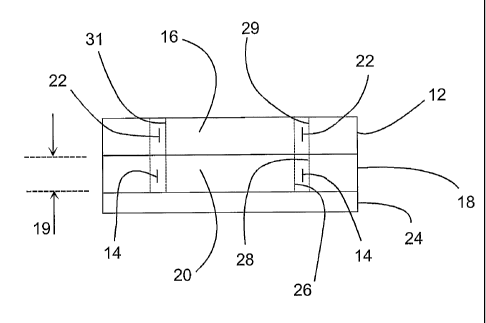
With reference to FIG. 13B, there are shown
essentially three layers stacked on each other. The
bottommost layer includes the non-conductive base member
24. The middle layer includes the inner electrode 20, the
fusion chamber 14, and the inner electrode 20. The
topmost layer includes the non-conductive outer electrode
cover member 12, the access channel 22, and the non-
conductive inner electrode cover member 16.
It is noted that the bottommost layer and the middle
layer, taken together, illustrate a first embodiment of
the apparatus of the invention. More specifically, the
first embodiment of the apparatus is provided for carrying
out fusion of biological cells and includes a non-
conductive base member 24. A conductive outer electrode
18 is supported on the base member 24, wherein the outer
electrode 18 includes a concave outer electrode surface 28
which has an outer electrode radius (r2) and has an
electrode height 19. A conductive inner electrode 20 is
supported on the base member 24, wherein the inner
electrode 20 includes a convex inner electrode surface 26
which has an inner electrode radius (r1) and has the
electrode height 19. The outer electrode surface 28 and
the inner electrode surface 26 are spaced apart from each
other by a gap which defines a fusion chamber 14.
As discussed above, the first electrode radius (r1),
the second electrode radius (r2), and the gap are selected
in accordance with a predetermined range of selectable
ratios (rl/r2) of the first electrode radius to the second
electrode radius, wherein the range of selectable ratios
(rl/r2) is from 0.7 to 0.9, wherein a selected gap is
CA 02546545 2006-05-17
WO 2005/066342
PCT/US2003/035982
-24-
limited by the range of selectable ratios (r1/r2), and
wherein a determined ratio (r1/r2) of the selectable
ratios is based on the selected gap, such that compression
between the biological cells 10 and permeability between
cell membranes are maximized and temperature rise is
minimized for providing cell fusion in the fusion chamber
14.
In accordance with a second embodiment of the
invention, also shown in FIG. 13B and in FIG. 13A as well,
the topmost layer is fixed to the middle layer. In this
respect, the second embodiment of the invention includes
all of the bottommost layer, the middle layer, and the
topmost layer in FIG. 13B.
More specifically, with respect to the second
embodiment of the invention a non-conductive outer
electrode cover member 12 is supported by the outer
electrode 18. A non-conductive inner electrode .cover
member 16 is supported by the inner electrode 20, wherein
the outer electrode cover member 12 and the inner
electrode cover member 16 define an access channel 22, and
wherein the access channel 22 is in communication with the
fusion chamber 14.
Preferably, the non-conductive outer electrode cover
member 12 includes a concave outer cover member surface 29
which has an outer cover member radius. Also, preferably,
the non-conductive inner electrode cover member 16
includes a convex inner cover member surface 31 which has
an inner cover member radius. Preferably, the outer cover
member radius is equal to the outer electrode radius, and
the inner cover member radius is equal to the inner
electrode radius, whereby the access channel 22 is in
registration with the fusion chamber 14.
Non-conductive nylon screws can be used to attach the
inner electrode 20 to the base plate 24 and to attach the
inner electrode cover member 16 to the inner electrode.
Conductive metal screws can be used to attach the outer
electrode 18 to the base plate 24 and to attach the outer
CA 02546545 2006-05-17
WO 2005/066342
PCT/US2003/035982
-25 -
electrode cover member 12 to the outer electrode 18.
The outer electrode 18 and the inner electrode 20 can
be made from stainless steel.
A third embodiment of a coaxial chamber is
illustrated in FIG. 14. Generally, this chamber is a half
of a coaxial chamber mounted vertically. When the
alignment AC voltage is applied, cell motion will be
counter to gravity. This prevents cells from settling to
the bottom of the chamber while the waveforms are applied.
This chamber may be open or closed with sterile ports and
filter relief ports to fill and empty the chamber.
More specifically, this third embodiment of the
apparatus includes a non-conductive support member 40. A
conductive outer electrode 43 is supported in a horizontal
orientation by the support member 40. The outer electrode
43 includes a conductive concave outer electrode surface
42 which has an outer electrode radius (r2) and has an
electrode width. A conductive inner electrode 45 is
supported in a horizontal orientation by the support
member 40 above the outer electrode 43. The inner
electrode 45 includes a conductive convex inner electrode
surface 44 which has an inner electrode radius (r1) and
has the electrode width. A pair of non-conductive
vertically oriented end walls are located at ends of the
outer electrode 43 and the inner electrode 45. The outer
electrode surface 42 and the inner electrode surface 44
are spaced apart from each other by a gap. The gap and
the vertically oriented end walls define a fusion chamber
46. The level of cell fusion medium in the fusion chamber
46 is at level 56.
Preferably, the outer electrode 43 includes a non-
conductive outer electrode support portion 48 which
supports the conductive outer electrode surface 42, and
the inner electrode 45 includes a non-conductive inner
electrode support portion 50 which supports the conductive
inner electrode surface 44. The conductive electrode
surfaces 42 and 44 can be a gold film plated onto the
CA 02546545 2006-05-17
WO 2005/066342
PCT/US2003/035982
-26-
respective non-conductive support portions 48 and 50.
In addition, the apparatus can further include an
input/output port 52 supported by the support member 40,
wherein the input/output port 52 is in communication with
the fusion chamber 46.
In addition, the apparatus can further include a filter
pressure relief valve 54 supported by the support member
40, wherein the filter pressure relief valve 54 is in
communication with the fusion chamber 46.
Preferably, the non-conductive support member 40,
the non-conductive outer electrode support portion 48, the
non-conductive inner electrode support portion 50, and the
non-conductive vertically oriented end walls are formed as
an integrated molded plastic unit.
The values of the ratio rl/r2 and the gap are
determined by the above method. The chamber may be open
or closed. The cells to be fused are placed in a quantity
of low conductivity medium and then placed in the gap
between the two conducting electrode materials. An AC
waveform generator and pulse generator are then connected
to the center (inner) conducting electrode and outer
conducting electrode.
For electric field generation a voltage waveform
generator such as the Cyto Pulse PA-4000/PA-101 computer
controlled waveform generator is. After the alignment,
compression, fusing and holding waveforms are applied, a
cell culture medium is added in the nonconducting volume
of the electrode. This culture medium increases the cell
viability while the fused cells are recovering.
The apparatus can have large volume research,
clinical, and commercial applications. The apparatus can
be packed in sterile packaging. Also, the apparatus can
be manufactured as single-use disposable units. In all
embodiments the volume may be increased by increasing the
electrode height. Temperature increase is not a function
of electrode height.
While the present invention has been shown in the
CA 02546545 2006-05-17
WO 2005/066342
PCT/US2003/035982
-27-
drawings and fully described above with particularity and
detail in connection with what is presently deemed to be
the most practical and preferred embodiments of the
invention, it will be apparent to those of ordinary skill
in the art that many modifications thereof may be made
without departing from the principles and concepts set
forth herein, including but not limited to, variations in
size, materials, shape, form, function and manner of
operation assembly and use.