Note: Descriptions are shown in the official language in which they were submitted.
CA 02550846 2016-08-23
CA2550846
TISSUE ABLATION WITH IRREVERSIBLE ELECTROPORATION
FIELD
100011 This specification resides in the fields of electroporation of
tissue and to treatments
whereby tissue is destroyed by irreversible electroporation.
BACKGROUND
100021 In many medical procedures, such as the treatment of benign or
malignant tumors, it
is important to be able to ablate the undesirable tissue in a controlled and
focused way without
affecting the surrounding desirable tissue. Over the years, a large number of
minimally invasive
methods have been developed to selectively destroy specific areas of
undesirable tissues as an
alternative to resection surgery. There are a variety of techniques with
specific advantages and
disadvantages, which arc indicated and contraindicated for various
applications. For example,
cryosurgery is a low temperature minimally invasive technique in which tissue
is frozen on contact
with a cryogen cooled probe inserted in the undesirable tissue (Rubinsky, B.,
ed. Cryosurgery.
Annu. Rev. Biomed. Eng. Vol. 2. 2000. 157-187.). The area affected by low
temperature therapies,
such as cryosurgery, can be easily controlled through imaging. However, the
probes are large and
difficult to use. Non-selective chemical ablation is a technique in which
chemical agents such as
ethanol are injected in the undesirable tissue to cause ablation (Shiina, S.,
et al., Percutaneotts
ethanol injection therapy fbr hepatocellular carcinoma: results in 146
patients. AJR. 1993. 160: p.
1023-8). Non-selective chemical therapy is easy to apply. However, the
affected area cannot be
controlled because of the local blood flow and transport of the chemical
species. Elevated
temperatures are also used to ablate tissue. Focused ultrasound is a high
temperature non-invasive
technique in which the tissue is heated to coagulation using high-intensity
ultrasound beams
focused on the undesirable tissue (Lynn, .1.G., et al., A new method for the
generation of use of
lbcused ultrasound in experimental biology. J.Gen Physiol., 1942. 26: p. 179-
93; Foster, R.S., et al.,
High-intensitylbcused ultrasound in the treatment of prostatic disease. Eur.
Urol., 1993. 23: p. 44-
7). Electrical currents are also commonly used to heat tissue. Radiofrequency
ablation (RF) is a
high temperature minimally invasive technique in which an active electrode is
introduced in the
undesirable tissue and a high frequency alternating current of up to 500 kHz
is used to heat the
tissue to coagulation (Organ, L. W., Electrophysiological principles of
radiofrequency lesion
making. App!. Neurophysiol., 1976. 39: p. 69-76). In addition to RF heating
traditional Joule
1
CA 02550846 2016-08-23
CA2550846
heating methods with electrodes inserted in tissue and de or ac currents are
also common, (Erez, A.,
Shitzer, A. (Controlled destruction and temperature distribution in biological
tissue subjected to
monoactive electrocoagulation) J. Biomech Eng. 1980- 102(1):42-9).
Interstitial laser coagulation
is a high temperature thermal technique in which tumors are slowly heated to
temperatures
exceeding the threshold of protein denaturation using low power lasers
delivered to the tumors by
optical fibers (Bown, S.G., Phototherapy of tumors. World. J. Surgery, 1983.7:
p.700-9). High
temperature thermal therapies have the advantage of ease of application. The
disadvantage is the
extent of the treated area is difficult to control because blood circulation
has a strong local effect on
the temperature field that develops in the tissue. The armamentarium of
surgery is enhanced by the
availability of the large number of minimally invasive surgical techniques in
existence, each with
their own advantages and disadvantages and particular applications. This
document discloses
another minimally invasive surgical technique for tissue ablation,
irreversible electroporation. We
will describe the technique, evaluate its feasibility through mathematical
modeling and demonstrate
the feasibility with in vivo experimental studies.
(0003] Electroporation is defined as the phenomenon that makes cell
membranes
permeable by exposing them to certain electric pulses (Weaver, J.C. and V.A.
Chizmadzhev,
Theory of electroporation: a review. Bioelectrochem. Bioenerg., 1996. 41: p.
135-60).
Electroporation pulses are defined as those electrical pulses that through a
specific combination of
amplitude, shape, time length and number of repeats produce no other
substantial effect on
biological cells than the permeabilization of the cell membrane. The range of
electrical parameters
that produce cicctroporation is bounded by: a) parameters that have no
substantial effect on the cell
and the cell membrane, b) parameters that cause substantial thermal effects
(Joule heating) and c)
parameters that affect the interior of the cell, e.g. the nucleus, without
affecting the cell membrane.
Joule heating, the thermal effect that electrical currents produce when
applied to biological
materials is known for centuries. It was noted in the previous paragraph that
electrical thermal
effects which elevate temperatures to values that damage cells are commonly
used to ablate
undesirable tissues. The pulse parameters that produce thermal effects are
longer and/or have higher
amplitudes than the electroporation pulses whose only substantial effect is to
permeabilize the cell
membrane.
[00041 There are a variety of methods to electrically produce thermal
effects that ablate
tissue. These include RF, electrode heating, and induction heating. Electrical
pulses that produce
thermal effects are distinctly different from the pulses which produce
electroporation. The
2
CA 02550846 2016-08-23
CA2550846
distinction can be recognizing through their effect on cells and their
utility. The effect of the
thermal electrical pulses is primarily on the temperature of the biological
material and their utility is
in raising the temperature to induce tissue ablation through thermal effects.
[0005] The effect of the electroporation parameters is primarily on the
cell membrane and
their utility is in permeabilizing the cell membrane for various applications.
Electrical parameters
that only affect the interior of the cell, without affecting the cell membrane
were also identified
recently. They are normally referred to as -nanosecond pulses". It has been
shown that high
amplitude, and short (substantially shorter than eleetroporation pulses -
nanoseconds versus
millisecond) length pulses can affect the interior of the cell and in
particular the nucleus without
affecting the membrane. Studies on nanosecond pulses show that they are
"distinctly different than
electroporation pulses" (Beebe SJ. Fox PM. Rec U. Somers K. Stark RH.
Schoenbach KH.
Nanosecond pulsed electric field (nsPEF) effects on cells and tissues:
apoptosis induction and
tumor growth inhibition. PPPS-2001 Pulsed Power Plasma Science 2001. 28th IEEE
International
Conference on Plasma Science and 13th IEEE International Pulsed Power
Conference. Digest of
Technical Papers (Cat. No.01CH37251). IEEE. Part vol.1, 2001, pp.211-15 vol.l.
Piscataway,NJ,
USA. Several applications have been identified for nano-second pulses. One of
them is for tissue
ablation through an effect on the nucleus (Schoenbach, K.H., Beebe, S.J.,
Buescher, K.S. Method
and apparatus for intracellular electro-marnpulation U.S. Patent Application
Pub No. US
2002/0010491 Al, Jan 242002). Another is to regulate genes in the cell
interior, (Gunderson, M.A.
et al. Method for intracellular modification within living cells using pulsed
electrical fields -
regulate gene transcription and entering intracellular US Patent application
2003/0170898 Al,
Sept 11, 2003). Electrical pulses that produce intracellular effects are
distinctly different from the
pulses which produce electroporation. The distinction can be recognizing
through their effect on
cells and their utility. The effect of the intracellular electrical pulses is
primarily on the intracellular
contents of the cell and their utility is in manipulating the intracellular
contents for various uses -
including ablation. The effect of the electroporation parameters is primarily
on the cell membrane
and their utility is in permeabilizing the cell membrane for various
applications, which will be
discussed in greater detail later.
[0006] Electroporation is known for over half a century. It was found that
as a function of
the electrical parameters, electroporation pulses can have two different
effects on the permeability
of the cell membrane. The permeabilization of the membrane can be reversible
or irreversible as a
function of the electrical parameters used. In reversible electroporation the
cell membrane reseals a
3
CA 02550846 2016-08-23
CA2550846
certain time after the pulses cease and the cell survives. In 'rreversible
electroporation the cell
membrane does not reseal and the cell lyses. A schematic diagram showing the
effect of electrical
parameters on the cell membrane permeabilization (electroporation) and the
separation between:
no effect, reversible electroporation and irreversible electroporation is
shown in Figure 1 (Dev,
S.B., Rabussay, D.P., Widera, G., Hofmann, G.A., Medical applications of
electroporation, IEEE
Transactions of Plasma Science, Vo128 No 1, Feb 2000, pp 206 ¨ 223) Dielectric
breakdown of the
cell membrane due to an induced electric field, irreversible electroporation,
was first observed in
the early 1970s (Neumann, E. and K. Rosenheck, Permeability changes induced by
electric
impulses in vesicular membranes. J. Membrane Biol., 1972. 10: p. 279-290;
Crowley, J.M.,
Electrical breakdown of biomolecular lipid membranes as an electromechanical
instability.
Biophysical Journal, 1973. 13: p. 711-724; Zimmermann, U., J. Vienken, and G.
Pilwat, Dielectric
breakdown of cell membranes,. Biophysical Journal, 1974. 14(11): p. 881-899).
The ability of the
membrane to reseal, reversible electroporation, was discovered separately
during the late 1970s
(Kinosita Jr, K. and T.Y. Tsong, Hemolysis of human erythrocytes by a
transient electric field.
Proc. Natl. Acad. Sci. USA, 1977. 74(5): p. 1923-1927; Baker, P.F. and D.E.
Knight, Calcium-
dependent exocytosis in bovine adrenal medullary cells with leaky plasma
membranes. Nature,
1978. 276: p. 620-622; Gauger, B. and F.W. Bentrup, A Study of Dielectric
Membrane Breakdown
in the Fucus Egg,. J. Membrane Biol., 1979. 48(3): p. 249-264).
[0007] The mechanism of electroporation is not yet fully understood. It is
thought that the
electrical field changes the electrochemical potential around a cell membrane
and induces
instabilities in the polarized cell membrane lipid bilayer. The unstable
membrane then alters its
shape forming aqueous pathways that possibly are nano-scale pores through the
membrane, hence
the term "electroporation" (Chang, D.C., et al.. Guide to Electroporation and
ElectrolUsion. 1992,
San Diego, CA: Academic Press, Inc.). Mass transfer can now occur through
these channels under
electrochemical control. Whatever the mechanism through which the cell
membrane becomes
permeabilized, electroporation has become an important method for enhanced
mass transfer across
the cell membrane.
[0008] The first important application of the cell membrane permeabilizing
properties of
electroporation is due to Neumann (Neumann, E., et al., Gene transfer into
mouse lyoma cells by
electroporation in high electric _fields. J. EMBO, 1982. 1: p. 841-5). He has
shown that by applying
reversible electroporation to cells it is possible to sufficiently
permeabilize the cell membrane so
that genes, which are macromolecules that normally are too large to enter
cells, can after
4
CA 02550846 2016-08-23
CA2550846
electroporation enter the cell. Using reversible electroporation electrical
parameters is crucial to the
success of the procedure, since the goal of the procedure is to have a viable
cell that incorporates
the gene.
[0009] Following this discovery electroporation became commonly used to
reversible
permeahilize the cell membrane for various applications in medicine and
biotechnology to
introduce into cells or to extract from cells chemical species that normally
do not pass, or have
difficulty passing across the cell membrane, from small molecules such as
fluorescent dyes, drugs
and radioactive tracers to high molecular weight molecules such as antibodies,
enzymes, nucleic
acids, HMW dextrans and DNA. It is important to emphasize that in all these
applications
electroporation needs to be reversible since the outcome of the mass transport
requires for the cells
to be alive after the electroporation.
100101 Following work on cells outside the body, reversible
electroporation began to be
used for permeabilization of cells in tissue. Heller. R., R. Gilbert, and M.J.
Jaroszeski, Clinical
applications of electrochemotherapy. Advanced drug delivery reviews, 1999. 35:
p. 119-129.
"I issue electroporation is now becoming an increasingly popular minimally
invasive surgical
technique for introducing small drugs and macromolecules into cells in
specific areas of the body.
This technique is accomplished by injecting drugs or macromolecules into the
affected area and
placing electrodes into or around the targeted tissue to generate reversible
permeabilizing electric
field in the tissue, thereby introducing the drugs or macromolecules into the
cells of the affected
area (Mir, L. M., Therapeutic perspectives of in vivo cell
electropermeabilization.
Bioelectrochemistry, 2001. 53: p. 1-10).
[0011] The use of electroporation to ablate undesirable tissue was
introduced by Okino and
Mohri in 1987 and Mir et al. in 1991. They have recognized that there are
drugs for treatment of
cancer, such as bleomycin and cys-platinum, which are very effective in
ablation of cancer cells but
have difficulties penetrating the cell membrane. Furthermore, some of these
drugs, such as
bleomycin, have the ability to selectively affect cancerous cells which
reproduce without affecting
normal cells that do not reproduce. Okino and Mori and Mir et al. separately
discovered that
combining the electric pulses with an impermeant anticancer drug greatly
enhanced the
effectiveness of the treatment with that drug (Okino, M. and H. Mohri, Effects
of a high-voltage
electrical impulse and an anticancer drug on in vivo growing tumors. Japanese
Journal of Cancer
Research, 1987. 78(12): p. 1319-21; Mir, L.M., et al., Electrochernotherapy
potentiation of
antitztmour effect of bleomycin by local electric pulses. European Journal of
Cancer, 1991. 27: p.
CA 02550846 2016-08-23
CA2550846
68-72). Mir et al. soon followed with clinical trials that have shown
promising results and coined
the treatment electrochemotherapy (Mir, L.M., et al., Electrochemotherapy, a
novel antitumor
treatment: first clinical trial. C. R. Acad. Sci., 1991. Ser. III 313(613-8)).
100121 Currently, the primary therapeutic in vivo applications of
electroporation are
antitumor electrochemotherapy (ECT), which combines a cytotoxic nonpermeant
drug with
permcabilizing electric pulses and electrogenetherapy (EGT) as a form of non-
viral gene therapy,
and transdermal drug delivery (Mir, L.M., Therapeutic perspectives of in vivo
cell
electropermeabilization. Bioelectrochemistry, 2001. 53: p. 1-10). The studies
on
electrochemotherapy and electrogenetherapy have been recently summarized in
several publications
(Jaroszeski, M.J., et al., In vivo gene delivery by electroporation. Advanced
applications of
electrochemistry, 1999. 35: p. 131-137; Heller, R., R. Gilbert, and M.J.
Jaroszeski, Clinical
applications of electrochemotherapy. Advanced drug delivery reviews, 1999. 35:
p. 119-129; Mir,
L.M., Therapeutic perspectives of in vivo cell electropermeabilization.
Bioelectrochemistry, 2001.
53: p. 1-10; Davalos, R.V., Real Time Imaging for Molecular Medicine through
electrical
Impedance Tomography of Electroporation, in Mechanical Engineering. 2002,
University of
California at Berkeley: Berkeley. p. 237). A recent article summarized the
results from clinical
trials performed in five cancer research centers. Basal cell carcinoma (32),
malignant melanoma
(142), adenocarcinoma (30) and head and neck squamous cell carcinoma (87) were
treated for a
total of 291 tumors (Mir, L.M., et al., Effective treatment of cutaneous and
subcutaneous malignant
tumours by electrochemotherapy. British Journal of Cancer, 1998. 77(12): p.
2336-2342).
[0013] Electrochemotherapy is a promising minimally invasive surgical
technique to
locally ablate tissue and treat tumors regardless of their histological type
with minimal adverse side
effects and a high response rate (Dev, S.B., etal., Medical Applications of
Electroporation. IEEE
Transactions on Plasma Science, 2000. 28(1): p. 206-223; Heller, R., R.
Gilbert, and M.J.
Jaroszeski, Clinical applications of electrochemotherapy. Advanced drug
delivery reviews, 1999.
35: p. 119-129). Electrochemotherapy, which is performed through the insertion
of electrodes into
the undesirable tissue, the injection of cytotoxic dugs in the tissue and the
application of reversible
electroporation parameters, benefits from the ease of application of both high
temperature treatment
therapies and non-selective chemical therapies and results in outcomes
comparable of both high
temperature therapies and non-selective chemical therapies.
[0014] In addition, because the cell membrane permeabilization electrical
field is not
affected by the local blood flow, the control over the extent of the affected
tissue by this mode of
6
CA 02550846 2016-08-23
CA2550846
ablation does not depend on the blood flow as in thermal and non-selective
chemical therapies. In
designing electroporation protocols for ablation of tissue with drugs that are
incorporated in the cell
and function in the living cells it was important to employ reversible
electroporation: because the
drugs can only function in a living cell. Therefore, in designing protocols
for electrochemotherapy
the emphasis was on avoiding irreversible electroporation. The focus of the
entire field of
electroporation for ablation of tissue was on using reversible pulses, while
avoiding irreversible
electroporation pulses, that can cause the incorporation of selective drugs in
undesirable tissue to
selectively destroy malignant cells. Electrochemotherapy which employs
reversible electroporation
in combination with drugs, is beneficial due to its selectivity however, a
disadvantage is that by its
nature, it requires the combination of chemical agents with an electrical
field and it depends on the
successful incorporation of the chemical agent inside the cell.
SUMMARY
[0015] The present inventors have recognized that irreversible
electroporation, whose
ability to lyse various types of cells outside the body has been known for at
least five decades, has
never been used for tissue ablation in the body and in fact was considered
detrimental to
conventional electrochemotherapy. Although irreversible electroporation of
tissue is not as selective
as reversible electroporation with drug incorporation the present inventors
have found it to be
effective in ablating volumes of undesirable tissues in a way comparable to
other non-
discriminating bulk ablative methods such as cryosurgery, thermal methods or
alcohol injection.
[0016] The present specification comprises a method for the ablation of
undesirable tissue,
involving the placement of electrodes into or near the vicinity of the
undesirable tissue with the
application of electrical pulses causing irreversible electroporation of the
cells throughout the entire
undesirable region. The electric pulses irreversibly permeate the membranes,
thereby invoking cell
death. The length of time of the electrical pulses, the voltage applied and
the resulting membrane
permeability are all controlled within defined ranges. The irreversibly
permeabilized cells may be
left in situ and may be removed by natural processes such as the body's own
immune system. The
amount of tissue ablation achievable through the use of irreversible
electroporation without
inducing thermal damage is considerable, as disclosed and described here.
100171 This concept of irreversible electroporation in tissue to destroy
undesirable tissues is
different from other forms of electrical therapies and treatments.
Irreversible electroporation is
different from intracellular electro-manipulation which substantially only
affects the interior of the
7
CA 02550846 2016-08-23
CA2550846
cell and does not cause irreversible cell membrane damage. Irreversible
electroporation is not
electrically induced thermal coagulation ¨ which induces cell damage through
thermal effects but
rather a more benign method to destroy only the cell membrane of cells in the
targeted tissue.
Irreversible electroporation which irreversible destroys the cell membrane is
also different from
electrochemotherapy in which reversible electroporation pulses are used to
introduce drugs into the
living cells and in which the drugs subsequently affect the living cell.
[0018] An electrical pulse can either have no effect on the cell membrane,
effect internal
cell components, reversibly open the cell membrane after which cells can
survive, or irreversibly
open the cell membrane, after which the cells die. Of these effects,
irreversible electroporation of
tissue was (prior to present invention) generally considered undesirable due
to the possibility of
instantaneous necrosis of the entire tissue affected by the electrical field,
regardless of its diseased
or healthy state. Irreversible electroporation is detrimental in certain
applications, such as gene
therapy or electrochemotherapy, where the sole purpose of the electric pulses
is to facilitate the
introduction of the drug or gene into the cells of a tissue without killing
the cell (Mir., L.M. and S.
Orlowski, The basis of electrochemotherapy, in Electrochemotherapy,
electrogenetherapy, and
transdermal drug delivery: Electrically mediated delivery of molecules to
cells, M.J. Jaroszeski, R.
Heller, R. Gilbert, Editors, 2000, Humana Press, p. 99-118).
[0019] In contrast, irreversible electroporation of the type described
here, solely uses
electrical pulses to serve as the active means for tissue destruction by a
specific means, i.e. by
fatally disrupting the cell membrane. Electrochemotherapy may be selective,
but it does require the
combination of chemical agents with the electrical field. Irreversible
electroporation, although non-
selective, may be used for the ablation of undesirable tissue (such as a
tumor) as a minimally
invasive surgical procedure without the use of adjuvant drugs. Its non-
selective mode of tissue
ablation is acceptable in the field of minimally invasive surgery and provides
results which in some
ways are comparable to cryosurgery, non-selective chemical ablation and high
temperature thermal
ablation.
[0020] An aspect of this specification is a method whereby cells of tissue
are irreversibly
electroporated by applying pulses of very precisely determined length and
voltage. This may be
done while measuring and/or observing changes in electrical impedance in real
time and noting
decreases at the onset of electroporation and adjusting the current in real
time to obtain irreversible
cellular damage without thermal damage. In embodiments where voltage is
applied, the monitoring
of the impedance affords the user knowledge of the presence or absence of
pores. This
8
CA 02550846 2016-08-23
CA2550846
measurement shows the progress of the pore formation and indicates whether
irreversible pore
formation, leading to cell death, has occurred.
[0021] An aspect of this specification is that the onset and extent of
electroporation of cells
in tissue can be correlated to changes in the electrical impedance (which term
is used herein to
mean the voltage over current) of the tissue. At a given point, the
electroporation becomes
irreversible. A decrease in the resistivity of a group of biological cells
occurs when membranes of
the cells become permeable due to pore formation. By monitoring the impedance
of the biological
cells in a tissue, one can detect the average point in time in which pore
formation of the cells
occurs, as well as the relative degree of cell membrane permeability due to
the pore formation. By
gradually increasing voltage and testing cells in a given tissue one can
determine a point where
irreversible electroporation occurs. This information can then be used to
establish that, on average,
the cells of the tissue have, in fact, undergone irreversible electroporation.
This information can
also he used to control the electroporation process by governing the selection
of the voltage
magnitude.
[0022] This specification provides the simultaneous irreversible
electroporation of
multitudes of cells providing a direct indication of the actual occurrence of
electroporation and an
indication of the degree of electroporation averaged over the multitude. The
discovery is likewise
useful in the irreversible electroporation of biological tissue (masses of
biological cells with
contiguous membranes) for the same reasons. The benefits of this process
include a high level of
control over the beginning point of irreversible electroporation.
100231 A feature of this specification is that the magnitude of electrical
current during
electroporation of the tissue becomes dependent on the degree of
electroporation so that current and
pulse length are adjusted within a range predetermined to obtain irreversible
electroporation of
targeted cells of the tissue while minimizing cellular damage to surrounding
cells and tissue.
[0024] An aspect of this specification is that pulse length and current
are precisely adjusted
within ranges to provide more than mere intracellular electro-manipulation
which results in cell
death and less than that which would cause thermal damages to the surrounding
tissues.
[0025] Another aspect of this specification is that the electroporation is
carried out without
adding drugs, DNA, or other materials of any sort to be brought into the
cells.
[0026] Another feature of this specification is that measuring current (in
real time) through
a circuit gives a measurement of the average overall degree of electroporation
obtained.
[0027] Another aspect of this specification is that the precise electrical
resistance of the
9
CA 02550846 2016-08-23
CA2550846
tissue is calculated from cross-time voltage measurement with probe electrodes
and cross-current
measurement with the circuit attached to electroporation electrodes.
[0028] Another aspect of this specification is that the precise electrical
resistance of the
tissue is calculated from cross-time voltage measurement with probe electrodes
and cross-current
measurement with the circuit attached to electroporation electrodes.
[0029] Another aspect of this specification is that electrical
measurements of the tissue can
be used to map the electroporation distribution of the tissue.
[0030] Unlike electrical impedance tomography for detection of reversible
electroporation
which needs to be done during or close to the time the reversible
electroporation pulses are applied
¨ because of the transient nature of the reversible electroporation; in
irreversible electroporation it
is possible and perhaps even preferential to perform the current or EIT
measurements a substantial
time (several minutes or more) after the electroporation to verify that it is
indeed irreversible.
[0031] These and further features, advantages and objects will be better
understood from
the description that follows.
[0032] The claimed invention pertains to a device for ablating tissue in a
living mammal,
comprising: electroporation means for electroporation of cells in a tissue
zone in the mammal;
electroporation monitoring means for monitoring electroporation; and
electroporation controlling
means for controlling electroporation of the cells in the tissue zone, wherein
the electroporation
means and electroporation controlling means comprise: first and second
electrodes adapted to be
positioned near the tissue zone, and a voltage generator for applying
electrical pulses of controlled
length, number and voltage between the first and second electrodes to provide
an electric field
around the tissue zone which has been predetermined to be sufficient to
perform irreversible
electroporation of the cells in the tissue zone while minimizing thermal
damage to the cells in the
tissue zone such that substantially all of the cells in the tissue zone are
killed by non-thermal
irreversible electroporation. The claimed invention also pertains to a device
for ablating tissue in a
living mammal, comprising: electroporation means for electroporation of cells
in a tissue zone in
the mammal, wherein the tissue zone comprises an outer tissue zone surrounding
an inner tissue
zone; electroporation monitoring means for monitoring electroporation; and
electroporation
controlling means for controlling electroporation of the cells in the tissue
zone, wherein the
electroporation means and electroporation controlling means comprise: first
and second electrodes
adapted to be positioned near the tissue zone, and a voltage generator for
applying electrical pulses
of controlled length, number and voltage between the first and second
electrodes to provide an
CA2550846
electric field around the tissue zone which has been predetermined to be
sufficient to perform
irreversible electroporation of cells in the inner tissue zone while
minimizing thermal damage to
cells in the outer tissue zone such that substantially all of the cells in the
inner tissue zone are killed
by non-thermal irreversible electroporation. Also claimed is use of such a
device for said ablating
of tissue in the mammal.
[0033] The claimed invention also pertains to a device for ablating tissue
in a living
mammal, comprising: electroporation means for electroporation of cells in a
tissue zone in the
mammal, wherein the tissue zone comprises an outer tissue zone surrounding an
inner tissue
zone; electroporation monitoring means for monitoring electroporation; and
electroporation
controlling means for controlling electroporation of the cells in the tissue
zone, wherein the
electroporation means and electroporation controlling means comprise: one or
more electrodes
adapted to be positioned near the tissue zone, and a voltage generator for
applying electrical
pulses of controlled length, number and voltage between the first and second
electrodes to
provide an electric field around the tissue zone which has been predetermined
to be sufficient to
raise the temperature of cells in the inner tissue zone to above 50 c thereby
killing some cells in
the inner zone with thermal damage and whereby in the outer tissue zone are
subjected to
minimal thermal damage. The claimed invention also pertains to a device for
ablating tissue in a
living mammal, comprising: electroporation means for electroporation of cells
in a tissue zone
in the mammal, wherein the tissue zone comprises an outer tissue zone
surrounding an inner
tissue zone; electroporation monitoring means for monitoring electroporation;
and
electroporation controlling means for controlling electroporation of the cells
in the tissue zone,
wherein the electroporation means and electroporation controlling means
comprise: one
or more electrodes adapted to be positioned near the tissue zone, and a
voltage generator for
applying electrical pulses of controlled length, number and voltage between
the first and second
electrodes in an amount to selectively induce irreversible electroporation of
cells in the tissue
zone and selectively raise tissue temperature in the tissue zone, said amount
being determined
according to a bioheat equation selected from the group consisting of Penne's
bioheat equation
and Arrhenius bioheat equation.
11
CA 2550846 2017-08-01
CA2550846
BRIEF DESCRIPTION OF THE DRAWINGS
[0034] The invention is best understood from the following detailed
description when read
in conjunction with the accompanying drawings. It is emphasized that,
according to common
practice, the various features of the drawings are not to scale. On the
contrary, the dimensions of
the various features are arbitrarily expanded or reduced for clarity. Included
in the drawings are the
following figures:
[0035] Fig 1. is a graph showing a schematic relationship between field
strength and
pulselength applicable to the electroporation of cells.
[0036] Figures 2 A, 2B and 2C are each images of irreversibly
electroporated areas for
two-electrode configurations using 10 mm center-to-center spacing as following
for Figures 2A, 13
and C: (2A) 0.5mm (857V); (2B) 1.0mm (1295V); (2C) I.5mm (1575V) diameter
electrodes with
a 680V/cm threshold for irreversible electroporation.
[0037] Figures 3A, 3B, and 3C are images showing irreversibly
electroporated regions
1 1 a
CA 2550846 2017-08-01
CA 02550846 2016-08-23
CA2550846
using a 680 V/cm threshold for a two-electrode confirmation with lmm diameter
and 876V and
5mm spacing for Figures 3A; 1116V and 7.5mm for Figure 3B; and 1295V and lOmm
spacing for
Figure 3C.
[0038] Figures 4A, 4B and 4C are images showing the effect of electrode
diameter for a 4-
electrode configuration with 1 Omm spacing wherein Figure 4A is for 0.5mm
diameter and 940V;
Figure 4B is for 1.0mm diameter and 1404V and Figure 4C is for 1.5mm and 1685
V.
[0039] Figures 5A, 5B and 5C are images showing the effect of electrode
spacing for a 4-
electrode configuration wherein the electrode is lmm in diameter and Figure 5A
shows results with
a 5mm and 910V; Figure 5B 7.5mm and 1175V and Figure 5C lOmm and 1404V.
[0040] Figure 6 is an image showing the irreversible (1295V, 680V/cm
threshold) as
compared to the reversible region (1300V, 360V/cm threshold) using virtually
the same electrical
parameters. 1300V is the most common voltage applied across two electrodes for
ECT. The most
common voltage parameters are eight 100 s pulses at a frequency of 1Hz.
Applying a single 800iis
pulse provides a conservative estimate of the heating associated with a
procedure. The one second
space normally between pulses will enlarge an area amount of heat to be
dissipated through the
tissue.
[0041] Figure 7 is an image showing reversible electroporation with 1mm
electrodes,
1 Omm spacing. A voltage of 189V applied between the electrodes induces
reversible
electroporation without any irreversible electroporation by not surpassing the
680V/cm irreversible
electroporation threshold anyone in the domain. The shaded area is greater
than 360 V/cm.
[0042] Figures 8A and 8B show a comparison of the effect of blood flow and
metabolism
on the amount of irreversible electroporation. Figure 8A no blood flow or
metabolism. Figure 8B
wb=lkg/m3, Ch = 3640 J/(kg K), Th = 37 C, and q" = 33.8kW/m3.
[0043] Figure 9 is a schematic view of a liver between two cylindrical
Ag/AgC1 electrodes.
The distance between the electrodes was 4mm and the radius of the electrodes
is 1 Omm. The
electrodes were clamped with special rig parallel and concentric to each
other. The liver lobe was
compressed between the electrodes to achieve good contact.
[0044] Figure 10 is a photo of a view of a liver which was electroporated
by irreversible
electroporation with two cylindrical surface electrodes of lOmm in diameter.
Histology shows that
the dark area is necrotic.
[0045] Figure 11 is a photo of a cross section through an electroporated
liver. Histology
shows that the dark area is necrotic. The distance between the two Al plates
that hold the liver is
12
CA 02550846 2016-08-23
CA2550846
exactly 4 mm. The electroporation electrodes were 10 mm in diameter and
centered in the middle
of the lesion.
[0046] Figure 12 shows the liver of calculated temperature distribution
(C), upper panel,
and electrical potential gradient (electroporation gradient) (V/cm), lower
panel, for the in vivo
experiment. The Figure 12 also shows conditions through a cross section of a
liver slab through the
center of the electroporated area. Height of thc slab is 4 mm.
[0047] Figure 13 combines Figures 11 and 12 to show a comparison between
the extent of
tissue necrosis (dark area) and the temperature and voltage gradient
distribution in the
electroporated tissue. The photo of Figure 11 is shown schematically at the
bottom on Figure 13. It
is evident that most of the dark area was at a temperature of about 42 C
following the 40
milliseconds electroporation pulse. The edge of the dark area seems to
correspond to the 300 V/cm
electroporation gradient line.
DETAILED DESCRIPTION
[0048] Before the present methods, treatments and devices are described,
it is to be
understood that this subject matter is not limited to particular embodiments
described, as such may,
of course, vary. It is also to be understood that the terminology used herein
is for the purpose of
describing particular embodiments only, and is not intended to be limiting,
since the scope of the
present invention will be limited only by the appended claims.
[0049] Where a range of values is provided, it is understood that each
intervening value, to
the tenth of the unit of the lower limit, unless the context clearly dictates
otherwise, between the
upper and lower limits of that range is also specifically disclosed. Each
smaller range between any
stated value or intervening value in a stated range and any other stated or
intervening value in that
stated range is encompassed within the invention. The upper and lower limits
of these smaller
ranges may independently be included or excluded in the range, and each range
where either,
neither or both limits are included in the smaller ranges is also encompassed
within the invention,
subject to any specifically excluded limit in the stated range. Where the
stated range includes one
or both of the limits, ranges excluding either or both of those included
limits are also included in the
invention.
[0050] Unless defined otherwise, all technical and scientific terms used
herein have the
same meaning as commonly understood by one of ordinary skill in the art to
which this invention
belongs. Although any methods and materials similar or equivalent to those
described herein can
13
CA 02550846 2016-08-23
CA2550846
be used in the practice or testing of the present invention, the preferred
methods and materials are
now described.
[0051] The publications discussed herein are provided solely for their
disclosure prior to
the filing date of the present application. Nothing herein is to be construed
as an admission that the
present invention is not entitled to antedate such publication by virtue of
prior invention. Further,
the dates of publication provided may be different from the actual publication
dates which may
need to be independently confirmed.
DEFINITIONS
[0052] The term "reversible electroporation" encompasses permeabilization
of the cell
membrane through the application of electrical pulses across the cell. In
"reversible
electroporation" the permeabilization of the cell membrane ceases after the
application of the pulse
and the cell membrane permeability reverts to normal. The cell survives -
reversible
electroporation." It is used as a means for introducing chemicals, DNA, or
other materials into
cells.
[0053] The term "irreversible electroporation" also encompasses the
permeabilization of
the cell membrane through the application of electrical pulses across the
cell. However, in
-irreversible electroporation" the permeabilization of the cell membrane does
not cease after the
application of the pulse and the cell membrane permeability does not revert to
normal. The cell
does not survive "irreversible electroporation" and the cell death is caused
by the disruption of the
cell membrane and not merely by internal perturbation of cellular components.
Openings in the cell
membrane are created and/or expanded in size resulting in a
13a
CA 02550846 2006-06-21
WO 2005/065284 PCT/US2004/043477
fatal disruption in the normal controlled flow of material across the cell
membrane. The cell
membrane is highly specialized in its ability to regulate what leaves and
enters the cell.
Irreversible electroporation destroys that ability to regulate in a manner
such that the cell can
not compensate and as such the cell dies.
INVENTION IN GENERAL
)054] The invention provides a method and a system for destruction
(ablation) of undesirable
tissue. It involves the insertion (bringing) electroporation electrodes to the
vicinity of the
undesirable tissue and in good electrical contact with the tissue and the
application of electrical
pulses that cause irreversible electroporation of the cells throughout the
entire area of the
undesirable tissue. The cells whose membrane was irreversible permeabilized
may be left in
situ (not removed) and as such may be gradually removed by the body's immune
system. Cell
death is produced by inducing the electrical parameters of irreversible
electroporation in the
undesirable area.
)055] Electroporation protocols involve the generation of electrical fields
in tissue and are
affected by the Joule heating of the electrical pulses. When designing tissue
electroporation
protocols it is important to determine the appropriate electrical parameters
that will maximize
tissue permeabilization without inducing deleterious thermal effects. It has
been shown that
substantial volumes of tissue can be electroporated with reversible
electroporation without
inducing damaging thermal effects to cells and has quantified these volumes
(Davalos, R.V.,
B. Rubinsky, and L.M. Mir, Theoretical analysis of the thermal effects during
in vivo tissue
electroporation. Bioelectrochemistry, 2003. Vol. 61(1-2): p. 99-107).
1056] The electrical pulses required to induce irreversible
electroporation in tissue are larger
in magnitude and duration from the electrical pulses required for reversible
electroporation.
Further, the duration and strength of the pulses required for irreversible
electroporation are
different from other methodologies using electrical pulses such as for
intracellular electro-
manipulation or thermal ablation. The methods are very different even when the
intracellular
(nano-seconds) electro-manipulation is used to cause cell death, e.g. ablate
the tissue of a
tumor or when the thermal effects produce damage to cells causing cell death.
1057] Typical values for pulse length for irreversible electroporation are
in a range of from
about 5 microseconds to about 62,000 milliseconds or about 75 microseconds to
about 20,000
milliseconds or about 100 microseconds 10 microseconds. This is
significantly longer than
the pulse length generally used in intracellular (nano-seconds) electro-
manipulation which is 1
14
CA 02550846 2006-06-21
WO 2005/065284 PCT/US2004/043477
microsecond or less ¨ see published U.S. application 2002/0010491 published
January 24,
2002.
1058] The pulse is at voltage of about 100 V/cm to 7,000 V/cm or 200 V/cm
to 2000 V/cn or
300 V/cm to 1000 V/cm about 600 V/cm 10% for irreversible electroporation.
This is
substantially lower than that used for intracellular electro-manipulation
which is about 10,000
V/cm, see U.S. application 2002/0010491 published January 24, 2002.
1059] The voltage expressed above is the voltage gradient (voltage per
centimeter). The
electrodes may be different shapes and sizes and be positioned at different
distances from each
other. The shape may be circular, oval, square, rectangular or irregular etc.
The distance of
one electrode to another may be 0.5 to 10 cm., 1 to 5 cm., or 2-3 cm. The
electrode may have
a surface area of 0.1 ¨5 sq. cm. or 1-2 sq. cm.
1060] The size, shape and distances of the electrodes can vary and such can
change the
voltage and pulse duration used. Those skilled in the art will adjust the
parameters in
accordance with this disclosure to obtain the desired degree of
electroporation and avoid
thermal damage to surrounding cells.
1061] Thermal effects require electrical pulses that are substantially
longer from those used in
irreversible electroporation (Davalos, R.V., B. Rubinsky, and L.M. Mir,
Theoretical analysis
of the thermal effects during in vivo tissue electroporation.
Bioelectrochemistry, 2003. Vol.
61(1-2): p. 99-107). Figure 1 is showing that irreversible electroporation
pulses are longer and
have higher amplitude than the reversible electroporation pulses. When using
irreversible
electroporation for tissue ablation, there may be concern that the
irreversible electroporation
pulses will be as large as to cause thermal damaging effects to the
surrounding tissue and the
extent of the tissue ablated by irreversible electroporation will not be
significant relative to that
ablated by thermal effects. Under such circumstances irreversible
electroporation could not be
considered as an effective tissue ablation modality as it will act in
superposition with thermal
ablation.
1062] The present invention evaluates, through mathematical models and
experiment, the
maximal extent of tissue ablation that could be accomplished by irreversible
electroporation
prior to the onset of thermal effects. The models focused on electroporation
of liver tissue with
two and four needle electrodes and on electroporation of liver tissue with two
infinite parallel
plates using available experimental data. The experiment (EXAMPLE 3) evaluates
irreversible
electroporation between two cylindrical electrodes, also in the liver. The
liver was chosen
because it is considered a potential candidate for irreversible
electroporation ablation. The
CA 02550846 2006-06-21
WO 2005/065284 PCT/US2004/043477
results show that the area that can be ablated by irreversible electroporation
prior to the onset
of thermal effects is comparable to that which can be ablated by
electrochemotherapy,
validating the use of irreversible electroporation as a potential minimally
invasive surgical
modality.
0063] Earlier studies have shown that the extent of electroporation can be
imaged in real time
with electrical impedance tomography (EIT) (Davalos, R.V., B. Rubinsky, and
D.M. Otten, A
feasibility study for electrical impedance tomography as a means to monitor
tissue
electroporation for molecular medicine. IEEE Transactions on Biomedical
Engineering, 2002.
49(4): p. 400-403). In irreversible electroporation the electroporated area
persists indefinitely
after the electroporation pulse, showing that irreversible electroporation may
be imaged
leisurely with EIT. Irreversible electroporation, therefore, has the advantage
of a tissue
ablation technique that is as easy to apply as high temperature ablation,
without the need for
adjuvant chemicals as electrochemotherapy and with real-time control of the
affected area with
electrical impedance tomography.
EXAMPLES
00641 The following examples are put forth so as to provide those of
ordinary skill in the art
with a complete disclosure and description of how to make and use the present
invention, and
are not intended to limit the scope of what the inventors regard as their
invention nor are they
intended to represent that the experiments below are all or the only
experiments performed.
Efforts have been made to ensure accuracy with respect to numbers used (e.g.
amounts,
temperature, etc.) but some experimental errors and deviations should be
accounted for.
Unless indicated otherwise, parts are parts by weight, molecular weight is
weight average
molecular weight, temperature is in degrees Centigrade, and pressure is at or
near atmospheric.
EXAMPLE 1
0065] The mathematical model provided here shows that irreversible tissue
ablation can affect
substantial volumes of tissue, without inducing damaging thermal effects. To
this end, the
present invention uses the Laplace equation to calculate the electrical
potential distribution in
tissue during typical electroporation pulses and a modified Pennes (bioheat),
(Pennes, H.H.,
Analysis of tissue and arterial blood flow temperatures in the resting
forearm. J of Appl.
Physiology., 1948. 1: p. 93-122), equation to calculate the resulting
temperature distribution. It
is important to note that there are several forms of the bioheat equation
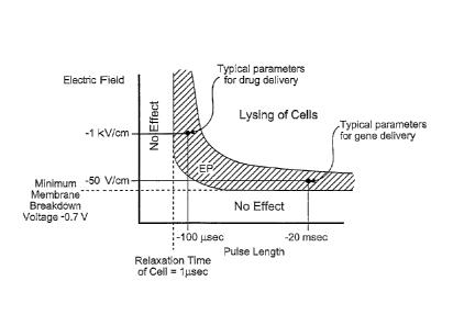
which have been
16
CA 02550846 2006-06-21
WO 2005/065284 PCT/US2004/043477
reviewed (Carney, C.K., Mathematical models of bioheat transfer, in
Bioengineering heat
transfer, Y.I. Choi, Editor. 1992, Academic Press, Inc: Boston. p. 19-152;
Eto, T.K. and B.
Rubinsky, Bioheat transfer, in Introduction to bioengineering, S.A. Berger, W.
Goldsmith, and
E.R. Lewis, Editors. 1996, Oxford Press). While the Pennes equation is
controversial, it is
nevertheless commonly used because it can provide an estimate of the various
biological heat
transfer parameters, such as blood flow and metabolism. The modified Pennes
equation in this
study contains the Joule heating term in tissue as an additional heat source.
066] The electrical potential associated with an electroporation pulse is
determined by
solving the Laplace equation for the potential distribution:
V = (oVO) = 0 (1)
1)671 where 0 is the electrical potential and a- is the electrical
conductivity. The electrical
boundary condition of the tissue that is in contact with the leftmost
electrode(s) on which the
electroporation pulse is applied is:
0 = Vo
(2)
1)68] The electrical boundary condition at the interface of the rightmost
electrode(s) is:
0 = 0
(3)
1)69] The boundaries where the analyzed domain is not in contact with an
electrode are
treated as electrically insulative to provide an upper limit to the electrical
field near the
electroporation electrodes and an upper limit to the temperature distribution
that results from
electroporation:
50 n
¨ (4)
an
1)70] Solving the Laplace equation enables one to calculate the associated
Joule heating, the
heat generation rate per unit volume from an electrical field (p):
P '1\ 7 012 (5)
1)71] This term is added to the original Pennes equation, (Pennes, H.H.,
Analysis of tissue
and arterial blood flow temperatures in the resting forearm. J of Appl.
Physiology., 1948. 1: p.
93-122) to represent the heat generated from the electroporation procedure:
V = (WT)+}vbcb(Ta ¨T)+ q + p = pc ¨aT
(6)
1:172] To solve equation (4) it is assumed that the entire tissue is
initially at the physiological
temperature of 37 C:
17
CA 02550846 2006-06-21
WO 2005/065284 PCT/US2004/043477
T(x,y,z,0). 37
(7)
1073]
The outer surface of the analyzed domain and the surfaces of the electrodes
are taken to
be adiabatic, which should produce an upper limit to the calculated
temperature distribution in
the tissue:
-aT 0 on the electrodes boundary and the outer surface
domain (8)
an
10741 The analysis modeled conditions typical to tissue electroporation in
the liver. The liver
was chosen because it is the organ that most minimally invasive ablation
techniques treat since
cancer in the liver can be resolved by extirpation of the diseased area while
surgical resection
is not possible in many cases for this organ (Onik, G., B. Rubinsky, and et
al., Ultrasound-
Guided Hepatic Cryosurgery in the Treatment of Metastatic Colon Carcinoma.
Cancer, 1991.
67(4): p. 901-907).. The electroporation parameters, i.e. pulse parameters for
reversible and
irreversible electroporation where obtained from rat liver data (Miklavcic,
D., et al., A
validated model of in vivo electric field distribution in tissues for
electrochemotherapy and for
DNA electrotransfer for gene therapy. Biochimica et Biophysica Acta, 2000.
1523(1): p. 73-
83; Suzuki, T., et al., Direct gene transfer into rat liver cells by in vivo
electroporation. FESS
Letters, 1998. 425(3): p. 436-440), but biological parameters corresponding to
the human liver
were used in the analysis. Tissue thermal properties are taken from reference
(Duck, F.A.,
Physical Properties of Tissues: A Comprehensive Reference Book. 1990, San
Diego: Academic
Press) and the electrical properties from reference (Boone, K., D. Barber, and
B. Brown,
Review - Imaging with electricity: report of the European Concerted Action on
Impedance
Tomography. J. Med. Eng. Technol., 1997. 21: p. 201-232) and are listed in
table 1. The tissue
is assumed isotropic and macroscopically homogeneous.The intent of the
analysis was to
determine the extent of the region in which reversible or irreversible
electroporation is induced
in the liver for various electroporation voltages and durations while the
maximal temperature
in the tissue is below 50 C. Thermal damage is a time-dependent process
described by an
Arhenius type equation (Henriques, F.C. and A.R. Moritz, Studies in thermal
injuries: the
predictability and the significance of thermally induced rate processes
leading to irreversible
epidermal damage. Arch Pathol., 1947. 43: p. 489-502; Diller, K.R., Modeling
of bioheat
transfer processes at high and low temperatures, in Bioengineering heat
transfer, Y.I. Choi,
Editor. 1992, Academic Press, Inc: Boston. p. 157-357),
RT dt
(9)
18
CA 02550846 2006-06-21
WO 2005/065284 PCT/US2004/043477
1075] Where Q is a measure of thermal damage, is the frequency factor, Ea
is the activation
energy and R is the universal gas constant. A detailed description on the
various degrees of
thermal damage as described in Equation (9) above can be found in (Diller,
K.R., Modeling of
bioheat transfer processes at high and low temperatures, in Bioengineering
heat transfer, Y.I.
Choi, Editor. 1992, Academic Press, Inc: Boston. p. 157-357).
W76] A careful examination shows that the thermal damage is a complex
function of time,
temperature and all the parameters in Equation (9) above and that there are
various degrees of
thermal damage. In various applications or for various considerations it is
possible to design
irreversible electroporation protocols that induce some degree of thermal
damage, either in part
of the electroporated region or at a reduced level throughout the
electroporated region.
However, in this example we have chosen 50 C as the target temperature for
several reasons.
Thermal damage begins at temperatures higher than 42 C, but only for prolonged
exposures.
Damage is relatively low until 50 C to 60 C at which the rate of damage
dramatically
increases (Diller, K.R., Modeling of bioheat transfer processes at high and
low temperatures,
in Bioengineering heat transfer, Y.I. Choi, Editor. 1992, Academic Press, Inc:
Boston. p. 157-
357). Therefore 50 C will be a relatively low bound on the possible thermal
effects during
irreversible electroporation. It is anticipated that the electrical parameters
chosen for
irreversible electroporation without a thermal effect could be substantially
longer and higher
than those obtained from an evaluation for 50 C in this example. Furthermore,
since the
Laplace and bioheat equations are linear, the results provided here can be
extrapolated and
considered indicative of the overall thermal behavior.
10771 The analyzed configurations have two needles or four needle
electrodes embedded in a
square model of the liver. Needle electrodes are commonly used in tissue
electroporation and
will be most likely also used in the liver (Somiari, S., et al., Theory and in
vivo application of
electroporative gene delivery. Molecular Therapy, 2000. 2(3): p. 178-187). The
square model
of the liver was chosen large enough to avoid outer surface boundary effects
and to produce an
upper limit for the temperature, which develops during electroporation in the
liver. For each
configuration the surface of one electrode is assumed to have a prescribed
voltage with the
other electrode set to ground. The effect of the spacing between the
electrodes was investigated
by comparing distances of 5, 7.5 and 10 mm, which are typical. The electrodes
were also
modeled with typical dimensions of 0.5, 1 and 1.5 mm in diameter. The blood
flow perfusion
rate was taken to zero or 1.0 kg/m3 s (Deng, Z.S. and J. Liu, Blood perfusion-
based model for
characterizing the temperature fluctuations in living tissue. Phys A STAT Mech
Appl, 2001.
19
CA 02550846 2006-06-21
WO 2005/065284 PCT/US2004/043477
300: p. 521-530). The metabolic heat was taken to be either zero or 33.8 kW/m3
(Deng, Z.S.
and J. Liu, Blood perfusion-based model for characterizing the temperature
fluctuations in
living tissue. Phys A STAT Mech Appl, 2001. 300: p. 521-530).
078] The calculations were made for an electroporation pulse of 800 ts.
This pulse duration
was chosen because typically, reversible electroporation is done with eight
separate 100 us
pulses, (Miklavcic, D., et al., A validated model of in vivo electric field
distribution in tissues
for electrochemotherapy and for DNA electrotransfer for gene therapy.
Biochimica et
Biophysica Acta, 2000. 1523(1): p. 73-83) and therefore the value we chose is
an upper limit
of the thermal effect in a pulse time frame comparable to that of reversible
electroporation.
Consequently, the results obtained here are the lower limit in possible lesion
size during
irreversible electroporation. It should be emphasized that we believe
irreversible
electroporation tissue ablation can be done with shorter pulses than 800 1.1S.
To evaluate the
thermal effect, we gradually increased in our mathematical model the applied
pulse amplitude
for the 800 ps pulse length until our calculations indicated that the
electroporation probe
temperature reached 50 C, which we considered to be the thermal damage limit.
Then, we
evaluated the electric field distribution throughout the liver.
079] A transmembrane potential on the order of 1V is required to induce
irreversible
electroporation. This value is dependent on a variety of conditions such as
tissue type, cell size
and other external conditions and pulse parameters. The primary electrical
parameter affecting
the transmembrane potential for a specific tissue type is the amplitude of the
electric field to
which the tissue is exposed. The electric field thresholds used in estimating
the extent of the
region that was irreversibly electroporated were taken from the fundamental
studies of
Miklavcic, Mir and their colleagues performed with rabbit liver tissue
(Miklavcic, D., et al., A
validated model of in vivo electric field distribution in tissues for
electrochemotherapy and for
DNA electrotransfer for gene therapy. Biochimica et Biophysica Acta, 2000.
1523(1): p. 73-
83). In this study, that correlated electroporation experiments with
mathematical modeling,
they have found that the electric field for reversible electroporation is 362
+/- 21 V/cm and is
637 +/- 43 V/cm n for irreversible electroporation for rat liver tissue.
Therefore, in the analysis
an electric field of 360 V/cm is taken to represent the delineation between no
electroporation
and reversible electroporation and 680 V/cm to represent the delineation
between reversible
and irreversible electroporation.
1080] All calculations were performed using MATLAB's finite element solver,
Femlab v2.2
(The MathWorks, Inc. Natick, MA). To ensure mesh quality and validity of
solution, the mesh
CA 02550846 2006-06-21
WO 2005/065284 PCT/US2004/043477
was refined until there was less than a 0.5% difference in solution between
refinements. The
baseline mesh with two lmm electrodes, lOmm spacing had 4035nodes and 7856
triangles.
The simulations were conducted on a Dell Optiplex GX240 with 512MB of RAM
operating on
Microsoft Windows 2000.
RESULTS and DISCUSSION
1081] Figures 2 and 3 examine the effect of the electrode size and spacing
on the ablated area
in a two-needle electroporation configuration. In obtaining these figures, we
ignored the effect
of the blood flow and metabolism in the heat transfer equation, which should
give an upper
limit for the estimated ablation area. Figure 2 compares the extent of the
irreversible
electroporated area for electroporation electrode sizes of 0.5, 1 and 1.5 mm
in diameter and a
distance between electrodes of 10 mm. The strong effect of the electrode size
is evident. It is
seen that for the smaller electrodes, the irreversibly electroporated area is
not contiguous, while
for a 1.5 mm electrode the area of potential tissue, ablation has an
elliptical shape with
dimensions of about 15 mm by 10 mm. In the brackets, we give the
electroporation voltage for
which the probe temperature reaches 50 C in these three configurations. It is
seen that the
range is from 857V for the 0.5 mm probe to 1575V for the 1.5 mm probe. This is
within the
typical range of tissue electroporation pulses. Figure 3 evaluates the effect
of the spacing
between the electrodes. It is observed that in the tested range, the small
dimension of the
contiguous elliptical shape of the ablated lesion remains the same, while the
larger dimension
seems to scale with the distance between the electrodes.
1082] Figures 2 and 3 demonstrate that the extent of tissue ablation with
irreversible
electroporation is comparable to that of other typical minimally invasive
methods for tissue
ablation, such as cryosurgery (Onik, G.M., B. Rubinsky, and et. al.,
Ultrasound-guided hepatic
cryosurgery in the treatment of metastatic colon carcinoma. Cancer, 1991.
67(4): p. 901-907;
Onik, G.M., et al., Transrectal ultrasound-guided percutaneous radical
cryosurgical ablation
of the prostate. Cancer, 1993. 72(4): p. 1291-99). It also shows that varying
electrode size and
spacing can control lesion size and shape. The shape and size of the ablated
lesion can be also
controlled by varying the number of electrodes used. This is shown in Figures
4 and 5, for a
four-electrode configuration. These figures also compare the effect of probe
size and spacing
and the results were also obtained by ignoring the effect of blood flow and
metabolism in the
energy equation. Again, it is seen that larger electrodes have a substantial
effect on the extent
21
CA 02550846 2006-06-21
WO 2005/065284
PCT/US2004/043477
of the ablated region and that the extent of ablation scales with the spacing
between the
electrodes.
10831 A comparison between reversible and irreversible electroporation
protocols can be
achieved from Figures 6 and 7. In Figure 6, an 800 ps, 1295 V pulse was
applied between two
1.5 mm diameter electrodes placed 10 mm apart. This produces a tissue
temperature lower than
50 C. The figure plots the margin of the irreversibly electroporated region,
i.e. the 680 V/cm
voltage-to-distance gradients and that of the reversible electroporated
region, the 360 V/cm
gradients. Figure 7 was obtained for two 1 mm electrodes placed 10 mm apart.
In this figure,
we produced an electroporated region that was only reversibly electroporated,
i.e. with electric
fields lower than 360 V/cm. In comparing Figures 6 and 7, it is obvious that
the extent of the
ablated area possible through electrochemotherapy alone is substantially
smaller than that
through irreversible electroporation alone.
084] The effect of blood flow and metabolism on the extent of irreversible
electroporation is
illustrated in Figure 8. The figures compare a situation with metabolism and a
relatively high
blood flow rate to a situation without blood flow or metabolism. It is obvious
that metabolism
and blood perfusion have a negligible effect on the possible extent of
irreversible tissue
electroporation. This is because the effect of the Joule heating produced by
the electroporation
current is substantially larger than the effects of blood flow or metabolism.
085] An even more conservative estimate for the thermal damage can be
obtained by
assuming that the tissue reaches 50 C instantaneously, during the
electroporation pulses such
that the damage is defined as
Q = t 1,e-6EIRT
(10)
086] Several values taken from the literature for activation energy and
frequency factor were
applied to equation (10) with the pulse lengths calculated in the examples
above. Because the
application of the pulse is so short, the damage would be near zero, many
times less than the
value (Q=0.53) to induce a first degree burn (Diller, K.R., Modeling of
bioheat transfer
processes at high and low temperatures, in Bioengineering heat transfer, Y.I.
Choi, Editor.
1992, Academic Press, Inc: Boston. p. 157-357) regardless of the values used
for activation
energy and frequency factor.
087] Currently, tissue ablation by electroporation is produced through the
use of cytotoxic
drags injected in tissue combined with reversible electroporation, a procedure
known as
electrochemotherapy. The present invention shows that irreversible
electroporation by itself
produces substantial tissue ablation for the destruction of undesirable
tissues in the body. The
22
CA 02550846 2006-06-21
WO 2005/065284 PCT/US2004/043477
concern was that higher voltages required for irreversible electroporation
would cause Joule
heating and would induce thermal tissue damage to a degree that would make
irreversible
electroporation a marginal effect in tissue ablation. Using a mathematical
model for calculating
the electrical potential and temperature field in tissue during
electroporation, the present
invention shows that the area ablated by irreversible tissue electroporation
prior to the onset of
thermal effects is substantial and comparable to that of other tissue ablation
techniques such as
cryosurgery. Our earlier studies have shown that the extent of electroporation
can be imaged
in real time with electrical impedance tomography (Davalos, R.V., B. Rubinsky,
and D.M.
Often, A feasibility study for electrical impedance tomography as a means to
monitor tissue
electroporation for molecular medicine. IEEE Transactions on Biomedical
Engineering, 2002.
49(4): p. 400-403; Davalos, R.V., et al., Electrical impedance tomography for
imaging tissue
electroporation. IEEE Transactions on Biomedical Engineering, 2004).
Irreversible
electroporation, therefore, has the advantage of being a tissue ablation
technique, which is as
easy to apply as high temperature ablation, without the need for adjuvant
chemicals as required
in electrochemical ablation and electrochemotherapy. In addition, a unique
aspect of
irreversible electroporation is that the affected area can be controlled in
real time with
electrical impedance tomography.
EXAMPLE 2
1088] This example was developed to produce a correlation between
electroporation pulses
and thermal effects. The system analyzed is an infinitesimally small control
volume of tissue
exposed to an electroporation voltage gradient of V (Volts/cm).The entire
electrical energy is
dissipated as heat and there is no conduction of heat from the system. The
calculations produce
the increase in temperature with time during the application of the pulse and
the results are a
safe lower limit for how long a certain electroporation pulse can be
administered until a certain
temperature is reached. To generate the correlation an energy balance is made
on a control
volume between the Joule heating produced from the dissipation of heat of the
V (volt/cm)
electrical potential gradient (local electrical field) dissipating through
tissue with an electrical
conductivity of o- (ohm-cm) and the raise in temperature of the control volume
made of tissue
with a density p (g/cc) and specific heat, c, (J/g K). The calculation
produces the following
equation for the raise in temperature (T) per unit time (t) as a function of
the voltage gradients
and the thermal and electrical properties of the liver.
23
CA 02550846 2006-06-21
WO 2005/065284 PCT/US2004/043477
dT V2 cr
(2-1)
dt pc
10891 The table below was obtained for the liver with the following
properties:
1090] Electrical resistivity of liver - 8.33 Ohm-meter
10911 Specific heat of liver - Jig K
10921 Density of liver - 1 g/cc
1093] We obtain the following table:
TABLE 1
Voltage Gradient - V Time per degree C rise time from 37 C to 65 C
(V/cm) (ms) (ms)
50 1199.52 33586.56
100 299.88 8396.64
150 133.28 3731.84
200 74.97 2099.16
250 47.98 1343.46
300 33.32 932.96
350 24.48 685.44
400 18.74 524.79
450 14.81 414.65
500 12.00 335.87
550 9.91 277.57
600 8.33 233.24
650 7.10 198.74
700 6.12 171.36
750 5.33 149.27
800 4.69 131.20
850 4.15 116.22
900 3.70 103.66
950 3.32 93.04
1000 3.00 83.97
1050 2.72 76.16
1100 2.48 69.39
1150 2.27 - 63.49
1200 2.08 58.31
1250 1.92 53.74
1300 1.77 49.88
1350 1.65 46.07
24
CA 02550846 2006-06-21
WO 2005/065284 PCT/US2004/043477
Voltage Gradient - V Time per degree C rise time from 37 C to 65 C
(V/cm) (ms) (ms)
1400 1.53 42.84
1450 1.43 39.94
1500 1.33 37.32
094] The second column of Table 1 gives the amount of time it takes for the
temperature of
the liver to raise 1 C, when the tissue experiences the electroporation pulse
in column 1. The
time for even a relatively high electroporation voltage of 1500V/cm is of the
order of 1.33
millisecond for 1 C rise and 37.32 millisecond until a temperature of 65 C is
reached. Using
the equation (2-1) or Table 1 it is possible to evaluate the amount of time a
certain pulse can be
applied without inducing thermal effects. Considering the typical
electroporation parameters
reported so far there is no limitation in the electroporation length from
thermal considerations.
Column 3 of Table 1 shows the time required to reach 65 C, which is where
thermal damage
may begin. The calculations in this example give a lower limit for the extent
of time in which a
certain thermal effects will be induced by electroporation pulses. For more
precise calculations
it is possible to use the equation developed in this example with equation (9)
or (10) from
Example 1.
EXAMPLE 3
095] The goal of this experiment was to verify the ability of irreversible
electroporation
pulses to produce substantial tissue ablation in the non-thermal regime. To
this end we have
performed experiments on the liver of Spraque-Dawley male rats (250g to 350 g)
under an
approved animal use and care protocol. After the animals were anesthetized by
injection of
Nembutal Sodium Solution (50mg/m1 Pentobarbital) the liver was exposed via a
midline
incisions and one lobed clamped between two cylindrical electrodes of Ag/AgC1,
with a
diameter of 10 mm (In Vivo Metric, Healdsburg, CA). The electrodes had their
flat surface
parallel; they were concentric and the liver between the electrodes was
compressed so that the
lobes were separated by 4 mm. A schematic of the electrodes and the liver is
shown in Figure
9. The liver was exposed to a single electroporation pulse of 40 milliseconds.
One electrode
was set to 400 V and the other grounded. The rest of the liver was not in
contact with any
media and therefore is considered electrically insulated. After
electroporation the rat was
maintained under controlled anesthesia for three hours. Following
exsanguination the liver was
flushed with physiological saline under pressure and fixed by perfusion with
formaldehyde.
The liver was resected through the center of the electroporated region and
analyzed by
CA 02550846 2011-12-01
histology. Figures 10 and 11 show the appearance of the liver. Histology has
determined that
the dark area corresponds to the region of tissue necrosis. The electrical
field in the
electroporated liver and the temperature distribution were calculated using
the equations in
Example 1, subject to one electrode at a voltage of 400V and the other
grounded, for 40
milliseconds. The liver was modeled as an infinite slab of 4 mm thickness,
with concentric
cylindrical electrodes (see Figure 9). The results are shown in Figure 12.
Figure 12 shows
lines of constant voltage gradients (V/cm) and lines of constant temperature.
It is evident that in
the majority of the electroporated tissue the temperature is about 42 C
immediately after the
pulse. The highest temperature occurs near the edge of the cylindrical
electrodes, where it is
about 50 C. Figure 13 was obtained by bringing together Figures 11 and 12.
Superimposing the
calculated results on the histological measurements reveals that the dark
(necrotic) area margin
corresponds to electroporation parameters of about 300 V/cm. The results
demonstrate that
irreversible electroporation can induce substantial tissue necrosis without
the need for chemical
additives as in electrochemotherapy and without a thermal effect.
[0096] The preceding merely illustrates the principles of the invention. It
will be
appreciated that those skilled in the art will be able to devise various
arrangements which,
although not explicitly described or shown herein, embody the principles of
the invention and
are included within its spirit and scope. Furthermore, all examples and
conditional language
recited herein are principally intended to aid the reader in understanding the
principles of the
invention and the concepts contributed by the inventors to furthering the art,
and are to be
construed as being without limitation to such specifically recited examples
and conditions.
Moreover, all statements herein reciting principles, aspects, and embodiments
of the invention
as well as specific examples thereof, are intended to encompass both
structural and functional
equivalents thereof Additionally, it is intended that such equivalents include
both currently
known equivalents and equivalents developed in the future, i.e., any elements
developed that
perform the same function, regardless of structure. The scope of the present
invention,
therefore, is not intended to be limited to the exemplary embodiments shown
and described
herein. Rather, the scope of the present invention is embodied by the appended
claims.
26