Note: Descriptions are shown in the official language in which they were submitted.
CA 02553940 2006-07-25
WO 2005/074814 PCT/US2005/003126
DEVICES, SYSTEMS, AND METHODS FOR CLOSURE OF CARDIAC OPENINGS
Cross Reference to Related Applications
[0001] This application incorporates by reference, and claims priority to and
the benefits of, U.S.
Provisional Patent Applications Serial Nos. 60/540,474, 60/540,827, and
60/540,821, each of
which were filed on January 30, 2004.
Technical Field
(0002) The invention generally relates to devices, systems, and related
methods for closing
cardiac openings. More particularly, the invention features devices, systems,
and related
methods for the percutaneous transluminal closure of patent foramen ovales and
left atrial
appendages.
Background
[0003] The human heart is divided into four compartments or chambers. The left
and right
atria are located in the upper portion of the heart and the left and right
ventricles are located in
the lower portion of the heart. The left and right atria are separated from
each other by a
muscular wall, the intraatrial septum, while the ventricles are separated by
the intraventricular
septum.
(0004] Either congenitally or by acquisition, abnormal openings, holes, or
shunts can occur
between the chambers of the heart or the great vessels, causing blood to
inappropriately flow
therethrough. Such deformities are usually congenital and originate during
fetal life when the
heart forms from a folded tube into a four chambered, two-unit system. The
septal deformities
result from the incomplete formation of the septum, or muscular wall, between
the chambers of
the heart and can cause significant problems.
[0005) One such septal deformity or defect, a patent foramen ovate, is a
persistent, one-way,
usually flap-like opening in the wall between the right atrium and left atrium
of the heart. Since
left atrial pressure is normally higher than right atrial pressure, the flap
typically stays closed.
Under certain conditions, however, right atrial pressure exceeds left atrial
pressure, creating the
possibility for right to left shunting that can allow blood clots to enter the
systemic circulation.
This is particularly problematic for patients who are prone to forming venous
thrombus, such as
those with deep vein thrombosis or clotting abnormalities.
CA 02553940 2006-07-25
WO 2005/074814 PCT/US2005/003126
[0006] Moreover, certain patients are prone to atrial arrhythmias (i. e.,
abnormal heart
rhythms which can cause the heart to pump less effectively). In a common such
abnormality,
atrial fibrillation, the two upper chambers of the heart (i. e., the left
atria and the right atria),
quiver instead of beating effectively. Because the atria do not beat and empty
cleanly during
atrial fibrillation, blood can stagnate on the walls and form clots that can
then pass through the
heart and into the brain, causing a stroke or a transient ischemic attack.
These clots typically
form in a cut-de-sac in the heart called the left atrial appendage due to its
tendency to have low
or stagnant flow.
[0007] Nonsurgical (i. e., percutaneous) closure of a patent foramen ovate and
similar cardiac
openings such as an atrial septal defect or a ventricular septal defect, and
obliteration of a left
atrial appendage can be achieved using a variety of mechanical closure
devices. These closure
devices typically consist of a metallic structural framework with a scaffold
material attached
thereto. Currently available closure devices, however, are often complex to
manufacture, are
inconsistent in performance, require a technically complex implantation
procedure, lack
anatomic conformability, and lead to complications (e.g., thrombus formation,
chronic
inflammation, residual leaks, perforations, fractures, and conduction system
disturbances).
[0008] Improved devices, systems, and related methods for closing cardiac
openings, such
as, for example, a patent foramen ovate, and for obliterating cardiac cut-de-
sacs, such as, for
example, a left atrial appendage, are, therefore, needed.
Summary of the Invention
[0009] The present invention provides devices, compounds, systems, and related
methods for
closing cardiac openings. A device of the invention may include, for example,
a patch with an
adhesive and/or a removable frame. The patch can be placed across a cardiac
opening, such as a
patent foramen ovate or a left atrial appendage, to substantially occlude the
cardiac opening.
Alternatively, in another aspect, the device includes a U-shaped patch,
together with an adhesive,
that is specifically configured for attachment to a septum secundum and
closure of a patent
foramen ovate.
[0010] Moreover, in another aspect, a compound may be used to assist the
device in closing,
or may be used on its own to close, a cardiac opening. For example, a compound
that includes
an adhesive and a plurality of composite particles disposed within the
adhesive may be used in
that regard. In one embodiment, the plurality of composite particles disposed
within the
adhesive expand upon contact with blood and/or water, thereby locking the
compound into place
in the cardiac opening to substantially occlude the cardiac opening.
CA 02553940 2006-07-25
WO 2005/074814 PCT/US2005/003126
[0011] In using the devices and compounds of the invention to close cardiac
openings, the
aforementioned disadvantages associated with the closure devices known in the
art are
minimized or eliminated.
[0012] In one aspect, the invention provides a closure device for percutaneous
transvascular
closure of a cardiac opening. The closure device includes a patch, an adhesive
coated on the
patch, and at least one hollow channel enclosed within the patch.
[0013] Various embodiments of this aspect of the invention include the
following features.
The patch may include a bioresorbable material and the adhesive may be a light
activated
adhesive, such as, for example, an adhesive curable with ultraviolet light.
The hollow channel
enclosed within the patch may, for its part, be a conduit for light. The
closure device may further
include a fiber optic cable, and/or a removable frame, enclosed within the
hollow channel. In
another embodiment, the closure device includes a divider that has first and
second surfaces.
The first surface is coupled to the adhesive and the second surface is coated
with a primer.
[0014] In another aspect, the invention relates to a method for percutaneous
transluminal
closure of a cardiac opening in a patient. The method includes inserting a
closure device as
described above into a heart of the patient and positioning the closure device
across the cardiac
opening to substantially occlude the cardiac opening.
[0015] In various embodiments of this aspect of the invention, positioning the
closure device
across the cardiac opening includes coupling the closure device to a tissue
surface of the patient
proximate the cardiac opening. The cardiac opening may be, for example, a
patent foramen
ovate or a left atrial appendage. Coupling the closure device to the tissue
surface may include
providing light to the hollow channel enclosed within the patch and activating
the adhesive
coated on the patch with the provided light. In another embodiment, coupling
the closure device
to the tissue surface includes applying a primer to the tissue surface.
[0016] In yet another aspect, the invention provides a closure device for
percutaneous
transluminal closure of a cardiac opening. The closure device includes a
patch, at least one
hollow channel enclosed within the patch, and a removable frame enclosed
within the hollow
channel.
[0017] In one embodiment of this aspect of the invention, the patch is made
from a collagen
material. In another embodiment, the frame is constructed from a shape memory
alloy, such as,
for example, nitinol.
[0018] In still another aspect, the invention relates to a method for
percutaneous transluminal
closure of a cardiac opening in a patient. The method includes inserting a
closure device as
CA 02553940 2006-07-25
WO 2005/074814 PCT/US2005/003126
immediately described above into a heart of the patient and positioning the
closure device across
the cardiac opening to substantially occlude the cardiac opening.
[0019] In various embodiments of this aspect of the invention, positioning the
closure device
across the cardiac opening includes coupling the closure device to a tissue
surface of the patient
proximate the cardiac opening. The cardiac opening may be, for example, a
patent foramen
ovale or a left atrial appendage. In one embodiment, coupling the closure
device to the tissue
surface includes thermally welding the closure device to the tissue surface.
In another
embodiment, the frame of the closure device is removed from within the hollow
channel after the
closure device is thermally welded to the tissue surface.
[0020] In another aspect, the invention provides a closure device for
percutaneous
transluminal closure of a cardiac opening. The closure device includes a
housing, a releasable
patch coupled to the housing, and an adhesive coated on the releasable patch.
[0021] In one embodiment of this aspect of the invention, the housing is
substantially
sonically shaped. In another embodiment, the releasable patch includes a
bioresorbable material.
The adhesive may be a light activated adhesive, such as, for example, an
adhesive curable with
ultraviolet light. In yet another embodiment, the closure device includes a
light source enclosed
within the housing. The light source may be, for example, a light bulb or a
fiber optic cable. In
still another embodiment, the closure device includes a divider that has first
and second surfaces.
The first surface is coupled to the adhesive and the second surface is coated
with a primer.
[0022] In yet another aspect, the invention relates to a method for
percutaneous transluminal
closure of a cardiac opening in a patient. The method includes inserting a
closure device as
immediately described above into a heart of the patient and positioning the
releasable patch of
the closure device across the cardiac opening to substantially occlude the
cardiac opening.
[0023) In various embodiments of this aspect of the invention, positioning the
releasable
patch of the closure device across the cardiac opening includes coupling the
releasable patch to a
tissue surface of the patient proximate the cardiac opening. The cardiac
opening may be, for
example, a patent foramen ovale or a left atrial appendage. Coupling the
releasable patch of the
closure device to the tissue surface may include providing a light source
emitting light within the
housing and activating the adhesive coated on the releasable patch with the
emitted light. In
another embodiment, coupling the releasable patch to the tissue surface
includes applying a
primer to the tissue surface. In yet another embodiment, coupling the
releasable patch to the
tissue surface includes separating the releasable patch from the housing.
[0024) Additionally, in another aspect, the invention provides a closure
device for
percutaneous transvascular closure of a patent foramen ovate. The closure
device includes a U-
CA 02553940 2006-07-25
WO 2005/074814 PCT/US2005/003126
shaped patch configured for attachment to a septum secundum and an adhesive
coated on the U-
shaped patch.
[0025] In one embodiment of this aspect of the invention, a substance for
stimulating tissue
in-growth into the closure device is coated on the U-shaped patch. The
substance may be, for
example, a growth factor, a pharmacological agent to stimulate tissue growth,
an irritant to
encourage an inflammatory response, cells, or genes. In another embodiment, a
substance for
increasing endothelization, or, alternatively, a substance for decreasing
thrombogenicity, such as,
for example, heparin, is coated on the U-shaped patch. In yet another
embodiment, the closure
device includes at least one barrier coupled to the U-shaped patch. The
barrier may be a right
atrial barrier for blocking an opening to the patent foramen ovale from the
right atrium, or,
alternatively, the barrier may be a left atrial barrier for blocking an
opening to the patent foramen
ovate from the left atrium.
[0026] The U-shaped patch may include a biological material, a bioresorable
material, a
synthetic material, a polymeric material, a shape memory material, and/or a
metallic mesh
material. The adhesive may be, for example, cyanoacrylate and/or a fibrin
based adhesive.
[0027] In a further aspect, the invention provides a method for percutaneous
transluminal
closure of a patent foramen ovate in a patient. The method includes inserting
a closure device
into a heart of the patient and coupling the closure device to the septum
secundum to
substantially occlude the patent foramen ovate. The closure device includes a
U-shaped patch
configured for attachment to a septum secundum and an adhesive coated on the U-
shaped patch.
[0028] In one embodiment of this aspect of the invention, coupling the closure
device to the
septum secundum includes gluing the closure device to the septum secundum.
[0029] In another aspect, the invention relates to a compound for percutaneous
transluminal
closure of a cardiac opening. The compound includes an adhesive and a
plurality of composite
particles disposed within the adhesive. The composite particles are capable of
expansion upon
contact with blood and/or water.
[0030] In various embodiments of this aspect of the invention, the adhesive is
a fibrin based
adhesive. The composite particles may be, for example, gelatin particles,
biological particles,
bioresorbable particles, and/or foam particles.
[0031] In yet another aspect, the invention provides a method for percutaneous
transluminal
closure of a cardiac opening in a patient. The method includes providing a
compound as
described above and injecting the compound into the cardiac opening to
substantially occlude the
cardiac opening.
CA 02553940 2006-07-25
WO 2005/074814 PCT/US2005/003126
[0032] In one embodiment of this aspect of the invention, the method further
includes
positioning a patch or a barrier across an end of the cardiac opening, which
may be, for example,
a patent foramen ovate or a left atrial appendage.
[0033] A device of the invention may further include specially designed
balloons together
with adhesives and/or substances for stimulating tissue growth coated on, or
contained within,
the specially designed balloons. According to one feature of the invention,
the specially
designed balloons ensure that the adhesives are only exposed once the balloons
are located
within the cardiac openings. Advantageously, the adhesives are exposed only to
the tissue
surface of the cardiac openings and not to a patient's blood prior to locating
the balloons within
the cardiac openings. By minimizing the exposure of the adhesives to blood,
the risk of
thrombus formation is reduced.
[0034] According to another feature of the invention, closure systems employ
one or more
locators for initially locating the cardiac openings and then properly
positioning the balloons of
the invention within the cardiac openings. Knowing that a balloon is properly
positioned within
a cardiac opening allows a physician to release the adhesive contained within
the balloon at the
appropriate time. As such, the risk of exposing the adhesive prior to locating
the balloon within
the cardiac opening, and the consequent risk of thrombus formation, is again
reduced.
[0035] In one aspect, the invention provides a closure device for percutaneous
transluminal
closure of a cardiac opening. The closure device includes a balloon, which has
an outer surface,
and an adhesive. The balloon is inflatable between a deflated state and an
inflated state. In the
deflated state, the outer surface of the balloon involutes to form a cavity
and the adhesive is
coated on a surface of the cavity. In the inflated state, the cavity unfolds
to form the outer
surface of the balloon and the adhesive is coated on the outer surface of the
balloon.
[0036] In one embodiment of this aspect of the invention, the cavity is formed
around a mid-
portion of the balloon, which may be tubularly-shaped. In another embodiment,
the closure
device further includes a substance for stimulating tissue growth. In the
deflated state of the
balloon, the growth substance is coated on the surface of the cavity. In the
inflated state of the
balloon, the growth substance is coated on the outer surface of the balloon.
[0037] In another aspect, the invention relates to a method for percutaneous
transluminal
closure of a cardiac opening in a patient. The method includes inserting a
closure device as
described above into a heart of the patient, positioning the closure device
within the cardiac
opening with the balloon of the closure device deflated, and inflating the
balloon to expose the
adhesive coated on the outer surface of the balloon to the cardiac opening. In
one embodiment
CA 02553940 2006-07-25
WO 2005/074814 PCT/US2005/003126
of this aspect of the invention, the balloon of the closure device is removed
from the patient after
the adhesive is exposed to the cardiac opening.
[0038] In yet another aspect, the invention provides a closure device that
includes a balloon
having an outer surface, a porous band encircling only a portion of the outer
surface of the
balloon, and an adhesive disposed between the outer surface of the balloon and
the porous band.
The porous band has a plurality of openings.
[0039] In one embodiment of this aspect of the invention, the porous band
encircles a center
portion of the balloon, which may be, for example, tubularly-shaped. In
another embodiment, a
substance for stimulating tissue growth is disposed between the outer surface
of the balloon and
the porous band.
[0040] In still another aspect, the invention relates to a method that
includes inserting a
closure device as just described into a heart of the patient, positioning the
closure device within
the cardiac opening, and applying a pressure to the balloon of the closure
device to expose the
adhesive through the plurality of openings of the porous band to the cardiac
opening. In one
embodiment of this aspect of the invention, the balloon and the porous band of
the closure device
are removed from the patient after the adhesive is exposed through the
plurality of openings of
the porous band to the cardiac opening.
[0041] Additionally, in another aspect, the closure device includes an outer
balloon that has a
plurality of first holes, an inner balloon that has a plurality of second
holes, and an adhesive.
The adhesive is contained within the inner balloon, which is itself contained
within the outer
balloon.
[0042] In various embodiments of this aspect of the invention, at least one of
the plurality of
first holes and the plurality of second holes includes pores. Alternatively,
in another
embodiment, at least one of the plurality of first holes and the plurality of
second holes includes
slits. In yet another embodiment, at least one of the inner balloon and the
outer balloon is
tubularly-shaped. In another embodiment, a substance for stimulating tissue
growth is contained
within the inner balloon.
[0043] In a further aspect, the invention relates to a method that includes
inserting a closure
device as just described into a heart of the patient, positioning the closure
device within the
cardiac opening, applying a first pressure to the inner balloon to express the
adhesive through the
plurality of second holes, and applying a second pressure to the outer balloon
to express the
adhesive through the plurality of first holes to the cardiac opening. In one
embodiment of this
aspect of the invention, the outer balloon and the inner balloon of the
closure device are removed
CA 02553940 2006-07-25
WO 2005/074814 PCT/US2005/003126
from the patient after the adhesive is expressed through the plurality of
first holes to the cardiac
opening.
[0044] In another aspect, the closure device includes a balloon and an
adhesive. The balloon
has a membrane constructed from a wicking material and the adhesive is
contained within the
membrane of the balloon.
[0045] In one embodiment of this aspect of the invention, the balloon is
tubularly-shaped. In
another embodiment, a substance for stimulating tissue growth is contained
within the membrane
of the balloon. At least a portion of the adhesive and/or the substance for
stimulating tissue
growth may be absorbed within the membrane of the balloon.
[0046] In yet another aspect, the invention relates to a method that includes
inserting a
closure device as just described into a heart of the patient, positioning the
closure device within
the cardiac opening, and contacting a tissue surface of the cardiac opening
with the membrane of
the balloon to apply the adhesive to the tissue surface of the cardiac
opening. In one
embodiment of this aspect of the invention, the balloon of the closure device
is removed from the
patient after the adhesive is applied to the tissue surface of the cardiac
opening.
[0047] In various embodiments of the foregoing aspects of the invention, the
adhesives are
cyanoacrylates, fibrin based adhesives, albumin gluteraldehyde type adhesives,
or light activated
adhesives. Moreover, the substances for stimulating tissue growth may be, for
example, growth
factors, pharmacological agents for stimulating tissue growth, irritants for
encouraging an
inflammatory response, cells, or genes. The cardiac opening is, for example, a
patent foramen
ovate or a left atrial appendage.
[0048] In still another aspect, the invention relates to a method for
percutaneous transluminal
closure of a left atrial appendage in a patient. The method includes inserting
a closure device
into a heart of the patient and positioning the closure device within the left
atrial appendage. The
closure device includes a balloon having a plurality of holes and an adhesive
contained within
the balloon. The method further includes applying a pressure to the balloon to
separate the
plurality of holes and to expose the adhesive to the left atrial appendage.
The method also
includes coupling the balloon of the closure device to the left atrial
appendage with the exposed
adhesive.
[0049] Additionally, in another aspect, the invention provides a closure
device that includes
a balloon with an outer surface, a first adhesive coated on the outer surface
of the balloon, and a
light source located within the balloon.
[0050] In one embodiment of this aspect of the invention, the closure device
further includes
a second adhesive coated on an inner surface of the balloon. At least one of
the first adhesive
CA 02553940 2006-07-25
WO 2005/074814 PCT/US2005/003126
and the second adhesive may be a light activated adhesive. In another
embodiment, the closure
device further includes a divider having first and second surfaces. The first
surface of the divider
may be coupled to the first adhesive and the second surface of the divider may
be coated with a
primer. The balloon may be made of an elastomer, or, alternatively, a
biological material, which
may be, for example, a collagen or a bioresorbable polymer. The balloon may be
tubularly-
shaped.
(0051] In a further aspect, the invention relates to a method that includes
inserting a closure
device as just described into a heart of the patient, positioning the closure
device within the
cardiac opening, and coupling the closure device to the cardiac opening to
substantially occlude
the cardiac opening.
[0052] In various embodiments of this aspect of the invention, coupling the
closure device to
the cardiac opening includes inflating the balloon, emitting light from the
light source located
within the balloon, and activating the adhesive coated on the outer surface of
the inflated balloon
with the emitted light. The inflated balloon may then be deflated and left
behind in the cardiac
opening. Coupling the closure device to the cardiac opening may also include
applying a primer
to a tissue surface of the cardiac opening. The cardiac opening may be, for
example, a patent
foramen ovale or a left atrial appendage.
[0053] In another aspect, the invention provides a percutaneous transluminal
system for
positioning a closure device in a cardiac opening. The system includes a
catheter, a closure
device coupled to the catheter, and a first locator coupled to at least one of
the catheter and the
closure device. The first locator is for positioning the closure device within
the cardiac opening.
[0054] In various embodiments of this aspect of the invention, the first
locator is a disk, a
plurality of arms, a rod, or a balloon. The first locator may be, for example,
a right atrial locator
or a left atrial locator. In one embodiment, an adhesive, such as, for
example, a cyanoacrylate, a
fibrin based adhesive, or an albumin gluteraldehyde type adhesive, is coated
on the first locator.
In another embodiment, the system further includes a second locator coupled to
at least one of
the catheter and the closure device. The second locator is also for
positioning the closure device
within the cardiac opening.
[0055] In one embodiment, the system further includes an adhesive coupled to
the closure
device. Again, the adhesive may be, for example, a cyanoacrylate, a fibrin
based adhesive, or an
albumin gluteraldehyde type adhesive. The adhesive coupled to the closure
device may
alternatively be a light activated adhesive and the system may further include
a light source
coupled to the catheter for activating the light activated adhesive.
CA 02553940 2006-07-25
WO 2005/074814 PCT/US2005/003126
[0056] In another embodiment, the closure device is a balloon, which may be,
for example,
tubularly-shaped. In one embodiment, the balloon includes a first end, a
second end, and a
lumen extending from the first end to the second end. In another embodiment,
the balloon
includes a first opening at the first end of the balloon and a second opening
at the second end of
the balloon. In yet another embodiment, the balloon comprises a plurality of
holes. An adhesive
may be coated on an outer surface of the balloon, coated on an inner surface
of the balloon, or
simply contained within the lumen of the balloon.
[0057] In still another aspect, the invention relates to a method for
delivering a closure
device to a cardiac opening in a patient. The method includes inserting, into
a heart of the
patient, a system for positioning the closure device within the cardiac
opening. The system is as
just described. The first locator of the system is used to locate the cardiac
opening and also to
position the closure device within the cardiac opening.
(0058] In various embodiments of this aspect of the invention, the method
further includes
coupling the closure device to the cardiac opening to substantially occlude
the cardiac opening.
The method may also include coupling the first locator to a tissue surface of
the patient that is
proximate the cardiac opening. The cardiac opening may be, for example, a
patent foramen
ovate or a left atrial appendage.
[0059] Additionally, in another aspect, the invention provides a percutaneous
transluminal
system for closing a cardiac opening. The system includes a first catheter
having a proximal
end, a distal end, and a lumen extending from the proximal end to the distal
end, a second
catheter at least partially enclosed within the lumen of the first catheter,
and a lining coupled to
the first and second catheters. The second catheter is movable between a
retracted state and a
deployed state. In the retracted state of the second catheter, the lining is
positioned within the
lumen of the first catheter. In the deployed state of the second catheter, the
lining inverts and is
positioned outside the lumen of the first catheter.
[0060] In various embodiments of this aspect of the invention, the lining is
sock-shaped.
Moreover, adhesives and/or substances for stimulating tissue growth, of the
types described
above, may be coated on a surface of the lining and/or contained within the
lining itself.
[0061] In a further aspect, the invention relates to a method for percutaneous
transluminal
closure of a cardiac opening in a patient. The method includes inserting a
system as just
described into a heart of the patient, positioning the system proximate the
cardiac opening with
the second catheter in a retracted state, and deploying the second catheter to
invert the lining and
position the lining within the cardiac opening.
to
CA 02553940 2006-07-25
WO 2005/074814 PCT/US2005/003126
[0062] In one embodiment of this aspect of the invention, the system further
includes an
adhesive coated on a surface of the lining and the adhesive is exposed to the
cardiac opening
when the second catheter is deployed. In another embodiment, the lining
includes a plurality of
holes and the system further includes an adhesive contained within the lining.
In such an
embodiment, the adhesive is exposed through the plurality of holes to the
cardiac opening when
the second catheter is deployed. In yet another embodiment, the system is
removed from the
patient after the adhesive is exposed to the cardiac opening, which may be,
for example, a patent
foramen ovate.
[0063] The foregoing and other aspects, features, and advantages of the
invention will
become more apparent from the following description taken in conjunction with
the
accompanying drawings.
Brief Description of the Drawings
[0064] In the drawings, like reference characters generally refer to the same
parts throughout
the different views. Also, the drawings are not necessarily to scale, emphasis
instead generally
being placed upon illustrating the principles of the invention.
[0065] FIG. 1 is a cutaway view of a heart illustrating a patent foramen
ovate.
[0066] FIG. 2 is a partial cross-sectional view of another heart illustrating
a left atrial
appendage.
[0067] FIG. 3 is a schematic perspective view of a system, including a
delivery catheter and
a closure device, for the percutaneous transluminal closure of a cardiac
opening according to an
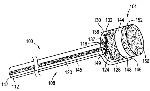
illustrative embodiment of the invention.
[0068] FIGS. 4A-4B illustrate extended and folded configurations of a frame
for the closure
device illustrated in FIG. 3, according to an illustrative embodiment of the
invention.
[0069] FIG. 5 is a schematic perspective view of a system, including a
delivery catheter and
a closure device, for the percutaneous transluminal closure of a cardiac
opening according to
another illustrative embodiment of the invention.
[0070] FIG. 6. is a schematic cross-sectional view of a system, including a
delivery catheter
and a closure device, for the percutaneous transluminal closure of a cardiac
opening according to
another illustrative embodiment of the invention.
[0071] FIG. 7 is a schematic side view of a closure device for the
percutaneous transluminal
closure of a cardiac opening according to another illustrative embodiment of
the invention.
11
CA 02553940 2006-07-25
WO 2005/074814 PCT/US2005/003126
[0072] FIG. 8 is a schematic side view of a closure demce, according to
another illustrative
embodiment of the invention, coupled to the septum secundum and the septum
primum of a
patent foramen ovate.
[0073] FIG. 9 is a schematic side view of a closure device, according to
another illustrative
S embodiment of the invention, coupled to the septum secundum and the septum
primum of a
patent foramen ovate.
[0074] FIG. 10 is a schematic side view of a closure device, according to
another illustrative
embodiment of the invention, coupled to the septum secundum and the septum
primum of a
patent foramen ovate.
[0075] FIGS. 11A-11C illustrate the stages, according to an illustrative
embodiment of the
invention, for closing a patent foramen ovate in a patient.
[0076] FIG. 11 D illustrates a left atrial appendage closed according to an
illustrative
embodiment of the invention.
[0077] FIG. 11 E-11 F illustrates the stages, according to another
illustrative embodiment of
the invention, for closing a patent foramen ovate in a patient.
[0078] FIG. 12 is a schematic side view of the illustrative closure device of
FIG. 7 coupled
to the septum secundum of a patent foramen ovate.
[0079] FIG. 13 illustrates a compound for the percutaneous transluminal
closure of a cardiac
opening according to an illustrative embodiment of the invention.
[0080] FIG. 14 is a schematic side view of a closure system according to an
illustrative
embodiment of the invention.
[0081] FIG. 15 is a schematic perspective view of a closure system according
to another
illustrative embodiment of the invention.
[0082] FIG. 16 is a schematic side view of a closure system according to
another illustrative
embodiment of the invention.
[0083] FIG. 17 is a schematic side view of a closure system according to
another illustrative
embodiment of the invention.
(0084] FIG. 18 is a schematic perspective view of an inflated balloon
according to an
illustrative embodiment of the invention.
[0085] FIG. 19 is a schematic perspective view of the illustrative balloon of
FIG.17 deflated
according to an illustrative embodiment of the invention.
[0086] FIG. 20 is a schematic cross-sectional view of the illustrative balloon
of FIG. 19
taken along the line 19-19.
12
CA 02553940 2006-07-25
WO 2005/074814 PCT/US2005/003126
[0087] FIG. 21 is a schematic cross-sectional view of the illustrative balloon
of FIG. 20
inflated according to an illustrative embodiment of the invention.
(0088] FIG. 22 is a schematic perspective view of a deflated balloon according
to another
illustrative embodiment of the invention.
(0089] FIG. 23 is a schematic perspective view of a balloon according to
another illustrative
embodiment of the invention.
[0090] FIG. 24 is a schematic cross-sectional view of the illustrative balloon
of FIG. 23
taken along the line 23-23.
[0091] FIG. 25 is a schematic cross-sectional view of a balloon according to
another
illustrative embodiment of the invention.
[0092] FIG. 26 is a schematic cross-sectional view of concentric balloons
according to
another illustrative embodiment of the invention.
[0093] FIG. 27 is a schematic cross-sectional view of a balloon according to
another
illustrative embodiment of the invention.
[0094] FIG. 28 is a schematic cross-sectional view of a balloon according to
another
illustrative embodiment of the invention.
[0095] FIG. 29 is a schematic cross-sectional view of the illustrative balloon
of FIG. 28
taken along the line 28-28.
[0096] FIG. 30 is a schematic side view of a closure system, including a
retracted sock
catheter, according to another illustrative embodiment of the invention.
[0097] FIG. 31 is a schematic side view of the illustrative closure system of
FIG. 30,
including a deployed sock catheter.
[0098] FIGS. 32A-32D illustrate the stages, according to an illustrative
embodiment of the
invention, for closing a patent foramen ovate in a patient.
[0099] FIGS. 33A-33E illustrate the stages, according to another illustrative
embodiment of
the invention, for closing a patent foramen ovate in a patient.
[0100] FIGS. 34A-34B illustrate the stages, according to another illustrative
embodiment of
the invention, for closing a patent foramen ovate in a patient.
Description
[0101] The present invention features devices, systems, and related methods
for closing
cardiac openings, such as, for example, the patent foramen ovate described
below, and for
obliterating cardiac cut-de-sacs, such as, for example, the left atrial
appendage described below.
13
CA 02553940 2006-07-25
WO 2005/074814 PCT/US2005/003126
(0102] FIG. 1 depicts a cutaway view of a heart 2U. The heart ZU includes a
septum 24 that
divides a right atrium 26 from a left atrium 32. The septum 24 includes a
septum secundum 36
and a septum primum 40. An exemplary cardiac opening, a patent foramen ovale
44, that is to
be corrected by the devices, systems, and related methods of the present
invention is located
between the septum secundum 36 and the septum primum 40. The patent foramen
ovale 44
provides an undesirable fluid communication between the right atrium 26 and
the left atrium 32
and, under certain conditions, allows for the shunting of blood between the
right atrium 26 and
the left atrium 32. If the patent foramen ovale 44 is not closed or obstructed
in some manner, a
patient is placed at a higher risk for an embolic stroke in addition to other
circulatory
abnormalities.
[0103] FIG. 2 depicts a partial cross-sectional view of another heart 60. The
heart 60
includes an aorta 64, a left ventricle 68, a left atrium 72, and a fossa
ovalis 76. The heart 60 also
includes an exemplary cardiac cul-de-sac, a left atrial appendage 80, that is
to be obliterated by
the devices, systems, and related methods of the present invention. Under
certain conditions,
blood clots may form in the left atrial appendage 80. If the left atrial
appendage 80 is not closed
or obstructed in some manner, a patient is placed at a higher risk of having
the blood clots pass
from the heart 60 and into the vasculature of the brain, causing a stroke or a
transient ischemic
attack.
[0104] In broad overview, embodiments of the devices of the invention
typically include a
patch or a balloon. Referring to embodiments that include a patch, an adhesive
may be coated on
the patch and the adhesive may require activation (e.g., light activation) to
bond the patch to a
patient's tissue surface. In one embodiment, to close a patient's cardiac
opening, the patch is
placed across the cardiac opening and the adhesive activated to bond the patch
to the patient's
tissue. The cardiac opening is thereby substantially occluded.
[0105] In another embodiment, a removable frame is enclosed within the patch.
In one such
embodiment, to substantially occlude the cardiac opening, the patch is placed
across the cardiac
opening and thermally welded to the patient's tissue. The frame is then
removed from the patch.
[0106] In yet another embodiment, the patch is a U-shaped patch that is bonded
to a septum
secundum of a patent foramen ovate. The U-shaped patch includes, for example,
a barrier that is
attached to a septum primum to substantially occlude the patent foramen ovate.
Alternatively,
the U-shaped patch includes, for example, a substance that stimulates tissue
growth from the
septum secundum andlor the septum primum. In such a case, the patent foramen
ovate is
encouraged to heal itself.
14
CA 02553940 2006-07-25
WO 2005/074814 PCT/US2005/003126
[0107] Compounds of the invention may be employed on their own, or in
conjunction with
the devices of the invention, to occlude the cardiac openings described
herein. Typically, the
compounds are first physically injected or otherwise applied into the cardiac
openings and
thereafter expand to substantially occlude the cardiac openings.
[0108] FIG. 3 depicts a system 100, capable of being used for the percutaneous
transluminal
closure of a cardiac opening, according to an illustrative embodiment of the
invention. The
system 100 includes a closure device 104 and a delivery catheter 108 that is
used to deliver the
closure device 104 to the cardiac opening in a patient's heart. In one
embodiment, the delivery
catheter 108 includes a proximal end 112 (i. e., an end that is closest to a
physician when the
physician is using the system 100), an opposite, distal end 116, and a lumen
120 that extends
from the proximal end 112 to the distal end 116.
[0109] For its part, in one embodiment, the closure device 104 includes a
patch 124 and at
least one hollow channel 136 enclosed within the patch 124. For example, as
illustrated, the
patch 124 includes a plurality of hollow channels 136 extending from a common
center similar
to spokes of a wheel. The closure device 104 is coupled to the distal end 116
of the delivery
catheter 108 such that the lumen 120 of the delivery catheter 108 is
contiguous with the hollow
channels 136 enclosed within patch 124. In one embodiment, the closure device
104 is
releasably coupled to the distal end 116 of the delivery catheter 108. For
example, the closure
device 104 is coupled to the distal end 116 of the delivery catheter 108 so
that it may be
separated from the delivery catheter 108 through the application of a force,
such as a torsional
force applied by the physician to the proximal end 112 of the delivery
catheter 108 and
transmitted along the delivery catheter 108 to the point of coupling with the
closure device 104.
[0110] The lumen 120 of the catheter 108 and the hollow channels 136 may be
used, for
example, as conduits to channel light through the delivery catheter 108 and
the patch 124. In one
embodiment, for example, a physician using the system 100 positions a light
source (not shown)
proximal to the proximal end 112 of the delivery catheter 108, or at some
other point within the
lumen 120 of the delivery catheter 108, and projects light down the lumen 120
and through the
hollow channels 136 of the patch 124. Alternatively, in another embodiment,
the lumen 120 and
the hollow channels 136 enclose one or more fiber optic cables for delivering
light through the
delivery catheter 108 and the patch 124. In such a case, the fiber optic
cables are connected at
their proximal ends to a source of illumination. The light serves to activate
adhesive 128 to bond
the patch 124 to a patient's tissue.
[0111] Referring to FIG. 4A, in yet another embodiment, the lumen 120 and the
hollow
channels 136 enclose a continuous frame 110. The frame 110 may be constructed
from a shape
CA 02553940 2006-07-25
WO 2005/074814 PCT/US2005/003126
memory alloy, such as, for example, from nitinol or, alternatively fiom a
polymer, stainless steel,
or any combination of the above materials. In one embodiment, the frame 110 is
used as a
means for expanding the patch 124 of the closure device 104 and as a means for
holding the
patch 124 flush against a patient's tissue surface proximate the cardiac
opening. In a further
embodiment, the frame 110 may include a plurality of arms having springs or
resilient coils 113
that cause the closure device 104 to expand. Referring to FIG. 4B, in one
embodiment, a
physician advances a sheath 400 into a patient's heart and positions the
distal end 404 of the
sheath proximate the cardiac opening. This is described below with reference
to FIGS. 11A and
11B. During advancement of the closure device 104 through the sheath 400, the
arms 111 of
frame 110 may bend at the springs or resilient coils 113 to facilitate passage
of the closure device
through the sheath 400. The frame 110 may also be coupled at its proximal end
to a power
supply and used to deliver radio frequency energy to a tissue surface
proximate the cardiac
opening.
[0112] In a particular embodiment, the fiber optic cables and/or the frame 110
may be
removable from the patch 124 after the patch 124 is coupled to a patient's
tissues proximate the
cardiac opening. For example, the fiber optic cables and/or the frame 110 may
be retracted from
the hollow channels 136 of the patch 124 into the contiguous lumen 120 of the
delivery catheter
108.
[0113] Referring again to FIG. 3, in one embodiment, an adhesive 128 is
applied to the patch
124 as a coating. For example, the adhesive 128 is coated on a distal side 132
of the patch 124,
or, alternatively, on a proximal side 130 of the patch 124 (not shown). The
adhesive 128 may
be, for example, a light activated adhesive, such as an adhesive curable with
ultraviolet light. To
bond the patch 124 to a patient's tissue surface proximate the cardiac
opening, light may be
delivered through the delivery catheter 108 and the patch 124 to the adhesive
128 and used to
activate the adhesive 128.
[0114] Alternatively in still other embodiments, the adhesive 128 may be a
heat activated
adhesive, a chemically activated adhesive, or a bioreactive adhesive. In such
alternative
embodiments, the lumen 120 and hollow channels 136 are used to deliver heat,
chemicals, or
biological agents, respectively, to the adhesive 128. For example, the lumen
120 and hollow
channels 136 may enclose a pipe to bidrectionally carry hot water proximate a
heat activated
adhesive 128. Alternatively, radio frequency energy (delivered, for example,
by the frame 110
enclosed within the lumen 120 and the hollow channels 136), electrical
resistance, ultrasound
energy, laser energy, or chemical energy may be supplied to the heat activated
adhesive 128.
16
CA 02553940 2006-07-25
WO 2005/074814 PCT/US2005/003126
[0115] In yet another embodiment, the adhesive 128, rather than being
initially coated on the
distal side 132 or on the proximal side 130 of the patch 124, is introduced to
the distal side 132
or to the proximal side 130 of the patch 124 via the lumen 120 and the hollow
channels 136. For
example, in one embodiment illustrated in FIG. 3, holes 137, which pass from
the hollow
channels 136 to the surface of the patch 124, are present on the distal side
132 or on the proximal
side 130 of the patch 124 in the region of the hollow channels 136. When the
physician is ready
to adhere the patch 124 to the patient's tissues proximate the intracardiac
defect, the physician
injects the adhesive 128 through the lumen 120, through the hollow channels
136, and through
the holes 137 to the surface of the distal side 132 or the surface of the
proximal side 130 of the
patch 124.
[0116] In embodiments where the patch 124 includes the adhesive 128, the patch
124 may be
made, either entirely or in part, from a biological material, a bioresorbable
material (e.g.,
polylactide, glycolide, or caprolactone), a synthetic material (e.g.,
polyester, expanded
polytetrafluoroethylene (ePTFE), or polyvinyl alcohol), a polymeric material,
a shape memory
material (e.g., a shape memory alloy), a metal mesh, or other suitable
material for closing a
cardiac opening, such as combinations of these materials. Moreover, portions
of the patch 124
proximate the hollow channels 136 may be made from a translucent material.
[0117] In some embodiments, the closure device 104 is devoid of the adhesive
128. In such
embodiments, radio frequency energy is delivered via the frame 110 and the
patch 124 is
thermally welded to a patient's tissue surface proximate the cardiac opening.
In such
embodiments, the patch 124 is typically made from a biological material. For
example, the patch
124 is made from a collagen based material derived from the intestine,
stomach, skin, bladder, or
pericardium of a porcine animal, a bovine animal, and/or a human.
[0118] Referring still to FIG. 3, the patch 124 may be disk-shaped and have a
circular cross-
section. Alternatively, the patch 124 may have a variety of other cross-
sectional shapes suitable
for closing a cardiac opening, including, but not limited to, rectangular and
triangular. The patch
124 may also include one or more radio-opaque markers or radio-opaque fillers
to indicate its
position within a patient's body.
(0119] FIG. 5 depicts a system 100, capable of being used for the percutaneous
transluminal
closure of a cardiac opening, according to another illustrative embodiment of
the invention. As
shown, the closure device 104 of the system 100 further includes a removable
divider 144, such
as, for example, a non-reactive sheet 144, having a first surface 148 and a
second surface 152. In
the context of divider 144, non-reactive means that the divider does not
appreciably adhere to
adhesive 128, nor interact with material, such as a primer, that may be coated
onto a surface of
17
CA 02553940 2006-07-25
WO 2005/074814 PCT/US2005/003126
divider 144. The first surface 148 of the removable divider 144 contacts the
adhesive 128 of the
closure device 104. Coated on the second surface 152 of the removable divider
144 is a primer
156. In one embodiment, the primer 156 prepares the tissue surface of the
patient to which the
closure device 104 will be adhered during the process of closing the patient's
cardiac opening.
In another embodiment, the primer 156 helps to activate the adhesive 128
and/or bond the
adhesive 128 to the patient's tissue surface. After application of the primer
156 to the tissue
surface proximate the cardiac opening, the removable divider 144 may be
removed from the rest
of the closure device 104. In one embodiment, sutures 145, illustrated in FIG.
5, are attached to
the removable divider 144 at a point 146 on the edge of the removable divider
144. The
physician may remove the removable divider 144 from the rest of the closure
device 104 by
applying traction to the proximal end 147 of the suture, and withdrawing the
removable divider
144 through a perforation 149 in the delivery catheter 108.
[0120] In one embodiment, illustrated in FIG. 5, the adhesive 128 is coated on
a distal side
132 of the patch 124. The removable divider 144 and the primer 156 are
therefore also located
1 S distal to the patch 124. Alternatively, in another embodiment, the
adhesive 128 is coated on a
' proximal side 130 of the patch 124 (not shown). In such an embodiment, the
removable divider
144 and the primer 156 are located proximal to the patch 124.
[0121] FIG. 6 depicts a system 200, capable of being used for the percutaneous
transluminal
closure of a cardiac opening, according to another illustrative embodiment of
the invention. The
system 200 includes a closure device 204 and a delivery catheter 208 that is
used to deliver the
closure device 204 to the cardiac opening in a patient's heart. The delivery
catheter 208 includes
a proximal end 212 (i. e., an end that is closest to a physician when the
physician is using the
system 200) and an opposite, distal end 216. In one embodiment, the closure
device 204
includes a housing 222, a releasable patch 224 coupled to a distal surface 226
of the housing 222,
and an adhesive 228 coated on a distal side 232 of the releasable patch 224.
[0122] In one embodiment, as illustrated in FIG. 6, the housing 222 is
conically shaped, with
the distal surface 226 of the housing 222 forming the base of the cone and the
apex 218 of the
cone being coupled to the distal end 216 of the delivery catheter 208.
Alternatively, the housing
222 may be otherwise shaped, for example as a tetrahedron with the distal
surface 226 of the
housing 222 forming the triangular base of the tetrahedron and the apex 218 of
the tetrahedron
being coupled to the distal end 216 of the delivery catheter 208. Enclosed
within the housing
222 is, in one embodiment, a light source 236. The light source 236 may be,
for example, a light
bulb or a fiber optic cable that is used to deliver light to, for example, a
light activated adhesive
228 located at the distal surface 226 of the housing 222.
18
CA 02553940 2006-07-25
WO 2005/074814 PCT/US2005/003126
[0123] As described above for the closure device 104, the releasable patch 224
of the closure
device 204 may be made, either entirely or in part, from biological materials,
bioresorbable
materials, synthetic materials, polymeric materials, shape memory materials,
and/or metal
meshes. Moreover, portions of the releasable patch 224 may be made from a
translucent
material and may include one or more radio-opaque markers or radio-opaque
fillers to indicate
the anatomical position of the releasable patch 224 within a patient's body.
(0124] Referring still to FIG. 6, the releasable patch 224 of the closure
device 204 may be
disk-shaped and have a circular cross-section to match the shape of the distal
surface 226 of the
housing 222. Alternatively, the releasable patch 224 may have a variety of
other cross-sectional
shapes suitable for closing a cardiac opening. Where, for example, the housing
222 is shaped as
a triangular prism, the releasable patch 224 may have a triangular or
rectangular cross-section to
match the shape of the distal surface 226 of the housing 222.
[0125] In one embodiment according to the invention, the adhesive 228, coated
to the distal
side 232 of the releasable patch 224, is a light activated adhesive. For
example, the adhesive 228
is an adhesive curable with ultraviolet light. Alternatively, in other
embodiments, the adhesive
228 may be a heat activated adhesive, a chemically activated adhesive, or a
bioreactive adhesive.
In such alternative embodiments, the light source 236 is replaced by other
devices. For example,
to deliver heat to a heat activated adhesive 228, a pipe may be used to
bidirectionally carry hot
water proximate the heat activated adhesive 228. Alternatively, electrical
resistance, radio
frequency energy, ultrasound energy, laser energy, or chemical energy is
delivered to a heat
activated adhesive 228. In still other embodiments, chemicals are delivered to
a chemically
activated adhesive 228 or biological agents are delivered to a bioreactive
adhesive 228.
[0126] As described above with respect to FIG. 5 for the closure device 104,
the closure
device 204 may similarly further include a removable divider 244 having a
primer 256 coated on
its second surface 252. As illustrated in FIG. 6, the removable divider 244
separates the
adhesive 228 from the primer 256.
[0127] FIG. 7 depicts a closure device 304, capable of being used for the
percutaneous
transluminal closure of a patent foramen ovale, according to another
illustrative embodiment of
the invention. As illustrated, the exemplary closure device 304 includes a U-
shaped patch 324
and an adhesive 328. In one embodiment, the U-shaped patch 324 includes an
outer surface 306
and an inner surface 310 to which the adhesive 328 is coated. The U-shaped
patch is specifically
configured for attachment to a septum secundum 36 of a patent foramen ovate.
[0128] The U-shaped patch 324 may be made from the biological materials, the
bioresorbable materials, the synthetic materials, the polymeric materials, the
shape memory
19
CA 02553940 2006-07-25
WO 2005/074814 PCT/US2005/003126
materials, and/or the metal meshes described above, or from other suitable
materials for closing a
patent foramen ovate, such as combinations of these materials. For its part,
the adhesive 328
may be, for example, a cyanoacrylate, a fibrin based adhesive, and/or a light
activated adhesive.
[0129] In one embodiment, the U-shaped patch 324 further includes on its outer
surface 306,
i.e., convex surface, and/or on its inner surface 310, i.e., concave surface,
a substance that
stimulates in-growth of the patient's tissue into the patent foramen ovate
following placement of
the closure device 304 on the septum secundum 36 of the patent foramen ovate.
In one
embodiment, the growth substance is, for example, a growth factor, such as a
vascular
endothelial growth factor, a basic fibro growth factor, or an angiogenic
growth factor. In another
embodiment, the growth substance is a pharmacological agent for stimulating
tissue growth,
such as, for example, growth of cells or expression of genes. Alternatively,
in another
embodiment, the growth substance is a topical irritant for encouraging an
inflammatory
response, such as, for example, cotton seed oil or alcohol.
[0130] In one embodiment, because the closure device 304 is placed on the
septum
secundum 36, the growth substance is delivered to, or impregnated within, the
septum secundum
36 and the tissue in-growth into the patent foramen ovate therefore occurs
from the septum
secundum 36. In another embodiment, the natural hydraulic pressure difference
between the
right atrium 26 and the left atrium 32 eventually causes the septum primum 40
to contact the
closure device 304 that has been coupled to the septum secundum 36. In such a
case, the growth
substance coated on the outer surface 306 of the closure device 304 would
contact the septum
primum 40 and be delivered to, or impregnated within, the septum primum 40.
Tissue in-growth
into the patent foramen ovate would therefore occur from the septum primum 40.
The newly
grown tissue leads to the closure of the patent foramen ovate.
(0131] In yet another embodiment, a substance for increasing endothelization,
or,
alternatively, a substance for decreasing thrombogenicity, such as, for
example, heparin, is
coated on the outer surface 306 and/or on the inner surface 310 of the U-
shaped patch 324.
[0132] FIGS. 8, 9, and 10 depict, according to further illustrative
embodiments of the
invention, the exemplary closure device 304 of FIG. 7 coupled to the septum
secundum 36 of a
patent foramen ovate. As shown in each of FIGS. 8, 9, and 10, the closure
device 304 may
further include at least one barrier 314 coupled to the U-shaped patch 324.
For example, the
closure device 304 may include a right atrial barrier 314A, as shown in FIG.
8, for blocking an
opening to the patent foramen ovate from the right atrium 26, a left atrial
barrier 314B, as shown
in FIG. 9, for blocking an opening to the patent foramen ovate from the left
atrium 32, or,
alternatively, both a right atrial barrier 314A and a left atrial barrier
314B, as shown in FIG. 10.
CA 02553940 2006-07-25
WO 2005/074814 PCT/US2005/003126
In one embodiment, the right atrial barrier 314A and/or the felt atrial
barrier 31413 includes) an
adhesive for bonding the barrier 314 to the septum primum 40, as shown. In
another
embodiment, the right atrial barrier 314A and/or the left atrial barrier 314B
include(s), as
described above for the U-shaped patch 324, a substance that stimulates tissue
in-growth into the
S closure device 304 following placement of the closure device 304 on the
septum secundum 36 of
the patent foramen ovate.
[0133] In another aspect, the invention provides methods for percutaneously
closing a
cardiac opening in a patient. FIGS. 1 1A-11 C depict the steps of an
illustrative method for
closing a cardiac opening in a patient using the closure device 104 of the
invention. Similar
steps, with appropriate differences described below, are also performed in
closing a cardiac
opening in a patient using the closure device 204 of the invention. The
cardiac opening
illustrated in FIGS. 1 lA-11C is a patent foramen ovate. However, as described
below, the
methods of the invention may also be used to close or obliterate a left atrial
appendage.
[0134] Referring to FIG. 11A, in one embodiment, an operator such as a
physician advances
a sheath 400 into the patient's heart and positions a distal end 404 of the
sheath 400 proximate
the cardiac opening. The physician then advances the system 100, including the
closure device
104 and the delivery catheter 108, into and through a lumen 408 of the sheath
400. The
physician continues to advance the system 100 though the lumen 408 of the
sheath 400 until the
closure device 104 exits the distal end 404 of the sheath 400 and expands to a
position proximate
the cardiac opening, as illustrated in FIG. 11 B. The closure device 104 may
be made to expand
by any of a variety of means. For example, the shape memory frame 110
described above may
cause the closure device 104 to expand. Alternatively, the patch 124 of the
closure 104 may
itself be made from a shape memory material, such as a shape memory alloy.
[0135] Where the closure device 104 includes both the adhesive 128 and the
removable
divider 144 containing the primer 156 (see FIG. 5), in order to couple the
closure device 104 to a
tissue surface of the patient proximate the cardiac opening, the physician
first applies the primer
156 to the tissue surface. In one embodiment, the physician advances the
closure device 104
distally to contact the patient's tissue surface with the primer 156 contained
on the second
surface 152 of the removable divider 144. The physician then withdraws the
closure device 104
proximally to separate it from the patient's tissues and removes the removable
divider 144 from
about the rest of the closure device 104.
[0136] After applying the primer 156 to the patient's tissues proximate the
cardiac opening
and removing the removable divider 144, the physician advances the closure
device 104 to
contact the patient's tissue proximate the cardiac opening with the distal
side 132 of the patch
21
CA 02553940 2006-07-25
WO 2005/074814 PCT/US2005/003126
124. In one embodiment, the adhesive 128 is coated on the surface of the
distal side 132 of patch
124 and is therefore immediately applied to the patient's tissues. In another
embodiment, after
contacting the patient's tissues with the distal side 132 of the patch 124,
the physician injects the
adhesive 128 through the lumen 120, through the hollow channels 136, and
through holes 137 on
the distal side 132 of the patch 124 to apply the adhesive 128 to the
patient's tissue.
[0137] With the adhesive 128 of the closure device 104 in contact with the
patient's tissues
proximate the cardiac opening, the physician activates the adhesive 128 to
cure the adhesive 128
to the patient's tissues. Specifically, for a light activated adhesive 128,
the physician provides
light to the hollow channels 136 enclosed within the patch 124, thereby
activating the adhesive
128. In another embodiment, where the physician uses the closure device 204 to
close the
cardiac opening (see FIG. 6), the physician causes the light source 236
enclosed within the
housing 222 of the closure device 204 to emit light. The housing 222 prevents
the blood in the
area surrounding the closure device 204 from blocking, or otherwise
interfering with, the passage
of emitted light. The housing 222 therefore ensures that the emitted light
reaches the adhesive
228 to activate the adhesive 228.
[0138] Once the adhesive 128 has cured to the patient's tissue proximate the
cardiac
opening, the physician separates the patch 124 of the closure device 104 from
the delivery
catheter 108 of the system 100, or, alternatively, separates the releasable
patch 224 of the closure
device 204 from the housing 222 of the closure device 204. For example, the
physician causes
the patch 124 or the releasable patch 224 to break away from the delivery
catheter 108 or the
housing 222, respectively, by applying a torque. Alternatively, a variety of
other mechanical
means may be used to separate the patch 124 from the delivery catheter 108 or
the releasable
patch 224 from the housing 222. Accordingly, the patch 124 of the closure
device 104, or the
releasable patch 224 of the closure device 204, is positioned across the
cardiac opening to
substantially occlude the cardiac opening. For instance, as illustrated in
FIG. 11 C, the patch 124
is positioned across a patent foramen ovale. In another embodiment, steps
similar to those
described above are performed to position the patch across a left atrial
appendage 80, as
illustrated in FIG. 11 D.
[0139] Alternatively, in another embodiment, as described above, the hollow
channels 136 of
the patch 124 of the closure device 104 enclose the frame 110, but the closure
device 104 does
not also include the adhesive 128 or the removable divider 144 containing the
primer 156. In
such an embodiment, following the exit, and the expansion, of the closure
device 104 from the
distal end 404 of the sheath 400, as illustrated in FIG. 11B, the physician
contacts the patient's
tissues proximate the cardiac opening with the patch 124 of the closure device
104 and thermally
22
CA 02553940 2006-07-25
WO 2005/074814 PCT/US2005/003126
welds the patch 124 to the patient's tissues. More specifically, in one
embodiment, the physician
generates a radio frequency current through the frame 110 enclosed within the
hollow channels
136 of the patch 124. The resultant radio frequency energy applied to the
patient's tissues
proximate the cardiac opening, and to the patch 124 itself, heats the
patient's tissues and the
biological material from which the patch 124 is made. By applying this heat,
and by also
pressing the patch 124 against the patient's tissues proximate the cardiac
opening, the physician
fuses the patch 124 to the patient's tissues. Accordingly, the patch 124 of
the closure device 104
is positioned across the cardiac opening to substantially occlude the cardiac
opening. In one
embodiment, the physician then retracts the frame 110 from within the patch
124 and removes
the frame 110, along with the sheath 400 and the delivery catheter 108, from
the patient's body.
[0140] In accordance with the methods described above, where the cardiac
opening under
repair is a patent foramen ovale, the closure device 104 may be deployed in
the right atrium 26,
as illustrated in FIG. 11 B, and the patch 124 may be bonded to the right
atrial walls of the
septum primum 40 and the septum secundum 36, as illustrated in FIG. 11C.
Alternatively, in
another embodiment in accordance with the methods described above, the sheath
400 is
advanced through the patent foramen ovate and the closure device 104 is
deployed in the left
atrium 32, as illustrated in FIG. 11E. In such an embodiment, by proximally
withdrawing the
closure device 104 to contact the left atrial walls of the septum primum 40
and the septum
secundum 36, the patch 124 may be bonded thereto, as illustrated in FIG. 11F,
in any of the
manners described above.
[0141] Alternatively, in yet another embodiment, to substantially occlude a
cardiac opening
or to obliterate a left atrial appendage, the physician places the patch 124
within the cardiac
opening or the left atrial appendage, and bonds it thereto.
[0142] To percutaneously close a patent foramen ovate using the closure device
304 of the
invention, the physician first performs essentially the same steps as
illustrated and described
above with respect to FIGS. 11A and 11B. More specifically, in one embodiment,
the physician
positions the distal end 404 of the sheath 400 proximate the patent foramen
ovate and advances
the closure device 304, by means of a delivery catheter attached to the
closure device 304, into
and through the lumen 408 of the sheath 400 until the closure device 304 exits
the distal end 404
of the sheath 400 and expands to a position proximate the patent foramen
ovate.
[0143] Because the septum secundum 36 is rather thick in comparison to the
septum primum
40, the physician then couples the inner surface 310 of the closure device
304, which contains
the adhesive 328, to the septum secundum 36. Once the adhesive 328 has cured
and glued to the
septum secundum 36, the physician removes the delivery catheter from about the
U-shaped patch
23
CA 02553940 2006-07-25
WO 2005/074814 PCT/US2005/003126
324 of the closure device 304, leaving the closure device 304 attached to the
patient's septum
secundum 36, as illustrated in FIG. 12.
[0144] As described above, the U-shaped patch 324 may include a substance that
stimulates
in-growth of the patient's tissue into the closure device 304 from either the
septum secundum 36,
the septum primum 40, or both the septum secundum 36 and the septum primum 40.
Following
placement of the closure device 304 on the septum secundum 36, as illustrated
in FIG. 12, this
tissue in-growth may be relied upon to substantially occlude the patent
foramen ovate.
Alternatively, as illustrated in FIGS. 8, 9, and 10, the closure device 304
may be further provided
with either a right atrial barrier 314A, a left atrial barrier 314B, or both
the right atrial barrier
314A and the left atrial barrier 314B to assist in closing the patent foramen
ovate. The barriers
314A, 314B may include adhesives and may be bonded to the septum primum 40, as
shown.
Moreover, the barriers 314A, 314B may include substances that stimulate tissue
in-growth into
the closure device 304 from either the septum secundum 36, the septum primum
40, or both the
septum secundum 36 and the septum primum 40.
1 S [0145] In yet another aspect, the invention provides a compound for
percutaneous
transluminal closure of a cardiac opening, such as a patent foramen ovate, or
for percutaneous
transluminal obliteration of a cardiac cut-de-sac, such as a left atrial
appendage. In one
embodiment, the compound is used alone to close the cardiac opening or to
obliterate the cardiac
cut-de-sac. In another embodiment, the compound is used together with a
closure device 104,
204, or 304.
[0146] FIG. 13 depicts an exemplary compound 500 in accordance with this
aspect of the
invention. As illustrated, the compound S00 includes an adhesive 504 and a
plurality of
composite particles 508 disposed within the adhesive 504. In one embodiment,
the plurality of
composite particles 508 are capable of expansion upon contact with blood
and/or water. The
composite particles 508 are, for example, gelatin particles, biological
particles, bioresorbable
particles, and/or foam particles that swell upon contact with blood and/or
water. In one
embodiment, the adhesive 504 is a fibrin based adhesive. Alternatively, in
other embodiments,
the compound S00 includes other types of adhesives 504. Moreover, the adhesive
504 may be a
permanent adhesive in the sense that, following placement of the adhesive 504
into the cardiac
opening, the adhesive 504 permanently remains in the cardiac opening over
time. Alternatively,
in another embodiment, the adhesive 504 is a temporary adhesive that gradually
disappears over
time after having been placed in the cardiac opening. Mixed into the adhesive
504 may be a
substance that promotes tissue in-growth into the cardiac opening over time.
24
CA 02553940 2006-07-25
WO 2005/074814 PCT/US2005/003126
[0147] In one embodiment, a physician positions the distal end of the sheath
proximate the
cardiac opening. The physician then advances, for example, a delivery catheter
containing the
compound 500 through a lumen of the sheath, until the delivery catheter exits
the distal end of
the sheath to lie within the cardiac opening. The physician then injects the
compound 500 into
the cardiac opening. Once injected into the cardiac opening and upon contact
with the
surrounding blood and/or water, the plurality of composite particles 508
disposed within the
adhesive 504 of the compound 500 expand. By expanding, the plurality of
composite particles
508 help to lock the adhesive 504 into place and to prevent the adhesive 504
from being washed
away by the surrounding blood. More specifically, upon being injected into the
cardiac opening,
the adhesive 504 of the compound 500 cures both to the patient's surrounding
tissue and to the
plurality of expanding composite particles 508. As a result, the compound 500
substantially
occludes the cardiac opening such as a patent foramen ovate. Similar steps may
be performed to
substantially obliterate an intra-cardiac cut-de-sac, such as the left atrial
appendage.
[0148] In addition to closing the cardiac opening or obliterating the cardiac
cut-de-sac on its
own, the compound 500 may also be used in conjunction with the closure devices
104, 204, and
304 described above. For instance, after the compound 500 is injected into a
patent foramen
ovate or a left atrial appendage, or as the compound 500 is being injected,
the patch 124 of the
closure device 104 or the releasable patch 224 of the closure device 204 may
be positioned
across the cardiac opening, for example the patent foramen ovate or across the
intra-cardiac cul-
de-sac such as the left atrial appendage and coupled to the proximate tissue
surface.
Alternatively, prior to injecting the compound S00 into a patent foramen
ovate, the closure
device 304, including either or both the right atrial barrier 314A and the
left atrial barrier 314B,
described above, may be bonded to the septum secundum 36, as described above.
The above-
described patch 124 of the closure device 104, the releasable patch 224 of the
closure device
204, and/or the atrial barriers 314A, 314B of the closure device 304 can thus
be used to ensure
that the adhesive 504 of the compound 500 remains in the cardiac opening and
can also be used
to aid the compound 500 in occluding the cardiac opening or the cardiac cut-de-
sac, or in
obliterating the cardiac cut-de-sac.
[0149] Referring now to embodiments of closure devices that include a balloon,
FIG. 14
depicts an exemplary percutaneous transluminal system 600 for positioning a
balloon 700 in, for
example, the patent foramen ovate 44 or the left atrial appendage 80 described
above. In one
embodiment the closure system 600 includes the balloon 700 coupled to a
balloon catheter 612.
In accordance with the invention, either an adhesive, a substance for
stimulating tissue growth,
or both an adhesive and a substance for stimulating tissue growth is coated
on, or contained
CA 02553940 2006-07-25
WO 2005/074814 PCT/US2005/003126
within, the balloon 700. In use, the balloon 700 may be placed within a
cardiac opening, such as
a patent foramen ovale of the patient. Once placed within the cardiac opening,
the balloon 700
may be manipulated to deliver the adhesive and/or the substance for
stimulating tissue growth to
the tissue surface of the cardiac opening. In one embodiment, the balloon 700
is then released
from the balloon catheter 612 and left behind in the cardiac opening, while
the balloon catheter
612 is removed from the patient. In such a case, the balloon 700 assists in
closing the cardiac
opening. Similar steps may be performed to close a cardiac cul-de-sec.
Alternatively, in another
embodiment, the balloon 700 is, after having delivered the adhesive and/or the
substance for
stimulating tissue growth to the tissue surface of a cardiac opening, removed
from the cardiac
opening and withdrawn from the patient along with the balloon catheter 612. In
this case, the
cardiac opening is encouraged to heal itself.
[0150] Referring briefly to FIGS. 15-17, to help locate a cardiac opening and
properly
position the balloon 700 within the cardiac opening, the closure system 600
includes, in some
embodiments, a proximal locator 628 (i. e., a locator that is closest to a
physician when the
physician is using the closure system 600), an opposite, distal locator 630,
or both the proximal
locator 628 and the distal locator 630. In use, the locator 628 locates a
proximal end of a cardiac
opening. The locator 630 locates a distal end of the cardiac opening.
[0151] Referring again to FIG. 14, the balloon 700 includes a proximal end 614
and an
opposite, distal end 616. In one embodiment, the balloon 700 also includes a
proximal opening
633 at its proximal end 614, a distal opening 635 at its distal end 616, and a
lumen 631 extending
from its proximal end 614 to its distal end 616. For its part, the balloon
catheter 612 includes a
proximal end 618, an opposite, distal end 620, and a lumen 619 extending from
the proximal end
618 to the distal end 620. In the illustrative embodiment shown, the balloon
catheter 612
extends through the proximal opening 633, through the lumen 631, and through
the distal
opening 635 of the balloon 700 so that the entire balloon 700 is located
between the proximal
end 618 and the distal end 620 of the balloon catheter 612. In such an
embodiment, the portion
of balloon catheter 612 located within the balloon 700 includes a plurality of
holes 611.
Accordingly, the lumen 619 of the balloon catheter 612 is in fluid
communication with the
lumen 631 of the balloon 700. In another embodiment, the distal end 620 of the
balloon catheter
612 is coupled to the proximal end 614 of the balloon 700 such that the lumen
619 of the balloon
catheter 612 is contiguous with the lumen 631 of the balloon 700.
Alternatively, in yet another
embodiment, the distal end 620 of the balloon catheter 612 extends through the
proximal
opening 633 and is located within the lumen 631 of the balloon 700.
26
CA 02553940 2006-07-25
WO 2005/074814 PCT/US2005/003126
[0152] In one embodiment, the balloon 700 is releasably coupled to the balloon
catheter 612.
For example, the balloon 700 is coupled to the balloon catheter 612 so that it
may be separated
from the balloon catheter 612 by applying a force, such as compression,
tension, torsion, or any
other type of force. In this embodiment, the balloon 700 is left within the
cardiac opening to
assist in closing the cardiac opening. In another embodiment, the balloon 700
is permanently
coupled to the balloon catheter 612 for removal from the patient's body along
with balloon
catheter 612.
[0153] FIG. 14 also depicts a delivery catheter 622 having a lumen 624. In one
embodiment,
to deliver the balloon 700 to a cardiac opening in a patient, the physician
first places a distal end
626 of the delivery catheter 622 in the patient's heart proximate the cardiac
opening. The
physician then places the closure system 600 in the lumen 624 of the delivery
catheter 622 and
advances the closure system 600 through the lumen 624 of the delivery catheter
622 until the
closure system 600 exits the distal end 626 of the delivery catheter 622, as
shown. Methods of
delivering the balloon 700 to the patient are described further below.
[0154] FIGS. 15-17 depict the percutaneous transluminal closure system 600 for
positioning
the balloon 700 in a cardiac opening of a patient according to alternative
illustrative
embodiments of the invention. As depicted in FIG. 15, the closure system 600
includes, in one
embodiment, an expandable proximal locator 628 for locating a proximal end of
a cardiac
opening. The locator 628 may be, as shown, coupled to the balloon catheter
612, to the proximal
end 614 of the balloon 700, or to both the balloon catheter 612 and the
balloon 700. In one
embodiment, the locator 628 is a right atrial locator for locating a patent
foramen ovale 44 from
the right atrium 26. When expanded, the locator 628 is configured to abut the
tissue surfaces of
the septum secundum 36 and the septum primum 30 from the right atrium 26 as
the balloon 700
is placed within the patent foramen ovate 44. Accordingly, the locator 628
locates the patent
foramen ovate 44. In another embodiment, the locator 628 is used for locating
an exterior tissue
surface of a left atrial appendage 80. In this case, the locator 628 is
configured to abut the
exterior tissue surface of the left atrial appendage 80 as the balloon 700 is
placed within the left
atrial appendage 80.
[0155] In another embodiment, as depicted in FIG. 16, the closure system 600
includes an
expandable distal locator 630 for locating a distal end of a cardiac opening.
More specifically,
the locator 630 is a left atrial locator for locating a patent foramen ovate
44 from the left atrium
32. The locator 630 may be, as shown, coupled to the balloon catheter 612, to
the distal end 616
of the balloon 700, or to both the balloon catheter 612 and the balloon 700.
In use, a physician
advances the balloon 700 from the right atrium 26, through the patent foramen
ovate 44, to the
27
CA 02553940 2006-07-25
WO 2005/074814 PCT/US2005/003126
left atrium 32. The physician then expands the locator 630 in the left atrium
32 and withdraws
the balloon 700 from the left atrium 32 back into the patent foramen ovale 44.
The locator 630 is
configured to abut the tissue surfaces of the septum secundum 36 and the
septum primum 40
from the left atrium 32 as the balloon 700 is placed within the patent foramen
ovate 44.
Accordingly, the locator 630 locates the patent foramen ovate 44 and properly
positions the
balloon 700 within the patent foramen ovate 44.
[0156] In yet another embodiment, and with reference now to FIG. 17, the
closure system
600 includes two locators, a proximal locator 628 and a distal locator 630.
[0157] The locators 628, 630 may be made to expand by any of a variety of
means. For
example, in one embodiment, the locators 628, 630 include a plurality of
springs or resilient coils
that cause them to expand. In another embodiment, the locators 628, 630 are
balloons that are
inflated. The locators 628, 630 may be made from an elastomer material, such
as a polyurethane
or a silicone, from a biological material, such as a collagen or a
bioresorbable polymer, or from
other materials, such as synthetic materials. In yet another embodiment, the
locators 628, 630
are made from a metallic material or a shape memory material, such as a shape
memory alloy.
[0158] A plurality of arms, as illustrated for the proximal locator 628 in
FIG. 15, may form
the locators 628, 630. Alternatively, the locators 628, 630 may each be shaped
as a disk, as
illustrated for the distal locator 630 in FIG. 16, as a balloon, as
illustrated for the distal locator
630 in FIG. 17, or as a rod, as illustrated for the proximal locator 628 in
FIG. 17. Any other
geometry deemed suitable by one skilled in the art, such as, for example, a
spiral wire, may also
be used for the locators 628, 630. In one embodiment, the locators 628, 630
are releasably
coupled to the balloon 700 and/or to the balloon catheter 612. For example,
the locators 628,
630 are coupled to the balloon 700 and/or to the balloon catheter 612 so that
they may be
separated from the balloon 700 and/or the balloon catheter 612 by applying a
force, such as
compression, tension, torsion, or any other type of force. In another
embodiment, the locators
628, 630 are permanently coupled to the balloon 700 and/or to the balloon
catheter 612 for
removal from the patient's body along with the balloon 700 and/or the balloon
catheter 612.
[0159] In another embodiment, an adhesive 632 is coated on the locators 628,
630. The
adhesive 632 may be used, for example, to bond the locators 628, 630 to the
wall of the septum
secundum 36 and/or to the wall of the septum primum 40 when the balloon 700 is
used to close a
patent foramen ovate 44. Alternatively, the adhesive 632 may be used to bond
the proximal
locator 628 to a tissue surface proximate a left atrial appendage 80 when the
balloon 700 is used
to obliterate the left atrial appendage 80. The adhesive 632 may be, for
example, a
cyanoacrylate, a fibrin based adhesive, or an albumin gluteraldehyde type
adhesive.
28
CA 02553940 2006-07-25
WO 2005/074814 PCT/US2005/003126
[0160] FIG. 18 depicts an inflated balloon, generally 700, having an outer
surface 648. In
one embodiment, an adhesive 656 is coated on the outer surface 648 of the
balloon 700x. FIG.
19 depicts the balloon 700a deflated or partially deflated and FIG. 20 depicts
a schematic cross-
sectional view of the balloon 700a of FIG. 19 taken along the line 19-19.
Typically, the balloon
700a of the invention is coupled to the balloon catheter 612, as described
previously, and is
initially delivered to a cardiac opening in a heart of a patient with the
balloon 700a deflated or
partially deflated.
[0161] Referring to FIG. 20, the outer surface 648 of the deflated or
partially deflated
balloon 700a is involuted to form a cavity 660 around the circumference of the
outer surface 648
of the balloon 700x. In one embodiment, the midline 676 of the outer surface
648 of the balloon
700a is pushed towards the inside of the balloon 700a to form the cavity 660.
The edges 664, 672
of the cavity 660 are folded to contact or overlap one another, as
illustrated. By folding the
balloon 700a as such, the outer surface 648 of the balloon 700a seals the
cavity 660 from
exposure to an outside environment 684.
[0162] In one embodiment, adhesive 656 is coated on the surface of the cavity
660 of the
deflated or partially deflated balloon 700x. Accordingly, when the balloon
700a is deflated or
partially deflated, the adhesive 656 is also sealed from exposure to the
outside environment 684.
By delivering the balloon 700a to the patient's cardiac opening with the
balloon 700a deflated,
the adhesive 656 is not exposed to the patient's blood and, according to one
advantage of the
invention, the risk of thrombus formation is thereby minimized. Once properly
positioned within
a patient's cardiac opening, the balloon 700a may be inflated, thereby causing
the involuted
cavity 660 to unfold, as illustrated in FIG. 21, and exposing the adhesive 656
to the patient's
tissues within the cardiac opening. Although the cavity 660 has been shown
encircling the mid-
portion of the balloon 700x, the cavity 660 may encircle in any orientation
any portion of the
outer surface 648 of the balloon 700a and in fact may be restricted to being
simply an involuted
pocket in a portion of the outer surface 648 of the balloon 700x, as
illustrated in FIG. 22.
[0163] FIG. 23 depicts an embodiment of a balloon 700b. The balloon 700h has
an outer
surface 748. An expandable porous band 752 (e.g., an elastic band 752), having
a plurality of
openings 756, encircles, in one embodiment, only a portion of the outer
surface 748 of the
balloon 700b, such as the center portion 728 of the balloon 700b. In
alternative embodiments, the
expandable porous band 752 encircles other portions of the balloon 700b. In
one embodiment, an
adhesive 716 is disposed between the outer surface 748 of the balloon 7006 and
the expandable
porous band 752. The expandable porous band 752 is designed such that, until
the balloon 700b
is sufficiently inflated, the openings 756 of the porous band 752 are too
small to allow any of the
29
CA 02553940 2006-07-25
WO 2005/074814 PCT/US2005/003126
adhesive 716 to pass therethrough. By ensuring that the balloon 700° is
not sufficiently inflated,
a physician can prevent the adhesive 716 from being inadvertently exposed to a
patient's blood
and can thereby avoid thrombus formation.
[0164] FIG. 24 depicts a schematic cross-sectional view of the balloon 700b of
FIG. 23 taken
S along the line 23-23. When nearly sufficiently inflated, a cross-sectional
diameter 760 of the
center portion 728 of the balloon 700b is, in one embodiment, slightly less
than the inner
circumference of the cardiac opening or the cardiac cul-de-sac that the
balloon 700b is to close.
For example, where the balloon 700b is used to close a patent foramen ovate
44, the center
portion 728 of the balloon 700b is designed to have, when nearly sufficiently
inflated, a cross-
sectional diameter 760 between approximately 1 millimeter and approximately 25
millimeters.
Alternatively, when the balloon 700b is used to close a left atrial appendage
80, the center
portion 728 of the balloon 700b is designed to have, when nearly sufficiently
inflated, a cross-
sectional diameter 760 between approximately 5 millimeters and approximately
25 millimeters.
[0165] FIG. 25 depicts another embodiment of a balloon 700°. Contained
within the lumen
862 of the balloon 700° is an adhesive 816. The balloon 700 has a
plurality of holes 864. In
various embodiments, the holes 864 are pores or slits. Until sufficiently
inflated, the balloon
700° will not have expanded and/or stretched to a point where the holes
864 are large enough to
allow the adhesive 816 to pass therethrough.
[0166] FIG. 26 depicts yet another illustrative embodiment of the invention.
This
embodiment includes two concentric balloons 700aA, 700aB and an adhesive 916
located within
the lumen 962B of the inner balloon 700aB. In one embodiment, the outer
balloon 700aA and
the inner balloon 700aB each include a plurality of holes (e.g., pores or
slits) 964A and 964B,
respectively.
[0167] Once the balloon 700b'°, ~' a is placed within the cardiac
opening or the cardiac cut-de-
sac of a patient and is nearly sufficiently inflated, the further application
of pressure to the
balloon 700b° °> °' a enlarges the openings 756 of the
porous band 752, the holes 864, or the holes
964, respectively. The adhesive is thereby forced through the openings 756,
the holes 864, or the
holes 964 to the tissue surface of the cardiac opening or the cardiac cut-de-
sac. A physician may
apply the further pressure to the balloon 700b' °' °~ a by
further inflating the lumen of the balloon
700b° ~, °r a. For example, to further inflate the lumen 862 of
the balloon 700°, the physician
pumps additional adhesive 816 through the lumen 619 of the balloon catheter
612 into the lumen
862 of the balloon 700°. Alternatively, compression of a part of the
balloon 700b° °, °r a by~ for
example, contacting the tissue surface of the cardiac opening or the cardiac
cut-de-sac with that
part of the balloon 700b° ~> °~ a, thus further inflating the
remaining portions of the balloon 7006° °, ~'
CA 02553940 2006-07-25
WO 2005/074814 PCT/US2005/003126
°, will also cause the adhesive to be exposed to the tissue surface. In
certain embodiments, the
adhesive is only exposed to the patient's tissues in the area where the
balloon 700b, ~, °r a contacts
the patient's tissues. For example, in one embodiment, the adhesive is only
exposed in the area
where the patient's tissues compress the balloon 700b, °, °r a.
[0168] FIG. 27 depicts a balloon 700e having a membrane 1088 constructed from
a wicking
material. The wicking material may be, for example, a natural fiber, such as
cotton. In one
embodiment, an adhesive 1016 is absorbed within the wicking material of the
balloon membrane
1088. The adhesive 1016 is naturally drawn, by capillary action, to the outer
surface 1008 of the
balloon 700e. Additionally, as shown, the adhesive 1016 may also be contained
within a lumen
1062 of the balloon 700e.
[0169] Once the balloon 700e is placed within, for example, the cardiac
opening, contacting
the tissue surface of the cardiac opening with the membrane 1088 of the
balloon 700e draws, by
capillary action, further adhesive 1016 absorbed within the membrane 1088 to
the outer surface
1008 of the balloon 700e. The tissue surface of the cardiac opening is thereby
coated with the
adhesive 1016.
[0170] FIG. 28 depicts still another embodiment of the invention. As shown, a
balloon 700f
has a first adhesive 1116A coated on an outer surface 1108 of the balloon
700f. A light source
1168 is located within the lumen 619 of the balloon catheter 612 and within a
lumen 1162 of the
balloon 700f. The balloon catheter 612 may be translucent, or, alternatively,
may includes holes
in the region of the light source 1168, to allow the light emitted by the
light source 1168 to
propagate outside the balloon catheter 612. The light source 1168 is, in one
embodiment, a light
bulb coupled through the balloon catheter 612 to a power supply. In another
embodiment, the
light source 1168 is an optical fiber connected at its other end to a source
of illumination.
[0171] In yet another embodiment, a second adhesive 1116B is coated on an
inner surface
1112 of the balloon 700f. The first adhesive 1116A and/or the second adhesive
1116B may each
be, for example, a light activated adhesive, such as an adhesive curable with
ultraviolet light.
The first adhesive 1116A and the second adhesive 1116B may cover only a
portion of the outer
surface 1108 of the balloon 700f and the inner surface 1112 of the balloon
700f, respectively, as
shown, or they may cover the entire outer surface 1108 and inner surface 1112,
respectively. In
one embodiment, the balloon 700f is translucent.
[0172] FIG. 29 depicts a schematic cross-sectional view of the balloon 700f of
FIG. 28, taken
along the line 28-28. As shown in FIGS. 28 and 29, the balloon 700f further
includes, in one
embodiment, a first non-reactive removable divider 1172. The divider 1172 is
coupled to the
balloon 700f so that one surface 1176 of the divider 1172 contacts the first
adhesive 1116A
31
CA 02553940 2006-07-25
WO 2005/074814 PCT/US2005/003126
coated on the outer surface 1108 of the balloon 700'. Coated on the second
surface 1108 of the
divider 1172 is a primer 1184. In one embodiment, the primer 1184 prepares the
tissue surface
of the patient to which the adhesive 1116A will be applied. In another
embodiment, the primer
1184 helps to activate the adhesive 1116A and/or bond the adhesive 1116A to
the patient's tissue
surface.
[0173] In another embodiment, a second removable divider or temporary membrane
(not
shown) covers the primer 1184 to protect it against premature exposure to
blood. The second
removable divider or temporary membrane may have attached to it sutures that
can be pulled
upon by the physician to remove it from the primer 1184 when the physician is
ready to apply
the primer 1184 to the tissues of the patient's cardiac opening. Similarly, in
one embodiment,
the first removable divider 1172 has attached to it sutures that can be pulled
upon by the
physician to remove it from the adhesive 1116A when the physician is ready to
apply the
adhesive 1116A to the tissues of the patient's cardiac opening.
[0174] The balloons 700 described above may be tubularly-shaped. In
alternative
embodiments, the balloons 700 have other shapes, such as, for example,
circular or rectangular
shapes. The adhesives coated on, or contained within, the balloons 700 of the
invention may be,
for example, cyanoacrylates, fibrin based adhesives, albumin gluteraldehyde
type adhesives, or
light activated adhesives. Alternatively, other adhesives, known to those
skilled in the art, may
be used.
[0175] In some embodiments, the balloons 700 include a substance for
stimulating tissue
growth. The growth substance may be combined with the adhesives of the
balloons 700 or be
used independently. In fact, the growth substance may be applied to, or be
positioned within, the
balloons 700 in the same manner as described above for the adhesives of the
balloons 700. In
one embodiment, the growth substance is, for example, a growth factor, such as
a vascular
endothelial growth factor, a basic fibro growth factor, or an angiogenic
growth factor. In another
embodiment, the growth substance is a pharmacological agent for stimulating
tissue growth,
such as, for example, cells or genes. Alternatively, in another embodiment,
the growth substance
is an irritant for encouraging an inflammatory response, such as, for example,
cotton seed oil or
alcohol.
[0176] The balloons 700x° b, c, a, and f described above are, in one
embodiment, made from an
elastomer material, such as, for example, a polyurethane or a silicone. In
another embodiment,
the balloons 700 a° b' ~, a, ana r ~.e made from a biological material,
such as, for example, a collagen
or a bioresorbable polymer. Alternatively, the balloons 700 ~ b°
°, a, ~,a f are made from other
materials.
32
CA 02553940 2006-07-25
WO 2005/074814 PCT/US2005/003126
[0177] FIG. 30 depicts a closure system 600 according to still another
illustrative
embodiment of the invention. As illustrated, the exemplary closure system 600
includes the
delivery catheter 622 described above, a sock catheter 612', and a sock-shaped
lining 634. For
its part, the lining 634 has an open end 636, a closed end 637, a first
surface 638, and a second
S surface 629. In one embodiment, the open end 636 of the lining 634 is
coupled to the distal end
626 of the delivery catheter 622 and the closed end 637 of the lining 634 is
coupled to the distal
end 620 of the sock catheter 612' . When the sock catheter 612' is retracted,
as depicted in FIG.
30, the lining 634 is contained within the lumen 624 of the delivery catheter
622, the first surface
638 forms an inner surface of the sock-shaped lining 634, and the second
surface 629 forms an
outer surface of the sock-shaped lining 634. When the sock catheter 612' is
deployed, as
illustrated in FIG. 31, the lining 634 inverts. Consequently, the first
surface 638 now forms an
outer surface of the sock-shaped lining 634 and the second surface 629 forms
an inner surface of
the sock-shaped lining 634.
[0178] In the illustrative embodiment shown in FIGS. 30 and 31, an adhesive
639, similar to
any of the adhesives used for the balloons 700 described above, is coated on
the first surface 638
of the lining 634. Typically, a physician advances the closure system 600 with
the sock catheter
612' retracted, as illustrated in FIG. 30, and, once the closure system 600 is
proximate a cardiac
opening, the physician deploys the sock catheter 612' so that it exits the
distal end 626 of the
delivery catheter 622, as illustrated in FIG. 31. In one embodiment, the
physician deploys the
sock catheter 612' by advancing (e.g., pushing) it distally or by withdrawing
(e.g., pulling) the
delivery catheter 622 proximally. Alternatively, in another embodiment, the
sock catheter 612'
is itself a balloon that may be expanded to exit the distal end 626 of the
delivery catheter 622.
By deploying the sock catheter 612', the lining 634 inverts to expose the
first surface 638 and the
adhesive 639 coated thereon to the patient's tissues.
[0179] In another embodiment, the lining 634 includes a plurality of holes
(not shown). In
one such embodiment, the adhesive 639 is coated on the second surface 629 of
the lining 634
and/or is contained (e.g., absorbed) within the lining 634 itself. By
deploying the sock catheter
612' as shown in FIG. 21, the lining 634 inverts and stretches, thereby
enlarging the plurality of
holes. Accordingly, the adhesive 639 may pass through the plurality of holes
to the first surface
638 of the lining 634 for application to the patient's tissues.
(0180] In yet another embodiment, a substance for stimulating tissue growth,
as described
above, is combined with the adhesive 639 or is used independently.
[0181] In another aspect, the invention features methods for delivering a
balloon 700 to a
cardiac opening or a cardiac cul-de-sac in a patient and also methods for
percutaneously closing
33
CA 02553940 2006-07-25
WO 2005/074814 PCT/US2005/003126
the cardiac opening or obliterating the cardiac cul-de-sac using the delivered
balloon 700. FIGS.
32A-32D depict the steps of an illustrative method for delivering a balloon
700 of the invention
to a cardiac opening in a patient. The cardiac opening illustrated in FIGS.
32A-32D is a patent
foramen ovale 44. However, as described below, the methods of the invention
may also be used
to obliterate a left atrial appendage 80.
[0182] Referring to FIG. 32A, in one embodiment, a physician advances the
delivery
catheter 622 into the patient's heart and positions the distal end 626 of the
delivery catheter 622
proximate the cardiac opening. The physician then advances the balloon 700 and
the balloon
catheter 612 into and through the lumen 624 of the delivery catheter 622. The
physician
continues to advance the balloon 700 until it and a distal portion of the
balloon catheter 612 exit
the distal end 626 of the delivery catheter 622 adjacent the cardiac opening,
as illustrated in FIG.
32B. In one embodiment, a deflated balloon 700 exits the distal end 626 of the
delivery catheter
622 and is kept deflated until appropriately positioned within the cardiac
opening. The balloon
700 is then inflated. In another embodiment, the balloon 700 (e.g., the
balloon 700f depicted in
FIGS. 28 and 29) exits the distal end 626 of the delivery catheter 622
inflated, or, alternatively,
exits deflated and is inflated by the physician prior to being positioned
within the cardiac
opening.
[0183] Referring now to FIG. 32C, after the balloon 700 exits the distal end
626 of the
delivery catheter 622 and/or is inflated adjacent the cardiac opening by the
physician, the
physician, in one embodiment, expands the proximal locator 628 adjacent the
cardiac opening.
In one embodiment, with the locator 628 expanded as illustrated in FIG. 32C,
the physician
advances the balloon 700 into the cardiac opening. The physician continues to
advance the
balloon 700 into the cardiac opening until the locator 628 abuts a tissue
surface proximate
cardiac opening. For example, as depicted in FIG. 32D, the physician advances
the balloon 700
into the patent foramen ovale 44 until the locator 628 abuts the proximal
walls of the septum
secundum 36 and the septum primum 40. Alternatively, where the left atrial
appendage 80 is to
be obliterated, the physician advances the balloon 700 into the left atrial
appendage 80 until the
locator 628 abuts the tissue surface of the heart proximate the left atrial
appendage 80. Having
used the locator 628 to locate the cardiac opening, the physician then uses
the locator 628 to
correctly position the balloon 700 within the cardiac opening. In one
embodiment, the balloon
700 is correctly positioned within the cardiac opening when the locator 628
abuts the tissue
surface proximate the cardiac opening. In another embodiment, the physician
proximally
withdraws the locator 628 by a fixed amount from the tissue surface of the
cardiac opening to
correctly position the balloon 700 within the cardiac opening.
34
CA 02553940 2006-07-25
WO 2005/074814 PCT/US2005/003126
[0184] The methods described above for positioning the balloon 700 within a
cardiac
opening apply equally to positioning the balloon 700 within a patent foramen
ovale 44 or a left
atrial appendage 80. Alternatively, other illustrative methods, specific to
positioning a balloon
700 within a patent foramen ovate 44, are illustrated in FIGS. 33A-33E.
[0185] Referring first to FIG. 33A, in one embodiment, the physician advances
the delivery
catheter 622 into the patient's heart and through the patent foramen ovate 44,
thereby positioning
the distal end 626 of the delivery catheter 622 in the left atrium 32. The
physician then advances
the balloon 700 into and through the lumen 624 of the delivery catheter 622
until the balloon 700
and a distal portion of the balloon catheter 612 exit the distal end 626 of
the delivery catheter
622 into the left atrium 32, as illustrated in FIG. 33B. After the balloon 700
exits the delivery
catheter 622, the physician, in one embodiment, expands the distal locator
630, as illustrated in
FIG. 33C.
[0186] With the locator 630 expanded as illustrated in FIG. 33C, the physician
withdraws the
delivery catheter 622 and the balloon 700 proximally into the patent foramen
ovate 44. The
physician continues to withdraw the delivery catheter 622 and the balloon 700
into the patent
foramen ovate 44 until the locator 630 abuts the distal walls of the septum
secundum 36 and the
septum primum 40, as illustrated in FIG. 33D.
[0187] . Alternatively, in another embodiment, after the physician positions
the distal end 626
of the delivery catheter 622 in the left atrium 32, as illustrated in FIG.
33A, rather than both
deploying the balloon 700 and expanding the distal locator 630 in the left
atrium 32, as
illustrated in FIG. 33C, the physician only expands the locator 630 in the
left atrium 32. The
physician then positions the locator 630 to abut the distal walls of the
septum secundum 36 and
the septum primum 40. Once the locator 630 is positioned as such, the
physician removes the
delivery catheter 622 from about the balloon 700 to deploy the balloon 700
within the patent
foramen ovate 44, as illustrated in FIG. 33D.
[0188] Optionally, when the balloon 700 is deployed within the patent foramen
ovate 44 as
illustrated in FIG. 33D, the physician then expands the proximal locator 628
in the right atrium
26, as illustrated in FIG. 33E. In one particular embodiment, the locators 628
and 630 are both
balloons that are inflated in the right atrium 26 and the left atrium 32,
respectively. Together, the
locators 628 and 630 may be used to correctly locate and position the balloon
700 within the
patent foramen ovate 44.
[0189) After the physician correctly positions the balloon 700 within the
cardiac opening, as
depicted in FIGS. 32D and 33E, the physician exposes the adhesive of the
balloon 700 to the
cardiac opening. As described above, the physician may accomplish this step in
a variety of
CA 02553940 2006-07-25
WO 2005/074814 PCT/US2005/003126
manners, depending on the type of balloon 700 that has been positioned within
the cardiac
opening.
[0190] In an exemplary embodiment, a deflated balloon 700f is positioned
within the cardiac
opening and, subsequent to placement within the cardiac opening, is inflated,
as illustrated in
FIG. 28. Where the balloon 700f includes the divider 1172 containing the
primer 1184, the
physician first applies the primer 1184 to the patient's tissue surface within
the cardiac opening.
In one embodiment, the physician does so by contacting the patient's tissues
within the cardiac
opening with the primer 1184 contained on the second surface 1180 of the
divider 1172. The
physician then removes the divider 1172 from about the balloon 700f.
[0191] After applying the primer 1184 to the tissue surface of the cardiac
opening and
removing the divider 1172, the physician further inflates the balloon 700f to
contact the tissue
surface of the cardiac opening with the adhesive 1116A. Alternatively, where
the balloon 700f
does not include the divider 1172 and the primer 1184, the physician simply
inflates the balloon
700f to contact the tissue surface of the cardiac opening with the adhesive
1116A immediately
following the placement of the balloon 700f within the cardiac opening.
[0192] With the adhesive 1116A of the balloon 700f in contact with the
patient's tissues in
the cardiac opening, the physician activates the adhesive 1116A to cure the
adhesive 1116A to
the patient's tissues. In one embodiment, the physician illuminates the
adhesive 1116A to
activate the adhesive 1116A.
[0193] In various embodiments, once the adhesive is exposed to the tissue
surface of the
cardiac opening or the cardiac cul-de-sac, the physician allows the adhesive
to cure and to
thereby glue the balloon 700 to the tissue surface within the cardiac opening
or the cardiac cul-
de-sac. As such, the entire balloon 700 is coupled to the cardiac opening or
the cardiac cul-de-
sac to substantially occlude the cardiac opening or obliterate the cardiac cul-
de-sac. Where, for
example, the cardiac cul-de-sac is the left atrial appendage 80, the physician
may couple the
entire balloon 700 to the tissue surface of the left atrial appendage 80 to
substantially obliterate
the left atrial appendage 80. As another example, where the physician has
positioned the balloon
700f within a patent foramen ovale 44 and activated the adhesive 1116A, the
physician allows
the adhesive 1116A to cure and to thereby couple the entire balloon 700f to
the tissue surface of
the patent foramen ovate 44. The physician may then separate and remove the
balloon catheter
612 from the balloon 7001 and deflate the balloon 700f to draw the septum
secundum 36 and the
septum primum 40 together. Once the balloon 700f is deflated, the adhesive
1116B, which is
coated on the inner surface 1112 of the balloon 700 ; glues portions of the
inner surface 1112 of
the balloon 700f together.
36
CA 02553940 2006-07-25
WO 2005/074814 PCT/US2005/003126
[0194] Where the physician couples the entire balloon 700 to the cardiac
opening, the
physician may also couple the locators 628, 630 to the tissue surface of the
patient proximate the
cardiac opening. In one embodiment, for example, the physician uses the
adhesive 632 to glue
the locators 628, 630 to the tissue surface of the patient proximate the
cardiac opening. The
locators 628, 630 therefore also aid in closing the cardiac opening.
Alternatively, one or both
locators 628, 630 may be removed from the patient's body, even though the
entire balloon 700 is
coupled to the cardiac opening. For example, in one embodiment, the physician
collapses the
proximal locator 628 and removes it, along with the balloon catheter 612, from
the patient's
body. In another embodiment, the distal locator 630 is collapsed and
proximally withdrawn,
along with the balloon catheter 612, through the distal opening 635, through
the lumen 631, and
through the proximal opening 633 of the balloon 700 for removal from the
patient's body.
[0195] In another embodiment, where the cardiac opening being closed is a
patent foramen
ovale 44, after the adhesive is exposed to the tissue surface of the patent
foramen ovale 44 and is
applied thereto, but before the adhesive has cured, the physician removes the
balloon 700, the
balloon catheter 612, and the one or more locators 628, 630 from the patent
foramen ovate 44.
In such a case, the natural pressure difference between the right atrium 26
and the left atrium 32
will eventually cause the septum secundum 36 to contact the septum primum 40.
Because the
septum secundum 36 and the septum primum 40 are coated with the adhesive, they
will
eventually bond together, thereby permanently closing the patent foramen ovate
44.
Alternatively, in other embodiments, while removing the balloon 700 from the
patent foramen
ovate 44, the physician permanently glues one or both locators 628 and 630 to
the tissue surface
of the patient proximate the patent foramen ovate 44. The locators 628, 630
therefore also aid in
closing the patent foramen ovate 44.
[0196] In yet another embodiment, the substance for stimulating tissue growth
is combined
with the adhesive of the balloon 700, or is used in place of the adhesive, and
is delivered to, or
impregnated within, the tissue surface of the patient's cardiac opening in a
manner similar to that
described above for the adhesives. In one embodiment, for example, the balloon
700 is used to
deliver the growth substance to a patient's tissue surface located within a
patent foramen ovate
44. The balloon 700 is then removed from the patent foramen ovate 44. The
growth substance,
having been applied to the septum secundum 36 and the septum primum 40, then
stimulates
tissue growth within the patent foramen ovate 44. The newly grown tissue leads
to the closure of
the patent foramen ovate 44.
[0197] In yet another aspect, the invention provides methods for
percutaneously closing a
patent foramen ovate 44 using the exemplary closure system 600 depicted in
FIGS. 30 and 31.
37
CA 02553940 2006-07-25
WO 2005/074814 PCT/US2005/003126
In one embodiment, a physician advances the closure system 600 into the
patient's heart with the
sock catheter 612' retracted, as illustrated in FIG. 30. The physician then
positions the distal end
626 of the delivery catheter 622 in the right atrium 26 proximate the patent
foramen ovate 44, as
illustrated, for example, in FIG. 34A. With the closure system 600 positioned
as such, the sock
catheter 612' is deployed to invert the lining 634 within the patent foramen
ovate 44.
Accordingly, as described above, the adhesive 639 and/or the substance for
stimulating tissue
growth is exposed to the patient's tissue surface located within the patent
foramen ovate 44, as
illustrated in FIG. 34B, and is applied thereto. The physician then retracts
the sock catheter 612'
to remove the lining 634 from the patent foramen ovate 44. Accordingly, as
described above, the
natural pressure difference between the right atrium 26 and the left atrium 32
causes the septum
primum 36 and the septum secundum 30 to bond together, and/or natural tissue
growth is
stimulated within the patent foramen ovate 44, thereby leading to closure of
the patent foramen
ovate 44.
[0198] Variations, modifications, and other implementations of what is
described herein will
occur to those of ordinary skill in the art without departing from the spirit
and the scope of the
invention. The invention is not to be defined only by the preceding
illustrative description.
[0199] What is claimed is:
38