Note: Descriptions are shown in the official language in which they were submitted.
CA 02554872 2010-07-30
75341-40
APPARATUS AND METHOD FOR DETERMINING EFFECTS OF A
SUBSTANCE ON AN ORGAN
BACKGROUND OF THE INVENTION
1. Field of Invention
The invention relates to an apparatus and method for perfusing one or more
organs to monitor, sustain and/or restore the viability of the organ(s) and/or
for
transporting and/or storing the organ(s). This invention further relates to
determining if
the organ(s) is/are a viable candidate for transplantation. Particularly, if
the organ(s)
is/are not viable transplantation candidates, then this invention further
relates to
perfusing the organ(s) with a fluid to acquire data regarding the organ(s)
and/or fluid.
2. Description of Related Art
Preservation of organs by machine perfusion has been accomplished at
hypothermic temperatures with or without computer control with crystalloid
perfusates and without oxygenation. See, for example, U.S. Patents Nos.
5,149,321,
5,395,314, 5,584,804, 5,709,654 and 5,752,929 and U.S. Patent
No. 5,827,222 to Klatz et al.
Hypothermic temperatures provide a decrease in organ metabolism, lower the
energy
requirements, delay the depletion of high energy phosphate reserves and
accumulation
of lactic acid and retard the morphological and functional deterioration
associated
with disruption of blood supply. Oxygen can not be utilized efficiently by
mitochondria below approximately 20 C to produce energy, and the reduction in
catalase/superoxide dismutase production and ascorbyl and glutathione
regeneration at
low temperatures allows high free radical formation. The removal of oxygen
from
perfusates during low temperature machine perfusion has even proven helpful in
improving organ transplant results by some investigators.
CA 02554872 2006-07-31
WO 2005/074681 PCT/US2005/003008
Reduction in potential oxygen damage is also accomplished via the addition of
antioxidants to the perfusate. In particular, this has proven useful in
reducing organ
damage after long warm ischemia times. Numerous other perfusate additives have
also been reported to improve the outcome of machine perfusion.
Ideally organs would be procured in a manner that limits their warm ischemia
time to essentially zero. Unfortunately, in reality, many organs, especially
from non-
beating heart donors, are procured after extended warm ischemia time periods
(i.e., 45
minutes or more). The machine perfusion of these organs at low temperature has
demonstrated significant improvement (Transpl Int 1996 Daemen). Further, prior
art
teaches that the low temperature machine perfusion of organs is preferred at
low
pressures (Transpl. Int 1996 Yland) with roller or diaphragm pumps delivering
the
perfusate at a controlled pressure. Numerous control circuits and pumping
" configurations have been utilized to achieve this objective and to
machine perfuse
organs in general. See, for example, U.S. Patents Nos. 5,338,662 and 5,494,822
to
Sadri; U.S. Patent No. 4,745,759 to Bauer et al.; U.S. Patents Nos. 5,217,860
and
5,472,876 to Fahy et al.; U.S. Patent No. 5,051,352 to Martindale et al.; U.S.
Patent
No. 3,995,444 to Clark et al.; U.S. Patent No. 4,629,686 to Gruenberg; U.S.
Patents
Nos. 3,738, 914 and 3,892,628 to Thorne et al.; U.S. Patents Nos. 5,285,657
and
5,476,763 to Bacchi et al.; U.S. Patent No. 5,157,930 to McGhee et al.; and
U.S
Patent No. 5,141,847 to Sugimachi et al. However, in some situations the use
of such
pumps for machine perfusion of organs may increase the risk of
ovetpressurization of
the organ should the organ perfusion apparatus malfunction. High pressure
perfusion
(e.g., above about 60 mm Hg) can wash off the vascular endothelial lining of
the
organ and in general damages organ tissue, in particular at hypothermic
temperatures
where the organ does not have the neurological or endocrinal connections to
protect
itself by dilating its vasculature under high pressure.
Furthermore, the techniques used for assessment of the viability of these
machine perfused organs have been a critical factor in limiting the organs
from greater
use. While increased organ resistance (i.e., pressure/flow) measurements
during
machine perfusion are a useful indicator, they demonstrate only the worst case
situations.
During low temperature machine perfusion of organs that have been damaged
by warm ischemia time or by the machine perfusion itself, the organs will
elute
2
CA 02554872 2006-07-31
WO 2005/074681 PCT/US2005/003008
intracellular and endothelial as well as membrane constituents. Over the years
the
appearance of various ubiquitous intracellular enzymes, such as lactic
dehydrogenase
(LDH) and alkaline phosphatase, in the perfusate has been used as a biomarker
of
organ damage. Recently, the determination of the presence of alpha glutathione-
S-
transferase (a-GST) and Pi glutathione-S-transferase (p-GST) in low
temperature
machine perfusion perfusates has proven a satisfactory indicator in predicting
the
functional outcome of non-beating heart donor kidney grafts before
transplantation
(Transpl 1997 Daemen).
The prior art has also addressed the need to restore or maintain an organ's'
physiological function after preservation for an extended period of time at
hypothermic temperatures. In particular, U.S. Patent No. 5,066,578 to Wilanan-
Coffelt discloses an organ preservation solution that contains large amounts
of
pyruvate. Wikman-Coffelt teaches that flooding of the organ with pyruvate
bypasses
glycosis, the step in the cell energy cycle that utilizes adenosine
triphosphate (ATP) to
produce pyruvate, and pyruvate is then available to the mitochondria for
oxidative
phosphorylation producing ATP. Wikman-Coffelt teaches perfusing or washing an
organ at a warm temperature with a first preservation solution containing
pyruvate for
removal of blood or other debris from the organ's vessels and to vasodilate,
increase
'flow and load the cells with an energy supply in the form of a clean
substrate, namely
the pyruvate. Wikman-Coffelt teaches that the pyruvate prevents edema,
ischemia,
calcium overload and acidosis as well as helps preserve the action potential
across the
cell membrane. The organ is then perfused with a second perfusion solution
containing pyruvate and a small percentage of ethanol in order to stop the
organ from
working, vasodilate the blood vessels allowing for full vascular flow,
continue to load
the cells with pyruvate and preserve the energy state of the organ. Finally
the organ is
stored in a large volume of the first solution for 24 hours or longer at
temperatures
between 4 C and 10 C.
However, the mitochondria are the source of energy in cells and need
significant amounts of oxygen to function. Organs naturally have significant
pyruvate
levels, and providing an organ with additional pyruvate will not assist in
restoring
and/or maintaining an organ's full physiological function if the mitochondria
are not
provided with sufficient oxygen to function. Further, briefly flooding an
organ with
3
CA 02554872 2006-07-31
WO 2005/074681 PCT/US2005/003008
pyruvate may, in fact, facilitate tearing off of the vascular endothelial
lining of the
organ.
U.S. Patent No. 5,599,659 to Brasile et al. also discloses a preservation
solution for warm preservation of tissues, explants, organs and endothelial
cells.
Brasile et al. teaches disadvantages of cold organ storage, and proposes warm
preservation technology as an alternative. Brasile et al. teaches that the
solution has
an enhanced ability to serve as a medium for the culture of vascular
endothelium of
tissue, and as a solution for organs for transplantation using a warm
preservation
technology because it is supplemented with serum albumin as a source of
protein and
colloid; trace elements to potentiate viability and cellular function;
pyruvate and
adenosine for oxidative phosphorylation support; transferrin as an attachment
factor;
insulin and sugars for metabolic support and glutathione to scavenge toxic
free
radicals as well as a source of impermeant; cyclodextrin as a source of
impermeant,
scavenger, and potentiator of cell attachment and growth factors; a high Mg++
concentration for microvessel metabolism support; mucopolysaccharides,
comprising
primarily chondroitin sulfates and heparin sulfates, for growth factor
potentiation and
hemostasis; and ENDO GROTM as a source of cooloid, impermeant and specific
vascular growth promoters. Brasile et al. further teaches warm perfusing an
organ for
up to 12 hours at 30 C, or merely storing the organ at temperatures of 25 C in
the
preservation solution.
However, flooding an organ with such chemicals is insufficient to arrest or
repair ischemic injury where the mitochondria are not provided with sufficient
oxygen
to function to produce energy. The oxygen needs of an organ at more than 20 C
are
substantial and cannot be met by a simple crystalloid at reasonable flows.
Further,
assessment of the viability of an organ is necessary before the use of any
type of
solution can be determined to have been fruitful.
WO 88/05261 to Owen discloses an organ perfusion system including an
organ chamber that is supplied with an emulsion fluid or physiological
electrolyte that
is transported through a perfusion system. The chamber contains a synthetic
sac to
hold the organ. Perfusate enters the organ through a catheter inserted into an
artery.
The perfusate is provided by two independent fluid sources, each of which
includes
two reservoirs.
4
CA 02554872 2006-07-31
WO 2005/074681 PCT/US2005/003008
SUMMARY OF THE INVENTION
The present invention focuses on avoiding damage to an organ during
perfusion while monitoring, sustaining and/or restoring the viability of the
organ and
preserving the organ for storage, transport, transplantation or other use. The
invention
is directed to an apparatus and method for perfusing an organ to monitor,
sustain and/or
restore the viability of the organ and/or for transporting and/or storing
and/or using the
organ. More particularly, the organ perfusion apparatus and method according
to the
invention monitor, sustain and/or restore organ viability by perfusing the
organ at
hypothermic temperature (hypothermic perfusion mode) and/or normothermic
temperatures (normothermic perfusion mode) preferably after flushing of the
organ
such as by hypothermic flushing followed by static organ storage and/or organ
perfusion at hypothermic temperatures for transport and/or storage of the
organ.
The restoring of organ viability may be accomplished by restoring high energy
nucleotide (e.g., adenosine triphosphate (ATP)) levels and enzyme levels in
the organ,
which were reduced by warm ischemia time and/or hypoxia, by perfusing the
organ
with an oxygenated medical fluid, such as an oxygenated cross-linked
hemoglobin-
based bicarbonate medical fluid, at normothermic or near-normothermic
temperatures.
The organ may be flushed with a medical fluid prior to perfusion with the
oxygenated
medical fluid. Such perfusion can be performed at either normothermic or
hypothermic temperatures, preferably at hypothermic temperatures. For
hypothermic =
flush, static storage and hypothermic perfusion, the medical fluid preferably
contains
little or no oxygen and preferably includes antioxidants, both molecular
(e,g.,
2-ascorbic acid tocopherol) and enzymatic (e.g., catalase and superoxide
dismutase
(SOD)). Normothermic and/or hypothermic perfusion, and preferably hypothermic
perfusion, can be performed in vivo as well as in vitro. Such perfusion
arrests
ischemic injury in preparation for transport, storage and/or transplant of the
organ.
The normothermic treatment is preferably employed after an organ has been
= subjected to hypothermic temperatures, statically and/or under perfusion.
Such initial
hypothermic exposure can occur, for example, during transport and/or storage
of an
organ after harvesting. The treatment is also suitable for organs that will
ultimately be
stored and/or transported under hypothermic conditions. In other words, the
treatment
can be applied to organs prior to cold storage and/or transport.
5
CA 02554872 2006-07-31
WO 2005/074681 PCT/US2005/003008
In the normothermic perfusion mode, gross organ perfusion pressure is
preferably provided by a pneumatically pressurized medical fluid reservoir
controlled
in response to a sensor disposed in an end of tubing placed in the organ,
which may be
used in combination with a stepping motor/cam valve or pinch valve which
provides
for perfusion pressure fine tuning, prevents overpressurization and/or
provides
emergency flow cut-off. Alternatively, the organ may be perfused directly from
a
pump, such as a roller pump or a peristaltic pump, with proper pump control
and/or
sufficiently fail-safe controllers to prevent overpressurization of the organ,
especially
as a result of a system malfunction. Substantially eliminating
overpressurization
prevents and/or reduces damage to the vascular endothelial lining and to the
organ
tissue in general. Viability of the organ may be monitored, preferably
automatically,
in the normothermic perfusion mode, preferably by monitoring organ resistance
(pressure/flow) and/or pH, p02, pCO2, LDH, T/GST,Tprotein, lactate, glucose,
base
excess and/or ionized calcium levels in the medical fluid that has been per-
fused
through the organ and collected.
Normothermic perfusion may be preceded by and/or followed by hypothermic
perfusion. In the hypothermic mode, the organ is perfused with a medical fluid
containing substantially no oxygen, preferably a simple crystalloid solution
that may
preferably be augmented with antioxidants, intermittently or at a slow
continuous flow
rate. Hypothermic perfusion also can be performed in vivo as well as in vitro
prior to
removal of the organ from the donor. Hypothermic perfusion reduces the organ's
metabolic rate, allowing the organ to be preserved for extended periods of
time. The
medical fluid is preferably fed into the organ by pressure from an
intermediary tank
which has a low pressure head so overpressurization of the organ is avoided.
Alternatively, in embodiments, gravity can be used to feed the medical fluid
into the
organ from the intermediary tank, if appropriate. Alternatively, the organ may
be
perfused directly from a pump, such as a roller pump or a peristaltic pump,
with
proper pump control and/or sufficiently fail-safe controllers to prevent
overpressurization of the organ, especially as a result of a system
malfunction.
Substantially eliminating overpressurization prevents or reduces damage to the
vascular endothelial lining of the organ and to the organ tissue in general,
in particular
at hypothermic temperatures when the organ has less ability to protect itself
by
vascular constriction. Viability of the organ may also be monitored,
preferably
6
CA 02554872 2006-07-31
WO 2005/074681 PCT/US2005/003008
automatically, during the recovery process, preferably by monitoring organ
resistance
(pressure/flow) and/or pH, p02, pCO2, LDH, T/GST,Tprotein, lactate, glucose,
base
excess and/or ionized calcium levels in the medical fluid that has been
perfused
through the organ and collected.
An organ diagnostic apparatus may also be provided to produce diagnostic
data such as an organ viability index. The organ diagnostic apparatus includes
features of an organ perfusion apparatus, such as sensors and temperature
controllers,
as well as cassette interface features, and provides analysis of the organ and
input and
output fluids in a perfusion system. Typically, the organ diagnostic apparatus
is a
simplified perfusion apparatus providing diagnostic data in a single pass, in-
line
perfusion.
An organ viability index may be provided taking into account the various
measured factors identified above, such as vascular resistance, pH etc. The
index may
be organ specific, or may be adaptable to various organs. The index compiles
the
monitored parameters into a diagnostic summary to be used for making organ
therapy
decisions and deciding whether to transplant the organ. The index may be
automatically generated and provided to the physician.
Embodiments of this invention include a control system for automatically
controlling perfusion of one or more organs by selecting between perfusion
modes and
control parameters. Automatic perfusion may be based on sensed conditions in
the
system or manually input parameters. The system may be preprogrammed or
programmed during use. Default values and viability checks are utilized.
The perfusion apparatus may be used for various organs, such as the kidneys,
hearts, and lungs and may be adapted to more complex organs, such as the
liver,
having multiple vasculature structures, for example, the hepatic and portal
vasculatures of the liver.
The present invention also provides an organ cassette which allows an organ to
be easily and safely moved between apparatus for perfusing, storing, analyzing
and/or
transporting the organ. The organ cassette may be configured to provide
uninterrupted
sterile conditions and efficient heat transfer during transport, recovery,
analysis and
storage, including transition between the transporter, the perfusion apparatus
and the
organ diagnostic apparatus.
7
CA 02554872 2006-07-31
WO 2005/074681 PCT/US2005/003008
The present invention also provides an organ transporter which allows for
transportation of an organ over long distances. The organ transporter may be
used for
various organs, such as the kidneys, and may be adapted to more complex
organs,
such as the liver, having multiple vasculature structures, for example, the
hepatic and
portal vasculatures of the liver. The organ transporter includes features of
an organ
perfusion apparatus, such as sensors and temperature controllers, as well as
cassette
interface features.
The present invention focuses on avoiding damage to an organ during
perfusion while monitoring, sustaining and/or restoring the viability of the
organ and
preserving the organ for storage and/or transport and/or transplantation
and/or other
use. For various reasons, it may be decided that the organ should not be
transplanted.
= Because of the difficulty in obtaining organs from donors and restoring
their viability,
it is preferable that no organ should be completely discarded. As such,
according to
further exemplary embodiments of this invention, even though an organ might
not be
suitable for transplanting, the same organ can be used for other purposes such
as
screening the organ with bioactive agents for drug research or the like.
= According to exemplary embodiments of the invention, the perfusion,
diagnostic and transporter apparatus of the invention may be used in
conjunction with
the above techniques and methods and/or in conjunction with further techniques
and
methods, to perform research with an organ or tissue. Except where otherwise
specified, organ in the present application includes tissue. During the period
in which
the organ is preserved and/or maintained, various drug research and
development
activities may be performed on and/or with the organ. The organ may be
perfused
with a medical fluid which may contain a substance such as a drug or other
bioactive
agent or other test substance, to obtain data regarding the interaction of the
medical
fluid and/or the substance and the organ. The data may then be used to provide
information regarding efficacy, toxicity or other properties of the substance,
for
example in support of regulatory filings for new drugs or new uses thereof.
The perfusion, diagnostic and/or transporter apparatus may be used to perfuse
a medical fluid through an organ while monitoring the organ and the organ
outflow to
analyze the condition of the organ and/or to determine the effect on it from
the
introduction of the medical fluid and/or substance such as a drug or other
bioactive
agent.
8
CA 02554872 2009-08-26
75341-40
The data of the organ, the medical fluid and the interaction therebetween can
be compiled. Additionally, an organ data index may be provided to belised for
storing the data generated from per-.fusing the organ. The data allows for
ready
research of organ and medical fluid and information may also be directly
recovered
from the perfusion, diagnostic or transporter apparatus to monitor the organ
status.
Various types of data and information may be grouped into sub-records or sub-
directories to Assist in data management and transfer. All the sub-records may
be
combined to form an overall organ screening record, which may be disseminated
to or
retrievable by physicians, scientists-or other organizations for research
purposes.
The perfusion apparatus, transporter, cassette, and organ diagnostic apparatus
may be networked to permit remote management, tracking and monitoring of the
location and therapeutic and diagnostic parameters of the organ or organs
being stored
or transported. The information systems may be used to compile historical data
of
organ transport and storage, and provide cross-referencing with hospital and
United
Network for Organ Sharing (UNOS) data on the donor and recipient. The systems
may also provide outcome data to allow for ready research of perfusion
parameters
and transplant outcomes.
9
81713055
The invention as claimed relates to:
- a method of using at least one ex vivo organ to determine effects of at
least
one test substance, the method comprising: acquiring at least one ex vivo
human organ that
has been perfused ex vivo with at least one medical fluid that restores and/or
maintains pre-
ischemia energy and enzyme levels thereof, and subsequently predetermined to
be unsuitable
for transplantation based on diagnostic data obtained by sensing tissue and/or
fluid
characteristics indicative of viability of the at least one organ after
perfusing: perfusing the at
least one organ with a first medical fluid to preserve the at least one organ;
and exposing the
at least one perfused organ to at least one test substance by perfusing the
organ with a second
medical fluid containing the test substance, wherein the test substance is a
drug, and wherein
the first and second medical fluids are different, wherein at least one of the
at least one test
substance-exposed organ and an effluent from the organ is monitored by a
sensor that senses
characteristics of at least one of the effluent and the at least one test
substance-exposed organ,
and wherein the sensed characteristics relate to at least one of absorption,
distribution,
metabolism, excretion, pharmacokinetics, pharmacodynamics and toxicity;
- a method of using at least one ex vivo organ to determine effects of at
least
one test substance, the method comprising: perfusing at least one organ ex
vivo with at least
one medical fluid, wherein the at least one medical fluid restores and/or
maintains pre-
ischemia energy and enzyme levels of the at least one organ; sensing tissue
and/or fluid
characteristics indicative of viability of the at least one organ by a first
sensor to obtain
diagnostic data; analyzing the diagnostic data to predetermine whether the at
least one organ
is unsuitable for transplanation; and based on a predetermination from the
diagnostic data
indicating that the at least one organ is unsuitable for transplantation,
subsequently: perfusing
the at least one organ with a first medical fluid to preserve the at least one
organ; and
contacting the at least one organ with at least one test substance by
perfusing the organ with a
second medical fluid containing the test substance, wherein the test substance
is a drug, and
wherein the first and second medical fluids are different, wherein at least
one of the at least
one test substance-exposed organ and an effluent from the organ is monitored
by a second
sensor that senses characteristics of at least one of the effluent and the at
least one test
substance-exposed organ, and wherein the sensed characteristics relate to at
least one of
9a
CA 2554872 2017-06-21
81713055
absorption, distribution, metabolism, excretion, pharmacokinetics,
pharmacodynamics and
toxicity; and
- a method of using at least one ex vivo organ to determine effects of at
least
one test substance, the method comprising: perfusing at least one organ ex
vivo with at least
one medical fluid, wherein the at least one medical fluid restores and/or
maintains pre-
ischemia energy and enzyme levels of the at least one organ; sensing tissue
and/or fluid
characteristics indicative of viability of the at least one organ by a first
sensor to obtain
diagnostic data; analyzing the diagnostic data to predetermine whether the at
least one organ
is unsuitable for transplanation; and based on a predetermination from the
diagnostic data
indicating that the at least one organ is unsuitable for transplantation,
subsequently: perfusing
the at least one organ with a first medical fluid to preserve the at least one
organ; contacting
the at least one organ with at least one test substance by perfusing the organ
with a second
medical fluid containing the test substance, wherein the first and second
medical fluids are
different; and gathering data regarding at least one of the at least one
organ, the at least one
test substance, and interaction between the at least one organ and the at
least one test
substance.
9b
CA 2554872 2017-06-21
CA 02554872 2016-06-17
75341-40
BRIEF DESCRIPTION OF THE DRAWINGS
These and other aspects and advantages of the invention will become apparent
20 from the following detailed description of embodiments when taken in
conjunction with
the accompanying drawings, in which:
Fig. 1 is an organ perfusion apparatus according to the invention;
Fig. 2 is a schematic diagram of the apparatus of Fig. 1;
Fig. 3 is a diagram of the electronics of the apparatus of Fig. 1;
25 Fig. 4 is an exploded view of a first pump module of a combined
pump,
filtration, oxygenation and/or debubbler apparatus according to the invention;
Fig. 5 is an exploded view of a filtration module of a combined pump,
filtration,
oxygenation and/or debubbler apparatus according to the invention;
Fig. 6 is an exploded view of an oxygenation module of a combined pump,
30 filtration, oxygenation and/or debubbler apparatus according to the
invention;
Fig. 7 is an exploded view of a debubbler module of a combined pump,
filtration, oxygenation and/or debubbler apparatus according to the invention;
9c
CA 02554872 2006-07-31
WO 2005/074681 PCT/US2005/003008
Fig. 8 is an exploded view of a second pump module of a combined pump,
filtration, oxygenation and/or debubbler apparatus according to the invention;
Fig. 9 is an exploded perspective view showing the modules of Figs. 4-8
assembled together;
Fig. 10 is a front perspective view of an assembled modular combined pump,
filtration, oxygenation and/or debubbler apparatus according to the invention;
Figs. 11A - 11D show side perspective views of varibus embodiments of an
organ cassette according to the invention;
Fig. 12 is a schematic diagram of an organ perfusion apparatus configured to
simultaneously perfuse multiple organs;
Figs. 13A and 13B show a stepping motor/cam valve according to the
invention;
Figs. 14A - 14F show another stepping motor/cam valve according to the
invention;
Fig. 15 shows a block diagram that schematically illustrates a control system
according to the invention;
Fig. 16 shows an exemplary diagram of possible processing steps according to
the invention;
Figs. 17 and 17A show an embodiment of an organ cassette of the present
invention;
Figs. 18 and 18A show an embodiment of an organ chair according to the
present invention;
Fig. 19 shows an exterior perspective view of an organ transporter according
to the present invention;
Fig. 20 shows a cross-section view of an organ transporter of Fig. 19;
Fig. 21 shows a block diagram of an organ transporter of Fig. 19;
Fig. 22 shows operation states of an organ transporter of Fig. 19;
Fig. 23 shows an alternative cross-section view of an organ transporter of
Fig.
19;
Fig. 24 shows data structures and information transfer schemes of a perfusion
and organ transplant system of the present invention;
Figs. 25 and 25A show motor control of a perfusion pump according to the
present invention;
CA 02554872 2006-07-31
WO 2005/074681 PCT/US2005/003008
Fig. 26 shows a liver perfusion apparatus according to the present invention;
Fig. 27 shows a close-up view of a peristaltic pump for use in a perfusion
apparatus according to Fig. 26;
Fig. 28 shows an overall view of an organ diagnostic system according to the
present invention;
Fig. 29 shows a perspective view of an organ evaluation instrument for use in
an organ diagnostic system according to Fig. 28;
Fig. 30 shows an in-line perfusion system for use in an organ diagnostic
system according to Fig. 28; and
Fig 31 shows a logic circuit for an organ diagnostic system according to Fig.
28.
DETAILED DESCRIPTION OF PREFERRED EM1:sUli1M.h1N 1 S
For a general understanding of the features of the invention, reference is
made
to the drawings. In the drawings, like reference numerals have been used
throughout
to designate like elements.
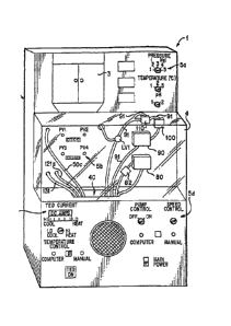
Figure 1 shows an organ perfusion apparatus 1 according to the invention.
Figure 2 is a schematic illustration of the apparatus of Fig. 1. The apparatus
1 is
preferably at least partially microprocessor controlled, and pneumatically
actuated.
The microprocessor 150 connection to the sensors, valves, thermoelectric units
and
pumps of the apparatus 1 is schematically shown in Fig. 3. Microprocessor 150
and
apparatus 1 may be configured to and are preferably capable of further being
connected to a computer network to provide data sharing, for example across a
local
area network or across the Internet.
The organ perfusion apparatus 1 is capable of perfusing one or more organs
simultaneously, at both normothermic and hypothermic temperatures
(hereinafter,
normothermic and hypothermic perfusion modes). All medical fluid contact
surfaces
are preferably formed of or coated with materials compatible with the medical
fluid
used, more preferably non-thrombogenic materials. As shown in Fig. 1, the
apparatus
1 includes a housing 2 which includes front cover 4, which is preferably
translucent,
and a reservoir access door 3. The apparatus preferably has one or more
control and
display areas 5a, 5b, 5c, 5d for monitoring and controlling perfusion.
As schematically shown in Fig. 2, enclosed within the housing 2 is a reservoir
10 which preferably includes three reservoir tanks 15a, 15b, 17. Two of the
reservoir
11
CA 02554872 2006-07-31
WO 2005/074681 PCT/US2005/003008
tanks 15a, 15b are preferably standard one liter infusion bags, each with a
respective
pressure cuff 16a, 16b. A pressure source 20 can be provided for pressurizing
the
pressure cuffs 16a, 16b. The pressure source 20 is preferably pneumatic and
may be
an on board compressor unit 21 supplying at least 10 L1311/1 external cuff
activation via
gas tubes 26,26a,26b, as shown in Fig. 2. The invention, however, is not
limited to
use of an on board compressor unit as any adequate pressure source can be
employed,
for example, a compressed gas (e.g., air, CO2, oxygen, nitrogen, etc.) tank
(not shown)
= preferably with a tank volume of 1.5 liters at 100 psi or greater for
internal
pressurization. Alternatively, an internally pressurized reservoir tank (not
shown)
may be used. Reservoir tanks 15a, 15b, 17 may, in embodiments, be bottles or
other
suitably rigid reservoirs that can supply perfusate by gravity or can be
pressurized by
compressed gas.
Gas valves 22-23 are provided on the gas tube 26 to allow for control of the
pressure provided by the onboard compressor unit 21. Anti-back flow valves
24a, 24h
may be provided respectively on the gas tubes 26a, 26b. Pressure sensors P5,
P6 may
be provided respectively on the gas tubes 26a, 26b to relay conditions therein
to the
microprocessor 150, shown in Fig. 3. Perfusion, diagnostic and/or transporter
apparatus may be provided with sensors to monitor perfusion fluid pressure and
flow
in the particular apparatus to detect faults in the particular apparatus, such
as pressure
elevated above a suitable level for maintenance of the organ. Gas valves GV1
and
GV2 may be provided to release pressure from the cuffs 16; 16b. One or both of
gas
valves GVI and GV2 may be vented to the atmosphere. Gas valve GV4 in=
communication with reservoir tanks 15a, 15b via tubing 18a, 18b may be
provided to
vent air from the reservoir tanks 15a, 15b through tubing 18. Tubing 18, 18a,
18b, 26,
26a and/or 26b may be configured with filters and/or check valves to prevent
biological materials from entering the tubing or from proceeding further along
the
fluid Path. The check valves and/or filters may be used to prevent biological
materials
from leaving one organ perfusion tubeset and being transferred to the tubeset
of a
subsequent organ in a multiple organ perfusion configuration. The check valves
and/or filters may also be used to prevent biological materials, such as
bacteria and
viruses, from being transferred from organ to organ in subsequent uses of the
perfusion apparatus in the event that such biological materials remain in the
perfusion
apparatus after use. The check valves and/or filters prevent contamination
problems
12
CA 02554872 2009-08-26
' 75341-40
associated with reflux in the gas and/or vent lines. For example, the valves
may be
configured as anti-reflux valves to prevent reflux. The third reservoir tank
17 is
preferably pressurized by pressure released from one of the pressure cuffs via
gas
valve GV2.
The medical fluid may be blood or a synthetic fluid and may, for example, be a
simple crystalloid solution, or may be augmented with an appropriate oxygen
carrier.
The oxygen carrier may, for example, be washed, stabilized red blood cells,
cross-
linked hemoglobin, pegolated hemoglobin or fluorocarbon based emulsions. The
medical fluid may also contain antioxidants known to reduce peroxidation or
free
radical damage in the physiological environment and specific agents known to
aid in
tissue protection. As discussed in detail below, an oxygenated (e.g., cross-
linked
hemoglobin-based bicarbonate) solution is preferred for the normothermic mode
while
a non-oxygenated (e.g., simple crystalloid solution preferably augmented with
antioxidants) solution is preferred for the hypothermic mode. Trie specific
medical
fluids used in both the normothennie and hypothermic modes are designed to
reduce
or prevent the washing away of or damage to the vascular endothelial lining of
the
organ. For the hypothermic perfusion mode, as well as for flush And/or static
storage,
a preferred solution is the solution.disclosed in U.S. Patent No. 6,492,103.
Examples of additives which may be used in perfusion solutions for the
present invention are also disclosed in U.S. Patent No. 6,046,046 to Hassanein
Of course, other suitable solutions and materials may be used, as is known in
the art.
The perfusion solution may be provided in a perfusion solution kit, for
example, a saleable package preferably containing at least one first container
holding a
first perfusion solution for norrnothermic perfusion and at least one second
container
holding a second, different perfusion solution. for hypothermic perfusion,
optionally
the box 10 shown in Fig. 2. The firstperfusion solution may contain at
least.one
oxygen carrier, may be oxygenated and/or may be selected from the group
consisting
of a cross-linked hemoglobin and stabilized red blood cells. The second
perfusion
solution may be non-oxygenated, may contain at least one anti-oxidant, and/or
may
contain at least one vasodilator. Additionally, the solution preferably
contains no
more than 5 rnM of dissolved pyruvate salt. Also, the first container and the
second
13
CA 02554872 2006-07-31
WO 2005/074681 PCT/US2005/003008
container may be configured to be operably connected to a perfusion machine as
perfusion fluid reservoirs in fluid communication with perfusate conduits of
said
perfusion Machine. Further, one of the first and second containers may be
compressible to apply pressure to the perfusion solution therein. Furthermore,
at least
one of the first and second containers may include a first opening for passage
of a
contained perfusion solution out of the container and a second opening passage
of a
compressed gas into the container. The package may be a cassette configured to
be
operably connected to a perfusion machine for connection of the first and
second
containers within the cassette in fluid communication with perfusate conduits
or
tubing of the perfusion machine.
In other embodiments, the perfusion solution kit may contain at least one
first
container holding a first perfusion solution for hypothermic perfusion at a
first
temperature and at least one second container holding a second, different
perfusion
solution for hypothermic perfusion at a second temperature lower than the
first
temperature. In the kit, the first perfusion solution may contain at least a
crystalloid
and may contain at least one vasodilator. The second perfusion solution may be
oxygen carrier enhanced, where the oxygen carrier is selected from the group
consisting of a hemoglobin and stabilized red blood cells. In addition, the
second
perfusion solution may, if desired, contain at least one anti-oxidant or free
radical
scavenger. Preferably, the second solution contains no more than 5 mM of
dissolved
pyruvate salt. As above, the first container and the second container may be
configured to be operably connected to a perfusion machine as perfusion fluid
reservoirs in fluid communication with perfusate conduits of said perfusion
machine.
Further, one of the first and second containers may be compressible to apply
pressure
to the perfusion solution therein. Furthermore, at least one of the first and
second
containers may include a first opening for passage of a contained perfusion
solution
out of the container and a second opening passage of a compressed gas into the
container. The package may be a cassette configured to be operably connected
to a
perfusion machine for connection of the first and second containers within the
cassette
in fluid communication with perfusate conduits or tubing of the perfusion
machine.
The medical fluid within reservoir 10 is preferably brought to a predetermined
temperature by a first thermoelectric unit 30a in heat transfer communication
with the
reservoir 10. A temperature sensor T3 relays the temperature within the
reservoir 10
14
CA 02554872 2009-08-26
75341-40
to the microprocessor 150, which adjusts the thermoelectric unit 30a to
maintain a
desired temperature within the reservoir 10 and/or displays the temperature on
a
control and display areas 5a for manual adjustment. Alternatively or in
addition, and
preferably where the organ perfusion device is going to be transported, the
medical
fluid within the hypothermic perfusion fluid reservoir can be cooled utilizing
a
cryogenic fluid heat exchanger apparatus such as that disclosed in
U.S. Patent No. 6,014,864.
An organ chamber 40 is provided which supports a cassette 65, as shown in
Fig. 2, which holds an organ to be perfused, or a plurality of cassettes
,5,65,65, as
shown in Fig. 12, preferably disposed one adjacent the other. Various
embodiments of
the cassette 65 are shown in Figs. 11A-11D. The cassette 65 is preferably
formed of a
material that is light but durable so that the cassette 65 is highly portable.
The material
may also be transparent to allow visual inspection of the organ.
Preferably the cassette 65 includes side walls 67a, a bottom wall 67b and an
organ supporting surface 66, which is preferably formed of a porous or mesh
materiatto
allow fluids to pass therethrough. The cassette 65 may also include a top 67d
and May
be provided with an opening(s) 63 for tubing (see, for example, Fig. 11D). The
opening(s) 63 may include seals 63a (e.g., septum seals or o-ring seals) and
optionally
be provided with plugs (not shown) to prevent contamination of the organ and
maintain
a sterile environment. Also, the cassette 65 may be provided with a closeable
air vent 61
(see, for example, Fig. ID). Additionally, the cassette 65 may be provided
with tubing
for connection to the organ or to remove medical fluid from the organ bath and
a
connection device(s) 64 for connecting the tubing to, for example, tubing 50c,
81, 82, 91
and/or 132 (see, for example, Fig. 11D). The cassette 65, and more
particularly the organ
support, opening(s), tubing(s) and/or connection(s), may be specifically
tailored to the
type of organ and/or size of organ to be perfused. Outer edges 67c of the Side
support
walls 67a can be used to support the cassette 65 disposed in the organ chamber
40. The
cassette 65 may further include a handle portion 68 which allows the cassette
65 to be
easily handled, as shown, for example, in Figs. 11C and 11D. Each cassette 65
may also
be provided with its own stepping motor/cam valve 75 (for example, in the
handle
portion 68, as shown in Fig. 11C) for fine tuning the pressure of medical
fluid perfused
into the organ 60 disposed therein, discussed in more detail below.
Alternatively,
pressure may, in embodiments, be controlled by way of a pneumatic chamber,
such as
CA 02554872 2006-07-31
WO 2005/074681 PCT/US2005/003008
an individual pneumatic chamber for each organ (not shown), or by any suitable
variable
valve such as a rotary screw valve or a helical screw valve.
= Fig. 17 shows an alternative embodiment of cassette 65. In Fig. 17,
cassette 65
is shown with tubeset 400. Tubeset 400 can be connected to perfusion apparatus
1 or to
an organ transporter or an. organ diagnostic apparatus, and allows cassette 65
to be
moved between various apparatus without jeopardi7ing the sterility of the
interior of
cassette 65. Preferably, cassette 65 is made of a sufficiently durable
material that it can
withstand penetration and harsh impact. Cassette 65 is provided with a lid,
preferably
two lids, an inner lid 410 and an outer lid 420. The lids 410 and 420 may be
removable
or may be hinged or otherwise connected to the body of cassette 65. Clasp 405
provides
a mechanism to secure lids 410 and 420 to the top of cassette 65. Clasp 405
may
additionally be configured with a lock to provide further security, and
stability. A biopsy
port 430 may additionally be included in inner lid 410 or both inner lid 410
and outer lid
420. Biopsy port 430 provides access to the organ to allow for additional
diagnosis of
the organ with minimal disturbance of the organ. Cassette 65 may also have an
overflow trough 440 (shown in Fig. 17A). Overflow trough 440 is a channel
present in
the top of cassette 65. When lids 410 and 420 are secured on cassette 65,
overflow
trough 440 provides a region that is easy to check to determine if the inner
seal is
leaking. Perfusate may be poured into and out of cassette 65 and may be
drained from
cassette 65 through a stopcock or removable plug. =
Cassette 65 and/or both lids 410 and 420 may be constructed of an optically
clear material to allow for viewing of the interior of cassette 65 and
monitoring of the
organ and to allow for video images or photographs to be taken of the organ.
Perfusion
apparatus 1 or cassette 65 may be wired and fitted with a video camera or a
photographic camera, digital or otherwise, to record the progress and status
of the organ.
The captured images may be made available over a computer network such as a
local
area network or the Internet to provide for additional data analysis and
remote
monitoring. Cassette 65 may also be provided with a tag that would signal,
e.g., through
a bar code, magnetism, radio frequency, or other means, the location of the
cassette, that
the cassette is in the apparatus, and/or the identity of the organ to the
perfusion apparatus
or transporter. Cassette 65 may be sterile packaged and/or may be packaged or
sold as a
single-use disposable cassette, such as in a peel-open pouch. A single-use
package
containing cassette 65 may also include tubeset 400.
16 = -
CA 02554872 2006-07-31
WO 2005/074681 PCT/US2005/003008
Cassette 65 may additionally be provided with an organ chair 1800 shown in
Figs. 18 and 18A. Organ chair 1800 is removable and provides a support=
surface for the
organ within cassette 65. Utilizing a removable organ chair 1800 allows the
organ to be
cannulated and secured under cold conditions when the organ is recovered from
a donor
.5 before being placed into cassette 65. Organ chair 1800 may be reusable
or single-use.
Organ chair 1800 may be constructed specifically to correspond to each type of
organ,
such as the kidney, heart or liver. Organ chair 1800 is preferably designed to
be form
fitting to the organ but to allow for the full anthropometric range of organ
sizes.
Preferably, organ chair 1800 is at least partially perforated to allow fluids
t6 pass
through organ chair 1800. The perforations in organ chair 1800 may be sized to
catch
organ debris, or an additional filter layer, preferably constructed of cloth,
fabric, nylon,
plastic, etc., to catch organ debris of at least 15 microns in diameter. In
addition, a
separate filter may be used on the tubing that intakes fluid directly from the
perfusate
bath to prevent organ debris of a predetermined size, for example at least 10
to 15
microns in diameter, from entering the perfusion tubing.
Organ chair 1800 may also be configured with a venous outflow sampler 1810.
Organ chair 1800 funnels the venous outflow into venous outflow sampler 1810.
Venous outflow sampler 1810 provides a readily available source for capturing
the
venous outflow of the organ. Capturing the venous outflow in this manner
permits
analysis of the perfusate leaving the organ without cannulating a vein and
enables organ
viability to be measured with a high degree of sensitivity by analyzing
differentially the
perfusate flowing into and out of the organ. Alternatively, venous outflow may
be
captured directly by cannulating a vein, but this method increases the risk of
damaging
the vein or the organ. Organ chair 1800 may also be raised and lowered within
cassette
65 to facilitate sampling from venous outflow sampler 1810. Alternatively, a
sufficient
amount of the organ bath may be drained from cassette 65 to obtain access to
venous
outflow sampler 1810 or to capture venous outflow before the outflow mixes
with the
rest of the perfusate in the organ bath.
Organ chair 1800 is preferably additionally configured with a cannula 1820
that
attaches to the perfused artery, such as the renal artery. Cannula 1820 may be
reusable
or may be suitable for single-use, preferably provided in a sterile package
with cassette
65, organ chair 1800 and tubeset 400. Cannula 1820 is provided with a cannula
clamp
1830 to secure cannula 1820 around the perfused artery and to preferably
provide leak-
17
CA 02554872 2009-08-26
75341-40
tight perfusion. A straight-in flanged cannula may also be used, however
clamping
around the artery is preferable to prevent contact with the inner surface of
the artery,
which is easily damaged. Cannula 1820 may also be configured with additional
branching connections for accessory arteries. Multiple cannula and cannula
clamp sizes
5. may be used to accommodate various artery sizes or an adjustable cannula
and cannula
clamp may be used to accommodate various sized arteries. Cannula clamp 1830
may be
a clam-shell configuration or may be a two-part design. Cannula clamp 1830 may
be
configured with integral or separate means for tightening cannula clamp 1830
to the
proper pressure to provide leak-tight perfusion. In addition, cannula 1820 may
be
provided with a snap 1840 to hold cannula 1820 closed. Cannula 1820 may also
be
provided with a vent 1850 to remove air bubbles from cannula 1820,
Organ chair 1800 preferably has a detented region 1860 that corresponds to
protrusions 1870 on cannula 1820. Such detents, tracks or grooves on organ
chair 1800
allow cannula 1820 to be positioned at several locations to provide various
tensions on
the perfused artery. This allows the ideal minimum tension to be set for each
artery.
Cannula clamp 1830 secures the perfusate tubing to the perfused artery.
Cannula 1820
is adjustably secured to organ chair 1800 to provide for positioning the
perfused artery to
accommodate variations in organ size and artery length to prevent stretching,
twisting,
sagging or kinlcing of the artery. The combination of organ chair 1800,
cannula 1820
and additional straps or wide belts provides a secure platform to transport
the organ and
to transfer the organ between the cassette and the surgical field.
Organ chair 1800, cannula 1820 and/or cannula clamp 1830 may be constructed
of an optically clear material to facilitate monitoring of the organ and
perfusion status.
The cassette 65 is configured such that it may be removed from the organ
perfusion apparatus 1 and transported to another organ perfusion apparatus in
a portable
transporter apparatus, such as, for example, a conventional cooler or a
portable container
such as that disclosed in simultaneously filed co-pending
U.S. Patent No. 6,209,343, or U.S. Patent No. 5,586,438 to Fahy.
In embodiments, when transported, the organ is disposed on the organ
supporting surface 66 and the cassette 65 is preferably enclosed in a
preferably sterile
bag 69, a; shown, for example, in Fig. 11A. When the organ is perfused with
medical
fluid, effluent medical fluid collects in the bag 69 to form an organ bath.
Alternatively,
18
CA 02554872 2006-07-31
WO 2005/074681 PCT/US2005/003008
the cassette 65 can be formed with a fluid tight lower portion in which the
effluent
medical fluid may collect, or the effluent medical fluid may collect in the
organ chamber
40 to form the organ bath. In either alternative case, the bag 69 would
preferably be
removed prior to inserting the cassette into the organ chamber 40. Further,
where a
plurality of organs are to be perfused, an organ chamber may be provided for
each organ.
Alternatively, cassette 65 can be transported in the dual-lid cassette of Fig.
17 and
additionally carried within a portable organ transporter.
Fig. 19 shows an external view of an embodiment of transporter 1900 of the
invention. The transporter 1900 of Fig. 19 has a stable base to facilitate an
upright
position and handles 1910 for carrying transporter 1900. Transporter 1900 may
also be
fitted with a shoulder strap and/or wheels to assist in carrying transporter
1900. A
control panel 1920 is preferably also provided. Control panel 1920 may display
characteristics, such as, but not limited to infusion pressure, power on/off,
error or fault
condition, flow rate, flow resistance, infusion temperature, bath temperature,
pumping
time, battery charge, temperature profile (maximums and minimums), cover open
or=
closed, history log or graph, and additional status details and messages,
which are
preferably further transmittable to a remote location for data storage and/or
analysis.
Flow and pressure sensors or transducers in transporter 1900 may be used to
calculate
various organ characteristics including pump pressure and vascular resistance
of an
organ, which can be stored in computer memory to allow for analysis of, for
example,
vascular resistance history, as well as to detect faults in the apparatus,
such as elevated
pressure.
Transporter 1900 has latches 1930 that require positive user action to open,
thus
avoiding the possibility that transporter 1900 inadvertently opens during
transport.
Latches 1930 hold top 1940 in place on transporter 1900. Top 1940 or a portion
thereof
may be constructed with an optically clear material to provide for viewing of
the cassette
and organ perfusion status. Transporter 1900 may be configured with a cover
open
detector that monitors and displays if the cover is open or closed.
Transporter 1900 may
be configured with an insulating exterior of various thicknesses to allow the
user to
configure transporter 1900 for varying extents and distances of transport. In
embodiments, compartment 1950 may be provided to hold patient and organ data
such
as charts, testing supplies, additional batteries, hand-held computing devices
and/or
other accessories for use with transporter 1900. Transporter 1900 may also be
19
CA 02554872 2006-07-31
WO 2005/074681 PCT/US2005/003008
configured with means for displaying a UNOS label and/or identification and
return
shipping information.
Fig. 20 shows a cross-section view of a transporter 1900. Transporter 1900
contains cassette 65 and pump 2010. Cassette 65 may be placed into and taken
out of
transporter 1900 without disconnecting tubeset 400 from cassette 65, thus
maintaining
sterility of the organ. Sensors in transporter 1900 can detect the presence of
cassette 65
in transporter 1900, and depending on the sensor, can read the organ identity
from a
barcode or radio frequency or other smart tag that may be integral to cassette
65. This
allows for automated identification and tracking of the organ and helps
monitor and
control the chain of custody. A global positioning system may be added to
transporter
1900 and/or cassette 65 to facilitate tracking of the organ. Transporter 1900
can be
interfaced to a computer network by hardwire connection to a local area
network or by
wireless communication while in transit. This interface allows perfusion
parameters,
vascular resistance, and organ identification and transporter and cassette
location to be
tracked and displayed in real-time or captured for future analysis.
Transporter 1900 also preferably contains a filter 2020 to remove sediment and
other particulate matter, preferably ranging in size from 0.05 to 15 microns
in diameter
or larger, from the perfusate to prevent clogging of the apparatus or the
organ.
Transporter 1900 also contains batteries 2030, which may be located at the
bottom of
transporter 1900 or beneath pump 2010 or at any other location that provides
easy access
to change batteries 2030. Batteries 2030 may be rechargeable outside of
transporter
1900 or while intact within transporter 1900 and/or are preferably hot-swapp
able one at
a time. Batteries 2030 are preferably rechargeable rapidly and without full
discharge.
Transporter 1900 may also provide an additional storage space 2040 at the
bottom of
transporter 1900 for power cords, batteries and other accessories. Transporter
1900 may
also include a power port for a DC hookup to a vehicle such as an automobile
or
airplane and/or for an AC hookup.
Fig. 21 shows a block diagram of transporter 1900. Transporter 1900 of Fig. 21
is intended to provide primarily hypothermic perfusion, and may operate at any
temperatures, for example in the range of -25 to 60 C, approximately 0 to 8
C,
preferably approximately 4 C. The temperature may be adjusted based on the
particular fluids used and adapted to the particular transport details, such
as length of
time of transport. Transporter 1900 is cooled by coolant 2110, which may be an
ice and
CA 02554872 2006-07-31
WO 2005/074681 PCT/US2005/003008
water bath or a cryogenic material. In embodiments using cryogenic materials,
the
design should be such that organ freezing is prevented. The temperature of the
perfusate
bath surrounding the organ is monitored by temperature transducer 2115.
Transporter
1900 also contains filters 2020 to remove sediment and particulate, ranging in
size from
0.05 to 15 microns in diameter or larger, from the perfusate to prevent
clogging of the
apparatus or the organ. Using a filter 2020 downstream of pump 2010 allows for
capturing inadvertent pump debris and also dampens pressure spikes from pump
2010.
The flow of perfusate within transporter 1900 is controlled by pump 2010,
which
is preferably a peristaltic or roller pump. Pump 2010 is preferably not in
contact with
the perfusate to help maintain sterility. In addition, tubeset 400 may be
attached to
pump 2010 without opening the tubing circuit. Pump 2010 is controlled by a
computer
or microcontroller. The computer can actively modulate the angular velocity of
pump
2010 to reduce the natural pulse actions of pump 2010 to a low level,
resulting in =
essentially non-pulsatile flow. Further computer control can impose a
synthesized
pressure pulse profile that can be sinusoidal or physiological or otherwise.
The average
flow rate and pressure can be made independent of pulse repetition rate by
pulse width
modulating or amplitude modulating the synthesized pressure pulses. Control
over
some or all of the pulse parameters can be made available to users through
control panel
1920 or over a network. Pulse control can be organ specific. In the case of a
liver, a
single pump can provide continuous flow to the portal vein at, for example, 1
to 3 liters
per minute while providing pulsatile flow to the hepatic artery at, for
example, 100 to
300 ml per minute. Synchronizing the shunt valves to the pump controller
allows
independent pressure regulation of the two flows.
The flow of the perfusate into the organ is monitored by flow sensor 2125.
Pressure transducers 2120 may be present to monitor the pressure the perfusate
places
on the tubing. Pressure transducers 2120 may be used to monitor the pump
pressure
and/or the infusion pressure. A pressure transducer 2120 may be present just
upstream
of the organ to monitor the organ infusion pressure. Transporter 1900 may be
configured with a bubble detector 2125 to detect bubbles before the perfusate
enters
bubble trap 2130. Bubble detectors, such as bubble detector 2125, may be used
to detect
bubbles in, for example, the infuse line and/or in the pump output line.
Bubble trap
2130 removes air bubbles from the perfusate and vents the bubbles into the
wash tube.
Bubble trap 2130 may be disposable and may be constructed integral to tubeset
400.
21
CA 02554872 2006-07-31
WO 2005/074681 PCT/US2005/003008
Perfusate exiting bubble trap 2130 can either continue through infuse valve
2140 or
wash valve 2150. Wash valve 2150 is normally open and infuse valve 2140 is
normally
closed. Preferably, wash valve 2150 and infuse valve 2140 operate dependently
in an
on/off manner, such that if one valve is open, the other valve is closed.
Although infuse
valve 2140 is normally closed, if the sensor and monitors all report suitable
perfusion
parameters present in transporter 1900, then infuse valve 2140 may be opened
to allow
organ perfusion. In the occurrence of a fault, such as elevated perfusion
pressure above
a suitable level for the organ, infuse valve 2140 switches back to closed and
wash valve
2150 is opened to divert fluid flow into the perfusate bath surrounding the
organ. This
provides a failsafe mechanism that automatically shunts perfusate flow and
prevents
organ perfusion in case of a power failure or computer or electronics
malfunction. A
pressure transducer 2120, such as designated by P2, may be hardwired,
redundant to the
computer and software control, to wash valve 2150 and infuse valve 2140 to
quickly
deliver a default message to the valves in the case of a pressure malfunction.
In
embodiments, the diverted fluid may be separately collected in another
container or
compartment.
Fig. 22 shows various operation states of transporter 1900. For example, using
the controls provided on control panel 1920, a user may select operations such
as
perfuse, idle, wash and prime. Fig. 22 shows various options depending on the
present
state of transporter 1900. The labels idle, prime, wash, perfuse and error
handling
indicate the state of transporter 1900 that is preferably displayed on control
panel 1920
during the corresponding operation. For example, when transporter 1900 is in a
wash
operation, control panel 1920 displays the wash operation indicator, such as
an LED
display. The arrows connecting the various operations of transporter 1900
indicate the
manual and automatic actions that may occur to transition transporter 1900
between
operation states. Manual actions require the user to act, for example by
pressing a
button or turning a knob or dial. Fig. 22 exemplifies pressing a button or
other indicator,
for example, to move from a perfusion operation to an idle operation by
pressing the
stop button (Press Stop). To move directly into a perfuse operation from an
idle
operation, a user presses the perfuse button (Press Perfuse).
Automatic operations may be controlled by the passage of time and/or by an
internal monitor within transporter 1900. Such automatic operation is shown in
Fig. 22,
for example, connecting the prime operation to the idle operation. If the
prime operation
22
CA 02554872 2006-07-31
WO 2005/074681 PCT/US2005/003008
has been completed according to the internal transporter program parameters
before the
wash button has been pressed, transporter 1900 returns to an idle operation.
Another
automatic operation occurs during a perfuse operation if a fault or error
occurs, such as
overpressurization of the organ. When an error or fault occurs, transporter
1900 can
move to an error handling operation to determine the extent or degree of the
fault or
error. If the fault or error is determined to be a small or correctable error,
transporter
1900 moves into awash operation. If transporter 1900 can then adjust'the
system
parameters to handle the fault or error, transporter 1900 moves back to
perfuse (Error
Recovery). If transporter 1900 can not adjust the system parameters to handle
the fault
or error, transporter 1900 moves to an idle operation. If the error or fault
detected is
determined to be substantial, tranporter 1900 may move directly into an idle
operation.
Fig. 23 shows an alternative cross-section of transporter 1900. Transporter
1900
may have an outer enclosure 2310 constructed of metal, or preferably a plastic
or
synthetic resin that is sufficiently strong to withstand penetration and
impact.
Transporter 1900 contains insulation 2320, preferably a thermal insulation
made of, for
example, glass wool or expanded polystyrene. Insulation 2320 may be various
thicknesses ranging from 0.5 inches to 5 inches thick or more, preferably 1 to
3 inches,
such as approximately 2 inches thick. Transporter 1900 is cooled by coolant
2110,
which may be, e.g., an ice and water bath or a cryogenic material. In
embodiments
using cryogenic materials, the design should be such that organ freezing is
prevented.
An ice and water mixture is preferably in an initial mixture of approximately
1 to 1,
however, in embodiments the ice and water bath may be frozen solid.
Transporter 1900
can be configured to hold various amounts of coolant, preferably up to 10 to
12 liters.
An ice and water bath is preferable because it is inexpensive and can not get
cold
enough to freeze the organ. Coolant 2110 preferably lasts for a minimum of 6
to 12
hours and more preferably lasts for a minimum of 30 to 50 hours without
changing
coolant 2110. The level of coolant 2110 may be viewed through a transparent
region of
transporter 1900 or may be automatically detected and monitored by a sensor.
Coolant
2110 can be replaced without stopping perfusion or removing cassette 65 from
transporter 1900. Coolant 2110 is maintained in a watertight compartment 2115
of
transporter 1900. Compartment 2115 prevents the loss of coolant 2110 in the
event
transporter 1900 is tipped or inverted. Heat is conducted from the walls of
the perfusion
reservoir and cassette 65 into coolant 2110 enabling control within the
desired
23
CA 02554872 2006-07-31
WO 2005/074681 PCT/US2005/003008
temperature range. Coolant 2110 is a failsafe cooling mechanism because
transporter
1900 automatically reverts to cold storage in the case of power loss or
electrical or
computer malfunction. Transporter 1900 may also be configured with a heater to
raise
the temperature of the perfusate.
Transporter 1900 may be powered by batteries or by electric power provided
through plug 2330. An electronics module 2335 is also provided in transporter
1900.
Electronics module 2335 is cooled by vented air convection 2370, and may
further be
cooled by a fan. Preferably, electronic module 2335 is positioned separate
from the
perfusion tubes to prevent the perfusate from wetting electronics module 2335
and to
avoid adding extraneous heat from electronics module 2335 to the perfusate.
Transporter 1900 has a pump 2010 that provides pressure to perfusate tubing
2360 to
deliver perfusate 2340 to organ 2350. Transporter 1900 may be used to perfuse
various
organs such as a kidney, heart, liver, small intestine and lung. Transporter
1900 and
cassette 65 may accommodate various amounts of perfusate 2340, for example up
to 3
to 5 liters. Preferably, approximately 1 liter of a hypothermic perfusate 2340
is used to
perfuse organ 2350. Organ 2350 may be various organs, including but not
limited to a
kidney, heart, lung, liver or small intestine.
Cassette 65 and transporter 1900 are preferably constructed to fit or mate
such
that efficient heat transfer is enabled. The geometric elements of cassette 65
and
transporter 1900 are preferably constructed such that when cassette 65 is
placed within
transporter 1900, the elements are secure for transport.
Fig. 24 shows various data structures and information connections that can be
facilitated to assist in the overall communication and data transfers that may
be
beneficial before, during and after organ transplantation. The perfusion
apparatus,
transporter, cassette, and organ diagnostic apparatus may be networked to
permit
remote management, tracking and monitoring of the location and therapeutic and
diagnostic parameters of the organ or organs being stored or transported. The
information systems may be used to compile historical data of organ transport
and
storage, and provide cross-referencing with hospital and IJNOS data on the
donor and
any recipient and/or information on why transplant my be innappropriate. The
systems may also provide outcome data to allow for ready research of perfusion
= parameters and transplant outcomes. For example, information regarding
the donor
may be entered at the location where an organ is recovered from a donor.
Information
24 -
CA 02554872 2009-08-26
75341-40
,
=
may also be directly recovered from the perfusion, diagnostic or transporter
apparatus
to monitor organ status and location. Various types of information may be
grouped
into sub-records or sub-directories to assist in data management and transfer.
All the
sub-records may be combined to form an overall transplant record, which may be
disseminated to or retrievable by physicians, scientists or other
organizations for
tracking and monitoring purposes.
Preferred embodiments of transporter 1900 can automatically log much or all
of the perfusion process data and transporter 1900 events into an internal
database. A
radio frequency or barcode labeled tag or the like for each cassette 65 allows
transporter 1900 to reference the data uniquely to each organ. When
transporter 1900
reaches a docking port, transporter 1900 can upload data to a main database
computer
over a LAN. Transporter 1900 can also provide real-time status whenever
transporter
1900 is connected to the LAN. Transporter 1900 can also be configured with a
wireless communications setup to provide real-time data transfer during
transport.
Perfusion apparatus 1 can also be connected to the LAN and since perfusion
apparatus
is generally stationary, data uploads can occur continuously and in real-time.
The data
can be cross-referenced with UNOS data to utilize the UNOS data on organ
identification, donor condition, donor logistics, recipient logistics and
recipient
outcomes. Data may be displayed and accessed on the Internet to facilitate
monitoring
=
from any location.
Within the perfusion, diagnostic and/or transporter apparatus, the organ bath
is
preferably cooled to a predetermined temperature by a second thermoelectric
unit 30b,
as shown in Fig. 2, in heat transfer communication with the organ chamber 40.
Alternatively and preferably where the organ perfusion device is going to be
transported, the medical fluid within reservoir 10 can be cooled utilizing a
heat
transfer device such as an ice and water bath or a cryogenic fluid heat
exchanger
apparatus such as that disclosed in U.S. Patent No. 6,014,864. A temperature
sensor T2 within the organ chamber 40 relays the temperature of the organ 60
to
the microprocessor 150, which adjusts the thermoelectric unit 30b to maintain
a
desired organ temperature and/or displays the temperature on the control and
display areas 5c for manual adjustment.
Medical fluid may be fed from the bag 15a directly to an organ 60 disposed in
the organ chamber 40 through tubing 50a,50b,50c or from bag 15b through tubing
CA 02554872 2006-07-31
WO 2005/074681 PCT/US2005/003008
50d,50e,50c by opening valve LV4 or LV3, respectively. Conventional medical
fluid
bag and tubing connections may be utilized. All tubing is preferably
disposable, easily
replaceable and interchangeable. Further, all tubing is preferably formed of
or coated
with materials compatible with the medical fluids used, more preferably non-
thrombogenic materials. An end of the tubing 50c is inserted into the organ
60. The
tubing may beconnected to the organ(s) with conventional methods, for example,
with
sutures. The tubing may include a lip to facilitate connection to the organ.
Alternatively, cannula 1820 described above may be used with or without
connection to
an organ chair 1800. However, the specific methods and connection depend on
the type
of organs(s) to be perfused.
The microprocessor 150 preferably controls the pressure source 20 in response
to signals from the pressure sensor P1 to control the pressure of the medical
fluid fed
into the organ 60. The microprocessor 150 may display the pressure on the
control
and display areas 5a, optionally for manual adjustment. A fluid flow monitor
Fl may
also be provided on the tubing 50c to monitor the flow of medical fluid
entering the
organ 60 to indicate, for example, whether there are any leaks present in the
organ.
Alternatively, the medical fluid may be fed from the reservoir tank 17 via
tubing 51 into an intermediary tank 70 preferably having a pressure head of
approximately 5 to 40 mm Hg. Medical fluid is then fed by gravity or,
preferably,
pressure, from the intermediary tank 70 to the organ 60 along tubing 50c by
activating
a valve LV6. A level sensor 71 may be provided in the intermediary tank 70 in
order
to maintain the pressure head. Where a plurality of organ chambers 40 and
organs 60
are provided, the organs 60 are connected in parallel to the reservoir 10
utilizing suitable ,
tubing duplicative of that shown in Fig. 2. See, for example, Fig. 12. The use
of
pneumatically pressurized and gravity fed fluid pumps configured to avoid
oveipressurization even in cases of system failure reduces or prevents general
tissue
damage to the organ and the washing away of or damage to the vascular
endothelial
lining of the organ. Thus, organ perfusion in this system can be performed,
e.g., with
either hydrostatic perfusion (gravity or pressure fed flow) or peristaltic
perfusion by
introducing flow to the organ from a peristaltic (roller) pump.
A bubble detection system may be installed to sense bubbles in the perfusate.
An air sensor and sensor board are preferably used. The output of the sensor
activates
a debubbler system, such as an open solenoid valve, to rid bubbles from the
perfusate
26
CA 02554872 2006-07-31
WO 2005/074681 PCT/US2005/003008
flow prior to organ introduction. As with all of the sensors and detectors in
this
system, the bubble detector may be positioned at any point in the system that
is
effective based on the particular parameters Cr design characteristics of the
system.
For, example, a bubble detector and debubbler system BD may be positioned
between
the cam valve 205 and pressure sensor Pl, as shown in Fig. 1.
A stepping motor/cam valve 205, or other suitable variable valve such as a
rotary screw valve, may be arranged on the tubing 50c to provide pulsatile
delivery of
the medical fluid to the organ 60, to decrease the pressure of the medical
fluid fed into
the organ 60, and/or to stop flow of medical fluid into the organ 60 if the
perfusion
pressure exceeds a predetermined amount. Alternatively, a flow ciiverter or
shunt line
may be provided in the perfusion apparatus to which the fluid flow is diverted
in the
occurrence of a fault, such as excess pressure, for example by opening and
closing a
valve or a series of valves. Specific embodiments of the stepping motor/cam
valve are
shown in Figs. 13A-13B and 14A-14F. Figs. 13A-13B show a stepping
motor/rotational type cam valve.
Fig. 13A is a top view of the apparatus. Tubing, for example, tubing 50c, is
interposed between a support 203 and cam 200. Cam 200 is connected by a rod
201
to stepping motor 202. Fig. 13B is a side view of the apparatus. The dashed
line
shows the rotational span of the cam 200. In Fig. 13B, the cam 200 is in its
non-
occluding position. Rotated 180 degrees, the cam 200 totally occludes the
tubing 50c
= with varying degrees of occlusion therebetvveen. This stepping motor/cam
valve is
relatively fast, for example, with respect to the embodiment shown in Figs.
14A - 14F;
however, it requires a strong stepping motor.
Figs. 14A - 14F disclose another stepping motor/cam valve 210 according to
the invention. Fig. 14A is a side view of the apparatus while Fig. 14C is a
top view.
Tubing, for example, tubing 50c, is interposed between cam 220 and support
223.
The cam 220 is connected to stepping motor 222 by supports 221a - 221d and
helical
screw 225, which is connected to the stepping motor 222 via plate 222a. Fig.
14B
shows the supports 221a and plate 222a in front view. As shown in Fig. 14D,
where
the support 221d is to the left of the center of the helical screw 225, the
tubing 50c is
not occluded. However, as the helical screw 225 is turned by the stepping
motor 222,
the support 221d moves to the left (with respect to Figs. 14D - 14F) toward a
position
27
CA 02554872 2006-07-31
WO 2005/074681 PCT/US2005/003008
where the cam 220 partially or fully occludes the tubing 50c. Such apparatus
is
slowerthan the apparatus of Figs. 13A - 13B, but is more energy efficient
Medical fluid expelled from the organ 60 which has collected in the bottom of
the bag 69 (the cassette 65 or the organ chamber 40) is either pumped out
through
tubing 81 by a pump 80 for filtration, passing through a filter unit 82 and
being
returned to the organ bath, or is pumped out by a pump 90 for circulation
through
tubing 91. The pumps 80, 90 are preferably conventional roller pumps or
peristaltic
pumps; however, other types of pumps may also be appropriate.
Fig. 25 shows a simplified schematic of a pump and pulse controller 2500' and
the interaction of the pump and pulse controller with a perfusion apparatus,
such as
shown in Fig. 1. Pump and pulse controller 2500 receives pressure sensor data
input
2510 from pressure sensor P and tachometer data input 2520. A tachometer may
be
used to set the phase angle of the active wave. Pump and pulse controller 2500
converts this information to motor drive output 2530, which powers pump 2540.
Fig.
25A shows various modes of operation that pump and pulse controller 2500 can
provide and how pump and pulse controller 2500 eliminates pressure pulse waves
from the perfusate flow and how it modulates perfusate flow rate while
maintaining a
constant pressure pulse rate.
A peristaltic pump driven at a constant speed provides a constant pressure
wave in the associated tubing. Fig. 25A shows in the first mode of operation
the
waveforms that result from a constant drive speed applied to a peristaltic
pump. The
second mode of operation, called active continuous, shows how the pressure
pulse
wave can be eliminated or canceled out by applying a motor drive wave that is
opposite to the pressure wave of the pump. In the third mode of operation,
called
active waveform amplitude modulating, the pump pressure pulse wave is canceled
by
the motor drive wave, and a selected wave is added with a new amplitude as
compared to the original pressure pulse wave amplitude. In the fourth mode of
operation, called active waveform pulse width modulating, the pump pressure
pulse
wave is canceled by the motor drive wave, and a selected wave is added with a
new
pulse width as compared to the original pressure pulse wave width. In an
alternative
mode of operation, the frequency may be modulated by adding a new frequency
wave
to the canceled waves.
28
CA 02554872 2006-07-31
WO 2005/074681 PCT/US2005/003008
= A level sensor L2 in communication with the microprocessor 150 (see Fig.
3)
ensures that a predetermined level of effluent medical fluid is maintained
within the
organ chamber 40. As shown in Fig. 2, a temperature sensor Ti disposed in the
tubing 91 relays the temperature of the medical fluid pumped out of the organ
bath
along tubing 91 to the microprocessor 150, which monitors the same. A pressure
sensor P2 disposed along the tubing 91 relays the pressure therein to the
microprocessor 150, which shuts down the system if the fluid pressure in the
tubing
91 exceeds a predetermined limit, or activates an alarm to notify the operator
that the
=
system should be shut down, for example, to clean filters or the like.
As the medical fluid is pumped along tubing 91 it preferably passes through a
filter unit 95 (e.g., 25 , 8j.t, 2 , 0.814 0.2 and/or 0.1 filters); a CO2
scrubber/02
membrane 100 and an oxygenator 110, for example, a JOS l'RATm oxygenator. The
CO2 scrubber/02membrane 100 is preferably a hydrophobic macroporous membrane
with a hydrophilic (e.g., Hypol) coating in an enclosure. A vacuum source (not
shown) is utilized to apply a low vacuum on a side opposite the hydrophilic
coating by
the activation of valve WI. A hydrostatic pressure of approximately 100 mm Hg
is
preferred for aqueous passage through the membrane. The mechanical relief
valve
(not shown) prevents the pressure differential from attaining this level.
Immobilized
pegolated carbonic anhydrase may be included in the hydrophilic coating. This
allows
bicarbonate to be converted to CO2 and subsequently removed by vacuum venting.
However, with organs such as kidneys which have the ability to eliminate
bicarbonate,
this may be unnecessary except in certain cases.
The oxygenator 110 is preferably a two stage oxygenator which preferably
includes a hydrophilically coated low porosity oxygen permeable membrane. A
portion of the medical fluid is diverted around the oxygenator along tubing
111 in
which is disposed a viability sensor V1, which senses fluid characteristics,
such as
organ resistance (pressure/flow), pH, p02, pCO2, LDH, T/GST, Tprotein,
lactate,
glucose, base excess and ionized calcium levels indicative of an organ's
viability. The
viability sensor V1 is in communication with the microprocessor 150 and allows
the
organ's viability to be assessed either automatically or manually. One of t-wo
gases,
preferably 100% oxygen and 95/5% oxygen/carbon dioxide, is placed on the
opposite
side of the membrane depending on the pH level of the diverted medical fluid.
Alternatively, another pump (not shown) may be provided which pumps effluent
29
CA 02554872 2006-07-31
WO 2005/074681 PCT/US2005/003008
medical fluid out of the organ chamber 40 and through a viability sensor
before
returning it to the bath, or the viability sensor can be placed on tubing 8.1
titilizing
pump 80. In embodiments, the fluid characteristics may be analyzed in a
separate
diagnostic apparatus and/or analyzer as shown in Figs. 28-31.
The sensed fluid characteristics, such as organ resistance (pressure/flow),
pH,
P02, pCO2, LDH, T/GST, Tprotein, lactate, glucose, base excess and ionized
calcium
levels may be used to analyze and determine an organ's viability and/or the
effect of
applied bioactive or other test substance thereon. The characteristics may be
analyzed
individually or multiple characteristics may be analyzed to determine the
effect of .
various factors. The characteristics may be measured by capturing the venous
outflow
of the organ and comparing its chemistry to the perfusate inflow. The venous
outflow
may be captured directly and measured or the organ bath may, be measured to
provide
a rough approximation of the fluid characteristics for comparisons over a
period of
=
time.
In embodiments, an organ viability index is provided taking into account the
various measured factors identified above, such as vascular resistance, pH,
etc. The
index may be organ specific, or may be adaptable to various organs. The index
compiles the monitored parameters into a diagnostic summary which may be used
for
making organ therapy decisions and deciding whether to transplant the organ or
make
other use of it. The index may be automatically generated and provided to the
physician. The index is preferably computer generated via a connection to the
perfusion apparatus, transporter, cassette and/or organ diagnostic apparatus.
The
additional information, such as donor specific information, may be entered
into a
single computer at the site of the perfusion apparatus, transporter, cassette
and/or
organ diagnostic apparatus or may be entered in a remote computer and linked
to the
perfusion apparatus, etc. In embodiments, the index may be made available over
a
computer network such as a local area network or the Internet for quick
comparison,
remote analysis and data storage.
The organ viability index provides measurements and nonnal ranges for each
characteristic, such as vascular resistance and perfusate chemistry
characteristics
based on pH, p02, pCO2, LDH, T/GST, Tprotein, lactate, glucose, base excess
and
ionized calcium levels. For example, at approximately 5 C, normal pH may be
from
7.00 and 8.00, preferably from 7.25 and 7.75 and more preferably from 7.50 and
7.60
CA 02554872 2009-08-26
, .
75341-40
and base excess may be in the range of from -10 to -40, preferably from -15 to
-30,
and more preferably from -20 to -25. Measurements that are outside the normal
range
may be indicated visually, e.g., by an asterisk or other suitable notation,
aurally or by
machine perceivable signals. The characteristics give the physician insight
into the
metabolism of the organ, such as stability of the metabolism, consumption of
glucose,
creation of lactic acid and oxygen consumption.
The index may also provide identifying information, such as age, gender,
blood type of the donor and any expanded criteria; organ information, such as
organ
collection date and time, warm isehemia time, cold ischemia time and vascular
resistance; apparatus information, such as flow rate, elapsed time the pump
has been
operating and pressure; and other identifiers such as UNOS number and
physician(s)
in charge. The index may additionally provide temperature corrections if
desired.
Returning to Fig. 2 and the flow andJor treatment of the medical fluid or
perfu.sate in perfusion apparatus 1, alternative to the pump 90, filter unit
95, the CO2
scrubber/02 membrane 100 and/or the oxygenator 110, a modular combined pump,
filtration, oxygenation andJor debubbler apparatus may be employed such as
that
described in detail in simultaneously filed co-pending
U.S. Patent No. 6,241,945. As shown in Figs. 4 ¨ 10, the
apparatus 5001 is formed of stackable modules. The apparatus 5001 is capable
of
pumping a fluid through a system as well as oxygenating, filtering and/or
debubbling the
fluid. The modules are each formed of a plurality of stackable support members
and are
easily combinable to form a compact apparatus containing desired components.
Filtration, oxygenation and/or degassing membranes are disposed between. the
support
members.
Figures 4-8 show various modules that may be stacked to form a combined
pump, filtration, oxygenation and/or debubbler apparatus, such as the combined
pump, filtration, oxygenation and debubbler apparatus 5001 shown in Figs. 9-
10. As
depicted in these figures, the combined pump, filtration, oxygenation and
debubbler
apparatus 5001 is preferably formed of a plurality of stackable support
members
groupable to form one or more modules.
Interposed between the plurality of stackable support member are filtration,
oxygenation and/or degassing membranes depending on a particular user's needs.
The
filtration, oxygenation and/or degassing membranes are preferably commercially
31
CA 02554872 2006-07-31
WO 2005/074681 PCT/US2005/003008
available macro-reticular hydrophobic polymer membranes hydrophilically
grafted in
a commercially known way, such as, for example, ethoxylation, to prevent
protein
deprivation, enhance biocompatibility with, for example, blood and to reduce
clotting
tendencies. The filtration membrane(s) is preferably hydrophilically grafted
all the
way through and preferably has a porosity (pore size) within a range of 15 to
35p,
more preferably 20 to 30g, to filter debris in a fluid, preferably without
filtering out
cellular or molecular components of the fluid. The degassing membrane(s) and
oxygenation membrane(s) are hydrophilically surface treated to maintain a
liquid-gas
boundary. The degassing membrane(s) and oxygenation membrane(s.) preferably
have
a porosity of 15p or less, more preferably 10t or less. -
The modules may include a first pump module 5010, as shown in exploded
view in Fig. 4; a filtration module 5020, as shown in exploded view in Fig. 5;
an
oxygenation module 5030, as shown in exploded view in Fig. 6; a debubbler
module
5040, as shown in exploded view in Fig. 7; and a second pump module 5050, as
shown in exploded view in Fig. 8. The pump modules are each connected to a
source
of pump fluid and are actuated either manually or by the microprocessor. The
support
members are preferably similarly shaped. For example, the support members may
each be plate-shaped; however, other shapes may also be appropriate. As shown
in
Fig. 10, the support members are preferably removably connected by screws or
bolts
5065; however, other fasteners for assembling the apparatus may also be
appropriate.
The first pump module 5010 preferably includes a first (end) support member
5011, a second support member 5012 with a cut-out center area 5012c, a
diaphragm
5013 and a third support member 5014. The support members of this module and
each of the other modules are preferably thin and substantially flat (plate-
like), and
can be formed of any appropriate material with adequate rigidity and
preferably also
biocompatibility. For example, various resins and metals may be acceptable.. A
preferred material is an acrylic/polycarbonate resin.
The first (end) support member 5011 is preferably solid and provides.support
for the pump module 5010. The first (end) support member 5011 preferably
includes
a domed-out cavity forreceiving pump fluid such as air. Tubing 5011t is
provided to
allow the pump fluid to enter the pump module 5010. The diaphragm 5013 may be
made of any suitable elastic and preferably biocompatible material, and is
preferably
polyurethane. The third support member 5014 includes a domed-out fluid cavity
32
CA 02554872 2006-07-31
WO 2005/074681
PCT/US2005/003008
5014d and tubing 5014t for receiving fluid, such as, for example, blood or an
artificial
perfusate, into the cavity 5014d of the pump module 5010. The first pump
module, or
any of the other modules, may also include a port 5014p for sensors or the
like.
Preferably hemocompatible anti-backflow valves serve to allow unidirectional
flow
through the pump module 5010.
The filtration module 5020 preferably includes a filtration membrane 5021m
which forms a boundary of cavity 5014d, a first support member 5022 with a cut-
out
center area 5022c, a degassing membrane 5022m and second and third support
members 5023 and 5024. The filtration membrane 5021m is preferably a 25u macro-
reticular filtration membrane modified to enhance biocompatibility with, for
example,
blood and to reduce clotting tendencies (like the other supports, filters and
membranes
in the device). The degassing membrane 5022m is preferably a 0.2 - 3 macro-
reticular degassing membrane with a reverse flow aqueous pressure differential
of at
least 100 mmHg for CO2 removal surface modified to enhance biocompatibility.
The first support 5022 includes tubing 5022t for forwarding fluid into the
oxygenation module 30, or another adjacent module, if applicable, after it has
passed
through the filtration membrane 5021m and along the degassing membrane 5022m.
The second support member 5023 of the filtration module 5020 includes a domed-
out
fluid cavity 5023d and tubing 5023t through which a vacuum may be applied to
the
cavity 5023d to draw gas out of the fluid through degassing membrane 5022m.
The
fourth support member 5024 is preferably solid and provides support for the
filtration
module 5020. The third support member can also include tubing 5024t through
which
a vacuum may be applied to draw gas out of the fluid through the degassing
membrane 5031m of the oxygenation module 5030 as discussed below. The
filtration
module 5020, or any of the other modules, may also include a port 5023p for
sensors
or the like.
The oxygenation module 5030 includes a degassing membrane 5031m, a first
support member 5032, a filtration membrane 5033m, an oxygenation membrane
5034m, a second support member 5034 with a cut-out center area 5034c, and
third and
fourth support members 5035, 5036. The degassing membrane 5031m is preferably
a
0.2 - 311 macro-reticular degassing membrane with a reverse flow aqueous
pressure
differential of at least 100 mmHg surface modified to enhance
biocompatibility.
33
CA 02554872 2006-07-31
WO 2005/074681 PCT/US2005/003008
The first support member 5032 includes a domed-out fluid cavity 5032d. The
surface of the domed-out fluid cavity 5032d preferably forms a tortuous path
for the
fluid, which enhances the oxygenation and degassing of the fluid. The
filtration
membrane 5033m is preferably a 25 macro-reticular filtration membrane
modified to
enhance biocompatibility. The oxygenation membrane 5034m is preferably a 0.2 L
11.1
macro-reticular oxygenation membrane with a reverse flow aqueous pressure
differential of at least 100 mmHg surface modified to enhance
biocompatibility.
The second support member 5034 includes tubing 5034t for forwarding fluid
out of the oxygenation module 5030 into the debubbler module 5040, or another
adjacent module, if applicable. The third support member 5035 includes a domed-
out
cavity 5035d and tubing 5035t for receiving oxygen from an external source.
The
fourth support member 5036 is preferably solid and provides support for the
oxygenation module 5030.
The debubbler module 5040 includes a first support member 5041, a filtration
membrane 5042m, a degassing membrane 5043m, a second support member 5043
having a cut-out center area 5043c, and a third support member 5044. The first
support member 5041 has a domed-out fluid cavity 5041d.
The filtration membrane 5042m is preferably a 25 macro-reticular filtration
membrane modified to enhance biocompatibility. The degassing membrane 5043m is
preferably a 0.2 - 3 macro-reticular degassing membrane with a reverse flow
aqueous
pressure differential of at least 100 mmHg surface modified to enhance
biocompatibility. The second support member 5043 has tubing 5043t for
forwarding
fluid out of the debubbler module 5040 into the pump module 5050, or another
adjacent module, if applicable. The third support member 5044 includes a domed-
out
cavity 5044d and tubing 5044t through which a vacuum may be applied to draw
gas
out of the fluid through the degassing membrane 5043m.
= The second pump module 5050 may correspond to the first pump
module 5010. It preferably includes a first support member 5051, a diaphragm
5052,
a second support member 5053 with a cut-out center area 5053c, and a third
(end)
support member 5054. The first support member 5051 includes a domed out fluid
cavity 5051d and tubing 5051t for allowing fluid to exit the pump module. The
diaphragm 5052 is preferably a polyurethane bladder. =
34
CA 02554872 2006-07-31
WO 2005/074681 PCT/US2005/003008
The third (end) support piece member 5054 is preferably solid and provides
support for the pump module 5050. Support member 5054 preferably includes a
domed out cavity (not shown) for receiving pump fluid. Tubing 5054a is
provided to
allow the pump fluid such as air to enter the pump module 5050. Preferably
hemocompatible anti-backflow valves may serve to allow unidirectional flow
through
the pump module 5050.
In operation, blood and/or other medical fluid enters the first pump module
5010 through tube 5014t passes through the filtration membrane 5021m and along
the
degassing membrane 5022m. A small vacuum is applied through tubing 5023t to
draw gas through the degassing membrane 5022m. Next, the blood and/or medical
fluid travels into the oxygenation module 5030 via internal tubing 5022t,
passing
along the degassing membrane 5031m, through the filtration membrane 5033m and
along the oxygenation membrane 5034m. Oxygen is received into the domed-out
cavity 5035d of the third support member of the oxygenation module 5030 via
tubing
5035t and passes through the oxygenation membrane 5034m into the blood and/or
=
other medical fluid as the blood and/or other medical fluid travels along its
surface.
After being oxygenated by the oxygenation module 5030, the blood and/or
other medical fluid then travels via internal tubing 5034t into the debubbler
module
5040. The blood and/or other medical fluid passes through the filtration
membrane
5042m and along the degassing membrane 5043m. A small vacuum force is applied
through tubing 5044t to draw gas out of the blood and/or other medical fluid
through
the degassing membrane 5043m. After passing through the degassing module 5040,
the blood and/or other medical fluid travels into the second pump module 5050
through tubing 5041t, and exits the second pump module 5050 via tubing 5051t.
= 25 After passing through the oxygenator 110, or alternatively
through the
combined pump, oxygenation, filtration and/or degassing apparatus 5001, the
recirculated medical fluid is selectively either directed to the reservoir 15a
or 15b not
= in use along tubing 92a or 92b, respectively, by activating the
respective valve LV2
and LV5 on the tubing 92a or 92b, or into the organ chamber 40 to supplement
the
organ bath by activating valve LV1. Pressure sensors P3 and P4 monitor the
pressure
of the medical fluid returned to the bag 15a or 15b not in use. A mechanical
safety
valve MV2 is provided on tubing 91 to allow for emergency manual cut off of
flow
therethrough. Also, tubing 96 and manual valve MVi are provided to allow the
CA 02554872 2006-07-31
WO 2005/074681
PCT/US2005/003008
apparatus to be drained after use and to operate under a single pass mode in
which
perfusate exiting the organ is directed to waste rather than being
recirculated
(recirculation mode.)
A bicarbonate reservoir 130, syringe pump 131 and tubing 132, and an
excretion withdrawal unit 120, in communication with a vacuum (not shown) via
vacuum valve VV2, and tubing 121a, 122a are also each provided adjacent to and
in
communication with the organ chamber 40.
The present invention also provides for perfusion apparatus adapted for organs
with complex vasculature structures, such as the liver. Using the liver as an
exaniple,
Fig. 26 shows perfusion apparatus 2600. Perfusion apparatus 2600 has a single
pump
2610, which is preferably a roller pump or peristaltic pump. The tubing splits
into
two or more directions with, for example, three tubes going toward the portal
vein
side of the liver (portal tubing 2625) and one tube going toward the hepatic
artery side
of the liver (hepatic tubing 2626). The portal side of perfusion apparatus
2600 has
more tubes because the portal side of the liver uses three to ten times the
flow that the
hepatic side uses. Fig. 27 shows a perspective view of pump 2610 and the
tubing split
into portal tubing 2625 and hepatic tubing 2626.
Both the portal side and the hepatic side of perfusion apparatus 2600
preferably have a filter 2630, bubble trap 2640, pressure transducer 2650,
temperature
transducer 2660, and flow sensor 2670. An additional temperature transducer
2660
may be present in fluid return tubing 2620. The organ may be cooled as
discussed
above, for example by an ice and water bath 2680 or by a cryogenic fluid. In
embodiments using cryogenic fluids, the design should be such that organ
freezing is
prevented.
Multiple pumps may be used; however, utilizing multiple pumps generally
increases the size and cost of the apparatus. Utilizing a single pump 2610 for
both
vasculature systems provides a variety of modes that can be used to perfuse a
liver.
After each bubble trap 2640, the tubing splits into two directions. On the
hepatic side,
hepatic infusion valve 2685 controls the flow to the hepatic side of the liver
and
hepatic wash valve 2686 controls the flow into the organ bath. On the portal
side,
portal infusion valve 2695 controls the flow to the portal side of the liver
and portal
wash valve 2696 controls the flow into the organ bath. Preferably, each pair
of
infusion valves and wash valves operates in an on/off or either/or manner. In
other
36
CA 02554872 2006-07-31
WO 2005/074681
PCT/US2005/003008
words, when, for example, the portal side is set to infuse, the portal wash
valve 2696
is closed. The following table shows various modes of operation for perfusion
apparatus 2600.
MODES OF PORTAL HEPATIC DOMINANT NOTES
OPERATION VALVES VALVES PRESSURE
Portal Only Infuse Wash Portal No.hepatic
perfusion
Portal Priority Infuse Infuse Portal Hepatic slave to
portal
Hepatic Only Wash Infuse Hepatic No portal perfusion
Hepatic Priority Infuse Infuse Hepatic Portal slave to
hepatic
Alternating Infuse Switching Alternating Wavy portal flow;
pulsed hepatic flow
The modes of operation identified in the table above show options for infusing
a liver. In the first mode, Portal Only, the portal side of the liver is
infused.
Therefore, the portal valves are set to infuse, which means that portal
infusion valve
2695 is open and portal wash valve 2696 is closed. Also, in a Portal Only
mode,
hepatic infusion valve 2685 is closed and hepatic wash valve 2686 is open. In
a Portal
Only mode, the portal pressure is dominant, which means the pressure is
controlled by
the pressure transducer 2650 on the portal side. In this mode, there is no
hepatic
infusion.
In a Portal Priority mode, the portal valves and the hepatic valves are set to
infuse. The portal pressure is dominant; and therefore, the hepatic side is a
slave to
the portal side. In an Alternating mode, the portal valves are set to infuse
and the
hepatic valves switch between an infuse setting and a wash setting. In an
Alternating
mode, when the hepatic valves are set to infuse, the hepatic side provides the
dominant pressure. When the hepatic valves are set to wash, the portal side
provides
the dominant pressure. This type of alternating pressure control provides the
portal
side with a wavy flow and provides the hepatic side with a pulsed flow.
The present invention also provides an organ diagnostic system 2800 shown in
Fig. 28. Organ diagnostic system 2800 has a computer 2810 and an analyzer
2820.
37
CA 02554872 2006-07-31
WO 2005/074681 PCT/US2005/003008
Connected to both computer 2810 and analyzer 2820 is an organ evaluation
instrument 2830, also shown in Fig. 29. Organ diagnostic system 2800.is
preferably
provided with suitable displays to show the status of the system and the
organ. Organ
evaluation instrument 2830 has a perfusate chamber 2840 and an organ chamber
2850.
, 5 Connecting analyzer 2820 and organ evaluation instrument 2830 is a
transfer line
2860. Organ diagnostic system 2800 provides analysis of an organ and produces
an
organ viability index quickly and in a sterile cassette, preferably
transferable from
perfusion apparatus 1 and/or transporter 1900. The organ viability index is
preferably
produced by flow and temperature programmed single-pass perfusion and in-line'
automatic analysis. The analysis may be performed in a multi-pass system. The
multi-pass system will recirculate the flow for analysis while sustaining and
evaluating the organ. Flow may be controlled by a valve (not shown) and may
recirculate back to the beginning of the system prior to reaching the analyzer
2820.
A beneficial aspect of the single-pass system is that it can be configured
with a
limited number of sensors and requires only enough perfusate to perform the
analysis.
Single-pass perfusion also allows for an organ inflow with a perfusate having
a known
and predetermined chemistry. This increases the flexibility of types and
contents. of
perfusates that may be delivered such as blood or a synthetic blood carrier or
a
combination thereof, which can be tailored and modified to the particular
analysis in
process.
Fig. 29 shows a perspective view of organ evaluation instrument 2830. Organ
evaluation instrument 2830 has a perfusate chamber 2840 and an organ chamber
2850.
Organ chamber 2850 may be insulated and preferably has a lid 2910 that may be
removable or may be hinged. Organ chamber 2850 is preferably configured to
receive
cassette 65, preferably without opening cassette 65 or jeopardizing the
sterility of the
interior of cassette 65. Cassette 65 and organ chamber 2850 are preferably
constructed
to fit or mate such that efficient heat transfer is enabled. The geometric
elements of.
cassette 65 and organ chamber 2850 are preferably constructed such that when
cassette
65 is placed within organ chamber 2850, the elements are secure for analysis.
A port
2920 is also provided to connect transfer line 2860.
Fig. 30 shows a single-pass fluid system of organ diagnostic system 2800. The
initial perfusion fluids 3000 are contained in a chamber 3010. Chamber 3010 is
preferably temperature controlled by a heating and cooling system. Fluid flow
within
38
CA 02554872 2006-07-31
WO 2005/074681 PCT/US2005/003008
the system is monitored by flow sensor 3020 and controlled by signaling to
pinch
valves 3030 and pumps 3040. The fluid system also provides a bubble trap 3050,
a
pressure transducer 3060 and a temperature transducer 3070. Heat exchanger
3080
provides temperature control and heating and cooling to the fluid within the
system
5. prior to organ perfusion. The organ is perfused in cassette 65. The
fluid in the organ
bath may be collected, or the venous outflow may be captured, to be analyzed.
The
fluid is collected and passed via transfer line 2860 to analyzer 2820.
Transfer line
2860 may also be provided with a separate heating and cooling unit. After the
fluid is
analyzed, it may be collected in a waste receptacle 3090.
Fig. 31 shows a logic circuit for organ diagnostic system 2800. The computer
provides control parameters and receives results and data from the analyzer.
The logic
circuit shows inputs from the sensors to the microcontroller and outputs to
hardware
elements, such as perfusate coolers, perfusate heaters, pinch valves, pumps,
=
transferline heater/cooler and displays.
The method according to the invention preferably utilizes apparatus such as
that discussed above to perfuse an organ to sustain, monitor and/or restore
the
viability of an organ and/or to transport and/or store the organ. Preservation
of the
viability of an organ is important to a successful organ transplant or other
use of the
organ. Organs are often deprived of oxygen (known as ischemia) for extended
periods
of time due to disease or injury to the donor body; during removal of the
organ from
the donor body and/or during storage and/or transport of the organ. The
perfusion,
diagnostic, and/or transporter apparatus of the present invention have the
ability to
detect the cell chemistry of an organ to be transplanted in order to adjust
the perfusate
and control the cellular metabolism to repair ischemic damage to the organ and
to
prevent reperfusion injury. One specific outcome of ischemic injury may be
apoptosis
or programmed cell death. Specific agents and additives provided to an organ
by the
perfusion, diagnostic and/or transporter apparatus, under conditions
controlled by the
particular apparatus, may interrupt, decrease and/or reverse apoptosis.
In preferred methods of the present invention, an organ or tissue is treated
ex
vivo by mechanical, physical, chemical or genetic manipulation and/or
modification to
treat disease and/or treat damage to and/or enhance the properties of the
organ or
tissue. An organ or tissue sample may be removed from a first body, modified,
treated
and/or analyzed outside the first body and returned to the first body or
transplanted to
39
CA 02554872 2009-08-26
v. 75341-40
a second body or otherwise used. An advantage of the apparatus is that it
enlarges the
time that an organ may be available for ex vivo treatment, e.g., for hours
(e.g. 2, 4, 6,
8, 10, 12 or more hours) or even days (e.g. 2, 4, 6, 8, 10, 12 or more days)
or weeks
(e.g. 1, 2, 3, 4, 5, 6, 7, 8 or more weeks). In preferred embodiments, the
perfusion,
diagnostic and/or transporter apparatus of the present invention may be used
to
provide particular solutions or chemicals or agents to an organ or tiSsue or
May be
tised to perform particular treatments including flushing or washing an organ
or tissue
with particular solutions or chemicals. Ex vivo treatments may be performed on
tissue or an organ to be transplanted, may be performed on tissue or an organ
that has
been removed from a patient and is to be returned to the patient after the
desired
procedure is performed, or may be performed on tissue or an organ that is to
be used
in substance testing or the like. Ex vivo treatments include but are not
limited to
treatment of tissue or an organ that has endured a period .or periods of
ischemia and/or
apoxia. Ex vivo treatments may involve performing surgical techniques on an
organ,
such as cutting and suturing an organ, for example to remove necrotic tissue.
Any
surgical or other treatment technique that may be performed on tissue or an
organ in
vivo may also be .performed on tissue or an organ ex vivo. The benefit of such
ex
vivo treatment may be seen, for example, in the application of radiation or
chemotherapy to treat a tumor present in or on an organ, to prevent other
portions of
the patient from being subjected to extraneous radiation or chemotherapy
during
treatment. The perfusion and transporter apparatus of the present invention
also
provide additional time for a physician to maintain the tissue or organ
before, during
and/or after performing a particular technique on the tissue or organ.
Particles trapped in an organ's vasculature may prevent the organ from
perfusing properly, or may cause the organ to function improperly, before
and/or after
transplantation. Perfusion, diagnostic and transporter apparatus of the
invention
provide ex vivo techniques include perfusing, flushing or washing an organ
with
suitable amounts of a thrombolytic agent, such as streptokinase, to dissolve
blood
clots that have formed or to prevent the formation of blood clots in an organ
and to
open the vasculature of the organ. Such techniques are disclosed, for example,
in U.S.
Patent Application Publication No. 20020051779.
CA 02554872 2009-08-26
, =
75341-40
Another concern with organ transplantation is the degree to which a recipient
may be medicated to prevent organ rejection. In organ transplantation, a
further ex
vivo technique involves modifying the organ to avoid having it activate the
immune
system of the donee to prevent or reduce organ rejection and to limit or
prevent the
need to suppress the donee's immune system before, during and/or after organ
transplantation so as to increase the tolerance of the donee to the
transplanted organ.
Modifications of an organ may, for example, encourage the donee body to
recognize
the transplanted organ as autologous.
The perfusion, diagnostic and/or transporter apparatus of the present
invention
may deliver substances such as chemical compounds, natural or modified
antibodies,
immunotoxins or the like, to an organ and may assist the organ to adsorb,
absorb or
metabolize such substances to increase the likelihood that the organ will not
be
rejected. These substances may also mask the organ by blocking, killing,
depleting
and/or preventing the maturation of allostimulatory cells (dendritic cells,
passenger
leukocytes, antigen presenting cells, etc.) so that the recipient's immune
system does
not recognize it or otherwise recognizes the organ as autologous. An organ may
be
treated just prior to transplantation or may be pretreated hours, days or
weeks before
transplantation.
Substances, such as modified or unmodified immunoglobulin, steroids and/or
a solution containing polyethylene glycol (PEG) and an antioxidant such .as
glutathione, may also be provided to an organ or tissue to mask the organ or
to treat
the onset of intimal hyperplasia during cryopreservation and/or organ or
tissue
transplantation. These solutions may be provided to an organ or tissue by
perfusion,
diagnostic and/or transporter apparatus of the invention. Exemplary such
solutions
and methods are disclosed in U.S. Patent No. 6,280,925.
As discussed above, the present invention involves avoiding damage to an
organ during perfusion while monitoring, sustaining and/or restoring the
viability of
the organ and preserving the organ for storage and/or transport. However, not
all
organs that are donated and perfused according to the exemplary embodiments
discussed above, are ultimately transplanted in a donee. After careful
analysis, a
41
CA 02554872 2006-07-31
WO 2005/074681 PCT/US2005/003008
determination might be made that the organ might not be suitable for
transplanting.
The organ, however, should not be unnecessarily discarded. That is, that same
organ
determined not to be suitable for transplantation may serve another purpose.
According to further exemplary embodiments of this invention, the organ may
be perfused with medical fluids for the purpose of screening bioactive or
other test
agents and providing data for research and development. Since the organ or
tissue
may be maintained and/or analyzed at or near physiologic parameters, an organ
may
be tested for the effects of various treatments using substances such as
bioactive
= agents.or drugs, on the organ or tissue, ex vivo. The ex vivo treatment
can be utilized
for organs of small mammals, large mammals including livestock animals such as
cattle, pigs, sheep, and goats, and/or humans. Further, the ex vivo treatment
of organs
may be used for various organs, such as the kidneys, gut, pancreas, heart and
lungs,
and may be adapted to more complex organs, such as the liver, having multiple
vasculature structures, for example, the hepatic and portal vasculatures of
the liver.
The perfusion, diagnostic and transporter apparatus of the invention maybe,
used in conjunction with the above techniques and methods and/or in
conjunction
with further techniques and methods, to perform research on an organ or
tissue. The
various apparatus may preserve and/or maintain the organ and allow the organ
to be
available for ex vivo use.
During the period in which the organ is preserved and/or maintained, various
activities may be performed on and/or with the organ. For example, the organ,
or
multiple organs simultaneously, may be perfused with a fluid containing a
substance,
such as one or more bioactive agents or other (e.g. putatively inert agents)
to obtain
data regarding the behavior of the substance and/or the organ and/or the
interaction
between the substance and the organ.
The perfusion, diagnostic and/or transporter apparatus may be used to gather
data by perfusing blood, a synthetic blood substitute or a combination
thereof, or
blood cells mixed with a perfusate having known or unknown chemical
properties,
through an organ while monitoring the organ, organ vascular outflow or other
organ
outflow, to perform research and to analyze the condition of the organ and/or
to
determine the effect on the organ by screening with the substance. It
For example, as discussed with respect to Figures 28-31 of the present.
invention, the organ diagnostic system 2800 has a computer 2810 and an
analyzer
42
CA 02554872 2006-07-31
WO 2005/074681 PCT/US2005/003008
2820. Connected to both computer 2810 and analyzer 2820 is an organ evaluation
instrument 2830 to provide automatic sampling. The systems and method of the
invention allow for manual sampling. The organ diagnostic system 2800 provides
analysis of an organ and the perfusate. According to embodiments of this
invention,
perfusion of the organ allows for an organ inflow with a perfusate having a
known and
predetermined chemistry. This increases the flexibility of types and contents
of
perfusates that may be delivered such as blood or a synthetic blood carrier or
a
combination thereof, which may be tailored and/or modified to the particular
analysis
in process.
Fluid flow within the system is monitored by flow sensor 3020. The fluid is
collected and passed via transfer line 2860 to analyzer 2820. The sensed
characteristics may be measured by capturing any measurable outflow of the
organ,
such as venous, bile, intraluminal, and urine outflow, and airway measurements
from
organs such as the lungs and comparing the sensed characteristics, for
example, to
characteristics of the inflow or of other actual or idealized organs. The
venous
outflow may be captured directly and measured or the organ bath may be
measured to
provide a rough approximation of the fluid characteristics for comparisons.
As discussed above, the organ and medical fluid characteristics may optionally
.
be analyzed, for example, in a separate diagnostic apparatus and/or analyzer
as shown
in Figs. 28-31. The sensed characteristics provide researchers a determination
of how
much of a test substance went into the organ and how much came out. Further,
the
test substance can be labeled with radioisotopes to help track it and its
interaction, if
any, with the organ. The radioisotopes can be tracked with instruments such as
a mass
spectrometer. The result of such sensed characteristics may allow the
researcher to
analyze organ screening results such as absorption, distribution, metabolism,
excretion, pharrnacokinetics, pharmacodynamics and toxicity may be used to
provide
data, for example, for drug development in which the data ultimately may help
determine drug efficacy and/or toxicity. The sensed characteristics may be
analyzed
individually or multiple characteristics may be analyzed to determine the
effect upon
and/or interaction between the medical fluid containing a substance and the
organ.
While, as discussed above, the organ diagnostic system 2800 analyzes the
organ and/or the perfusate and/or the interaction there between, data may be
generated
regarding the outcome of the analysis. As discussed above, Fig. 31 shows a
logic
43
CA 02554872 2006-07-31
WO 2005/074681 PCT/US2005/003008
circuit for organ diagnostic system 2800. The computer provides control
parameters
and receives results and data from the analyzer 2820. The computer further,
controls
features of the present invention such as auto sampling, control over
maintenance of
sampling and other quality assurance features. Data gathered in accordance
with
embodiments of the invention provide for efficiency in gathering of data
regarding for
example, absorption, distribution, metabolism and excretion (ADME). =The data
can
be generated and displayed in real time, stored, transmitted to a remote site,
and/or
transferred to a recording medium. Gathering of this type of data allows for
scientists
and researchers to determine what the substance is doing to the organ and
converiely,
what the organ is doing to the substance. With this data, researchers are able
to
contribute to a more effective and safe research process and analyze
substances and
their effects, if any, on organs prior to testing of such substances on a
whole animal
level. Additionally, data that may be determined according to the various
exemplary
embodiments discussed above, includes data relating to presystemic
absorption and drug delivery, pharmacokinetics and metabolism,
pharmacodynamics,
toxicoldnetics, drug-drug interactions, and the like. It is within the spirit
and scope of
the present invention that the various exemplary embodiments of this invention
allow
for the gathering of any data relating to the substances, the organ, and the
interaction
therebetween.
Various data structures and information connections and analysis sub records
can be facilitated to assist in the overall communication and data transfers
that may be
beneficial before, during and.afler treatment of an organ. The perfusion
apparatus,
transporter, and organ diagnostic apparatus may be networked to permit remote
management and monitoring of the organ, medical fluids and test substances.
The
information systems may be used to compile data of the organ, the medical
fluid, the
test substance, and the interaction therebetween. The systems may also be used
for
compiling data regarding chemical cleanliness and chemical integrity of the
systems
themselves and providing information regarding trace amounts of chemical in
the
system. The systems may also provide outcome data to allow for ready research
of
organ and medical fluid and substance, and information may also be directly
recovered from the perfusion, diagnostic or transporter apparatus to monitor
such data.
Various types of data and information may be grouped into sub-records or sub-
directories to assist in data management and transfer. All the sub-records may
be
44 44
CA 02554872 2006-07-31
WO 2005/074681 PCT/US2005/003008
combined to form an overall record, which may be disseminated to or
retrievable by
physicians, scientists or other organizations for research purposes.
Preferred embodiments of the perfusion, diagnostic and transporter can
automatically log much or all of the data into an internal database. The
apparatus may
also be connected to a LAN and data uploads can occur intermittently or
continuously
and in real-time. Data may be displayed and accessed on the Internet to
facilitate
monitoring from any location.
According to exemplary embodiments, an organ data index is generated taking
into account the various measured and analyzed factors identified above. The
data
index may be organ specific, or may be adaptable to various organs. The data
index
compiles the sensed characteristics and data into a diagnostic summary to be
used for
making organ treatment and research decisions. The data index may be
automatically
generated and provided to the researcher or physician. The index is preferably
computer generated via a connection to the perfusion apparatus, transporter,
and/or
organ diagnostic apparatus. In embodiments, the index may be made available
over a
computer network such as a local area network or the internet for quick
comparison,
remote analysis and data storage. The organ data index may provide
measurements
and normal ranges for each characteristic, such as for absorption,
distribution,
metabolism, excretion, pharrnacolcinetics, pharmacodynamics and toxicity.
Measurements that are outside the normal range may be indicated visually,
e.g., by an
asterisk or other suitable visible notation, aurally or by machine perceivable
signals.
The characteristics give the physician or researcher insight into effects such
as the
metabolism of the organ, such as stability of the metabolism, consumption of
glucose,
creation of lactic acid and oxygen consumption.
The methods according to the invention preferably utilize apparatus such as
that discussed above to perfuse an organ to sustain, monitor and/or restore
the
viability of an organ and/or to transport and/or store the organ. Organs are
often
deprived of oxygen (known as ischemia) for extended periods of time due to
disease
or injury to the donor body, during removal of the organ from the donor body
and/or
during storage and/or transport. The perfusion, diagnostic, and/or transporter
apparatus of the present invention have the ability to detect the cell
chemistry of an
organ in order to adjust the perfusate and control the cellular metabolism to
repair
ischemic damage to the organ and to prevent reperfusion injury. One specific
CA 02554872 2006-07-31
WO 2005/074681 PCT/US2005/003008
outcome of ischemic injury may be apoptosis or programmed cell death. Specific
agents and additives provided to an organ by the perfusion, diagnostic and/or
transporter apparatus, under conditions controlled by the particular
apparatus, may
interrupt, decrease and/or reverse apoptosis.
Preferred methods according to the present invention focus on three concepts
in order to preserve an organ's viability prior to transplant of the organ
into a donee
body, or prior to use of the organ for research and development: treating the
cellular
mitochondria to maintain and/or restore pre-ischemia energy and enzyme levels,
= preventing general tissue damage to the organ, and preventing the washing
away of or
damage to the vascular endothelial lining of the organ.
The mitochondria are the energy source in cells. They need large amounts of
oxygen to function. When deprived of oxygen, their capacity to produce energy
is
reduced or inhibited. Additionally, at temperatures below 20 C the
mitochondria are
unable to utilize oxygen to produce energy. By perfusing the organ with an
oxygen
rich medical fluid at normothermic temperatures, the mitochondria are provided
with
sufficient amounts of oxygen so that pre-ischemia levels of reserve high
energy
nucleotide, that is, ATP levels, in the organ reduced by the lack of oxygen
are
maintained and/or restored along with levels of enzymes that protect the
organ's cells
from free radical scavengers. Pyruvate rich solutions, such as that disclosed
in U.S.
Patent No. 5,066,578, are incapable of maintaining and/or restoring an organ's
pre-
ischemia energy levels and only function in the short term to raise the level
of ATP a
small amount. That is, organs naturally have significant pyruvate levels.
Providing an
organ with additional pyruvate will not assist in restoring and/or maintaining
the
= organ's pre-ischemia energy levels if the mitochondria are not provided
with
- sufficient oxygen to produce energy. Thus, the normothennic perfusion
fluid may
contain pyruvate but may also contain little or no pyruvate. For example, it
can
contain less than 6 rnM of pyruvate, 5 mM, 4 mM, or even no pyruvate. Other
known
preservation solutions, such as that disclosed in U.S. Patent No. 5,599,659,
also fail to
contain sufficient oxygen to restore and/or maintain pre-ischemia energy and
enzyme
levels.
After maintaining and/or restoring the organ's pre-ischemia energy levels by
per-fusing the organ with an oxygen rich first medical fluid at normothermic
or near-
normothermic temperatures (the normothermic mode), the organ may be perfused
46
CA 02554872 2006-07-31
WO 2005/074681 PCT/US2005/003008
with a second medical fluid at hypothermic temperatures (the hypothermic
mode).
The hypothermic temperatures slow the organ's metabolism and conserve energy
during storage and/or transport of the organ. The medical fluid utilized in
the
hypothermic mode contains little or no oxygen, which cannot be utilized by
mitochondria to produce energy below approximately 20 C. The medical fluid may
include antioxidants and other tissue protecting agents, such as, for example,
ascorbic
acid, glutathione, water soluble vitamin E, catalase, or superoxide cliSmutase
to protect
against high free radical formation which occurs at low temperatures due to
the
reduction in catalase/superoxide dismutase production. Further, various drugs
and
agents such as hormones, vitamins, nutrients, antibiotics and others may be
added to
either solution where appropriate. Additionally, vasodilators, such as, for
example,
peptides, may be added to the medical fluid to maintain flow even in condition
of
injury.
Prior to any norrnothennic perfusion with the oxygen rich first medical fluid
at
normothermic temperatures, the organ may be flushed with a medical solution
containing little or no oxygen and preferably containing antioxidants. The
flushing is
usually performed at hypothermic temperatures but can, if desired and/or as
necessary,
be performed at normothermic or near-normothermic temperatures. Flushing can
be
followed by one or more of hypothermic perfusion, normothermic perfusion,
and/or
static storage, in any necessary and/or desired order. In some cases,
normothermic
perfusion or hypothermic perfusion may not be necessary.
The normothermic perfusion, with or without prior hypothermic flushing, may
also be performed on an organ that has already been subjected to hypothermic
temperatures under static or perfusion conditions, as well as on normothermic
organs,
The organ may be perfused at normothermic or near-normothermic
temperatures to sustain, monitor and/or restore its viability prior and/or
subsequent to
being perfused at hypothermic temperatures for storage and then may be
transported
without or preferably with hypothermic perfusion. Also, the normothermic
perfusion
may be performed in vivo prior to removal of the organ from the donor body.
Further,
the organ may be perfused at normothermic temperatures to sustain, monitor
and/or
restore its viability prior to being perfused at hypothermic temperatures
preparatory to
storage and/or transport. Then the organ may be transplanted into a donee body
or
used for other research while remain;ng at hypothermic temperatures, or it may
first
47
CA 02554872 2009-08-26
= 75341-40
. =
be subjected to normothermic perfusion to help it recover from the effects of
storage
and/or transport. In the latter case, it may then be transplanted or used at
normothermic temperatures, or preferably, be hypothermically perfused again
for
transplantation at hypothermic temperatures. After transplant, the organ may
optionally again be perfused at normothermic temperatures in vivo, or allowed
to
.warm up from the circulation of the donee. Substance research is Preferably
conducted at normothermic temperatures. Further, it is preferable to conduct
substance testing in conditions that are close to normal physiological
conditions. For
example, temperature, oxygen levels, and the like.
By way of example only, and without being limited thereto, Fig. 16 shows an
exemplary diagram of possible processing steps according to the invention. =
The
Figure shows various possible processing steps of multiple organ recovery
(MOR)
from organ explant from the organ donor through implant in the donee (or other
use),
including possible WIT (warm ischemia time.) and hypoxia damage assessment.
Several exemplary scenarios are set forth in the following discussion.
For example, in one embodiment of the present invention, the organ can be
harvested from the donor under beating heart conditions. Following harvesting,
the
organ can be flushed, such as with any suitable solution or material
including, but not
limited to VIASPAN (a preservation solution available from DuPont), other
crystalloid solution, dextran, HES (hydroxyethyl starch), solutions described
in U.S.
Patent No. 6,492,103. The organ can then be stored statically, for example, at
ice
temperatures (for example of from about 1 to about 10 C).
In another embodiment, such as where the organ has minimal WIT and
minimal vascular Occlusion, a different procedure can be used. Here, the organ
can
again be harvested under beating heart conditions, followed by flushing,
preferably at
hypothermic temperatures. If necessary, the organ can be stored in a suitable
transporter at, for example, ice temperatures. Flow to the organ can be
controlled by a
set pressure maximum, where preset pressure minimum and pressure Maximum
values control the pulse wave configuration. If necessary to store the organ
for a
longer period of time, such as for greater than 24 hours, the organ can be
placed in the
MOR. In the MOR, a suitable perfusate can be used, such as a crystalloid
solution,
dextan or the like, and preferably at hypothermic temperatures. Preferably,
the
48
CA 02554872 2006-07-31
WO 2005/074681 PCT/US2005/003008
hypothermic temperatures are from about 4 to about 10 C, but higher or lower
temperatures can be used, as desired and/or necessary. Preferably, the
perfusate
solution contains specific markers to allow for damage assessment, although
damage
= assessment can also be made by other known procedures. When desired, the
organ
can then be returned to the transporter.
As a variation of the above procedure, an organ having minimal WIT and
minimal vascular occlusion can be harvested under non-beating heart
conditions.
Here, the organ can flushed, preferably at hypothermic temperatures and, if
necessary,
stored for transport in a suitable transporter at, for example, ice
temperatures. As
above, flow to the organ can be controlled by a set pressure maximum; where
preset
pressure minimum and pressure maximum values control the pulse wave
configuration. The organ can be placed in the MOR, either for extended storage
and/or for damage assessment and/or repari. In the MOR, a suitable perfusate
can be
used, such as a crystalloid solution, dextran or the like, and preferably at
hypothermic
temperatures. Preferably, the hypothermic temperatures are from about 4 to
about
10 C, but higher or lower temperatures can be used, as desired and/or
necessary.
Preferably, the perfusate solution contains specific markers =to allow for
damage =
assessment, although damage assessment can also be made by other known
procedures. Following hypothermic perfusion, a second perfusion can be
utilized,
preferably at normothermic temperatures. Any suitable perfusion solution can
be used
for this process, including solutions that contain, as desired, oxygenated
media, .
nutrients, and/or growth factors. Preferably, the normothermic temperatures
are from
about 12 to about 24 C, but higher or lower temperatures, including about 37 C
can
be used, as desired and/or necessary. The normothermic perfusion can be
conducted
for any suitable period of time, for example, for from about 1 hour to about
24 hours.
Following recovery from the normothermic perfusion, the organ is preferably
returned
to a hypothermic perfusion Using, for example, a suitable solution such as a
crystalloid
solution, dextran or the like, and preferably at hypothermic temperatures.
When
desired, the organ can then be returned to the transporter.
In embodiments where the organ has high WIT, and/or where there is a high
likelihood of or actual vascular occlusion, variations on the above processes
can be
used. For example, in the case where the organ is harvested under non-beating
heart
conditions, the organ can be flushed as described above. In addition, however,
free
49
CA 02554872 2006-07-31
WO 2005/074681 PCT/US2005/003008
radical scavengers can be added to the flush solution, if desired. As above,
the organ
can be stored for transport in a suitable transporter at, for example, ice
temperatures,
where flow to the organ can be controlled by a set pressure maximum, and where
preset pressure minimum and pressure maximum values control the pulse wave
configuration. The organ can be placed in the MOR, either for extended storage
and/or for damage assessment and/or repari. In the MOR, a suitable perfusate
can be
Used, such as a crystalloid solution, dextran or the like, and preferably at
hypothermic
temperatures. Preferably, the hypothermic temperatures are from about 4 to
about
C, but higher or lower temperatures can be used, as desired and/or necessary.
10 Preferably, the perfusate solution contains specific markers to allow
for damage
assessment, although damage assessment can also be made by other known
procedures. Following hypothermic perfusion, a second perfusion can be
utilized,
preferably at normothermic temperatures. Any suitable perfusion solution can
be used
for this process, including solutions that contain, as desired, oxygenated
media,
nutrients, and/or growth factors. Preferably, the normothermic temperatures
are from
about 12 to about 24 C, but higher or lower temperatures can be used, as
desired
and/or necessary. The normothermic perfusion can be conducted for any suitable
period of time, for example, for from about 1 hour to about 24 hours. If
desired, and
particularly in the event that vascular occlusion is determined or assumed to
be
present, a further perfusion can be conducted at higher normothermic
temperatures,
for example of from about 24 to about 37 C. This further perfusion can be
conducted
using a suitable solution that contains a desired material to retard the
vascular
occlusion. Such materials include, for example, clotbusters such as
streptokinase. ,
Following recovery from the normothermic perfusion(s), the organ may be
returned to
a hypothermic perfusion using, for example, a suitable solution such as a
crystalloid
solution, dextran or the like, and preferably at hypothermic temperatures.
When
desired, the organ can then be returned to the transporter. =
The organ cassette according to the present invention allows an organ(s) to be
easily transported to an organ recipient and/or between organ perfusion,
diagnostic
and/or portable transporter apparatus, such as, for example, transporter 1900
described
above or a conventional cooler or a portable container such as that disclosed
in co-
pending U.S. Application No. 09/161,919. Because the organ cassette may be
provided
with openings to allow the insertion of tubing of an organ perfusion,
transporter or
CA 02554872 2006-07-31
WO 2005/074681 PCT/US2005/003008
diagnostic apparatus into the cassette for connection to an organ disposed
therein, or
may be provided with its own tubing and connection device or devices to allow
connection to tubing from an organ perfusion, transporter or diagnostic
apparatus and/or
also with its own valve, it provides a protective environment for an organ for
storage,
analysis and/or transport while facilitating insertion of the organ into
and/or connection
of an organ to the tubing of an organ perfusion, transporter or diagnostic
device.
Further, the organ cassette may also include a handle to facilitate transport
of the
cassette and may be formed of a transparent material so the organ may be
visually
monitored.
Optionally, transporter 1900 and/or cassette 65 may include a Global
Positioning
System (GPS) (not shown) to allow tracking of the location of the organ(s).
The
apparatus may also include a data logger and/or transmitter (not shown) to
allow
monitoring of the organ(s) at the location of the apparatus or at another
location.
The method of the invention will be discussed below in terms of the operation
of the apparatus shown in Fig. 2. However, other apparatus may be used to
perform
the inventive method.
As previously discussed, the apparatus discussed above can operate in two
modes: a normothermic perfusion mode and a hypothermic perfusion mode. The
nonnothennic perfusion mode will be discussed first followed by a discussion
of
hypothermic perfusion mode. Repetitive description will be omitted as much as
possible.
In the normothermic or near-normothermic perfusion mode, an organ is
perfused for preferably 1/2. to 6 hours, more preferably Vito 4 hours, most
preferably Y2
to 1 hour, with a medical fluid maintained preferably within a range of
approximately
10 C to 38 C, more preferably 12 C to 35 C, most preferably 12 C to 24 C or 18
C
to 24 C (for example, room temperature 22-23 C) by the thermoelectric unit 30a
disposed in heat exchange communication with the medical fluid reservoir 10.
As discussed above, in this mode, the medical fluid is preferably an
oxygenated cross-linked hemoglobin-based bicarbonate solution. Cross-linked
hemoglobin-based medical fluids can deliver up to 150 times more oxygen to an
organ
per perfusate volume than, for example, a simple University of Wisconsin (UW)
gluconate type perfusate. This allows normothennic perfusion for one to two
hours to
partially or totally restore depleted ATP levels. However, the invention is
not limited
51
CA 02554872 2009-08-26
=
75341-40
to this preservation solution. Other preservation solutions, such as those
disclosed in
U.S. Patents Nos. 5,149,321, 5,234,405 and 5,395,314 and co-pending
U.S. Patent Nos. 5,827,222 and 6,492,103.
In the normothermic perfusion mode, the medical fluid is fed directly to an
organ disposed within the organ chamber 40 from one or the .other of bags 15a,
15b
via tubing 50a,50b,50c or 50d,50e,50c, respectively. The organ is perfused at
flow
rates preferably within a range of approximately 3 to 5 ml/gram/min. Pressure
sensor
131 relays the perfusion pressure to the microprocessor 150, which varies the
pressure
supplied by the pressure source 20 to control the perfusion pressure and/or
displays
the pressure on the control and display areas 5a for manual adjustment. The
pressure
is preferably controlled within a range of approximately 10 to 100 mm Hg,
preferably
50 to 90 mm Hg, by the combination of the pressure source 20 and pressure cuff
15a,
I 5b in use and the stepping motor/cam valve 65. The, compressor and cuffs
provide
gross pressure control. The stepping motor/cam valve 65 (or other variable
valve Or
pressure regulator), which is also controlled by the operator, or by the
microprocessor
150 in response to signals from the pressure sensor PI, further reduces and
fine tunes
the pressure and/or puts a pulse wave on the flow into the organ 60. If the
perfusion
pressure exceeds a predetermined limit, the stepping motor/cam valve 65 may be
activated to shut off fluid flow to the organ 60.
The specific pressures, flow rates and length of perfusion time at the
particular
temperatures will vary depending on the particular organ or organs being
perfused.
For example, hearts and kidneys are preferably perfused at a pressure of
approximately 10 to 100 mm Hg and a flow rate of approximately 3 to 5
ml/gram/min.
for up to approximately 2 to 4 hours at normothermic temperatures to Maintain
and/or
restore the viability of the organ by restoring and/or maintaining pre-
ischemia energy
levels of the organ, and are then preferably perfused at a pressure of
approximately 10
to 30 nun Hg and a flow rate of approximately 1 to 2 ml/gram/min. for as long
as
approximately 72 hours to 7 days at hypothermic temperatures for storage
and/or
transport. However, these criteria will vary depending on the condition of the
particular organ, the donor body and/or the donee body, the intended use,
and/or on
52
CA 02554872 2006-07-31
WO 2005/074681 PCT/US2005/003008
the size of the particular organ. One of ordinary skill in the art can select
appropriate
conditions without undue experimentation in view of the guidance set forth
herein.
Effluent medical fluid collects in the bottom of the organ chamber 40 and is
maintained within the stated temperature range by the second thermoelectric
unit 30b.
The temperature sensor T2 relays the organ temperature to the microprocessor
150,
which controls the thermoelectric unit 30a to adjust the temperature of the
medical
fluid and organ bath to maintain the organ 60 at the desired temperature,
and/or
displays the temperature on the control and display areas 5c for manual
adjustment.
Collected effluent medical fluid is pumped out by the pump 80 via tubing 81
through the filter unit 82 and then returned to the organ bath. This filters
out surgical
and/or cellular debris from the effluent medical fluid and then returns
filtered medical
fluid to act as the bath for the organ 60. Once the level sensor L2 senses
that a
predetermined level of effluent medical fluid is present in the organ chamber
40
(preferably enough to maintain the organ 60 immersed in effluent medical
fluid),
additional effluent medical fluid is pumped out by the pump 90 through tubing
91.
The temperature sensor T1 relays the temperature of the organ bath to the
microprocessor 150, which controls the thermoelectric unit 30b to adjust the
temperature of the medical fluid to maintain the organ 60 at the desired
temperature
and/or displays the temperature on the control and display area 5c for manual
=
adjustment and monitoring.
As noted above, the medical fluid can be directed to waste in a single pass
mode or recirculated eventually back to the organ and/or bath (recirculation
mode.)
Along tubing 91, the recirculated medical fluid is first pumped through the
filter unit 95. Use of a cross-linked hemoglobin medical fluid allows the use
of sub-
micron filtration to remove large surgical debris and cellular debris, as well
as
bacteria. This allows the use of minimal antibiotic levels, aiding in
preventing organ
damage such as renal damage.
Next, the recirculated medical fluid is pumped through the CO2 scrubber/02
membrane 100. The medical fluid passes over the hydrophobic macroporous
membrane with a hydrophilic coating (for example, Hypol) and a low vacuum is
applied on the opposite side by activating valve VVI which removes CO2 from
the
recirculated medical fluid.
53
CA 02554872 2006-07-31
WO 2005/074681 PCT/US2005/003008
Subsequently, a portion of the medical fluid then enters the oxygenator 110
(for example, a JOSTRATm oxygenator) and a portion is diverted therearound
passing
via tubing 111 though the pH, p02, pCO2, LDH, T/GST and Tprotein sensor VI. At
this point two gases, preferably 100% oxygen and 95/5% oxygen/carbon dioxide,
are
respectively placed on the opposite sides of the membrane depending on the pH
level
of the diverted medical fluid. The gases are applied at a pressUre of up to
200 mm Hg,
preferably 50 to 100 mm Hg, preferably through a micrometer gas valve GV3. The
cross-linked hemoglobin-based bicarbonate medical fluid may be formulated to ,
require a pCO2 of approximately 40 mm Hg to be at the mid point (7.35) of a
preferred
pH range of 7.25-7.45.
lithe medical fluid exiting the oxygenator is within the preferred pH range
(e.g., 7.25-7.45), 100% oxygen is delivered to the gas exchange chamber, and
valve
LVI is then not opened, allowing the perfusate to return to the reservoir 10
into the
bag 15a or 15b not in use. lithe returning perfusate pH is outside the range
on the
acidic side (e.g., less than 7.25), 100% oxygen is delivered to the gas
exchange
chamber and valve LVI is then opened allowing the perfusate to return to the
organ
chamber 40. Actuation of syringe pump 131 pumps, for example, one cc of a
bicarbonate solution from the bicarbonate reservoir 130, via tubing 132 into
the organ
bath. Medical fluids with high hemoglobin content provide significant
buffering
capacity. The addition of bicarbonate aids in buffering capacity and providing
a
reversible pH control mechanism.
lithe returning perfusate pH is outside the range on the basic side (e.g.,
greater
than 7.25), 95/5% oxygen/carbon dioxide is delivered to the gas exchange
chamber
and valve LVI is not actuated, allowing the perfusate to return to the bag 15a
or 15b
not in use. The bag 15a or 15b not in use is allowed to degas (e.g., any
excess
oxygen) through valve GV4. When the bag 15a or 15b in use has approximately
250m1 or less of medical fluid remaining therein, its respective cuff 16a, 16b
is
allowed to vent via its respective gas valve GVI, GV2. Then, the respective
cuff 16a,
16b of the bag 15a or 15b previously not in use is supplied with gas from the
compressed gas source 20 to deliver medical fluid to the organ to continue
perfusion
of the organ.
In the hypothermic mode, an organ is perfused with a cooled medical fluid,
preferably at a temperature within a range of approximately 1 C to 15 C, more
54
CA 02554872 2006-07-31
WO 2005/074681 PCT/US2005/003008
preferably 4 C to 10 C, most preferably around 10 C. The medical fluid is
preferably a crystalloid perfusate without oxygenation and preferably
supplemented
with antioxidants and other tissue protecting agents, such as, for example,
ascorbic
acid,. glutathione, water soluble vitamin E, catalase, or superoxide
dismutase.
Instead of feeding the medical fluid directly to the organ, the medical fluid
may be fed from the reservoir tank 17 via tubing 51 into an intermediary tank
70
preferably having a pressure head of approximately 5 to 40 mm Hg, more
preferably
to 30 mm Hg, most preferably around 20 mm Hg. Medical fluid is then fed by
gravity or, preferably, pressure, from the intermediary tank 70 to the organ
60 along
10 tubing 50c by activating a valve LV6. The level sensor 71 in the
intermediary tank 70
is used to control the feed from reservoir tank 17 to maintain the desired
pressure
head. Because the medical fluid is fed to the organ by gravity or, preferably,
pressure,
in the hypothermic mode, there is less perfusion pressure induced damage to
the=
delicate rnicrovasculature of the organ. In fact, the pressure at which the
organ is
perfused is limited by the pressure head to at most 40 mm Hg.
The stepping motor/cam valve 205 (or other variable valve or pressure
regulator) may be arranged on the tubing 50c to provide pulsatile delivery of
the
medical fluid to the organ 60, to decrease the pressure of the medical fluid
fed into the
organ 60 for control purposes, or to stop flow of medical fluid into the organ
60, as
described above.
Further, in the hypothermic mode, because the organ 60 has less of a demand
for nutrients, the medical fluid may be provided to the organ 60
intermittently (e.g.,
every two hours at a flow rate of up to approximately 100 mUmin.), or at a
slow
continuous flow rate (e.g., up to approximately 100 ml/min.) over a long
period of
time. Intermittent perfusion can be implemented in the single pass mode or
recirculation mode. The pump 80, filter unit 82 and tube 81 may be used to
filter the
organ bath along with use of the pH, p02, pCO2, LDH, T/GST and Tprotein
sensor;
however, because the organ is unable to utilize oxygen at hypothermic
temperatures,
the oxygenator is not used. If desired and/or necessary, adequate oxygen can
be
obtained from filtered room air or other suitable source.
Both the perfusate flow and the temperature regulation can be automatically
controlled. Such automatic control allows a rapid and reliable response to
perfusion
conditions during operation. Automatic flow control can be based on the
parameters
CA 02554872 2006-07-31
WO 2005/074681 PCT/US2005/003008
measured from the system, including the perfusate flow rate, the perfusate pH
exiting
the organ, the organ inlet pressure or timed sequences such as pre-selected
flow rates
or switching between perfusate modes. Preferably, the flow control is based on
pressure monitoring of the perfusate inflow into the organ. The benefits of
automatic '
flow control include maintaining proper oxygenation and pH control while
operating
under continuous flow or controlled intermittent flow. Thermal control of the
thermoelectric devices (TED) can regulate the temperature of the organ
cassette or
container and the perfusate reservoir. The thermal control is based on thermal
measurements made for example by thermistor probes in the perfusate solution
or
inside the organ or by sensors in the TED.
The automatic control is preferably effected by an interactive control program
using easily operated menu icons and displays. The parameters may be prestored
for
selection by a user or programmed by the user during operation of the system.
The
control program is preferably implemented on a programmed general purpose
computer.
However, the controller can also be implemented on a special purpose computer,
a
programmed microprocessor or microcontroller and peripheral integrated circuit
elements, an ASIC or other integrated circuit, a digital signal processor, a
hardwired
electronic or logic circuit such as a discrete element circuit, a programmable
logic
device such as a PLD, PLA, FPGA or PAL, or the like. In general, any device
capable
of implementing a finite state machine that is in turn capable of implementing
the
control process described herein may be used. The control program is
preferably
implemented using a ROM. However, it may also be implemented using a PROM, an
EPROM, an EEPROM, an optical ROM disk, such as a CD-ROM or DVD-ROM, and
disk drive or the like. However, if desired, the control program may be
employed using
static or dynamic RAM. It may also be implemented using a floppy disk and disk
drive,
a writable optical disk and disk drive, a hard drive, flash memory or the
like.
= In operation, as seen in Fig. 15, the basic steps of operation to control
= perfusion of one or more organs include first inputting organ data. The
organ data
includes at least the type of organ and the mass. Then, the program will
prompt the
user to select one or more types of perfusion modes. The types of perfusion
modes,
discussed above, include hypothermic perfusion, normothermic perfusion, and
sequential perfusion using both normothermic and hypothermic perfusion. When
both
normothermic and hypothermic perfusion are employed, the user can select
between
56
CA 02554872 2006-07-31
WO 2005/074681 PCT/US2005/003008
medical fluids at different temperatures. Of course, the system includes
default values
based on previously stored values appropriate for the particular organ. The
user may
also select intermittent perfusion, single pass perfusion, and recirculation
perfusion.
Depending on the type of perfusion selected, aerobic or anaerobic medical
fluids may
be specified.
Next, the type of flow control for each selected perfusion mode is set. The
flow control selector selects flow control based on at least one of perfusate
flow rate,
perfusate pH, organ inlet pressure and timed sequences. In the preferred
embodiment,
the flow control is based on detected pressure at the perfusion inlet to the
organ. The
flow of the medical fluid is then based on the selected perfusion mode and
flow
control.
During operation the conditions experienced by the system, in particular by
the
organ and the perfusate, are detected and monitored. The detected operating
conditions are compared with prestored operating conditions. A signal can then
be
generated indicative of organ viability based on the comparison. The various
detectors, sensors and monitoring devices are described above, but include at
least a
pressure sensor, a pH detector, an oxygen sensor and a flow meter.
The control system may also include a thermal controller for controlling
temperature of at least one of the perfusate and the organ. The thermal
controller can
control the temperature of the medical fluid reservoirs and the organ
container by
controlling the TEDs. As noted above, temperature sensors are connected to the
controller to facilitate monitoring and control.
The control system may be manually adjusted at any time or set to follow
default settings. The system includes a logic circuit to prevent the operator
from
setting parameters that would compromise the organ's viability. As noted
above, the
system may also be operated in a manual mode for sequential hypothermic and/or
normothermic perfusion, as well as in the computer controlled mode for
sequential
hypothermic and/or nonnothermic perfusion.
The above described apparatus and method may be used for child or small
organs as well as for large or adult organs with modification as needed of the
cassettes
and or of the pressures and flow rates accordingly. As previously discussed,
the organ
cassette(s) can be configured to the shapes and sizes of specific organs or
organ sizes.
57
CA 02554872 2006-07-31
WO 2005/074681 PCT/US2005/003008
The apparatus and method can also be used to provide an artificial blood
supply to,
such, for example, artificial placentas cell cultures, for growing/cloning
organ(s).
= While the invention has been described in conjunction with specific
embodiments thereof, it is evident that many alternatives, modifications and
variations
may be apparent to those skilled in the art. Accordingly, the preferred
embodiments
of the invention as set forth herein.are intended to be illustrative, not
limiting.
Various changes may be made without departing from the spirit and scope of the
invention.
58