Note: Descriptions are shown in the official language in which they were submitted.
CA 02558602 2010-02-11
FORWARD SCANNING IMAGING OPTICAL FIBER PROBE
BACKGROUND OF THE INVENTION
[00021 The present invention relates generally to optical probes and more
particularly to
optical probes for use with Optical Coherence Tomography (OCT) and other
optical imaging
modalities.
[00031 OCT is a laser based imaging modality that uses near infrared or
infrared laser light
to non-destructively image subsurface tissue structures. An imaging depth on
the order of
millimeters (mm), with a spatial resolution of a few micrometers ( m) is
relatively easily
achieved using OCT at practical light fluence levels on the order of 100 W.
OCT is
therefore very useful for in vitro and in vivo tissue structure imaging
applications such as may
be used during minimally invasive surgical procedures. Currently, both side-
imaging
endoscope systems and forward imaging endoscope systems are known.
[0004) The construction of a needle endoscope that is capable of performing
forward OCT
. imaging presents very significant design challenges. Current endoscopes are
typically more
than 5mm thick. The thickness of such probes, especially when compared with
their en face
imaging area, e.g., about 2mm wide, makes them undesirable as a needle
endoscope for
image-guided surgical procedures. One major challenge of making a thin
endoscope lies with
the difficulty of designing a probe beam deflection system that is capable of
covering a
-sufficient scan volume while constraining the probe diameter to be less than
about 2 mm to
minimize the invasiveness of the probe. A reasonable OCT scan volume for
providing
sufficient image information would be a conical volume that is about 3 mm in
length and
about 2 mm in diameter at its maximum circumference.
(00051 Therefore it is desirable to provide probes such as forward imaging
endoscope
needles useful for OCT imaging of a scan volume that overcome the above and
other
problems.
CA 02558602 2006-09-06
WO 2005/094449 PCT/US2005/009334
BRIEF SUMMARY OF THE INVENTION
[0006] The present invention provides forward imaging optical endoscope probes
useful in
imaging applications, and in particular in imaging applications using OCT as
the imaging
modality. The endoscope probes of the present invention advantageously allow
for improved
high-resolution imaging of non-transparent tissue structures in the immediate
vicinity of the
endoscope needle tip.
[00071 According to the present invention, a probe includes an optical fiber
having a
proximal end and a distal end and defining an axis, with the proximal end of
the optical fiber
being proximate a light source, and the distal end having a first angled
surface. A refractive
lens element is positioned proximate the distal end of the optical fiber. The
lens element and
the angled fiber end are both configured to separately rotate about the axis
so as to image a
conical scan volume when light is provided by the source. Reflected light from
a sample
under investigation is collected by the fiber and analyzed by an imaging
system. Such probes
maybe very compact, e.g., having a diameter 1 mm or less, and are advantageous
for use in
minimally invasive surgical procedures.
[0008] According to one aspect of the present invention, an optical apparatus
is provided
that typically includes an optical fiber including a proximal end and a distal
end and defining
an axis, wherein the proximal end of the optical fiber is proximate a light
source, and wherein
the distal end comprises a first angled surface. The apparatus also typically
includes a
refractive lens element proximate the distal end of the optical fiber, wherein
the lens element
and the optical fiber are both configured to rotate about the axis, and
wherein the optical fiber
and the lens are configured to rotate relative to each other about the axis.
[0009] According to another aspect of the present invention, an optical
apparatus is
provided that typically includes an optical fiber having a proximal end and a
distal end and
defining an axis, wherein the proximal end of the optical fiber is proximate a
light source, and
wherein the distal end is proximal a first refractive lens element. The
apparatus also typically
includes a second refractive lens element proximate the first lens element,
wherein the second
lens element is configured to rotate about the axis, and wherein the first
lens element is
configured to rotate about the axis separate from the second lens element.
2
CA 02558602 2006-09-06
WO 2005/094449 PCT/US2005/009334
[0010] According to yet another aspect of the present invention, a method is
provided for
imaging a forward scan volume of a tissue sample using a forward scanning
probe that
typically includes an optical fiber including a proximal end and a distal end
and defining an
axis, wherein the proximal end of the optical fiber is proximate a light
source, and wherein
the distal end is proximal a first refractive lens element. The probe further
typically includes
an imaging end having a second refractive lens element positioned proximate
the first lens
element, wherein the second lens element is configured to rotate about the
axis, and wherein
the first lens element is configured to rotate about the axis separate from
the second lens
element. The method typically includes positioning the imaging end of the
probe proximal a
tissue sample to be imaged, providing a light beam to the proximal fiber end
from the light
source, rotating the inner tube at a first rate, and simultaneously rotating
the outer tube at a
second rate different from the first rate so as to image a conical scan volume
of the tissue
sample.
[0011] Reference to the remaining portions of the specification, including the
drawings and
claims, will realize other features and advantages of the present invention.
Further features
and advantages of the present invention, as well as the structure and
operation of various
embodiments of the present invention, are described in detail below with
respect to the
accompanying drawings. In the drawings, like reference numbers indicate
identical or
functionally similar elements.
BRIEF DESCRIPTION OF THE DRAWINGS
[0012] FIG. 1 illustrates a side view of a probe design including a fiber and
a lens element
according to one embodiment.
[0013] FIG. 2 illustrates a side view of a lens element design according to
one embodiment.
[0014] FIG. 3 illustrates another embodiment of a lens element design.
[0015] FIG. 4 illustrates an orientation of the elements of FIG. 1 that
results in a maximum
angle of the forward light beam with respect to the forward axis.
[0016] FIG. 5a illustrates a side view of a probe design according to another
embodiment
of the present invention.
[0017] FIG. 5b illustrates an orientation of the elements of FIG. 5a that
results in a zero
angle of the forward light beam with respect to the forward axis.
3
CA 02558602 2006-09-06
WO 2005/094449 PCT/US2005/009334
[00181 FIG. 5c illustrates a rotation actuation system according to one
embodiment.
DETAILED DESCRIPTION OF THE INVENTION
[00191 The present invention provides novel probes, and systems and methods
for optically
scanning a conical volume in front of a probe, for use with an imaging
modality, such as
Optical Coherence Tomography (OCT). Other useful imaging modalities for which
probes of
the present invention are useful include Optical Doppler Tomography (ODT), and
Speckle
Decorrelation Tomography (SDT).
[00201 A probe 10 according to one embodiment is shown in FIG. 1. As shown,
probe 10
includes an optical fiber 20 and a lens element 30 proximal the end of fiber
20. A tube 40
encloses fiber 20. Tube 40 is also coupled to lens element 30 to facilitate
rotation of lens
element 30 relative to fiber 20. Fiber 20 may itself be rotated separately
from tube 40, in one
aspect, as will be described in more detail below with reference to FIG. 5.
[00211 In one aspect, fiber 20 includes a single mode fiber (although
multimode fibers can
be used if desired) having an end that is angled cut at an angle of 0 as shown
in FIG. 1. Input
light from a light source (not shown) positioned proximal a distal end of
fiber 20 enters fiber
and exits at the end of fiber 20 proximal lens element 30. The light exiting
from the fiber
20 will be incident on focusing lens element 30. In one aspect, it is
preferred that the light
20 source provides collimated light in the infrared (IR) or near-IR wavelength
range. Of course,
other wavelengths may be used as desired. One example of a useful light source
is a laser or
a diode laser that emits in the IR or near-IR wavelength range. FIGS. 2 and 3
show examples
of two possible ways the focusing lens element 30 may be constructed.
100221 According to one embodiment, as shown in FIG. 2, lens element 30
includes a
(cylindrical) GRIN lens 31 that is cut and polished at one end to have an
angle of 01. The
angle 01 is chosen so that when the GRIN lens 31 and the end of fiber 20 are
oriented in the
manner shown in FIG 1, the exiting light beam from the GRIN lens 31 is focused
in the
forward direction. In one aspect, therefore, the angle 01 should be
substantially close (e.g.,
within 1 or 2 ) to 0, the angle at the fiber end.
[00231 According to another embodiment, as shown in FIG. 3, lens element 30
includes a
(cylindrical) GRIN lens 32 and an angled glass wedge element 34 attached to
the GRIN lens
4
CA 02558602 2006-09-06
WO 2005/094449 PCT/US2005/009334
32. Wedge element 34 is preferably formed (e.g., cut and polished) from a
cylindrical glass
element. Wedge element 34 may be glued or otherwise secured to GRIN lens 32.
The choice
of angle cut presented by the wedge 34 is determined by the same
considerations as described
above. For example, the angle 01 should be substantially close (e.g., within 1
or 2 ) to 0, the
angle at the fiber end.
[00241 In one aspect, rotation of the GRIN lens element 30 shown in FIG. 2 (or
the GRIN-
wedge construction shown in FIG. 3) with respect to a fixed fiber orientation
will vary the
angle of the forward light beam from zero degrees to a certain angle with
respect to the
forward axis. Zero angle is achieved when the two elements are oriented as
shown in FIG. 1.
The maximum angle is achieved when the two elements are oriented as shown in
FIG. 4. A
visualization of the zero angle and maximum angle can be seen in FIG. 5b and
5a,
respectively, which illustrate a slightly different probe configuration. The
continuous
rotation of the lens element 30 between those two orientations will complete a
span of the
angle between the zero angle and maximum angle values. Therefore, in one
aspect, rotation
of both elements will allow for a conical scan volume to be imaged. For
example, rotating
the fiber 20 at one rate and the GRIN lens 30 of FIG. 2 (or GRIN-wedge
construction of FIG.
3) at a different rate allows for a forward conical scan volume to be taken.
[00251 The focal length of the lens element 30 and the distance from the tip
of fiber 20 is
preferably selected so that the output light forms a focus at an appropriate
desired distance in
the foreground. For example, in an OCT imaging system, the focal point can be
chosen to be
at half the penetration depth of the OCT imaging capability. A useful focus
length for many
applications is about 2.0 mm, however, it should be understood that a focal
length of between
about 0.1 mm and about 10 mm or more can be implemented.
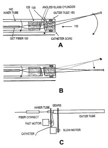
[0026) FIG. 5 illustrates a probe 110, and a probe scan system, according to
another
embodiment of the present invention. In the embodiment shown, optical probe
110 includes
a pair of GRIN lenses and a pair of cylindrical glass elements that are cut at
an appropriate
angle 0. As shown, probe 110 includes an optical fiber 120 and a fiber lens
element 125
proximal the end of fiber 120. A first tube 140 ("inner tube") encloses fiber
120. Inner tube
140 is also coupled to fiber lens element 125 to facilitate rotation of lens
element 125. A
second rotatable tube 150 ("outer tube") encloses tube 140 and refractive lens
element 130 to
facilitate rotation of lens element 130 relative to fiber lens element 125.
Input light from a
light source (not shown) at a distal end of fiber 120 enters fiber 120 and
exits the fiber end
5
CA 02558602 2006-09-06
WO 2005/094449 PCT/US2005/009334
internal to inner tube 140 as shown. In one aspect, the optical fiber 120 is
fixed at the focal
point of fiber lens element 125 within the inner tube. In preferred aspects,
lens element 125
includes a GRIN lens. The GRIN lens maybe cut at an angle or it may be coupled
with an
angled wedge element (e.g., similar to wedge 34 discussed above with reference
to FIG. 3)
as shown. In this case, the light output is collimated by the GRIN lens and
angularly
displaced by the angled glass wedge element. The tilted beam is brought to a
focus by lens
element 130, which in one aspect as shown includes a second glass wedge
element and GRIN
lens pair, and which is attached to the outer tube.
[00271 The rotation of lens element 130 with respect to fiber lens element 125
will change
the angle of the forward light beam with respect to the forward axis. For
example, FIG. 5a
shows the orientations that provide a maximum angle, and FIG. 5b show the
orientations that
provide a zero angle. If the angular difference between the orientation of the
first and second
angled surfaces is given by Liq) (AO = 0 when the cylinders are oriented as
shown in FIG.
5b), the angle made by the output beam to the forward axis is approximately
given by:
yr -- 8 (yl-1)2(1_cos(A1b)2)+sin(Ig5)2 (6)
where n is the refractive index of the cylinders. By rotating fiber lens
element 125 with
respect to lens element 130, the angle yr made by the output beam relative to
the forward
axis can be changed from 0 to 2(n -1) rads. Rotating both lens elements in
synchrony scans
the output beam in a complete circular cone. If the focal point of the output
is 2 mm from the
probe tip and it is desirable to cover a scan area 2 mm in diameter at that
distance, the angular
cut, 0, should be about 0.19 rads (about 11 ). Given the smallness of the
angle, in one aspect,
the design is further simplified by simply cutting the GRIN lenses with the
given angular tilt,
eliminating the need for glass wedge elements.
[00281 In one embodiment, the outer and inner tubes (holding lens element 130
and fiber
120, respectively) are preferably mounted to two different motors via gears as
shown in FIG.
Sc. In the embodiment of FIG. 1, tube 40 and fiber 20 may similarly be coupled
to different
motors. In both cases, the complete rotation of the refractive lens element
and the fiber end
with respect to a reference plane will complete a conical sweep. Therefore,
the combination
of these two motions will create a scan volume equal to a solid cone with a
maximum angle
from the forward axis given by the considerations described above. Each motor
preferable
provides one or multiple rotational speeds in the range of a fraction of a HZ
to about 1KHz or
more. Also, each motor may rotate the coupled elements in the same or opposite
direction as
6
CA 02558602 2011-12-28
the other motor. Further, the fiber 120 need not rotate with the fiber lens
element 125; that is
inner tube may rotate without rotation of fiber 120. It should also be
appreciated that a single
motor may be used to rotate both the inner and outer tubes. In this case, a
ratchet and pawl
type mechanism coupling the motor to both tubes may be used to rotate the
tubes at different
rotational speeds. Examples of a similar rotation actuation system and a fiber
connection to
an OCT imaging system for a side scanning probe is shown in "Scanning single-
mode fiber
optic catheter-endoscope for optical coherence tomography", Optics Letters, V2
1, pg. 543
(1996).
[0029] By using OCT imaging to create depth resolved imaging along each light
beam path
orientation, a three dimensional image of the structure in front of the
imaging needle (probe)
can be constructed. For example, an imaging Fourier Domain OCT (FDOCT) engine
can be
used with the probes of the present invention to acquire toniographic images
of the forward
scan volume. Given the large forward scan volumes possible (e.g., about 3-4 mm
forward
and an area of diameter 4 mm at the 4 mm forward distance point), a needle
endoscope
according to the present invention provides unprecedented forward imaging
capability. For
example, by rotating the inner tube at 100 Hz and the outer tube at 1 Hz, a 3
dimensional
image with a total of 108 voxel per second can be generated'-with an OCT
imaging system
that is capable of acquiring 100 kHz rate A-scans with 1,000 pixels each.
[0030] This innovative and yet elegantly simple design enables very compact
probes to be
built, e.g., probes of diameter 1 mm or less (e.g., 500 microns or less). Such
devices provide a
dramatic improvement over existing endoscopic imaging technology. The compact
size and
forward tomographic imaging capability of the probes of the present invention
make image
guidance of minimally invasive surgical procedure possible.
[0031] While the invention has been described by way of example and in terms
of the
specific embodiments, it is to be understood that the invention is not limited
to the disclosed
embodiments. To the contrary, it is intended to cover various modifications
and similar
arrangements as would be apparent to those skilled in the art. For example,
rather than
having a flat end face, a GRIN lens may be angled cut and a wedge element may
be attached
thereto and cut so as to provide the desired angled surface, e. g., 0 or 01.
Additionally, the
tubes holding the lens elements and fibers may comprise a flexible or rigid,
material.
Therefore, the scope of the appended claims should be accorded the broadest
interpretation so
as to encompass all such modifications and similar arrangements.
7