Note: Descriptions are shown in the official language in which they were submitted.
CA 02570305 2012-05-18
Systems, Methods and Devices Relating to Implantable Supportive Slings
10 Field of the Invention
The invention relates generally to implantable supportive slings. More
particularly, in various embodiments, the invention is directed to aspects of
soft tissue
anchors, adjustable length/tension slings, interconnects between slings and
soft tissue
anchors, delivery devices and systems for implanting supportive slings, and
methods
relating to anchoring, adjusting and implanting supportive slings.
Background of the Invention
Urinary incontinence occurs in both men and women. Various types of
incontinence are caused by different conditions and call for different
treatments. For
example, stress urinary incontinence (SUI) is known to be caused by at least
two
conditions, intrinsic sphincter deficiency (ISD) and hypermobility. These
conditions may
occur independently or in combination. In ISD, the urinary sphincter valve,
located
within the urethra, fails to close properly (coapt), causing urine to leak out
of the urethra
during stressful activity. Hypermobility is a condition in which the pelvis
floor is
distended, weakened or damaged, causing the bladder neck and proximal urethra
to rotate
and descend in response to increases in intra-abdominal pressure (for example,
due to
sneezing, coughing, straining, etc.). As a result, the patient's response time
becomes
insufficient to promote urethral closure and, consequently, the patient
suffers from urine
leakage and/or flow.
Page 1 of 63
CA 02570305 2006-12-14
WO 2005/122954
PCT/US2005/021267
A popular treatment of SUI uses a surgical sling placed under the bladder neck
or
the mid-urethra to provide a urethral platform. Placement of the sling limits
the
endopelvis fascia drop. One disadvantage of prior art approaches is that
certain mid-
urethral sling stabilization procedures typically require incisions in
addition to those
made in the vaginal wall. By way of example, some procedures require abdominal
incisions, while others require groin incisions.
Accordingly, there is a need for improved systems, devices and methods for
treating urinary incontinence.
Summary of the Invention
The invention addresses the deficiencies in the prior art by, in various
embodiments, providing improved systems, devices and methods relating to
urinary
incontinence. More particularly, in some embodiments, the invention provides
improved
sling assemblies that make it easier for a medical operator to adjust the
length and thus,
the tension of the sling during implantation. In other embodiments, the
invention
provides improved soft tissue anchors for affixing supportive slings at a
desired
anatomical location. I further embodiments, the invention provides dilators
sized
similarly to tissue anchors, which are used to deliver sling ends to an
anatomical location,
such as into or through the obturator membrane, but subsequently dissolve,
leaving only
the sling end embedded in the obturator membrane to hold the sling in place.
In
additional embodiments, the invention provides improved mechanisms for
attaching or
otherwise associating soft tissue anchors and/or anchor sized dilators to the
ends of sling
assemblies to further facilitate sling length/tension adjustment. In further
embodiments,
the invention provides improved delivery devices, systems and methods for
implanting
supportive slings and their associated soft tissue anchors and/or anchor sized
dilators to
desired anatomical sites.
According to one aspect, the invention is directed to an improved implantable
supportive sling for treating urinary incontinence. According to one
embodiment, the
supportive sling of the invention includes a pocket formed at a first end. The
pocket is
2
CA 02570305 2006-12-14
WO 2005/122954
PCT/US2005/021267
sized and shaped for receiving a distal end of a delivery device shaft.
According to a
further embodiment, the supportive sling includes a second pocket formed at a
second
end, also sized and shaped for receiving a distal end of a delivery device
shaft. In one
implementation, a medical operator may insert a distal end of a shaft into the
first end of
the sling, and then insert the distal end of the delivery device shaft and
sling end into the
body of a patient, for example, via an incision in the vaginal wall, to
deliver the first end
of the sling to a desired anatomical location. The medical operator may
deliver the
second end of the sling to another anatomical location, for example, on a
contralateral
side of the patient's body, with the same or a second delivery device using
the same or a
similar approach, to implant the sling under a location to be supported, such
as a mid-
urethral location.
The sling may be made from any suitable material, and may include portions
having smooth or tanged edges or a combination of smooth and tanged edges. In
one
configuration, the sling is formed from a mesh material. The sling assemblies
are
generally short, e.g., from about 5 cm to about 20 cm long. According to one
construction, the sling end pockets are formed by folding over the sling
material onto itself
and sealing the edges. In some configurations, the entire edges of the pockets
are sealed.
However, in other configurations, only a portion of one or both edges is
sealed. According
to one feature, portions of the sling ends are left unsealed to allow for tabs
to be inserted at
the entrance to each end pocket.
According to another aspect, the invention provides a plurality of anchor
sized
tissue dilators, which may dissolve subsequent to implantation. In other
aspects, the
invention provides a plurality of soft tissue anchor configurations. In some
embodiments,
the tissue anchors of the invention have relatively smooth outer surfaces and
rely, for
example, on orientation and/or features on a sling for anchoring within the
tissue. In some
such embodiments, these tissue anchors are used primarily as tissue dilators
during
implantation, and subsequently dissolve, leaving just the sling ends or other
tissue ingrowth
sites along the sing to hold the sling in place.
3
CA 02570305 2006-12-14
WO 2005/122954
PCT/US2005/021267
In other embodiments, the tissue anchors of the invention include barbs
projecting
from anchor bodies and are oriented for passing the anchors into tissue and
for resisting
backing the anchors out of tissue. In some embodiments, the barbs project
radially from
discrete locations along the anchor bodies. In other embodiments, the barbs
are formed as
rings projecting radially around the entire circumference of the anchors. In
some
configurations, one or more of the barbs substantially aligns axially. In
other
configurations, one or more of the barbs substantially aligns radially along a
common
circumference. In further configurations, the barbs are arranged so as not to
align axially or
radially, but instead to be staggered in both directions. In some
configurations, the anchors
include only a single row of barbs substantially radially aligned along a
circumference of
the anchor. According to some embodiments, the barbs are formed by building up
material
onto the outer body of an anchor. In other embodiments, the barbs are molded
into the
anchor. In further embodiments, the barbs are carved into the outer body of
the anchor. In
some embodiments, the barbs are formed from pealing back portions of an outer
surface of
the anchor.
According to some configurations, the barbs are narrow and bristle-like. In
some
such configurations, the bristles are relatively short (e.g., less than about
2 millimeters in
length). However, in other such configurations, the barbs are longer (e.g.
between about 2
millimeters and about 5 millimeters in length). According to some embodiments,
the barbs
have pointed tips. However, in other configurations, the barbs may have
rounded tips.
According to some embodiments, the barbs are relatively narrow (e.g., less
than about 1
millimeter in width/diameter). In other embodiments, the barbs are relatively
wide (e.g.,
between about 1 millimeter and about 2 millimeters in width/diameter).
According to one feature, the soft tissue anchors and/or anchor sized dilators
of the
invention include an aperture sized and shaped for interfitting over a distal
tip of a delivery
device shaft. In some configurations, the aperture extends axially from a
proximal end of
the anchor part way to a distal end of the anchor. In other configurations,
the anchor and/or
anchor sized dilator includes a through-passage extending axially between the
proximal
and distal ends of the anchor thus forming a hollow anchor.
4
CA 02570305 2006-12-14
WO 2005/122954
PCT/US2005/021267
According to various embodiments, the anchors and anchor sized dilators of the
invention are generally elongated. In some configurations, they are between
about 1
centimeter and about 4 centimeters long. According to one configuration, they
are between
about 2.5 centimeters and about 3.5 centimeters long. According to other
embodiments,
they have an outside diameter (not including the barbs) of between about 2
millimeters and
about 4 millimeters. However, in some embodiments, they have an outside
diameter (not
including the barbs) of less than about 2 millimeters.
According to other embodiments, the distal tips of the anchors and/or anchor
sized
dilators may have any suitable configuration. In some embodiments, the distal
tips are
sharp enough to pierce human tissue. However, in other embodiments, the tips
may be
rounded. According to some configurations, the distal ends are tapered into a
conical shape
to provide for tissue dilation during sling implantation.
The anchors and/or anchor sized dilators of the invention may attach to sling
ends
by any suitable mechanism. In some configurations, the proximal end is, for
example,
glue-, heat- or shrink tube-bonded to each end of a sling. In other
configurations, a
proximal portion includes a slot for interfitting with a sling end. Each sling
end may be
suitably bonded into a proximal slot of a respective anchor or anchor sized
dilator. In some
configurations, the slot extends distally from the proximal end of the anchor
or anchor
sized dilator along a cross-sectional diameter of the anchor or anchor sized
dilator.
According to various constructions, the slings to which the anchors or anchor
sized dilators
attach are between about 5 centimeters and about 8 centimeters long. In one
embodiment,
they are about 6 centimeters long. According to a further construction, the
total (i.e.,
anchor/dilator tip to opposite anchor/dilator tip) sling assembly length is
between about 8
centimeters and about 14 centimeters. In one embodiment, the total sling
assembly length
is about 12 centimeters.
According to some aspects, the tissue anchors/dilators of the invention are
configured for attaching to a sling end in a sling length/tension adjustable
manner. For
example, in one embodiment, an anchor/dilator includes a radial aperture in a
side wall
near its proximal end. A first end of a filament threads through the aperture
and a second
5
CA 02570305 2006-12-14
WO 2005/122954
PCT/US2005/021267
end of the filament threads through an aperture in a sling end. The aperture
in the sling
may be, for example, a gap in a mesh or may be separately formed, and
optionally
reinforced. The length of the filament, and thus the overall length of the
sling assembly
(i.e., from anchor/dilator distal tip to opposite anchor/dilator distal tip),
may be adjusted by
pulling on the filament terminal ends and securing them. In some
configurations, the
filament terminal ends may be secured together, for example, by clipping,
tying, gluing or
other suitable mechanism. By way of example, in one configuration, the
filament ends are
tied together in a one-way slip knot, which easily slides to be tightened, but
not to be
loosened.
According to one embodiment, the filament threads through the aperture or
other
suitable structure in the anchor/dilator. Then, each end of the filament
threads through a
separate aperture in the sling end. The further the filament ends are drawn
through the
sling end apertures, the closer to the sling end the anchor is drawn, once
again adjusting the
overall length of the sling assembly. As in the prior example, the filament
ends may be
secured together to hold the sling assembly length constant. The filament ends
may be
secured, for example, by tying, tying in a one-way slip knot, glued, clipped,
or passed
through a one-way adjustable holder.
In a further embodiment, the anchor/dilator attaches to a sling end, and the
filament
ends thread through respective apertures in the sling end. Then, each of the
filament ends
interweaves with the sling material along at least a partial length of the
sling. In one
configuration, one filament end interweaves with the sling material along one
long edge of
the sling, and the other filament end interweaves with the sling material
along the other
long edge of the sling material. In response to pulling on the terminal ends
of the filament,
the sling material accordions to reduce its effective length. In some
configurations, the
interwoven filament is employed only at one end of a sling assembly, with the
other end
remaining at a fixed location. In some such embodiments, the filament-
interwoven, and
thus accordionable sling section extends for substantially the entire length
of the sling. In
other embodiments, the filament is interwoven with half or less of the length
of the sling.
In further embodiments, the sling assembly employs such interwoven filaments
at both
6
CA 02570305 2006-12-14
WO 2005/122954
PCT/US2005/021267
ends. In some constructions, the interwoven filaments pass first through an
aperture or
other suitable structure on an anchor, for example, to attach the
anchor/dilator to the sling.
According to alternative embodiments, a tissue anchor/dilator of the invention
includes a loop, for example, extending from a proximal end. A sling end may
slidably
interfit within the loop and the anchor/dilator may be placed at any desired
location along
the length of the sling. Once placed, the anchor/dilator may be secured in
position. The
anchor/dilator may be secured in place, for example, with a vascular or any
other suitable
clip, a suture, or a staple. In the case of the clip or staple, they may be
placed on a sling-
end side of the anchor to stop the anchor/dilator from sliding in a
lengthening direction or
sliding off the sling altogether. In some configurations, the loop may include
angled spikes
or teeth that are oriented to enable the loop, and thus the anchor/dilator, to
slide onto the
sling, but not allow it to slide in an opposite (e.g., lengthening) direction.
In other
configurations, a portion of the sling may include one-way bristles or spikes
that are
oriented to enable the sling end to be inserted into the anchor/dilator loop,
but inhibit
sliding the anchor/dilator back off the sling in a sling-lengthening
direction. In a variation
of this configuration, the sling assembly includes an elongated, anchor-like
element
attached to the sling end. This element includes the directionally oriented
spikes, bristles
or other projections positioned to slide into the anchor/dilator loop and to
impede sliding
out of the anchor/dilator loop. The anchor/dilator may be slid along the
length of this
anchor-like attachment to adjust the overall (anchor/dilator distal tip to
anchor/dilator distal
tip) length of the sling assembly.
In other configurations, the sling assembly may include a one way buckle, such
as
that employed on backpacks, for passing the sling end through and adjusting
the sling
length/tension. In some configurations, the buckle may be, or may be attached
to, the
anchor/dilator loop. Alternatively, the buckle may be formed into the body of
the
anchor/dilator. In other configurations, the buckle is located on the sling
end, independent
from the sling end passing through a loop or other suitable structure on the
anchor/dilator.
In further configurations, the one way buckle may be placed at any suitable
location along
the length of the sling.
7
CA 02570305 2006-12-14
WO 2005/122954
PCT/US2005/021267
In another embodiment, an anchor/dilator of the invention includes a hollow
portion extending axially from a proximal end at least part way to a distal
end of the
anchor/dilator, and a bar or other structure extending radially across the
hollow portion
inside the anchor/dilator. In this embodiment, a sling end may pass into the
hollow portion
via a proximal opening in the anchor/dilator, then loop around the bar and
back out of the
proximal end of the anchor/dilator. In some configurations, the bar may
include spikes,
bristles or other projections for allowing the sling end to pass through the
hollow portion in
a sling shortening direction, but impeding the sling from passing in an
opposite sling-
lengthening direction. In other configurations, the sling end may be secured,
for example,
by way of a clip, staple or suture, outside the anchor/dilator subsequent to
the
anchor/dilator being placed at a desired location along the sling length. As
in all of the
described embodiments, excess sling-end material may be trimmed off.
In some embodiments, sling assemblies of the inventions are formed in two
sections. In various configurations, one end of each section includes a tissue
anchor/dilator
and the other end of each section may be affixed together to achieve a desired
sling
assembly length. In one implementation, one or both of the non-anchor/dilator
ends of the
two sling assembly sections are cut to length and then attached, for example,
by way of
suturing, tying, clipping, stapling or heat melting/bonding. In another
implementation, the
anchor/dilator end of one of the sections is passed through an aperture near
the non-
anchor/dilator end of the other section. The anchor/dilator is pulled through
to a desired
length and is then secured in place near the aperture. In some configurations,
the sling
assembly section being passed through the aperture includes projections for
resisting that
section from being pulled back out of the aperture in the opposite direction.
According to another aspect, the invention is directed to stackable tissue
anchors/dilators. In one embodiment, a first tissue anchor/dilator attaches to
a sling end.
Then, a second tissue anchor/dilator may slidably interfit over a distal end
of the first
anchor/dilator to effectively create a longer anchor/dilator with a new distal
end. By
stacking anchors/dilators in this fashion, the overall (anchor/dilator distal
tip to
anchor/dilator distal tip) length of the sling assembly may be increased.
Previously stacked
8
CA 02570305 2006-12-14
WO 2005/122954
PCT/US2005/021267
anchors/dilators may be unstacked to reduce the length of the sling assembly.
According to
one feature, each anchor/dilator one or more radially extending apertures in
its side wall
near a proximal end, and one or more corresponding radial projections in its
side wall near
a distal end. The distal radial projections of the first anchor/dilator snap
fit into the
proximal radial apertures of the second anchor/dilator to hold the two
anchors/dilators
together when stacked. Any number of anchors/dilators may be stacked in this
fashion.
In other aspects, the invention provides devices and/or systems for delivering
a
sling assembly to anatomical locations within the body of a patient. Delivery
systems
include, for example, a sling assembly having at least one tissue
anchor/dilator, along with
a suitable delivery device. According to one embodiment, a delivery device of
the
invention includes a handle and a shaft extending distally from a distal end
of the handle.
A distal end of the shaft may, for example, be sharp enough for piercing
tissue, conical in
shape for tunneling, or rounded blunt. The shaft may have one or more
substantially
straight sections and/or one or more curved sections. The shaft may be formed
substantially in a single plane, substantially in two planes, or in more than
two planes. In
one configuration, the delivery device is sized and shaped for delivering
sling ends (and
tissue anchors/dilators) transvaginally to a suprapubic location (e.g. on the
posterior /
bladder side of the pubic bone). In other configurations, the delivery device
is sized and
shaped for delivering the sling ends (and tissue anchors/dilators)
transvaginally to a
prepubic location (e.g. a location between the pubic bone and the abdominal
wall on the
anterior side of the pubic bone). This approach has the advantage that there
is considerably
less risk of inadvertently puncturing the bladder during placement. In further
configurations, the delivery device is sized and shaped for delivering the
sling ends (and
tissue anchors/dilators) transvaginally near, into or through the obturator
membrane. In a
variation of this configuration, the delivery devices may be sized and shaped
for initiating
this procedure by inserting a distal end of the delivery device into the
patient's body via a
vaginal wall incision, or alternatively, via an inner thigh incision.
According to one embodiment, a delivery device of the invention includes a
narrowed distal end configured for interfitting with an aperture, a hollow
through passage
9
CA 02570305 2006-12-14
WO 2005/122954
PCT/US2005/021267
or other suitable feature on a tissue anchor/dilator. Optionally, a shoulder
is formed near
the distal end of the shaft. When inserted into the anchor/dilator, the
shoulder of the
delivery device shaft abuts the proximal end of the anchor/dilator. In various
configurations, the narrowed distal portion is between about 2 centimeters and
about 4
centimeters long. In other configurations it is between about 1 centimeter and
about 3
centimeters long. In further configurations, the narrowed distal portion has
an outside
diameter of between about .03 inch and about .05 inch. In one embodiment, it
has an
outside diameter of about .04 inches. According to other configurations, the
portion of the
shaft forming the shoulder has an outside diameter of between about .07 inch
and about .1
inch. In one implementation the outside diameter of this portion of the shaft
is about .09
inch. According to one configuration, the total shaft length is between about
7 centimeters
and about 20 centimeters. In other configurations, the total length of the
shaft is between
about 8 centimeters and about 12 centimeters.
According to a further embodiment, the delivery device includes an inner shaft
and
an outer cannula. In one configuration, a distal end of the outer cannula
forms a radially
extending shoulder around the inner shaft. Additionally, the narrow inner
shaft extends
distally from the outer cannula (similar to the above described narrowed
distal shaft
portion) with the outer cannula in a retracted position. According to some
embodiments,
the delivery device includes a pusher near a distal end of the handle for
sliding the outer
cannula axially over the inner shaft. In operation, with the pusher retracted,
an
anchor/dilator is interfitted over the narrowed distal portion of the shaft.
Subsequent to
anchor/dilator placement, the medical operator slides the pusher distally to
push the
anchor/dilator off of the narrowed distal portion, and withdraws the delivery
device from
the patient.
In an alternative embodiment, outer cannula remains fixed and the inner shaft
is
slidable. More particularly, the delivery device of the invention includes a
slidable shaft
actuator located on the handle, for enabling an operator to alternatingly
extend and retract
the distal portion of the shaft from the distal end of the cannula. In
operation of this
embodiment, an operator extends the distal portion of the shaft to insert it
into the tissue
CA 02570305 2006-12-14
WO 2005/122954
PCT/US2005/021267
anchor/dilator. Subsequent to anchor/dilator placement, the operator retracts
the distal
portion of the shaft to disengage it from the anchor/dilator, and withdraws
the delivery
device from the patient.
According to another embodiment, a delivery device of the invention includes a
dilator, a pusher and a guide member. In operation a dilator is inserted
through an incision
in the vaginal wall until its distal tip-reaches a location at or near to
where an anchor/dilator
is to be implanted. The guide member, optionally a guide wire, is inserted
axially through
the dilator until it extends out of the distal tip of the dilator. The dilator
is then slid
proximally along the guide wire to remove the dilator from the patient's body.
A hollow
anchor/dilator of a sling assembly is then slid over a proximal end of the
guide wire and
slid distally along the guide wire. A pusher is then slid over the proximal
end of the guide
wire and also slid distally along the guide wire to advance the tissue
anchor/dilator along
the wire until it reaches a desired location within the body of the patient.
The pusher and
the guide wire are then removed to leave the tissue anchor/dilator in place.
In another embodiment, a delivery device of the invention includes a hollow
insertion shaft and a push wire. In this embodiment, a tissue anchor/dilator
of a sling
assembly interfits over a distal end of the insertion shaft. The distal end of
the shaft with
the anchor/dilator so interfitted is inserted into the body of the patient via
a vaginal
incision. The shaft is advanced distally until the anchor/dilator is located
at the desired site
of implantation. The push wire is then inserted into a proximal end of the
shaft and
advanced distally until a distal end of the push wire abuts the tissue
anchor/dilator. The
push wire is then further advanced distally to push the anchor/dilator off of
the insertion
shaft to implant the anchor/dilator at the desired location. The insertion
shaft and the push
wire are then removed from the patient.
As mentioned above, according to some embodiments, the methods of the
invention deliver a tissue anchor/dilator of a sling assembly to the obturator
foramen. In
one approach, the anchor/dilator is delivered to a location in front of the
obturator
membrane. In another approach, the anchor/dilator is delivered into the
obturator
membrane. The anchor/dilator may also be fixed to the obturator membrane. In a
further
11
CA 02570305 2006-12-14
WO 2005/122954
PCT/US2005/021267
approach, the anchor/dilator is delivered through the obturator membrane. In
some
practices, the anchor/dilator is delivered through the obturator membrane to
about 2.5
centimeters into the obturator foramen. In other practices, the anchor/dilator
is delivered
through the obturator membrane about 1 centimeter to about 2.5 centimeters
into the
obturator formen.
These and other features, embodiments and aspects of the invention will be
further
understood with reference to the description of the illustrative embodiments.
Brief Description of the Drawings
Illustrative embodiments of the invention are described below with reference
to
the appended drawings, in which like parts have like reference designations
and in which
the various depicted parts may not be drawn to scale. The depicted embodiments
are to
be understood as illustrative of the invention and not as limiting in any way.
Figures 1A-1B depict different views of a mesh sling including end pockets
according to an illustrative embodiment of the invention.
Figures 2A-2E each depict various soft tissue anchors/dilators according to
illustrative embodiments of the invention.
Figures 3A-3B depict an approach for affixing a sling end to a soft tissue
anchor/dilator according to an alternative illustrative embodiment of the
invention.
Figures 4A-4B depict an approach for affixing a sling end to a soft tissue
anchor/dilator according to another alternative illustrative embodiment of the
invention.
Figures 5A-5G depict various hollow soft tissue anchors/dilators according to
illustrative embodiments of the invention.
Figures 6A-6D depict approaches for affixing a soft tissue anchor/dilator to a
sling end in such a way as to provide for sling assembly length/tension
adjustment
according to various illustrative embodiments of the invention.
12
CA 02570305 2006-12-14
WO 2005/122954
PCT/US2005/021267
Figures 7A-7C depict approaches for length/tension adjusting an implantable
sling assembly using filaments, for example, tied in one way knots and/or
interwoven
with the sling material according to illustrative embodiments of the
invention.
Figures 8A-8B are perspective and side views, respectively, of a sling
assembly
end including a soft tissue anchor/dilator with an internal bar about which
sling
length/tension may be adjusted according to an illustrative embodiment of the
invention.
Figures 9A-9B show a soft tissue anchor/dilator having a loop for receiving a
ridged/jagged element attached to an end of a sling for providing sling
length/tension
adjustment according to another illustrative embodiment of the invention.
Figure 10 shows a sling assembly including two sections attached together at
an
intermediate location to provide for adjustable sling/tension according to
another
illustrative embodiment of the invention.
Figure 11 shows another two section sling assembly adapted for adjustably
interfitting at an intermediate location to provide for adjustable sling
length/tension
according to a further illustrative embodiment of the invention.
Figures 12A-12B show adjustable length/tension sling assemblies having an end
clip for affixing a sling end to a soft tissue anchor/dilator at a desired
sling length
according to another illustrative embodiment of the invention.
Figures 13A-13B depict soft tissue anchor/dilator having a buckle in a side
wall
for interthreading with a sling end to provide for adjustable sling
length/tension according
to an additional embodiment of the invention.
Figures 14A-14B depict an arrangement of interlocking stackable soft tissue
anchors/dilators for providing adjustable sling assembly length/tension
according to
another embodiment of the invention.
Figures 15A-15B show a delivery device including a shaft having a narrowed
distal tip for interfitting with a soft tissue anchor/dilator of a sling
assembly according to
an illustrative embodiment of the invention.
13
CA 02570305 2006-12-14
WO 2005/122954
PCT/US2005/021267
Figure 16 shows a delivery device having a slidable inner shaft for engaging
with
a soft tissue anchor/dilator of a sling assembly according to an illustrative
embodiment of
the invention.
Figure 17 shows a delivery device having a slidable outer cannula and a
narrowed
inner shaft for engaging with a soft tissue anchor/dilator of a sling assembly
according to
an illustrative embodiment of the invention.
Figure 18 shows a delivery system for implanting a sling assembly including a
hollow soft tissue anchor/dilator to an anatomical site according to an
illustrative
embodiment of the invention.
Figures 19A-19B show a delivery system for implanting a sling assembly
including a soft tissue anchor/dilator to an anatomical site according to
another
illustrative embodiment of the invention.
Figure 20 shows a delivery system employing a delivery device having a spiral
shaft according to an illustrative embodiment of the invention.
Figure 21 shows a delivery device having a curved shaft for delivering a sling
assembly, for example, transobturally according to an illustrative embodiment
of the
invention.
Figures 22A-22C show various views of a delivery device having a halo shaft
for
delivering a sling assembly, for example, transobturally according to another
illustrative
embodiment of the invention.
Figures 23A-23C depict an approach for delivering a sling assembly
transobturally using the delivery system of Figure 18 according to an
illustrative
embodiment of the invention.
Figures 24A-24C depict an approach for delivering a sling assembly
transobturally using the delivery system of Figures 22A-22C according to an
illustrative
embodiment of the invention.
14
CA 02570305 2006-12-14
WO 2005/122954
PCT/US2005/021267
Figures 25A-25C show a detailed view of placing a soft tissue anchor/dilator
of a
sling assembly according to an illustrative embodiment of the invention.
Figures 26A-26B show a detailed view of an alternative placement of a soft
tissue
anchor/dilator of a sling assembly according to another illustrative
embodiment of the
invention.
Illustrative Description
As described above in summary, the invention addresses deficiencies in the
prior
art by, in various illustrative embodiments, providing improved systems,
methods and
devices related to implanting supportive slings within the human body. In
particular
illustrative embodiments, the systems, methods and devices of the invention
are
particularly sized, shaped and adapted for delivering a sling to periurethral
tissue to
provide urethral, bladder, and/or bladder neck support for treating urinary
incontinence.
As described below in further detail, some of the illustrative embodiments are
directed to
improved sling and sling assemblies. Other illustrative embodiments are
directed to
improved tissue anchors, such as soft tissu9 anchors, for anchoring one or
both ends of a
sling or sling assembly at a desired anatomical location. Further illustrative
embodiments, are directed to anchor sized dilators, which in various
implementations
may be sized and shaped like any of the described anchors, except with
substantially
smooth outer surfaces. In some of these illustrative embodiments, the
dilator/anchor
relies on dilator/anchor orientation, rather than barbs for anchoring. In
other illustrative
embodiments, the anchor/dilator dissolves and is bioabsorbed, leaving only the
sling ends
or other locations along the sling itself to hold the sling in place. In some
illustrative
embodiments, the improved anchors/dilators include, for example, improved
anchoring
structures, improved interfittings with delivery devices, improved features
for attaching
the anchors to the sling assembly in a length/tension adjustable manner, and
the like.
Additional illustrative embodiments are directed to improved delivery devices
and sling
delivery systems. The illustrative delivery systems include, for example, a
sling
assembly along with a delivery device. Other illustrative embodiments describe
CA 02570305 2006-12-14
WO 2005/122954
PCT/US2005/021267
exemplary procedures for implanting a supportive sling employing features of
the
invention.
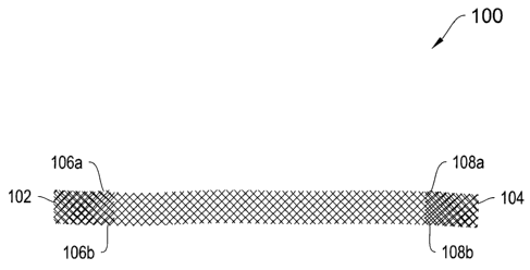
Turning to the depicted illustrative embodiments, Figures 1A and 1B depict top
and side views, respectively, of a sling 100 including end pockets 102 and
104. The end
pockets 102 and 104 are formed by folding a short length of an end of the
sling, e.g.,
about 0.5 inches to about 1.0 inches, over onto itself and closing the edges.
For example,
the edges 106a and 106b are closed to form the pocket 102 and the edges 108a
and 108b
are closed to form the pocket 104. The edges may be closed, for example, by
way of
suturing, gluing or heat sealing. The edge closures may extend for the entire
length of
the folded over portions, or as most clearly shown in Figure 1B, a portion 109
and 110 of
the respective folder over portions may be left without its edges closed to
provide for free
tabs at the entrance to the pockets 102 and 104.
In certain illustrative embodiments, the pockets 102 and 104 of the sling 100
may
be coated or otherwise treated with a material to stiffen and/or strengthen
them.
According to the illustrative embodiment, the sling 100 is between about 5
centimeters
and about 20 centimeters long. According to a feature of the sling 100, a
distal end of a
delivery device shaft may be inserted into either pocket 102 or 104 and then
inserted
through a vaginal incision to deliver a sling end to an anatomical site. With
the delivery
device removed, either or both of the pockets 102 and 104 may employed as a
soft tissue
anchor. By way of example, the pocket 102 and/or 104 may be implanted into or
through
a obturator membrane or other tissue, muscle, ligament or suitable anatomical
structure.
The folded over sling material then resists the pulling of the end pocket back
out of the
membrane or other structure to anchor the sling end in place.
The sling 100 may be formed from any suitable materials and configurations.
For
example, the sling 100 may be formed from an omnidirectional material, a
material that
has equivalent tensile strength from any direction, such as pericardium or
dermis.
Alternatively, the material may be an oriented material, a material that has a
single
direction where the tensile strength of the material is the highest. Oriented
materials may
include rectus fascia and/or facia lata.
16
CA 02570305 2006-12-14
WO 2005/122954
PCT/US2005/021267
The edge or other regions of the sling 100 can be configured differently
depending on their intended placement in the body of the patient. For example,
a middle
section of the sling 100 is typically located where an anatomical site, such
as a
midurethral or bladder neck location in the periurethral tissue, needs to be
supported. In
one illustrative embodiment, a middle section of the sling 100 has smooth or
rounded
edges, hereinafter also referred to as "non-tanged." According to a further
illustrative
embodiment, other sections of the sling 100 may include tangs (e.g., sharp
projections or
frayed edges). The tangs are generally useful for anchoring the sling 100 and
encouraging tissue growth into the sling 100. Anchoring the sling 100 in this
manner
generally obviates the need for additional sutures to hold the sling 100 in
place.
Anchoring the sling 100 via its tangs is especially useful for anchoring the
sling 100 on a
tissue and facilitating the removal of the sleeve according to the invention
by pulling on
the center tab of the sleeve while the sling 100 stays in place, without the
need for
additional incisions in order to hold the sling 100 external to the body while
the sleeve is
being removed through pulling.
The tanged and non-tanged edges of the sling 100 may be formed in a plurality
of
ways. For example, the sling 100 can be cut from a woven sheet, in which case
the edges
would be initially tanged along the entire length of the sling 100. One or
more non-
tanged sections may be formed by any process that smoothes, rounds or removes
the
sharp edges of the tangs. For example, the tangs rimy be heat-smoothed by
burning or
melting the tangs. Providing one or more non-tanged sections, which may be in
close
proximity to a sensitive anatomical site in the patient, can enhance the
comfort level of
the patient and reduce the potential for the edges of the tangs to erode or
irritate the
urethra. Alternatively, the sling 100 can be produced from a woven tape having
the
approximate finished width of the sling 100. The smooth sides of the tape can
then be
trimmed off to produce the tanged sections.
The sling 100 used with the invention may be fabricated from any suitable
material(s), preferably biocompatible materials. In certain illustrative
embodiments, the
material may include, for example, synthetic mesh or other synthetic material;
it may also
17
CA 02570305 2006-12-14
WO 2005/122954
PCT/US2005/021267
or alternatively include non-synthetic material, such as cadaver, human or
animal tissue;
it may also include any combinations thereof. In examples employing synthetic
material
for the sling 100, it may be derived from any suitable synthetic material.
Such material
could include, for example, polymeric material such as, for example, as
Polytetrafluorethylene (Goretex), polypropylene (Marlex), polyethylene
(Mersiline),
silastic, or impregnated collagen matrix (Protegen). In certain illustrative
embodiments,
one or more suitable materials for the sling 100 may include, for example,
nylon,
polyethylene, polyester, polypropylene, fluoropolymers, copolymers thereof,
combinations thereof, or other suitable synthetic material(s). The material
may be, for
example, a synthetic material that is absorbable by the patient's body.
Suitable
absorbable synthetic materials can include polyglycolic acid, polylactic acid,
and other
suitable absorbable synthetic materials. The sling 100 material may be
fabricated from
one or more yarns, which yarns may be made from one or more materials.
Alternatively, the materials for the sling 100 may employ non-synthetic or
natural
materials, for example materials from human fascia, cadaveric fascia or skin
mammalian
tissue(s). Human tissues may be used in certain embodiments and may be
derived, for
example, from human cadaveric or engineered human tissue. Animal tissues may
be
derived, for example, from porcine, ovine, bovine, and equine tissue sources.
In certain
embodiments the materials for the sling 100 may include a combination of non-
synthetic
(e.g., mammalian tissue(s)) and synthetic material(s).
According to a further illustrative embodiment, any or all of the sling 100
may be
configured to be biodegradable/bioabsorbable. According to another feature, at
least a
portion of the sling 100 is biodegradable and may also dissolve and/or be
absorbed into
the patient's tissues. For example, in some embodiments, only a section of the
sling 100
is biodegradable/bioabsorbable, such as, for example, an intermediate portion.
Examples
of biodegradable/bioabsorbable materials that may be used for the sling 100
include,
without limitation, polylactic acid (PLA), polyglycolic acid (PGA), poly-L-
lactic acid
(PLLA), human dermis and decellularized animal tissue.
18
CA 02570305 2006-12-14
WO 2005/122954
PCT/US2005/021267
Exemplary biodegradable/bioabsorbable materials, in addition to those listed
above, which may be employed for the sling 100 include, but are not limited
to,
polylactic acid, polyglycolic acid and copolymers and mixtures thereof, such
as poly(L-
lactide) (PLLA), poly(D,L-lactide) (PLA), polyglycolic acid [polyglycolide
(PGA)],
poly(L-lactide-co-D,L-lactide) (PLLA/PLA), poly(L-lactide-co-glycolide)
(PLLA/PGA),
poly(D,L-lactide-co-glycolide) (PLA/PGA), poly(glycolide-co-trimethylene
carbonate)
(PGA/PTMC), poly(D,L-lactide-co-caprolactone) (PLA/PCL), and poly(glycolide-co-
caprolactone) (PGA/PCL); polyethylene oxide (PEO); polydioxanone (PDS);
polypropylene fumarate; polydepsipeptides, poly(ethyl glutamate-co-glutamic
acid),
poly(tert-butyloxy-carbonylmethyl glutamate); polycaprolactone (PCL),
poly(hydroxy
butyrate), polycaprolactone co-butylacrylate, polyhydroxybutyrate (PHBT) and
copolymers of polyhydroxybutyrate; polyphosphazenes, poly(phosphate ester);
maleic
anhydride copolymers, polyiminocarbonates, poly[(97.5% dimethyl-trimethylene
carbonate)-co-(2.5% trimethylene carbonate)], cyanoacrylate,
hydroxypropylmethylcellulose; polysaccharides, such as hyaluronic acid,
chitosan and
regenerate cellulose; poly(amino acid) and proteins, such as gelatin and
collagen; and
mixtures and copolymers thereof.
According to a further illustrative embodiment, the sling 100 may incorporate
or
be coated with one or more agents to provide a therapeutic effect, for
example, to reduce
discomfort, to reduce the chance of infection and/or to promote tissue growth.
The sling
100 may be treated or coated with any suitable material. For example, in some
illustrative embodiments, suitable treatment materials may include
bioabsorbable/dissolvable materials which may include, but are not limited to,
alginates,
sugar based formulations, starches, gelatins, cellulose, polyvinyl alcohol,
polyglycolic
acid (PGA), polylactic acid (PLA), polydioxinone (PDO), and/or other synthetic
or
natural polymers including combinations thereof The treatment materials are
preferably
biocompatible, and the biocompatible protective treatment may cover any
portion or all
of the sling 100. In one particular configuration, the protective treatment
encapsulates or
substantially encapsulates at least portion of the sling 100. According to one
feature, the
protective treatment is formed from lubricious material and reduces the
friction between
19
CA 02570305 2006-12-14
WO 2005/122954
PCT/US2005/021267
the sling 100 and the patient's periurethral tissues. In this way, the
protective treatment
can provide a relatively smooth tissue contact surface to otherwise tanged or
ragged sling
edges to reduce the likelihood of the sling 100 irritating the patient's
tissues during
implantation.
The protective treatment may be applied to the sling 100 by any suitable
approach, for example, by way of spraying, brushing or dipping the portion of
the sling
100 to be treated. According to another illustrative embodiment, the
protective treatment
is formed as a sheet of material that can be affixed to the portion of the
sling 100 to be
treated. According to another feature, the protective treatment may be
configured to
dissolve within a particular time range. The protective treatment may be
configured, for
example, to substantially absorb into the patient's tissues within about 30,
15, 10 or 5
minutes from the time the sling 100 is implanted. Alternatively, the
protective treatment
may be configured to substantially absorb into the patient's tissues over a
time span of
hours, days, weeks, or months.
According to another illustrative feature, the sling 100 may also include an
agent
for release into the patient's tissues. One illustrative agent promotes, when
applied to the
patient's tissues in a pharmaceutically acceptable amount, well-organized
collagenous
tissue growth, such as scar tissue growth, preferably, in large quantities.
According to
one feature, the agent may or may not block or delay the dissolvability of the
protective
treatment. This may be controlled by selecting differing methods for loading
the agent
onto the sling 100. The tissue growth factor may include natural and/or
recombinant
proteins for stimulating a tissue response so that collagenous tissue such as
scar tissue
growth is enhanced. Exemplary growth factors that may be used include, but are
not
limited to, platelet-derived growth factor (PDGF), fibroblast growth factor
(FGF),
transforming growth factor-beta (TGF-beta), vascular endothelium growth factor
(VEGF), activin/TGF and sex steroid, bone marrow growth factor, growth
hormone,
insulin-like growth factor 1, and combinations thereof. The agent may also
include a
hormone, including but not limited to estrogen, steroid hormones, and other
hormones to
promote growth of appropriate collagenous tissue such as scar tissue. The
agent may also
CA 02570305 2006-12-14
WO 2005/122954
PCT/US2005/021267
include stem cells or other suitable cells derived from the host patient.
These cells may
be fibroblast, myoblast, or other progenitor cells to mature into appropriate
tissues.
In various illustrative embodiments, the agent may include one or more
therapeutic agents. The therapeutic agents may be, for example, anti-
inflammatory
agents, including steroidal and non-steroidal anti-inflammatory agents,
analgesic agents,
including narcotic and non-narcotic analgesics, local anesthetic agents,
antispasmodic
agents, growth factors, gene-based therapeutic agents, and combinations
thereof.
Exemplary steroidal anti-inflammatory therapeutic agents (glucocorticoids)
include, but are not limited to, 21-acetoxyprefnenolone, alclometasone,
algestone,
amicinonide, beclomethasone, betamethasone, budesonide, chloroprednisone,
clobetasol,
clobetasone, clocortolone, cloprednol, corticosterone, cortisone, cortivazol,
deflazacort,
desonide, desoximetasone, dexamethasone, diflorasone, diflucortolone,
difluprednate,
enoxolone, fluazacort, flucloronide, flumehtasone, flunisolide, fluocinolone
acetonide,
fluocinonide, fluocortin butyl, fluocortolone, fluorometholone, fluperolone
acetate,
fluprednidene acetate, fluprednisolone, flurandrenolide, fluticasone
propionate,
formocortal, halcinonide, halobetasol priopionate, halometasone, halopredone
acetate,
hydrocortamate, hydrocortisone, loteprednol etabonate, mazipredone, medrysone,
meprednisone, methyolprednisolone, mometasone furoate, paramethasone,
prednicarbate,
prednisolone, prednisolone 25-diethylaminoacetate, prednisone sodium
phosphate,
prednisone, prednival, prednylidene, rimexolone, tixocortal, triamcinolone,
triamcinolone
acetonide, triamcinolone benetonide, triamcinolone hexacetonide, and
pharmaceutically
acceptable salts thereof.
Exemplary non-steroidal anti-inflammatory therapeutic agents include, but are
not
limited to, aminoarylcarboxylic acid derivatives such as enfenamic acid,
etofenamate,
flufenamic acid, isonixin, meclofenamic acid, mefanamic acid, niflumic acid,
talniflumate, terofenamate and tolfenamic acid; arylacetic acid derivatives
such as
acemetacin, alclofenac, amfenac, bufexamac, cinmetacin, clopirac, diclofenac
sodium,
etodolac, felbinac, fenclofenac, fenclorac, fenclozic acid, fentiazac,
glucametacin,
ibufenac, indomethacin, isofezolac, isoxepac, lonazolac, metiazinic acid,
oxametacine,
proglumetacin, sulindac, tiaramide, tolmetin and zomepirac; arylbutyric acid
derivatives
21
CA 02570305 2006-12-14
WO 2005/122954
PCT/US2005/021267
such as bumadizon, butibufen, fenbufen and xenbucin; arylcarboxylic acids such
as
clidanac, ketorolac and tinoridine; arylpropionic acid derivatives such as
alminoprofen,
benoxaprofen, bucloxic acid; carprofen, fenoprofen, flunoxaprofen,
flurbiprofen,
ibuprofen, ibuproxam, indoprofen, ketoprofen, loxoprofen, miroprofen,
naproxen,
oxaprozin, piketoprofen, pirprofen, pranoprofen, protizinic acid, suprofen and
tiaprofenic
acid; pyrazoles such as difenamizole and epirizole; pyrazolones such as
apazone,
benzpiperylon, feprazone, mofebutazone, morazone, oxyphenbutazone,
phenybutazone,
pipebuzone, propyphenazone, ramifenazone, suxibuzone and thiazolinobutazone;
salicylic acid derivatives such as acetaminosalol, aspirin, benorylate,
bromosaligenin,
calcium acetylsalicylate, diflunisal, etersalate, fendosal, gentisic acid,
glycol salicylate,
imidazole salicylate, lysine acetylsalicylate, mesalamine, morpholine
salicylate, 1-
naphthyl salicylate, olsalazine, parsalmide, phenyl acetylsalicylate, phenyl
salicylate,
salacetamide, salicylamine o-acetic acid, salicylsulfuric acid, salsalate and
sulfasalazine;
thiazinecarboxamides such as droxicam, isoxicam, piroxicam and tenoxicam;
others such
as Eacetamidocaproic acid, s-adenosylmethionine, 3-amino-4-hydroxybutyric
acid,
amixetrine, bendazac, benzydamine, bucolome, difenpiramide, ditazol,
emorfazone,
guaiazulene, nabumetone, nimesulide, orgotein, oxaceprol, paranyline,
perisoxal,
pifoxime, proquazone, proxazole and tenidap; and pharmaceutically acceptable
salts
thereof.
Exemplary narcotic analgesic therapeutic agents include, but are not limited
to,
alfentanil, allylprodine, alphaprodine, anileridine, benzylmorphine,
bezitramide,
buprenorphine, butorphanol, clonitazene, codeine, codeine methyl bromide,
codeine
phosphate, codeine sulfate, desomorphine, dextromoramide, dezocine,
diampromide,
dihydrocodeine, dihydrocodeinone enol acetate, dihydromorphine, dimenoxadol,
dimepheptanol, dimethylthiambutene, dioxaphetyl butyrate, dipipanone,
eptazocine,
ethoheptazine, ethylmethylthiambutene, ethylmorphine, etonitazene, fentanyl,
hydrocodone, hydromorphone, hydroxypethidine, isomethadone, ketobemidone,
levorphanol, lofentanil, meperidine, meptazinol, metazocine, methadone
hydrochloride,
metopon, morphine, myrophine, nalbuphine, narceine, nicomorphine,
norlevorphanol,
normethadone, normorphine, norpipanone, opium, oxycodone, oxymorphone,
22
CA 02570305 2006-12-14
WO 2005/122954
PCT/US2005/021267
papaveretum, pentazocine, phenadoxone, phenazocine, pheoperidine, piminodine,
piritramide, proheptazine, promedol, properidine, propiram, propoxyphene,
rumifentanil,
sufentanil, tilidine, and pharmaceutically acceptable salts thereof.
Exemplary non-narcotic analgesic agents that may be combined with the sling
100 include, but are not limited to, aceclofenac, acetaminophen,
acetaminosalol,
acetanilide, acetylsalicylsalicylic acid, alclofenac, alminoprofen, aloxiprin,
aluminum
bis(acetylsalicylate), aminochlorthenoxazin, 2-amino-4-picoline,
aminopropylon,
aminopyrine, ammonium salicylate, amtolmetin guacil, antipyrine, antipyrine
salicylate,
antrafenine, apazone, aspirin, benorylate, benoxaprofen, benzpiperylon,
benzydamine,
bermoprofen, brofenac, p-bromoacetanilide, 5-bromosalicylic acid acetate,
bucetin,
bufexamac, bumadizon, butacetin, calcium acetylsalicylate, carbamazepine,
carbiphene,
carsalam, chloralantipyrine, chlorthenoxazin(e), choline salicylate,
cinchophen,
ciramadol, clometacin, cropropamide, crotethamide, dexoxadrol, difenamizole,
diflunisal,
dihydroxyaluminum acetylsalicylate, dipyrocetyl, dipyrone, emorfazone,
enfenamic acid,
epirizole, etersalate, ethenzamide, ethoxazene, etodolac, felbinac,
fenoprofen,
floctafenine, flufenamic acid, fluoresone, flupirtine, fluproquazone,
flurbiprofen, fosfosal,
gentisic acid, glafenine, ibufenac, imidazole salicylate, indomethacin,
indoprofen,
isofezolac, isoladol, isonixin, ketOprofen, ketorolac, p-lactophenetide,
lefetamine,
loxoprofen, lysine acetylsalicylate, magnesium acetylsalicylate,
methotrimeprazine,
metofoline, miroprofen, morazone, morpholine salicylate, naproxen, nefopam,
nifenazone, 5' nitro-2' propoxyacetanilide, parsalmide, perisoxal, phenacetin,
phenazopyridine hydrochloride, phenocoll, phenopyrazone, phenyl
acetylsalicylate,
phenyl salicylate, phenyramidol, pipebuzone, piperylone, prodilidine,
propacetamol,
propyphenazone, proxazole, quinine salicylate, ramifenazone, rimazolium
metilsulfate,
salacetamide, salicin, salicylamide, salicylamide o-acetic acid,
salicylsulfuric acid,
salsalte, salverine, simetride, sodium salicylate, sulfamipyrine, suprofen,
talniflumate,
tenoxicam, terofenamate, tetradrine, tinoridine, tolfenamic acid, tolpronine,
tramadol,
viminol, xenbucin, zomepirac, and pharmaceutically acceptable salts thereof.
Exemplary local anesthetic therapeutic agents include, but are not limited to,
ambucaine, amolanone, amylocaine hydrochloride, benoxinate, benzocaine,
betoxycaine,
23
CA 02570305 2006-12-14
WO 2005/122954
PCT/US2005/021267
biphenamine, bupivacaine, butacaine, butaben, butanilicaine, butethamine,
butoxycaine,
carticaine, chloroprocaine hydrochloride, cocaethylene, cocaine,
cyclomethycaine,
dibucaine hydrochloride, dimethisoquin, dimethocaine, diperadon hydrochloride,
dyclonine, ecgonidine, ecgonine, ethyl chloride, beta-eucaine, euprocin,
fenalcomine,
fomocaine, hexylcaine hydrochloride, hydroxytetracaine, isobutyl p-
aminobenzoate,
leucinocaine mesylate, levoxadrol, lidocaine, mepivacaine, meprylcaine,
metabutoxycaine, methyl chloride, myrtecaine, naepaine, octacaine, orthocaine,
oxethazaine, parethoxycaine, phenacaine hydrochloride, phenol, piperocaine,
piridocaine,
polidocanol, pramoxine, prilocaine, procaine, propanocaine, proparacaine,
propipocaine,
propoxycaine hydrochloride, pseudococaine, pyrrocaine, ropavacaine, salicyl
alcohol,
tetracaine hydrochloride, tolycaine, trimecaine, zolamine, and
pharmaceutically
acceptable salts thereof.
Exemplary antispasmodic therapeutic agents include, but are not limited to,
alibendol, ambucetamide, aminopromazine, apoatropine, bevonium methyl sulfate,
bietamiverine, butaverine, butropium bromide, n-butylscopolammonium bromide,
caroverine, cimetropium bromide, cinnamedrine, clebopride, coniine
hydrobromide,
coniine hydrochloride, cyclonium iodide, difemerine, diisopromine, dioxaphetyl
butyrate,
diponium bromide, drofenine, emepronium bromide, ethaverine, feclemine,
fenalamide,
fenoverine, fenpiprane, fenpiverinium bromide, fentonium bromide, flavoxate,
flopropione, gluconic acid, guaiactamine, hydramitrazine, hymecromone,
leiopyrrole,
mebeverine, moxaverine, nafiverine, octamylamine, octaverine, oxybutynin
chloride,
pentapiperide, phenamacide hydrochloride, phloroglucinol, pinaverium bromide,
piperilate, pipoxolan hydrochloride, pramiverin, prifinium bromide,
properidine,
propivane, propyromazine, prozapine, racefemine, rociverine, spasmolytol,
stilonium
iodide, sultroponium, tiemonium iodide, tiquizium bromide, tiropramide,
trepibutone,
tricromyl, trifolium, trimebutine, n-trimethy1-3, 3-diphenyl-propylamine,
tropenzile,
trospium chloride, xenytropium bromide, and pharmaceutically acceptable salts
thereof.
The agent may be associated with the sling 100 in a variety of manners. For
example, the agent may be chemically or physically attached to the surface of
the sling
100. In one illustrative embodiment, the surface of the sling 100 and the
agent, for
24
CA 02570305 2006-12-14
WO 2005/122954
PCT/US2005/021267
example, in solution, have complementary ionic charges. As such, when placed
on the
sling 100, the agent ionically bonds to its surface. In another illustrative
embodiment,
before application of the agent, the protective treatment is applied to the
sling 100.
According to another illustrative embodiment, the protective treatment and the
agent are
mixed to form a single treatment and then applied to the sling 100 in a one
step process.
According to the invention, any suitable process may be employed for
associating the
agent with the sling 100, such that the agent can leach to tissue in the
region of the
implanted sling 100 and/or the protective treatment can dissolve and/or leach
into the
tissue in the region of the implanted sling 100.
Figures 2A-2E show tissue anchors/dilators according to various illustrative
embodiments of the invention. It should be noted that any of the anchoring
structures
described herein with radial projections for anchoring, may also be
implemented without
such projections or with such projections, rounded, smoothed or otherwise
reduced in
side, so as to accentuate tissue dilation as opposed to tissue anchoring.
Thus, although
features of the invention may be described below with regard tissue anchors,
all such
features may be employed with similarly sized tissue dilators. As also
mentioned below,
any of the illustrative anchors/dilators may be configured to be
bioabsorbable/biodegradable, so that they dissolve subsequent to implantation
into a
patient, leaving only the sling ends, or other portions of the sling to hold
itself in place.
Similarly, as also mentioned below, portions of the sling may also be
bioabsorbable/biodegradable so it dissolves subsequent to implantation,
leaving scar
tissue in its place as a naturally formed sling/platform.
According to various configurations, the below described anchors/dilators are
generally elongated. In some configurations, the anchors/dilators are between
about 1
centimeter and about 4 centimeters long. According to other configurations,
the
illustrative anchors/dilators are between about 2.5 centimeters and about 3.5
centimeters
long. According to additional configurations, the illustrative
anchors/dilators have an
outside diameter (not including the barbs) of between about 2 millimeters and
about 4
millimeters. However, in some configurations, the illustrative
anchors/dilators of the
CA 02570305 2006-12-14
WO 2005/122954
PCT/US2005/021267
invention have an outside diameter (not including the barbs) of less than
about 2
millimeters.
As described below, in some of the illustrative embodiments, the tissue
anchors/dilators of the invention have relatively smooth outer surfaces, and
rely on
orientation and/or features on an attached sling for anchoring within the
patient's tissues.
However, in other illustrative embodiments, the anchors of the invention
include radial
projections for resisting removal from a patient's tissue once implanted. The
radial
projections may have any of a plurality of configurations. According to some
configurations, the projections form barbs (also referred to as tines) that
are narrow and
bristle-like. In some of these configurations, the bristles are relatively
short (e.g., less than
about 2 millimeters in length). However, in other such configurations, the
barbs are longer
(e.g. between about 2 millimeters and about 5 millimeters in length).
According to some
embodiments, the radial projections have pointed tips. However, in other
configurations,
the projections may have rounded tips. According to some illustrative
embodiments, the
projections are relatively narrow (e.g., less than about 1 millimeter in
width/diameter). In
other illustrative embodiments, the projections are relatively wide (e.g.,
between about 1
millimeter and about 2 millimeters in width/diameter). In some instances, the
radial
projections are wide enough to extend all the way around a circumference of
the anchor.
Turning now to the drawings, Figure 2A depicts a tissue anchor 112 having
distal
114 and proximal 116 ends. The anchor 112 includes an aperture at the proximal
end
116 for receiving a distal end of a delivery device shaft. The aperture may or
may not
extend through-lumen all the way through to the distal end 114. The anchor 112
includes
a plurality of radial projections 118a-118c. In this particular embodiment,
the projections
118a-118c are formed as a build up of a polymer material onto an original
anchor body.
Each of the projections 118a-118c circumscribes the circumference of the
anchor 112,
and is curved/oriented to facilitate insertion into tissue, but each also
includes a
respective proximally facing surface 119a-119c (oriented substantially normal
to the long
axis of the anchor 112) for resisting removal from the tissue. The distal end
114 may
have any suitable shape, including being sharpened to pierce tissue or being
rounded
26
CA 02570305 2006-12-14
WO 2005/122954
PCT/US2005/021267
blunt. As shown in Figure 2A, the anchor 112 includes a conical shaped distal
end 120
for providing tissue dilation during insertion.
As is the case with any of the anchors/dilators described herein, the anchor
112
may be inserted into any suitable soft tissue in a patient, including
ligaments, muscles,
cartilage, fibro-fatty tissue, organs, and soft portions of bones or bone
coatings. As is
also the case with any of the tissue anchors/dilators of the invention, the
anchor 112 may
be formed from any suitable biocompatible material, such as any suitable
polymer
material. As described below in more detail, the anchors/dilators may also be
coated or
otherwise treated with any suitable material, and may be partially or entirely
0 biodegradable / bioabsorbable.
Figure 2B shows an alternative tissue anchor 122. As in the case of the anchor
112, the anchor 122 includes distal 124 and proximal 126 ends, and an aperture
at the
proximal end 126 for receiving a distal end of a delivery device shaft. As is
the case
with all of the anchors of Figures 2A-2E, the aperture may or may not extend
as a
through-lumen all the way through to the distal end 124. The anchor 122
includes a
plurality of radial projections 128a-128c, which are similar in configuration
to the radial
projections 118a-118c, in that they are sloped/oriented to facilitate
insertion into tissue
and include proximally facing surfaces 129a-129c for resisting removal from
the tissue.
In contrast to the radial projections 118a-118c, the radial projections 128a-
128c are
molded into the body of the anchor 122. Each of the projections 128a-128c also
circumscribes the circumference of the anchor 122. The distal end 126 has a
similar
conical shape to that of the anchor 112.
Figure 2C shows another illustrative tissue anchor 132 having all of the
described
properties of the anchors 112 and 122, but having an alternative barb
configuration.
More specifically, the anchor 132 includes distal 134 and proximal 136 ends
configured
similarly to those of the anchors 112 and 122. The anchor 123 also includes a
conical
distal tip 144. However, the anchor 132 includes three radially aligned rows
of discrete
radially projecting barbs. Specifically, the anchor 136 includes a first row
of radial
projecting barbs 138a-138c, a second row of radially projecting barbs 140a-
140c, and a
27
CA 02570305 2006-12-14
WO 2005/122954
PCT/US2005/021267
third row of radially projecting barbs 142a-142c. According to one feature of
this
illustrative embodiment, the barbs also substantially align axially. For
example, the
barbs 138a, 140a and 142a all substantially align axially. Similarly, the
barbs 138b,
140b, and 142b also substantially align. The same is the case for the barbs
138c, 140c
and 142c. The barbs of this embodiment have squared edges and relatively flat
surfaces.
Their tips are formed as blunt flat surfaces. As in the prior examples, they
are sloped to
facilitate insertion into tissue and to resist removal from the tissue.
Figure 2D shows a further illustrative tissue anchor 146. As in the prior
examples, the anchor 46 includes distal 148 and proximal 150 ends and a
conical tip 156.
However in this illustrative embodiment, the anchor 146 includes two axial
rows of barbs
as opposed to three, and each row includes two barbs; 152a and 152b in the
first row and
154a and 154b in the second row. Another difference in this embodiment is that
the
barbs 152a and 154a do not align axially. Instead they are rotationally offset
from each
other. The same is true for the barbs 152b and 154b. According to the
particular
illustrative embodiment, the first and second rows of barbs are offset from
each other by
about 90 , but other rotational offsets may be employed. Another feature of
the anchor
146 is that the barbs are cut away and peeled back from the body of the anchor
146. For
example, the barb 152b is cut away from the anchor body at location 153.
Figure 2E shows another illustrative tissue anchor 158. The anchor 158 is
similar
to the anchor 146 in that it includes distal 160 and proximal 162 ends and a
conical tip
170. It also includes barbs 164a, 164b, 166a, 166b, 168a, and 168b, which are
cut away
and peeled back from the body of the anchor 146. However, in the configuration
of
Figure 2E, the peeled back barbs align both in radially and axially extending
rows. For
example, barbs 164a and 164b align radially along a circumference of the
anchor 158,
and the barbs 164a, 166a, and 168a all align axially.
In certain illustrative embodiments, any materials described above for use
with
the sling 100 may also be used for any of the anchors/dilators. For example,
any or all of
the anchors/dilators may be configured from synthetic materials, non-synthetic
materials,
or both. The anchors/dilators may also be configured to be
bioabsorbable/biodegradable,
28
CA 02570305 2014-02-20
either in whole or in part, and such configurations may employ any of the
materials
referenced above in reference to materials used for the sling 100. Moreover,
the
anchors/dilaors may be prepared to include a protective coating or treatment,
as described
above in reference to the sling 100, and may also be configured to contain an
agent for
release into the patient's tissues, again as described above in reference to
the sling 100.
Any of such configurations may adopt any of the materials suitable for the
sling 100 for
use with the anchors/dilators of the invention.
Figures 3A-4B show alternative approaches for attaching a sling end to a
proximal end of an anchor/dilator, such as the tissue anchors described above
with regard
to Figures 2A-2E, those described below with respect to other illustrative
embodiments,
and their smooth surfaced dilator counterparts. Figure 2A shows an exemplary
sling
assembly end 180 including a sling end 184 and an exemplary tissue anchor 182.
As
shown, the mesh end 184 wraps around and affixes to a proximal end 186 of the
anchor
182. Such affixation may, for example, be by way of gluing, heat bonding,
shrink tubing,
or any other suitable mechanism. As shown in the cross-sectional view of FIG.
3B, the
anchor 184, like the anchors of Figures 2A-2E, includes an aperture 188 in its
proximal
end 186 for receiving a distal end of a delivery device shaft. In the
alternative illustrative
embodiment of Figures 4A and 4B, the anchor 194 includes a slot 196 extending
distally
from its proximal end 197. As shown in the cross sectional view of Figure 4B,
the slot
196 extends radially across the entire width of the anchor 194 to enable the
end 198 of
the sling 192 to slidably interfit within the slot 196. The slot 196 may be
configured to
be tight enough to capture the sling end 198. Alternatively, it may include
one or more
textured surfaces or include projections for capturing the sling end 198. In
other
illustrative embodiments, the sling end 198 may be affixed within the slot
196, for
example, by way of gluing, heat bonding, shrink tubing, or any other suitable
mechanism.
As shown in Figure 4B, the anchor 194 also includes an aperture 199 in its
proximal end
197 for receiving a delivery device. As shown, the slot 196 may be offset from
an axis of
the aperture 199.
Figures 5A-5E depict additional illustrative tissue anchors according to the
invention. Although they are depicted as having hollow bodies (e.g., a through
lumen
29
CA 02570305 2006-12-14
WO 2005/122954
PCT/US2005/021267
extending axially from a proximal end to distal end), this need not be the
case. Each of
the anchors of Figures 5A-5E may include any of the operable properties of any
other
anchors described herein and may be similarly sized to those of Figures 2A-2E.
Turning
to the drawings, Figure 5A shows a hollow anchor 200 including a body 201 and
various
types of barbs 202, which in the illustrated embodiment include spikes and
bristles 202.
The anchor 200 includes a through-passage 203 extending between proximal and
distal
ends and may attach to a sling, such as the depicted sling 205 by any suitable
mechanism.
The barbs 202 of the anchor 200 are both axially and radially aligned, in a
similar fashion
to those described above with regard to Figures 2A-2E. The barbs 202 are
relatively
short (e.g., less than about 2 millimeters in length) and relatively wide
(e.g., between about
1 millimeter and about 2 millimeters in width/diameter). Additionally, they
have
relatively flat (as opposed to pointed) terminal ends 207. According to
another feature,
the barbs 202 are flexible enough and oriented to compress against the body
201 of the
anchor 200 during insertion, and are oriented to lift up and extend radially
from the body
201 to resist removal from the tissue.
Figure 5B shows a tissue anchor 209 according to another illustrative
embodiment of the invention. In a similar fashion to the anchor 200, the
anchor 209
includes an axially extending through passage 206. It also includes radially
projecting
barbs 208, which are oriented relative to the body 204 in a similar fashion to
the way the
projections 202 are oriented relative to the body 201 of the anchor 200. As in
the case of
the projections 202, the projections 208 are also flexible enough to
deflect/compress
against the body 204 of the anchor 209 during insertion into tissue and to
lift up and
resist reverse movement out of the patient's tissue. The projections 208 are
of similar
length as, but much narrower than the projections 202, being for example less
than about
a millimeter in diameter/width. As in the case of the anchor 200, the
projections 208
align both axially and radially.
Figure 5C shows an anchor 210 according to a further illustrative embodiment
of
the invention. As shown, the anchor 210 includes a through-aperture 214, a
body 212,
and a single row of axially projecting bristles 216 of the type shown at 208
in Figure 5B.
CA 02570305 2006-12-14
WO 2005/122954
PCT/US2005/021267
In this illustrative embodiment, the anchor 210 is but one-third the length of
the anchors
200 and 209. More particularly, according to the illustrative embodiment, the
anchor 210
is less than about 1.5 centimeters in length.
Figure 5D shows an anchor 218 according to an alternative embodiment of the
invention. In this embodiment, the anchor 218 includes a through-aperture 222,
a body
220 and two rows of radial projections 224. The radial projections 224 have
the same
deflection and tissue engagement properties as those discussed with regard to
Figures
5A-5D and, like the projections 202 of Figure 5A, are relatively short (e.g.,
less than
about 2 millimeters in length) and relatively wide (e.g., between about 1
millimeter and
about 2 millimeters in width/diameter).
Figure 5E shows an anchor 226 according to another alternative embodiment of
the invention. As shown, the anchor 226 also includes a through aperture 230,
a body
228 and two rows of radial projections 232. The only difference between this
embodiment and the embodiment of Figure 5D is that the projections 232 are
relatively
long (e.g., greater than or equal to about 2 millimeters in length) and
relatively wide (e.g.,
between about 1 millimeter and about 2 millimeters in width/diameter).
Figure 5F shows an anchor 234 according to a further illustrative embodiment
of
the invention. As shown, the anchor 234 includes a body 236, an axially
extending
through-aperture 238 and radial projections 240. The anchor 234 has
substantially the
same properties as the anchors 218 and 226 of Figures 5D and 5E, respectively,
except that
the radial projections 240 are pointed rather than being squared-off like the
projections 202,
224, and 232. According to the illustrative embodiment, the projections have
an initial
width at a base 237 comparable to the width of the projections 224 and 232,
and have a
length similar to that of the projections 224. The pointed tips of the
projections 240 further
facilitate the projections 240 engaging with tissue to oppose being pulled out
of the
patient's tissue once implanted.
Figure 5G shows another illustrative anchor 242, in which the anchor includes
a
relatively long (e.g., between about 2.5 centimeters and about 3.5
centimeters) body 244
and five rows of relatively long (e.g., greater than about 5 millimeters)
radial projections
31
CA 02570305 2006-12-14
WO 2005/122954
PCT/US2005/021267
248. As in the case of the above described examples, the anchor 242 includes a
radially
extending through-passage 246.
Having described various illustrative tissue anchor configurations, Figures 6A-
6D
show approaches for affixing a soft tissue anchor/dilator to a sling end in
such a way as
to provide for sling assembly length/tension adjustment according to various
illustrative
embodiments of the invention. More specifically, Figure 6A shows a sling
assembly 250
including a sling 252 and an anchor 254. The anchor 254 includes a radial
aperture 256, or
other suitable aperture, in its side wall near its proximal end. A first end
260a of a filament
260 threads through the aperture 256, and a second end 260b of the filament
260 threads
through an aperture 258 in the sling end 252a. The aperture 258 in the sling
end 252a may
be, for example, a gap in a mesh or may be purposefully formed and optionally
reinforced.
The length of the filament 260, and thus the overall length of the sling
assembly 250 (i.e.,
from anchor/dilator distal tip to anchor/dilator distal tip) may be adjusted
by pulling on the
filament terminal ends 260a and 260b and securing them. In some
configurations, they
may be secured together, for example, by clipping, tying, gluing or applying
another
suitable mechanism. By way of example, in one configuration, the filament ends
260a and
260b are tied together in a one-way slip knot, which easily slides to reduce
the length of the
section of the filament 260 extending between the sling end 252 and the anchor
254, but
not to extend the length of that section.
Figure 6B shows an adjustable-length sling assembly 262 according to another
illustrative embodiment of the invention. In this illustrative embodiment, the
sling
assembly 262 includes a sling 264 and first 266 and second 270 tissue anchors.
The first
tissue anchor 266 affixes to a first end 268 of the sling 264 in any suitable
manner. The
second tissue anchor 270 includes a loop 272 at its proximal end. In
operation, the second
end 274 of the sling 264 is inserted through the loop 272 and the anchor 270
is slid into a
desired position along the length of the sling 264. Once the anchor is in
position, the
anchor 270 may be secured in place, for example, with: a snap-fit clip 276a
having, for
example, a textured inner surface projection from its inner surface; a
vascular clip 276b; or
a staple 276c. As shown, the clip 276a or 276b, or the staple 276c, may be
placed on the
32
CA 02570305 2006-12-14
WO 2005/122954
PCT/US2005/021267
sling 264 next to the anchor 270 on the sling-end 274 side of the anchor 270
to stop the
anchor 270 from sliding along the sling 264 in a lengthening direction and/or
from sliding
off the sling 264 altogether. In other configurations, the loop 272 may be
sutured, glued or
otherwise secured in place on the sling 264. Once the anchor 270 is secured,
the excess
sling material may be trimmed from the sling end 274.
As shown in Figure 6C, the loop 272 may include angled spikes or teeth 286
that
are oriented and sized to enable the loop 272, and thus the anchor 270, to
slide onto the
sling 264, but not allow the loop 272 to slide in an opposite (e.g., a
lengthening) direction.
Figure 6D shows another alternative assembly 288 in which a one-way buckle 292
is employed to adjust the length/tension of a sling 290. The buckle 292 may be
similarly
shaped to those employed on backpacks, but sized-reduced for the sling 290. As
depicted,
the buckle 292 includes first 294a and second 294b slots, defined by a cross
bar 296. The
surfaces forming the first slot 294a are substantially smooth, while the slot
294b includes a
serrated surface 298. In operation, a sling end 290 passes into the slot 294b
from the
bottom of the buckle 292 over the cross bar 296 and down into the slot 294a.
According to
the illustrative embodiment, the buckle 292 may be, or may be attached to, the
anchor loop
272. Alternatively, the buckle 292 may be formed integrally with or otherwise
attached to
the body of an anchor/dilator such as anchor 270, or its
projectionless/projection reduced
dilator counterpart. In other configurations, the buckle 292 is located on a
sling end, such
as the sling end 290, independent from the sling end 290 passing through a
loop 272 or
other suitable structure on the anchor 270. In further configurations, the one-
way buckle
292 may be placed at any suitable location along the length of a sling.
Figures 7A-7C depict approaches for length/tension adjusting of an implantable
sling assembly using filaments, for example, tied in one-way knots and/or
interwoven
with the sling material according to illustrative embodiments of the
invention. More
particularly, Figure 7A shows a sling assembly 300 including a sling 302, two
tissue
anchors 308 and 310, and two filaments 312 and 314. As shown, according to the
illustrative embodiment, the filament 312 threads through an aperture 309 or
other suitable
structure in the anchor 308. Each end 312a and 312b of the filament 312
threads through a
33
CA 02570305 2006-12-14
WO 2005/122954
PCT/US2005/021267
respective aperture 304a and 304b in the sling end 302a. The further the
filament ends
312a and 312b are drawn through the sling end apertures 304a and 304b, the
closer to the
sling end 302a the anchor 308 is drawn, once again adjusting the overall
(e.g., anchor tip to
anchor tip) length of the sling assembly 300. As in the prior example, the
filament ends
312a and 312b may be secured together to hold the length of sling assembly 300
constant.
The filament ends 312a and 312b may be secured, for example, by tying, tying
in a one-
way slip knot (shown at 316), gluing, clipping, crimping, or passing through a
one-way
adjustable holder, such as the holder 318 or through another suitable
mechanism. The
anchor 310 may be affixed directly to the sling end 302b in any suitable
manner or, as
shown, tethered to the sling end 302b in a similar fashion to the anchor 308.
More
specifically, the filament 314 may be passed through the aperture 311 of the
anchor 310.
The filament ends 314a and 314b may then be passed through respective sling
end
apertures 306a and 306b and secured through the one-way suture holder 318.
Once the
filament lengths have been adjusted, excess filament may be trimmed off. Any
suitable
filament may be employed, including any suitable suture material. According to
one
illustrative embodiment, the sling assembly 300 employs a 3-0 prolene suture
for the
filaments 312 and 314.
Figures 7B and 7C show a sling assembly 320 in a first elongated state and in
a
second accordioned state, respectively. Referring first to Figure 7B, the
assembly 320
includes a sling 322, two tissue anchors 328 and 330, and two filaments 332
and 334. As
shown, each anchor 328 and 330 attaches to a respective sling end 324 and 326
by any
suitable mechanism. The filament ends 332a and 332b thread through respective
apertures
(which may be openings in a mesh sling material or may be specifically formed
for that
purpose) in the sling end 324. Similarly, the filament ends 334a and 334b
thread through
corresponding respective apertures in the sling end 326. The filament end 332a
interweaves with the sling material toward the second sling end 326 along at
least a portion
of the length of the sling 322 near a first sling edge 325a. Similarly, the
filament end 332b
interweaves with the sling material toward the second sling end 326 along at
least a portion
of the length of the sling 322 near a second sling edge 325b. The filament 334
similarly
34
CA 02570305 2006-12-14
WO 2005/122954
PCT/US2005/021267
interweaves with the sling material in a direction from the second sling end
326 toward the
first sling end 324.
Referring also to Figure 7C, in response to pulling on the terminal ends 332a
and
332b of the filament 332, a section 336 of the sling 322 accordions to reduce
the length of
the sling 322. Similarly, in response to pulling on the terminal ends 334a and
334b of the
filament 334, a section 338 of the sling 322 accordions to further reduce the
length of the
sling 322. The filament ends may be secured by any suitable mechanism,
including any of
those described herein once a desired sling length is achieved. In some
applications, the
sling assembly 320 is inserted into the body in the expanded state of Figure
7B, and
shortened to a desired length subsequent to placement. In other applications,
the sling
assembly is inserted into the body in the accordioned state of Figure 7C, and
lengthened to
a desired length during placement. In other applications, a combination of
lengthening and
shortening is employed during sling placement.
In some configurations, an interwoven filament is employed in only one end of
the
sling assembly 320, with the other end remaining at a fixed location. In some
such
embodiments, the filament-interwoven, and thus accordionable sling section may
extend
for substantially the entire length of the sling 322. In other illustrative
embodiments, the
filament is interwoven with half or less of the length of the sling 322. In
some illustrative
embodiments, the interwoven filaments pass first through an aperture or other
suitable
structure on an anchor/dilator, for example, to attach the anchor 328 or 330
to the sling.
Figures 8A-8B show perspective and side views, respectively, of a sling
assembly
340 including a soft tissue anchor 344 with an internal bar 348 about which
the
length/tension of the sling 342 may be adjusted according to an illustrative
embodiment
of the invention. As shown, an anchor 344 includes a hollow inner portion 346
extending
axially from a proximal end 347 at least part way to a distal end 349 of the
anchor 344, and
a bar 348 or other structure extending radially across the hollow portion 346
inside the
anchor 344. In this embodiment, a sling end 342a passes into the hollow
portion 346 via a
proximal opening in the anchor 344, loops around the bar 348 and back out of
the proximal
end 347 of the anchor 349. In some configurations, the bar 348 may include
spikes,
CA 02570305 2006-12-14
WO 2005/122954
PCT/US2005/021267
bristles or other projections, such as those described above, for allowing the
sling end 342a
to pass through the hollow portion 346 in a sling shortening direction, but
impedes the sling
342 from passing in an opposite sling lengthening direction. In other
configurations, the
sling end 342 may be secured, for example, by way of a clip, staple or suture,
outside the
anchor 344 (e.g., in a similar fashion to that discussed with reference to
Figure 6B)
subsequent to the anchor 344 being placed at a desired location along the
length of the sling
342. As in the other illustrative embodiments, excess sling material may be
trimmed off
subsequent to securing the position of the anchor 344.
Figures 9A-9B shows an end of a sling assembly 350, including soft tissue
anchor/dilator 352 having a loop 356 for receiving an elongated ridged/jagged
element
360 or 366 attached to an end of a sling 362 or 364 for providing sling
length/tension
adjustment according to another illustrative embodiment of the invention. As
mentioned
above and as shown in Figures 9A and 9B, the structure 352 need not have any
radial
projections, and may instead either rely on orientation for anchoring, or
dissolve
altogether to leave the sling ends to hold the sling in place. According to
this illustrative
embodiment, the elements 360 and 366 may have any anchor-like configuration,
for
example, including directionally oriented spikes, bristles or other
projections positioned to
slide into the anchor loop 356, and to impede sliding out of the loop 356. The
anchor/dilator 352 may be slid along the length of the anchor-like element 360
or 366 to
adjust the overall (anchor/dilator distal tip to anchor/dilator distal tip)
length of the sling
assembly 350. The elements 360 and 366 may attach to the respective slings 362
and 364
by way of any suitable mechanism, such as any of those employed to attach a
tissue
anchor/dilator to a sling end.
Figure 10 shows a sling assembly 370 including two sling assembly sections 372
and 374 attached together at an intermediate location 373 to provide for
adjustable
sling/tension according to another illustrative embodiment of the invention.
According to
the illustrative embodiment, the assembly section 372 includes an
anchor/dilator end 376
and a free end 380. Similarly, the assembly section 374 includes an
anchor/dilator end 378
and a free end 382. One or both of the free ends 380 and 382 may be cut to a
desired
36
CA 02570305 2006-12-14
WO 2005/122954
PCT/US2005/021267
length, and then attached, for example, by way of the clip 384. In other
illustrative
embodiments, the free ends 380 and 384 are attached, for example, by way of
suturing,
tying, stapling, gluing or heat melting/bonding, or other suitable mechanism.
Figure 11 shows another two-section adjustable length/tension sling assembly
386. As in the case of the illustrative embodiment of Figure 10, the assembly
section 388
includes an anchor/dilator end 392 and a free end 393, and the assembly
section 390
includes an anchor/dilator end 394 and a free end 395. The assembly section
388 also
includes bristles, spikes or tangs 398 extending from its edges along at least
a portion of
its length. The bristles may be formed, for example, from sling mesh
filaments, which
to optionally are treated to stiffen them. In other embodiments, the
bristles may be bonded
to the edges of the sling assembly section 388. The assembly section 390
includes an
aperture 396 near its free end 395. The aperture 396 may be, for example, a
gap between
filaments of a mesh sling material or may be purposefully formed, and
optionally
reinforced.
In operation, the anchor/dilator end 392 interfits through the aperture 396.
The
aperture 396 is sized to be large enough to pass the anchor/dilator end 392
but small
enough to engage the projections 398. The projections 398 are oriented for and
flexible
enough to pass through the aperture 396 behind the anchor/dilator end 392 in
the direction
of the arrow 397, but inhibit the assembly section 388 from sliding back
through the
aperture 396 in the direction of the arrow 399.
Figures 12A-12B show an end of an adjustable length/tension sling assembly
400,
including a sling 408 having an end 406 and employing a soft tissue
anchor/dilator 402
with a loop section 404 according to another illustrative embodiment of the
invention.
As shown in Figure 12B, the sling 408 may be secured within the loop 404 by
placing
any suitable securing mechanism 410 next to the loop 404 on the free end 406
side of the
anchor/dilator 402, in a similar fashion to that described with regard to
Figure 6B.
However, as shown in the alternative embodiment of Figure 12A, the clip 410
may be
placed around both the sling 408 and the end section 406. In some
implementations, the
securing mechanism of Figure 12A may be a suitably-sized elastic band or 0-
ring. In
37
CA 02570305 2006-12-14
WO 2005/122954
PCT/US2005/021267
this way, the fastener 410 need not be a permanent fastener and may continue
to allow for
sling length adjustment even after the fastener 410 is slid into place, in
much the same
way as a buckle arrangement would allow.
Figures 13A-13B depict soft tissue anchor/dilator 412 having a buckle formed
in
a side wall for interthreading with an end of a sling, such as the end 424 of
the sling 422,
to provide for adjustable sling length/tension according to an additional
illustrative
embodiment of the invention. As shown in Figures 13A and 13B, the anchor 420
includes a through-passage 420 extending between proximal 421 and distal 423
ends of
the anchor/dilator 412. The anchor/dilator 412 also includes two substantially
rectangular through-apertures 414 and 418 in the wall of the anchor/dilator
412. The
apertures 414 and 418 are separated by a cross bar 416. As shown in Figure
13B, the
sling end 424 threads into the through-passage 420 at the proximal end 421 of
the
anchor/dilator 412. It then exits the through-passage 420 through the aperture
414, loops
over the cross bar 416, and re-enters the through-passage 420 through the
aperture 418.
By pulling on the sling end 424 in the direction of the arrow 425, the length
of the sling
422 may be shortened. Similarly, by feeding the end 424 in an opposite
direction, the
sling 422 may be lengthened. The buckle structure of Figures 13A and 13B may
also
include any of the operable features described above with regard to Figure 6D.
Additionally, although the passage 420 is depicted and described as a through-
passage, this
need not be the case.
Figures 14A-14B depict a sling assembly 426 employing an arrangement of
interlocking, stackable soft tissue anchors/dilators 430, 440 and 446 for
providing
adjustable sling assembly length/tension according to another embodiment of
the
invention. As shown in Figure 14A, a first tissue anchor/dilator 430 attaches
to an end of a
sling 428 in any suitable manner and includes opposing apertures 434a and 434b
in its side
wall near a proximal end. It also includes small radially extending barbs 436a
and 436b in
its side wall near a distal end 433. The anchor/dilator 430 also includes
tissue engaging
projections 432 of the type described above. Similarly, the anchor/dilator 440
includes
opposing apertures 438a and 438b in its side wall near its proximal end 441,
axially
38
CA 02570305 2006-12-14
WO 2005/122954
PCT/US2005/021267
extending barbs 444a and 444b in its side wall near its distal end 443. As may
be the case
with any of the anchor/dilator structures discussed herein, the anchor/dilator
440 includes
no tissue engaging projections. In operation, the proximal end 441 of the
anchor/dilator
440 interfits over the distal end 433 of the anchor/dilator 430, with the
barbs 436a and 436b
snap fitting into the apertures 438a and 438b. In this way the two
anchors/dilators 430 and
440 may be stacked to extend the overall length of the sling assembly 426.
In a similar fashion, the anchor/dilator 446 includes apertures 452a and 452b
in its
side wall near its proximal end 457 and radially projecting barbs 450a and
450b in its side
wall near its distal end 453. It also includes tissue-engaging barbs 448. In
operation, the
proximal end 457 of the anchor/dilator 446 can interfit over the distal end
443 of the
anchor/dilator 440, with the proximal apertures 452a and 452b snap fitting
onto the radial
projections 444a and 444b to further extend the anchoring/dilating mechanism
and further
increase the overall length of the sling assembly 426. According to another
feature, the
anchors/dilators 446 and/or 440 may also be removed from the anchor/dilator
430 to
shorten the sling assembly 426. According to one illustrative embodiment, each
of the
anchors/dilators 430, 440 and 446 may be between about 2 centimeters and about
4
centimeters long. Additionally, they may be of differing sizes to provide for
differing
increments by which to adjust the sling-assembly length. The same or similar
anchor/dilator configuration may be employed at the other end of the sling
assembly 426,
or alternatively, the other end of the sling assembly may employ a fixed
length
configuration.
Figures 15A-15B show a delivery device 460 for delivering a sling to an
anatomical site within the body of a patient. The illustrative delivery device
460 includes
a handle 466 and a shaft 464. The handle 466 includes a proximal end 468 and a
distal
portion 470 and extends substantially along an axis 472. The handle 466, as
depicted, is
substantially straight and tapers inward from the proximal end 470 to a distal
location
474. The distal portion 470 of the handle 466 tapers outward from the distal
location 474
to help prevent a medical operator's hand from slipping distally while using
the device by
grasping the handle 466. The shaft 464, as depicted, includes a first
substantially straight
39
CA 02570305 2006-12-14
WO 2005/122954
PCT/US2005/021267
proximal portion 476 attached to and extending distally from the distal end
470 of the
handle 466, substantially along the axis 472. The illustrated shaft 464 also
includes a
curved portion 478 that extends distally from and curves away from the
substantially
straight proximal portion 476, and a second substantially straight portion 480
extending
distally from the curved portion 478 and substantially along an axis 482. In
illustrative
embodiments, the axis 472 and 482 form a non-orthogonal angle relative to each
other
and lie substantially in a single plane. However, this need not be the case,
and any
suitable angle may be employed with the invention.
In certain embodiments, the shaft 464 may be, for example, substantially
straight
or may include one or more curved sections. Additionally, the shaft 464 may
lie
substantially in one plane or may be shaped to lie in multiple planes. The
shaft 464 may
be of substantially constant outside diameter 484 or may include portions of
differing
outside diameters. In various embodiments, the shaft 464 may include hooked
and/or
helical portions. The shaft may also be configured in various ways and/or
include
various features as described in the patents and patent applications mentioned
and
incorporated by reference herein.
Figure 15B shows a magnified view of the distal section 486 of the shaft 464.
The distal section 486 includes part of the substantially straight section 480
having an
outside diameter 484, and a reduced diameter distal section 488 extending
distally from
the section 480 and having an outside diameter 490. The distal section 488
terminates in
a distal tip 492. In illustrative embodiments, the diameter 490 is
substantially smaller
than the diameter 484, forming a radially extending shoulder 494 between the
section 488
and the section 480. According to one feature, the shoulder 494 inhibits or
impedes a
tissue anchor/dilator located at the end of a sling assembly slidably engaged
or associated
with the section 488 from sliding proximally along the curved section 478 and
first
straight section 476 of the shaft 464 during implantation of the sling
assembly. The distal
tip 492 may be sharp enough to pierce tissue, or alternatively, relatively
blunt.
Additionally, the distal tip 492 may have a conical or any other suitable
shape.
CA 02570305 2006-12-14
WO 2005/122954
PCT/US2005/021267
In one configuration, the shoulder 494 extends around the entire circumference
of
the shaft 464. In other configurations, the shoulder 494 extends around only a
portion of
the circumference. In both cases, the shoulder 494 extends far enough to
provide a
protuberance of sufficient size to impede the sling assembly end from sliding
proximally
along a substantial portion of the length of the shaft 464.
In alternative illustrative embodiments, the portion 488 may taper towards the
distal end 492, or may have constant outside diameter 490, except, for
example, for a
conical tip. In other embodiments, the portion 486 may have an outside
diameter that
gradually decreases distally, instead of an abrupt decrease in outside
diameter, such as the
abrupt decrease between diameters 484 and 490 depicted in Figure 1B.
According to various illustrative embodiments, the length 496 of the section
488
is between about 2 centimeters and about 4 centimeters long. In other
illustrative
embodiments, it is between about 1 centimeter and about 3 centimeters long. In
further
illustrative embodiments, the narrowed distal portion 488 has an outside
diameter 490 of
between about .03 inches and about .05 inches. In one illustrative embodiment,
it has an
outside diameter 490 of about .04 inches. According to other configurations,
the portion
480 of the shaft 464 forming the shoulder 494 has an outside diameter 484 of
between
about .07 inches and about .1 inches. In one implementation, the outside
diameter 484 of
this portion 480 of the shaft is about .09 inches. According to one
configuration, the total
length of the shaft 464 is between about 7 centimeters and about 20
centimeters. In other
configurations, the total length of the shaft 464 is between about 8
centimeters and about
12 centimeters.
Figure 16 shows a delivery device 500 for delivering a sling assembly to an
anatomical location in the body of a patient according to another illustrative
embodiment
of the invention. The illustrative delivery device 500 includes a handle 502,
a fixed
position cannula 504 and a movable shaft 506. The handle 502 includes a
proximal end
508 and a distal portion 510. The handle 502, as depicted, is substantially
straight and
tapers inward from the proximal end 508 to a distal location 512. The distal
portion 510
of the handle 502 tapers outward from the distal location 512 to help prevent
a medical
41
CA 02570305 2006-12-14
WO 2005/122954
PCT/US2005/021267
operator's hand from slipping distally while grasping the handle 502. The
cannula 504
has a proximal end 504a and distal end 504b, and extends distally from a
distal most end
514 of the handle 502. The cannula 504 is substantially straight, but this
need not be the
case. In alternative embodiments, it may include any combination of curved
sections and
straight sections, and may extend into one, two or more planes. The shaft 506
interfits
within the cannula 504 and mechanically couples at a proximal end to a slider
516 on the
handle 502. An operator may slide the slider 516 axially within the slot to
retract and
extend the shaft 505 in and out of the cannula 504. With the shaft 506
extended, a distal
most end 520 of the cannula 504 forms a shoulder 522 similar to the shoulder
494 formed
between the shaft sections 480 and 488 of Figures 15A and 15B. According to
some
embodiments, the distal end of the shaft 506 may be sized and shaped similarly
to the
shaft section 488, while the cannula 504 may be sized and shaped similarly to
any of the
shaft sections 476, 478 and 480.
In operation, a tissue anchor/dilator of a sling assembly of the type
described
above interfits onto the distal end of the shaft 506 with the slider 516, and
thus the shaft
506, in an extended position. The distal end of the delivery device 500, with
a tissue
anchor/dilator so interfitted may then be inserted into the body of a patient,
for example,
by way of an incision in the vaginal wall. The delivery device is advanced
until the
interfitted anchor/dilator is placed at a desired location. The slider 512 is
then retracted
to retract the shaft 506 into the cannula 504 and out of the tissue
anchor/dilator. The
delivery device is then removed from the patient to leave the tissue
anchor/dilator and
sling-assembly end placed at the desired location within the patient. The
procedure may
be employed with the other end of the sling assembly on the contralateral side
of the
body with the same or a different delivery device.
According to the illustrative embodiment, in an extended position, the exposed
distal section of the shaft 506 is between about 2 centimeters and about 4
centimeters long.
In other illustrative embodiments, it is between about 1 centimeter and about
3 centimeters
long. In further illustrative embodiments, the narrowed distal section of the
shaft 506 has
an outside diameter of between about .03 inches and about .05 inches. In one
illustrative
42
CA 02570305 2006-12-14
WO 2005/122954
PCT/US2005/021267
embodiment, it has an outside diameter of about .04 inches. According to other
configurations, the outside diameter of the cannula 504 at the distal end 520
is between
about .07 inches and about .1 inches. In one implementation, the outside
diameter of this
portion of the cannula is about .09 inches. According to one configuration,
the total
distance from the distal end 514 of the handle 502 to the distal most tip 506a
of the shaft
506, with the shaft extended is between about 7 centimeters and about 20
centimeters. In
other configurations, the total distance is between about 8 centimeters and
about 12
centimeters.
Figure 17 shows a delivery device 524 for delivering a sling assembly to an
anatomical location in the body of a patient according to another illustrative
embodiment
of the invention. The illustrative delivery device 524 includes a handle 526,
a fixed
position shaft 528 and a movable pusher assembly 530. The handle 526 includes
a
proximal end 532 and a distal portion 536. The handle 526 is substantially
straight and
tapers inward from the proximal end 532 to a distal location 534. The distal
portion 536
of the handle 526 tapers outward from the distal location 534 to help prevent
a medical
operator's hand from slipping distally while grasping the handle 525.
The pusher assembly 530 includes a user actuator 538 and to a cannula 540
extending distally from the user actuator 538 and over the shaft 528. The
cannula 540
has a proximal end 540a and a distal end 540b, and is substantially straight,
but this need
not be the case. In alternative embodiments, the cannula 540 and the shaft 528
many
include any combination of curved sections and straight sections, and may
extend into
one, two or more planes. An operator may slide the pusher actuator 538 axially
to extend
and retract the cannula 540 over the shaft 528 to alternatingly cover and
uncover the
distal end 528a of the shaft 528. With the cannula 540 retracted, a distal-
most end 540b
of the cannula 540 forms a shoulder 542 similar to the shoulder 494 formed
between the
shaft sections 480 and 488 of Figures 15A and 15B. According to some
embodiments,
the distal end of the shaft 528 may be sized and shaped similarly to the shaft
section 488,
while the cannula 540 may be sized and shaped similarly to any of the shaft
sections 476,
478 and 480.
43
CA 02570305 2006-12-14
WO 2005/122954
PCT/US2005/021267
In operation, a tissue anchor/dilator of a sling assembly of the type
described
above interfits onto the distal end of the shaft 528 with the pusher actuator
538, and thus
the cannula 540, in a retracted position. The distal end of the delivery
device 524, with a
tissue anchor/dilator so interfitted may then be inserted into the body of a
patient, for
example, by way of an incision in the vaginal wall. The delivery device is
then advanced
until the interfitted anchor/dilator is placed at a desired location. The
pusher actuator 538
is then slid distally along the shaft 528 to cause the shoulder 542 of the
cannula 540 to
abut a proximal end of the anchor/dilator and push it off the distal end of
the shaft 528.
The delivery device 524 is then removed from the patient to leave the tissue
anchor/dilator and sling-assembly end placed at the desired location within
the patient.
The procedure may be employed with the other end of the sling assembly on the
contralateral side of the body with the same or a different delivery device.
According to the illustrative embodiment, with the pusher assembly 530 in a
retracted position, the exposed distal section of the shaft 528 is between
about 2
centimeters and about 4 centimeters long. In other illustrative embodiments,
the exposed
distal section of the shaft 528 is between about 1 centimeter and about 3
centimeters long.
In further illustrative embodiments, the narrowed distal section of the shaft
506 has an
outside diameter of between about .03 inches and about .05 inches. In one
illustrative
embodiment, it has an outside diameter of about .04 inches. According to other
configurations, the outside diameter of the cannula 540 at the distal end 540b
is between
about .07 inches and about .1 inches. In one implementation, the outside
diameter of this
portion of the cannula is about .09 inches. According to one configuration,
the total
distance from the distal end of the cannula 540 to the distal most tip 528a of
the shaft 528,
with the pusher assembly 530 retracted is between about 7 centimeters and
about 20
centimeters. In other configurations, the total distance is between about 8
centimeters and
about 12 centimeters.
Figure 18 shows a delivery system 550 for implanting a sling assembly 552 at
an
anatomical location within the body of a patient according to another
illustrative
embodiment of the invention. The delivery system 550 includes a dilator 554, a
pusher
44
CA 02570305 2006-12-14
WO 2005/122954
PCT/US2005/021267
556, and a guide member 558. The dilator 550 is a hollow tube having an
aperture 562 at a
distal end 564, an aperture 566 at a proximal end 568 and through-lumen 570
extending
therebetween. The depicted dilator is substantially straight, yet this need
not be the case.
In alternative embodiments, it may include any combination of curved and/or
straight
sections, and may extend into one, two or more planes. As depicted, the
dilator 554 has a
conical distal end 564 for dilating tissue during tunneling and terminating in
a tip that is
sharp enough to pierce tissue. However, in other embodiments, the distal end
may be
dulled or have any other suitable configuration. The dilator 554 may be
substantially rigid
or flexible. Preferably, it is rigid enough to tunnel through tissue without
bending or
otherwise deflecting. The guide member 558 is sized and shaped to interfit
with and
axially pass through the dilator lumen 570. The guide member 558 may be
substantially
rigid or flexible enough to accommodate any curves or bends in the dilator
554.
The pusher 556 includes a handle 560 and a cannula 562. The handle 560
includes
a distal end 572, a proximal end 574 and a through-lumen 576 extending axially
there
between. The cannula 562 extends axially from the distal end 572 of the handle
560, and
includes a distal end 578, a proximal end 580, and a through-lumen extending
therebetween. The through-lumen of the cannula 562 axially aligns with the
through-
lumen 576 of the handle 560.
In operation, the dilator 554 is inserted at the distal end 564 first through
an
incision, for example, in the vaginal wall of a patient until the distal tip
564 reaches a
location at or near to the anatomical site at which the anchor 582 of the
sling assembly 552
is to be implanted. The guide member 558, optionally a guide wire, is inserted
axially into
the proximal aperture 566 of the dilator 554 and advanced through the dilator
lumen 570
and out the distal aperture 562. Optionally, the distal end 584 of the guide
member 558
may be extended into the tissue of the patient past the anatomical site at
which the anchor
582 is to be implanted. The dilator 554 is then slid proximally along the
guide member
558 to remove it from the patient's body. The hollow anchor 582 of a sling
assembly 552
is then slid distal end first over a proximal end 586 of the guide member 558,
and slid
distally along the guide member 558. The distal end 578 of the cannula 562 of
the pusher
CA 02570305 2006-12-14
WO 2005/122954
PCT/US2005/021267
556 is then slid over the proximal end 586 of the guide member 558, and slid
distally along
the guide member 558 to abut the distal end 578 of the cannula 562 against a
proximal end
of the anchor 582. The pusher 556 is then slid farther distally along the
guide member 558
to advance the tissue anchor 582 along the guide member 558 until it reaches
the desired
implantation location within the body of the patient. The pusher 556 and the
guide
member 558 may then be removed to leave the tissue anchor 582 and the
corresponding
end of the sling assembly 552 in place. The delivery system 550 is discussed
further below
with reference to Figures 23A-23C, 25A-25C, and 26A-26B.
Figures 19A-19B show a delivery system 600 for implanting a sling assembly 602
including a soft tissue anchor/dilator to an anatomical site in the body of a
patient
according to another illustrative embodiment of the invention. As shown, the
delivery
system 600 includes a hollow insertion shaft 606 and a push wire 608. The
insertion shaft
606 may be configured similarly to the dilator 554 of Figure 18 and includes a
distal
aperture 610, a proximal aperture 612 and a through-lumen extending there
between. The
push wire 608 has proximal 614 and distal 616 ends and may be of any suitable
length.
The push wire 608 may be configured as a substantially rigid rod or may be
flexible
enough to accommodate any curves in the insertion shaft 606.
In operation and as shown in Figure 19B, the tissue anchor 604 of the sling
assembly 602 interfits over a distal end 618 of the insertion shaft 606. The
distal end 618
of the shaft 606, with the anchor 604 so interfitted, is inserted into the
body of the patient,
for example via a vaginal wall incision, and advanced distally until the
anchor 604 is
positioned at the desired implantation site. The push wire 608 is then
inserted at distal end
614 first into the proximal aperture 612 of the shaft 606 and advanced
distally through the
shaft 606 until the distal end 614 of the push wire 608 abuts the tissue
anchor 604 and
pushes it off the insertion shaft 606 to implant the anchor 604 and the
associated sling
assembly end at the desired anatomical location. The insertion shaft 606 and
the push wire
608 may then removed from the patient.
Figure 20 shows a delivery system 620 for delivering a tissue anchor/dilator
of a
sling assembly 624 to an anatomical location in the body of a patient
according to a
46
CA 02570305 2006-12-14
WO 2005/122954
PCT/US2005/021267
further illustrative embodiment of the invention. The delivery system 620
includes a
delivery device 626 having a handle 628 and a shaft 630 extending distally
from a distal
end 632 of the handle 628. According to this illustrative embodiment, the
shaft 630
includes a first straight section 634 extending distally from the distal end
632 of the
handle 628 and a spiral section 636 extending and spiraling away from the
first straight
section 634. The spiral section 636 extends into more than two planes and
terminates
distally in a second straight section, the tip 636 of which is shown extending
from the
hollow anchor 622. Although not shown, the illustrative shaft 630 forms a
shoulder
between the second straight section and the spiral section 636 near the
proximal end 638
of the 622. This shoulder is similar to the shoulder 494 of the device 460 of
Figures 15A
and 15B. The system 620, with the shaft 630 inserted into the anchor 636 may
be
employed, for example, to deliver the anchor, and thus the associated sling
assembly end,
to or through a obturator membrane via an inside out (e.g., insert through an
incision in
the vaginal wall rather than an incision in the inner thigh) transobtural
procedure.
Figure 21 shows another illustrative delivery 640 device particularly sized
and
shaped for transobtural placement of an implantable sling employing a soft
tissue
anchor/dilator, such as the anchors/dilators described above. The delivery
device 640
includes a handle 642, a shaft 644, and a transitional portion 646 extending
distally
between a distal end 642a of the handle 642 and a proximal end 648 of the
shaft 644.
The transitional portion 646 includes a first straight section 646a, a curved
section 646b,
and a second straight section 646c, all lying substantially in a single plane,
and may be
formed as either part of the shaft 644 or as part of the handle 642. The shaft
644 includes
a curved section 644a, a straight section 644b and a reduced diameter section
644c, all
lying substantially in the same plane as the transitional portion 646. In the
illustrative
embodiment, the first straight section 646a of the transitional portion 646
attaches to the
distal end 642a of the handle 642, extends distally along a first axis 650,
and preferably
has a substantially constant diameter. The curved section 646b of the
transitional portion
646 extends from a distal end of the first straight section 646a, curves away
from the first
axis 650, and also preferably has a substantially constant diameter. The
second straight
section 646c extends from a distal end of the curved section 646b along a
second axis
47
CA 02570305 2006-12-14
WO 2005/122954
PCT/US2005/021267
652, and preferably has a diameter that decreases from its proximal end to its
distal end to
provide increased structural stability to the shaft 644. The curved section
644a,
preferably, has a substantially constant diameter, smaller than the diameter
of the curved
section 646b of the transitional portion 646, and extends from the distal end
of the second
straight section 646c of the transitional portion 646, curves back toward the
first axis 650,
and terminates at a distal end approximately at an intersection with the first
axis 650.
The straight section 644b, preferably, has a substantially constant diameter
and extends
from the distal end of the curved section 644a along a third axis 656, which
crosses the
first axis 650. According to the illustrative embodiment, the reduced diameter
section
644c extends distally from the straight section 644b. In various illustrative
embodiments,
the dimensions for the shaft 644 may be the same or similar to those discussed
with
regard to the shaft 464 of Figures 15A and 15B. For example, the outside
diameters of
the second straight section 644b and the reduced diameter section 644c may be
the same
or similar to the dimensions given for the shaft section 480 and reduced
diameter shaft
section 488, respectively. Similarly, the length of the section 644c may also
be the same
or similar to the length ranges given for the shaft section 488.
Figures 22A-22C shows another illustrative delivery device 660 particularly
sized
and shaped for transobtural placement of an implantable sling, and employable,
without
limitation, with any of the illustrative embodiments described herein. More
particularly,
the delivery devices 660 includes a handle 662 with first 662a and second 662b
substantially straight sections located substantially in a first plane and
angled relative to
each other, a transitional portion 665 extending out of a distal end 663 of
the handle 662,
and a shaft 664 extending from a distal end of the transitional portion 665.
The shaft 664
includes curved section 664a, a straight section 664b, and terminates in a
reduced
diameter section 604c, comparable to the reduced diameter sections 488 and
644c of
Figures 15B and 21, respectively.
The transitional portion 665 interfits and extends axially out of the distal
end 663
of the second straight handle section 662b to affix the shaft 664 to the
handle 662. As a
result, the transitional portion 665 is substantially co-planer with the
handle 662 in the
48
CA 02570305 2006-12-14
WO 2005/122954
PCT/US2005/021267
first plane. The curved section 664a of the shaft 664 extends from a distal
end of the
transitional portion 665. The straight section 664b of the shaft 664 extend
from a distal
end of the curved section 664a. The curved section 664a and the straight
section 664b
are substantially coplanar in a second plane. According to the illustrative
embodiment of
Figures 22A-22C, the first and second planes are substantially orthogonal to
each other.
However, the first and second planes may be at any suitable angle (e.g., about
10, 20, 30,
45, 60, 70 or 80 degrees) to each other.
To provide structural reinforcement, the sections 662a and 662b have a cross
sectional diameter that tapers to be smaller at the distal end 603 of the
handle 602.
Additionally, rather than having a tapered transitional portion 665, the
transitional portion
655 is formed as part of the shaft 604, as shown in Figure 22A, the tapered
portions 662b
and 662b of the embodiment of Figures 22A-22C are formed as part of the handle
662.
According to one feature, this configuration reduces the length of the
transitional portion
665 and thus, provides improved structural support for the curved section
664a.
Preferably, in operation, neither the handle 662 nor the
intermediate/transitional portion
665 extends into the body of the patient, and provides a positive stop against
this
occurring.
Figures 23A-23C illustrate an approach for delivering a sling assembly
transobturally using the delivery system 550 of Figure 18 according to an
illustrative
embodiment of the invention. Referring also to Figure 18, as shown in Figure
23A, the
dilator 554 is inserted distal end 564 first through an incision, for example,
in the vaginal
wall of a patient until the distal tip 564 reaches an obturator foramen 670.
With reference
to Figures 23A and 23B, the guide member 558 is then inserted axially into the
proximal
aperture 566 of the dilator 554 and advanced through the dilator lumen 570 and
out the
distal aperture 562 into the obturator foramen 670. The dilator 554 is then
slid proximally
along the guide member 558 to remove it from the patient's body. As shown in
Figure
23B, the hollow anchor 582 of the sling assembly 552 is then slid, distal end
first, over a
proximal end 586 of the guide member 558, and slid distally along the guide
member 558.
The distal end 578 of the cannula 562 of the pusher 556 is then slid over the
proximal end
49 ,
CA 02570305 2006-12-14
WO 2005/122954
PCT/US2005/021267
586 of the guide member 558, and slid distally along the guide member 558 to
abut the
distal end 578 of the cannula 562 against a proximal end of the anchor 582.
The pusher
556 is then further slid distally along the guide member 558 to advance the
tissue anchor
582 along the guide member 558 until it enters the obturator foramen 670. As
shown in
Figure 23C, the pusher 556 and the guide member 558 are then removed to leave
the tissue
anchor 582 and the corresponding end of the sling assembly 552 in place in the
obturator
foramen 670. As shown in Figure 23C, the procedure is repeated on the other
side of the
body to implant the second anchor 583 and its associated sling assembly end in
the
obturator foramen 672.
According to the illustrative embodiment of Figures 23A-23C, the anchors 582
and
583 are delivered through respective obturator membranes to about 2.5 cm into
the
obturator foramen. However, in other illustrative embodiments, the anchors 582
and 583
are delivered through the respective obturator membranes about 1 centimeter to
about 2.5
centimeters into the obturator formen. The anchors 582 and 583 may also be
fixed to the
obturator membrane.
Figures 24A-24C depict an approach for delivering a sling assembly 702
transobturally using the delivery system of Figures 22A-22C according to an
illustrative
embodiment of the invention. The delivery device 664 is employed to describe
this ,
embodiment. However, the delivery device 640 may be employed in a similar
fashion.
Additionally, as in the previously described embodiment, the barbed tissue
anchor, may
be replaced with a relative smooth tissue anchor and/or with a anchor-sized
tissue dilator.
To begin, an incision is made in the anterior vaginal wall and dissected
bilaterally to the
interior portion of the inferior pubic ramus. The soft tissue anchor 704
attached to an end
of the sling 706 is interfitted over the reduced diameter portion 664c of the
delivery
device shaft 664. Referring to Figures 24A and 24B, a medical operator grasps
the
handle 662 and inserts the delivery device shaft portion 664c with the anchor
102
installed through the vaginal incision. With a lateral motion, the medical
operator passes
the curved portion 664a of the shaft 664 behind the ischiopubic ramus 712 and
pierces
the obturator membrane 708. The delivery device shaft 664 can then be
withdrawn from
CA 02570305 2006-12-14
WO 2005/122954
PCT/US2005/021267
the body leaving the anchor 704 implanted in or through the obturator membrane
708
and, optionally, fixed to the obturator membrane 708. As indicated in Figure
24C, this
process is repeated with the same or a second delivery device having an
opposite
curvature and the second soft tissue anchor 705 to implant the second soft
tissue anchor
705 in or through the obturator membrane 714 on the contralateral side of the
body. As
shown in Figure 24C the sling 706 forms a supportive platform under the
urethra 716.
Figures 25A-25C show a more detailed view of how an anchor/dilator may be
placed in the obturator membrane 670 using the procedure of Figures 23A-23C
according
to an illustrative embodiment of the invention. As shown in Figures 23A-23C,
the anchor
582 is placed over the guide member 558, which has been previously inserted
through the
obturator membrane 670. Then, the pusher 556 is advanced behind it to advance
the
anchor 582 distally along the guide member 558. As seen in the illustrative
embodiment
of Figure 23C, the anchor 552 may be advanced into the obturator membrane 670.
The
radial projections on the anchor 582 inhibit the anchor 582 from being
retracted out of the
membrane 670 once inserted. Regardless of the procedure employed, the anchor
582
may be advanced into the membrane until the desired sling tension is achieved
under the
urethra. Alternatively, any of the above described approaches may be employed
to adjust
sling tension.
Figures 26A-26B show a detailed view of an alternative placement of a soft
tissue
anchor/dilator of a sling assembly according to another illustrative
embodiment of the
invention. In this embodiment, an anchor/dilator 800 includes aperture 802 in
its side
wall about midway along its length, for example, similar to the buckle
arrangement of
Figures 13A and 13B. As shown in Figure 26B, subsequent to being advanced
along the
guide member 558 through the obturator membrane 670 and the guide member 558
being
removed, tension applied to the sling 804 causes the anchor 800 to pivot
horizontally
until braced against the obturator membrane 670. The lengthwise orientation of
the
anchor 800 further prevents withdrawal of the anchor 800 and ensures that the
sling 804
will be held securely in place. As mentioned above, in some implementations,
the
51
CA 02570305 2012-05-18
anchor/dilator 800 may dissolve to leave only the sling end embedded in the
obturator
membrane to hold the sling in place.
Variations, modifications, and other implementations of what is described may
occur without departing from the spirit and the scope of the invention. By way
of
example, and without limitation, examples of slings, sling assemblies, sling
delivery
devices and approaches, sling assembly-to-delivery device association
mechanisms, and
sling anchoring mechanisms including features that may be employed with the
above
described invention are disclosed in U.S. Patent No. 6,042,534, entitled
"Stabilization
sling for use in minimally invasive pelvic surgery," U.S. Patent No.
6,755,781, entitled
"Medical slings," U.S. Patent No. 6,666,817, entitled "Expandable surgical
implants and
methods of using them," U.S. Patent No. 6,042,592, entitled "Thin soft tissue
surgical
support mesh," U.S. Patent No. 6,375,662, entitled "Thin soft tissue surgical
support
mesh," U.S. Patent No. 6,669,706, entitled "Thin soft tissue surgical support
mesh," U.S.
Patent No. 6,752,814 entitled "Devices for minimally invasive pelvic surgery,"
US Patent
Application Publication No. 20050096499, entitled "Surgical Slings," US Patent
Application Publication No. 20040116944, entitled "Spacer for sling delivery
system," US
Patent Application Publication No. 20050038452, entitled "Medical slings," US
Patent
Application Publication No. 20050038451, entitled "Medical slings," US Patent
Application Publication No. 20040073234, entitled "Medical implant," US Patent
Application Publication No. 20040039246, entitled "Medical slings," US Patent
Application Publication No. 20050027220, entitled "Bioabsorbable easing for
surgical
sling assembly," US Patent Application Publication No. 20020138025, entitled
"Medical
slings," US Patent Application Publication No. 20050090706, entitled "Devices
for
minimally invasive pelvic surgery," US Patent Application Publication No.
20040230207,
entitled "Devices for minimally invasive pelvic surgery," US Patent
Application
Publication No. 20040230206, entitled "Devices for minimally invasive pelvic
surgery,"
US Patent Application Publication No. 20020055748, entitled "Devices for
minimally
invasive pelvic surgery," US Patent Application Publication No. 20060089524,
entitled
"Systems and methods for sling delivery and placement," US Patent Application
Publication No. 20050075660, entitled "Systems and methods for delivering a
medical
implant to an anatomical location in a patient," US Patent Application
Publication No.
Page 52 of 63
CA 02570305 2012-05-18
20050090706, entitled "Devices for minimally invasive pelvis surgery," US
Patent
Application Publication No. 20050096499, entitled "Surgical sling," US Patent
Application Publication No. 20040225181, entitled "Systems and methods for
sling
delivery and placement," US Patent Application Publication No. 20050131393,
entitled
"Systems, methods and devices relating to delivery of medical implants," US
Patent
Application Publication No. 20050131392, entitled "Systems, methods and
devices
relating to delivery of medical implants," US Patent Application Publication
No.
20050131391, entitled "Systems, methods and devices relating to delivery of
medical
implants," US Patent Application Publication No. 20040087970, entitled
"Systems,
methods and devices relating to delivery of medical implants," US Patent
Application
Publication No. 20030009181, entitled "System for implanting an implant and
method
thereof," US Patent Application Publication No. 20020151910, entitled System
for
implanting an implant and method thereof," US Patent Application Publication
No.
20020156489, entitled "System for implanting an implant and method thereof,"
US Patent
Application Publication No. 20020156488, entitled "System for implanting and
method
thereof," US Patent Application Publication No. 20020151909, entitled "System
for
implanting an implant and method thereof," and US Patent Application
Publication No.
20020156487, entitled "System for implanting an implant and method thereof."
Page 53 of 63