Note: Descriptions are shown in the official language in which they were submitted.
CA 02570336 2006-12-14
WO 2005/122916 PCT/EP2005/052751
AN IMAGELESS ROBOTIZED DEVICE AND METHOD
FOR SURGICAL TOOL GUIDANCE
FIELD OF THE INVENTION
The present invention relates to the field of robotic-aided surgical systems
and methods. It applies in particular to mechanical guidance for an
oscillating
saw blade or a drill in a variety of surgical applications. For instance, in a
total
knee replacement surgery, the present invention improves the accuracy of
implant installation and its longevity providing a reliable guidance system.
BACKGROUND OF THE INVENTION
Many surgical procedures in various specialities (orthopaedics,
neurosurgery, maxillofacial, etc.) require precise bone cutting or drilling.
It is the
case for example for surgeries around the knee (knee arthroplasty, tibial or
femoral osteotomy, ligamentoplasty), in spine surgery (pedicular screws
placement) or in neurosurgery.
These procedures are traditionally carried out using motorized
instruments (surgical drill, oscillating saw, etc.) positioned and maintained
either
directly by the surgeon or using basic mechanical guides.
However, there are many studies in the literature showing that existing
techniques do not guarantee a good and foreseeable result. They suggest that a
more precise execution of cuts and drillings would lead to better post-
operative
results.
It would be desirable to provide improved systems and methods for
performing surgical gestures that would perfectly match surgeon's operative
plans. Crucial issues in such surgical gestures include the necessity to
obtain
perfect alignments of cuts or drillings with respect to patient anatomy as
well as
relative alignments of cuts or drillings.
Total knee replacement (TKR) is an example of a surgical procedure that
requires accurate cuts. In TKR, the surgeon resects the distal femur and the
proximal tibia and replaces them with prosthetic components to restore correct
functionality of the knee. Theses components have to be properly aligned with
respect to the mechanical axes of the bones. Otherwise, the result can lead to
CA 02570336 2006-12-14
WO 2005/122916 PCT/EP2005/052751
2
poor knee kinematics or loosening of the components. Misalignment can occur in
many different ways: orientations along three axes (varus/valgus,
flexion/extension, internal/external) and translation along three axes
(medial/lateral, proximal/distal, anterior/posterior). Currently, conventional
TKR
involves a complex jig system of cutting blocks and alignment rods. It is
difficult
for the surgeon to correctly position the cutting blocks with alignment rods
laid
along the estimated axes.
There are evidence in the literature that theses techniques are not
satisfactory. According to studies such as "Navigation in total-knee
arthroplasty:
CT-based implantation compared with the conventional technique.", Perlick L,
et
al., Acta Orthop Scand. 2004, vol. 4, pp 464-470, and "The effect of surgeon
experience on component positioning in 673 PFC posterior cruciate-sacrificing
total knee arthroplasties", by Mahaluxmivala J, et al., J. Arthroplasty 2001,
vol.5,
pp 635-640, almost one third of such operations are outside the alignment
limits
(between 3 degrees varus and 3 degrees valgus from ideal postoperative leg
axis). Perlick L., et al., in "Useability of an image based navigation system
in
reconstruction of leg alignment in total knee arthroplasty", Biomed Tech
(Berlin)
20,03, vol. 12, pp 339-343, found in a study of 50 knees that only 70 percent
were inside the alignment limits. Conventional instrumentation is of some
assistance to the surgeon in achieving the correct alignment between the leg
axis and the implant but the result depends highly on the surgeon's
experience.
Different approaches have been proposed to assist the surgeon during
TKR. Navigation systems are based on a tracking system that locates the
spatial
position of trackers. Trackers are fixed on the femur, on the tibia and on
mechanical devices such as cutting blocks and pointing tools. The surgeon can
visually follow the relative position of the tool with respect to the bones.
In a first
step, the surgeon registers anatomical landmarks and surfaces with a tracked
pointer and defines the center of the hip joint by a kinematic procedure. The
navigation system is then able to compute the mechanical axes of the bones and
the optimal position for the different cuts. Implanting pins, the surgeon
fixes the
cutting blocks on the bone with the visual help provided by the navigation
system. Drawbacks of such systems are their complexity, the longer procedure
CA 02570336 2006-12-14
WO 2005/122916 PCT/EP2005/052751
3
time required, and their lack of assistance for the actual surgical gesture
realization. There can also be significant loss of accuracy in the positioning
of
cutting blocks at the very moment when the surgeon looks away from the
navigation system screen to implant the fixation pins. Therefore, these
navigated
solutions still mainly rely on surgeon's skill.
Robotic systems have also been proposed to improve bone cutting during
knee replacement surgery. T.C. Kienzle in 'Total Knee Replacement, IEEE
Engineering in Medicine and Biology, vol. 14, no. 3, 1995-05-01, describes a
computer-assisted surgical system using a calibrated robot. The system uses a
workstation which displays a 3D model of the patient's bones obtained from a
CT
scan of the leg and a modified industrial robot which directs the placement of
prosthetic components. Positions of fiducial markers fixed on the bones are
measured with a probe attached to the robot mounting flange. They serve to
register the preoperative image data (CT scan frame) with the position of the
patient (robot reference frame). After computing the optimal placement of the
prosthesis component, the robot positions a drill guide where the holes for
the
cutting block are to be placed. Main drawback of this system is that surgeon
has
to perform a pre-operative surgical procedure to, place invasive pins in the
patient's femur and tibia before carrying out a CT-scan of the leg.
Another robotic device is disclosed in U.S Pat. No. 5,403,319. This device
comprises a bone immobilization device, an industrial robot and a template
attached to the robot mounting flange. The template has a functional interior
surface corresponding to the exterior surface of the femoral component of a
knee
prosthesis. In the first step, the surgeon positions the template in the
desired
position of the prosthesis and the robot registers the position. In the second
step,
the system combines the registered position with a geometric database to
generate coordinate data for each cutting task. The robot then positions a
tool
guide perfectly aligned for each specific task. The actual surgical task is
carried
out by the surgeon through the tool guide. One of the main drawbacks of this
system is that its accuracy entirely relies on an unlikely hypothesis:
surgeon's
ability to determine visually the optimal spatial position of the prosthesis.
Practically, it is almost impossible even for a high-skilled surgeon to
position
CA 02570336 2006-12-14
WO 2005/122916 PCT/EP2005/052751
4
freehand a prosthesis template with an accuracy sufficient to obtain a good
post-
operative result. Authors describe some rudimentary alignment means such as
cut guide marks, alignment tabs and reference rods that could be used for
evaluating the position and orientation of the prosthesis relative to the
bone.
These means are far less accurate than conventional instrumentation.
Therefore,
this system would be certainly less accurate than conventional jig systems.
Another main drawback is that this system anticipates one prosthesis template
for each type and size of implant component. As there are around a hundred
different models of prosthesis commercialized and around 5 to 7 sizes for each
model, this solution seems rather unadapted to operating room constraints.
Other robotic systems have been proposed for performing total knee
replacement, many of them using pre-operative image data of the patient.
ROBODOC (TM) and CASPAR (TM) surgical systems are active robots that mill
automatically the bones, realizing autonomously the surgical gesture. The
Acrobot (TM) surgical system is a semi-active robot assisting the surgeon
during
the milling. All these systems are image based.
Other automated systems are proposed in combination with a navigation
system. It is the case for the Praxiteles (TM) device from PRAXIM, the Galileo
(TM) system from Precision Implants and the GP system (TM) from Medacta
International (TM). All these systems are bone-mounted, requiring a large
incision, and cannot work without a navigation system.
Other surgeries around the knee like tibial osteotomy and ligament repairs
share the same issues as TKR: accurate cuts or drillings are required to
restore
knee functionality. In a tibial osteotomy for example, a bone wedge is removed
from the tibia to change the axis of the bone. The angular correction is
determined pre-operatively on an X-ray. As for TKR, conventional
instrumentation includes very basic mechanical guides. There is a need for
assistance in precise bone cut.
SUMMARY OF THE INVENTION
The present invention provides an imageless system and method for
surgical tool guidance by accurately positioning a guide mounted to a robot
arm,
CA 02570336 2012-04-13
typically a cutting guide used in knee replacement surgery for guiding an
oscillating saw.
The method of using it comprises the steps of: collecting anatomical
landmarks with a robot arm; combining landmarks data with geometric planning
5 parameters to generate a position data; automatically positioning a tool
guide
mounted to the robot arm.
In one preferred embodiment, the device is a robotized surgical device
used for the optimal positioning of a cutting or drilling guide.
The robotized device is rigidly attached to the operating table by a specific
fixation device.
Preferably, the robot arm presents at least six degrees of freedom and is
adapted to receive a cutting and/or drilling guide and/or a pointing tool.
Same
instrument can be used both for pointing and guiding.
The robotized device accurately positions the guide at the place where
cutting or drilling must be carried out. Bone cutting or drilling is realized
through
the guide by a surgeon using an oscillating saw or a surgical drill.
In one preferred embodiment, the robot arm comprises a force sensor and
can work in a cooperative mode in which the user has the ability to move the
robot arm manually by grabbing it by its final part.
In another preferred embodiment, movements of the guide in the
cooperative mode can be restricted either to a plane for a cutting guide or to
an
axis for a drilling guide.
In another preferred embodiment, the system such as briefly exposed
above comprises a display monitor provided with a user communication interface
to receive planning parameters from a user.
Anatomical landmarks data and planning parameters are combined to
define the optimal position of the guide. For example, in TKR, the internal
rotation of the femoral component is a planning parameter for implant
positioning. The user communication interface could be, for example, a
keyboard, a touch screen and/or a mouse.
In another embodiment, the device also comprises an interface with a
surgical navigation system being able to work from preoperative images of the
CA 02570336 2006-12-14
WO 2005/122916 PCT1EP20051052751
6
bone (CT scan, radiography...) or from intra-operative data. Data provided by
the
surgical navigation system are then Used to generate position data for the
guide.
In this case, the use of a navigation system supplements the step of
collecting
anatomical landmarks with the robot. Data is provided from the navigation
system through a communication interface in accordance to a predefined
protocol. The robotized device object of the invention is then a peripheral
for
precise execution of the surgical planning realized by means of the surgical
navigation system-
Preferably, the guiding tool comprises limited surfaces to reduce contact
and friction with an oscillating saw while preserving an efficient guidance.
In another preferred embodiment, the robotized device comprises a limb D
fixation device adWed to ensure immobilization of the leg at two levels: at
the
level of the ankle with a toothed rack; at the level of the knee with two pins
screwed In the femoral or tibial epiphysis.
75 These means of fixation of the limb ensure the immobility of the leg during
the steps of anatomical landmarks collection and bone cutting and/or drilling.
Other advantages, goals and characteristic of this invention will arise from
the following description.
BRIEF DESCRIPTION OF THE DRAWINGS
For a better understanding of the nature, objects and function of the
present invention, reference should be made to the following detailed
description
in conjunction with the accompanying drawings, in which:
FIG. 7 is an overview of the system of the present invention showing a
mobile base, a robot arm with a force sensor and a tool mounted on, and a
display monitor;
FIG. 2A is a perspective view of the pointing tool;
FIG. 2B is a perspective view of the guiding tool;
FIG. 2C is a perspective view of a pointing and guiding too(;
FIG. 3 is a perspective view of a fixation device for rigidly fixing the
mobile
base to the operating table;
FIG. 4A is a perspective view of a limb fixation device that rigidly holds the
leg to the operating table:
CA 02570336 2006-12-14
WO 2005/122916 PCT/EP2005/052751
7
FIG. 4B is a perspective view of the plate of the limb fixation device
described in FIG. 4A;
FIG. 4C is a perspective view of the knee part of the limb fixation device
described in FIG. 4A;
FIG. 4D is a perspective view of the ankle part of the limb fixation device
described in FIG. 4A;
FIG. 5 is an exploded view of the pointing tool, the force sensor and the
robot arm mounting flange;
FIG. 6 is an overview of the system of the present invention including a
patient positioned on an operating table; and
FIG. 7 is a block diagram showing various modules of the control
software.
DETAILED DESCRIPTION OF THE SPECIFIC EMBODIMENTS
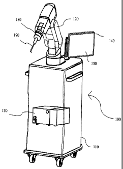
With reference to FIG. 1, it can be seen that a preferred embodiment of
the present invention generally includes a robotized device 100 comprising a
mobile base 110; a robot arm 120; a control unit 130 inside the mobile base,
that
controls the robot arm 120 and allows a surgeon to manually input data through
the use of an interface 150 that can be a touch screen, a mouse, a joystick, a
keyboard or the like; a display monitor 140; a tool 190 and a force sensor 180
mounted to the robot arm mounting flange; and specific fixation device 170 to
fix
the robotized device 100 to an operating table (not represented here).
Mobile base 110 ensures easy handling of the robotized device 100 with
its wheels and handles. Mobile base 110 is also preferably provided with
immobilization pads or equivalent.
Robot arm 120 is a six joint arm. Each joint is provided with an encoder
which measures its angular value. These data, combined with the known
geometry of the six joints, allow to compute the position of the robot arm
mounting flange and the position of the tool mounted to the robot arm, either
a
pointing tool, a guiding tool or a pointing and guiding tool.
FIG. 2A illustrates a pointing tool 190. The pointing tool 190 comprises a
base plate 200; a handle 210; and a pointing sphere 220.
CA 02570336 2006-12-14
WO 2005/122916 PCT/EP2005/052751
8
FIG. 2B illustrates a cutting guide. The cutting guide comprises a base
plate 230; a handle 240 and a slit 250 to guide a saw blade.
FIG. 2C illustrates a pointing and guiding tool. It comprises a base plate
260; a handle 270; a slit 280 to guide a saw blade and a pointing sphere 290.
The tools described in FIGS. 2A to 2C are just three examples of pointing
and/or guiding tools that may be utilized with the device shown in FIG. 1.
Preferably, robot arm 120 is rigidly attached to the operating table by a
specific base fixation device. As shown in FIG. 3, a base fixation device
includes
two sets of clamps 300 adapted to the operating table rail 310 and U-shape
bars
320. Initially, the user installs one clamp 300 on the operating table rail
310 and
another clamp on the mobile base rail 330. When clamps are in place, the user
inserts the U-shape bar in the cylindrical holes of the clamps, locks the
clamps in
place and locks the U-shape bar inside the clamps using the knobs.
In a preferred embodiment of the invention, the system comprises a limb
fixation device (see FIGS. 4A, 4B, 4C and 4D) to ensure the immobility of the
leg
during the procedure. This limb fixation device allows an immobilization of
leg at
two levels: at the level of the ankle with a toothed rack (FIG. 4D); at the
level of
the knee with two pins screwed on femo.ral or tibial epiphysis (FIG. 4C).
FIG. 4B shows the main plate 400 of the limb fixation device. Main plate
400 is fixed on the operating table with two clamps 300. The knee fixation
part
410 and the ankle fixation part 420 can slide along the main plate 400 and be
locked in place by screws.
FIG. 4C is a front view of the means of immobilizing patient's leg at the
level of the knee. Knee rests on the support bar 440. As bones are exposed in
a
knee replacement surgery, two pins 430 are screwed either in the femoral
epiphysis or in the tibial epiphysis. The position of the support bar 440 can
be
adjusted vertically and locked with two knobs. The orientation can be adjusted
from 0 to 90 by rotating around the main axis 450 and locked with one knob.
The whole system can slide along the plate.
FIG. 4D illustrates the means of immobilizing patient's leg at the level of
the ankle. Patient foot and ankle are rigidly fixed with surgical tape or
other
sterile means to lock the foot in the boot 460. The boot 460 is adapted to be
CA 02570336 2006-12-14
WO 2005/122916 PCT/EP2005/052751
9
clamped in a carriage 470 that can slide along the main plate 400 and be
locked
in place with a knob.
Both parts of the limb fixation device (ankle part and knee part) are
independent but are used in combination to guarantee immobilization of the
lower limb during the procedure.
In a preferred embodiment of the invention, control unit 130 can set the
robot arm 120 in a cooperative mode in which a user is able to move the robot
arm 120 manually by grabbing it by its final part. With reference to FIG. 5,
the
system of the present invention comprises a force sensor 180 mounted to the
robot arm mounting flange 125. Force sensor 180 is adapted to receive a tool
like the pointing tool 190. When the user grabs the tool and tries to move it
in a
direction, the control unit 130 receives efforts measured by the force sensor
180
and combines them with the position of the robot arm 120 to generate the
movement desired by the user.
Once the robotized device has been fixed to the operating table, the first
step of the procedure is collecting anatomical landmarks on the patient. These
anatomical landmarks are known by the surgeon. For example, in a TKR
procedure, the malleoluses, the internal part of tibial tuberosity, the middle
of the
spines and the tibial plateaus are collected on the tibia; the notch middle
point,
the distal and posterior condyles and the anterior cortex are collected on the
femur. FIG. 6 illustrates positions of the patient and of the robotized device
100
at the beginning of the landmarks collection step for a TKR procedure.
During the landmarks collection step, the control unit 130 sets the robot
arm 120 in cooperative mode and indicates through the display monitor 140 the
anatomical landmarks to acquire. The surgeon moves the pointing tool 190 until
being in contact with the required anatomical landmark and validates the
acquisition of the point coordinates using the user interface 150. The control
unit
130 then memorizes the coordinates of the point and its anatomical
significance.
After the landmarks collection step, the surgeon inputs planning
parameters through the user interface 150. For example, in a TKR procedure,
the surgeon chooses the model and the size of the prosthesis components and
defines their positions and orientations relative to the mechanical axes of
the
CA 02570336 2006-12-14
WO 2005/122916 PCT/EP2005/052751
femur and the tibia. Typical geometric parameters are varus/valgus angle,
posterior slope and thickness of resection for the tibia and varus/valgus
angle,
flexion/extension angle, external rotation and thickness of resection for the
femur.
5 In another embodiment of the invention, control unit 130 comprises a
data-processing interface that enables the system to be connected with another
computer-assisted surgical system, like a navigation system. Navigation
systems
work with preoperative images of the bone (CT scan, X-ray, fluoroscopy, etc)
or
with intra-operative data. In the latter case, they use a 3D reconstruction
10 algorithm based on the digitalization of the bone. Data provided by the
navigation
system then replaces, or is combined with the landmarks collection step data.
Position of the guiding tool may be generated by the navigation system and
transmitted to the robotized device in accordance with a predefined
communication protocol.
Once the required position of the guide has been generated, the user
mounts the guiding tool to the robot arm. Preferably, a pointing and guiding
tool
is used, so that the user does not need to change the tool between the
landmarks collection step. and the cutting or drilling step.
The robotized device 100 accurately aligns the guide relative to patient's
anatomy, in accordance with surgeon's planning. If the guiding tool is a
cutting
guide for a saw blade, the robot arm 120 holds it in the chosen cutting plane.
If
the guiding tool is a drilling guide, the robot arm 120 holds it along the
chosen
drilling axis.
In a preferred embodiment of the invention, planar cooperative mode can
then be activated by the user to restrict movements of the guide in the plane.
Similarly, axial cooperative mode restricts movements of the guide along the
axis. The user moves the guiding tool to what he/she estimates is the optimal
position, as the control unit 130 restricts movements of the robot arm to a
plane
or an axis. Once this optimal position reached, the control unit 130 stops the
robot arm 120 that holds the guiding tool in place. Surgical task like bone
cutting
or drilling is carried out by the surgeon using a conventional instrument
(oscillating saw or surgical drill) through the guide.
CA 02570336 2006-12-14
WO 2005/122916 PCT/EP2005/052751
11
In a TKR procedure, the same guiding tool is used for the tibial cut and
the five femoral cuts. In a tibial osteotomy procedure, the same guiding tool
is
used for both tibial cuts.
With reference to FIG. 7, control unit 130 runs a control software 132, that
exchanges data with elements of the robotized device. Software communicates
with the user through the user interface 150 and the display monitor 140.
Software communicates with another computer-assisted surgical system as
described above through the data-processing interface. Software communicates
with the force sensor 180 to regularly measure the efforts exerted by the user
at
the tool mounted to the robot arm. Software communicates with the robot arm
120 to control its position.
Control software 132 comprises five independent modules 134 to 138.
Preferably, these modules run simultaneously under a real time environment and
use a shared memory to ensure a good management of the various tasks of the
control software. Modules have different priorities, safety module 134 having
the
highest.
Safety module 134 monitors the system status and stops the robot arm
.,_120 when a critical situation is detected (emergency, stop, software
failure,
collision with an obstacle, etc).
Interface module 135 manages the communication between the surgeon
and the control software through the user interface 150 and the display screen
140. Display screen 140 displays a graphical interface that guides the user
through the different steps of the procedure. User interface 150 enables the
user
to have permanent control during the procedure (validating landmarks
collection,
defining planning parameters, stopping the robot arm if needed, etc).
Force module 136 monitors the forces and torques measured by the force
sensor 180. Force module is able to detect a collision with an obstacle and
alert
the safety module.
Control module 137 manages the communication with the robot arm 120.
It receives data encoder values of each joint and sends position commands.
Calculations module 138 does all the calculations necessary for the
procedure. For example, in a TKR procedure, it reconstructs the mechanical
CA 02570336 2012-04-13
12
axes of the bones combining anatomical landmarks data and statistical data. It
also defines the trajectory of the robot arm 120 using direct and inverse
kinematics.
Present invention is not limited by what has been described above. It will
be appreciated that various changes can be made therein without departing from
the scope of the invention.