Note: Descriptions are shown in the official language in which they were submitted.
CA 02570539 2006-11-30
WO 2006/009887 PCT/US2005/021557
EVALUATION OF A TREATMENT TO DECREASE
THE RISK OF A PROGRESSIVE BRAIN DISORDER
OR TO SLOW BRAIN AGING
CROSS REFERENCES TO RELATED APPLICATIONS
This application claims priority to US Provisional Application Serial No.
60/580,762,
filed on June 18, 2004, titled "Method For Evaluating The Efficacy Of Putative
Primary And
Secondary Prevention Therapies In Cognitively Normai Persons At Risk For Brain
Disorders",
which is incorporated herein by reference.
FIELD OF INVENTION
This invention relates to brain disorders and treatments for brain disorders,
and is more
particularly related to strategies for evaluating the efficacy of treatments
for neurological,
psychiatric, and related disorders.
BACKGROUND
The present invention relates generally to methods that utilize imaging
techniques to
measure the activity and/or structural changes in the human brain to determine
the efficacy of
putative treaiments for brain-related disorders. More particularly, the
present invention relates to
methods to utilize structural or functional imaging techniques such as PET,
SPECT, MRI, or
amyloid imaging, as well as other measurements of change over time as
surrogate markers to
predict efficacy of putative treatments in improving clinical outcome in
persons susceptible to
Alzheimer's Dementia (AD), Mild Cognitive Impairment (MCI), or other
progressive brain
disorders and to evaluate the efficacy of putative treatment to slow age-
related changes in the
brain.
To facilitate indexing to references, square brackets below may indicate
reference numbers
in the section preceding the claims. No admission is being made by the
applicant as to the
pertinence of any of the listed references. A presentation is attached
following the claims and
comprises part of this disclosure.
Brain Disorders And Surrogate Markers
Brain disorders such as Alzheimer's dementia (AD) constitute a rapidly growing
public
health problem. Clinically, AD is characterized by a gradual and progressive
decline in memory
and other cognitive functions, including language skills, the recognition of
faces and objects, the
perfonnance of routine tasks, and executive functions, and it is frequently
associated with other
distressing and disabling behavioral problems [1-3]. Histopathological
features of AD include
1
CA 02570539 2006-11-30
WO 2006/009887 PCT/US2005/021557
neuritic and diffuse plaques (in which the major constituent is the A-amyloid
protein),
neurofibrillary tangles (in which the major constituent is the
hyperphosphorylated form of the
microtubule-associated protein tau), and the loss of neurons and synapses [4].
In addition to its
effects on patients, AD places a terrible burden on the family; indeed, about
half of the affected
persons' primary caregivers become clinically depressed [5]. According to one
community survey,
AD afflicts about 10% of those over the age of 65 and almost half of those
over the age of 85 [6].
As the population grows older, the prevalence and cost of AD is expected to
increase dramatically
[7]. For example, by 2050 the prevalence of AD in the United States has been
projected to
quadruple (from about 4 to 16 million cases, even without assuming an increase
in an affected
person's life expectancy) and the cost of caring for patients will quadruple
(from about 190 to 750
million dollars per year, even without any adjustment for inflation) [8]. An
AD prevention therapy
is urgently needed to avert an overwhelming public health problem.
Scientific progress has raised the hope of identifying treatments to halt the
progression and
prevent the onset of AD [9]. This progress includes the discovery of genetic
mutations and at least
one susceptibility gene that account for many cases of AD; the
characterization of other AD risk
factors and pathogenic molecular events that could be targeted by potential
treatments; the
development and use of improved research methods (e.g., in the fields of
genomics and
proteomics) for the identification of new therapeutic targets; the development
of promising animal
models, including transgenic mice containing one or more AD genes, which may
help clarify
disease mechanisms and screen candidate treatments; suggestive evidence that
several available
interventions (e.g., estrogen-replacement therapy, anti-inflammatory
medications, statins {e.g.
HNIG CoA Reductase inhibitors such as Crestor , Lipitor or Pravachol },
vitamin E, folic acid,
and gingko biloba), which might be associated with a lower risk and later
onset of AD; the
discovery of medications which at least modestly attenuate AD symptoms (e.g.,
several
acetyleholinesterase inhibitors and the N-methyl-D-aspartate [N1VIDA]
inhibitor memantine); and
the development of other potentially disease-modifying investigational
treatments (e.g.,
histopathological immunization therapies, drugs which inhibit the production,
aggregation, and
neurotoxic sequelae of A[i, drugs which inhibit the hyperphosphorylation of
tau, and drugs which
protect neurons against oxidative, inflammatory, excitatory, and other
potentially toxic events).
Even if a prevention therapy is only modestly helpfui, it could provide an
extraordinary
public health benefit. For instance, a therapy that delays the mean onset of
AD by only five years
might reduce the number of cases by half [10]. Unfortunately, it would require
thousands of
volunteers, many years, and great expense to determine whether or when
cognitively normal
persons treated with a candidate primary prevention therapy develop cognitive
impairment and
2
CA 02570539 2006-11-30
WO 2006/009887 PCT/US2005/021557
AD. One way to reduce the samples and time required to assess the efficacy of
an AD prevention
therapy is to conduct a clinical trial in patients with mild cognitive
impairment (MCI), who may
have a 10-15% rate of conversion to probable AD and commonly have
histopathological features
of AD at autopsy [11,12]. Randomized, placebo-controlled clinical trials in
patients with MCI
could thus help establish the efficacy of putative "secondary prevention"
therapies. Using clinical
outcome measures, the only practical way to establish the efficacy of
a"primary prevention"
therapy has been to restrict the randomized, placebo-controlled study to
subjects in advanced age
groups--a strategy which still requires extremely large samples, a study
duration of several years,
and significant cost. While these strategies are likely to play significant
roles in the identification of
effective prevention therapies, it rema.ins possible that subjects will
require treatment at a younger
age or at an even earlier stage of underlying disease for a candidate
prevention therapy to exert its
most beneficial effects. Those of skill in the art recognize the value of
developing putative primary
prevention therapies, and they are placing an increasing emphasis on the
earliest possible detection
of the brain changes associated with the predisposition to this disorder. A
new paradigm is needed
to reduce the subject samples, time, and cost required to establish the
efficacy of putative primary
prevention therapies, encourage industry and government agencies to sponsor
the required trials,
and prevent this growing problem without losing a generation along the way.
What is further
needed is a means to evaluate putative treatment modalities on additional
brain disorders other than
AD, including, but not limited to mild cognitive impairment (MCI) or decline
in cognitive ability
due to other age-related atrophy or other disorders.
Researchers have been using 18F-fluorodeoxyglucose (FDG) positron emission
tomography
(PET) and magnetic resonance imaging (MIZI) to detect and track changes in
brain fiuiction and
structure which precede the onset of brain disorder symptoms in cognitively
normal persons who
are at risk for developing brain disorders such as Alzheimer's. Suggested risk
factors for AD
include older age, female gender, lower educational level, a history of head
trauma, cardiovascular
disease, higher cholesterol and homocysteine levels, lower serum folate
levels, a reported family
history of AD; trisomy 21 (Down's syndrome), at leastl2 missense mutations of
the amyloid
precursor peptide (APP) gene on chromosome 21, at least 92 missense mutations
of the presenilin
1(PS1) gene on chromosome 14, at least 8 missense mutations of the presenilin
2 (PS2) gene on
chromosome 1, candidate susceptibility loci on chromosomes 10 and 12, and the
APOE s4 allele
on chromosome 19 [9,13,14]. Next to age, the APOE s4 allele is the best-
established risk factor
for late-onset AD and, thus, it is especially relevant to human brain imaging
studies. The APOE
gene has three major alleles, E2, s3, and s4 [22]. In comparison with the s3
allele (the most
3
CA 02570539 2006-11-30
WO 2006/009887 PCT/US2005/021557
common variant), the s4 allele is associated with a higher risk of AD and a
younger age at
dementia onset, whereas the ~2 allele may be associated with a lower risk of
AD and an older age
at dementia onset [15-18,23]. In one of the original case-control studies,
individuals with no
copies of the E4 allele had a 20% risk of AD and a median age of 84 at
dementia onset; those with
one copy of the E4, which is found in about 24% of the population [22], had a
47% risk of AD and
a median age of 76 at dementia onset; and those with two copies of the s4
allele (the s4/s4
genotype, found in 2-3% of the population [22]) had a 91% risk of AD by 80
years and a mean age
of 68 at dementia onset [17]. In another study, 100% of s4 carriers with
cognitive loss had neuritic
plaques at autopsy [24]. In a related study, 23% of their AD cases were
attributed to absence of the
E2 allele and another 65% of their cases were attributed to the presence of
one or more copies of
the s4 allele [23]. Case-control studies in numerous clinical,
neuropathological, and community
studies have confirmed the association between the s4 allele and AD. Farrer et
al conducted a
worldwide meta-analysis of data from 5930 patients with probable or autopsy-
confirmed AD and
8607 controls from various ethnic and racial backgrounds [18]. In comparison
with persons with
the genotype 0/0, the risk of AD was significantly increased in genotypes
s2/s4 (odds ratio
[ORI=2.6), E3/s4 (OR=3.2), and E4/s4 (OR=14.9), and the risk of AD was
significantly decreased
in genotypes s2/0 (OR=0.6), and s2/s2 (OR=0.6). Community-based, prospective
studies promise
to better characterize the absolute risk of AD in persons with each APOE
genotype.
Some imaging research has focused on demonstrating that baseline reductions in
structural
or functional performance with a single imaging measurement, predict
subsequent clinical
decline in patients with dementia, and that baseline measurements in MCI
predict higher rate of
conversion to AD. However, these findings are insufficient to demonstrate that
the selected
brain imaging technique is an adequate surrogate marker for demonstrating
prevention of or
delayed onset of a disease state. More specifically, the measurement protocols
must be able to
show that the surrogate marker correlates with clinical severity in patients,
and when a change in
measurements is attributable to administration of a treatment regimen, it also
predicts an
improvement in clinical outcome. Prior single baseline imaging techniques are
insufficient in
this regard.
Linlung Functional and Structural Brain Images
Neuroimaging researchers frequently acquire a combination of functional (e.g.,
positron
emission tomography [PET] or functional magnetic resonance imaging [flVIRI])
and structural
(e.g., volumetric MRI) brain images. The structural MRI data is usually used
in PET/flVIRI
4
CA 02570539 2006-11-30
WO 2006/009887 PCT/US2005/021557
studies for anatomical localization of functional alterations, definition of
regions of interest for
the co-registered PET/fMRI data extraction, and partial volume correction
(Ibanez et al. 1998).
While neuroimages have been most commonly analyzed using univariate methods,
multivariate analyses have also been used to characterize inter-regional
correlations in brain
imaging studies. Multivariate algorithms have included principal component
analysis (PCA)
(Friston 1994), the PCA-based Scaled Subprofile Model (SSM) (Moeller et al.
1987; Alexander
& Moeller 1994), and the Partial Least Squares (PLS) method (McIntosh et al.
1996). These
methods have typically been used to characterize regional networks of brain
function (and more
recently brain anatomy) and to test their relation to measures of behavior.
Such multivariate
methods, however, have not yet been used to identify patterns of regional
covariance between
functional and structural brain imaging datasets.
A major challenge to the multivariate analysis of regional covariance with
multiple
imaging modalities is the extremely high dimensionality of the data matrix
created by including
relatively high-resolution neuroimaging datasets. What is needed is a strategy
to make
computation dimensional datasets with covariance analysis using multivariate
methods feasible.
DISCLOSURE OF THE INVENTION
In view of the foregoing, it is an object of the present invention to improve
various
problems associated with the prior art. To this end, an object of the
invention is to provide a
method to evaluate putative therapies to improve clinical outcomes in patients
at risk for brain-
related disorders. It is to be understood that the following description is
exemplary and
explanatory only and is not restrictive of the invention, as claimed. Thus,
the present invention
comprises a combination of features, steps, and advantages that enable it to
overcome various
deficiencies of the prior art. The various characteristics described, as well
as other features, will
be readily apparent to those skilled in the art upon reading the following
detailed description of
the preferred embodiments of the invention, and by referring to the
accompanying drawings.
Longitudinal brain imaging studies have been conducted with E4 homozygotes, E4
heterozygotes (all with the E3/s4 genotype), and s4 non-carriers who were
initially late middle-
aged (i.e., younger than the suggested median onset of AD), cognitively
normal, and individually
matched for their gender, age, and educational level. Since individuals with
the 64/s4 genotype
have an especially high risk of AD, the study of this subject group is
intended to optimize the
power to characterize the brain and behavioral changes which precede the onset
of cognitive
impairment and eventually relate these changes to the subsequent onset of MCI
and AD. Since
individuals with the s3/E4 genotype have an increased risk of AD and comprise
about 20-23% of
the population [22], the study of this subject group extends the findings to a
larger segment of the
5
CA 02570539 2006-11-30
WO 2006/009887 PCT/US2005/021557
population and increases the number of individuals who would be eligible to
participate in future
clinical trials of putative primary prevention therapies. The study of 0
noncarriers who are
individually matched for gender, age, and educational level could optimize the
power to
characterize the brain and behavioral changes associated with normal aging and
permit us to
distinguish them from those age-related changes preferentially related to the
presence of the s4
allele and the subsequent onset of AD. As other risk factors are confirmed, it
should be possible to
extend the brain imaging paradigm of the present invention to the study of
cognitively normal
persons who are at differential risk for AD independent of (and in conjunction
with) their APOE
genotype.
PET In The Study Of AD
FDG PET, which provides measurements of the cerebral metabolic rate for
glucose
(CMRg1), is the most extensively used functional brain imaging technique in
the study, early
detection, and tracking of AD. FDG PET reveals characteristic abnormalities in
patients with AD,
including abnormally low posterior cingulate, parietal, and temporal CMRgI,
abnormally low
prefrontal and whole brain CMRgI in more severely affected patients, and a
progressive decline in
these and other measurements over time [25-39]. These abnormalities, which are
correlated with
dementia severity and predict subsequent clinical decline and the
histopathological diagnosis of
AD [28-31,33-35,37,38], could be related to a reduction in the activity or
density of terminal
neuronal fields or perisynaptic glial cells that innervate these regions [40-
42], a metabolic
dysfunction [42-44], or a combination of these factors. They do not appear to
be solely attributable
to the combined effects of atrophy and partial-volume averaging [36].
Brain abnormalities can be detected prior to the onset of dementia [8,9,44-
46]. In
comparison with the 0 noncarriers, the E4 homozygotes and heterozygotes each
have abnormally
low CMRgI in the same brain regions as patients with probable AD [9,46].
Despite no significant
differences in clinical ratings or neuropsychological test scores and no
significant interactions
between these measurements and time, the s4 heterozygotes have significantly
higher 2-year rates
of CMRgI decline [8]. Based on these data, we estimated the power of PET to
test the efficacy of
candidate prevention therapies to attenuate this decline in 2 years [8]. In
complementary PET
studies of non-demented 0 carriers and noncarriers, who were about 10 years
older, had memory
concerns, and had slightly lower MMSE scores; furthermore, lower CMRgI
measurements in the
posterior cingulate and parietal cortex were correlated with a subsequent
decline in memory
[45,47]. While it remains possible that the CMRgI abnormalities reflect
aspects of the 0 allele
unrelated to AD, PET studies suggest that these abnormalities are related to
the development of
6
CA 02570539 2006-11-30
WO 2006/009887 PCT/US2005/021557
this disorder. While there may be a few differences [48,49], patients with
probable AD appear to
have a similar pattern of reductions in regional CMRgI whether or not they
have the s4 allele
[50,51]; and, as previously noted, the CMRgI abnormalities in patients with
probable AD predict
the subsequent progression of dementia and the histopathological diagnosis of
AD [37,38], are
progressive [28-31,39], and are correlated with dementia severity [34].
Other promising PET radiotracer techniques have been developed for the study
of AD.
[11C] methylpiperidinyl propionate (PMP) PET provides estimates of
acetylcholinesterase activity
and has been used to detect deficits in patients with probable AD; this
radiotracer method could be
used to evaluate the extent of central inhibition by established or
investigational
acetylcholinesterase inhibitors and help optimize dosage schedules [52].
[11C](R)-PK11195 PET
provides estimates of peripheral benzodiazepine receptor binding, a putative
marker of
neuroinflammation; it has been used to detect abnormally increased
measurements and herald the
subsequent onset of atrophy in patients with probable AD, and it could be used
to track the course
of neuroinflammation in AD and characterize the central anti-inflammatory
effects of medications
[53]. Researchers have recently developed promising PET radiotracer methods
for the assessment
of AD histopathology [54,55]. Additional research is needed to further
evaluate these methods,
identify the most suitable radioligands and tracer-kinetic models, and use
them to characterize,
compare, and track measurements in patients with AD and normal controls.)
MRI In The Study Of AD
Volumetric MRi studies reveal abnormally high rates of brain atrophy in
patients with
probable AD, including progressive reductions in the volume of the
hippocampus, entorhinal
cortex, and whole brain and progressive enlargement of the ventricles and
sulci [56-85].
Embodiments of the MRI embodiment of the present invention comprise Tl-
weighted volumetric
MRI measurements of hippocampal, entorhinal cortex, and whole brain volume and
are used to
provide structural brain imaging measurements in the early detection and
tracking of AD; they
have roles in the assessment of candidate treatments to modify disease
progression. MRI studies
find significantly smaller hippocampal volumes in patients with probable AD
[56-73] and non-
demented persons at risk for AD[86-97], correlations between reduced
hippocampal volume and
the severity of cognitive impairment [60,64,65], and progressive declines in
hippocampal volume
during the course of the illness [61,77,92]. Methods for the reliable
characterization of entorhinal
cortex volume have recently been developed and used in the early detection and
tracking of MCI
and AD [68,73-76,79,80,92].
Fox et al. have developed a semi-automated method for the measurement of whole
brain
atrophy in individual human subjects following the coregistration and digital
subtraction (DS) of
7
CA 02570539 2006-11-30
WO 2006/009887 PCT/US2005/021557
MRI's [81-84]. They found significantly higher rates of whole brain atrophy in
patients with
probable AD than those associated with normal aging [81-84], as well as
significantly higher rates
of whole brain atrophy shortly before the onset of dementia in persons at risk
for AD [96,97], and
they have estimated the statistical power of this method to test the efficacy
of candidate treatments
to attenuate these atrophy rates [84]. We have recently developed and tested a
fully automated
algorithm for the measurement of brain atrophy from sequential MRI's using an
iterative principal
component analysis (IPCA), have applied it the study of patients with AD, our
cognitively normal
APOE c4 homozygotes, heterozygotes, and noncarriers, and transgenic mice [98-
102]. Other
embodiments for the analysis of volumetric MRI's include but are not limited
to the use of "voxel-
based morphometry (VBM) to create probabilistic brain maps to compute regional
alterations in
gray matter or white matter [103-106]; and the use of non-linear warping
algorithms to characterize
alterations in the size and shape of the hippocampus [107], multiple brain
regions [85], variations
in gyral and sulcal patterns [108], and reductions in gray matter [108,1091.
PET And MRI In The Evaluation Of Putative AD Treatments
Following Temple's commonly cited definition [110], "A surrogate endpoint of a
clinical
trial is a laboratory measurement or a physical sign used as a substitute for
a clinically meaningful
endpoint that measures directly how a patient feels, functions, or survives.
Changes induced by a
therapy on a surrogate endpoint are expected to reflect changes in a
clinically meaningful
endpoint." According to Fleming and DeMets [111], a valid surrogate endpoint
is not just a
correlate of the clinical outcome; rather, it should reliably and meaningfully
predict the clinical
outcome and it should fully capture the effects of the intervention on this
outcome. Citing several
examples, they note several ways in which an otherwise promising surrogate
endpoint might fail to
provide an adequate substitute for a clinical endpoint. Although few if any
surrogate endpoints
have been rigorously validated, the 1997 United States "FDA Modernization Act"
authorizes the
approval of drugs for the treatment of serious and life-threatening illnesses,
including AD, based
on its effect on an unvalidated surrogate [112]. In order to promote the study
and expedite the
approval of drugs for the txeatment of these disorders, "fast track" approval"
may be granted if the
drug has an effect on a surrogate marker that is "reasonably likely" to
predict a clinical benefit; in
this case, the drug sponsor may be required to conduct appropriate post-
marketing studies to verify
the drug's clinical benefit and validate the surrogate endpoint [112].
FDG PET measurements of posterior cingulate, parietal, temporal, and
prefrontal CMRgl
and volumetric MRI measurements of hippocampal, entorhinal cortex, and whole
brain volume are
established surrogate markers for the assessment of putative drugs in the
treatment of AD. These
surrogate endpoints are not rigorously validated, partly because validation
may actually require
8
CA 02570539 2006-11-30
WO 2006/009887 PCT/1JS2005/021557
demonstration of these endpoints to account for the predicted clinical effect
using several
established disease-modifying treatments. Still, these brain imaging
measurements are "reasonably
likely" to predict a drug's clinical benefit in the treatment of AD. They have
much greater
statistical power than traditional outcome measures [39], reducing the
potential cost of proof-of-
concept studies. They are "reasonably likely" to determine a drug's disease-
modifying effects,
helping to distinguish a drug's disease-modifying from symptomatic effects. As
discussed below,
these brain-imaging measurements may permit the efficient discovery of
prevention therapies in
non-demented persons at risk for AD [8,84], and they may assist in the pre-
clinical screening of
candidate treatments in transgenic mice and other putative animal models of AD
[102,103,133].
For all of these reasons, FDG PET and volumetric MRI have important and
emerging roles in the
evaluation of putative disease-modifying candidate drugs in the treatment and
prevention of AD.
When using FDG PET in a clinical trial of a putative drug for the treatment or
prevention
of AD, we recommend (a) the use of a state-of-the-art imaging system with an
axial field-of-view
that covers the entire brain; (b) data acquisition in the three-dimensional
mode, thus permitting the
use of lower radiation doses, (c) the use of a non-invasive, image-derived
input function, thus
permitting the computation of quantitative measurements (in case CMRgI
reductions are so
extensive that they affect measurements in the whole brain or relatively
spared regions, like the
pons, that would otherwise be used to normalize images for the variation in
absolute
measurements); (d) data acquisition in the "resting state" (e.g., eyes closed
and directed forward)
rather than during the performance of a behavioral task (since the resting
state has been used most
extensively to track the progression of CMRgI changes in patients with AD and
non-demented
persons at risk for the disorder and since any effects of a drug on task
performance could confound
interpretations about the drug's putative disease-modifying effects); (e) the
use of an automated
brain mapping algorithm to characterize and compare regional CMRgI declines in
the active
treatment and placebo treatment arms (to date, SPM99 has been the most
extensively used
algorithm for tracking CMRgI declines in patients with AD and non-demented
patients at risk for
the disorder; (f) quality assurance procedures to maximize the quality and
standardization of
image-acquisition and image-analysis procedures at different sites; and (g) a
single site for the
technical coordination and the centralized storage and analysis of data in
multi-center studies.
In the design of clinical imaging trials using FDG PET (and volumetric MRI),
we
recommend (a) efforts to control or account for potentially confounding
effects, such as medication
effects (e.g., stratifying samples for use of an approved medication,
discouraging the introduction
of new medications during the trial, and minimizing or accounting for the use
of medications prior
to the PET session) and changes in depression ratings; (b) the use of
baseline, early, and end-of-
9
CA 02570539 2006-11-30
WO 2006/009887 PCT/US2005/021557
treatment scans (performance of the early scan after a drug's steady state and
relevant
pharmacodynamic effects would help characterize and contrast a medication's
state-dependent
effects on local neuronal activity or glucose metabolism and its disease-
modifying effects; and (c)
the use of additional scans as indicated (e.g., to evaluate the time course of
an effect, increase
statistical power, or incorporate a randomized start or withdrawal design).
(d) Although not
required, a randomized start or withdrawal design [112] could be used to
further support a drug's
disease-modifying effects. In a randomized start design, patients initially
randomized to the
placebo arm and treated for an appropriate time are then re-randomized to
active medication or
placebo; a disease-modifying effect would be inferred if the change in the
surrogate endpoint
between the beginning and end of the study is significantly smaller in the
patients initially
randomized to the active treatment arm (i.e., treated longer) than those
subsequently randomized to
the active treatment arm. In a randomized withdrawal design, patients
initially randomized to the
active treatment arm and treated for an appropriate time are then re-
randomized to active
medication or placebo; a disease-modifying effect would be inferred if the
change in the surrogate
endpoint is significantly smaller in the patients who were initially
randomized to the active
treatment arm and subsequently randomized to placebo than those who were
treated with placebo
throughout the study. Practically, a randomized start design may be preferred
since it may be
difficult to justify drug discontinuation in those who believe that the
medication has been helpful.
(e) Even if the data is not necessary for accelerated drug approval, we
strongly recommend efforts
to relate a drug's short-term effects on surrogate endpoint (e.g., 6-month
effects in patients with
probable AD or 12-months effects in patients with MCI) to their subsequent
clinical course (e.g.,
subsequent clinical decline in patients with probable AD or 3-year conversion
rate to probable AD
in patients with MCI}--information that will help validate the use of these
surrogate markers (and
support the use of shorter study intervals) for candidate drug and others to
be studied in the future.
(f) We strongly encourage the combined use of FDG PET and volumetric MRI in
the study of a
candidate treatment. Using an individual brain imaging technique, there is a
small possibility that a
drug's effect on a surrogate endpoint might be unrelated to a disease-
modifying effect (e.g., an
increase in neuronal activity or brain swelling) or that a drug's effect on a
surrogate end-point
might actually mask its disease-modifying effect (e.g., a contraction in brain
size due to a drug's
osmotic or perhaps even plaque-clearing effects). The combined used of
complementary imaging
techniques would provide converging evidence in support of a drug's disease-
modifying effects. It
would further reduce the small possibility that the drug's effect on an
individual surrogate endpoint
is unrelated to its effect on disease progression (an advantage in seelcing
approval for a drug's
disease-modifying effect). It would minimize the chance that a drug effect on
one of the surrogate
CA 02570539 2006-11-30
WO 2006/009887 PCT/US2005/021557
endpoints would mask its disease-modifying effects (an advantage in proof-of-
concept studies).
Embedding both of the these imaging modalities in clinical trials would
maximize the chance of
validating one or both surrogate endpoints and help support their role in the
efficient discovery of
primary prevention therapies. We believe that these advantages far outweigh
the additional costs
and note that both of these imaging modalities are now widely available. (g)
Finally, we wish to
encourage the application of these imaging techniques to the study of
cognitively nonnal APOE E4
carriers in primary prevention trials. In order to conduct primary prevention
trials in these subjects,
researchers and ethicists may consider two ways to address the risk of
providing genetic
information to cognitively normal research participants: withholding
information from subjects
about their genetic risk with their prior informed consent and 'uicluding
persons with and without a
genetic risk for AD (as we have been done in our naturalistic studies) or (b)
counseling potential
research subjects about the uncertainties and risks involved in receiving
information about their
genetic status, obtaining their informed consent to receive this information,
and restricting the
study to persons at genetic risk for the disorder.
PET In The Study Of Cognitively Normal APOE E4 Carriers And Noncarriers
In order to study cognitively nornmal persons at differential genetic risk for
AD, we have
used newspaper ads to recruit persons who denied any memory concerns and were
medically well.
The subjects agreed that they would not receive any information about their
APOE genotype (since
this information cannot be used to predict with certainty whether or when a
person will develop
AD) and provided their informed consent. Blood samples were then drawn and
APOE genotypes
characterized. For each APOE 0 carriers who agreed to participate in our
imaging trials, one 0
noncarrier was matched for his or her gender, age (within 3 years), and
educational level (within 2
years). The subjects had quantitative FDG PET measurements of CMRgI as they
rested quietly
with their eyes closed, a volumetric TI-weighted MRI, a clinical examination,
structured
psychiatric interview, and depression rating scale, the Folstein Mini-Mental
State Examination
(MMSE), and batteries of neuropsychological tests and psycholinguistic tasks.
In our ongoing
longitudinal study, we have begun to acquire these data every 2 years in 160
cognitively normal
individually matched 0 homozygotes, heterozygotes, and noncarriers 47-68 years
of age with a
reported first-degree family history of probable AD. In other studies, we have
begun to
characterize and compare these measurements in cognitively normal e4 carriers
and noncarriers
20-80 years of age irrespective of their reported family history or probable
AD.
11
CA 02570539 2006-11-30
WO 2006/009887 PCT/1JS2005/021557
Baseline Measurements
We originally sought to test the hypothesis that cognitively normal, late
middle-aged APOE
s4 homozygotes, at a particularly high risk of AD, have abnormally low PET
measurements in the
same brain regions as patients with probable AD [46]. APOE genotypes were
characterized in
cognitively normal persons 50-65 years of age with a reported first-degree
family history of
probable AD. For each of the 11 e4 homozygotes who agreed to participate in
our imaging study, 2
s4 noncarriers were matched for their gender, age (within 3 years), and
educational level (within 2
years. The E4 homozygotes had a mean age of 55 (range 50-62), a mean MMSE
score of 29.4
(range 28-30), and no significant differences from the controls in their
clinical ratings or
neuropsychological test scores. To characterize regions of the brain with
abnormally low CMRgI
in patients with probable AD, an automated was initially used to create a
three-dimensional
stereotactic surface projection statistical map comparing the data from 37
patients with probable
AD and 22 normal controls (mean age 64) provided by researchers at the
University of Michigan
[32,34]. As previously demonstrated, the patients with probable AD had
abnormally low CMRgI
bilaterally in posterior cingulate, parietal, temporal, and prefrontal cortex,
the largest of which was
in the posterior cingulate corte. To characterize regions of the brain with
reduced CMRgI in the
cognitively normal 0 homozygotes, the same brain mapping algorithm was used to
create a three-
dimensional surface projection statistical map comparing the data from our
homozygotes and non-
carriers; this map was then superimposed onto the map of CMRgI abnormalities
in the patients
with probable AD (Figure 1) [46]. As predicted, the 0 homozygotes had
abnormally low CMRgI
bilaterally in the same posterior cingulate, parietal, temporal, and
prefrontal regions as the patients
with probable AD (figure 1) [46]. The largest reduction was in the posterior
cingulate cortex,
which is pathologically affected in AD and might provide the earliest
metabolic indicator of the
predisposition to Alzheimer's dementia [32]. The c4 homozygotes also had
abnormally low
CMRgI bilaterally in additional prefrontal regions (figure 1), which PET, MRI,
and
neuropathological studies suggest are preferentially affected during normal
aging [46,114-118]-
and which have led us to postulate that the APOE s4 allele accelerates normal
aging processes
which are necessary but not sufficient for the development of AD [46].
We subsequently sought to detect abnormalities in cognitively normal APOE E4
heterozygotes [8,9], thus providing a foundation for using PET to efficiently
test the potential of
candidate primary prevention therapies in this large segment of the
population. Eleven cognitively
normal c4 heterozygotes (50-63 years of age, all with the 0/0 genotype) who
reported family
history of probable AD in a first-degree relative were matched to our original
group of s4
12
CA 02570539 2006-11-30
WO 20061009887 PCT/US2005/021557
homozygotes and non-carriers for gender, age, and educational level [9]. The
e4 heterozygotes had
perfect scores on the MMSE and no impairments in their neuropsychological test
scores. Using the
same brain-mapping algorithm employed in our original study, the s4
heterozygotes had
significantly reduced CMRgI bilaterally in the same regions of posterior
cingulate, parietal, and
temporal cortex as patients with probable AD (figure 2) [9]. Like the s4
homozygotes, the largest
CMRgI reduction was located in the posterior cingulate cortex. Unlike the c4
homozygotes, the s4
heterozygotes did not have significant reductions in additional prefrontal
regions, which we
postulate will be affected at an older age than that observed in the E4
homozygotes.
We have recently extended these findings to 160 cognitively normal persons in
this age
group (including 36 s4 homozygotes, 46 E4 heterozygotes, and 78 noncarriers,
who enrolled in our
longitudinal study and followed every two years [119]. As in our earlier
reports, the s4 carriers had
abnonnally low CMRgl in the posterior cingulate, parietal, temporal, and
prefrontal cortex, which
were not solely attributable to the combined effects of atrophy and partial
volume-averaging [119].
Lower CMRgI in each of these regions was significantly correlated with s4 gene
dose, which has
been related to a higher risk of AD and a lower mean age at the onset of
dementia [119].
We have also extended our findings to the comparison of 10 cognitively normal
s4
heterozygotes and 15 E4 noncarriers 20-39 years of age, who were recruited
irrespective of their
reported family history of AD [120, 121]. The 0 heterozygotes had abnormally
low CMRgI in the,
same regions of posterior cingulate, parietal, temporal, and prefrontal
cortex, raising new questions
about the earliest brain changes involved in the predisposition to AD, new
questions about how
these early changes are related to the histopathological and physiological
brain changes found at
older ages [120], and raising the possibility that brain processes associated
with the
preredisposition to AD might be targeted by prevention therapies at a
parCicularly young age and a
potentially tractable preclinical stage of disease vulnerability.
We have also begun to characterize and compare MRI measurements in our APOE E4
carriers and noncarriers. Using volumetric MRI's from the 11 E4 homozygotes
and 22 0 non-
carriers included in our original analysis of PET date, well characterized
hippocampal landmarks,
and a technique used extensively by Mony deLeon and his colleagues at New York
University
[85], we investigated the possibility that cognitively normal persons at risk
for AD have reductions
in hippocampal volume [94]. After normalizi.ng regional measurements for the
variation in
supratentorial intracranial volume, 'mean left and right hippocampal volumes
were about 8%
smaller in the s4 homozygotes, but did not reach statistical significance.
Consistent with other
MRI studies, smaller left and right hippocampal volumes in the 33 subjects
were each significantly
13
CA 02570539 2006-11-30
WO 2006/009887 PCT/US2005/021557
correlated with lower long-term recall scores. As predicted, posterior
cingulate CMRg1
measurements continued to distinguish s4 homozygotes from non-carriers after
adjusting for left
and right hippocampal volumes in a stepwise logistic regression model. In
contrast, neither left nor
right hippocampal volumes significantly improved the ability to distinguish
the e4 homozygotes
and noncarriers in a model already including posterior cingulate glucose
metabolism. Thus, using
the image-acquisition and image-analysis techniques employed in this study,
PET tended to be
more sensitive than MRI in identifying cognitively normal persons at risk for
AD. While larger
samples and longitudinal assessment are required to confum our conclusions, we
suggest that PET
measurements of posterior cingulate CMRg1 begin to decline prior to the onset
of memory decline
in persons at risk for AD, and that MRI measurements of hippocampai volume
begin to decline
some time later, in conjunction with the onset of memory decline and shortly
before the onset of
AD [94].
It remains possible that other brain regions, other image-analysis strategies,
and
longitudinal comparisons could be used to detect abnormalities in MRI
measurements of brain
volume in cognitively normal persons at genetic risk for AD. We recently used
VBM (with
procedures optimized to remove the influence of non-brain tissue) to
investigate regional
abnormalities in gray matter density in the 11 c4 homozygotes, 11 s4
heterozytotes, and 22
noncarriers included in our original PET studies. An automated algorithm was
used to transform
the MRI's into the coordinates of a standard brain atlas, correct the images
for inhomogeneities,
segment them for gray matter, smooth them, and create a statistical map of
significant differences
in gray matter intensity [104]. A significance threshold of 0.005, uncorrected
for multiple
comparisons, was used for hypothesized regional effects. In comparison with
the E4 noncarriers,
the E4 homozygotes had significantly lower gray matter densities in the
vicinity of the right
posterior cingulate cortex, a right peri-hippocampal region, and the left
parahippocampal and
lingual gyri; and the s4 heterozygotes had significantly lower gray matter
density in the vicinity of
the left parahippocampal gyrus, the anterior cingulate cortex, and the right
temporal cortex [104].
In comparison with the s4 heterozygotes, the s4 homozygotes had significantly
lower gray matter
density in the vicinity of the left parahippocampal and lingual gyri and in
bilateral regions of
parietal cortex [104]. Lower measurements of gray matter density in the left
parietal and left
parahippocampaUlingual areas were correlated with poorer memory scores in the
aggregate c4
carrier group [104]. Thus, cognitively normal s4 carriers appear to have
abnormally low gray
matter density in heteromodal association and paralimbic regions that are
preferentially affected
14
CA 02570539 2006-11-30
WO 2006/009887 PCT/US2005/021557
early in AD. If, as our preliminary findings suggest, reductions in gray
matter density are
progressive [105], they could help in the efficient evaluation of primary
prevention therapies.
Longitudinal Changes
In our first longitudinal comparison, we characterized and compared 2-year
CMRgI
declines in 10 cognitively normal s4 heterozygotes and 15 s4 non-carriers 50-
63 years of age with
a reported first-degree family history of probable AD and we estimated the
power of PET to test
the efficacy of treatments to attenuate these declines [8]. There were no
significant differences
between the subject groups in scores on the MMSE or any of the
neuropsychological tests at the
time of either scan, no significant declines in these scores between these 2
times in either group,
and no significant Group x Time interactions. The s4 heterozygotes had
significant 2-year CMRgI
declines in the vicinity of temporal cortex, posterior cingulate cortex,
prefrontal cortex, basal
forebrain, parahippocampaUlingual gyri, and thalamus, and these declines were
significantly
greater than those in the s4 non-carriers [8]. (Like us, Small and his
colleagues found 2-year
CMRgI declines in their older E4 carriers with and without a reported family
history of probable
AD [45].) Although smaller in magnitude, significant declines in posterior
cingulate cortex,
parietal cortex, anterior cingulate cortex, and the caudate nucleus were found
in our group of 64
noncarriers [8]-apparent physiological markers of normal aging in this age
group.
Based on our findings, we have estimated the number of cognitively normal E4
heterozygotes 50-63 years of age per active and placebo treatment group are
needed to detect an
attenuation in these CMRgI declines in I or 2 years [8] (Table 2). (As a
complement to the power
estimates provided in our original report, the tables published here include
data for different effect
sizes, interpolated estimates of the subjects required in a 1-year study, and
information about the
number of subjects needed to detect an effect in at least one of the
implicated regions, [denoted in
the table as "combined"].)
In our ongoing longitudinal study, 2-year follow-up studies have currently
been performed
in 94 of our 47-68 year-old subjects, including (27 s4 homozygotes, 27 s4
heterozygotes, and 40
E4 noncarriers [119]. As in our earlier reports, the c4 noncarriers had only
modest CMRg1
declines, and the e4 carriers had significant CMRgI declines in the vicinity
of temporal, posterior
cingulate, and prefrontal cortex, basal forebrain, and the thalamus. The CMRgI
declines in the
temporal and prefrontal cortex in the e4 carriers were significantly greater
than those in the s4
noncarriers and were significantly correlated with s4 gene dose. Together,
these studies suggest
that PET could test the potential efficacy of primary prevention therapies
without having to study
CA 02570539 2006-11-30
WO 2006/009887 PCT/1JS2005/021557
thousands of research participants, restrict the study to elderly
participants, or wait many years to
determine whether or when they develop symptoms.
Using both Nick Fox's semi-automated method for the analysis of sequential
MRI's using
digital subtraction and our fully automated method for analysis of sequential
MRI's using IPCA in
independent analyses, we have now characterized 2-year rates of whole brain
atrophy in 36
cognitively normal subjects from our longitudinal study, including 10 s4
homozygotes, 10 s4
heterozygotes, and 16 s4 noncarriers [100]. Whole brain atrophy rates were
significantly correlated
with s4 gene dose and were significantly greater in the homozygotes than in
the noncarriers.
Our ongoing longitudinal PET and MRI study of late middle-aged s4 homozygotes,
heterozygotes, and noiicarriers is intended to characterize and contrast the
trajectory of decline in
brain function and structure in cognitively normal persons at differential
risk for AD and further
establish the role of our brain imaging strategy in the efficient evaluation
of primary prevention
therapies.
The following is a taxonomy for demonstrating one embodiment of the method of
the
present invention, including an illustrative set of test conditions:
l.a. A short term decline (for instance, over a period of 6 months to a year)
in structural
or functional brain imaging results in persons affected by AD predicts further
decline in those
individuals. That is, not a single baseline measurement, but the measurement
in the changes of
brain function or structure over a short-term period of time predicts ultimate
clinical decline.
l.b. A short term decline in brain imaging measurements in patients with MCI
predicts a
higher rate of conversion of those patients to AD. These markers of disease
progression predict
subsequent clinical outcome.
l.c. A two-year decline in imaging measurements in APOE e4 carriers predicts
subsequent clinical decline in MCI and AD.
2.a. Once a candidate disease-slowing treatment has been identified and
administered to
test subjects, then slowing the short term decline predicts subsequent
clinical improvement in
AD. Likewise, slowing the short term decline in MCI predicts subsequent rate
of conversion to
AD.
2.b. If the short term brain changes in AD or MCI-affected patients (or in
APOE s4
carriers) predicts subsequent clinical decline, then a disease slowing
treatment in AD and MCI
predicts subsequent clinical outcome.
As a result, one embodiment of the method of the present invention provides
that
sequential longitudinal declines in brain imaging measurements predict
subsequent cognitive
16
CA 02570539 2006-11-30
WO 2006/009887 PCT/US2005/021557
decline and increased rates of conversion to MCI and probable AD. Likewise, a
putative treatment
administered to study participants that slows the declines of brain imaging
measurements predicts
an improved clinical outcome, such as reduced or delayed conversion to MCI or
AD. Therefore,
using a surrogate marker such as longitudinal brain imaging studies via FDG-
PET or volumetric
MRI measurement, or a combination of two or more brain imaging data sets
processed through a
approach such as Partial Least Squares (PLS) analysis, a means is provided to
evaluate treatment
modalities to prevent or delay the onset of diseases such as MCI or AD, and to
evaluate the
efficacy of treatments to reduce the effects of aging on the brain in
cognitively normal individuals.
The efficacy both primary treatments and secondary treatments may be evaluated
through
sequential 'unaging surrogate markers; and one resulting treatrnent goal is
that putative primary
prevention therapy slows the decline in brain activity.
The surrogate markers identified in the present invention are not limited to
FDG PET,
volumetric MRI, or combination studies. In altemate embodiment of the present
invention,
longitudinal amyloid imaging measurements can be used to predict whether a
treatment modality
will be effective in delaying or preventing the onset of a brain disorder such
as MCI or AD.
Through administration of an imaging agent or dye such as Pittsburg Compound B
combined with
imaging via techniques such as PET, time-sequenced imaging studies of the
brain produce data
indicating rates of plaque accumulation/deposition that may be further used to
predict a the
likelihood of conversion to MCI or AD in a cognitively normal person at risk
for AD. Likewise,
the method of the present invention further comprises a method to evaluate
primary and secondary
putative treatments for brain disorders by monitoring amyloid imaging of
treated patients over an
interval of time such as six months to a year. If such treated patients show a
decline in the rate of
plaque deposition, for instance, the putative treatment will be evaluated as
positively affecting the
clinical progression of AD or MCI.
In an additional aspect of the present invention, if it can be shown that a
putative treatment
slows the decline in structural or functional brain measurements in
cognitively normal persons with
other risk factors for AD (e.g. APOE4 non-carriers who have higher cholesterol
levels (a possible
risk factor) or another susceptibility gene (to be detennined), that would
support the efficacy and
use of the drug in other persons at risk for AD (including those without the
APOE s4 gene).
Linlring Functional and Structural Brain Images
In another embodiment of the present invention, the combined use of PET and
MRI
imaging data can be used to correlate the effects of aging on the brain.
Partial least squares
linkage between the patterns of reductions of gray matter in MRI and the
patterns in glucose
metabolism in PET, for instance, provide greater power in testing any change
through the
17
CA 02570539 2006-11-30
WO 2006/009887 PCT/US2005/021557
combined imaging from two different modalities (e.g. structural via MRI, and
functional via
FDG PET).
Using Partial Least Squares (PLS) as one of a set of possible multivariate
network
analysis tools, the present invention utilizes the relation between two (or
more) image modalities
(i.e., inter-modality) to enhance the ability to detect time- or drug-related
effects on the brain by
examining the regional covariance between functional and structural
neuroimaging datasets.
Linearly combining variables in each of the two datasets to form a new
variable
(representing all variables in that dataset), PLS can identify newly formed
variable pairs (latent
variable pair), one from each dataset, that has maximal covariance. More
generally, PLS can
identify a series of paired latent variables such that the covariance of the
kth pair is the kth
largest among all possible pairs between the two datasets. Note that PLS
maximizes covariance,
not the correlation coefficient.
To perform this computationally intensive multivariate analysis, we developed
a strategy
to utilize submatrix operations that make the computation of high dimensional
datasets with
covariance analysis using multivariate methods, such as PLS, feasible.
In one approach, image pre-processing was performed using SPM99 (Wellcome
Department of Cognitive Neurology, London). Improved procedures were used to
optimize image
segmentation and spatial normalization (i.e., discounting the effects of non-
brain tissue when
generating gray tissue probability maps in the coordinates of the Montreal
Neurological Institute
WI] brain template). The MRI gray tissue maps were re-sampled into 26 slices
each is a 65x87
matrix of 2x2x4mm voxels. A common mask was generated such that voxels in this
mask had
20% or higher gray matter concentration for all subjects. PET data were also
transformed into the
MNI coordinates using the same image dimensions and the common mask created
above. Finally,
MRI/PET images were smoothed to final compatible resolutions. After pre-
processing individual
images, PET and MRI data matrix, X and Y, were formed. X and Y all have n
rows, one for each
subject. The i"' row of the matrix X (Y) represents the 3D MRI (PET) data for
subject i in the form
of a row vector; and j'h column consists the data from voxel j. Global mean
PET/MRI
measurements were statistically removed on a voxel basis using analysis of
covariance. In addition,
X and Y were standardized (i.e., such that meari=0 and STD=l).
The square root of the largest eigenvalue of the matrix SZ=[X'YY'X]
corresponds to the
largest covariance among atl possible latent variable pairs between X and Y.
The latent variable t
of X is expressed as twJx., where (wl w2 ... w4' is the column eigenvector of
SZ, and x; is the
i'y column of X. The corresponding latent variable u of Y is formed similarly.
The second largest
18
CA 02570539 2006-11-30
WO 2006/009887 PCT/US2005/021557
covariance can be obtained by first regressing t out of X and u out of Y, and
then repeating the
above procedure using the residual matrices. The same iteration procedure also
works for the 3'a
largest covariance etc. Subsequent statistical analysis of the PLS results
(the latent variable pair [its
value for each subject is referred to as subject scores below] and the
associated covariance) is an
important part of the PLS analysis and requires more dedicated tools (such as -
non-parametric
permutation tests). In one embodiment, the subject score pair was examined by
linear regression
and used to check their power to distinguish the young adult group from the
older group. The latent
variables were mapped back to MRI space (singular images) for visual
inspection.
In one embodiment of the present invention, to make the computation possible
for a high-
dimensional data matrix, we adopted the following strategy: a), we reduced the
number of voxels
by re-sampling the image data with larger voxel size; b) we partitioned each
of the matrices into a
series of small matrices; saved the small matrices on the hard disk (16 bits
with scaling factor);
only read one sub-matrix at a time into memory; and saved the calculated
results back to the hard
disk as a sub-matrix. To make this strategy work, we only used matrix
operations that can act
separately on sub-matrices and result in a sub-matrix form; c) we adopted a
power iterative
algorithm for computing latent variables. The only operations in each
iteration are matrix-by-
vector/scalar multiplications.
In a preliminary cross-sectional study, PLS was used to investigate the
regional covariance
between functional and structural brain imaging data from cognitively normal
15 younger (31.3
4.8 years old) and 14 older (70.7 3.5 years old) volunteers. 18 F-
fluorodeoxyglucose (FDG) PET
and volumetric Ti-weighted MRI data were acquired in each subject with his/her
informed
consent, and under guidelines approved by human-subjects committees at Good
Samaritan Medical
Center and the Mayo Clinic. PET was performed with the 951/31 ECAT scanner
(Siemens,
Knoxville, Tenn.) as the subjects, who had fasted for at least 4 hours, lay
quietly in a darkened
room with their eyes closed and directed forward. MRI data was acquired using
a 1.5 T Signa
system (General Electric, Milwaukee, WI) and Tl-weighted, 3D pulse sequence
(radio-frequency-
spoiled gradient recall acquisition) in the steady state. The pooled data from
the younger and older
subjects was analyzed by PLS without reference to the group age difference.
For the datasets used in this application, the computation of the first
singular image pair
took approximately 96 hours for a covariance matrix of 45,666 by 45,666. The
PLS algorithm was
implemented in MATLAB (MathWorks, MA) on an XP1000 Alpha station.
The PET and MRI subject scores were closely correlated (R=0.84, p<7.2e-09). As
indicated in FIG. 1, there was no overlap between the younger (diamonds) and
older subjects
19
CA 02570539 2006-11-30
WO 2006/009887 PCT/US2005/021557
(circles) using the combination of PET and MRI scores and, indeed, the
combination of scores
maximized the group separation.
Turning to FIG. 2, the first singular PET (left) and MRI images. Reduced
cerebral
metabolic rate for glucose (CMRgl) and gray matter concentration were each
observed in the
vicinity of medial frontal, anterior cingulate, bilateral superior frontal and
precuneus cortex; lower
CMRgI was observed in the absence of lower gray matter concentration in the
vicinity of the
posterior cingulate and bilateral inferior frontal cortex; and measurements of
CMRgI and gray
matter concentration were each relatively preserved in the vicinity of
occipital cortex and the
caudate nucleus. Analyzing the paired PET and MRI images from normal older and
younger
adults, the PLS method revealed a regional pattern of association between
brain function and brain
structure that differed as a function of normal aging.
In a preliminary cross-sectional study, we characterized the regional
covariance or linkage
between cerebral metabolic and gray matter patterns that best accounted for
differences in brain
function and structure related to normal aging. The disclosed PLS method
facilitates the
investigation of relationships between brain function and brain structure,
providing increased
power in the diagnosis, early detection, and tracking of disease-related brain
changes and providing
increased power in the evaluation of a candidate treatments' disease-modifying
effects.
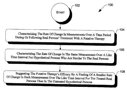
Given the above, the invention may be further characterized as a method for
evaluating of a
treatment to decrease the risk of a progressive brain disorder or to slow
brain aging. For real
persons at risk for Alzheimer's disease, a neurodegenerative disease, or brain
aging, a
measurement's rate of change can be characterized during or following the real
persons' treatment
with disease-preventing or neurological age-slowing therapy. For hypothetical
persons similar to
the real persons at risk for these conditions but who are not so treated, the
measurement's rate of
change can be characterized over a like time interval. The disease-preventing
or age-slowing
therapy's efficacy is suggested by a smaller measurement rate of change over
the like time interval
in the real persons treated than in the hypothetical persons not so treated,
even in the absence of
clinical decline over the time interval. Measurements of neurodegenerative
disease progression
will have significantly higher rates of change in persons clinically affected
by or at risk for the
disease than in those persons at lower risk for the neurodegenerative disease.
The treatment being evaluated can be putative AD prevention therapy, putative
neurodegenerative disease prevention therapy, a putative therapy to slow an
aspect of brain aging,
or a combination of the foregoing. These therapies, and methods for their
evaluation, are discussed
below.
CA 02570539 2006-11-30
WO 2006/009887 PCT/US2005/021557
Evaluation of An AD Prevention Therapy
To evaluate an AD prevention therapy, one or more measurements are taken in
real
persons at two or more different times each of which is found in the absence
of treatment to be
associated with statistically significant (i) rates of change in AD patients,
or (ii) greater rates of
change in MCI patients who subsequently show fiirther cognitive decline than
in MCI patients who
do not, or (iii) greater rates of change in persons thought to be at higher AD
risk that are
cognitively normal or not disabled by AD than persons thought to be at lower
AD risk that are
cognitively normal or not disabled by AD.
A method can use the measurements with respect to real persons who have an AD
risk
factor but do not have clinically significant cognitive impairment. The method
has a step that
characterizes the rate of change in each measurement over a time period during
or following the
real persons' treatment with a putative AD prevention therapy.
For hypothetical persons who are similar to the real persons in their risk for
AD, age, and
absence of clinically significant cognitive impairment but who are not treated
with the putative AD
prevention therapy, the method has a step that characterizes the rate of
change in the same
measurement over a like time interval.
From the foregoing method steps, the efficacy of the putative AD prevention
therapy is
suggested by a fmding of a statistically smaller rate of change in each
measurement over the like
time interval for the real persons treated with the putative AD prevention
therapy than in the
hypothetical persons that are not treated with the putative AD prevention
therapy.
Each of the measurements can be a brain imaging measurement, an
electrophysiological
measurement, a biochemical measurement, a molecular measurement, a
transcriptomic
measurement, a proteomic measurement, a cognitive measurement, a behavior
measurement, or a
combination of the foregoing.
One of the measurements can be the cerebral metabolic rate for glucose (CMRgI)
in brain
regions found to have a greater rate of CMRgI decline in cognitively normal
persons at higher risk
for AD than in those with a lower risk. Here, the CMRgI is measured using
fluorodeoxyglucose
(FDG) positron emission tomography (PET), where the real and hypothetical
persons each have at
least one copy of the APOE s4 allele.
Each measurement can be the rate of change in brain tissue volume or the rate
of change in
cerebrospinal fluid volume so as to provide information about the rate of
brain atrophy. The brain
tissue volume or the cerebrospinal fluid volume can be measured using magnetic
resonance
imaging (MRI). In such cases, the real and hypothetical persons will
preferably have at least one
copy of the APOE s4 allele.
21
CA 02570539 2006-11-30
WO 2006/009887 PCT/US2005/021557
In one embodiment, each of the measurements is suggested to provide an
indirect
assessment of the progression of AD pathology, where the AD pathology can be
the loss of intact
neurons or synapses, the formation of amyliod plaques, the formation of
neurofibrillary tangles, or
a combination of the foregoing.
Each measurement can be a concentration of amyloid proteins, a concentration
of amyloid
oligimers, a concentration of amyloid plaques, a concentration of tau, a
concentration of
phosphorylated tau proteins, a concentration of tangles, a concentration of F2-
isoprostanes, a
concentration of lipid peroxidation, a concentration of inflammatory,
activated microglial, a
molecular immune change, and a molecular change associated with the
progression of AD. Each
measurement can be a reflection of the activity or integrity of brain cells, a
reflection of the activity
or integrity of white matter tracks, or a combination of the foregoing. Each
measurement can be a
neurotransmitter characteristic, a neuroreceptor characteristic, a
neurochemical characteristic, a
molecular characteristic, a physiological characteristic, or a combination of
the foregoing. Each
measurement can be made by a brain imaging technique, a biological assay, and
combination of
the foregoing. Here, the biological assay can be performed using a sample that
is a body fluid,
cerebrospinal fluid, blood, saliva, urine, a body tissue. Here, the -brain
imaging technique can be
different PET and single photon emission tomography radiotracer methods, a
structural, functional,
perfusion-weighted, or diffusion-weighted MRI, x-ray computed tomography,
magnetic resonance
spectroscopy measurements of N-acetyl aspartic acid, myoinositol, and other
chemical compounds,
electroencephalography, quantitative electroencephalography, event-related
potentials, other
electrophysiological procedures, magnetoencephalography, an
electrophysiological method, or a
combination of the foregoing.
The AD risk factor can be a genetic risk factor, a non-genetic risk factor, or
a combination
of the foregoing. The genetic risk factor can be the presence of I or 2 copies
of the APOE s4
allele, the presence of other confirmed susceptibility genes, the presence of
a presenilin 1 mutation,
presenilin 2 mutation, amyloid precursor protein mutation, or other mutations
or gene shown to
cause AD, an aggregate genetic risk score that is based upon a person's number
of susceptibility
genes and their individual contribution to an AD risk, a family history of AD,
or a combination of
the foregoing. The non-genetic risk factor can be head trauma associated with
loss of
consciousness, a higher than normal cholesterol level, a higher than normal
homocysteine level, a
brain imaging measurement thought to be associated with a higher than normal
risk of subsequent
cognitive decline, MCI, or AD, being at least 60 years of age, a biological
marker associated with a
higher that normal risk of subsequent cognitive decline, MCI, or AD, a
cognitive measurement
thought to be associated with a higher than normal risk of subsequent
cognitive decline, MCI, or
22
CA 02570539 2006-11-30
WO 2006/009887 PCT/US2005/021557
AD, a behavioral measurement thought to be associated with a higher than
normal risk of
subsequent cognitive decline, MCI, or AD, or a combination of the foregoing.
The validity of each measurement as a "therapeutic surrogate" will preferably
be further
supported to suggest the efficacy of the putative AD prevention therapy by a
statistically
significant relationship between rates of change in each measurement over the
like time interval
and subsequent clinical decline in patients with AD or MCI or in cognitively
normal or non-
disabled persons at AD risk. Further, the validity of each measurement as a
"therapeutic
surrogate" will preferably be further supported to suggest the efficacy of the
putative AD
prevention therapy by a statistically significant showing of how the ability
of the putative AD
prevention therapy to slow the rate of change in each said measurement over
the like time interval
is associated with slower rates of subsequent clinical decline in patients
with AD or MCI or in
cognitively normal or non-disabled persons at AD risk.
The putative AD prevention therapy can be a pharmacological prescription, an
over-the-
counter medication, an immunization therapy, a biological therapeutic, a
dietary supplement, a
dietary change, a physical exercise, a mental exercise, a lifestyle change
intended to promote
healthy living, decrease the risk of cognitive decline, MCI, AD, or
cardiovascular disease, or a
combination of the foregoing. Note that the putative therapy can be applied to
a patient who has
AD, MCI, or is a cognitively nortnal or non-disabled person who has an AD risk
factor.
Evaluation of A Neurodegenerative Disease Prevention Therapy
To evaluate a neurodegenerative disease prevention therapy, one or more
measurements
are taken in real persons at two or more different times, each of which is
found in the absence of
treatment to be associated with statistically significant (i) rates of change
in patients having a
neurodegenerative disease or (ii) greater rates of change in persons at higher
risk for the
neurodegenerative disease but not disabled by the neurodegenerative disease
than those in persons
at lower risk for the neurodegenerative disease.
A method can use the measurements with respect to the real persons who have a
neurodegenerative disease risk factor but do not have clinically significant
neurological
impairment. The method has a step that characterizes the rate of change in
each measurement over
a time period during or following the real persons' treatment with a putative
neurodegenerative
disease prevention therapy.
For hypothetical persons who are similar to the real persons in their risk for
the
neurodegenerative disease, age, and absence of clinically significant
cognitive impairment but who
are not treated with the putative neurodegenerative disease prevention
therapy, the method has a
step that characterizes the rate of change in the same measurement over a like
time interval.
23
CA 02570539 2006-11-30
WO 2006/009887 PCT/US2005/021557
From the foregoing method steps, the efficacy of the putative
neurodegenerative disease
prevention therapy is suggested by a finding of a statically smaller rate of
change in each
measurement over the like time interval for the real persons treated with the
putative
neurodegenerative disease prevention therapy than in the hypothetical persons
that are not treated
with the putative neurodegenerative disease prevention therapy.
The neurodegenerative disease can be Alzheimer's disease, Dementia with Lewy
Bodies,
Parkinson's disease, Parkinson's dementia, a frontotemporal dementia, a
tauopathy, other
progressive dementias, amyotropic lateral sclerosis, other progressive
neuromuscular disorders,
multiple sclerosis, other progressive neuroimmunological disorders,
Huntington's disease, a focal
or generalized brain disorder which involves a progressive loss of brain
function over time, or a
combination of the foregoing.
Each repeated measurement can be a brain imaging measurement, an
electrophysiological
measurement, a biochemical measurement, a molecular measurement, a
transcriptomic
measurement, a proteomic measurement, a cognitive measurement, a behavior
measurement, or a
combination of the foregoing.
One of the measurements can be the cerebral metabolic rate for glucose (CMRgI)
in brain
regions found to have a greater rate of CMRgI decline in patients with
Parkinson's disease patients
who subsequently development Parkinson's dementia than in Parkinson's patients
who do not
subsequently develop Parkinson's dementia. Here, the CMRgI is measured using
fluorodeoxyglucose (FDG) positron emission tomography (PET). Preferably, the
real and
hypothetical persons each have Parkinson's disease but do not have dementia at
the beginning of
the like time intervaL
Each of the measurements can be a brain imaging measurement, an
electrophysiological
measurement, or a combination of the foregoing. Each measurement can be a
biochemical assay, a
molecular assay, or a combination of the foregoing. In one implementation, at
least one of the
measurements will preferably have a greater rate of change in persons at a
higher risk for the
neurodegeneragive disease that in persons at a lower risk for the
neurodegeneragive disease in the
absence of disabling symptoms of the neurodegeneragive disease.
The validity of each measurement as a "therapeutic surrogate" will preferably
be further
supported to suggest the efficacy of the putative neurodegenerative disease
prevention therapy by a
statistically significant relationship between rates of change in each said
measurement over the like
time interval and subsequent clinical decline in patients affected by or at
risk for the
neurodegenerative disease. Moreover, the validity of each measurement as a
"therapeutic
surrogate" will further be supported to suggest the efficacy of the putative
neurodegenerative
24
CA 02570539 2006-11-30
WO 2006/009887 PCT/US2005/021557
disease prevention therapy by a statistically significant showing of how the
ability of the putative
neurodegenerative disease prevention therapy to slow the rate of change in
each said measurement
over the like time interval is associated with slower rates of subsequent
clinical decline in patients
affected by or at risk for the neurodegenerative disease.
The putative neurodegenerative disease prevention therapy can be a
pharmacological
prescription, an over-the-counter medication, an immunization therapy, a
biological therapeutic, a
dietary supplement, a dietary change, a physical exercise, a mental exercise,
a lifestyle change
intended to promote healthy living, reduced the risk of the neurodegenerative
disorder or its
symptoms, or reduce the risk of cardiovasculare disease, or a combination of
the foregoing. The
person being treated with the neurodegenerative disease prevention therapy can
have a
neurodegenerative disease or can be a person without disabling symptoms of a
neurodegenerative
disease who has a neurodegenerative disease risk factor.
Evaluation of a Therapy to Slow An Aspect of Brain Aging
To evaluate a putative therapy to slow an aspect of brain aging, one or more
measurements
are taken in real persons at two or more different times. These measurements
will preferably be
found in the absence of treatment to be associated with statistically
significant rates of change
associated with aging in patients who do not have clinical signs or symptoms
of a progressive brain
disorder.
A method can use the measurements with respect to the real persons who do not
have
clinical signs or symptoms of a progressive brain disorder. The method has a
step that
characterizes the rate of change in each measurement over a time period during
or following the
real persons' treatment with a putative therapy to slow an aspect of brain
aging;
For hypothetical persons who are similar to the real persons their age and
absence of
clinically significant signs of symptoms of a brain disorder but who are not
treated with the
putative therapy to slow an aspect of brain aging, the method has a step that
characterizes the rate
of change in the same measurement over a like time interval.
From the foregoing method steps, the efficacy of the putative therapy to slow
an aspect of
brain aging is suggested by a finding of a statistically smaller rate of
change in each said
measurement over the like time interval for the real persons treated with the
putative therapy to
slow an aspect of brain aging than in the hypothetical persons that are not
treated with the putative
therapy to slow an aspect of brain aging. When the therapy is effective in
slowing down an aspect
of brain aging, there could be a delay in the onset of disorders that are
caused in part by those aging
changes and there could be a slower decline in cognitive or neurological
abilities that are adversely
affected by those aging changes.
CA 02570539 2006-11-30
WO 2006/009887 PCT/US2005/021557
One of the measurements can be the cerebral metabolic rate for glucose (CMRgI)
in brain
regions found to be affected by normal aging, healthy aging, or very health
aging. Here, the
CMRgI is measured using fluorodeoxyglucose (FDG) positron emission tomography
(PET).
"Normal aging" can be characterized by the absence of a brain disorder of the
absence of a
medical problem that could affect the brain. "Healthy aging" can be further
characterized by the
absence of any signs or symptoms of an age-related brain disorder. "Very
health aging" can be
further characterized by the absence of one or more known risk factors for an
age-related disorder.
For instance, a risk factor can be having a copy of the APOE s4 allele.
One of the measurements can be a brain imaging measurement, an
electrophysiological
measurement, or a combination of the foregoing. Each measurement can be a
biochemical assay, a
molecular assay, a measurement of oxidative stress, or a combination of the
foregoing.
The validity of each measurement as a "therapeutic surrogate" will preferably
be further
supported to suggest the efficacy of the putative therapy to slow an aspect of
brain aging by a
statistically significant showing that the rate of change in each said
measurement over the like time
interval is predictive of an age-related cognitive decline or a behavioral
decline. Further, the
validity of each measurement as a"therapeutic surrogate" will preferably be
further supported to
suggest the efficacy of the putative therapy to slow an aspect of brain aging
by a statistically
significant showing that the rate of change in each measurement over the like
time interval is
predictive of a subsequent age-related decline in cognitive, behavioral, or
other neurological
abilities. Still further, the validity of each measurement as a "therapeutic
surrogate" will preferably
be fflzrther supported to suggest the efficacy of the putative therapy to slow
an aspect of brain aging
by a statistically significant showing that the rate of change in each said
measurement over the like
time interval is predictive of one or more age-related disorders that are more
likely to be found in
aged individuals. In addition, the validity of each measurement as a
'therapeutic surrogate" will
preferably be further supported to suggest the efficacy of the putative
therapy to slow an aspect of
brain aging by a statistically significant showing that the rate of change in
each measurement over
the like time interval is associated with slower rates of age-related
cognitive decline, age-related
behavioral decline, other age-related neurological, neuropsychological, or
psychiatric declines, or
the onset of an age-related disorder.
The putative therapy to slow an aspect of brain aging can be a pharmacological
prescription, an over-the-counter medication, an immunization therapy, a
biological therapeutic, a
dietary supplement, a dietary change, a physical exercise, a mental exercise,
a lifestyle change
intended to promote healthy living, a lifestyle change intended to promote
healthy mental function,
a lifestyle change intended to decrease a risk of cardiovascular disease, or a
combination of the
26
CA 02570539 2006-11-30
WO 2006/009887 PCT/US2005/021557
foregoing. The person being treated with the putative therapy may or may not
have an age-related
disorder and may or may not have a risk factor for an age-related disorder.
While preferred embodiments of this invention have been shown and described,
modifications thereof can be made by one skilled in the art without departing
from the spirit or
teaching of this invention. The embodiments described herein are exemplary
only and are not
limiting. Many variations and modifications of the method and any apparatus
are possible and
are within the scope of the invention. One of ordinary skill in the art will
recognize that the
process just described may easily have steps added, taken away, or modified
without departing
from the principles of the present invention. Accordingly, the scope of
protection is not limited
to the embodiments described herein, but is only limited by the claims that
follow, the scope of
which shall include all equivalents of the subject matter of the claims.
Table of Number References:
1. McKhann G, Drachman D, Folstein M, Katzman R, Price D, Stadlan EM. Clinical
diagnosis of Alzheimer's disease: report of the NINCDS-ADRDA Work Group under
the
auspices of the Department of Health and Human Services Task Force on
Alzheimer's
Disease. Neurology 1984;34:939-944.
2. American Psychiatric Association. Diagnostic and statistical manual of
mental
disorders. 4th ed. Washington, DC: American Psychiatric Association, 1994.
3. Reiman EM, Caselli RJ. Alzheimer's Disease. Maturitas 1999;31:185-200.
4. Terry RD, Maskiah E, Hansen LA. The neuropathology of Alzheimer disease and
the
structural basis of its cognitive alterations. In: Terry RD, Katzman R, Bick
KL, and
Sisodia SS, eds. Alzheimer Disease, 2nd Ed. Philadelphia:Lippincott Williams &
Wilkins, 1999;187-206.
5. Cohen D, Eisdorfer C. Depression in family members caring for a relative
with
Alzheimer's disease. J Am Geriatr Soc 1988;36:885-889.
6. Evans DA, Funkenstein HH, Albert MS, et al. Prevalence of Alzheimer's
disease in a
community population of older persons: higher than previously reported. JAMA
1989;262: 2551-2556.
7. Katzman R, Kawas C. The epidemiology of dementia and Alzheimer disease. In:
Terry
RD, Katzman R, and Bick KL, eds. Alzheimer Disease. New York: Raven Press,
1994;105-122.
8. Reiman EM, Caselli RJ, Chen K, et al. Declining brain activity in
cognitively normal
apolipoprotein E E4 heterozygotes: a foundation for testing Alzheimer's
prevention
therapies. Proc Natl Acad Sci USA 2001;98:3334-3339.
9. Reiman EM, Caselli RJ, Alexander GE, Chen, K. Tracking the decline in
cerebral
glucose metabolism in persons and laboratory animals at genetic risk for
Alzheimer's
disease. Clinical Neuroscience Research 2001;1:194-206.
10. Khachaturian, Z S. The five-five, ten-ten plan for Alzheimer's disease
(editorial).
Neurobiol Aging 1992;13:197-198.
12. Petersen RC, Smith GE, Waring SC, Ivnik, RJ, Tangalos EG, Kokmen E. Mild
cognitive impairment: clinical characterization and outcome. Arch Neurol.
1999;56:303-
308.
27
CA 02570539 2006-11-30
WO 2006/009887 PCT/US2005/021557
12. Morris JC, Storandt M, Miller JP, et al. Mild cognitive impairment
represents early-
stage Alzheimer disease. Arch Neurol 2001;58:397-405.
13. Seshadri, S, et al. Plasma Homocysteine as a Risk Factor for Dementia and
Alzheimer's Disease. New Engl J Med; 346: 476-483.
14. Snowdon DA, Tully CL, Smith CD, Riley KP, Markesbery WR. Serum folate and
the
severity of atrophy of the neocortex in Alzheimer disease: findings from the
nun study.
Am J Clin Nutr 2000;71:993-998.
15. Strittmatter WJ, Weisgraber KH, Huang DY, et al. Binding of human
apolipoprotein
E to synthetic amyloid 0 peptide isoform-specific effects and implications for
late-onset
Alzheimer disease. =Proc Natl Acad Sci USA 1993;90:98-8102.
16. Saunders AM, Strittmatter WJ, Schmechel D, et al. Association of
apolipoprotein E
allele E4 with late-onset familial and sporadic Alzheimer's disease. Neurology
1993;43:1467-1472.
17. Corder EH, Saunders AM, Strittmatter WJ, et al. Gene dose of
apolipoprotein E type
4 allele and the risk of Alzheimer's disease in late onset families. Science
1993;261:921-
924.
18. Farrer LA, Cupples LA, Haines JL, Hyman B, Kukull WA, Mayeux R, Myers RH,
Pericak-Vance MA, Risch N, van Duijn CM. Effects of age, sex, and ethnicity on
the
association between apolipoprotein E genotype and Alzheimer disease. A meta-
analysis.
APOE and Alzheimer Disease Meta Analysis Consortium. JAMA 1997;278(16):1349-
56.
19. Wisniewski T, Castano EM, Golabek A, et al. Acceleration of Alzheimer's
Fibril
formation by apolipoprotein E in vitro. Am J Patho11994;145:1030-1035.
20. Miyata M, Smith JD. Apolipoprotein E allele-specific antioxidant activity
and effects
on cytotoxicity by oxidative insults and bamyloid peptides. Nat Genet
1996;14:55-61.
21. Strittmatter WJ, Weisgraber KH, Goedert M, et al. Hypothesis: microtubule
instability and paired helical filament formation in the Alzheimer disease
brain are related
to apolipoprotein E genotype. Exp Neurol 1994;125:163-171.
22. Mahley RW. Apolipoprotein E: cholesterol transport protein with expanding
role in
cell biology. Science 1988;240:622-30.
23. Corder EH, Saunders AM, Risch NJ, et al. Protective effect of
apolipoprotein E type
2 allele for late onset Alzheimer disease. Nature Genetics 1994;7:180-184.
24. Saunders AM, Hulette C, Welsh-Bohmer KA, et al. Specificity, sensitivity,
and
predictive value of apolipoprotein-E genotyping for sporadic Alzheimer's
disease.
Lancet 1996; 348:90-93.
25. de Leon MJ, Ferris SH, George AE, et al. Computed tomography and positron
emission transaxial evaluations of normal aging and Alzheimer's disease. J
Cereb Blood
Flow Metab 1983;3:391-394.
26. Foster NL, Chase TN, Fedio P, et al. Alzheimer's disease: Focal cortical
changes
shown by positron emission tomography. Neurology 1983;33:961-965.
27. Duara R, Grady CL, Haxby JV, et al. Positron emission tomography in
Alzheimer's
disease. Neurology 1986;36:879-887.
28. Jagust WJ, Friedland RP, Budinger TF, et al. Longitudinal studies of
regional cerebral
metabolism in Alzheimer's disease. Neurology 1988;38:909-912.
29. Haxby JV, Grady CL, Koss E, et al. Longitudinal study of cerebral
metabolic
asymmetries and associated neuropsychological patterns in early dementia of
the
Alzheimer type. Arch Neurol 1990;47:753-760.
30. McGeer EG, Peppard RP, McGeer PL, et al. 18 Fluorodeoxyglucose positron
emission tomography studies in presumed Alzheimer cases, including 13 serial
scans.
Can J Neurol Sci 1990;17:1-11.
28
CA 02570539 2006-11-30
WO 2006/009887 PCT/1JS2005/021557
31. Smith GS, de Leon MJ, George AE, et al. Topography of crosssectional and
longitudinal glucose metabolic defecits in Alzheimer's disease.
Pathophysiologic
implications. Arch Neurol 1992;49:1142-1150.
32. Minoshima S, Foster NL, Kuhl DE. Posterior cingulate cortex in Alzheimer's
disease.
Lancet 1994;344:895.
33. Mielke R, Herholz K, Grond M. Clinical deterioration in probable
Alzheimer's disease
correlates with progressive metabolic impairment of association areas.
Dementia
1994;5:36-41.
34. Minoshima S, Frey KA, Koeppe RA, et al., A diagnostic approach in
Alzheimer's
disease using three-dimensional stereotactic surface projections of fluorine-
18-FDG PET. J
Nucl Med 1995;36:123 8-48.
35. Mega MS, Cheii SS, Thompson PM, et al. Mapping histology to metabolism:
coregistration of stained whole-brain sections to premortem PET in Alzheimer's
disease.
Neuroimage 1997;5:147-153.
36. Pietrini P, Alexander GE, et a1. Abnormal metabolic patterns in
Alzheimer's disease
after correction for partial volume effects. Neurology 1998;50:1585-1593.
37. Hoffman JM, Welsh-Bohmer KA, Hanson M, et al. FDG PET imaging in patients
with pathologically verified dementia. J Nucl Med 2000;41:1920-1928.
38. Silverman DH, Small GW, Chang CY, et al. Positron emission tomography in
evaluation of dementia: Regional brain metabolism and long-term outcome. JAMA
2001;286;17:2120-7.
39. Alexander GE, Chen K, Pietrini P, Rapoport SI, Reiman EM. Longitudinal PET
evaluation of cerebral metabolic decline in dementia: a potential outcome
measure in
Alzheimer's disease treatment studies. Am J Psychiatry 159:738-745,2002.
40. Schwartz WI, Smith CB, Davidsen L, et al. Metabolic mapping of functional
activity
in the hypothalamic neurohypophysial system of the rat. Science 1979;205:723-
725.
41. Meguro K, Blaizot X, Kondoh Y, et al. Neocortical and hippocampal glucose
hypometabolism following neurotoxic lesions of the entorhinal and perirhinal
cortices in
the non-human primate as shown by PET. Implications for Alzheimer's disease.
Brain
1999;122:1519-31.
42. Magistretti PJ and Pellerin L. Cellular bases of brain energy metabolism
and their
relevance to functional brain imaging: evidence for a prominent role of
astrocytes. Cereb
Cortex 1996;6:50-61.
43. Piert M, Koeppe RA, Giordani B, et al. Diminished glucose transport and
phosphorylation in Alzheimer's disease determined by dynamic FDG-PET. J Nucl
Med
1996;37:201-8.
44. Mark RJ, Pang Z, Geddes JW, et al. Amyloid (3-Peptide Impairs Glucose
Transport in
Hippocampal and Cortical Neurons: Involvement of Membrane Lipid Peroxidation.
J
Neurosci 1997;17:1046-54.
45. Small GW, Mazziotta JC, Collins MT, et al. Apolipoprotein E type 4 allele
and
cerebral glucose metabolism in relatives at risk for familial Alzheimer
disease. J Am Med
Assoc 1995;273 :942-947.
46. Reiman EM, Caselli RJ, Yun LS, et al. Preclinical evidence of a genetic
risk factor
for Alzheimer's disease in apolipoprotein E type 4 homozygotes using positron
emission
tomography. N Engl J Med 1996;334:752-758.
47. Small GW, Ercoli LM, Silverman DHS, et al. Cerebral metabolic and
cognitive
decline in persons at genetic risk for Alzheimer's disease. Proc Natl Acad Sci
USA
2000; 97: 6037-6042.
48. Mielke R, Zerres K, Uhlhaas S, et al. Apolipoprotein E polymorphism
influences the
cerebral metabolic pattern in Alzheimer's disease. Neurosci Lett 1998;254:49-
52.
29
CA 02570539 2006-11-30
WO 2006/009887 PCT/US2005/021557
49. Higuchi M, Arai H, Nakagawa T, et al. Regional cerebral glucose
utilization is
modulated by the dosage of apolipoprotein E type 4 allele and alphal-
antichymotrypsin
type A allele in Alzheimer's disease. Neuroreport 1997;8:2639-43.
50. Corder EH, Jelic V, Basun H, et al. No difference in cerebral glucose
metabolism in
patients with Alzheimer disease and differing apolipoprotein E genotypes. Arch
Neurol
1997;54:273-7.
51. Hirono N, Mori E, Yasuda M, et al. Lack of association of apolipoprotein E
epsilon 4
allele dose with cerebral glucose metabolism in Alzheimer disease. Alzheimer
Dis Assoc
Disord 1998;12:362-7.
52. Kuhl DE, Koeppe RA, Minoshima S, et al. In vivo mapping of cerebral
acetylcholinesterase activity in aging and Alzheimer's disease. Neurology.
1999;52:691-
9.
53. Cagnin A, Brooks DJ, Kennedy AM, et al. In-vivo measurement of activated
microglia in dementia. Lancet. 2001;358:461-7.
54. Mathis CA, Bacskai BJ, Kajdasz ST, et al. A lipophilic thioflavin-T
derivative for
positron emission tomography (PET) imaging of amyloid in brain. Biorg Med Chem
Lett
2002;12:295-8.
55. Shoghi-Jadid K, Small GW, Agdeppa ED, et al. Localization of
neurofibrillary
tangles (NFTs) and beta-amyloid placques (APs) in the brains of living
patients with
Alzheimer's disease. Am J Geriatr Psychiatry 2002;10:24-35.
56. Seab J, Jagust W, Wong S, et al. Quantitative NMR measurements of
hippocampal
atrophy in Alzheimer's disease. Magn Reson Med 1988;8:200-208.
57. Jack CR, Petersen RC, O'Brien PC, Tangalos EG. MR-based hippocampal
volumetry
in the diagnosis of Alzheimer's disease. Neurology 1992;42:183-188.
58. Golomb J, de Leon MJ, Kluger A, et al. Hippocampal atrophy in normal aging
- an
association with recent memory impairment. Arch Neurol 1993;50:967-973.
59. de Leon MJ, George AE, Stylopoulos LA, Smith G, Miller DC. Early marker
for
Alzheimer's disease: the atrophic hippocampus. Lancet 1989;672-673.
60. Kesslak J, Nalcioglu 0, Cotman C. Quantification of magnetic resonance
scans for
hippocampal and parahippocampal atrophy in Alzheimer's disease. Neurology
1991;41:51-54.
61. Rusinek H, de Leon M, George A, et al. Alzeimer disease: measuring loss of
cerebral
gray matter with MRI imaging. Radiology 1991;178:109-134.
62. Killiany R, Moss M, Albert M, Tanmas S. Temporal lobe regions on magnetic
resonance imaging identify patients with early Alzheimer's disease. Arch
Neurol
1993;50:949-954.
63. Lehericy S, Baulac M, Chiras J, et al. Amygdalohippocampal MR volume
measurements in the early states of Alzheimer disease. Am JNeuroradiol
1994;15:927-
937.
64. Deweer B, Lehericy S, Pillon B, et al. Memory disorders in probable
Alzheimer's
disease: the role of hippocampal atrophy as shown with MRI. J Neurol Neurosurg
Psychiatry 1995;58:590-597.
65. Laakso M, Partanen K, Riekkinen P, et al. Hippocampal volumes in
Alzheimer's
disease, Parkinson's disease with and without dementia, and in vascular
dementia: an
MRI study. Neurology 1996;46:678-681.
66. de Leon MJ, Golomb J, George AE, et al. The radiologic prediction of
Alzheimer's
disease: the atrophic hippocarnpal formation. Am J Neuroradiol 1993;14:897-
906.
67. Krasuski JS, Alexander GE, Horwitz B, et al. Volumes of medial temporal
lobe
structure in patients with Alzheimer's disease and mild cognitive impairment
(and in
healthy controls). Biol Psychiatry 1998;43:60-9.
CA 02570539 2006-11-30
WO 2006/009887 PCT/US2005/021557
68. Juottonen K, Laakso MP, Partanen K, et al. Comparative MR analysis of the
entorhinal cortex and hippocampus in diagnosing Alzheimer disease. AJNR Am J
Neuroradiol 1999;20:13 9-44.
69. Jack CR Jr, Petersen RC, Xu Y, et al. Rate of medial temporal lobe atrophy
in typical
aging and Alzheimer's disease. Neurology 1998;51:993-9.
70. Laakso MP, Soininen H, Partanen K, et al. Volumes of hippocampus, amygdala
and
frontal lobes in the MRI-based diagnosis of early Alzheimer's disease:
correlation with
memory functions. J Neural Transm Park Dis Dement Sect 1995;9:73-86.
71. Schuv N, Amend D, Ezekiel F, et al. Change of hippocampal N-acetyl
aspartate and
volume in Alzheimer's disease. Neurology 1997;49:1513 21.
72. Jack CR Jr, Petersen RC, Xu YC, et al. Medial temporal atrophy on MRI in
normal
aging and very mild Alzheimer's disease. Neurology 1997;49:786-94.
73. Frisoni GB, Laakso MP, Beltramello A, et al. Hippocampal and entorhinal
cortex
atrophy in frontotemporal dementia and Alzheimer's disease. Neurology
1999;52:91-
100.
74. Juottonen K, Laakso MP, Insausti R, et al. Volumes of the entorhinal and
perirhinal
cortices in Alzheimer's disease. Neurobiol Aging 1998;19:15 22.
75. Bobinski M, de Leon MJ, Convit A, et al. MRI of entorhinal cortex in mild
Alzheimer's disease. Lancet 1999;353:38-40.
76. Xu Y, Jack CR Jr, O'Brien PC, et al. Usefulness of MRI measures of
entorhinal
cortex versus hippocampus in AD. Neurology, 2000;54:1760-7.
77. Jack CR, Jr., Petersen RC, Xu Y, O'Brien PC, Smith GE, Ivnik RJ, et al.
Rates of
hippocampal atrophy correlate with change in clinical status in aging and AD.
Neurology
2000;55(4):484-489.
78. Visser PJ, Scheltens P, Verhey FR, et al. Medial temporal lobe atrophy and
memory
dysfunction as predictors for dementia in subjects with mild cognitive
impairment. J
Neurol 1999;246:477-85.
79. Du AT, Schuff N, Zhu XP, et al. Atrophy rates of entorhinal cortex in AD
and
normal aging. Neurology 2003; 60:481-6.
80. Du AT, Schuff N, Kramer JH, et al. Higher atrophy rate of entorhinal
cortex than
hippocampus in Alzheimer's disease. Neurology 2003 (in press).
81. Fox NC, Freeborough PA, Rossor MN. Visualization and quantification of
rates of
atrophy in Alzheimer's disease. Lancet 1996;348:94-97.
82. Freebourough PA, Fox NC. The boundry shift integral : an accurate and
robust
measure of cerebral volume changes from registered repeat MRI. IEEE Trans Med
Imaging 1997;16:623-629.
83. Fox NC, Freeborough PA. Brain atrophy progression measured from registered
serial
MRI. J Magn Reson Imaging 1997;7:1069-1075.
84. Fox NC, Cousens S, Scahill R, et al. Using serial registered brain
magnetic resonance
imaging to measure disease progression in Alzheimer disease: power
calculations and
estimates of sample size to detect treatment effects. Arch Neurol 2000:57:333-
444.
85. Fox NC, Crum WR, Scahill RI, et al. Imaging of onset and progression of
Alzheimer's disease with voxel-compression mapping of serial magnetic
resonance
images. Lancet 2001; 358:201-205.
86. Kaye JA, Swihart T, Howieson D, et al. Volume loss of the hippocampus and
temporal lobe in the healthy elderly persons destined to develop dementia.
Neurology
1997;48:1297-304.
87. Fox NC, Warrington EK, Stevens JM, et al. Atrophy of the hippocampal
formation in
early familial Alzheimer's disease. A longitudinal MRI study of at-risk
members of a
31
CA 02570539 2006-11-30
WO 2006/009887 PCT/US2005/021557
family with an amyloid precursor protein 717Va1-Glymutation. Ann NY Acad Sci
1996;777:226-32.
88. Convit A, de Leon MJ, Tarshish C, et al. Hippocampal volume losses in
minimally
impaired elderly. Lancet 1995;345:266.
89. Convit A, de Leon MJ, Tarshish C, et al. Specific hippocampal volume
reduction in
individuals at risk for Alzheimer's disease. Neurobiol Aging 1997;18:131-8.
90. Fox NC, Warrington WK, Freeborough PA, et al. Presymptomatic hippocampal
atrophy in Alzheimer's disease: a longitudinal MRI study. Brain 1996;119:2001-
2007.
91. Jack Jr CR, Petersen RC, Xu YC, et al. Prediction of AD with MRI-based
hippocampal volume in mild cognitive impairment. Neurology 1999;52:1397-1403.
92. Du AT, Schuff N, Amend D, Laakso MP, Hsu YY, Jagust WJ, et al. Magnetic
resonance imaging of the entorhinal cortex and hippocampus iri mild cognitive
impairment and Alzheimer's disease. J Neurol Neurosurg Psychiatry 2001;71:441-
447.
93. Soininen H, Partanen, Pitkanen A, et al. Decreased hippocampal volume
asymmetry
on MRIs in nondemented elderly subjects carrying the apolipoprotein E4 allele.
Neurology 1995;45:391-392.
94. Reiman EM, Uecker A, Caselli RJ, et al. Hippocampal volumes in cognitively
normal
persons at genetic risk for Alzheimer's disease. Ann Neuro11998;44:288-291.
95. Killiany RJ, Gomez-Isla T, Moss M, et al. Use of structural magnetic
resonance
imaging to predict who will get Alzheimer's disease. Ann Neuro12000;47:430-
439.
96. Schott JM, Fox NC, Frost C, et al. Assessing the onset of structural
change in familial
Alzheimer's disease. Ann Neuro12003;53:181-188.
97. Scahill RI, Schott JM, Stevens JM, et al. Mapping the evolution of
regional atrophy in
Alzhiemer's disease: unbiased analysis of fluid-registered serial MRI. Proc
Natl Acad Sci
USA 2002;99:4703-4707.
98. Chen K, Reiman EM, Alexander GE, et al. Automated method using iteractive
principle component analysis for detecting brain atrophy rates from sequential
MRI in
persons with Alzheimer's disease, Soc Neurosci Abstr, 2001 [abstract].
99. Chen K, Reiman EM, Alexander G, et al. Whole brain atrophy rates in
cognitively
normal persons at genetic risk for Alzheimer's disease. Presented at the 8 th
Internat Conf
on AD and Related Dis, Stockholm, June 2002 [abstract].
100. Chen K, Reiman EM, Alexander GE, et al. Whole brain atrophy rates in
cognitively
normal persons at genetic risk for Alzheimer's Disease. Presented at the 8th
Intemat Conf
on AD and Related Dis, Stockholm, June 2002 [abstract].
101. Chen K. Reiman EM, He J, et al. Evaluation of an iterative principal
component
analysis for detecting whole brain volume change in small animal magnetic
resonance
imaging. Presented at the 8u' Intemat Conf on AD and Related Dis, Stockholm,
2002
[abstract].
102. Hauss-Wegrzyniak B, Chen K, Galons JP, et al. Detecting an experimentally
induced reduction in mouse brain volume using sequential high-resolution MRI's
and the
iterative PCA method. Presented at the 8th Internat Conf on AD and Related
Dis,
Stockholm, 2002 [abstract].
103. Ashburner J, Friston KJ. Voxel-based morphometry--the methods. Neuroimage
2000;11:805-821.
104. Alexander GE, Chen K, Reiman EM, et al. Effect of apolipoprotein E on
regional
brain atrophy in cognitively normal s4 homozygotes and heterozygotes using
voxel-based
MRI morphometry. Soc Neurosci Abstr, 2001 [abstract].
105. Alexander GE, Chen K, Reiman EM, et al. Longitudinal declines of gray
matter in
cognitively normal apolipoprotein E E4 homozygotes and heterozygotes evaluated
by
32
CA 02570539 2006-11-30
WO 2006/009887 PCT/US2005/021557
voxel based MRI Morphometry. Presented at the 8th Intemat Conf on AD and
Related
Dis, Stockholm, 2002 [absract].
106. Alexander GE, Chen K, Reiman EM, et al. Abnormalities in gray matter
density in
young adults at genetic risk for late-onset Alzheimer's disease. Presented at
the 8th
Intemat Conf on AD and Related Dis, Stockholm, June 2002 [abstract].
107. Csernansky JG, Wang L, Joshi S, et al. Early DAT is distinguished from
aging by
high-dimensional mapping of the hippocampus. Dementia of the Alzheimer type.
Neurology 2000 Dec 12;55(11):1636-43.
108. Thompson PM, Mega MS, Woods RP, et al. Cortical change in Alzheimer's
disease
detected with a disease-specific population-based brain atlas. Cereb Cortex
2001;11:1-16.
109. Thompson PM, Hayashi KM, de Zubicaray G, et al. Early DAT is
distinguished
from aging by high-dimensional mapping of the hippocampus. Dementia of the
Alzheimer type. J Neurosci. 2003 23:994-1005.
110. Temple R. A regulatory authority's opinion about surrogate endpoints. In:
Nimmo
WS, Tucker GT, eds. Clinical Measurement in Drug Evaluation. New York, NY:
John
Wiley & Sons, Ltd; 1995:3-22.
111. Fleming TR, DeMets DL. Surrogate end points in clinical trials: are we
being
niisled? Ann Intern Med 1996;125:605-613.
112. Div Neuropharmacological Drug Products, US Food and Drug Administration.
Background Document for Joint Advisory Committee Meeting of November 18, 2002:
Issues Related to the Role of Brain Imaging as an Outcome Measure in Phase III
Trials of
Putative Drugs for Alzheimer's Disease. 2002 [Memo].
113. Reiman EM, Uecker A, Gonzalez-Lima F, Minear D., Chen K, Callaway N L,
Berndt, JD, Games D. Tracking Alzheimer's disease in transgenic mice using
fluorodeoxyglucose autoradiography. NeuroReport 2000;11:987-991.
114. Kuhl DE, Metter EJ, Riege WH, Phelps ME. Effects of human aging on
patterns of
local cerebral glucose utilization determined by the 18F-fluorodeoxyglucose
method. J
Cereb Blood Flow Metab 1982;2:163-71.
115. Salmon E, Sadzot B, Maquet P, Dive D, Franck G. Decrease of frontal
metabolism
demonstrated by positron emission tomography in a population of healthy
elderly
volunteers. Acta Neurol Belg 1991;91:288-95.
116. Loessner A, Alavi A, Lewandrowski K, Mozley D, Souder E, Gur RE. Regional
cerebral function determined by FDG-PET in healthy volunteers: normal patterns
and
changes with age. J Nucl Med 1995; 36:1141-9.
117. Coffey CE, Wilkinson WE, Parashos IA. Quantitative cerebral anatomy of
the aging
human brain: a cross-sectional study using magnetic resonance imaging.
Neurology
1992; 42:527-36.
118. Terry RD, DeTeresa R, Hansen LA. Neocortical cell counts in normal human
adult
aging. Ann Neurol 1987; 21:530-9.
119. Reiman EM, Chen K, Alexander GE, Caselli RJ. Positron emission tomography
studies of cognitively normal persons at genetic risk for Alzheimer's disease.
Presented at
the IPSEN Foundation Conference on the Living Brain and Alzheimer's Disease,
Paris,
2003.
120. Reiman EM, Caselli EM, Chen K, et al. Effects of age on cerebral glucose
metabolism in APOE E E4 carriers and noncanriers. Presented at the 8U'
Internat Conf on
AD and Related Dis, Stockholm, June 2002 [abstract].
121. Reiman EM, Caselli RJ, Chen K, et al. Abnormalities in regional brain
activity in
young adults at genetic risk for late-onset Alzheimer's disease Presented at
the 8th
Internat Conf on AD and Related Dis, Stockholm, June 2002 [abstract].
33
CA 02570539 2006-11-30
WO 2006/009887 PCT/US2005/021557
122. Games D, Adams D, Alessandrini R, et al. Alzheimer-type neuropathology in
transgenic mice overexpressing V717F (3-amyloid precursor protein. Nature
1995;373:523-527.
123. Hsiao K, Chapman P, Nilsen S, et al. Correlative memory deficits, Ap
elevation, and
amyloid plaques in transgenic mice. Science 1996;274:99-102.
124. Holcomb L, Gordon MN, McGowan E, et al. Accelerated Alzheimer-type
phenotype
in transgenic mice carrying both mutant amyloid precursor protein and
presenilin 1
transgenes. Nature Med 1998;4:97-100.
125. Terry RD, Masliah E, Salmon DP, et al. Physical basis of cognitive
alterations in
Alzheimer's disease: synapse loss is the major correlate of cognitive
impairment. Ann
Neurol 1991; 30:572-5 80.
126. Bierer LM, Haroutunian V, Gabriel S, et al. Neocortical neurofibrillary
tangles
correlate with dementia severity in Alzheimer's disease. Arch. Neurol.
1995;52:81-88.
127. Morris JC, Olichney JM, Thal LJ, et al. Cerebral amyloid deposition and
diffuse
plaques in "normal" aging: Evidence for presymptomatic and very mild
Alzheimer's
disease. Neurology 1996;46:707-719.
128. Gonzalez-Lima F, Bemdt JD, Valla J, Games D, Reiman EM. Reduced corpus
callosum, fornix and hippocampus in PDAPP transgenic mouse model of
Alzheimer's
disease. NeuroReport 2001;12:2375-2379.
129. Valla J, Chen K, Berndt JD, et al. Effects of image resolution on
autoradiographic
measurements of posterior cingulate activity in PDAPP mice: Implications for
fiinctional
brain imaging studies in transgenic mouse models of Alzheimer's disease.
NeuroImage
2002;16:1-6.
130. Valla J, Lewandowski L, Duff K, Reiman EM. No evidence of significant
white
matter disruption in the TG2576 mouse model of Alzheimer's disease:
implications for in
vivo microimaging. Presented at the 8th Internat Conf on AD and Related Dis,
Stockholm,
June 2002 [abstract].
131. Valla J, Bemdt JD, Gonzalez-Lima F. Energy hypometabolism in posterior
cingulate
cortex of Alzheimer's patients: superficial laminar cytochrome oxidase
associated with
disease duration. J Neurosci. 2001;21:4923-30.
34