Note: Descriptions are shown in the official language in which they were submitted.
= CA 02579569 2013-07-22
NEUROSTIMMATION METHODS AND SYSTEMS
[00011 <deleted>
' FIELD OF TUE INVENTION
[0002] The present invention relates to neurostimulation methods and
systems that enable
more precise stimulation of the nervous system. In particular, embodiments of
the present
invention provide for the controlled stimulation of spinal and paraspinal
nerve root ganglion.
In one embodiment, the ganglion is a dorsal root ganglion (DRG) and in another
embodiment
the ganglion is part of the sympathetic nervous system.
BACKGROUND OF THE INVENTION
100031 Application of specific electrical energy to the spinal cord for the
purpose of managing
pain has been actively practiced since the 1960s. While a precise
understanding of the
interaction between the applied electrical energy and the nervous tissue is
not fully appreciated,
it is known that application of an electrical field to spinal nervous tissue
can effectively mask
certain types of pain transmitted from regions of the body associated with the
stimulated
nervous tissue. More specifically, applying particularized electrical pulses
to the spinal cord
associated with regions of the body afflicted with chronic pain can induce
paresthesia, or a
subjective sensation of numbness or tingling, in the afflicted bodily regions.
This paresthesia
can effectively inhibit the transmission of non-acute pain sensations to the
brain.
[0004] Electrical energy, similar to that used to inhibit pain perception,
may also be used to
manage the symptoms of various motor disorders, for example, tremor, dystonia,
spasticity,
and the like. Motor spinal nervous tissue, or nervous tissue from ventral
nerve roots, transmits
muscle/motor control signals. Sensory spinal nervous tissue, or nervous tissue
from dorsal
nerve roots, transmit pain signals. Corresponding dorsal and ventral nerve
roots depart the
spinal cord "separately"; however, immediately thereafter, the nervous tissue
of the dorsal and
ventral nerve roots are mixed, or intertwined. Accordingly, electrical
stimulation intended to
manage/control one condition (for example, pain) often results in the
inadvertent interference
with nerve transmission pathways in adjacent nervous tissue (for example,
motor nerves).
[0005] As illustrated in FIG. 1, prior art spinal column or spinal cord
stimulators (SCS)
commonly deliver electrical energy to the spinal cord through an elongate
paddle 5 or epidural
-1-
CA 02579569 2013-07-22
electrode array containing electrodes 6 positioned external to the spinal cord
dura layer 32.
The spinal cord dura layer 32 surrounds the spinal cord 13 and is filled with
cerebral spinal
fluid (CSF). The spinal cord 13 is a continuous body and three spinal levels
14 of the spinal
cord 13 are illustrated. For purposes of illustration, spinal levels 14 are
sub-sections of the
spinal cord 13 depicting that portion where the dorsal and ventral roots join
the spinal cord 13.
The peripheral nerve 44 divides into the dorsal root 42 and dorsal root
ganglion 40 and the
ventral nerve root 41 each of which feed into the spinal cord 13. An ascending
pathway 92 is
illustrated between level 2 and level 1 and a descending pathway 94 is
illustrated from level 2
to level 3. Spinal levels 14 can correspond to the vertebral levels of the
spine commonly used
to describe the vertebral bodies of the spine. For simplicity, each level
illustrates the nerves of
only one side and a normal anatomical configuration would have similar nerves
illustrated in
the side of the spinal cord 13 directly adjacent the paddle 5.
100061 Typically, SCS are placed in the spinal epidural space. Conventional
SCS systems are
described in numerous patents. Additional details of the placement and use of
SCS can be
found, for example, in US Patent 6,319,241.
In general, the paddle 5 is about 8mm wide and from 24 to 60mm long depending
upon how many spinal levels are stimulated. The illustrated electrode paddle 5
is adapted to
conventionally stimulate all three spinal levels 14. These exemplary levels
1,2 and 3 could be
anywhere along the spinal cord 13. Positioning a stimulation paddle 5 in this
manner results in
the electrodes 6 spanning a plurality of nerves, here the dorsal root ganglion
40, the ventral
root 41 and peripheral nerve 41 on multiple spinal levels.
[0007] Because the paddle 5 spans several levels the generated stimulation
energy 8 stimulates
or is applied to more than one type of nerve tissue on more than one level.
Moreover, these
and other conventional, non-specific stimulation systems also apply
stimulation energy to the
spinal cord and to other neural tissue beyond the intended stimulation
targets. As used herein,
non-specific stimulation refers to the fact that the stimulation energy is
provided to all spinal
levels including the nerves and the spinal cord generally and
indiscriminately. Even if the
epidural electrode is reduced in size to simply stimulate only one level, that
electrode will
apply stimulation energy indiscriminately to everything (i.e., all nerve
fibers and other tissues)
within the range of the applied energy 8. Moreover, larger epidural electrode
arrays may alter
cerebral spinal fluid (CSF) flow thus further altering local neural
excitability states.
100081 Another challenge confronting conventional neurostimulation systems
is that since
epidural electrodes must apply energy across a wide variety of tissues and
fluids (i.e., CSF
fluid amount varies along the spine as does pia matter thickness ) the amount
of stimulation
-2-
CA 02579569 2007-03-06
WO 2006/029257
PCT/US2005/031960
energy needed to provide the desired amount of neuro stimulation is difficult
to precisely
control. As such, increasing amounts of energy may be required to ensure
sufficient
stimulation energy reaches the desired stimulation area. However, as applied
stimulation
energy increases so too increases the likelihood of deleterious damage or
stimulation of
surrounding tissue, structures or neural pathways.
[0009] To achieve stimulation the targeted tissue, the applied
electrical energy should be
properly defined and undesired energy application to non-targeted tissue be
reduced or
avoided. An improperly defined electric field may not only be ineffective in
controlling/managing the desired condition(s) but may also inadvertently
interfere with the
proper neural pathways of adjacent spinal nervous tissue. Accordingly, a need
exists for
stimulation methods and systems that enable more precise delivery of
stimulation energy.
SUMMARY OF THE INVENTION
[0010] In one embodiment, there is provided a method of stimulating a
dorsal root ganglion by
implanting an electrode in proximity to the dorsal root ganglion; and
activating the electrode to
stimulate a portion of the dorsal root ganglion, or activating the electrode
to stimulate
substantially only the dorsal root ganglion.
[0011] In another embodiment, there is provided a method of stimulating
a nerve root ganglion
by implanting an electrode into the nerve root ganglion; and activating the
electrode to
stimulate the nerve root ganglion.
[0012] In another embodiment, there is provided, a method of
stimulating the spinal cord by
implanting an electrode into the spinal cord; and providing stimulation energy
to spinal cord
fibers using the electrode.
[0013] In another embodiment, there is provided a method of modulating
nervous tissue within
a dorsal root ganglion by implanting an electrode within a dorsal root
ganglion; and providing
electrical stimulation from the electrode to stimulate neural tissue within
the dorsal root
ganglion.
[0014] In another embodiment, there is provided a method of modulating
a neural pathway in
the sympathetic nervous system by stimulating a spinal dorsal root ganglion
upstream of at
least one ganglion of the sympathetic nerve chain to influence a condition
associated with the
at least one ganglion of the sympathetic nerve chain.
[0015] In yet another embodiment, there is provided a neuro stimulation
system having an
electrode adapted for stimulation of only a nerve root ganglion; a signal
generator coupled to
the electrode; and a controller to control the output of the signal generator.
-3-
CA 02579569 2007-03-06
WO 2006/029257
PCT/US2005/031960
[0016] In yet another embodiment, there is provided a method of
stimulating the spinal cord by
piercing the spinal dura matter; and placing an electrode into contact with a
portion of the
intra-madullary of the spinal cord.
[0017] In yet another embodiment, there is a method of stimulating the
nervous system by
implanting an electrode such that when the electrode is activated, the
electrode stimulates only
a nerve root ganglion.
[0018] In yet another embodiment, there is provided a method of
stimulating neural tissue to
treat a condition including stimulating an electrode implanted to stimulate
only a dorsal root
ganglion on a spinal level wherein the stimulation treats the condition.
[0019] In yet another embodiment, there is provided a pulse generator,
comprising at least one
switch connected to at least one implantable electrode having an impedance
greater than 2,500
ohms; a DC-DC converter adapted to provide a stimulation signal to the at
least one
implantable electrode; and a controller configured to control the output of
the DC-DC
converter.
[0020] In yet another embodiment, there is provided a stimulation
component, comprising a
proximal connector; a distal electrode configured to be implanted within the
body at a
stimulation site; an electrical lead connected to the proximal connector and
the distal electrode;
a strain relief mechanism in proximity to the stimulation site; and a fixation
element adapted to
reduce the amount of movement of the electrical lead proximal to a fixation
point in an
anatomical structure proximal to the stimulation site.
[0021] In another embodiment, there is provided a stimulation
component, comprising a
proximal connector; a distal electrode configured to be implanted within the
body at a
stimulation site; an electrical lead connected to the proximal connector and
the distal electrode;
a strain relief mechanism in proximity to the stimulation site; and a fixation
element adapted to
reduce the amount of movement of the electrical lead proximal to a fixation
point in an
anatomical structure proximal to the stimulation site.
[0022] In another embodiment, there is provided a stimulation system,
comprising a pulse
generator; an electrode connector having a flexible, elongate body with a
proximal end
electrically connected to the pulse generator and a distal end adapted to
connect to a
microelectrode lead, wherein the microelectrode lead connects proximally to
the electrode
connector distal end and has a distal microelectrode electrically connected to
the pulse
generator.
[0023] In yet another embodiment, there is provided a stimulation system,
comprising a
battery; a pulse generator separate from the battery; an electrical connection
between the
-4-
CA 02579569 2015-04-23
CA 2579569
battery and the pulse generator; a mieroelectrode lead connected proximally to
the pulse
generator and distally to a microelectrode.
[0024] In yet another embodiment, there is provided a neurostimulation
component,
comprising a body having a distal end and a proximal end and a length selected
to implant the
body within a targeted neural tissue; a tip on the distal end of the body
adapted to pierce
through the targeted neural tissue; and an electrode structure positioned on
the body adapted to
neurostimulate only the targeted neural tissue.
[0025] In yet another embodiment, there is provided a method of
neurostimulating
targeted neural tissue, comprising implanting an electrode in a position
adapted to
neurostimulate only targeted neural tissue; and providing a controlled
stimulation signal from a
signal generator coupled to the electrode.
[0026] The claimed invention includes an electrode and electrode lead
combination for
use in selective stimulation of a dorsal root ganglion in a patient, wherein
the electrode is sized
and configured to be positionable on, in or adjacent the dorsal root ganglion,
and wherein the
distal end of the lead is advanceable through a delivery device and is
flexible so that the lead is
advanceable through the delivery device in a substantially straightened
configuration, and once
advanced beyond a distal end of the delivery device the distal end of the lead
is positionable
about said dorsal root ganglion, so as to conform to and thereby follow the
bulbous shape of the
dorsal root ganglion.
[0026A] The claimed invention also includes a delivery device advanceable
into a
foramen from outside of a spinal column so that the delivery device is
directed toward a dorsal
root ganglion without entering an epidural space, the device comprising an
electrode as claimed
herein, wherein the lead is advanceable through the delivery device.
[0026B] The claimed invention also includes a neurostimulation system
comprising a
plurality of electrode and electrode lead combinations as claimed herein.
[0026C] The claimed invention also includes a neurostimulation system
comprising: at
least one electrode and electrode lead combination as claimed herein; a signal
generator; and a
controller to control the output of the signal generator to provide a signal
to the electrode so
that together the signal and the electrode provide for selective stimulation
of one or more dorsal
root ganglions while substantially excluding stimulation of an associated
ventral root.
- 5 -
CA 02579569 2013-07-22
BRIEF DESCRIPTION OF THE DRAWINGS
[0027] A better understanding of the features and advantages of the
various
embodiments of the present invention will be obtained by reference to the
following detailed
description and the accompanying drawings of which:
[0028] FIGURE 1 illustrates a conventional epidural electrode array
positioned external
to and stimulating a portion of the spinal cord;
[0029[ FIGURE 2A illustrates an embodiment an electrode implanted into a
spinal
dorsal root ganglion;
[0030] FIGURE 2B illustrates how selective stimulation techniques of
FIGURE 2A
may raise a response threshold;
[0031] FIGURE 3 A illustrates a stimulation system with an electrode
embodiment of
the present invention implanted into a dorsal root ganglion (DRG) of a spinal
level;
[0033] FIGURE 3B relates the spinal nerve roots to their respective
vertebral spinal
levels;
[0033] FIGURE 3 C illustrates the various derrnatomes of the body related
to their
respective nerve roots in FIGURE 3B;
- 5a-
CA 02579569 2007-03-06
WO 2006/029257
PCT/US2005/031960
[0034] FIGURE 4A illustrates a single electrode, single level activation
pattern and FIGURE
4B illustrates an exemplary corresponding dermatome to the stimulation pattern
of FIGURE
4A;
[00351 FIGURE 5A illustrates a single electrode per level, two level
activation pattern and
FIGURE 5B illustrates an exemplary corresponding dermatome to the stimulation
pattern of
FIGURE 5A;
[0036] FIGURE 6A illustrates a two electrode, single level activation
pattern and FIGURE 6B
illustrates an exemplary corresponding dermatome to the stimulation pattern of
FIGURE 6A;
[0037] FIGURE 7A illustrates a single electrode level and a two
electrode level activation
pattern and FIGURE 7B illustrates an exemplary corresponding dermatome to the
stimulation
pattern of FIGURE 7A;
[0038] FIGURE 8A is a section view of a spinal level with an electrode
being implanted into a
dorsal root ganglia and FIGURE 8B is the view of FIGURE 8A with the delivery
catheter
being withdrawn and the electrode implanted into the dorsal root ganglia;
[0039] FIGURE 9A is a section view of a spinal level with an electrode
being implanted into a
dorsal root ganglia using an approach that crosses a medial line of the level
of interest and
FIGURE 9B is an enlarged view of the DRG in FIGURE 9A with an implanted
electrode;
[0040] FIGURE 10A is a section view of a spinal level with an electrode
being implanted onto
or in the nerve root epinurium using an approach that crosses a medial line of
the level of
interest and FIGURE 10B is an enlarged view of the implanted electrode in
FIGURE 10A;
[0041] FIGURE 11 is a illustrates an alternative DRG implantation
technique using an
approach along the peripheral nerve;
[0042] FIGURE 12A illustrates an implantation technique using an
electrode and anchor
design illustrated in FIGURE 12B;
[0043] FIGURE 12C illustrates an alternative anchoring technique using
the surrounding
vertebral bone;
[0044] FIGURE 13A illustrates the monopolar stimulation component
embodiment illustrated
in FIGURE 13B implanted in a DRG;
[0045] FIGURE 14A illustrates the bi-polar stimulation component
embodiment illustrated in
FIGURE 14B implanted in a DRG;
[0046] FIGURE 15A is a chart illustrating the relationship between
impedance and electrode
surface area;
[0047] FIGURE 15B is a chart illustrating representative electrode
areas for stimulation
components of several embodiments of the invention;
-6-
CA 02579569 2007-03-06
WO 2006/029257
PCT/US2005/031960
[0048] FIGURES 16-20 are various alternative electrode embodiments;
[0049] FIGURE 20A illustrates an electrode adapted to pierce through and
anchor to targeted
neural tissue;
[0050] FIGURE 20B illustrates a securing ring adapted for use with the
electrode in FIGURE
20A;
[0051] FIGURE 20C illustrates a piercing electrode embodiment in
position to stimulate a
ganglion in the sympathetic chain;
[0052] FIGURE 20D illustrates a piercing electrode embodiment in
position to stimulate a
dorsal root ganglion;
[0053] FIGURE 21 illustrates a coated electrode implanted into a DRG;
[0054] FIGURE 22 illustrates the position of the DRG upstream of various a
number of
stimulation mechanisms;
[00551 FIGURE 23A illustrates a combination stimulation and agent
delivery electrode that
provides the threshold adjustment illustrated in FIGURE 23B;
[0056] FIGURE 23C and 23D illustrate combined stimulation and
pharmacological agent
delivery electrodes and systems;
[0057] FIGURE 24 is a table listing several exemplary pharmacological
agents and their uses;
[0058] FIGURE 25 is a illustration of Na and Ca channel blocking targets
to mitigate c-fiber
activity;
[0059] FIGURE 26 is a schematic drawing of an embodiment of a pulse
generator;
[0060] FIGURE 27 is a schematic drawing of an electrode connector
embodiment;
[0061] FIGURE 28 is an alternative single pulse generator stimulation
system embodiment;
[0062] FIGURE 29 is an alternative embodiment of a multi-pulse generator
stimulation system
with generators in a master-slave arrangement;
[0063] FIGURE 30 is an embodiment of a stimulation system adapted to
treat conditions in
spinal levels Cl-C3;
[0064] FIGURES 31A and 31B illustrate, respectively, the result of
stimulation provided by
embodiments of the present invention to increase sub-threshold signals above a
threshold level;
[00651 FIGURE 32 is an illustration of the sympathetic nervous system;
[0066] FIGURE 33 is an illustration of a portion of sympathetic nervous
system
neuromodulated by an stimulation system embodiment of the present invention;
[0067] FIGURE 34 is an illustration of embodiments of the present
invention implanted for the
direct stimulation of a single sympathetic nerve ganglion and a single dorsal
root ganglion on
the same spinal level;
-7-
CA 02579569 2007-03-06
WO 2006/029257
PCT/US2005/031960
[0068] FIGURE 35 is an illustration of an embodiment of the present
invention implanted for
the direct stimulation of the spinal cord;
[0069] FIGURE 36 is an illustration of two embodiments of the present
invention implanted
for the direct stimulation of the spinal cord;
[0070] FIGURE 37A-37C illustrate sealing embodiments used when implanting
electrodes
into the spinal cord; and
[0071] FIGURE 38 summarizes numerous alternative embodiments of the
stimulation system
of the present invention as applied to different portions of the spine and
dorsal root ganglion.
DETAILED DESCRIPTION OF THE INVENTION
[0072] Embodiments of the present invention provide novel stimulation
systems and methods
that enable direct and specific neurostimulation techniques. For example,
there is provided a
method of stimulating a nerve root ganglion comprising implanting an electrode
into the nerve
root ganglion and activating the electrode to stimulate the nerve root
ganglion. As discussed in
greater detail below, the nerve root ganglion may be a dorsal root ganglion in
some
embodiments while in other embodiments the nerve root ganglion may be a nerve
root
ganglion in the sympathetic nervous system or other ganglion or tissue. In
some embodiments,
implanting the electrode includes forming an opening in the epinurium of the
root ganglion and
passing the electrode through the opening and into the interior space or
interfascicular space of
the ganglion.
[0073] In other embodiments, portions of an electrode body pass completely
through a
ganglion while maintaining an active electrode area appropriately positioned
to deliver
stimulation energy to the ganglion. In still other embodiments of the
microelectrodes and
stimulation systems of the invention, the size, shape and position of a
microelectrode and the
stimulation pattern or algorithm is chosen to stimulated targeted neural
tissue and exclude
others. In other additional embodiments, the electrode stimulation energy is
delivered to the
targeted neural tissue so that the energy dissipates or attenuates beyond the
targeted tissue or
region.
[0074] Once the electrode is in place on, in or adjacent the desired nerve
root ganglion, the
activating step proceeds by coupling a programmable electrical signal to the
electrode. In one
embodiment, the amount of stimulation energy provided into the nerve ganglion
is sufficient to
selectively stimulate neural tissue. In a specific embodiment, the stimulation
energy provided
only stimulates neural tissue within the targeted DRG. Alternatively, the
stimulation energy
-8-
CA 02579569 2007-03-06
WO 2006/029257
PCT/US2005/031960
beyond the DRG is below a level sufficient to stimulate, modulate or influence
nearby neural
tissue.
[0075] In an example where the electrode is implanted into a dorsal
root ganglion, the
stimulation level may be selected as one that preferentially activates
myelinated, large diameter
fibers (such as Ap and Act fibers) over unmyelinated, small diameter fibers
(such as c-fibers).
In additional embodiments, the stimulation energy used to activate an
electrode to stimulate
neural tissue remains at an energy level below the level to used ablate,
lesion or otherwise
damage the neural tissue. For example, during a radiofrequency percutaneous
partial
rhizotomy, an electrode is placed into a dorsal root ganglia and activated
until a thermolesion is
formed (i.e., at a electrode tip temperature of about 67 C) resulting in a
partial and temporary
sensory loss in the corresponding dermatome. In one embodiment, the
stimulation energy
levels applied to a DRG remain below the energy levels used during thermal
ablation, RF
ablation or other rhizotomy procedures.
[0076] Tissue stimulation is mediated when current flow through the
tissue reaches a
threshold, which causes cells experiencing this current flow to depolarize.
Current is generated
when a voltage is supplied, for example, between two electrodes with specific
surface area.
The current density in the immediate vicinity of the stimulating electrode is
an important
parameter. For example, a current of lmA flowing through a 1mm2 area electrode
has the
same current density in its vicinity as 10mA of current flowing through a 10
mm2 area
electrode, that is lmA/mm2. In this example, cells close to the electrode
surface experience the
same stimulation current. The difference is that the larger electrode can
stimulate a larger
volume of cells and the smaller electrode can stimulate a smaller volume of
cells in proportion
to their surface area.
[0077] In many instances, the preferred effect is to stimulate or
reversibly block nervous
tissue. Use of the term "block" or "blockade" in this application means
disruption, modulation,
and inhibition of nerve impulse transmission. Abnormal regulation can result
in an excitation
of the pathways or a loss of inhibition of the pathways, with the net result
being an increased
perception or response. Therapeutic measures can be directed towards either
blocking the
transmission of signals or stimulating inhibitory feedback. Electrical
stimulation permits such
stimulation of the target neural structures and, equally importantly, prevents
the total
destruction of the nervous system. Additionally, electrical stimulation
parameters can be
adjusted so that benefits are maximized and side effects are minimized.
[0078] Figure 2A illustrates an embodiment of a stimulation system 100
of the present
invention in place with an electrode 115 implanted into a spinal dorsal root
ganglion 40. For
-9-
CA 02579569 2007-03-06
WO 2006/029257
PCT/US2005/031960
purposes of illustration, spinal level 14, a sub-section of the spinal cord
13, is used to depict
where the dorsal root 42 and ventral root 41 join the spinal cord 13,
indicated by 42H and 41H -
respectively. The peripheral nerve 44 divides into the dorsal root 42 and
dorsal root ganglion
40 and the ventral nerve root 41. For simplicity, the nerves of only one side
are illustrated and
a normal anatomical configuration would have similar nerves positioned on the
other side. The
spinal dura layer 32 surrounds the spinal cord 13 and is filled with cerebral
spinal fluid (CSF).
For clarity, the spinal dura layer or dura mater 32 alone is used to represent
the three spinal
meninges ¨ the pia mater, the arachnoid mater and the dura mater ¨ that
surround and protect
the spinal cord 13.
[00791 Note that the electrode 115 is implanted medial to the peripheral
nerve 44 after the
nerve root splits into the ventral nerve 41 containing the motor nerves and
the dorsal root 42
containing the sensory nerves. The electrode 115 is also implanted lateral of
the dura layer 32.
The advantageous placement of one or more electrode embodiments of the present
invention
enables selective stimulation of neural tissue, such as a nerve root ganglion,
without
stimulation of surrounding neural tissue. In this example, a dorsal root
ganglion 40 is
stimulated with little or imperceptible amounts of stimulation energy provided
to the motor
nerves within the ventral nerve root 44, portions of the spinal cord 13,
spinal level 14, or the
peripheral nerve 44. Embodiments of the present invention are particularly
well suited for
providing pain control since the sensory fibers running through the dorsal
root ganglion 40
may be specifically targeted. Advantageously, embodiments of the present
invention may
neuromodulate one or more the dorsal root ganglia for pain control without
influencing
surrounding tissue.
[00801 The stimulation system 100 includes a pulse generator that
provides stimulation energy
in programmable patterns adapted for direct stimulation of neural tissue using
small area, high
impedance microelectrodes. The level of stimulation provided is selected to
preferentially
stimulate the AP and Act fibers 52 over the c-fibers 54. Stimulation energy
levels used by
embodiments of the present invention utilize lower stimulation energy levels
than conventional
non-direct, non-specific stimulations systems because the electrode 115 is
advantageously
placed on, in or about a dorsal root ganglion 40. Based on conventional gate
control theory, it
is believed that by stimulating of the faster transmitting AP and Act fibers
52 by the stimulation
methods of the present invention, the signal 53 from the fibers 52 will
release opiates at the
junction of the dorsal root 42 and the spinal cord 13. This release raises the
response threshold
at that junction (elevated junction threshold 56). The later arriving c-fiber
signal 55 remains
below the elevated junction threshold 56 and goes undetected.
-10-
CA 02579569 2007-03-06
WO 2006/029257
PCT/US2005/031960
[00811 Accordingly, some embodiments of the present invention provide
selective stimulation
of the spinal cord, peripheral nervous system and/or one or more dorsal root
ganglia. As used
herein in one embodiment, selective stimulation means that the stimulation
substantially only
neuromodulates or neurostim.ulates a nerve root ganglion. In one embodiment,
selective
stimulation of a dorsal root ganglion leaves the motor nerves unstimulated or
Immodulated. In
addition, in other embodiments, selective stimulation can also mean that
within the nerve
sheath, the A-myelinated fibers are preferentially stimulated or
neuromodulated as compared to
the c-unmyelinated fibers. As such, embodiments of the present invention
advantageously
utilize the fact that A-fibers carry neural impulses more rapidly (almost
twice as fast) as c-
fibers. Some embodiments of the present invention are adapted to provide
stimulation levels
intended to preferentially stimulate A-fibers over c-fibers.
[00821 In additional embodiments, selective stimulation can also mean
that the electrode
(including an electrode coated with or adapted to deliver a pharmacological
agent, e.g., FIGs.
21, 23A, C and D) is in intimate contact with the tissue or other nervous
system component
that is the subject of stimulation. This aspect recognizes our advantageous
use of electrode
placement. In specific illustrative embodiments discussed further below, one
or more
stimulation electrodes are placed (1) against or in contact with the outer
sheath of a nerve root
ganglion; (2) within a nerve root ganglion; (3) within the root ganglion
interfascicular space;
(4) in contact with a portion of the spinal cord; (5) in a position that
requires piercing of the
epidural space, the dura, nerve root epinurium or a portion of the spinal
cord; (6) in contact
with a portion of the sympathetic nervous system or (7) in contact with neural
tissue targeted
for direct stimulation.
[00831 Moreover, selective stimulation or neuromodulation concepts
described herein may be
applied in a number of different configurations. Unilateral (on or in one root
ganglion on a
level), bi-lateral (on or in two root ganglion on the same level), unilevel
(one or more root
ganglion on the same level) or multi-level (at least one root ganglion is
stimulated on each of
two or more levels) or combinations of the above including stimulation of a
portion of the
sympathetic nervous system and one or more dorsal root ganglia associated with
the neural
activity or transmission of that portion of the sympathetic nervous system. As
such,
embodiments of the present invention may be used to create a wide variety of
stimulation
control schemes, individually or overlapping, to create and provide zones of
treatment.
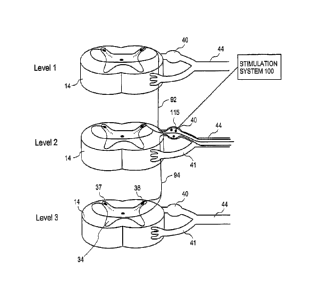
[0084] FIGURE 3A illustrates an embodiment of a stimulation system 100
of the present
invention with an electrode 115 implanted into a dorsal root ganglion (DRG)
40. The figure
illustrates three representative spinal levels 14 (i.e., spinal levels 1-3) of
the spinal cord 13.
-11-
CA 02579569 2007-03-06
WO 2006/029257
PCT/US2005/031960
The peripheral nerve 44 feeds into the dorsal root ganglion 40 and the ventral
nerve root 41
each of which feed into the spinal cord 13. The dorsal horns 37, 36 are also
indicated. For
clarity, the dura 32 and complete spinal cord 13 are not illustrated but are
present as described
elsewhere in this application and as occur in human anatomy. These exemplary
levels 1, 2 and
3 could be anywhere along the spinal cord 13. For simplicity, each level
illustrates the nerves
of only one side.
[0085] Using level 2 as a reference, an ascending pathway 92 is
illustrated between level 2 and
level 1 and a descending pathway 94 is illustrated from level 2 to level 3.
Application of
stimulation energy or signals to the DRG 40 in level 2 may be used to block
signals
progressing upstream from level 2 towards the path/pathways 92. Moreover,
modulation
applied to portions of level 2 but may also be used to effectively block the
neuron
paths/pathways from another level (here, alternatively using levels 1 and/or
3) from reaching
the brain. As such, application of stimulation to the level 2 DRG 40 using an
embodiment of
an apparatus and/or method of the present invention may advantageously provide
an effective
block of intrasegment pain pathways as well. It is to be appreciated that
while three
continuous levels are illustrated, some embodiments of the present invention
may be used to
stimulate 2 or more adjacent levels and still other embodiments may be used to
stimulate 2 or
more non-adjacent levels, or combinations thereof.
[0086] FIGURE 3B relates the spinal nerve roots to their respective
vertebral spinal levels.
The letter C designates nerves and vertebrae in the cervical levels. The
letter T designates
vertebrae and nerves in the thoracic levels. The letter L designates vertebrae
and nerves in the
lumbar levels. The letter S designates vertebrae and nerves in the sacral
levels. FIGURE 3C
illustrates the various dermatomes of the body related to their respective
nerve roots using the
designations in FIGURE 3B.
[0087] FIGURES 4-7 illustrate one embodiment of a stimulation system
activated under a
variety of control conditions to provide different levels and degrees of pain
control. FIGURES
4A, 5A, 6A and 7A all illustrate the stimulation system in various degrees of
activation.
FIGURES 4B, 5B, 6B and 7B illustrate a correspondingly influenced dermatome.
[0088] FIGURES 4A, 5A, 6A and 7A illustrate a stimulation system 100
having 3 electrodes
115 implanted into dorsal root ganglia 40 on two adjacent spinal levels. For
simplicity, each
spinal level illustrates a dorsal root ganglion 40, a ventral root 41 and a
peripheral nerve 44.
The exception is spinal level 3 that illustrates an additional dorsal root
ganglion 38, a ventral
root 39 and a peripheral nerve 42. The three electrodes 115 are designated
channels 1, 2 and 3
by the controller 106. Each electrode is activated to provide modulation
energy or signals
-12-
CA 02579569 2007-03-06
WO 2006/029257
PCT/US2005/031960
under the control of the controller 106. Exemplary electrodes for implantation
into a nerve
root ganglion are further described with regard to FIGs. 12A-13B. Level 3 is
an example of
bilateral electrode placement and level 2 is an example of unilateral
electrode placement. As
such, the illustrated embodiment is a multi-level, unilateral and bi-lateral
stimulation system.
Stimulation energy is provided by a pulse generator (not illustrated but
described in greater
detail below in FIGs. 26-29) under control of a suitable neurostimulation
controller 106. Those
of ordinary skill will recognize that any of a wide variety of known
neurostimulation
controllers may be used. Not illustrated in this view but present in the
system are suitable
connections between the various electrodes 115, electrode leads 110 and the
controller 106. In
the illustrations that follow, a line connecting the electrode lead 110 to the
controller 106
indicates "stimulation on" communication from the controller 106 to one
electrode 115 (see
FIG. 4A) or more than one electrode 115 (see FIG. 5A).
[0089] A signal of "stimulation on" indicates any of a wide variety of
stimulation patterns and
degrees of stimulation. The "stimulation on" signal may be an oscillating
electrical signal may
be applied continuously or intermittently. Furthermore, if an electrode is
implanted directly
into or adjacent to more than one ganglion, the oscillating electrical signal
may be applied to
one electrode and not the other and vice versa. One can adjust the stimulating
poles, the pulse
width, the amplitude, as well as the frequency of stimulation and other
controllable electrical
and signally factors to achieve a desired modulation or stimulation outcome.
[00901 The application of the oscillating electrical signal stimulates the
area of the nerve chain
where the electrode 115 is placed. This stimulation may either increase or
decrease nerve
activity. The frequency of this oscillating electrical signal is then adjusted
until the symptoms
manifest by physiological disorder being treated has been demonstrably
alleviated. This may
step may be performed using patient feedback, sensors or other physiological
parameter or
indication. Once identified, this frequency is then considered the ideal
frequency. Once the
ideal frequency has been determined, the oscillating electrical signal is
maintained at this ideal
frequency by storing that frequency in the controller.
[0091] In one specific example, the oscillating electrical signal is
operated at a voltage
between about 0.5 V to about 20 V or more. More preferably, the oscillating
electrical signal
is operated at a voltage between about 1 V to about 30 V or even 40V. For
micro stimulation,
it is preferable to stimulate within the range of 1V to about 20V, the range
being dependent on
factors such as the surface area of the electrode. Preferably, the electric
signal source is
operated at a frequency range between about 10 Hz to about 1000 Hz. More
preferably, the
electric signal source is operated at a frequency range between about 30 Hz to
about 500 Hz.
-13-
CA 02579569 2007-03-06
WO 2006/029257
PCT/US2005/031960
Preferably, the pulse width of the oscillating electrical signal is between
about 25
microseconds to about 500 microseconds. More preferably, the pulse width of
the oscillating
electrical signal is between about 50 microseconds to about 300 microseconds.
[0092] The application of the oscillating electrical signal may be
provided in a number of
different ways including, but not limited to: (1) a monopolar stimulation
electrode and a large
area non-stimulating electrode return electrode; (2) several monopolar
stimulating electrodes
and a single large area non-stimulating return electrode; (3) a pair of
closely spaced bi-polar
electrodes; and (4) several pairs of closely spaced hi-polar electrodes. Other
configurations are
possible. For example, the stimulation electrode(s) of the present invention
may be used in
conjunction with another non-stimulating electrode - the return electrode ¨ or
a portion of the
stimulation system may be adapted and/or configured to provide the
functionality of a return
electrode. Portions of the stimulation system that may be adapted and/or
configured to provide
the functionality of the return electrode include, without limitation, the
battery casing or the
pulse generator casing.
[0093] In the illustrated configuration, a stimulation pattern provided to
one of the electrodes
positioned in level 3 (i.e., channel #1 "ON") produces pain blocking/relief in
the indicated
region of the body (i.e., shaded area R1) in FIG. 4B.
[0094] It will be appreciated that embodiments of the present invention
can stimulate specific
dermatome distributions to probe which electrode or group of electrodes or
combination of
electrodes (including drug coated or delivery electrodes) is best positioned
or correlates most
closely to one or more specific areas of pain. As such, a stimulation system
according to an
embodiment of the present invention may be "fine tuned" to a specific area of
coverage or type
of pain. The results obtained from such testing can be used to one or more
stimulation or
treatment regimes (i.e., series of stimulations in the presence of or in
combination with a
therapeutic agent from a coated electrode) for a particular patent for a
particular type of pain.
These pain treatment regimes may be programmed into a suitable electronic
controller or
computer controller system (described below) to store the treatment program,
control and
monitor the system components execution of the stimulation regime as the
desired therapeutic
regime is executed.
[0095] FIG. 5A provides another example of distribution of pain relief
using a multi-channel
stimulation system and method. In the illustrated configuration and
stimulation pattern, a
stimulation pattern is provided to one electrode each in levels 2 and 3 via
channels #1 and #2.
This stimulation electrode pattern provides pain blocking/relief in the
indicated region of the
body (i.e., areas R1, R2) of FIG. 5B.
-14-
CA 02579569 2007-03-06
WO 2006/029257
PCT/US2005/031960
[0096] FIG. 6A provides another example of distribution of pain relief
using a multi-channel
- stimulation system and method. In the illustrated configuration and
stimulation pattern, a
stimulation pattern provided to both electrodes in level 3 via channels #1 and
#3 provides pain
blocking,/relief in the indicated region of the body (i.e., area R3) of FIG.
6B.
[0097] FIG. 7A provides another example of distribution of pain relief
using a multi-channel
stimulation system and method. In the illustrated configuration and
stimulation pattern, a
stimulation pattern is provided to all electrodes in the system via channels
#1, #2 and #3. This
stimulation electrode pattern provides pain blocking/relief in the indicated
region R4 of the
body (i.e., FIG. 7B). It is to be appreciated that the electrode placement and
blocking region
patterns illustrated by FIGs. 4A-7B may be modified using information such as
in FIGs. 3B
and 3C for targeted placement to specific portions of the body depending upon
individual
needs.
[0098] Micro-electrode and stimulation system embodiments of the
present invention may be
implanted into a single nerve root ganglion utilizing the implantation methods
of the present
invention. The implantation methods described herein provide numerous
advantages,
including but not limited to: low risk percutaneous access route similar to
other procedures,
direct delivery of localized quantities of pharmacological agents at the nerve
root when using
embodiment having electrodes coated with pharmacological agents, and electrode
placement
that enables preferential, selective nerve fiber stimulation.
[0099] FIG. 8A illustrates a cross section view of a spinal level.
Peripheral nerves 44, 42 feed
into dorsal root ganglia 40, 38 and ventral nerves 41, 39 respectively. A
vertebral body 70 and
two sympathetic nerve ganglia 62, 63 are also illustrated. In this embodiment,
the method
includes advancing a suitable catheter 107 medially towards the vertebral body
70, then along
the peripheral nerve 42 towards the dorsal root ganglion 38. The catheter 107
is advanced
using external imaging modalities for guidance such as fluoroscopy or other
suitable medical
imaging technique. The vertebral foramen offers a good landmark visible under
fluoroscopy
and useful in locating the DRG 38.
[00100] The electrode 115 is implanted in proximity to the dorsal root
ganglion by forming an
opening in the dorsal root ganglion epinurium and passing the electrode
through the opening
(FIG. 8A, 8B). The opening may be formed using conventional methods such as a
cutting
edge on or provided to the tip of the catheter 107, with an instrument
advanced through a
working channel within the catheter 107 or through the use of other suitable
endoscopic or
minimally invasive surgical procedure. Alternatively, the electrode body or
distal end may be
provided with a tissue cutting or piercing element to aid in piercing tissue
(see, e.g., tip 908 in
-15-
CA 02579569 2007-03-06
WO 2006/029257
PCT/US2005/031960
FIG. 20A). As the catheter 107 is withdrawn, the microelectrode leads 110 are
deployed and
attached, anchored or otherwise secured to the tissue, anatomy or bones
adjacent the DRG 38
to reduce the likelihood that electrode 115 will be pulled from the DRG 38. In
alternative
embodiments described below, the microelectrode leads 110 may be fixed prior
to electrode
implantation into a nerve root ganglion.
[00101] Note that the electrode 115 is sized and shaped to fit within the
DRG 38. A typical
DRG is generally spherical with a diameter of 3-5mm. Of course, a range of DRG
sizes occur
in humans and may vary in size depending on the age and sex of the individual
and other
factors. Electrode embodiments may be provided in a range of sizes to
accommodate the
specific anatomical characteristics of a patient. A number of factors are
considered when
selecting an appropriate DRG electrode embodiment for use in an individual.
[00102] Electrode placement within the DRG may be confirmed using
neurodiagnostic testing
techniques such as somatosensory evoked potential (SSEP) and electromyography
(EMG)
adapted for the methods and systems described herein. One illustrative example
includes the
placement of sensing electrodes in the sensory nervous system above and below
the DRG level
having the implanted electrode(s). Implant the electrode into the targeted
DRG. Apply a test
stimulation to the DRG and measure voltage potential at the sensory electrodes
above and
below the targeted DRG to confirm that the electrode is implanted in the
targeted DRG. A test
stimulation may range from 0.4 v to 0.8v at 50Hz or may be some other suitable
stimulation
level based on the evoked potential measurement technique used. In this way,
conventional
fluoroscopy techniques and instruments may be used to advance towards and
implant the
electrode into the DRG and confirm that the electrode is correctly implanted
and stimulating
the targeted DRG.
[00103] A number of different approaches are available for maneuvering an
electrode into
position on, in or about a DRG. Several exemplary approaches are provided in
FIGs. 8-10 in a
section view of the cauda equina portion of the spinal cord. In these
examples, electrodes 115
are placed on or in a ganglion on a representative sacral spinal level.
Sympathetic nervous
system ganglia 62, 63 are also indicated. DRG 40 and ventral root 41 are
associated with
peripheral nerve 44. DRG 38 and ventral root 39 are associated with peripheral
nerve 42.
[00104] FIGs. 8A and 8B illustrate a lateral approach to a DRG 38 using a
suitable catheter 107.
The catheter advances adjacent to the peripheral nerve 42 medially towards the
DRG 38. The
DRG dura is pierced laterally and the electrode 115 is advanced into the DRG
interior.
Thereafter, the electrode 115 is implanted into the DRG interior. Next, as is
illustrated in FIG.
8B, the catheter 107 is withdrawn from the DRG 38 and deploys the electrode
leads 110. The
-16-
CA 02579569 2007-03-06
WO 2006/029257
PCT/US2005/031960
electrode leads 110 may be anchored to the vertebral body 70 using suitable
fixation
techniques. The leads 110 are then connected to a pulse generator/controller
(not shown).
[00105] FIG. 9A is anatomically similar to FIGs. 8A and 8B. FIG. 9A
illustrates an alternative
DRG implantation approach that crosses the medial line inferior to the DRG of
interest. The
catheter 107 is advanced in a superior pathway towards the foramen and using
the foramen
under fluoroscopic guidance into the DRG. As illustrated in FIGs 9A and 9B,
there is provided
a method of stimulating a dorsal root ganglion by implanting an electrode
within the dorsal
root ganglion. In some embodiments, the implanting procedure includes passing
a portion of
the electrode through the spinal epidural space. Electrodes in systems of the
present invention
onto or in the nerve root epinurium 72 (FIG. 10A and 10B) or within the nerve
root (i.e., FIGs.
9A,B). Moreover, in some embodiments, there is also the step of forming an
opening in the
dorsal root ganglion epinurium 72 and then passing the electrode through the
opening (see, i.e.,
FIG. 9B).
[00106] FIG. 11 illustrates a section view through a portion of the spinal
cord 13 with another
alternative electrode implantation technique. In contrast to the earlier
described methods that
externally approach the DRG and involve piercing or entering the DRG epinurium
72, FIG. 11
illustrates an internal approach to the DRG interlascular from within the
nerve sheath of a
peripheral nerve 44. FIGURE 11 illustrates a section view of the nerve sheath
partially
removed to reveal the underlying nerve bundle 46. In this illustrative
example, an opening is
made in the peripheral nerve 44 sheath at a point 45 lateral to the DRG 40.
The microelectrode
115 enters the nerve 44 sheath through opening 45 using suitable endoscopic or
minimally
invasive surgical techniques. Next, the electrode 115 is advanced towards and
into the DRG
40.
[00107] As each of these illustrative embodiments make clear, the placement
of the electrode
relative to the DRG enables activating the electrode to selectively stimulate
sensory nerves.
Additionally, the placement of the electrode according to the methods of the
invention enable
activating the electrode to stimulate sensory nerves within the DRG or without
stimulating
motor nerves in the nearby ventral root. The control system described herein
also provides
stimulation levels that activate the electrode to stimulate at a level that
preferably stimulates
myelinated fibers over unmyelinated fibers.
[00108] In addition, as will be described in greater detail below, FIG. 11
illustrates an electrode
embodiment where the electrode tip and shaft may be coated with
pharmacological agents to
assist in the stimulation therapy or provide other therapeutic benefit. As
illustrated, the
electrode includes a tip coating 130 and a shaft coating 132. The
pharmacological agent in
-17-
CA 02579569 2007-03-06
WO 2006/029257
PCT/US2005/031960
each coating 130, 132 could be the same or different. One advantage of
implanting through the
nerve sheath is that the coated shaft 132 may include a pharmacological agent
active or
beneficial to neural activity in the ventral nerve root 41 since this coated
shaft is
advantageously positioned proximal to the ventral root 41. The shaft coating
132 may also be
selected to reduce inflammation or irritation caused by the presence of the
shaft within the
nerve sheath.
[00109] FIGs. 12A and 12B illustrate an embodiment of an exemplary anchor
body 171 with a
fixation hook 172 used to secure the leads 110 once the electrode 115 is
implanted into the
DRG 40. FIG. 12A is a section view of a portion of the spinal cord 13 showing
the dorsal root
42, ventral root 41, DRG 40 and peripheral nerve 44. In this illustrative
embodiment, a
catheter 70 is used to maneuver the electrode 115, leads 110 and anchor 171
about the DRG
40implantation site. Once a suitable site is identified, the hook 172 is
inserted into the fascia
layer of the DRG. The hook 172 may have various shapes and contours to adapt
it to engaging
with and securing to the outer DRG layer or within the outer DRG layer. FIG.
12B illustrates
an exemplary anchor body 171 and hook 172 mounted onto the distal end of a
catheter 70. The
anchor body 171 and hook 172 may be maneuvered into position using the
catheter 70 alone or
in combination with other suitable surgical , endoscopic or minimally invasive
tools.
Similarly, the electrode 115, leads 110 may be moved into position for
implantation on, in or
about targeted neural tissue. In other alternative electrode embodiments, the
electrode 115 is
implanted on, in or about a DRG is provided with a flexible tip that helps to
prevent or mitigate
chronic friction and ulceration.
[00110] Alternatively, the electrode leads 110 or other supporting or
anchoring structures may
be attached to the adjacent bony structure, soft tissue or other neighboring
anatomical
structures. In addition, there may also be provided a fixation, anchoring or
bonding structure
positioned proximal to the electrode anchor 172 that absorbs some or all
proximal movement
of the leads 110 so that the electrode is less likely to be pulled from or
dislodged from the
implantation site. The goal of the anchoring and other strain absorbing
features is to ensure the
electrode remains in place within or is less likely to migrate from the
implanted position
because of electrode lead 110 movement (i.e., lead 110 movement pulls the
electrode 115 from
the implantation site or disrupts the position of the electrode 115 within the
implantation site).
It is to be appreciated that numerous techniques are available to aid in
electrode placement
including percutaneous placement of single/multiple hooks or anchors,
vertebral anchor or
posts, micro-sutures, cements, bonds and other joining or anchoring techniques
known to those
of ordinary skill in the art. It is also to be appreciated that other
components of the stimulation
-18-
CA 02579569 2007-03-06
WO 2006/029257
PCT/US2005/031960
system embodiments described herein may also be adapted for attachment to
surrounding
tissue in proximity to the stimulation site or near the electrode implantation
site. Other
components include, for example, the stimulation controller, master
controller, slave controller,
pulse generator, pharmacological agent reservoir, pharmacological agent pump
and the battery.
[00111] FIG. 12C illustrates an exemplary anchoring of electrode leads 110
to bone surrounding
the electrode implantation site. FIG. 12C illustrates a section view through a
portion of the
spinal cord 13 showing the ventral root 41, the dorsal root 42 and dorsal root
ganglion 40.
FIG. 12C also illustrates the surrounding bone of the spine such as vertebral
body 1110, the
spinous process 1115, the pedicle 1120, the lamina 1125, the vertebral arch
1130, transverse
process 1135, and facet 1140. Electrode 115 is implanted into the DRG 40 and
the electrode
leads are held in place using a suitable anchor 111. In this embodiment, the
anchor 111 is
secured to the vertebral body 1110. The anchor 111 represents any suitable
manner of securing
the bony portions of the spine such as tacks, staples, nails, cement, or other
fixation methods
known to those in the surgical or orthopedics arts. A strain relief 122 is
present between
anchor 111 and the DRG 40 (see FIG. 13A and 14A). The strain relief 122 is
used to absorb
motion that may move the electrode 115 within the DRG 40 or remove the
electrode from the
DRG 40. In this illustrative embodiment, the strain relief 122 is a coiled
portion of the
electrode lead 110. One or more strain reliefs 122 may be provided between the
anchor 111
and the DRG 40 or between the anchor 111 and the battery or controller of the
stimulation
system (not shown).
[00112] FIGs. 13A -14B illustrate mono-polar and bi-polar stimulation
component
embodiments of the present invention. FIG. 13A illustrates a mono-polar
stimulation
component that has a proximal connector 126A adapted to be connected to a
pulse generator.
A distal electrode 115 is configured to be implanted within the body at a
stimulation site. The
distal electrode may be a mono-polar electrode 115A (FIG. 13B) or a bi-polar
electrode 115B
(FIG. 14B). The electrodes are sized for implantation into a nerve root
ganglion and will vary
according to the nerve root selected. In additional alternative embodiments,
the electrode leads
and electrode are adapted and sized to advance within a nerve sheath to a
nerve root ganglion.
The electrodes or their casing may be made of inert material (silicon, metal
or plastic) to
reduce the risk (chance) of triggering an immune response. Electrodes should
be studied for
suitability to MRI and other scanning techniques, including fabrication using
radio-opaque
materials as described herein.
[00113] Returning to FIG. 13A, an electrical lead 110 is connected to the
proximal connector
126A and the distal electrode 115. A strain relief mechanism 122 is connected
in proximity to
-19-
CA 02579569 2007-03-06
WO 2006/029257
PCT/US2005/031960
the stimulation site. The illustrated strain relief mechanism is formed by
coiling the electrical
lead 110. Other well known strain relief techniques and devices may be used. A
fixation
element 124 adapted to reduce the amount of movement of the electrical lead
proximal to a
fixation point is positioned in, on, or through an anatomical structure
proximal to the
stimulation site. Multiple elements are provided to mitigate or minimize
strain and force
transmission to the micro-leads 110 or the microelectrodes 115 because the
microelectrodes
and microelectrode leads used herein are very small and include fine, flexible
wires on the
order of lmm or less and in many cases less than 0.5 mm. Representative
electrode and lead
dimensions will be described in greater detail below (FIG. 15A, 15B). As such,
in some
embodiments, strain and movement may be absorbed or mitigated by the fixation
element 124,
the strain relief 122 and the electrode anchor 117 (if included). The fixation
element 124 may
be, for example, a loop, or a molded eyelet. The fixation element may be
sutured, tacked,
screwed, stapled, bonded using adhesives or joined using other techniques
known to those of
ordinary skill to secure the fixation element within the body for the purposes
described herein.
[00114] In one specific implantation embodiment, the method of implanting
the electrode is
modified based on consideration of the small size and delicate nature of the
microelectrode and
microelectrode leads. As such, high force actions are taken first followed by
light force
actions. In this way, the fine microelectrode and microelectrode lead
materials are not present
during high force operations. Consider an example where an electrode of the
present invention
will be implanted into a DRG. In an exemplary embodiment, the fixation element
124 is a
loop sized to allow passage of the electrode 115. Perform the high force
operation of
anchoring or otherwise fixing (i.e., adhesion) the fixation element into a
vertebral foramen
adjacent the selected DRG stimulation site. In general, the fixation site
should be as close as
practical to the stimulation site. In one specific embodiment, the fixation
site is within 3 cm to
cm of the stimulation site. Optionally, a guide wire attached to the loop
remains in place and
is used to guide the electrode and leads to the loop and hence to the implant
site. The electrode
and leads are passed through the loop (with or without use of a guide wire).
The electrode is
then implanted on or in the DRG. Optionally, an anti-strain device 122 may
also be positioned
between the electrode in the implantation site and the fixation element 124.
In one illustrative
embodiment, a section of microelectrode lead containing a plurality of loops
is used as an anti-
strain device 122. Finally, the microelectrode lead is secured to the loop
using a suitable
locking device. It is to be appreciated that the above method is only
illustrative of one method
and that the steps described above may be performed in a different order or
modified
depending upon the specific implantation procedure utilized.
-20-
CA 02579569 2007-03-06
WO 2006/029257
PCT/US2005/031960
[00115] In some embodiments, there may also be provided an anchoring
mechanism proximal
to the distal electrode 115. Examples of anchoring mechanisms include, for
example, anchors
117 illustrated in FIGs. 13B and 14B. In still further embodiments, the
anchoring mechanism
is adapted to anchor the distal electrode 115 within the stimulation site. For
example, the
anchor mechanism may remain stowed flat against the electrode body 118 during
implantation
and then deploy from within a nerve root ganglion to anchor against the
interior nerve root wall
to support the electrode and prevent electrode migration or pull-out. In some
embodiments the
anchoring mechanism and the distal electrode are integrally formed and in
other embodiments
they are separate components. In some embodiments, the anchoring mechanism is
formed
from a polymer or a silicone.
[00116] Selective nerve stimulation affords the use of smaller electrodes.
Smaller electrodes
create less impingement and are less susceptible to unwanted migration.
However, as
electrode surface area decreases the impedance of the electrode increases
(FIG. 15A). As such,
some electrode embodiments will have an impedance much greater than the
impedance of
conventional stimulation electrodes. In one embodiment, the impedance of a
microelectrode of
the present invention is more than 2500n. This difference in impedance also
impacts the
performance requirements of stimulation systems, pulse generators and the like
used to drive
the microelectrodes described herein.
[00117] Distal electrodes may come in a wide variety of configurations,
shapes and sizes
adapted for implantation into and direct stimulation of nerve root ganglion.
For example, the
distal electrode 115 may be a ring of conductive material attached the leads
110. Alternatively,
the distal electrode 115 may be formed from an un-insulated loop of electrical
lead. The loop
electrode is appealing and has improved wear properties because, unlike the
ring that must be
joined to the leads 110, the loop is formed from the lead and no joining is
needed. In still other
embodiments, the electrode may be an un-insulated portion of the lead.
[00118] Regardless of configuration, electrodes of the present invention
are sized and adapted
for implantation into, on or about a ganglion such as, for example, a dorsal
root ganglion or a
ganglion of the sympathetic nervous system. It is to be appreciated that the
size of the
electrode varies depending upon the implantation technique and the size of the
target ganglion.
An electrode implanted through the DRG dura (i.e., FIG. 9A) may be less than 5
mm since the
diameter of a DRG may be only 3-5 mm. On the other hand an electrode adapted
for
implantation along the peripheral nerve sheath (i.e., FIG. 11) may be longer
than the electrode
that passes through the dura but may face other design constraints since it
must advance
distally within the nerve sheath to reach the DRG. It is to be appreciated
that dimensions of
-21-
CA 02579569 2007-03-06
WO 2006/029257
PCT/US2005/031960
electrode embodiments of the present invention will be modified based on, for
example, the
anatomical dimensions of the implantation site as well as the dimensions of
the implantation
site based on implantation method.
[00119] FIG. 15B provides some exemplary electrode surface areas for
electrode embodiments
formed from wire diameters between 0.25mm to 1 mm, having widths of 0.25mm or
0.5 mm.
As such, embodiments of the present invention provide distal electrode surface
area that is less
than 0.5 mm2. In other embodiments, the distal electrode surface area is less
than 1 mm2. In
still other embodiments, the distal electrode surface area is less than 3 mm2.
[00120] The sizes of the electrodes of the present invention stand in
contrast to the conventional
paddle 5 having dimensions of about 8mm wide and from 24 to 60mm long (FIG.
1). One
result is that conventional stimulation electrodes have larger electrode
surface areas than
electrode embodiments of the present invention. It is believed that
conventional electrodes
have an impedance on the order of 500 to 1800C2 operated using a stimulation
signal generated
by a 10-12 volt pulse generator. In contrast, stimulation electrode
embodiments of the present
invention have an impedance on the order of 21d2 or about 2500 from 2k52 to
101M or
higher or even in the range of 10k.Q to 20ka As will be described in greater
detail below,
some pulse generator embodiments of the present invention operate with
voltages produced by
DC-DC conversion into ranges beyond conventional stimulation systems.
[00121] The electrodes may be formed from materials that are flexible and
have good fatigue
properties for long term use without material failure. The electrode material
should be formed
from a biocompatible material or coated or otherwise treated to improve bio
compatibility.
Additionally, electrode materials should be opaque to imaging systems, such as
fluoroscopy,
used to aid electrode placement during implantation procedures. Examples of
suitable
materials include but are not limited to Pt, Au, NiTi, PtIr and alloys and
combinations thereof.
Electrodes may also be coated with a steroid eluding coating to reduce
inflammation at the
implantation or stimulation site.
[00122] With the small surface areas, the total energy required for
stimulation of the DRG is
drastically reduced because we can achieve high current densities with low
currents. One
advantage of using microelectrodes is that only a small volume of tissues in
the immediate
vicinity of the electrodes is stimulated. Another advantage of using
microelectrodes is the
correspondingly smaller pulse generator and because of decreased battery size.
[00123] In addition to the implantable electrodes described above,
alternative electrode
embodiments may also be used to selectively stimulate a nerve root ganglion.
FIG. 16
illustrates an embodiment where conductive rings 205, 207 are positioned on
either end of a
-22-
CA 02579569 2007-03-06
WO 2006/029257
PCT/US2005/031960
dorsal root ganglion 40. When activated, the rings 205, 207 capacitively
couple stimulation
energy into the DRG 40. FIG. 17 illustrates an alternative capacitive
stimulation configuration
where the capacitive plates 210, 212 are attached to the DRG dura. Embodiments
of the
present invention are not limited to only one pair of capacitive plates but
more than one pair
may be used. FIG. 18 illustrates two pairs of capacitive plates attached to
the dura of a DRG
40. One pair includes plates 210, 212 and the other pair includes plate 214
and another plate
(not shown). As an alternative to attaching the plates directly to the dura,
the plates may be
attached to an electrode support element 230 adapted to slip around and engage
with the DRG
dura. Once the electrode support element 230 is in position about the DRG, the
plates are
properly positioned to selectively stimulate a DRG. The present invention is
not limited to
only capacitively coupled stimulation energy. FIG. 20 illustrates another
alternative
embodiment where a wire 235 is wrapped around a DRG 40 creating coils 236 that
may be
used to inductively couple stimulation energy into a nerve root ganglion. For
purposes of
discussion, these embodiments have been described in the context of
stimulation a DRG. It is
to be appreciated that the techniques and structures described herein may also
be used to
stimulate other nerve root ganglion, other neural structures or other
anatomical features.
[00124] FIGs. 20A and 20B illustrate another electrode embodiment adapted
for implantation
through neural tissue. Piercing electrode 900 has a body 902, a distal end
904, and a proximal
end 906. A electrode surface or component 912 receives stimulation signals and
energy from a
pulse generator/controller (not shown) via a suitable lead 914. The distal and
904 has a tip 908
adapted to pierce the targeted neural tissue. In addition, one or more anchors
910 are provided
at the distal end to help secure the electrode body 902 within the targeted
neural tissue. A
securing ring 920 (FIG. 20B) is provided to secure the electrode body 902 to
or relative to the
targeted neural tissue. The anchors 910 may be in a first or stowed position
against the
electrode body 902 during insertion through the neural tissue and then be
moveable into a
second or deployed position away from the electrode body 902. In the deployed
position (FIG.
20A, 20C and 20D) the anchors 910 resist the movement of the electrode 900 out
of the neural
tissue. Numerous alternative anchor configurations are possible. Anchor 910
could be a series
of individual struts arrayed in a circular pattern or struts with material
between them similar to
the construction of an umbrella. Anchor 910 could also be a single anchor.
[00125] The electrode 900 includes a body 902 adapted to pass completely
through targeted
neural tissue while positioning the electrode 912 within a portion of the
targeted neural tissue.
In this illustrative embodiments that follow, the electrode body 902 is
adapted to fit within a
DRG 40 (FIG. 20D) or a ganglion of the sympathetic chain (FIG. 20C). The
electrode 912
-23-
CA 02579569 2013-07-22
may be placed in any location on the electrode body 902 to obtain the desired
stimulation or
modulation level. Additionally, the electrode 912 may be placed so that
modulation or
stimulation energy patterns generated by the electrode 912 will remain within
or dissipate only
within the targeted neural tissue.
[00126] A securing ring 920 is used to hold the electrode body 902 in
position within and
relative to the targeted neural tissue. The securing ring 920 is ring shaped
having an annulus
922. In some embodiments, the inner surface 942 is used as a friction locking
surface to
engage and hold the electrode body 902. In other embodiments, the inner
surface 942 contains
a surface treatment to secure the electrode body. In still other embodiments,
the inner surface
942 is adapted to mechanically engage with and secure the electrode body 902.
The securing
ring 920 may be formed from a suitable elastic or inelastic material that may
be secured to the
electrode body 902 and the outer layer of the targeted neural tissue to help
prevent electrode
pull out or dislodgement. The securing ring 920 may be formed from a
biocompatible material
suited to gluing or mechanically affixing the ring 920 to the electrode body
902 and the tissue
outer layer. The securing ring 920 may be present during or positioned after
the electrode 900
is implanted into the targeted neural tissue. In one alternative embodiment,
the securing ring
920 is secured to the DRG outer layer and has a complementary engaging feature
positioned to
engage with an engaging feature on the electrode 900. The electrode body 902
advances
through the securing ring annulus 922 and into the DRG 40 until the
complementary engaging
features engage and stop further distal motion of the electrode body 902 into
the DRG. The
complementary engaging features may be used alone or in combination with
anchors 910 to
assist in electrode 900 placement within neural tissue such as a DRG or other
ganglion.
[00127] FIGs. 20C and 20D illustrate electrode embodiments adapted for
implantation through
targeted neural tissue illustrated in a section view of the spinal cord 13.
Additional details of
the various portions of the spinal cord section 14 are described below with
regard to FIG. 38.
Also illustrated in these views are exemplary sensory pathways 52/54 and motor
pathways 41P
within peripheral nerve 44 and roots 41/42 and entering the spinal cord.
Alternative
implantation sites and stimulation alternatives are described in US Patent
6,871,099,,,
[00128] In the illustrative embodiment of FIG. 20C, the electrode 900 is
positioned to remain in
a non-central location within the targeted neural tissue. In this embodiment,
the targeted neural
tissue is a ganglion 992 within the sympathetic chain 990. Additional details
and specific
targeted neural tissue within the sympathetic chain are described below with
regard to FIGs. 32
and 33. The electrode 912 is placed on or in the electrode body 902 so that
when the electrode
-24-
CA 02579569 2007-03-06
WO 2006/029257
PCT/US2005/031960
body 902 passes through the ganglion 992 and is seated within the securing
ring 920 the
electrode 912 is in the desired position within the interior of the ganglion
992. Other electrode
912 placement within the targeted neural tissue is possible, for example, by
varying the length
of the electrode body 902, the angle of penetration into the targeted neural
tissue or the
position of initial penetration into the targeted neural tissue.
[00129] In the illustrative embodiment of FIG. 20D, the electrode 900 is
positioned to remain in
a generally central location within the targeted neural tissue. In this
embodiment, the targeted
neural tissue is a DRG 40. The electrode 912 is placed on or in the electrode
body 902 such
that when the electrode body 902 is seated within the securing ring 920, then
the electrode 912
is in the middle of about the middle or center the DRG 40. As before the
securing ring 920 and
flat anchor 911 secure the electrode 900 in the desired position within the
DRG 40. The flat or
flap anchor 911 provides similar functionality as the anchor 910. The anchor
911 has flat
anchors rather than the curved anchors 910.
[00130] In some embodiments, the stimulation electrode tip may be
coated with a
pharmacological agent. In the embodiment illustrated in FIG. 21, a coating 130
covers that
portion of the electrode within the DRG 40. In other embodiments, less or more
of the
electrode or other implanted components may be suitably coated to achieve a
desired clinical
outcome. FIG. 21 also illustrates a coating 130 on the electrode shaft or
portion of the
electrode exterior to the DRG. The coating 132 may be the same or different
than the coating
130. For example, the tip coating 130 may include a distal coating containing
an agent to aid
in the effective stimulation of the DRG. The tip coating 130 may also include
a more proximal
coating portion (i.e., near where the electrode pierces the dura) that
contains an agent to
prevent fibrous growth about the electrode. In a further embodiment, the shaft
coating 132
would also contain an agent to prevent fibrous growth about the electrode.
Additionally, the
shaft coating 132 may be selected based on providing a pharmacological agent
to interact with
the tissue in the ventral root (i.e., the implantation technique in FIG. 11)
or within the
peripheral nerve sheath.
[001311 Examples of desired clinical outcomes provided by pharmacological
agents used as
coatings include but are not limited to reduction of scar tissue development,
prevention of
tissue growth or formation on the electrode, anti-inflammation, channel
blocking agents and
combinations thereof or other known pharmacological agents useful in treatment
of pain, or
neurological pathologies. In other alternative embodiments, the
pharmacological agent may
include other compounds that, when placed within the body, allow the
pharmacological agent
to be released at a certain level over time (i.e., a time released
pharmacological agent). In
-25-.
CA 02579569 2007-03-06
WO 2006/029257
PCT/US2005/031960
some embodiments, the pharmacological agent is an anti-inflammatory agent, an
opiate, a
COX inhibitor, a PGE2 inhibitor, combinations thereof and/or another suitable
agent to prevent
pathological pain changes after surgery. Other suitable pharmacological agents
that may be
used include those used to coat cardiac leads, including steroid eluding
cardiac leads or other
agents used to coat other implantable devices.
[00132] Embodiments of the present invention include direct stimulation of
a nerve root
ganglion or other neurological structure while releasing a pharmacological
agent from an
electrode used to provide stimulation. In one embodiment, the pharmacological
agent is
released before the electrode is activated. In other embodiments, the
pharmacological agent is
released after or during the electrode is activated. In still other
embodiments, the
pharmacological agent is pharmacologically active in the nerve root ganglion
during
stimulation of the nerve root ganglion. It is to be appreciated that
embodiments of the present
invention may be altered and modified to accommodate the specific requirements
of the neural
component being stimulated. For example, embodiments of the present invention
may be used
to directly stimulate a dorsal root ganglion or a nerve root ganglion of the
sympathetic system
using the appropriate pharmacological agents, agent release patterns and
amounts as well as
stimulation patterns and levels.
[00133] Turning now to FIG. 22, various stimulation mechanisms are shown.
While these
various mechanisms potentate pain, each of them acts on the primary sensory
neuron. The
primary modulator of this cell is its cell body, the DRG 40. One aspect of the
present
invention is to advantageously utilize the anatomical placement of the DRG 40
within the
nervous system to complement other treatment modalities. In another
embodiment,
stimulation of the DRG 40 as described herein is used in conjunction with a
substance acting
on a primary sensory neuron. As shown, the other mechanisms are nearer to the
illustrated
tissue injury than the DRG cell body 40. Put a different way, the DRG 40 is
upstream (i.e.,
closer to the brain/spinal cord 13) of the other pain mechanisms. Thus, this
is another
illustration of how upstream DRG stimulation may be used to block and/or
augment another
pain signals.
[00134] Electrophysiological studies suggest that Prostaglandin E2 (PGE2),
produced by COX
enzymes, increases the excitability of DRG neurons in part by reducing the
extent of
membrane depolarization needed to activate TTX-R Na+ channels. This causes
neurons to
have more spontaneous firing and predisposed them to favor repetitive spiking
(translates to
more intense pain sensation). Also illustrated here is how other pro-
inflammatory agents
(Bradykinin, Capsaicin on the Vanilloid Receptor [VR1]) converge to effect the
TTX-R NA+
-26-
CA 02579569 2007-03-06
WO 2006/029257
PCT/US2005/031960
channel. Opiate action is also upstream from the TTX-R Na+ channel modulation.
Embodiments of the present invention advantageously utilize aspects of the
pain pathway and
neurochemistry to modify electrophysiological excitability of the DRG neurons
where
electrical stimulation is coupled with pharmacological agents (electrical
stimulation alone or in
combination with a pharmacological agent) to optimize the efficacy of the
stimulation system.
[00135] Synergy of electrical and pharmacological modulation may also be
obtained using a
number of other available pharmacological blockers or other therapeutic agents
using a variety
of administration routes in combination with specific, directed stimulation of
a nerve root
ganglion, a dorsal root ganglia, the spinal cord or the peripheral nervous
system.
Pharmacological blockers include, for example, Na + channel blockers, Ca++
channel
blockers, NMDA receptor blockers and opoid analgesics. As illustrated in FIGs
23A and 23B,
there is an embodiment of a combined stimulation and agent delivery electrode.
Note the
bipolar electrodes 115B on the tip, the coating 130 and the beveled tip shape
for piercing the
dura during implantation. The electrode tip is within the DRG epinurium 72 and
well
positioned to modify and/or influence c-fiber 55 responsiveness. In the
illustration, circles
represent Na+ ions, triangles represent Na+ channel blockers (such as, for
example, dilantin ¨
[phenytoin], tegretol ¨ [carbamazapine] or other known Na+ channel blockers).
As the agent
is released from coating 130, receptors on c-fiber 55 are blocked thereby
decreasing the
response of the c-fiber below the response threshold (FIG. 23B). Because the
activation
potential of the c-fiber has been lowered, the larger diameter A-fiber is
preferentially
stimulated or the response of the A-fiber remains above the threshold in FIG.
23B.
[00136] Embodiments of the present invention also provide numerous
advantageous
combinational therapies. For example, a pharmacological agent may be provided
that acts
within or influences reactions within the dorsal root ganglia in such a way
that the amount of
stimulation provided by electrode 115B may be reduced and yet still achieve a
clinically
significant effect. Alternatively, a pharmacological agent may be provided
that acts within or
influences reactions within the dorsal root ganglia in such a way that the
efficacy of a
stimulation provided is increased as compared to the same stimulation provided
in the absence
of the pharmacological agent. In one specific embodiment, the pharmacological
agent is a
channel blocker that, after introduction, the c-fiber receptors are
effectively blocked such that a
higher level of stimulation may be used that may be used in the presence of
the channel
blocking agent. In some embodiments, the agent may be released prior to
stimulation. In other
embodiments, the agent may be released during or after stimulation, or in
combinations
thereof. For example, there may be provided a treatment therapy where the
agent is introduced
-27-.
CA 02579569 2013-07-22
alone, stimulation is provided alone, stimulation is provided in the presence
of the agent, or
provided at a time interval after the introduction of the agent in such a way
that the agent has
been given sufficient time to introduce a desired pharmacological effect in
advance of the
applied stimulation pattern. Embodiments of the stimulation systems and
methods of the
present invention enable fine tuning of C-fiber and AP-fiber thresholds using
microelectrodes
of the present invention having pharmacological agent coatings coupled with
electrical
stimulation. Representative pharmacological agents include, but are not
limited to: Nat
channel inhibitors, Phenytoin, Carbam.azapine, Lidocaine GDNF, Opiates,
Vicodin, Ultrarn,
and Morphine.
[00137] FIGs 23C and 23D illustrate alternative embodiments for combination
neurostimulation
and pharmacological agent delivery systems. Additional details of the
controller and pulse
generated systems suitable for these operations are described below With
reference to FIGs. 26-
29. While described using combined pump and reservoir delivery systems, it is
to be
appreciated that the pump for moving the pharmacological agent from the
reservoir to and out
of the electrode and the reservoir for storing the pharmacological agent
before delivery may be
two separate components that operate in a coordinated fashion. Pumps and
reservoirs may be
any of those suited for controlled delivery of the particular pharmacological
agent being
delivered. Suitable pumps include any device adapted for whole implantation in
a subject, and
suitable for delivering the formulations for pain management or other
pharmacological agents
described herein. In general, the pump and reservoir is a drug delivery device
that refers to an
implantable device that provides for movement of drug from a reservoir
(defined by a housing
of the pump or a separate vessel in communication with the pump) by action of
an operatively
connected pump, e.g., osmotic pumps, vapor pressure pumps, electrolytic pumps,
electrochemical pumps, effervescent pumps, piezoelectric pumps, or
electromechanical pump
systems. Additional details of suitable pumps are available in US Patents
3,845,770;
3,916,899; 4,298,003 and 6,835,194.
[00138] FIG. 23C illustrates a combined system controller and pulse
generator 105B adapted to
control the delivery of pharmacological agents from the agent reservoir and
pump 195. The
pharmacological agent pumped from the agent reservoir and pump 195 travels via
a dedicated
conduit into a common supply 110F, through a strain relief 122F and into the
agent and
stimulation electrode 2310. The common supply 110F may be a single line
containing both
electrode control and power signals from the controller 105B as well as agent
delivered from
-28-
CA 02579569 2007-03-06
WO 2006/029257
PCT/US2005/031960
the pump 195 or there could be two separate lines joined together. Regardless
of
configuration, common supply 110F simplifies implantation procedures because a
single line is
used to connect the electrode 2310 to the controller 105B and the pump 195.
[00139] The combination neurostimulation and pharmacological agent delivery
electrode 2310
includes a body 2312 adapted to fit within targeted neural tissue. In this
illustrative
embodiment, the electrode body 2310 is adapted to fit within a DRG 40. An
electrode 2318 is
positioned on or in the electrode body 2312 or may be the electrode body 2312.
The electrode
2318 is adapted to receive signals and power from the pulse generator 105B via
the common
supply 110F. The electrode 2318 may be placed in any location on the electrode
body 2312 to
obtain the desired stimulation or modulation level. Additionally, the
electrode 2318 may be
placed so that modulation or stimulation energy patterns generated by the
electrode will remain
within or dissipate only within the targeted neural tissue. In this
illustrative embodiment, the
electrode 2318 is positioned to remain in a generally central location within
the targeted neural
tissue. In this embodiment, the targeted neural tissue is a DRG 40. The
electrode 2318 is
placed on or in the electrode body 2312 such that when the electrode 2310 is
seated within the
securing ring (described below), then the electrode 2318 is in the middle of
about the middle or
center the DRG.
[00140] A securing ring 2315 is used to hold the electrode body 2312 in
position within and
relative to the DRG 40. The securing ring 2315 may be formed from a suitable
elastic or
inelastic material that may be secured to the electrode body 2312 and the
outer DRG layer to
help prevent electrode pull out or dislodgement. The securing ring 2315 may be
formed from a
biocompatible material suited to gluing or mechanically affixing the ring 2315
to the electrode
body 2312 and the DRG outer layer. The securing ring 2315 may be present
during or
positioned after the electrode 2310 is implanted into the DRG. In one
alternative embodiment,
the securing ring is secured to the DRG out layer and has a complementary
engaging feature
positioned to engage with an engaging feature on the electrode 2310. The
electrode body 2312
advances through the securing ring 2315 and into the DRG 40 until the
complementary
engaging features engage and stop further distal motion of the electrode body
2312 into the
DRG. The complementary engaging features may be used to prevent an electrode
2310
intended to be positioned within a DRG from piercing through a DRG.
[001411 There is at least one conduit or lumen (not shown) within the
electrode body 2312 that
provides communication from the portion of the common supply 110F containing
the
pharmacological agent to the distal opening 2316. In operation,
pharmacological agent(s)
within the pump/reservoir 195 are delivered, under the control of controller
105B, to the
-29-
CA 02579569 2007-03-06
WO 2006/029257
PCT/US2005/031960
common supply 110F, through the electrode body 2312 and out the distal opening
2316 into
the DRG interior. Note that this embodiment of the distal opening 2316
contains a beveled
edge that may be used to pierce the DRG during the implantation procedure.
[00142] FIG. 23D describes several alternative embodiments suited to
combined
neurostimulation and pharmacological agent delivery systems and electrodes.
[00143] In contrast to FIG. 23C that uses a combined controller, pulse
generator and battery
105B, the configuration in FIG. 23D provides a distributed system similar to
those described
with regard to FIGs. 28 and 29. A pulse generator and controller 105C and a
pharmacological
agent reservoir and pump 2395 receive power from battery 2830 using suitable
connections
2307 and 2305, respectively. The pharmacological agent reservoir and pump 2395
may have
its own controller operated independently of the controller/generator 105C,
have its own
controller operated under the control of the controller/generator 105C (i.e.,
in a master/slave
relationship) or be operated under the control of the controller/generator
105C. Electrode 912
receives stimulation power from generator 105c via leads 110. Perfusion ports
928 are
connected via one or more conduits (not shown) within the electrode body 902
and the conduit
2396 to the pharmacological agent reservoir and pump 2395.
[001441 The embodiment of electrode 900A is similar to the electrode 900 of
FIG. 20A.
Electrode 900A also includes perfusion ports 928 within the electrode body 902
that are in
communication with the contents of the pump and reservoir 2395 via the conduit
2396. The
electrode body 902 is long enough for implantation through targeted neural
tissue. While
illustrated implanted generally central to a DRG 40, it is to be appreciated
that the electrode
body 902 may be longer or shorter to accommodate different sizes of targeted
neural tissue or
different placement within neural tissue. For example, FIG. 20C illustrates an
embodiment of
electrode 900 implanted in a non-central position within a ganglion of the
sympathetic chain.
The electrode 900A includes a proximal end 904 with tip 908 and anchors 910. A
securing
ring 920 (described above) is provided to secure the electrode body 902 to or
relative to the
DRG 40. The anchors 910 may be in a first or stowed position against the
electrode body 902
during insertion through the DRG and then be moveable into a second or
deployed position
away from the electrode body 902. In the deployed position (FIG. 23D) the
anchors 910 resist
the movement of the electrode 900A out of the DRG 40. Numerous alternative
anchor
configurations are possible. Anchor 910 could be a series of individual struts
arrayed in a
circular pattern or struts with material between them similar to the
construction of an umbrella.
Anchor 910 could also be a single anchor.
-30-
CA 02579569 2007-03-06
WO 2006/029257
PCT/US2005/031960
[00145] The electrode 912 and perfusion ports 928 may be positioned along
the electrode body
902 in any position suited for the delivery of neurostimulation and
pharmacological agents. In
the illustrated embodiment, the electrode 912 is positioned generally central
within the DRG
and the perfusion ports 928 are near the distal end of the electrode body 902.
Other
configurations are possible and more or fewer electrodes and perfusion ports
may be used in
other embodiments. For example, a perfusion port 928 could be located near the
center of the
DRG while an electrode 912 could be located elsewhere on the electrode body
902 so as to
minimize the stimulation energy transmitted beyond the DRG and into
surrounding tissue.
One or more electrodes 912 could be positioned along the electrode body 902 so
that the
stimulation energy remained within (i.e., nearly completely attenuated within)
the DRG 40 or
other targeted neural tissue.
[00146] In one specific embodiment, the distal tip 908 has a point suited
for piercing the dura
layers to provide access for the electrode body 902 through the DRG. The tip
908 is advanced
through the DRG until the anchors 910 pass through the opening formed by the
tip 908 and
extend as shown in FIG. 23D. Once the anchors 910 are through the DRG and
extended, the
electrode body 902 may be withdrawn slightly to engage the anchors 910 against
the DRG
dura. Thereafter, the securing ring 920 is advanced into position around the
electrode body
902 and against the outer layer of DRG 40. When implanted into the DRG 40,
electrode 900A
is held in place using the anchors 910 and the securing ring 920. In other
embodiments, the
securing ring 920 may be used without the anchors 910. In another embodiment,
the anchors
910 are used without the securing ring 920 or the securing ring 920 is
replaced by another set
of anchors that are adapted to secure the proximal end of the electrode body
902 to or in
proximity to the DRG.
[00147] FIG. 24 is a table that includes several exemplary infusion
pharmacological agents.
The pharmacological agents are listed along the left side. Moving to the
right, closed circles
and open circles are used to indicate the level of support for using a
particular pharmacological
agent with a particular type of pain or other condition. Closed circles
indicate evidence from
controlled trials or several open-label trials and general acceptance or
utility. Open circles
indicate a less extensive base of evidence. For example in the treatment of
restless leg
syndrome (RLS), benzodiazepines have evidence of general acceptance or utility
while
gabapentin has a less extensive base of evidence. These and other
pharmacological agents may
be provided into the body to have a cooperative pharmacological result on the
neural tissue(s)
either alone or in combination with stimulation provided by embodiments of the
present
invention. In some embodiments, the pharmacological agent is provided at the
stimulation site
-31-
CA 02579569 2007-03-06
WO 2006/029257
PCT/US2005/031960
and in other embodiments the pharmacological agent is provided using a
stimulation electrode
embodiment adapted to deliver one or more pharmacological agents.
[00148] Consider the following specific example. Nociceptors express a
specific subclass of
voltage-gated sodium channel. These TTX-R Na+ channels are believed to
contribute
significantly to action potential firing rate and duration in small-diameter
sensory neurons (i.e.,
c-fibers). Embodiments of the present invention may provide the appropriate
channel blocker
to synergistically improve neurostimulation capabilities. For example, a
combination
stimulation and release of a pharmacological agent may be used to provide Na
channel
blockers directly within the dorsal root ganglia interfascicular space,
adjacent to c-fiber or
within a pharmacologically active position such that the agent interacts with
the channel.
[00149] Embodiments of the present invention also enable the advantageous
use of ion channels
in the nervous system as targets for pharmacological agents combined with
selective direct
stimulation. Na+ channels and gabapentin sensitive Ca2+ channels are
upregulated after
nerve -injury. Channel blockers can suppress abnormal C-fiber neural
excitability. Na+ and
Ca + channel targets distributed along the pain pathway are illustrated in
FIG. 25.
Embodiments of the present invention advantageously utilize the specific
anatomy and features
of the dorsal root ganglia (DRG) to improve the efficacy of pharmacological
agents. In one
specific example, note that the DRG contains both TTX-sensitive NA+ channels
(Nav1.3),
TTX-resistant Na+ channels (1.8,1.9), and gabapentin sensitive Ca2+ channels.
FIG. 25 shows
a number of dorsal root ganglia, peripheral nervous system and spinal cord
afferent pain
pathways. Note the alterations in voltage-dependent Na+ and Ca2+ channel
subunits after
chronic nerve injury associated with neuropathic pain. In addition, there is
an increase in the
expression of Nav1.3 channels and Na+ channel 3 (Nay 3) and Ca2+ channel 2 -1
(Cav 2 -1)
subunits in dorsal root ganglion neuron cell bodies, and in the expression of
Nav1.3 in second-
order nociceptive neurons in the spinal cord dorsal horn 37. The tetrodotoxin-
resistant Na+
channel subunits Nav1.8 and Nav1.9 are also redistributed from dorsal root
ganglion neuron
cell bodies to peripheral axons and pain receptors at the site of injury.
These changes are
thought to result in spontaneous ectopic discharges and lower the threshold
for mechanical
activation that leads to paraesthesias, hyperalgesia and allodynia.
[00150] In one aspect of the present invention, these channels are the
target of a stimulation
provided by embodiments of the systems and stimulation methods of the present
invention.
The stimulation may include electrical stimulation alone, a pharmacological
agent delivered
directly or via the DRG, a pharmacological agent delivered directly or via the
DRG in
-32-
CA 02579569 2013-07-22
combination with electrical stimulation, or electrical stimulation of the DRG
in combination
with the delivery of a phammcological agent elsewhere in the pain pathway. In
one particular
embodiment, delivery of a pharmacological agent elsewhere in the pain pathway
is upstream of
the dorsal root ganglion or the nerve root ganglion being stimulated. In
another embodiment,
delivery of a pharmacological agent elsewhere in the pain pathway is
downstream of the dorsal
root ganglion. In another specific embodiment, stimulation is provided to a
nerve ganglion in
the sympathetic nervous system and a dorsal root ganglion up stream of or
otherwise
positioned to influence or block signals originating from the nerve ganglion.
[00151] Alternative embodiments of the methods and systems of the present
invention may be
used to repair or assist in the repair of neurological tissue in the spinal
cord.
[00152] In another aspect of the present invention, there is provided
methods and systems for
the selective neurostimulation of the dorsal root ganglia for the regeneration
of neurological
tissue. For example, electrical stimulation may be provided selectively to the
DRG, a portion
of the DRO or in proximity to the DRG with or without a pharmacological agent
to produce
conditions within the DRG to assist in, encourage or otherwise promote the
regeneration of
neurological tissue.
[00153] In a specific embodiment where pharmacological agents may be
provided by
embodiments of the present invention, there is provided a method and/or system
to induce
intraganglionic cAMP elevation for the regeneration of sensory axons utilizing
the mechanisms
suggested by Neumann S, Bradke F, Tessier-Lavigne M, Basbaum AL in the article
entitled,
"Regeneration of Sensory Axons Within the Injured Spinal Cord Induced by
Intraganglionic
cAMP Elevation. (see Neuron. 2002 Jun 13;34(6):885-93).
The work of Neuman et al. demonstrated the regeneration of the central
branches
of sensory neurons in vivo after intraganglionic injection of db-cAMP.
Horizontal sections
through a lesion site taken from db-cAMP-injected animals shows regenerating
fibers. A
neurostimulation electrode adapted for delivery of a pharmacological agent may
be used for
intraganglionic delivery of db-cAMP. Intraganglionic delivery of db-cAMP may
be
accomplished using any of the techniques described herein for the delivery of
a
pharmacological agent including, for example, a coating on all or part of an
electrode body or
the use of suitably positioned perfusion ports.
[00154] FIG. 26 illustrates an embodiment of a pulse generator 105
according to one aspect of
the present invention. Similar to conventional stimulation pulse generators,
communication
electronics 102 have a receiver for receiving instructions and a transmitter
for transmitting
information. In one embodiment, the receiver and the transmitter are
implantable in the body
-33-
CA 02579569 2007-03-06
WO 2006/029257
PCT/US2005/031960
and adapted receive and transmit information percutaneously. The control
electronics 106
includes a microcontroller 103 having conventional features such as program
memory 103.1,
parameter and algorithm memory 103.2 and data memory 103.3. A battery 130 is
also
provided and may be located with and part of the pulse generator (i.e., FIG.
27) or implanted at
a location separate from the pulse generator (i.e., FIG. 28). Switches 109 are
provided to
couple stimulation energy from the DC-DC converter 113 to the stimulation
sites (i.e.,
electrodes located at STIM1 ¨ STIM4) under the control of the microcontroller
103.
[00155] Programmable parameters are modified in accordance with
transcutaneous RF
telemetry information received by communication electronics 102. The telemetry
information
is decoded and used by the control electronics to modify the pulse generator
105 output as
needed. The output of the pulse generator or a stimulation program may be
modified
dynamically. Pain often correlates to certain activities such as walking,
bending or sitting. An
activity level sensor may be used to detect the amount or degree of activity.
The level of
activity could be an input to dynamically modify the stimulation program to
determine the
appropriate level of stimulation. Alternatively or additionally, different pre-
programmed
stimulation algorithms may be designed for an individual patient based on that
specific
patient's pattern of activity. Pre-programmed stimulation algorithms may be
stored in an
appropriate medium for use by a stimulation system described herein.
Conventional
transcutaneous programming techniques may also be used to update, modify or
remove
stimulation algorithms.
[00156] Pain often correlates to certain positions such as standing or
laying down. A position
sensor may be used to detect position of the patient. The position of the
patient could be an
input to the stimulation control system to dynamically modify the stimulation
program to
determine the appropriate level of stimulation. One example of such a sensor
is a multi-axis
accelerometer. A conventional 3 or 4 axis accelerometer could be implanted
into a patient or
maintained on the patient to provide position, activity level, activity
duration or other
indications of patient status. The detected indications of patient status
could in turn be used in
determining stimulation level and pattern. The position sensor can be set up
or calibrated once
positioned or implanted on or in a person. The calibration aids the sensor in
correctly
recognizing the persons orientation and activity levels.
[00157] Optionally, a position sensor 108 is located within the same
physical housing as
implantable generator. If desired, the position sensor may be located
elsewhere on the body in
an implanted location or may be worn externally by the person. Position
information from the
position and/or activity sensor 108 is provided to the pulse generator 105
using suitable means
-34-
CA 02579569 2007-03-06
WO 2006/029257
PCT/US2005/031960
including direct connections or percutaneous transmission. Although a number
of
embodiments are suitable, the preferred mode employs, by way of example and
not to be
construed as limiting of the present invention, one or more accelerometers to
determine patient
state including, at least, the ability to sense whether the person is erect or
recumbent.
Additionally, the position sensor could be adapted to provide an indication of
activity or level
of activity such as the difference between walking and running. In another
embodiment, a
position sensor 108 may be positioned to sense specific motion such as
activity of a particular
part of the body to detect specific movement of a body part or limb that, for
example, is
undergoing post-surgical physical therapy. Using this position sensor
embodiment, when the
person started activity related to physical therapy, the sensor would detect
such activity and
provide the appropriate stimulation. In additional alternatives, the position
and/or activity
sensor includes one or more multi-axis accelerometers.
[00158] As discussed above, microelectrode embodiments of the present
invention have
electrode sizes and surface areas that are considerably smaller that
conventional stimulation
electrodes so that they may be implanted according to the methods described
herein. As
discussed above, the smaller electrode size leads to increased electrical
impedance and a need
for voltages above 15 volts, above 20 volts or even up to as much as 40 volts
in order to
provide sufficient stimulation current to the micro electrode. Conventional
pulse generators
employ capacitive switching arrays to provide voltages up to 12v from a 3v
battery for
conventional neurostimulation systems. It is believed that the large
electrical losses introduced
by the switches used in conventional capacitive systems would render them
incapable of
providing sufficient current to drive the microelectrodes of the present
invention. As such, the
pulse generator 105 departs from conventional pulse generators by using a DC-
DC converter to
multiply the battery voltage up to the ranges needed to operate the
stimulation systems
described herein.
[00159] In one embodiment of the pulse generator of the present invention,
there is at least one
switch 109 connected to at least one implantable electrode having an impedance
greater than
2,500 ohms. There is also provided a DC-DC converter adapted to provide a
stimulation signal
to the at least one implantable electrode under the control of the controller
103 that is
configured to control the output of the DC-DC converter 113. Additionally, the
pulse
generator, the at least one switch, the DC-DC converter and the controller are
implantable in
the body. In another aspect, the controller 103 controls the output of the DC-
DC converter 113
to deliver a stimulation signal according to an algorithm for blocking pain
signals. In one
aspect, the DC-DC converter is configured to provide a voltage from 0 volts to
30 volts. In
-35-
CA 02579569 2007-03-06
WO 2006/029257
PCT/US2005/031960
another aspect, the DC-DC converter is configured to provide a voltage from 0
volts to 40
volts.
[00160] FIG. 27 illustrates one embodiment of an electrode connector
according to the present
invention. The electrode connector 120 has a proximate end 123 adapted to
connect with a
pulse generator 105A and distal end 121 adapted to connect with the electrode
connector 126.
The electrode connector distal 121 end is adapted to connect to a plurality of
microelectrode
leads 110/connectors 126 depending upon how many microelectrodes 115 are used.
Optionally, a portion of the electrode connector 120 may be configured as a
return electrode in
some embodiments.
[00161] In conventional stimulation systems, the stimulation electrode
leads are connected
directly to the pulse generator resulting in an implantation procedure that
includes tunneling
multiple leads from the pulse generator to each electrode. This technique has
the added
shortcoming of multiple connection points into the pulse generator each one
required to be
sealed and a source of potential wear. In contrast, embodiments of the present
invention utilize
fine micro leads 110 and microelectrodes 115 that would likely hinder the
success of
conventional tunneling procedures. Rather than the conventional tunneling of
multiple
electrodes and their leads, the electrode connector 120 is a flexible
electrical connector used to
bridge the distance between the site where the pulse generator is implanted
and the one or more
stimulation sites where the microelectrodes will be implanted. It is to be
appreciated that the
electrode connector is sufficiently long to extend from the pulse generator
implanted at a first
anatomical site to the microelectrode implanted at a second anatomical site.
[00162] The pulse generator 105A differs from conventional pulse
generators in that is has a
single connection point to the electrode connector rather multiple connection
points to each
stimulation electrode. Advantageously, the fine micro leads and
microelectrodes are thus
implanted and span a distance now made much shorter by the electrode connector
120. The
microelectrode leads 110 now only span a distance between the electrode
connector distal end
121 and the microelectrode 115 at the nerve root ganglion implantation site.
[00163] FIG. 27 also illustrates an embodiment of a stimulation component.
The stimulation
component includes a proximal connector 126, a distal electrode 115 configured
to be
implanted within the body at a stimulation site and an electrical lead 110
connected to the
proximal connector and the distal electrode. The distal electrode may be, for
example, a mono-
polar electrode or a bi-polar electrode. In some embodiments, there is also
provided a strain
relief mechanism in proximity to the stimulation site and/or a fixation
element adapted to
reduce the amount of movement of the electrical lead proximal to a fixation
point in an
-36-
CA 02579569 2007-03-06
WO 2006/029257
PCT/US2005/031960
anatomical structure proximal to the stimulation site (See e.g., 12A/B, 13A,
14A). The
proximate connector 126 is adapted to connect with the electrode connector
distal end 121.
[00164] In still further embodiments, the stimulation component may also
include an anchoring
mechanism proximal to the distal electrode (e.g., deformable anchor 117 in
FIG. 13B, 14B). In
some embodiments, the anchoring mechanism is adapted to anchor the distal
electrode within
the stimulation site and may optionally be integrally formed with the distal
electrode. The
anchoring mechanism is formed from a polymer, a silicone or other flexible,
biocompatible
material. In some embodiments, the anchoring mechanism and/or the electrode
body is formed
from a flexible, biocompatible material that has been adapted to include a
radio opaque
material. Suitable biocompatible materials may biocompatible polymeric
biomaterials
featuring radio-opacity or other polymeric biomaterials made radio-opaque
through addition of
a 'contrast agent', usually a non-toxic salt or oxide of a heavy atom.
[00165] FIG. 28 illustrates another stimulation system embodiment of the
present invention. In
the illustrative embodiment, a pulse generator 2806 is connected to four
individually controlled
microelectrodes 115 implanted in four separate nerve root ganglion, here
dorsal root ganglions
DRG1 through DRG4. The innovative stimulation system of FIG. 28 differs from
conventional stimulation systems in that the battery 2830 is separate from the
pulse generator
2806. An electrical connection (e.g., wires 2804) suited to carry the battery
power extends
from the battery 2830 to the pulse generator 2806. A microelectrode lead 110
is connected
proximally to the pulse generator 2806 using connectors 2812 and distally to a
microelectrode
115. The pulse generator 2806 includes similar functionality of earlier
described pulse
generator embodiments such as a DC-DC converter configured to provide a
voltage from 0
volts to 30 volts, a voltage from 0 volts to 40 volts or other suitable
voltage ranges to drive
microelectrodes described herein. The battery 2830, the pulse generator 2806
separate from
the battery, the electrical connections 2804, the microelectrode lead 110 and
the microelectrode
115 are adapted to be implanted in the body.
[00166] Additional embodiments of the local pulse generator 2806 have a
compact size that
enables implantation of the pulse generator. 2806 in proximity to the
stimulation site.
Implanting the local pulse generator 2806 closer to the implantation site of
the microelectrodes
115 desirably allows the use of shorter microelectrode leads 110. Embodiments
of the pulse
generator 2806 are sufficiently small to allow implantation in the back near
the spinal levels to
be stimulated, the upper back near the Cl-C3 levels for migraine relief (FIG.
30). In one
specific embodiment, the pulse generator 2806 has an overall volume of less
than 200 mm3. In
-37-
CA 02579569 2007-03-06
WO 2006/029257
PCT/US2005/031960
another specific embodiment, at least one dimension of the pulse generator
2806 is 2 mm or
less or at least one dimension of the pulse generator 2806 is 10 mm or less.
[00167] One embodiment of a multiple pulse generator system is
illustrated in FIG. 29. The
multiple pulse generator embodiment is similar to the system of FIG. 28 with
the addition of a
second pulse generator 2806B connected to the first pulse generator 2806A at
connection
points 2810 using connectors 2814. As with the earlier system, the second
pulse generator
2806B is separate from the battery 2830. Additionally, there are provided
microelectrode leads
110 connected proximally using connectors 2812 to the second pulse generator
2806B and
distally to microelectrodes 115. The microelectrodes 115 are implanted within
nerve root
ganglia, here, dorsal root ganglia at implantation sites DRG5-DRG8. FIG. 29
illustrates eight
implanted electrodes in separate implantation sites that could include dorsal
root ganglion,
nerve root ganglion of the sympathetic nervous system or other stimulation
sites within the
body.
[001681 It is to be appreciated that in one aspect the pulse generator
2806 and the second pulse
generator 2806B are independently programmable. In another aspect, the pulse
generator
2806A and the second pulse generator 2806B are adapted to operate in a master-
slave
configuration. Numerous coordinated stimulation patterns are possible for each
electrode of a
pulse generator or of all the electrodes in the system. In still further
aspects, the activation of
one microelectrode is coordinated with the activation of a second
microelectrode. In one
specific aspect, the microelectrode and the second microelectrode are
activated by the same
pulse generator. In another specific aspect, the microelectrode is activated
by the pulse
generator 2806A and the second microelectrode by the second pulse generator
2806B in a
coordinated manner to achieve a therapeutic outcome. For example, the
microelectrode is
active when the second microelectrode is active or the microelectrode is
inactive when the
second microelectrode is active. In still further embodiments, the
microelectrode is implanted
in a dorsal root ganglion and the second microelectrode is implanted in a
nerve root ganglion
of the sympathetic nervous system. It is to be appreciated that the systems of
FIG. 27 and 28
may be configured as discussed above with regard to FIGs. 3-7.
[00169] In additional alternative aspects, specific embodiments of the
present invention may be
used to provide direct stimulation alone or in combination with released
therapeutic agents as
described herein for the treatment of headaches, migraine etc. As such,
embodiments of the
present invention may be used to provide direct, selective DRG, spinal cord
and/or peripheral
nervous system stimulation (using stimulation alone or in combination with the
delivery of a
therapeutic agent as described herein) to all, part or a combination of the Cl
-C3 levels to
-38-
CA 02579569 2007-03-06
WO 2006/029257
PCT/US2005/031960
provide relief, reduction or mitigation of pain resulting from headache,
migraine or other such
related conditions. There is provided a method of stimulating neural tissue to
treat a condition
by stimulating an electrode implanted to stimulate only a dorsal root ganglion
on a spinal level
wherein the stimulation treats the condition. As illustrated in FIG. 30, the
spinal level
comprises Cl, C2 or C3 and the condition is a headache, or more specifically,
a migraine
headache.
1001701 In another alternative aspect, embodiments of the present invention
provide sensory
augmentation as a treatment for diabetic neuropathy. In one embodiment, direct
stimulation of
the DRG, spinal cord and/or peripheral nervous system using the techniques
described herein
are provided to stimulate or otherwise generate a type of stochastic resonance
that will
improve, enhance or provide added neurological stimulation. Stochastic
resonance is the
addition of noise to a system to improve signal clarity. For example, the
introduction of direct
neurological stimulation to the appropriate DRG, group of DRG, the spinal cord
and/or
peripheral nervous system may provide, for example, improved vestibular
balance or other
improvement or mitigation of a condition induced by diabetic neuropathy. The
added
neurological stimulation (either stimulation alone or in combination with
therapeutic agent(s))
may be used, for example, to improve the nerve fiber function of nerve fibers
damaged,
improperly functioning or otherwise impaired as a result of diabetic
neuropathy. Exemplary
stimulation patterns induced utilizing direct stimulation techniques described
herein to help
raise the sub-threshold signal (FIG. 31A) to or above the threshold level
(FIG. 31B).
[00171] In other embodiments of the present invention there are provided
methods of treating
physiological disorders by implanting at least one stimulation electrode at a
specific location
along the sympathetic nerve chain. Preferably, the present invention provides
a method of
therapeutically treating a variety of physiological disorders or pathological
conditions by
surgically implanting an electrode adjacent or in communication to a
predetermined site along
the sympathetic nerve chain on the affected side of the body or, if clinically
indicated,
bilaterally. FIG. 32 illustrates a schematic of the autonomic nervous system
illustrating
sympathetic fibers and parasympathetic fibers, including several nerve root
ganglion.
[00172] Accordingly, embodiments of the present invention may be used in
conjunction with
other neurostimulation techniques by combining an upstream stimulation using
specific DRG
stimulation of the present invention with another stimulation acting
downstream of the DRG
stimulation. As used herein, downstream and upstream refer to pathways closer
to the brain
(i.e., upstream) or further from the brain (i.e., downstream). For example,
several stimulation
techniques are described by Rezai in US Patent Publication 2002/0116030 and US
Patent
-39-
CA 02579569 2013-07-22
6,438,423 and by Dobak in publication 2003/0181958.
En specific aspects, embodiments of the present invention may be used to
provide
electrical and combinational (i.e., with a pharmacological agent) stimulation
of the sympathetic
nerve chain as described by Rezai alone (i.e., using the appropriate DRG
stimulation or
implanting directly into a nerve root ganglion.). Alternatively or
additionally, embodiments of
the present invention provide specific, direct stimulation of one or more DRG
are used in
combination with the stimulation techniques described by Rezai (i.e.,
conventional stimulation
of the sympathetic chain using one or more of Rezai's techniques).
[00173] FIG. 33 illustrates how embodiments of the present invention may be
advantageously
utilized for neurostimulation of the sympathetic chain using direct
stimulation of the associated
DRG. This aspect of the present invention takes advantage of the anatomical
placement of the
DRG relative to the sympathetic chain in conjunction with gate control theory
described herein
to direct DRG stimulation for control of the sympathetic system. Thus,
selective
neurostimulation techniques of the present invention may be advantageously
employed to, for
example, provide and/or augment therapeutic tools in regards to weight
control, hormonal
regulation, vascular perfusion, etc. Additional alternative embodiments
include the use of
specific stimulation to provide organ system autonomic modulation. Through
implantation of
stimulation electrodes and systems of the present invention to stimulate the
appropriate DRG
upstream of the associated portion(s) of the sympathetic chain, the associated
system may be
controlled, modulated or influenced utilizing the electrical and/or
pharmacological agent
stimulation techniques described herein.
[00174] In one specific example, by stimulating the DRG 40 associated with
spinal level 13.3,
the portion of the sympathetic chain associated with hormonal regulation may
be altered,
modified, influenced or controlled. Similarly, by stimulating the DRG 40
associated with
spinal level 13.2 and/or level 13.1, the portion of the sympathetic chain
associated with the
gastrointestinal tract, or urinary incontinence (i.e., urinary bladder,
urethra, prostate, etc.) may
be altered, modified, influenced or controlled. Additionally, the direct
stimnlation techniques
described herein may be used to directly stimulate individual nerve ganglion
of the .
sympathetic nervous system, such as, for example, the celiac ganglion,
superior mesenteric
ganglion, inferior mesenteric ganglion and others listed in FIGs. 32, 33 or
known to those of
ordinary skill. It is to be appreciated that the stimulation systems, pulse
generators and
microelectrodes and other components are modified and sized as needed to allow
for direct
stimulation of the ganglion including implanting into the ganglion or within
adjacent nerve
sheaths leading to the ganglion. FIG. 34 illustrates the combined direct
stimulation of a DRG
-40-
CA 02579569 2007-03-06
WO 2006/029257
PCT/US2005/031960
38 with microelectrode 115 as well as a suitable sized microelectrode 115
implanted in a
sympathetic nerve root ganglion 63. The electrodes in FIG. 34 may stimulated
independently
or in a coordinated fashion to achieve the desired clinical outcome or other
desired result.
Similar to the discussion above for electrode placement in the DRG, electrode
placement for
the sympathetic chain may also be unilateral, bilateral, on adjacent portions
of the chain or
separate portions of the chain as needed.
[00175] One aspect of the present invention is a method of modulating a
neural pathway in the
sympathetic nervous system including stimulating a spinal dorsal root ganglion
upstream of at
least one ganglion of the sympathetic nerve chain to influence a condition
associated with the
at least one ganglion of the sympathetic nerve chain. In one specific
embodiment, stimulating
a spinal dorsal root ganglion comprises stimulating a spinal dorsal root
ganglion upstream of at
least one ganglion of the sympathetic nerve chain to influence functional
activation of a bodily
system associated with the at least one ganglion along the sympathetic nerve
chain, to
influence functional activation of an organ associated with the at least one
ganglion along the
sympathetic nerve chain, or to influence functional inhibition of a bodily
system associated
with the at least one ganglion along the sympathetic nerve chain. In specific
embodiments, the
ganglion of the sympathetic nerve chain is a cervical ganglion, a thoracic
ganglion, or a lumbar
ganglion.
[00176] In another aspect, the method of modulating a neural pathway in the
sympathetic
nervous system includes application of stimulation using an electrode exposed
to the spinal
dorsal root ganglion epinurium. In another aspect, the application of
stimulation is performed
using an electrode within the dorsal root ganglion. Alternatively, or in
addition, stimulation
may be applied to at least one ganglion along the sympathetic nerve chain
using an electrode
exposed to the at least one ganglion or using an electrode implanted within
the at least one
ganglion or applying stimulation along the sympathetic nerve chain.
[00177] FIGs. 35, 36 and 38 illustrate how embodiments of the, stimulation
system, methods and
microelectrodes described herein may be advantageously employed for direct
stimulation of
the spinal cord. Those of ordinary skill will appreciate that a pulse
generator, battery and other
stimulation system components described above would be used to drive the
spinal electrodes
described herein. As illustrated in FIG. 35, a microelectrode 115 has been
advanced through
. the epidural space 26 through the dura matter 32 and into the spinal
cord 13. In the illustrated
embodiment the electrode 13 is positioned in the spinal cord 13 with an anchor
124 in the
vertebral body 70 along with a strain reducing element 122 (i.e., a coil of
microelectrode lead
110). FIG. 36 illustrates two electrodes implanted into the spinal cord 13 for
direct
-41-
CA 02579569 2007-03-06
WO 2006/029257
PCT/US2005/031960
stimulation. Optionally or additionally, anchors and seals may also be
provided and are further
described below with regard to FIGs. 37A, B and C. While the illustrative
embodiments show
an electrode implanted at a depth into the spinal cord, electrodes may be
surface mounted as
well. For example, electrodes may be placed in positions that just pierce the
outer surface up
to a depth of 1 mm or alternatively at depths from 2 mm to 12 mm or as
otherwise needed to
accomplish the desired stimulation therapy or treatment.
[00178] Embodiments of the present invention provide a method of
stimulating the spinal cord
that includes implanting an electrode into the spinal cord and providing
stimulation energy to
spinal cord fibers using the electrode. In one aspect, the stimulation energy
is provided to the
spinal cord using the electrodes at a level below the energy level that will
ablate or otherwise
damage spinal cord fiber. In specific embodiments, the spinal microelectrode
is implanted into
the cuneate fascicle, the gracile fascicle, the cortico spinal tract, an
ascending neural pathway,
and/or a descending neural pathway.
[00179] In another specific embodiment, a method for stimulation of the
spinal cord includes
piercing the spinal dura matter and placing an electrode into contact with a
portion of the intra-
madullary of the spinal cord. Additionally, the portion of the intra-madullary
of the spinal cord
may include the cuneate fascicle, the gracile fascicle, the corticospinal
tract. Additionally or
optionally, the electrode may be implanted into the portion of the intra-
madullary of the spinal
cord including a portion of the intra-madullary that controls pain from the
upper extremities,
the lower extremities, an upper spinal cord pain pathway, or a lower spinal
cord pain pathway.
Additionally or optionally, an electrode may be implanted into and directly
stimulate a portion
of the intra-madullary of the spinal cord that influences control of an organ,
such as for
example, autonomic bladder stimulation, or other body function.
[001801 FIGs. 37A-37C illustrate alternatives to sealing the spinal dura 32
after the dura is
pierced during the electrode implantation procedure. In one aspect, the
present invention
provides methods of forming an opening in the spinal dura, passing the
electrode through the
opening in the spinal dura and sealing the opening in the spinal dura 32.
Additionally,
atraumatic anchors 3717 may also be provided distal to the electrode 3715 to
assist with
maintaining electrode position in the spinal cord 13 after implantation, as
well as resist pull
out. The anchors 3717 may be formed from any suitable biocompatible material
that is flexible
and will not contaminate the surrounding cerebral spinal fluid. In FIG. 37A, a
single fibrous
seal 3710 is disposed distal to the anchor 3717 against the interior wall of
the dura 32.
Examples of suitable seal materials for seals 3710, 3720 and 3725 include, for
example, tissue
glue, synthetic fibers, gel foam, hydro gels, hydrophilic polymers or other
materials having
-42-
CA 02579569 2007-03-06
WO 2006/029257
PCT/US2005/031960
fabric characteristics suited to sealing. Each of the seals described herein
may be separate
from or integrally formed with an anchor 3717. FIG. 37B illustrates an
embodiment where a
seal 3720 is provided on the exterior wall of the dura 32. FIG. 37C
illustrates the use of two
seals. A seal 3725 against the inner dura wall and a seal 3720 against the
outer dura wall.
Examples of suitable seal materials for seals 3720, 3725 include: vascular
suture pads,
polyurethane, fluorinated polymers, biodegradable polymers such as PLA/PGLA.
Seals as
described herein are adapted to prevent CSF leakage through the hole in the
dura formed
during electrode implantation. In alternative embodiments, the component
passing through the
dura after implantation (either a microelectrode shaft or microelectrode leads
depending upon
design) has a material or surface that engages with the seal 3717, 3720 and
assists in sealing
the dura. In one specific embodiment, the seal 3720 could be a fabric pad such
as a vascular
suture pad and the seal 3725 could be a polymer or a form of tissue glue.
[00181] FIG. 38 illustrates and summarizes numerous specific targets for
stimulation and
electrode placement within the nervous system. Nerves on only one side of the
spinal cord are
shown. FIG. 38 illustrates several alternative microelectrode placement
locations depending
upon desired stimulation, neural response or treatment of a condition.
Embodiments of the
present invention employ appropriately small sized microelectrodes thereby
enabling the
selective stimulation of numerous specific portions of the nervous system in
addition to the
specific embodiments described herein. Microelectrodes are illustrated in the
DRG dura (1),
within the DRG through the dura (2A), within the DRG by traversing the
peripheral nerve
sheath (2B). The spinal cord may be stimulated by implanting electrode(s) into
ascending
pathways 92, descending pathways 94 or fibers 96. Spinal cord stimulation may
also be
accomplished by placing microelectrodes into specific spinal cord regions such
as the cuneate
fascicle 3, gracile fascicle 4 or the cortico spinal tract 5. Additionally,
electrodes may be placed
in the spinal cord near the root entry into the cord, such as dorsal root 42H
and ventral root
41H. Embodiments of the present invention also enable microelectrode placement
and direct
stimulation can be advantageously positioned and applied so as to influence
and/or control
bodily function(s).
[00182] In some embodiments, direct stimulation refers to the application
of stimulation or
modulation energy to neural tissue by placing one or more electrodes into
contact with the
targeted neural tissue. In some specific embodiments, contact with the
targeted neural tissue
refers to electrode placement on or in a nerve ganglion. In other embodiments,
one or more
electrodes may be placed adjacent to one or more nerve ganglion without
contacting the nerve
ganglion. Electrode placement without contacting the nerve ganglion refers to
positioning an
-43..
CA 02579569 2013-07-22
electrode to stimulate preferentially only a nerve ganglion. = Stimulation of
preferentially only a
nerve ganglion refers to electrode placement or electrode energy delivery to
targeted neural
tissue without passing the neurostimulation or modulation energy through an
intervening
physiological structure or tissue.
[001831 Several advantages of the inventive stimulation system and methods
described herein
are made clear through contrast to existing conventional stimulation systems
such as those
described in, for example, US Patent 6,259,952; US Patent 6,319,241 and US
Patent 6,871,099.
[00184] Consider for example a conventional stimulation electrode placed
within a vertebral
body for stimulation of a dorsal root ganglion. A portion of the stimulation
energy provided by
an electrode so positioned will be attenuated or absorbed by the surrounding
bone structure.
As a result, the initial stimulation energy provided in this system must be
large enough to
compensate for propagation losses through the bone while still having
sufficient remaining
energy to accomplish the desired stimulation level at the dorsal root
ganglion. The stimulation
energy of this conventional system will also be non-specifically applied to
the intervening
physiological structures such as the spinal cord, peripheral nerves, dorsal
root, ventral root and
surrounding tissue, cartilage and muscle. Each of these intervening
physiological structures
will be subjected to the stimulation energy and may cause undesired
consequences. In
addition, each of these physiological structures will be subjected to and may
attenuate or
absorb the stimulation energy before the energy reaches the desired neural
tissue.
[001851 Consider the additional examples of conventional stimulation
electrodes placed (a)
within the dorsal root between the spinal dura and the spinal cord and (b)
within the peripheral
nerve. Neurostimulation of a dorsal root ganglion from these positions is
complicated by ways
similar to the above example. The stimulation energy provided by the electrode
must pass
through or may be absorbed by numerous surrounding physiological structures. A
portion of
the stimulation energy provided by an electrode in position (a) will be
attenuated or absorbed
by, for example, the surrounding dorsal root sheath, cerebral spinal fluid and
the spinal cord.
The stimulation energy provided in this system must be large enough to
compensate for
propagation losses through the dorsal root sheath, cerebral spinal fluid and
protective spinal
cord layers (i.e., the spinal meninges: pia mater, arachnoid mater and dura
mater) while still
having sufficient remaining energy to accomplish the desired stimulation level
in the dorsal
root ganglion. The stimulation energy will also be non-specifically applied to
the spinal cord.
A portion of the stimulation energy provided by an electrode in position (b)
will be attenuated
or absorbed by, for example, the peripheral nerve bundles including motor
nerve bundles. The
-44-
CA 02579569 2007-03-06
WO 2006/029257
PCT/US2005/031960
stimulation energy provided in this system must be large enough to compensate
for
propagation losses through the peripheral nerve while still having sufficient
remaining energy
to accomplish the desired stimulation level in the dorsal root ganglion.
Unlike the present
invention, the stimulation energy provided by electrode placement (b) will
also apply
stimulation energy to the motor nerves within the peripheral nerve. Electrode
placement in
positions (a) and (b) above each have intervening physiological structures
that are subjected to
the stimulation energy and may cause undesired consequences. In addition, each
of the
intervening physiological structures will be subjected to and may attenuate or
absorb the
stimulation energy before the energy reaches the desired neural tissue.
[00186] Embodiments of the present invention provide stimulation energy via
one or more
electrodes placed on, in or in proximity to the targeted neural tissue. The
intimate nature of the
electrode placement allows substantially less stimulation energy to be used to
achieve a
comparable neurostimulation level. One reason it is believed that that lower
power levels may
be used in the inventive techniques is that the lack of attenuation losses
caused by subjecting
intervening physiological structures to stimulation energy. Conventional
systems remain
concerned about the generation of heat and the possibility of heat induced
tissue damage
because conventional stimulation systems subject intervening tissues and
targeted tissues to
stimulation energy. Many conventional stimulation systems are provided with or
utilize tissue
temperature for control or feedback. Tissue temperature is a useful parameter
for these
conventional systems because they provide sufficient energy to substantially
or measurably
raise the temperature of the surrounding tissue or intervening structures.
These conventional
stimulation systems raise the temperature of surrounding tissue by tens of
degrees Celsius
while maintaining temperatures below the average temperature range that is
thermally lethal
such as that used by heat lesioning procedures (i.e., below 45C).
[00187] In contrast to systems that raise the temperature of both targeted
and surrounding tissue,
it is believed that the stimulation energy levels provided by embodiments of
the present
invention are low enough that the temperature of the targeted neural tissue
does not increase a
measurable amount or less than one degree Celsius. The stimulation levels
provided by some
embodiments of the present invention are within or below (a) the milliwatt
range; (b) the
millijoule range and/or (c) the microjoule range. It is also believed that the
stimulation levels
provided by some embodiments of the present invention are sufficiently low
that the
temperature of tissue surrounding an electrode is unaffected, increases by
less than 5 degrees
C, or less than 1 degree C. Moreover, it is believed that the stimulation
energy levels provided
by other embodiments of the present invention are low enough that the
temperature of the
-45-
CA 02579569 2013-07-22
surrounding tissue and other physiological structures is below'a measurable
amount using
conventional temperature measurement-techniques or below one degree Celsius.
It is to be
appreciated that the stimulation energy levels provided by embodiments of the
present
invention are substantially below those conventional stimulation systems that
measurably
elevate the temperature of surrounding tissue or operate at levels approaching
the level of
thermal ablation and lesioning.
1001881 It is to be appreciated that embodiments of the specific
stimulation techniques of the
present invention may be utilized alone to achieve the described stimulation
techniques or in a
combined upstream or downstream configurations with the described stimulation
techniques
and systems described in the following references:
US Patent 5,948,007 to Starkebaum; US Patent 5,417,719 to Hull; US Patent
6,658,302 to Kuzma; US Patent 6,606,521 to Paspa; and US Patent 5,938,690 to
Law,
[001891 While preferred embodiments of the present invention have been
shown and described
herein, it will be obvious to those skilled in the art that such embodiments
are provided by way
of example only. Numerous variations, changes, and substitutions will now
occur to those
skilled in the art without departing from the invention. It should be
understood that various
alternatives to the embodiments of the invention described herein may be
employed in
practicing the invention. It is intended that the following claims define the
scope of the
invention and that methods and structures within the scope of these claims and
their
equivalents be covered thereby.
-46-