Note: Descriptions are shown in the official language in which they were submitted.
CA 02579849 2009-07-27
1
SPECIFICATION
METHOD AND ASSEMBLY FOR DISTAL EMBOLIC PROTECTION
Cross-Reference to Related Application
This application is a continuation-in-part of, and claims priority to, co-
pending
U.S. Patent Publication Serial No. 2005/0075719, filed October 6, 2003 and co-
pending U.S. Patent Publication Serial. No. 2005/0119688, filed April 23,
2004.
Field of the Invention
The present invention relates generally to methods and systems for
cardiovascular surgery. More particularly, the invention relates to methods
and
systems for capturing embolic or other materials during cardiovascular
surgery.
Background of the Invention
Various surgical techniques may be used to repair a diseased or damaged
heart valve, such as annuloplasty (contracting the valve annulus),
quadrangular
resection (narrowing the valve leaflets), commissurotomy (cutting the valve
commissures to separate the valve leaflets), or decalcification of valve and
annulus
tissue. Alternatively, the diseased heart valve may be replaced by a
prosthetic valve.
Where replacement of a heart valve is indicated, the dysfunctional valve is
typically
removed and replaced with either a mechanical or tissue valve. Tissue valves
are
generally preferred over mechanical valves because they typically do not
require
long-term treatment with anticoagulants.
A number of different strategies have been used to repair or replace a
defective heart valve. Open-heart valve repair or replacement surgery is a
long and
tedious procedure and involves a gross thoracotomy, usually in the form of a
median
sternotomy. In this procedure, a saw or other cutting instrument is used to
cut the
sternum longitudinally and the two opposing halves of the anterior or ventral
portion
of the rib cage are spread apart. A large opening into the thoracic cavity is
thus
created, through which the surgeon may directly visualize and operate upon the
heart
and other thoracic contents. The patient must be placed on cardiopulmonary
bypass
for the duration of the surgery.
CA 02579849 2009-07-27
2
Open-chest valve replacement surgery has the benefit of permitting the direct
implantation of the replacement valve at its intended site. This method,
however, is
highly invasive and often results in significant trauma, risk of
complications, as well
as extended hospitalization and painful recovery period for the patient.
Minimally invasive percutaneous valve replacement procedures have
emerged as an alternative to open-chest surgery. Unlike open-heart procedures,
this
procedure is indirect and involves intravascular catheterization from a
femoral vessel
to the heart. Because the minimally invasive approach requires only a small
incision,
it allows for a faster recovery for the patient with less pain and the promise
of less
bodily trauma. This, in turn, reduces the medical costs and the overall
disruption to
the life of the patient.
The use of a minimally invasive approach, however, introduces new
complexities to surgery. An inherent difficulty in the minimally invasive
percutaneous
approach is the limited space that is available within the vasculature. Unlike
open
heart surgery, minimally invasive heart surgery offers a surgical field that
is only as
large as the diameter of a blood vessel. Consequently, the introduction of
tools and
prosthetic devices becomes a great deal more complicated. The device must be
dimensioned and configured to permit it to be introduced into the vasculature,
maneuvered therethrough, and positioned at a desired location. This may
involve
passage through significant convolutions at some distance from the initial
point of
introduction.
Accordingly, while heart valve surgery produces beneficial results for many
patients, numerous others who might benefit from such surgery are either
unable or
unwilling to undergo the trauma and risks of current techniques. Therefore,
what is
needed are methods and devices for performing heart valve repair or
replacement,
as well as other procedures within the heart and great vessels of the heart,
that
provide greater access to the heart valves than the currently minimally
invasive
techniques, while at the same time reducing the trauma, risks, recovery time
and
pain that accompany the more invasive techniques.
To this end, methods and systems for performing cardiovascular surgery by
accessing the heart or great vessels through the apical area of the heart are
disclosed in co-pending U.S. Patent Publication Serial No. 2005/0119688 filed
April
23, 2004. The unique anatomical structure of the apical area permits the
introduction
of various surgical devices and tools into the heart without significant
disruption of the
natural mechanical and electrical heart function.
CA 02579849 2007-03-08
WO 2006/031648 PCT/US2005/032164
3
While access to the heart through the femoral vessels in the conventional
percutaneous methods are limited to the diameter of the smallest vessel
through which
it must pass through (typically about 8 mm), access to the heart through the
apical
area permits a significantly larger and more direct working space (up to
approximately
25 mm). By directly access into the heart and great vessels through the apex,
there is
greater flexibility as to the type, size and capacity of surgical devices to
perform valve
replacement or repair surgery.
In any valve repair or replacement surgery, however, manipulation of the
heavily
calcified valves may result in dislodgment of calcium and valve or other
surrounding
tissue and debris, with subsequent embolization and blockage. Accordingly,
there is a
risk that embolic material will be dislodged by the procedure and will migrate
through
the circulatory system and cause clots and strokes. A need therefore exists
for safely
containing embolic material during cardiovascular surgery.
Various systems and techniques have been proposed for removing debris from
the circulatory system in order to prevent the debris from causing any harm.
One
technique involves temporarily obstructing the artery and then suctioning
embolic
material, debris and blood from the treatment site. This technique, however,
requires
that blood flow through the artery be obstructed, causing complete cessation
or at least
a substantial reduction in blood flow volume during a period of time which can
be
significant for organ survival. Another technique involves cutting the embolic
material
into small pieces such that they will not occlude vessels within the
vasculature. With
this technique, however, it is often difficult to control the size of the
fragments which
are severed and larger fragments may be severed accidentally.
Thus, there is a need for an apparatus and method for capturing debris that is
dislodged during valve repair or replacement surgery which substantially
reduces the
risk of embolic material escaping to the vessel and causing a blockage at a
downstream location. There is also a need for an apparatus and method that can
be
introduced through the apical area of the patient's heart and positioned in a
location
downstream from and distal to the area in which the valve repair or
replacement
surgery is to be performed.
Summary Of Invention
Methods and systems are provided for capturing embolic material in a blood
vessel or other body cavity during cardiovascular or valve replacement and
repair
surgery, wherein access is provided through the apical area of the patient's
heart. In
addition to capturing embolic material during cardiovascular procedures, the
distal
CA 02579849 2007-03-08
WO 2006/031648 PCT/US2005/032164
4
embolic protection assembly may also be used in connection with the removal of
native
valves, such as valve leaflets, and other valve components and materials which
may
become dislodged during surgical procedure.
In one embodiment, the distal embolic protection assembly generally comprises
a sleeve having a lumen, an actuating member having proximal and distal ends,
wherein the actuating member is movably disposed within the lumen of the
sleeve, and
a filter assembly coupled to the distal end of the actuating member. The
filter
assembly generally comprises a porous bag having an open proximal end, a
collapsible and expandable frame that is coupled to the open proximal end of
the
porous bag, and at least one support spine disposed at least a part of the
longitudinal
axis of the porous bag. The porous bag is configured such that it permits
blood to
perfuse freely through while capturing embolic material and other debris which
enters
through the open proximal end of the porous bag.
In another embodiment, the frame of the filter assembly is collapsible within
the
lumen of the sleeve and expandable to a deployed state when unconstrained by
the
sleeve to substantially conform to a vessel or other body lumen of the
patient. The
filter assembly may be delivered to the site of implantation within the
patient's body in a
collapsed state within the lumen of the sleeve. Once filter assembly is
positioned at
the site, the actuation member may be pushed distally to release the frame
from the
lumen of the sleeve and deploy the filter assembly within the vessel or other
body
lumen. After valve or other surgery has completed, the actuation member may be
pulled proximally to compress the frame of the filter assembly in a collapsed
state
within the lumen of the filter. When the frame of the filter assembly is
contained with
the lumen in this manner, embolic material or other debris contained within
the porous
bag is not likely to escape out of the porous bag.
In yet another embodiment, the frame of the filter assembly is selected such
that
it substantially engages open proximal end of the porous bag to the walls of
the blood
vessel or other body lumen. In one embodiment, the frame of the filter
assembly
comprises a substantially circular shape or a coil that may be formed from a
single
piece of shape memory material, such as Nitinol. The substantially circular or
coil
frames may be actuated between the collapsed and expanded state by
manipulation of
the actuation member, the sleeve, or relative motion of the actuation member
and the
sleeve toward one another.
In a further embodiment, the frame of the filter assembly may be a stent frame
having longitudinal arms that actuate the stent frame between the collapsed
and
expanded states. The stent frame may be actuated between the collapsed and
CA 02579849 2009-07-27
expanded state by manipulation of the actuation member, the sleeve, or the
relative
motion of the actuation member and the sleeve toward one another.
In yet a further embodiment, the frame of the filter assembly may be an
inflatable
balloon frame that is coupled to the open proximal end of the porous bag. The
inflatable
5 balloon frame is substantially donut shaped such that blood and embolic
material is
permitted to perfuse through the center of the inflatable balloon frame and
into the
porous bag. The inflatable balloon frame is in fluid communication with a
peripheral gas
or fluid reservoir through a conduit. Because the balloon may be deflated to a
collapsed
state, the filter assembly may be introduced into and removed from the vessel
or other
body lumen with or without the sleeve.
In an alternative embodiment, the frame of the filter assembly may comprise a
plurality of arms which converge at the distal end of the actuating member and
extend
radially outward and are coupled to the open proximal end of the filter
assembly. The
plurality of arms function to actuate between the collapsed state of the
filter assembly
when contacts the sleeve and is urged into the lumen of the sleeve. The
plurality of
arms is biased to an expanded and deployed state when the frame is released
from the
lumen of the sleeve.
The filter assemblies disclosed herein may further comprise a cloth covering
the
perimeter of the open proximal end of the porous bag. Used in this manner, the
cloth
covering will substantially form a seal between the open proximal end and the
walls of
the blood vessel. Such a seal will ensure that embolic material and debris
will not be
trapped in or be allowed to pass between the open proximal end of the porous
bag and
the walls of the blood vessel. In addition, the cloth covering will protect
the aortic wall
from becoming damaged by the expanding frame of the filter assembly.
Additionally and alternatively, the filter assemblies disclosed herein may
further
include a one-way valve at the open proximal end of the filter assembly to
serve the
dual function of acting as a temporary valve during valve replacement surgery
and
preventing embolic material and debris from escaping out from the filter. The
valve
permits the natural forward flow of blood and any embolic material into the
porous bag
and reduces the retrograde flow of blood and embolic material back out of the
porous
bag. In other words, blood and embolic material are allowed to flow
downstream, but
not upstream. The addition of a one-way valve also permits surgical
interventions on
the aortic valve on a beating heart and takes the function of the aortic valve
if it is
removed or becomes dysfunctional.
CA 02579849 2010-06-14
5a
According to another aspect of the present invention, there is provided a
distal
embolic protection assembly structured for introduction into a blood vessel,
the
assembly comprising:
a sleeve having a sleeve lumen with a stop latch matching groove;
an actuating member having proximal and distal ends, wherein the actuating
member is movably disposed within the sleeve lumen; and
a filter assembly coupled to the distal end of the actuating member, the
filter
assembly comprising a porous bag having an open proximal end, a collapsible
and
expandable frame comprising a stent made of a shape memory alloy coupled to
the
open proximal end of the bag;
wherein the actuating member may be pulled in a proximal direction to compress
and retract the frame within the lumen of the sleeve and retain the frame in a
collapsed
state and wherein the actuating member may be pushed in a distal direction out
of the
lumen to a deployed expanded state; and
further wherein the stent comprises a proximal collapsed end that remains
within
the sleeve lumen; a stop latch protruding from the stent and biased in a
distal direction
the stop latch adapted to mate with the stop latch matching groove when the
stent is
expanded to a deployed condition; an expanded distal end that is coupled to
and
supports the open proximal end of the porous bag; and longitudinal arms
joining the
proximal collapsed end and the expanded distal end, the longitudinal arms
being biased
to expand radially to a deployed state.
The various embodiments of the filter assembly described herein provide
various
advantages as a result of being deliverable through the apex of the heart. The
CA 02579849 2007-03-08
WO 2006/031648 PCT/US2005/032164
6
relative simplicity in the structure and mechanism of the distal protection
assembly
and, more particularly, the filter assembly, can be seen. For example, the
conventional
need for fixedly coupling both ends of the filter assembly to a catheter or
guidewire for
delivery and placement within the blood vessel is now obviated by the distal
protection
assemblies disclosed herein.
The above aspects and other objects, features and advantages of the present
invention will become apparent to those skilled in the art from the following
description
of the preferred embodiments taken together with the accompanying figures.
Brief Description Of The Drawings
FIG. 1 is a partial front view of a patient's chest showing a replacement
valve
deliver device introduced through the apex of the heart through the fifth
intercostal
space.
FIG. 2 shows the distal embolic protection assembly deployed in the aorta via
apical area access.
FIG. 3 depicts an embodiment of a distal embolic protection assembly having a
substantially circular frame in its deployed and expanded state. FIGS. 3A and
3B are
two side views of the distal embolic protection assembly.
FIG. 4 shows the retraction of the filter assembly of FIG. 3 into the lumen of
the
sleeve to facilitate removal of the distal embolic protection assembly from
the patient.
FIG. 4A shows the partial retraction and FIG. 4B shows the complete retraction
of the
frame of the filter assembly into the lumen.
FIG. 5 depicts an embodiment of the distal protection assembly having a stent
expandable frame. FIG. 5A shows the filter assembly in its deployed state and
FIG. 5B
shows the filter assembly in its collapsed state for insertion into or removal
from the
patient's body.
FIG. 6 illustrates an embodiment in which the frame of the filter assembly
comprising a plurality of arms. FIG. 6A shows the filter assembly in its
deployed state
and FIG. 6B shows the filter assembly in its collapsed state for insertion
into or removal
from the patient's body.
FIG. 7 depicts an embodiment of the distal protection assembly having an
inflatable balloon frame. FIG. 7A shows the filter assembly in its deployed
state and
FIG. 7B shows the filter assembly in its collapsed state for insertion into or
removal
from the patient's body.
FIG. 8 depicts an embodiment of the distal protection assembly of FIG. 3
having
a one-way valve at the open proximal end of the filter assembly. FIG. 8A
depicts the
CA 02579849 2009-07-27
7
filter assembly with a bileaflet valve and FIG. 8B depicts the filter assembly
with a
trileaflet valve.
Description of the Preferred Embodiments
The advantages of performing valve repair or replacement surgery through the
apical area of a patient's heart have been described in co-pending U.S. Patent
Publication Serial No. 2005/0119688, filed April 23, 2004. The apical approach
is
significantly less invasive than open-chest techniques and it provides a more
direct
surgical approach to the valves and great vessels of the heart than the
conventional
minimally invasive percutaneous techniques. Moreover, because the apical
approach
can accommodate a larger incision and does not require maneuvering through
long
convolutions of the vasculature from the femoral arteries, it is not limited
by the size
constraints of the percutaneous techniques. Moreover, percutaneous methods may
not
be suitable in patients with severe atherosclerosis in which the vasculature
is
substantially narrowed.
The apical approach to valve replacement surgery is particularly suited for
replacement of heart valves, such as the aortic, mitral, pulmonary, and
tricuspid valves.
For example, a trocar or other suitable device may be used to penetrate the
heart at or
near the apex of the heart. A delivery member, such as a catheter, can then be
movably disposed within the trocar. The delivery member may comprise a balloon
expansion member and a stented prosthetic valve collapsed around the balloon
expansion member. The delivery member may also comprise a number of other
devices useful in conjunction with performing valve replacement surgery, such
as a
valve removal device, valve sizer, and/or an imaging system.
After the trocar penetrates the apex of the patient's heart, the delivery
member
may be introduced therethrough. The stented prosthetic valve may then be
positioned
for implantation at a desired location within or near the heart. Once in
position, the
balloon expansion member is inflated by the infusion of gas or fluid,
preferably saline, to
expand and deploy the stented prosthetic valve at the desired location.
Self-expanding prosthetic valves may also be used in connection with the
apical
approach to valve replacement surgery. In this embodiment, a balloon expansion
member is not required since the valve stent is self-expanding. Instead, the
self-
expanding prosthetic valve is positioned around the delivery member and
introduced
through the apex of the heart and delivered to the site of implantation. Self-
expanding
stented prosthetic valves suitable for use in connection with apical valve
replacement
CA 02579849 2009-07-27
8
surgery are described more fully in co-pending U.S. patent publication Serial
No.
2005/0075719, filed October 6, 2003.
FIG. 1 shows the position through which the distal embolic protection assembly
may be delivered through patient's chest (11) and through the apex of the
patient's heart
(13) in relation to other anatomical landmarks, such as the sternum (15),
xiphoid (17),
ribs (23) and heart (13). The trocar (10) is depicted as entering the chest
(11) through
the fifth intercostal space (19) and through the apex of the heart (13). The
trocar (10)
may also enter the body cavity through various other locations (21 A, 21 B and
21 C) in
the patient's chest (11) in order to access the apex of the patient's heart
(13). Entry
through the apical area of the heart permits ease of access to the valves and
the great
vessels of the patient's heart.
A distal embolic protection assembly as disclosed herein may be implanted at a
location downstream from the site where valve repair or replacement surgery is
to be
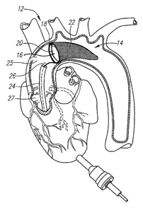
performed. One embodiment of the distal embolic protection assembly (12) is
depicted
in FIG. 2, which shows the filter assembly (14) positioned in the aorta (25)
and
downstream of the aortic valve (27). In this embodiment, the filter assembly
(14) is
comprised of a one-way valve (16) coupled to the frame (18) at the inlet
proximal end
(20) of the porous bag (22) extending therefrom.
The distal embolic protection assembly (12) provides distal embolic protection
and may be delivered by a sleeve (24), such as a catheter or cannula, and
deployed by
manipulation of either the sleeve (24) or the actuation member (26) that is
coupled to
the frame (18) of the filter assembly (14). After the filter assembly (14) is
deployed at its
desired location, it serves the dual functions of a temporary check valve and
a filter to
capture any loose emboli or debris during surgery.
In one embodiment, the distal embolic protection assembly is introduced
through
the apex of the patient's heart, advanced through the left ventricle and
across the aortic
valve into the ascending aorta. Once the inlet end of the filter assembly is
positioned in
the ascending aorta between the aortic valve and the brachiocephalic artery,
the frame
of the filter assembly is either actively or passively deployed to its
expanded state. As
herein described, the filter assembly may be utilized for capturing embolic
material that
is dislodged as a result of procedures involving the repair or replacement of
the aortic
and mitral valves.
In another embodiment, the distal protection assembly may be introduced
through the apex of the patient's heart, advanced through the right ventricle
and placed
downstream of the pulmonary valve and before the pulmonary trunk. At the
pulmonary
trunk, the pulmonary artery splits into the left and right pulmonary artery.
Thus, in an
CA 02579849 2007-03-08
WO 2006/031648 PCT/US2005/032164
9
alternative embodiment, two filter assemblies may each be placed in the left
and right
pulmonary artery. As herein described, the filter assembly may be utilized for
capturing embolic material that is dislodged as a result of procedures
involving the
repair or replacement of the pulmonary and tricuspid valves.
The distal embolic protection assembly, as disclosed herein, may be used to
place a filter assembly downstream of the valve before the commencement of
valve
repair or replacement surgery. Once in place, the filter assembly will allow
for the
capture and removal of embolic material and other debris from the patient
after surgery
has completed.
In one embodiment, the valve replacement system and the distal embolic
protection system, as disclosed herein, may comprise a single and integrated
piece of
equipment. In accordance with this embodiment, a single catheter or sleeve may
be
used both for providing a valve replacement system and for providing the
filter
assembly downstream of the valve replacement system.
In another embodiment, the valve replacement system and the distal embolic
protection system may comprise two separate pieces of equipment. As used in
this
manner, the catheter used to deliver the replacement valve is a structure that
is
separate from the distal embolic protection system. In one alternative
embodiment, the
filter assembly is first deployed at a location downstream from the valve that
is to be
replaced. The catheter comprising the replacement valve disposed thereon may
then
be provided on the sleeve or the actuating member of the distal embolic
protection
system. In this manner, a distinct advantage is conferred in that the sleeve
or
actuating member serves to guide the catheter comprising the replacement valve
to
the intended site of valve implantation.
A distinct advantage in using the apical approach over the percutaneous
approach for valve replacement and repair surgery, particularly with respect
to the
valves of the heart, is that the surgeon has direct access to the valves and a
larger
working area. Any surgical device that must be delivered to the heart in
connection
with the percutaneous approach must be contracted to a very small profile to
permit it
to be delivered through the vasculature. The apical approach relaxes this size
constraint considerably, as incisions of up to 25 mm may be made to the apical
area of
the heart.
The embodiment illustrated in FIGS. 3A and 3B show two perspective views of a
distal embolic protection assembly (30) generally comprising a sleeve (32)
having an
lumen (34), an actuating member (36) having a proximal end (36A) and a distal
end
(36B), and a deployed filter assembly (42) in a blood vessel (31). The filter
assembly
CA 02579849 2007-03-08
WO 2006/031648 PCT/US2005/032164
(42) comprises a porous bag (44) having an open proximal end (46) and a
collapsible
and expandable frame (48) coupled to the open proximal end (46) of the porous
bag
(44). The filter assembly (42) may optionally comprise one or more support
spines
(50) disposed along at least a part of the longitudinal axis of the porous bag
(44).
5 The sleeve (32) is configured and dimensioned to accommodate the actuating
member (36) and to restrain the frame (48) of the filter assembly (42) within
the lumen
(34) in a sufficiently low profile to facilitate the advancement and
retraction of the filter
assembly (42) through the apex of the heart and to the site where the filter
assembly
(42) is to be implanted.
10 The sleeve (32) may be made of any rigid, semi-rigid and flexible
biocompatible
materials, such as metals, alloys, polymers, and the like, depending on its
mode of
use. For example, in cases where the filter assembly is implanted in an area
of close
proximity to the apical area of the patient's heart, such as when the filter
assembly is
implanted in the aorta, the sleeve may be made of a rigid or semi-rigid
material. This is
because the pathway between the apex and the aorta of the heart is a
relatively short
and straight distance. In cases where the filter assembly is implanted in a
blood vessel
at a greater and more convoluted distance from the apex, it may be desirable
to use a
sleeve that is made of flexible material so as to permit the delivery of the
filter
assembly through the convolutions in the passageway.
The lumen (34) of the sleeve (32) is sized to receive the actuating member
(36)
and the frame (48) in its collapsed state. The lumen (34) of the sleeve (32)
may
comprise a coating of Teflon, high density polyethylene or other similar
material that
promotes the smooth insertion and retraction of the frame (48) into and out of
the
lumen (34) of the sleeve (32). The dimension of the sleeve (32) and the lumen
(34)
may be configured to accommodate the entire filter assembly (42) or only the
frame
(48) of the filter assembly (42).
The actuating member (36) may be constructed from any biocompatible
material, such as metal, alloys, polymers, and the like. Similarly as with the
materials
selected for the sleeve (32), the actuating member (36) may be constructed
from rigid
or semi-rigid material where the filter assembly is to be placed in relative
close
proximity to the apical area of the heart, such as the aorta. A rigid or semi-
rigid
actuating member (36) will permit greater control in the maneuvering and
placement of
the filter assembly (42) at its desired location. However, where the filter
assembly (42)
is to be implanted at a location that is. farther away from the apical area of
the heart, a
flexible material may be used.
CA 02579849 2007-03-08
WO 2006/031648 PCT/US2005/032164
11
The collapsible and expandable frame (48) may formed from a shape memory
material, such as Nitinol, that causes the frame (48) to expand to a pre-
determined
shape and diameter when it unrestrained or released from the sleeve (32). The
elasticity of the material causes the frame (48) to expand to a predetermined
shape
and size when outside of the sleeve (32) and to contract to a collapsed state
when
restrained within the lumen (34) of the sleeve (32).
In the embodiment shown in FIGS. 3A-B, the collapsible and expandable frame
(48) has a pre-determined substantially circular shape. The diameter of the
frame (48)
may be selected such that it substantially conforms to or is slightly larger
than the inner
diameter of the aorta or other vessel or body cavity in which placement of the
filter
assembly is desired.
In one embodiment of the frame depicted in FIGS. 3A-B, the actuating member
(36) and the substantially circular frame (48) may be formed from a single
piece of
shape memory metal, such as Nitinol. In this embodiment, the shape of the
substantially circular frame (48) and a angled kink (52) at the distal end
(36B) of the
actuating member (36) and the frame (48) are pre-shaped such that upon
deployment
of the filter assembly (42) from the lumen (34) of the sleeve (32), it assumes
the shape
that is depicted in FIGS. 3A-B.
A cloth or other protective covering may optionally be provided around the
frame
(48) to ensure that the open proximal end (46) of the filter assembly (42)
forms a seal
with the walls of the blood vessel and to prevent embolic material or debris
from
becoming trapped within or pass between the open proximal end (46) and the
walls of
the blood vessel (31). In addition, the cloth covering will protect the aortic
wall from
becoming damaged by the expanding frame (48) of the filter assembly (42).
The porous bag (44) of the filter assembly (42) may be a mesh of any size and
shape required to trap all of the embolic material while still providing
sufficient surface
area for providing satisfactory blood flow during use. The filter may be a
sheet or bag
of different mesh sizes. In a preferred embodiment, the mesh size is optimized
taking
into consideration such factors as flow conditions, application site, size of
filter bag,
and rate of clotting.
For example, the porous bag (44) may be made of a fine mesh material, such
as a screen, or may be a woven or knitted fabric, such as Dacron polyester or
nylon
mesh or other textile fabrics. The porous bag (44) may also be a nonwoven
fabric,
such as a spun bonded polyolefin or expanded polytetrafluoroethylene or other
nonwoven materials, or it may be a fine wire mesh or combination of any of the
CA 02579849 2007-03-08
WO 2006/031648 PCT/US2005/032164
12
aforementioned materials. Preferably, the porous bag (44) has a pore size that
permits
blood to perfuse freely through, while capturing embolic material and other
debris.
The porous bag (44) may have uniform pore size throughout or varying pore
sizes in different areas. In one embodiment, the pore size of the porous bag
(44) may
be in the range of 1 to 200 micrometers for capturing embolic material. Larger
pore
sizes may be selected for application in which the filter assembly is used to
capture
large debris, such as excised valve leaflets in connection with valve removal
surgery.
The porous bag (44) may further comprise one or more support spines (50) that
longitudinally extend at least a part of the length of the porous bag (44).
The support
spine (50) may be constructed into any shape and from any material of
sufficient
rigidity to support the porous bag (44) in substantially a lengthwise fashion
and prevent
porous bag (44) from collapsing into itself or from inverting inside-out. In
one
embodiment, the support spine (50) may simply be a rod that is coupled along
the
longitudinal axis of the porous bag (44). Further, the support spine (50) may
either
extend only partially along the porous bag (44) or extend the entire length of
the
porous bag (44).
The filter assembly (42) is movable between an expanded deployed state (as
shown in FIGS. 3A and 3B) and a collapsed state (as shown in FIGS. 4A and 4B)
to
permit the insertion and removal of the distal embolic protection assembly
(30) in the
patient. The frame (48) of the filter assembly (42) may be drawn into the
lumen (34) of
the sleeve (32) by manipulating the actuating member (36) in the proximal
direction as
indicated by the arrows in FIGS. 4A and 4B. This will retract and collapse the
frame
(48) and substantially close the open proximal end (46) of the - porous bag
(44).
Alternatively, the sleeve (32) may be pushed in a distal direction toward the
frame (48)
of the filter assembly (42) to urge and collapse the frame (48) within the
lumen (34).
Retraction of the frame (48) of the filter assembly (42) may also be
accomplished by
the simultaneous and relative motion of the sleeve (32) and the actuation
member (36)
connecting the frame (48) towards one another and in opposite directions.
The retraction of the filter frame (48) into the lumen (34) of the sleeve (32)
as
shown in FIG. 4B will substantially reduce the likelihood of embolic material
escaping
out of the porous bag (44) as the filter assembly (42) is removed from the
patient. The
filter assembly may be removed from the patient once the frame is fully
contained
within the lumen of the sleeve. In this embodiment, because only the frame
(48) of the
filter assembly (42), and not the porous bag (44), is retracted within the
lumen (34) of
the sleeve (32), there a reduced likelihood that embolic material contained
within the
porous bag (44) will become squeezed out through the pores of the porous bag
(44).
CA 02579849 2007-03-08
WO 2006/031648 PCT/US2005/032164
13
Alternatively, the entire filter assembly, including the porous bag (44), may
be fully
retracted into the lumen of the sleeve. Full retraction of the filter assembly
(42),
including the porous bag (44), may not be possible where large emboli or
debris, such
as excised valve leaflets, are contained in the porous bag (44). Accordingly,
where the
large emboli and debris is contained, only the frame (48) should be retracted
into the
lumen (34).
In another embodiment, the frame (48) of the filter assembly (42) may take the
form of a coil expansion frame that is made of a flexible polymer or shape
memory
material, such as Nitinol. The coil expansion frame may be configured such
that when
it is deployed to its expanded state, the diameter of the coil expansion frame
conforms
substantially to, or is slightly larger, than the diameter of the vessel in
which it is
placed.
The coil expansion frame further comprises a actuation member (36) which is
integral to the coil expansion frame and disposed within the lumen (34) of the
sleeve
(32), wherein the actuation member (36) may be pulled in a proximal direction
to
decrease coil diameter of the coil expansion frame to cinch the open proximal
end (46)
of the porous bag (44) closed in a manner similar to a draw string bag. In
this
embodiment, the sleeve (32) does not receive the filter assembly (42) into the
lumen
(34) but functions as a means by which the actuation member (36) of the coil
expansion frame may be pulled such that the coil expansion frame may be
cinched
closed.for removal of the filter assembly (42) from the body.
FIGS. 5A and 5B show another embodiment of the distal protection assembly
(60) comprising a sleeve (62) having an lumen (64), an actuating member (66)
having
a proximal end (66A) and a distal end (66B), and a filter assembly (72) in a
blood
vessel (59). The filter assembly (72) comprises a porous bag (74) having an
open
proximal end (76) and a collapsible and expandable stent frame (78) coupled to
the
open proximal end (76) of the porous bag (74). The filter assembly (72) may
optionally
comprise one or more support spines (80) disposed along at least a part of the
longitudinal axis of the porous bag (74).
FIG. 5A shows the filter assembly (72) in its deployed and expanded state and
FIG. 5B shows the stent frame of the filter assembly (72) in its collapsed
state for
placement and removal of the filter assembly (72) in the blood vessel (59).
The stent
frame (78) of the filter assembly (72) may be formed from a shape memory
material,
such as Nitinol, that causes the stent frame (78) to expand to a pre-
determined shape
and diameter when it unrestrained or released from the sleeve (62). The
elasticity of
the material causes the stent frame (78) to expand to a predetermined shape
and size
CA 02579849 2007-03-08
WO 2006/031648 PCT/US2005/032164
14
when outside of the sleeve (62) as shown in FIG. 5A and to contract to a
collapsed
state when restrained within the lumen (64) of the sleeve (62) as shown in
FIG. 5B.
The stent frame (78) may be constructed in any number of configurations
designed to substantially support the open proximal end (76) of the porous bag
(74)
open when it is in a deployed state. The deployed filter assembly (72)
depicted in
FIGS. 5A shows a stent frame (78) comprising a proximal collapsed end (78A)
that
remains within the lumen of the sleeve and an expanded distal end (78B) that
is
coupled to and supports the open proximal end (76) of the porous bag (76). The
longitudinal arms (77) of the stent frame (78) joins the proximal collapsed
end (78A)
and the expanded distal end (78B) and the longitudinal arms (77) are biased to
expand
radially to a deployed state. As the stent frame (78) is retracted into the
lumen (64) of
the sleeve (62), the longitudinal arms (77) are urged to radially compress the
expanded distal end (78B) of the stent frame (78) to a collapsed state.
The distal protection assembly (60) further comprises a means for preventing
the proximal collapsed end (78A) of the stent frame (78) from exiting the
lumen (64) of
the sleeve (62) when the filter assembly (72) is deployed. While a number of
different
methods and mechanisms may be used to prevent the stent frame from fully
exiting the
lumen of the sleeve, the distal protection assembly depicted in FIG. 5A shows
a stop
latch (82) that is biased in a forward direction on the proximal collapsed end
(78A) of
the stent frame (78) which mates with a matching groove (84) contained within
the
lumen (64) of the sleeve (62). The mating of the stop latch (82) to the grove
(84)
prevents the proximal collapsed end (78A) from exiting the lumen (64) during
deployment of the filter assembly (72) from the sleeve (62).
In FIG. 5B, the frame of the filter assembly (72) is shown in its retracted
state
and contained in the lumen (64) of the sleeve (62). The retraction of the
stent frame
(78) is accomplished in the same manner as described above for FIG. 4A and 4B.
As
the actuating member (66) is pulled in a proximal direction or as the sleeve
(62) is
pushed in a distal direction, the longitudinal arms (77) of the stent frame
(78) is radially
urged to a collapsed position to facilitate the retraction of the expanded
distal end
(78B) of the stent frame (78) into the lumen (64).
FIGS. 6A and 6B show yet another embodiment in which the frame (90) of the
filter assembly (92) comprises a plurality of arms (94) which converge at the
distal end
(98A) of the actuating member (98) and extend radially outward and are coupled
to the
open proximal end (100) of the filter assembly (92). As shown in FIG. 6A, the
arms
(94) are adapted to expand radially upon deployment to substantially engage
the open
proximal end (100) of the filter assembly (92) with the walls of the blood
vessel. The
CA 02579849 2007-03-08
WO 2006/031648 PCT/US2005/032164
plurality of arms (94) function to actuate between the collapsed state of the
filter
assembly (92) when it is retracted in the sleeve (102) (as shown in FIG. 6B)
and the
expanded and deployed state of the filter assembly (92) when it is outside of
the
sleeve (102) (as shown in FIG. 6A).
5 FIGS. 7A and 7B illustrate yet another embodiment of the distal embolic
protection assembly (110) comprising a sleeve (112) having an lumen (114), an
actuating member (116) having a proximal end (116A) and a distal end (116B),
and a
filter assembly (118) in a blood vessel (99). The filter assembly (118)
comprises a
porous bag (120) having an open proximal end (122) and an inflatable balloon
frame
10 (124) coupled to the open proximal end (122) of the porous bag (120). The
filter
assembly (118) may optionally comprise one or more support spines (126)
disposed
along at least a part of the longitudinal axis of the porous bag (120).
The inflatable balloon frame (124) is in fluid communication with a peripheral
saline reservoir through a conduit and may be substantially donut-shaped in
the
15 expanded and deployed state, as shown in FIG. 7A. Because the balloon frame
(124)
may be collapsed by deflating the balloon frame (124), the filter assembly
(118) may
be introduced into and removed from the patient in the collapsed state either
within the
sleeve (112) as shown in FIG. 7B or even without the sleeve (122).
In any one of the embodiments described herein, the filter assembly may
include a one-way valve at the open proximal end of the porous bag to provide
the
dual function of acting as a temporary valve during valve replacement surgery
and
preventing embolic material from escaping out from the porous bag. Adding the
one-
way valve prevents embolic material from escaping out of the porous bag, thus
reducing the incidence of embolization and blockage. A valve would
concurrently
provide a temporary valve for use during valve surgery. Combining both a
filter and a
valve in the same arrangement also creates a more compact device allowing more
space for conducting other procedures.
FIGS. 8A-B show one-way valves in connection with the distal protection
assembly of FIG. 3. FIG. 8A illustrates one embodiment of the sleeve (214)
having an
inner lumen (216) and a filter assembly (200) shown in FIG. 3 comprising a
porous bag
(202) having an open proximal end (204) and a substantially circular frame
(206)
coupled to the open proximal end (204). A bileaflet one-way valve (208) is
coupled to
the open proximal end (204) of the porous bag (202) to permit the
substantially
unidirectional blood flow through the filter assembly (200).
In the embodiment depicted in FIGS. 8A-B, the proximal end (212A) of the
actuating member (212) is manipulated in the distal direction to deploy the
frame (206)
CA 02579849 2009-07-27
16
to its deployed state and in the proximal direction to compress the frame
(206) in its
collapsed state. The distal end (212B) of the actuating member (212) is
attached to
the porous bag (202) at a location distal from the open proximal end (204) and
functions as a support spine for the porous bag (202) and for the bileaflet
valve (208).
An additional support rod (210) is provided on the porous bag (202) to support
the
bileaflet valve (208) to the porous bag (202).
FIG. 8B shows an embodiment of the filter assembly (200) having a trileaflet
valve (220) coupled to the open proximal end (204) of the porous bag. Because
this
valve has three leaflets, two support rods (210) are provided on the porous
bag to
support the trileaflet valve (220) to the porous bag (202). Although FIGS. 8A
and 8B
show the filter assembly in connection with bileaflet and trileaflet valves,
respectively,
a valve of any number of leaflets may be used so long as the valve permits
substantially the unidirectional flow of blood and materials through the
filter assembly
and substantially reduces the back flow of blood and materials in the reverse
direction.
Suitable valves may be constructed from a variety of flexible material such as
from natural tissue, polymers or plastics. The valves may be constructed to
the
frame at the open proximal end of the porous bag with commissural tabs that
attach
the valves to the frame. The construction of valves suitable for use in
connection
with the filter assemblies herein are disclosed in U.S. Pat. Nos. 6,736,846,
issued
May 18, 2004; 6,719,789, issued April 13, 2004; 6,719,788, issued April 13,
2004;
6,682,559, issued January 27, 2004; 5,344,442, issued September 6, 1994;
5,500,015, issued March 19, 1996; 6,805,711, filed June 17, 2002; and in U.S.
Patent Publication Serial Nos. 2004/0117009, filed September 23, 2003; and
2006/0025857, filed April 23, 2004.
Although FIGS. 8A and B show the one-way valve in connection with the filter
assembly of FIG. 3 having the substantially circular frame, the one-way valve
may
easily be adapted to a variety of frames in a number of ways. For example, the
valve
may be coupled to the stent frame of the filter assembly of FIG. 5 with
relative ease,
as the stent provides several points of attachment for the valve. The manner
of
including a one-way valve to stent frames is fully disclosed in co-pending
U.S. patent
publication Serial No. 2005/0075719, filed October 6, 2003.
Additionally, an imaging system to view the operating field may be used at
any time or throughout the duration of the surgery. Imaging systems are well-
known
to one of skill in the art and include transesophageal echo, transthoracic
echo,
CA 02579849 2009-07-27
17
intravascular ultrasound imaging (IVUS), or an injectable dye that is
radiopaque.
Cinefluoroscopy may also be utilized. In one embodiment, the imaging system is
deliverable through a catheter or cannula to the operating field.
Intravascular ultrasound (IVUS) uses high-frequency sound waves that are
sent with a device called a transducer. The transducer may be coupled to the
delivery member of the present invention. In this arrangement, the sound waves
bounce off of the walls of the vessel or heart and return to the transducer as
echoes.
Methods and systems for IVUS imaging for the placement of heart valves is
disclosed.
Although the invention has been described with reference to preferred
embodiments and specific examples, those of ordinary skill in the art will
readily
appreciate that many modifications and adaptations of the invention are
possible
without departure from the spirit and scope of the invention as claimed
hereinafter.