Note: Descriptions are shown in the official language in which they were submitted.
CA 02581677 2013-02-25
Occluder Device Double Securement System for
Delivery/Recovery of such Occluder Device
TECHNICAL FIELD
100021 This invention relates generally to occlusion devices for the
closure of physical
anomalies, such as an atrial septal defect, a patent foramen ovale, and other
septal and
vascular defects. The invention also relates to delivery systems and
mechanisms for such
devices.
BACKGROUND
100031 A patent foramen ovale (PFO), illustrated in FIG. 1, is a
persistent, one-way,
usually flap-like opening in the wall between the right atrium 11 and left
atrium 13 of the
heart 10. Because left atrial (LA) pressure is normally higher than right
atrial (RA)
pressure, the flap usually stays closed. Under certain conditions, however,
right atrial
pressure can exceed left atrial pressure, creating the possibility that blood
could pass from
the right atrium 11 to the left atrium 13 and blood clots could enter the
systemic
circulation. It is desirable that this circumstance be eliminated.
[0004] The foramen ovale serves a desired purpose when a fetus is
gestating. Because
blood is oxygenated through the umbilical cord, and not through the developing
lungs, the
circulatory system of the fetal heart allows the blood to flow through the
foramen ovale as a
physiologic conduit for right-to-left shunting. After birth, with the
establishment of
pulmonary circulation, the increased left atrial blood flow and pressure
results in functional
closure of the foramen ovale. This functional closure is subsequently followed
by anatomical
closure of the two over-lapping layers of tissue: septum primum
1
CA 02581677 2007-03-26
WO 2006/036837 PCT/US2005/034276
14 and septum secundum 16. However, a PFO has been shown to persist in a
number of
adults.
[0005] The presence of a PFO is generally considered to have no therapeutic
consequence in otherwise healthy adults. Paradoxical embolism via a PFO is
considered
in the diagnosis for patients who have suffered a stroke or transient ischemic
attack (TIA)
in the presence of a PFO and without another identified cause of ischemic
stroke. While
there is currently no definitive proof of a cause-effect relationship, many
studies have
confirmed a strong association between the presence of a PFO and the risk for
paradoxical embolism or stroke. In addition, there is significant evidence
that patients
with a PFO who have had a cerebral vascular event are at increased risk for
future,
recurrent cerebrovascular events.
[0006] Accordingly, patients at such an increased risk are considered for
prophylactic medical therapy to reduce the risk of a recurrent embolic event.
These
patients are commonly treated with oral anticoagulants, which potentially have
adverse
side effects, such as hemorrhaging, hematoma, and interactions with a variety
of other
drugs. The use of these drugs can alter a person's recovery and necessitate
adjustments in
a person's daily living pattern.
[0007] In certain cases, such as when anticoagulation is contraindicated,
surgery
may be necessary or desirable to close a PFO. The surgery would typically
include
suturing a PFO closed by attaching septum secundum to septum primum. This
sutured
attachment can be accomplished using either an interrupted or a continuous
stitch and is a
common way a surgeon shuts a PFO under direct visualization.
[0008] Umbrella devices and a variety of other similar mechanical closure
devices,
developed initially for percutaneous closure of atrial septal defects (ASDs),
have been
used in some instances to close PF0s. These devices potentially allow patients
to avoid
the side effects often associated with anticoagulation therapies and the risks
of invasive
surgery. However, umbrella devices and the like that are designed for ASDs are
not
optimally suited for use as PFO closure devices.
[0009] Currently available septal closure devices present drawbacks,
including
technically complex implantation procedures. Additionally, there are
significant
complications due to thrombus, fractures of the components, conduction system
2
CA 02581677 2007-03-26
WO 2006/036837 PCT/US2005/034276
disturbances, perforations of heart tissue, and residual leaks. Many devices
have high
septal profile and include large masses of foreign material, which may lead to
unfavorable
body adaptation of a device. Given that ASD devices are designed to occlude
holes,
many lack anatomic conformability to the flap-like anatomy of PF0s. Thus, when
inserting an ASD device to close a PFO, the narrow opening and the thin flap
may form
impediments to proper deployment. Even if an occlusive seal is formed, the
device may
be deployed in the heart on an angle, leaving some components insecurely
seated against
the septum and, thereby, risking thrombus formation due to hemodynamic
disturbances.
Finally, some septal closure devices are complex to manufacture, which may
result in
inconsistent product performance.
[0010] Various delivery systems have been used to deliver occluders and
other
medical devices through body lumens. Some delivery systems of the prior art
are used to
deliver devices that readily expand to a delivered configuration when removed
from the
delivery system. Such delivery systems are not generally suited for delivering
a device
that does not readily expand into the delivered configuration. Further, the
delivery
systems of the prior art may not allow verification of the position of the
device prior to
full deployment of the device. Finally delivery systems of the prior art may
not be
suitable to manipulate the configuration of the device in a secure manner to
allow for
complete deployment of the device.
[0011] The devices and techniques disclosed herein are designed to address
these
and other deficiencies of prior art septal closure devices and techniques for
delivering and
retrieving such devices.
SUMMARY OF THE INVENTION
[0012] These and other aspects and embodiments of the disclosure are
illustrated
and described below.
[0013] This description discloses several delivery devices and techniques
for
delivering an implant into a desired location within the body. This delivery
technique
relates particularly to, but is not limited to, a septal occluder made from a
polymer tube.
These delivery techniques, in addition to use with septal occluders, could be
applied to
3
CA 02581677 2007-03-26
WO 2006/036837
PCT/US2005/034276
other medical devices, such as other expandable devices constructed from an
underlying
tubular structure.
[0014] In one aspect, a delivery system is disclosed for delivering an
occluder that
closes an aperture in septal tissue. The occluder includes a first side
adapted to be
disposed on one side of the septal tissue and a second side adapted to be
disposed on the
opposite side of the septal tissue. The first and second sides are adapted to
occlude the
aperture upon deployment of the device at its intended delivery location. The
device also
includes a catch system that maintains the configuration of the device once it
has been
deployed.
[0015] According to at least some embodiments, the device is formed from a
tube.
According to some embodiments, the tube includes a material selected from the
group
consisting of metals, shape memory materials, alloys, polymers, bioabsorbable
polymers,
and combinations thereof. In particular embodiments, the tube includes a shape
memory
polymer. In particular embodiments, the tube includes nitinol. In some
embodiments, the
tube is formed by rolling a flat piece of material into a tubular form.
According to some
embodiments, the device is formed by cutting the tube. The device is placed in
its
deployment configuration by reducing the axial length of the device.
[0016] According to some embodiments, the catch system reduces and
maintains
the axial length of the device. Also, varied constructions could be used to
maintain the
axial dimension of the device. In one form, catch elements such as, for
example, balls,
attached to a delivery wire could be used to maintain the axial dimension of
the device.
In a different construction, a locking mechanism could be used. Preferably, if
a locking
mechanism is used, it secures both sides of the device in the locked position
with a single
locking element. In some embodiments, a catch element secures the ends of the
occluder
in a compressed position. Preferably, if a catch mechanism is used, it secures
both sides
of the device in the deployed position with a single element.
[OM] In
another aspect, the present invention provides a device for occluding an
aperture in septal tissue, including a first side adapted to be disposed on
one side of the
septal tissue and a second side adapted to be disposed on the opposite side of
the septal
4
CA 02581677 2007-03-26
WO 2006/036837 PCT/US2005/034276
tissue. The first and second sides are adapted to occlude the defect when the
device is
deployed at its intended delivery location. Each of the first and second sides
includes
loops. The device further includes a catch system that maintains the
configuration of the
device once it has been deployed. The loops of the first and second sides and
the catch
system cooperate to provide a compressive force to the septal tissue
surrounding the
aperture.
[0018] According to some embodiments, each of the first and second sides
includes
at least two loops. In particular embodiments, each of the first and second
sides includes
four or six loops. Of course, the most desirable number of loops on each side
will depend
on a variety of anatomical and manufacturing factors. According to some
embodiments,
the device also includes a central tube that connects the first and second
sides.
[0019] The delivery system may be used to deliver an occluder in which at
least one
of the first and second sides further includes a tissue scaffold. The tissue
scaffold
includes a material selected from the group consisting of polyester fabrics,
Teflon-based
materials, polyurethanes, metals, polyvinyl alcohol (PVA), extracellular
matrix (ECM) or
other bioengineered materials, synthetic bioabsorbable polymeric scaffolds,
collagen, and
combinations thereof. In particular embodiments, the tissue scaffold includes
nitinol.
[0020] The delivery system includes a first and a second securement
system. The
first securement system may be any one of a number of configurations. First, a
delivery
wire may be used to secure the distal end of the occluder onto the delivery
system. When
a delivery wire is used, the distal end of the delivery wire may be threaded
and cooperate
with a corresponding threaded portion on the occluder. In a preferred form,
the threaded
portion may have male threads on the occluder and female threads on the
delivery wire.
Alternatively, a ball and clasp, other interlocking system may be used.
[0021] The second securement system may be any one of a number of
configurations. In one aspect it may be a threaded connection between the
delivery
system and the occluder. In another aspect, the second securement system is a
collet
system that includes fingers, which are configured to fit within a groove in
the occluder
and thus secure the occluder to the delivery system when the fingers are
disposed in the
CA 02581677 2007-03-26
WO 2006/036837 PCT/US2005/034276
groove. A collet sheath is moveable with respect to the fingers and when the
collet
fingers are disposed within the collet sheath, the fingers are configured to
fit within the
groove provided on the occluder.
[0022] In one aspect, a delivery system for the device is provided within
(and
includes) a delivery sheath. In certain embodiments, the delivery system
includes a first
securement system for securing a first end of the occluder and a second
securement
system for securing a second end of the occluder. The securement systems
connect the
occluder to first and second catheters contained in the delivery system and
enable
deployment and/or retrieval of the occluder. The catheters are preferably able
to move
relative to each other. The securement systems enable pushing and pulling of
respective
ends of the occluder by manipulating the catheters to expand and contract the
device. The
first securement system may employ a threaded connection and the second
securement
system may employ a suture connection. The securement systems are detached
when the
device has been properly positioned.
[0023] In a further aspect of the invention, the first securement system
secures a
distal end of a catching system of the device and the second securement system
secures a
proximal end of the device. A first catheter connects to the first securement
system and a
second catheter connects to the second securement system. In certain
embodiments, the
second catheter encloses the first catheter in its central lumen. In one
aspect, the device is
deployed by inserting the delivery system, removing the sheath, expanding the
petals of a
distal portion of the device, and expanding the petals of a proximal portion
of the device.
The delivery system can be detached by detaching the first and second
securement
systems, e.g., by unscrewing the first securement system and by cutting and
removing the
sutures. In another aspect, the deployed device is retrieved by contracting
the petals of a
proximal portion of the device using the second catheter, advancing the sheath
over a
proximal portion of the device, contracting the petals of a distal portion of
the device
using the first catheter and advancing the sheath over the distal portion of
the device. The
occluder can then be repositioned or removed.
[0024] In another aspect, a delivery system is disclosed for delivering
an occluder
that closes an aperture in septal tissue. The occluder includes a first side
adapted to be
disposed on one side of the septal tissue and a second side adapted to be
disposed on the
6
CA 02581677 2013-10-01
opposite side of the septal tissue. The first and second sides are adapted to
occlude the
aperture upon deployment of the device at its intended delivery location. The
device also
employs a catch system that maintains the configuration of the device once it
has been
deployed. The occluder may be held in its deployment configuration by the
catch
element.
[0025] In one aspect of the invention, there is provided a collapsible
medical device for
occluding an aperture in a body and a delivery system. The medical device has
a first
configuration as a reduced profile and a second configuration as an expanded
profile, the
medical device being adapted to be delivered through the delivery system into
a desired
delivery location. The medical device comprises a tube having a proximal side
and a distal
side, each side having slits and a first threaded attachment for directly
securing the device to
a delivery wire of the delivery system and a second attachment for securing
the proximal
side of the device to the delivery system. The relative movement of the first
attachment and
second attachment changes the configuration of the device from the reduced
profile
configuration to the expanded profile configuration. The first attachment or
the second
attachment is threaded. The relative movement of the first attachment and the
second
attachment results in the shortening of axial length of the tube.
[0026] In another aspect of the invention, the shortening of the axial
length of the tube
forms (i) a first strut that bows and twists outwardly defining a first
proximal loop, and (ii) a
second strut that bows and twists outwardly defining a first distal loop.
[0026A] In another aspect of the invention, the medical device is
constructed from
materials which are bioresorbable.
[0027] These and other aspects and embodiments of the disclosure are
illustrated and
described below.
7
CA 02581677 2007-03-26
WO 2006/036837 PCT/US2005/034276
BRIEF DESCRIPTION OF THE DRAWINGS
In the Drawings:
[0028] FIG. 1 is a schematic representation of a human heart including
various
septal defects;
[0029] FIGS. 2A-2D are isometric views of an embodiment of an occluder for
use
with disclosed delivery systems and techniques;
[0030] FIG. 2E illustrates a deployed occluder according to an aspect of
the
disclosure;
[0031] FIG. 3A illustrates insertion of an occluder in a human subject
using a
delivery system in accordance with an aspect of the disclosure;
[0032] FIG. 3B illustrates introduction of the occluder in a human heart
using a
delivery system in accordance with an aspect of the disclosure;
[0033] FIGS. 4A-4D are side views of a delivery assembly for delivering an
occluder to a septal defect according to an aspect of the disclosure;
[0034] FIG. 5 is a side elevational view of a delivery system attached to
an occluder
in deployed configuration according to an aspect of the disclosure;
[0035] FIG. 6 is an exploded cross-sectional side view of a delivery system
attached
to an occluder in deployed configuration according to an aspect of the
disclosure;
[0036] FIG. 7 is an exploded cross-sectional side view of the control
portion of a
delivery system according to an aspect of the disclosure;
[0037] FIG. 8 is an enlarged cross-sectional side view of the catheter
portion of a
delivery system attached to an occluder according to an aspect of the
disclosure;
[0038] FIG. 9 is a cross-sectional side view of the catheter portion of the
delivery
system attached to a collapsed occluder according to an aspect of the
disclosure;
[0039] FIG. 10 is a cross-sectional side view of one step in a deployment
sequence
according to an aspect of the disclosure;
[0040] FIG. 11 is a cross-sectional side view of one step in a deployment
sequence
according to an aspect of the disclosure;
8
CA 02581677 2007-03-26
WO 2006/036837 PCT/US2005/034276
[0041] FIG. 12 is a cross-sectional side view of one step in a deployment
sequence
according to an aspect of the disclosure;
[0042] FIG. 13 is a cross-sectional side view of one step in a detachment
sequence
according to an aspect of the disclosure;
[0043] FIG. 14 is a cross-sectional side view of one step in a detachment
sequence
according to an aspect of the disclosure;
[0044] FIG. 15 is a cross-sectional side view of one step in a detachment
sequence
according to an aspect of the disclosure;
[0045] FIG. 16 is a cross-sectional side view of one step in a retrieval
sequence
according to an aspect of the disclosure;
[0046] FIG. 17 is a cross-sectional side view of one step in a retrieval
sequence
according to an aspect of the disclosure;
[0047] FIG. 18 is a cross-sectional side view of one step in a retrieval
sequence
according to an aspect of the disclosure; and
[0048] FIG. 19 is a cross-sectional side view of one step in a retrieval
sequence
according to an aspect of the disclosure.
[0049] FIG. 20 illustrates a cross-sectional schematic of a deployed
occluder
according to an aspect of the disclosure;
[0050] FIG. 21 illustrates a cross-sectional side view of several
components of the
delivery system according to one embodiment of disclosure;
[0051] FIG. 22 is an axial cross-sectional drawing of an occluder, in a
delivery
configuration, according to an embodiment of the disclosure;
[0052] FIG. 23 is a detail view of the delivery wire according to an aspect
of one
embodiment of the disclosure;
[0053] FIG. 24 is a configuration for a first securement system according
to an
embodiment of the disclosure;
[0054] FIGS. 25A, 25B, 26A, 26B, 27A and 27B are alternative configurations
for
the first securement system according to aspects of the disclosure;
9
CA 02581677 2007-03-26
WO 2006/036837 PCT/US2005/034276
[0055] FIGS. 28 and 29 are detail cross-sectional side view of the delivery
system
during two steps in the deployment process according to one aspect of the
disclosure;
[0056] FIG. 30 is a detail view of the collet finger according to one
aspect of the
disclosure;
[0057] FIG. 31 is a detail cross-sectional side view of the collet system
in the
splayed configuration;
[0058] FIG. 32A is a detail cross-sectional view of the collet system in
the
constrained configuration;
[0059] FIG. 32B is a cross-section taken along lines 32B-32B in FIG. 32A;
[0060] FIG. 33 is a detail sectional view of another embodiment of the
collet system
according to the disclosure;
[0061] FIGS. 34-37 are sectional views of an alternative delivery system
according
to an aspect of the disclosure;
[0062] FIG. 38A is a front cross-sectional view of a delivery catheter
according to
one embodiment of the disclosure;
[0063] FIG. 38B is a side cross-sectional view of a delivery catheter with
sutures
according to one embodiment of the disclosure;
[0064] FIG. 38C is an elevational view of a delivery catheter with sutures
secured
to an occluder according to one embodiment of the disclosure;
[0065] FIG. 38D is an elevational end view of delivery catheter along lines
38D of
FIG. 38C; and
[0066] FIG. 39 is a sectional view of a delivery assembly during a step in
the
deployment process according to one aspect of the disclosure.
CA 02581677 2007-03-26
WO 2006/036837 PCT/US2005/034276
DETAILED DESCRIPTION
[0067] The present disclosure provides devices, delivery/retrieval systems
and
techniques for delivering such devices intended to occlude an aperture within
body tissue.
In particular and as described in detail below, the described occluder may be
used for
closing an ASD or PFO in the atrial septum of a heart. Although the
embodiments are
described with reference to an ASD or PFO, one skilled in the art will
recognize that the
device and methods of the present invention may be used to treat other
anatomical
conditions. As such, the invention should not be considered limited in
applicability to
any particular anatomical condition. In addition, the systems and methods for
delivery
and retrieval, and for catching a device in a deployed state, which are
aspects of the
present invention may also be used in connection with other types of devices
besides an
occluder, in particular, devices having tubular profiles.
[0068] FIG. 1 illustrates a human heart 10, having a right atrium 1 1 and
a left
atrium 13 and including various anatomical apertures 18a and 18b. The atrial
septum 12
includes septum primum 14 and septum secundum 16. The anatomy of the septum 12
varies widely within the population. In some people, septum primum 14 extends
to and
overlaps with septum secundum 16. The septum primum 14 may be quite thin. When
the
anatomical apertures 18a is present, blood could travel through the anatomical
aperture
18a between septum primum 14 and septum secundum 16 (referred to as "the PFO
tunnel"). Additionally or alternatively, the presence of an ASD could permit
blood to
travel through an aperture in the septal tissue, such as through the
anatomical aperture
18b.
[0069] In this application, "distal" refers to the direction away from a
catheter
insertion location and "proximal" refers to the direction nearer the insertion
location.
Additionally, the term "delivery configuration" refers to the configuration of
a device,
such as an occluder, when it has a reduced profile in a delivery catheter. The
term
"deployed configuration" refers to the configuration of the device, such as an
occluder,
when it has deployed from the catheter, such as at the desired implantation
location.
[0070] FIGS. 2A-D illustrates an exemplary occluder with which systems and
techniques disclosed herein may be used. An occluder 70, for example, can be
formed by
cutting a series of slits on tube 25. As shown in FIGS. 2A-2D, distal petals
32 are
produced by cutting slits 31 in the upper portion of tube 25 according to the
cutting
11
CA 02581677 2007-03-26
WO 2006/036837 PCT/US2005/034276
pattern shown in FIG. 2A. As shown in FIG. 2B, the distal portion of the tube
25 is cut in
half to form half sections 91a and 91b. The half sections 91a and 91b are
further cut tc a
proximal distance from distal tip 39 into quarter sections 92a, 93a, 92b, and
93b. The
cuts are discontinued and quarter sections 92a and 92b form half section 94a
at distal tip
39, and quarter sections 93a and 93b form half section 94b at distal tip 39.
Upon
application of force Fd to distal tip 39, struts defined by slits 31 bow and
twist outward to
form distal petals 32 in distal side 30, as shown in FIGS. 2C-2D. The movement
of thc
struts during deployment is such that the struts rotate in an orthogonal plane
relative to
the axis of the device. Central tube 22 may be constrained during the
application of force
Fd, or any combination of forces sufficient to reduce the axial length of the
tube 25 may
be applied. One end of each of distal petals 32 originates from central tube
22, while the
other end originates from distal tip 39 (FIGS. 2B-2C). Proximal petals 42 may
be formed
in proximal side 40, as shown in FIGS. 2B-2D, making slits 41 between central
tube 22
and proximal end 44, using the same cutting pattern described above.
[0071] The tube(s) 25 forming occluder 70 may be formed from a
biocompatible
metal or polymer. In at least some embodiments, the occluder 70 is formed of a
bioabsorbable polymer, or a shape memory polymer. Shape memory polymers can be
advantageous so that the structure of the device assists in pressing the PFO
tunnel closd.
In other embodiments, the occluder 70 is formed of a biocompatible metal, such
as a
shape memory alloy (e.g., nitinol). The thermal shape memory and/or
superelastic
properties of shape memory polymers and alloys permit the occluder 70 to
resume and
maintain its intended shape in vivo despite being distorted during the
delivery process.
Alternatively, or additionally, the occluder 70 may be formed of a
bioabsorbable metal,
such as iron, magnesium, or combinations of these and similar materials.
Exemplary
bioabsorbable polymers include polyhydroxyallcanoate compositions, for example
poly-4-
hydroxybutyrate (P4HB) compositions, disclosed in U.S. Patent No. 6,610,764,
entitled.
Polyhydroxyalkanoate Compositions Having Controlled Degradation Rate and U.S.
Patent No. 6,548,569, entitled Medical Devices and Applications of
Polyhydro.xyalkanoate Polymers, both of which are incorporated by reference in
their
entirety.
12
CA 02581677 2013-02-25
[0072] The cross-sectional shape of tube 25 may be circular or polygonal,
for example
square, or hexagonal. The slits 31 and 41 may be disposed on the face of the
polygon (i.e.,
the flat part) or on the intersection of the faces.
[0073] The tube can be injection molded, extruded, or constructed of a
sheet of material
and rolled into a tube. The sheet of material could be a single ply sheet or
multiple ply. The
slits that form the struts could be cut or stamped into the sheet prior to
rolling the sheet into a
tube to connect the ends to form an enclosed cross section. Various
geometrical cross
sections are possible including circular, square, hexagonal and octagonal and
the joint could
be at the vertex or along the flat of a wall if the cross section is of a
particular geometery.
Various attachment techniques could be used to join the ends of the sheet to
form a tube,
including welding, heat adhesives, non-heat adhesives and other joining
techniques suitable
for in-vivo application.
[0074] The petal configuration is the deployed configuration. The occluder
70 can be
secured in the petal configuration by a catch system that holds the ends of
the tube 25
together, certain embodiments of which are described below. Use of the terms
distal and
proximal sides or portions 30 and 40, respectively, include the petals that
are formed on the
distal and proximal sides.
100751 The embodiment described in conjunction with FIGS. 2A-2D has
similarities to
the device disclosed in U.S. Patent No. 7,678,123 entitled Tubular Patent
Foramen Ovale
(PFO) Closure Device with Catch System, filed on July 14, 2004; U.S. Patent
Publication
No. US 2011-0112633 entitled Delivery/Recovery System for Septal Occluder;
U.S. Patent
Publication No. US 2006-0265004 entitled Catch Member for PFO Occluder; U.S.
Patent
Publication No. US 2006-0122647 entitled Occluder Device Double Securement
System for
Delivery/Recovery System of Such Occluder Device; all of which have the same
assignee as
the present application. These documents describe how a device can be formed
by making
cuts or slits in a tube and compressing the ends, and how to deliver such a
device.
[0076] The transformable design of occluder 70 enables occluder 70 to be
delivered in a
low profile, tubular form and to be converted readily, i.e., by reducing the
axial
13
CA 02581677 2007-03-26
WO 2006/036837 PCT/US2005/034276
length, in place to the high-profile deployed configuration. Moreover, the
conversion can
readily be effected by forcing distal end 39 and proximal end 44 together. For
example,
distal side 30 and proximal side 40 of occluder 70 may be deployed in separate
steps, or
both distal side 30 and proximal side 40 of occluder 70 may be exposed (e.g.,
out of the
delivery catheter) prior to engaging the catch system and deployed together as
the catch
element is engaged. Use of the terms distal and proximal side 30 and 40,
respectively,
include the loops or other geometries and configurations that are formed on
the distal and
proximal sides, respectively.
[0077] Occluder 70 may be prepared for delivery to an aperture 18 in any
one of
several ways. Slits 31 and 41 may be cut such that tube 25 bends into its
intended
configuration following deployment in vivo. Specifically, slits 31 and 41 may
be cut to
produce struts 32 and 42 of a thickness that facilitates the bending and
formation of loops
32 and 42 upon the application of forces Fd and/or Fp during deployment. See
FIGS. 2B
and 2C. Alternatively and/or additionally, a tube 25 formed of a shape memory
material
may be preformed into its intended configuration ex vivo so that it will
recover its
preformed shape once deployed in vivo. According to at least some embodiments,
this
preforming technique produces more reliable deployment and bending of occluder
70 in
vivo. An intermediate approach may also be used: tube 25 may be only slightly
preformed ex vivo such that it is predisposed to bend into its intended shape
in vivo upon
application of forces Fd and F.
[0078] FIG. 2E shows a deployed occluder 70 in a human heart with a catch
element 50 engaged. The term "catch system" describes the portion/aspect of
the device
that secures the device in the deployed configuration, it may be a single
piece or a group
of connected or assembled pieces. The catch element is the portion of the
catch system
that engages with the occluder to hold the occluder in the deployed
configuration and is
described in more detail below. The configuration illustrated is a simplified
schematic
view of the occluder 70 illustrated in FIGS. 2A-2D. This particular type of
occluder 70
and catch element 50 are described for purposes of illustration and
explanation; of course,
other types of occluders (with different types of catch elements or systems)
can be
deployed using the deployment systems described herein. The catch element 50,
as
illustrated, is disposed in an axially central location in the occluder 70 and
is
schematically illustrated as a separate piece than the occluder 70. In a
preferred
14
CA 02581677 2007-03-26
WO 2006/036837 PCT/US2005/034276
embodiment, the catch element may be fixed to one end of the tube 25 that
forms
occluder 70. For example, a flange 52 may be fixed to the distal tip 39 of the
tube 25 that
forms the distal and proximal petals 32 and 42.
[0079] In general, references to "occluder 70" herein may be inclusive of
catch
element 50, depending on the context, for example, unless separately listed or
otherwise
stated. One end of tube 25 is able to move with respect to the catch element
50 (and
especially the catch system) so that the distal and proximal petals 32 and 42
can move
from the delivery configuration to the deployed configuration. The inside
surface of the
tube 25 is able to slide over the catch element 50 so that, when the proximal
end 44 of the
occluder 70 rests against the surface of the proximal flange 56, the occluder
70 is secured
in its deployed configuration. The catch element 50 is included in the catch
system that
includes a portion for connection to the delivery/recovery system, including,
for example,
a threaded section illustrated in FIG. 2E. The threaded section is an
adaptation designed
to fit with the desired type of securement system according to a preferred
embodiment
discussed herein and is not necessarily an inherent feature of the catch
element 50.
Occluder 70 also includes an additional feature, such as threads or a groove
72 (as
illustrated) to provide another connection between the occluder and the
delivery/recovery
system.
[0080] FIG. 3A illustrates the insertion of an occluder in a human subject
122 using
a delivery assembly 124 in accordance with an aspect of the disclosure. A
portion of
delivery assembly 124, including an occluder and a delivery mechanism for the
occluder,
which can be externally manipulated by a clinician, is inserted into the
subject through an
incision point 126. The distal end of the delivery assembly is advanced toward
and into
the heart 10 until the distal end is in proximity to the defect to be closed,
as seen in FIG.
3B.
[0081] Figure 4A illustrates the occluder 70 in the distal end of the
delivery
assembly 124, which includes a delivery system 140. A delivery system
generally
includes a delivery catheter, a delivery wire and a delivery sheath. Because
the occluder
70 is delivered percutaneously, the device is secured to the delivery system
140 so that
the occluder 70 can be placed accurately at the desired delivery location.
Securement
systems are provided that attach the occluder to the delivery components. The
securement systems are configured to provide accurate delivery of the occluder
to the
CA 02581677 2007-03-26
WO 2006/036837 PCT/US2005/034276
desired delivery location and allow for a controlled deployment so that the
position of the
device as it is being deployed can be monitored. Also, a device deployed
according to
this system is able to be retrieved and repositioned until the final stage of
the deployment
process. In some circumstances, after the final stage of the deployment
process, the
device can be retrieved. The manner in which the occluder is secured to the
delivery
system 140 and the process for deployment and/or retrieval of the occluder 70
are
described in detail below.
[0082] As illustrated in FIG. 4A, the delivery system 140 includes a
delivery sheath
144 and a delivery catheter 148. A delivery string or wire 150 extends the
length of the
delivery assembly to the distal end of the occluder 70. The delivery system
140
constrains the occluder 70 in its elongated delivery configuration. As shown
in FIG. 4B,
a delivery sheath 144 containing the occluder 70 is first inserted into the
right atrium 11
of the patient's heart.
[0083] The delivery system, including the delivery sheath 144, may next be
inserted
through aperture 18 located in the septa' tissue 12 (which, in this example,
is a PFO
tunnel) and into the left atrium 13. Distal side 30 of occluder 70 is then
exposed into the
left atrium 13 by withdrawing the delivery sheath 144 then pulling force F1 is
applied to
delivery string or wire 150 such that, for example, a catch element 50 passes
through the
central tube 22, thereby securing distal side 30 into its deployed state.
Delivery sheath
144 is withdrawn further through the aperture 18 and into the right atrium 11,
such that
central tube 22 is positioned through the aperture 18. As shown in FIG. 4C,
proximal
side 40 of occluder 70 is then exposed into the right atrium 11, and a
relative force
between the proximal end 44 of the occluder 70 and the delivery string or wire
150 is
applied such that a catch element 50 passes through the proximal end 44 of the
occluder
70, thereby securing the proximal side 40 of the occluder into its deployed
state. Of
course, the occluder 70 should remain in position during deployment of each
side of the
occluder 70 and pulling forces on the septum tissue should be avoided.
[0084] As shown in FIG. 4D, when properly deployed, occluder 70 is disposed
through the aperture 18 with a portion of the device on the proximal side and
another
portion of the device on the distal side. The distal side 30 and proximal side
40 exert a
compressive force against septum primum 14 and septum secundum 16 in the left
13 and
right 11 atria, respectively, to close the aperture 18, e.g. the PFO. When the
occluder 70
16
CA 02581677 2007-03-26
WO 2006/036837 PCT/US2005/034276
is properly located, the securement systems are detached releasing the
occluder from the
delivery system. This delivery system is then removed from the heart. In the
event
occluder 70 is not properly deployed after performing the delivery sequence,
the occluder
70 may be recovered by reversing the steps of the delivery sequence.
[0085] As mentioned above, during the deployment of the occluder 70 in the
delivery system 140 described in connection with FIGS. 4A-4D, the occluder 70
is
secured to the delivery system 140 at two locations on the occluder 70 so that
the
occluder 70 can be formed (i.e., compressed) into its deployed configuration.
In a
preferred form, there are two securements to the delivery assembly 140. A
first
securement is toward the distal end of the occluder 70 whereby the occluder 70
is held by
the delivery string or wire 150. The second securement is at the proximal end
44 of the
occlude 70 whereby the occluder 70 is held by the delivery catheter 148. The
first and
second securements allow the proximal and distal ends of the occluder 70 to be
forced
together so that the occluder 70 can move from the delivery configuration to
the deployed
configuration. They also allow the occluder 70 to be forced back into its low
profile
delivery configuration for redeployment or retrieval. Even if the occluder 70
were
construced from shape memory material (e.g., Nitinol), the occluder 70 would
preferably
be secured to the delivery assembly 140 by first and second securements.
[0086] Both securement systems are able to move relative to one another
during the
delivery process and as a result, both securement systems cause the occluder
70 to move
into the deployed configuration. In the process of delivering the occluder 70,
the second
securement system is typically released and the first securement system is
held while the
position of the occluder 70 is evaluated by, for example, fluoroscopy, and if
the position
of the occluder 70 is appropriate, the first securement system is released.
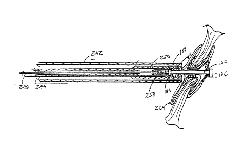
[0087] FIG. 5 shows delivery assembly 200, which includes a delivery system
220
with an occluder 224 to be delivered. The attached occluder 224 is shown in a
deployed
configuration for convenience only. Prior to deployment, the occluder 224
would
normally be in a low-profile configuration, contained within a delivery sheath
242. FIG.
6 shows delivery assembly 200 in an exploded cross-sectional side view. For
convenience, the illustrations have been divided into two parts comprising a
control
portion 230 of the delivery system 220, and a catheter portion 250 of the
delivery system
220 with the attached occluder 224, with the connection indicated by broken
line Ll. The
17
CA 02581677 2007-03-26
WO 2006/036837 PCT/US2005/034276
control portion 230 extends from a delivery wire control rod 232 to a delivery
sheath
control 240. The catheter portion 250 extends from the delivery sheath control
240 to the
end of the delivery system 220 where the occluder 224 is attached. The control
portion
230 remains external to the patient and incorporates the features provided for
operation of
the catheter portion 250 of the delivery system 220. FIG. 7 shows an enlarged
cross-
sectional side view of the control portion 230. FIG. 8 shows an enlarged cross-
sectional
side view of the catheter portion 250 and the occluder 224. The basic
components of the
delivery system 220 are described below by reference to FIGS. 5-8
collectively.
[0088] For convenience in describing the function of the controls, the
catheter
portion 250 is discussed first. Now, referring to FIG. 6, in the catheter
portion 250, a
delivery sheath 242 encloses the components that are used to deliver occluder
224. A
delivery catheter 244 contains an inner delivery wire 246. Both of the
delivery catheter
244 and delivery wire 246 connect to the occluder 224 during delivery.
Although it may
be considered advantageous to eliminate the central lumen in certain
embodiments, in
other embodiments the delivery wire 246 could also be tubular. The delivery
wire 246
should have sufficient tensile and compressive stiffness to withstand the
steps required for
the deployment and retrieval sequence. In this embodiment, the delivery wire
246 has a
stiffer proximal portion and a more flexible distal portion. The delivery
catheter 244 also
has a stiffer proximal portion and a more flexible distal portion. The
combination of
stiffness and flexibility facilitates delivery and positioning of the occluder
224. Both the
delivery catheter 244 and the delivery wire 246 may be made of two lengths of
two
different materials joined together in order to provide the requisite degree
of stiffness in
each portion of the element. Alternatively, the variation of stiffness can be
the result of
annealing, or some other material treatment process. The more flexible distal
portion
prevents undue distortion of the septal tissue during the delivery sequence.
The delivery
wire is further described infra.
[0089] Still referring to FIG. 6, the control portion of the delivery
system 230
includes respective controls for the delivery sheath 242, the delivery
catheter 244 and the
delivery wire 246. The delivery wire 246 can be advanced and retracted
linearly, in the
direction indicated by arrow D', and rotated with respect to the linear axis
of the delivery
system 220, in the direction indicated by arrow C'. The delivery wire control
rod 232 is a
rod-like element that provides both linear and rotational control for the
delivery wire 246.
18
CA 02581677 2007-03-26
WO 2006/036837 PCT/US2005/034276
The delivery wire control rod 232 slides linearly in the direction indicated
by arrow C and
rotates, with respect to the linear axis of the delivery system 220, in the
direction
indicated by arrow D to provide the corresponding motion in the delivery wire
246. The
delivery catheter 244 can be advanced and retracted linearly, in the direction
indicated by
arrow A', and rotated, with respect to the linear axis of the delivery system
220, in the
direction indicated by arrow B'. A delivery catheter control 234 is a tubular
element that
provides linear control for the delivery catheter 244, by sliding linearly in
the direction
indicated by arrow A. A delivery catheter rotational control 238 provides
rotational
control of the delivery catheter 244, by rotating, with respect to the linear
axis of the
delivery system 220, in the direction indicated by arrow B. The delivery wire
control rod
232 connects to the delivery wire 246 inside the delivery catheter control
234. A
perfusion port 236 is provided to permit introduction of fluids into the
delivery sheath
242. The delivery sheath 242 can also be rotated, with respect to the linear
axis of the
delivery system 220, in the direction indicated by arrow F' and extended and
retracted
linearly along the direction indicated by arrow E'. A delivery sheath control
240 provides
linear and rotational control of the delivery sheath 242. The delivery sheath
control 240
can be rotated, with respect to the linear axis of the delivery system 220, in
the direction
indicated by arrow F and slided linearly in the direction indicated by arrow E
to induce
the corresponding motion in the delivery sheath 242. Thus, all three of the
delivery
sheath 242, delivery catheter 244 and delivery wire 246 can be independently
extended
and retracted along and rotated around the longitudinal axis of the delivery
system 220
relative to each other using the appropriate controls. The controls are
preferably designed
to ergonomic specifications. Coordinated operation of the delivery sheath 242,
delivery
catheter 244 and delivery wire 246 allows for delivery (or retrieval) of the
occluder 224.
Although in the illustrated embodiment, each element of the catheter portion
250 can be
manipulated individually and directly by the user of the delivery system 220,
in alternate
embodiments, the required operations could be partially or completely
automated or
synchronized.
[0090] Since the occluder 224 is delivered percutaneously, the delivery
system 220
must be able to be secured so that the occluder 224 can be placed accurately
at the desired
delivery location and transformed into its deployed configuration. Securement
systems
are provided that attach the delivery components to the occluder 224. The
securement
19
CA 02581677 2007-03-26
WO 2006/036837 PCT/US2005/034276
systems are typically released serially after proper placement of the occluder
224 is
confirmed. The securernent systems are configured to provide accurate delivery
of the
occluder 224 to the desired delivery location and allow for a controlled
deployment.
Also, a device deployed according to this mechanism is able to be retrieved
and
repositioned until the final stage of the deployment process. It is also
possible to retrieve
the device once it has been fully released.
[0091] Referring to FIG. 8, the delivery catheter 244 and delivery wire 246
both
contain features of securement systems on their distal ends for connecting to
the occluder
224 and a catch system 180. The delivery wire 246 terminates in a threaded
portion 258
having a funnel-like profile. The threaded portion 258 screws onto a mated
threaded
portion 182 provided on the proximal flange 184 of the catch element 188 for
the
occluder 224. These two threaded portions cooperatively form the first
securement
system. The delivery catheter 244 terminates in a threaded portion 256 having
a funnel-
like profile. The threaded portion 256 screws onto a mated threaded portion
226 provided
on the frame of occluder 224. These two threaded portions cooperatively form
the second
securement system. The first securement system in effect secures the distal
end of the
occluder to the delivery system 220. The second securement system secures the
proximal
end 44 of the occluder 224 to the delivery system 220. The two-securement
systems
cooperatively allow the ends of the occluder 224 to be forced together or
apart for
deployment or retrieval_ The funnel-like profile is useful for locating the
corresponding
threaded portion of the occluder 224 or the catch element 188 for attachment.
The funnel
provides a channeling or guiding function. The funnel also helps the delivery
system 220
attach to the occluder 224 at extreme angles. The specific geometry of the
funnel tips can
be modified to achieve better alignment with the device. Application of torque
in the
appropriate direction engages or disengages each securement system by screwing
together
or unscrewing the respective elements from each other. The terms "distal" and
"proximal" generally refer to the disposition of the securement locations
while the
occluder 224 is in the delivery configuration in a delivery sheath, but the
orientation of
the securement systems may change during or after the delivery process.
[0092] Still referring to FIG. 8, in a presently preferred embodiment, the
threaded
portions 256 and 258 are both female threaded, while the corresponding
threaded portion
182 of the proximal flange 184 and threaded portion 226 are male threaded.
This
CA 02581677 2007-03-26
WO 2006/036837 PCT/US2005/034276
configuration has several advantages. First, a male thread in the occluder
eliminates a
cavity in the occluder 224 in which blood can stagnate and promote clotting.
Second, the
profile of the occluder 224 is reduced by using the male thread. Finally, the
female
connectors on the delivery system 220 can be provided with the funnel-like
guides
described above. In alternate embodiments, the male threads may be disposed on
threaded portions 256 and 258. Also, threaded portions 256 and 258 need not
have the
same type of threads.
[0093] Deployment of the occluder to a desired site is typically a multi-
step
operation. In FIGS. 5 and 6, the occluder 224 is shown outside the delivery
catheter for
purposes of illustration. As shown in FIG. 9, the delivery sheath 242 contains
occluder
224 in its elongated, delivery form, with the catch element 188 disengaged. As
discussed
above with reference to FIGS. 3A and 3B, the distal end of the delivery sheath
242 with
the enclosed occluder 224 is first inserted into the right atrium 11 of the
patient's heart.
The distal end of the delivery sheath 242 with the enclosed occluder 224 may
next be
inserted through the anatomical aperture 18a located in the septal tissue 12,
and into the
left atrium 13. The distal side 30 of occluder 224 is then deployed into the
left atrium 13.
The deployment process is described further below. As shown in FIG. 10, the
delivery
sheath 242 is withdrawn through the anatomical aperture 18a into the right
atrium 11,
such that central tube 22 of the occluder 224 is positioned through the
anatomical aperture
18a. As shown in FIG. 11, the proximal side 40 of the occluder 224 is then
deployed into
the right atrium 11. When properly deployed, the central tube 22 is disposed
at the
anatomical aperture 18a, and the distal side 30 and proximal side 40 exert a
compressive
force against septum primum 14 in the left atrium 13 and septum secundum 16 in
the
right atrium 11, respectively, to close the anatomical aperture 18a, e.g. the
PFO. When
the occluder 224 is properly deployed, the delivery system 220 is detached
from the
occluder 224, and the delivery sheath 242 with the delivery catheter 244 and
delivery
wire 246 are then withdrawn from the heart. In the event that the occluder 224
is not
properly deployed after performing the procedure described above, the occluder
224 may
be recovered by reversing the steps of the delivery sequence. These sequences
are
described in more detail below.
[0094] FIG. 9 illustrates the initial step for a typical delivery sequence
in
accordance with one aspect of the disclosure, a high level view of which is
shown in FIG.
21
CA 02581677 2007-03-26
WO 2006/036837 PCT/US2005/034276
3B. The occluder 224 and catch system 180 are secured to the delivery wire 246
and to
the delivery catheter 244, respectively. The female threaded portion 256 of
the delivery
catheter 244 is screwed onto the male threaded portion 226 of the occluder
224. The
female threaded portion 258 of the delivery wire 246 is screwed onto the male
threaded
portion 182 of the catch element 188 of the occluder 224. The distal end of
the delivery
sheath 242 with the enclosed occluder 224 is inserted through the aperture to
be occluded,
such as the anatomical aperture 18a of FIG. 1, to approximately the midpoint
of the
occluder 224.
[0095] Referring now to FIG. 10, the distal side 30 of the occluder 224 is
deployed
on the distal side of the aperture in the left atrium 13. The distal portion
30 is deployed
by first retracting the delivery sheath 242 to expose the distal portion 30 of
the occluder
224. The axial length of the occluder 224 is then reduced by applying pulling
force F1 on
delivery wire 246 with sufficient force to cause the catch element 188 to be
pulled
through the central tube 22 of the occluder 224 and the distal portion 30 of
the occluder
224 to compress and distal petals 32 to form. Force F2 is simultaneously
applied to the
delivery catheter 244 to hold the occluder 224 stationary. The central tube 22
of the
occluder 224 catches on the catch element 188. This holds the distal petals 32
in place
while the remainder of the deployment sequence is carried out.
[0096] Referring now to FIG. 11, the proximal side 40 of the occluder 224
is
deployed on the proximal side of the aperture in the right atrium 11. The
proximal
portion 40 is deployed by first retracting the delivery sheath 242 to expose
the proximal
portion 40 of the occluder 224. The proximal petals 42 are then deployed by
simultaneously advancing the delivery catheter 244 by applying force F4 and
retracting
the delivery wire 246 by applying force F5 to maintain the position of the
occluder 224.
Eventually, the proximal end 44 of the occluder 224 is pushed over the
proximal end 44
of the catch element 188 and the occluder 224 is caught on the proximal flange
184 of the
catch element 188. The final configuration is illustrated in FIG. 12. The
occluder 224
can now be evaluated for proper deployment at the desired location.
[0097] The occluder 224 can be evaluated for proper deployment with the
delivery
system 220 attached or at least partially detached. The delivery system 220
can be
partially detached by releasing one of the securemen_t systems provided by the
delivery
catheter 244 and the delivery wire 246. As shown in FIG. 13, according to one
preferred
22
CA 02581677 2007-03-26
WO 2006/036837 PCT/US2005/034276
embodiment, to evaluate the proper deployment of the occluder, if desired, the
delivery
sheath 242 can be further retracted and the delivery catheter 244 can be
detached from the
occluder 224. The delivery catheter 244 can be detached by applying torque to
unscrew
the delivery catheter 244 from the proximal threaded portion 226 of the
occluder 224 and
retracting the delivery catheter 244. The delivery wire 246 continues to
secure the
occluder 224, as illustrated in FIG. 14. This affords the clinician a
substantially
unobstructed view of the occluder delivery site in order to evaluate the
placement of the
occluder 224. In addition, the more flexible distal portions of the delivery
catheter 244
and the delivery wire 246 allow the distal end of the delivery system 220 and
the
deployed occluder to be re-positioned so that the view is not obstructed. The
positioning
of the occluder 224 can be evaluated using fluoroscopy or other appropriate
techniques.
If the delivery or deployment is not satisfactory, then the delivery system
220 can be used
to retrieve the occluder 224. If delivery catheter 244 has been detached, it
is reattached
by advancing the threaded portion 256 of the delivery catheter 244 toward the
threaded
portion 226 of the occluder 224 and applying torque until the delivery
catheter 244 is
threaded onto the occluder 224. As mentioned before, the funnel-like shape of
the
threaded portion 256 of the delivery catheter 244 helps to guide the
reattachment of this
securement system. A similar technique is used to reattach the delivery wire
246 if
needed.
[0098] Once the occluder 224 is successfully deployed, the delivery system
220 can
be detached in the sequence shown in FIGS. 13-15. As illustrated in FIG. 13,
the delivery
sheath 242 is partially retracted by applying force F12. Also, the delivery
catheter 244 is
detached by applying torque F14 to unscrew the threaded portion 256 of the
delivery
catheter 244 from the threaded portion 226 of the occluder 224. Force F13 is
then applied
to retract the delivery catheter 244 while simultaneously advancing the
delivery wire 246
by applying force F15 to maintain the position of the occluder 224. 'The
occluder 224
remains attached to the delivery system 220 by the second securement system
provided
by the delivery wire 246. As discussed above, if retrieval is desired for any
reason, the
occluder 224 can readily be returned to its low-profile configuration and
removed at this
point. As shown in FIG. 14, the delivery catheter 244 can be further retracted
by
applying force F16 to provide an unobstructed view of occluder 224, again
while the
delivery wire 246 remains attached. As illustrated in FIG. 15, if the
deployment is
23
CA 02581677 2007-03-26
WO 2006/036837 PCT/US2005/034276
successful, then the delivery wire 246 can be detached by applying torque F17
to unscrew
the threaded portion 258 of the delivery wire 246 from the threaded portion
182 of the
catch element 188. The torque applied to remove the delivery wire 246 and the
delivery
catheter 244 can be either clockwise or counterclockwise depending on the
design of the
device. The delivery wire 246 can be retracted by applying force F18. The
occluder 224
is now fully deployed.
[0099] Referring now to FIG. 16, if retrieval is desired, the process
involves
reattaching the delivery catheter 244 and delivery wire 246 as mentioned
above. Then
force F6 is applied to the delivery catheter 244 to pull the proximal portion
40 of the
occluder 224 over the proximal end of the catch element 188. As the axial
length of the
occluder 224 is increased, the proximal petals 42 are unformed and the
proximal portion
40 of the occluder 224 returns to its tubular profile. Referring to FIG. 17,
force Fg is
applied to the delivery sheath 242 to advance the delivery sheath 242 over the
proximal
portion 40 of the occluder 224 and retain the proximal portion 40 of the
occluder 2.24 in
the low-profile configuration. Also, force F7 is applied to delivery wire 246
in order to
release the distal portion 30 of the occluder 224 and further increase the
axial length of
the occluder 224. Referring now to FIG. 18, the distal portion 30 of the
occluder 224 is
fully extended back into it low-profile configuration and forces F9 and F10
are applied to
the delivery sheath 242 and the delivery catheter 244 in order to retrieve the
occluder 224
back into delivery sheath 242. Referring to FIG. 19, the delivery sheath 242
and enclosed
occluder 224 are removed from the anatomical aperture 18a and can further be
fully
removed from the heart 10 by applying force F11. This step can also be used as
a starting
point for redeployment of the occluder 224, i.e., the sequence shown beginning
in FIG. 9.
[00100] The components of an alternate preferred embodiment of the
invention are
described in connection with FIGS. 20-24. FIG. 20 illustrates an occluder 310
with a
distal side 30 and a proximal side 40 that are connected by central tube 22.
The
configuration illustrated is a simplified schematic view of the occluder
illustrated in
FIGS. 2A-2D. Of course, other types of occluders can be deployed using this
delivery
system. The occluder includes a catch system 320 that includes a distal flange
322, a
catch body 324 and a catch element 326 in the shape of a cone. The catch
system .320 is
disposed in an axially central location in the occluder 310. Although
schematically
illustrated as a separate piece than the proximal side and distal side loops
40 and 30,
24
CA 02581677 2007-03-26
WO 2006/036837 PCT/US2005/034276
respectively, of the occluder, the catch system 320 may be a single piece, or
even fixed to
one end of the tube that forms the proximal and distal loops by an adhesive,
ultrasonic
welding, or the like. For example, the flange 322 may be fixed to the end of
the tube that
forms the loops. The device can be formed from a single component or multiple
components that are fixed together. The catch body 324 is disposed axially
within the
inside surface of the tube that forms the loops. The tube is able to move with
respect to
the catch system (and the catch body) so that the petals can move from the
delivery
configuration to the deployed configuration. The inside surface of the tube
335 is able to
slide over the catch elemen 326 so that, when the proximal tip of the occluder
310 rests
against the flat surface 326a of the catch element 326, the occluder 310 is
secured in its
deployed configuration.
[00101] As shown in FIG. 20, the first securement system 330 includes a
threaded
component 332, illustrated as a male thread, and corresponding threads on a
corresponding female portion described below in connection with FIGS. 22 and
23. The
second securement system 340 includes a groove 314 on the proximal portion 40
of the
occluder 310 that cooperates with a collet system 344 described below in
connection with
FIGS. 21 and 22. As shown in FIG. 21, the collet system 344 also includes
collet fingers
346 that are configured to have ends that fit within the groove 314 on the
occluder 310.
The collet system also includes a collet tube 348 onto which the collet
fingers 346 are
mounted and a collet sheath 350 that is movable with respect to the collet
tube 348. In
one embodiment, the collet fingers 346 are constructed of nitinol and have a
splayed
configuration when at rest as illustrated in FIG. 21. More detail regarding
the
construction of the construction of the collet fingers 346 is provided below.
As the end of
the collet sheath 350 is moved over the collet fingers 346, the collet fingers
346 are
moved radially inward and when occluder 310 is being positioned in the
delivery system,
the collet fingers 346 are moved radially inward and engage the groove 314 on
the
occluder 310 (illustrated on the left side of FIG. 22). The collet sheath 350,
collet tube
348 and collet fingers 346 are described in more detail below.
[00102] FIG. 22 illustrates a delivery system of a preferred embodiment of
the
invention. Specifically, the occluder 310 is disposed within the delivery
sheath 356.
Within the delivery sheath 356 are the components that are used to secure the
occluder
310 during delivery and are (typically) released serially after proper
placement of the
CA 02581677 2007-03-26
WO 2006/036837 PCT/US2005/034276
occluder 310 is confirmed. The first securement system 330 and the second
securement
system 340 are each illustrated as securing the occluder 310 for delivery to
the desired
delivery location within the body. The securement systems 330 and 340 are
configured to
provide accurate delivery of the occluder 310 to the desired delivery location
and allow
for a controlled deployment so that the position of the device as it is being
deployed can
be monitored. Also, an occluder 310 deployed according to this system is able
to be
retrieved and repositioned until the final stage of the deployment process.
Even after the
final stage of the deployment process, the occluder 310 can be retrieved.
[00103] FIG. 22 also illustrates the second securement system 340 in an
engaged
configuration. Specifically, the collet fingers 346 are disposed in the collet
sheath 350 so
that the collet fingers 346 engage groove 314 on the occluder 310. When the
collet
sheath 350 is disposed in this configuration, the occluder 310 is secured by
the collet
fingers 346 against axial motion with respect to the collet sheath 350 and
collet tube 348.
Similarly, when the delivery wire 380 is secured in an engaged configuration,
the
occluder 310 is secured against axial motion with respect to the delivery wire
380. Thus,
the occluder 310 is secured during delivery and the controlled motion of the
collet sheath
350/collet tube 348 and the delivery wire 380 can deploy the occluder 310.
[00104] As illustrated in FIG. 22, the delivery wire 380 is threaded into
the first
securement system 330 by a threaded connection. As illustrated in FIG. 22, the
female
threads can be disposed on the delivery wire 380 and the male threads can be
disposed on
the occluder 310. FIG. 24 illustrates an alternative embodiment of a first
securement
system, designated 390, in which the male threaded portion 392 is disposed on
the
delivery wire 380 and the female threaded portion 394 is disposed on the
occluder 310.
[00105] In a presently preferred embodiment, the male threads are disposed
on the
occluder 310 and the female threads are disposed on the delivery wire 380.
This
configuration has several advantages. First, the occluder 310 does not need a
female
connector and there is no cavity in which blood can stagnate and promote
clotting.
Second, the space required for the threaded connector 392 on the occluder 310
is
diminished. Finally, a female connector on the delivery wire 380 may allow for
a more
smooth deployment of the occluder 310.
[00106] The first securement system interconnects the delivery wire 380 to
the
threaded portion on the occluder 310. Representative embodiments of the first
26
CA 02581677 2007-03-26
WO 2006/036837 PCT/US2005/034276
securement system and its components are illustrated in more detail in FIGS.
23 and 24.
In FIG. 23, the threaded portion 386, interconnects the delivery wire 380 and
the threaded
portion 332 on the occluder 310, illustrated in FIG. 20.
[00107] Referring again to FIG. 23, the delivery wire 380 has a more rigid
section
382 and a more flexible section 384. In general, the flexible section 384 is
distal to the
more rigid section and is provided on the delivery end of the delivery wire
380. The
delivery wire 380 can be any kind of flexible elongate member such as a wire,
tube,
hypotube, coil, or other hollow or solid constructions. The delivery wire 380
can be made
from any material suitable for medical applications. Exemplary materials
include metals
and alloys suitable for medical applications, including stainless steel (such
as "304
Stainless") and MP35N, polymers (such as nitinol), or any other suitable
materials. The
variation of stiffness can be the result of annealing; other material
treatment process, or it
may be a result of different materials being joined together. The amount of
flexibility, or
rigidity, can vary depending on the type of occluder being delivered and the
delivery
location within the body. The length of the flexible section 384 would
typically be about
the length of the occluder 310 in its delivery configuration. That is, the
occluder 310 in
the delivery configuration would surround the flexible portion of the delivery
wire 380.
The length of the flexible section 384, however, can be varied. The distal end
of the
delivery wire 380 includes a threaded attachment portion 386 on the end of the
flexible
section 384, described in detail below. The threaded portion 386 is
illustrated as a female
thread.
[00108] FIGS. 25A, 25B, 26A, 26B, 27A, and 27B illustrate alternative
embodiments of the first securement system 330. Generically, all of the
securement
embodiments described can be properly described as interlocking systems. Each
of these
embodiments of the first securement system can be used with the threaded or
collet
connection for the second securement system and provide alternatives which may
be
appropriate for different kinds of occluding devices or other devices that
could be
delivered by the delivery system described in this application
[00109] FIGS. 25A and 25B illustrate a ball and claw type attachment. In
place of a
screw type attachment, a ball 410 is disposed on the occluder and two or more
claws 412
are sized to secure the ball 410 within the claws 412. The claws 412 are
disposed at the
distal end of the delivery wire 380. Two claws 412 are illustrated in FIG.
25B. The
27
CA 02581677 2007-03-26
WO 2006/036837
PCT/US2005/034276
claws 412 operate under a similar principle as the collet design described
previously.
Specifically, there is a claw sheath 414 that is axially movable with respect
to the claws
412. As illustrated in FIG. 25B the claws 412 splay out in the at rest
configuration.
When the claws 412 are in the claw sheath 414, the claws 412 are sized to
secure the ball
410. Thus the configuration allows for a secure placement of the occluder on
the delivery
system. When the occluder is ready to be released claw sheath 414 is withdrawn
and the
claws 412 splay out to the at rest confiduration. Thus the occluder is
released from the
first securement system.
[00110] FIGS. 26A and 26B illustrate a pin-through-hole connector 420. In
this
embodiment, fingers 422 includes pins 424 that are disposed in an aperture in
the
occluder. As the example illustrates, the transverse aperture 428 is formed in
the occluder
and the transverse aperture 428 is sized to receive the pins 424. When the
fingers 422
including pins 424 are in a sheath 426, the pins 424 are secured within the
transverse
aperture 428. Thus the configuration allows for a secure placement of the
occluder on the
delivery system. When the occluder is ready to be released a sheath 426 is
withdrawn
and the pins 424 spring back to the unbiased position similar to the fingers
in the collet
system. Thus the occluder is released from the first securement system.
[00111] In another embodiment of the first securement system, illustrated
in FIGS.
27A and 27B, a pair of cooperating configurations are secured when disposed
within a
sheath and separable when the sheath is withdrawn. This is a type of
interlocking system
440. In this example, the lock is achieved using a combination of two C-shaped
elements.
Specifically, as illustrated, the occluder has a portion 442 that extends in
an axial
direction and is adapted to mate with a delivery wire 444. The portion 442 and
the
delivery wire 444 have cooperating extensions 446, 448 respectively that are
able to
interlock as illustrated in FIG. 27A. The system as illustrated has an
interlocking
elbow/arm attachment 450, 452 on each of the protrusion and the delivery wire.
A
variety of interlocking configurations are possible and the concept should not
be limited
to the configuration illustrated. When the interlocking system is disposed
within a sheath
454, the cooperating extension cannot move with respect to each other. Thus
the
configuration allows for a secure placement of the occluder on the delivery
system.
When the cooperating extensions are extended beyond the sheath 454, the
interlocking
system can release and the occluder is released from the first securement
system.
28
CA 02581677 2007-03-26
WO 2006/036837 PCT/US2005/034276
[00112] Any of the previous embodiments described in FIGS. 20-27B can be
deployed in a manned illustrated in FIGS. 28 and 29. FIG. 28 illustrates the
occluder 310
in its deployed configuration. To deploy the occluder 310, the delivery sheath
356 is
withdrawn to expose the distal side loops 30 and then the proximal side loops
40 into the
defect to be occluded. During this time the catch element 326 is engaged so
that the
occluder 310 is secured in the deployed configuration. Once the occluder 310
is in the
deployed configuration the collet sheath 350 is withdrawn and the collet
fingers 346 are
unconstrained by the collet sheath 350 and are allowed to move radially
outward to the
unbiased condition, as illustrated in FIG. 29. Once the collet fingers 346
move radially
outward the tips of the collet fingers 346 move away from the groove 314 in
the occluder
310. Accordingly, the occluder 310 is only attached to the delivery by the
first
securement system 330. In this position, the clinician is able to evaluate the
position of
the occluder 310 to make sure that the device is properly positioned.
[00113] The process of retrieving an occluder varies based on the state of
the
delivery when the decision to retrieve the occluder is made. If the second and
first
securement systems are still attached and the catch system has not secured the
device in
the deployed configuration, then the retrieval process is simply a reversal of
the
deployment process. The second securement system is pulled and the device can
be
withdrawn into delivery sheath 356 and removed from the body.
[00114] If the catch system has secured the device in a deployed
configuration, and
the second and first securement systems are still attached, the process is the
same with the
addition of moving the catch element of the occluder relative to the second
securement so
that the device can be elongated. Once that occurs, the device can be
withdrawn as
described above.
[00115] The retrieval process for an occluder in which the second
securement system
is a collet system, which has been disengaged, requires an additional step.
The collet
system is advanced until the collet fingers are in alignment with the groove
on the
occluder. Next the collet sheath is advanced over the collet fingers such that
the
fingertips fit within the groove on the occluder. By pulling on the collet
tube with the
occluder thinly secured, the device can be returned to its collapsed state and
retrieved
into the delivery assembly. From this point the delivery process can be
restarted.
29
CA 02581677 2007-03-26
WO 2006/036837 PCT/US2005/034276
[00116] FIG. 30 illustrates a detail view of a collet finger 346 and the
collet sheath
350. The collet finger 346 is configured to be about 20 degrees from the base
of the
collet finger 346, the dimension identified as 0 in FIG. 30. The collet finger
346 can
extend from the collet tube 348 approximately 0.25 in., the dimension
identified as "a" in
FIG. 30. The distance "a" can be from 0.1 in. to 0.5 in. The angle 0 can vary
from low
single digits to approximately 70 degrees. In general, as the length of "a" is
decreased,
the angle desired for 0 would increase. The collet finger 346 includes a
radially inwardly
extending protrusion 347, which is formed by a bend in the nitinol finger. The
bend is
preferably 90 degrees and the dimensions of the protrusion are selected to
securely fit
within the groove 314. As illustrated in FIG. 20, supra, the groove 314, for
example,
could be 0.02 ¨ 0.04 in. in axial length and 0.005-0.020 in radial depth. The
groove 314
is illustrated as a circumferential groove; alternatively, recesses can be
formed in part of
the occluder 310 to receive the collet fingers 346. It is preferable that the
collet fingers
346 have a close fit but not an interference fit in the axial direction. This
assures that the
collet system can move the device without significant slippage. It is also
preferable that
the protrusion does not come into contact with the bottom of the groove 314
(the inner-
most radial surface). This assists the deployment of the occluder.
[00117] FIGS. 31, 32A, and 32B illustrate alternative embodiments of the
second
securement system. The fingers are formed by cutting sections from a nitinol
hypotube
that has, for example, a .0075 in. wall thickness. The inner diameter could
be, for
example, 0.098 in. and the outer diameter could be 0.117 in. The thickness of
the
hypotube could be as large as 0.050 in.or more. Nitinol is a desirable
material due to its
superelastic characteristics. Other superelastic materials or simply springy
material may
be used. Of course, the materials would have to be suitable for use in a
medical device.
The nitinol hypotube is cut so that the fingers extend from one side and the
hypotube ring
is uncut at the other end. As an example, FIG. 31 illustrates a cross section
where the
hypotube is disposed on an end of the collet tube 348. The nitinol ring 355 is
disposed on
the outside surface of the collet tube. The nitinol ring 355 may be affixed to
the collet
tube 348 by a variety of known techniques such as a suitable adhesive.
[00118] FIGS. 32A and 32B illustrate the side and end view of
representative collet
fingers 346. In a preferred embodiment, there are four collet fingers 346 that
are used to
secure the occluder in the delivery system. In alternate embodiments, there
may be as
CA 02581677 2007-03-26
WO 2006/036837 PCT/US2005/034276
few as two collet fingers 346 or as many as 8. One practical limitation is the
circumferential size of the collet fingers 346 and the rigidity of the collet
fingers 346 as
they are used to deploy the occluder. In the embodiment illustrated in FIG.
32B, the four
collet fingers 346 are formed by cutting away a 118th section of the cross
section and
forming four equally spaced collect fingers 346. During the formation process
the
roundness of the collet finger 346 along the circumference can be modified to
adjust the
bendability of the collet fingers 346.
[00119] FIG. 33 illustrates another embodiment of the collet fingers 346.
In this
embodiment, the collet fingers 346 include a bend 358 between the base ring
355 and the
protrusion 347. As illustrated the bend 358 is in the approximate halfway
between the
base ring 355 and the protrusion 347. The bend 358 can be almost any
configuration but
the bend 358, as illustrated, allows for force to be applied to the occluder
and have the
configuration of the collet fingers 346 be such that it does not extend the so
far away from
the collet tube 348 in the radial direction. This allows the occluder to have
a more
controlled delivery because of the increased forces applied and a more compact
system
because the collet fingers 346 do not extend radially away from the collet
tube 348 as far. ,
[00120] Another embodiment of the second and first securement system of the
delivery system, illustrated in FIGS. 34-37, uses a filament instead of a
delivery wire. As
shown in FIG. 34, the second securement system 340 is illustrated as the
collet system
344, which is largely the same as in the previous embodiment. Of course, other
securement systems included a threaded connection can be used. The first
securement
system includes an eyelet 510 around which a flexible filament 512 can be
fastened or
looped. The flexible filament can be a suture thread (monofilament or
polyfilament), a
thin metallic wire or other flexible material that can withstand a tension
load.
[00121] The deployment of the occluder is effected by withdrawing the
delivery
sheath 356 to expose and let the distal petals 30 on the distal side of the
occluder expand
as illustrated in FIG. 35. Once the distal petals 30 are deployed by the catch
element 326,
the delivery sheath 356 is repositioned to the deployment site of the proximal
petals 40
and the proximal petals 40 are exposed as illustrated in FIG. 36. The filament
is pulled to
cause the catch element at the proximal side of the occluder to be secured.
Once the catch
element secures the occluder in the deployed configuration, the collet system
is released
in the manner described above and illustrated in FIG. 37. Once the collet
system is
31
CA 02581677 2007-03-26
WO 2006/036837 PCT/US2005/034276
released, the position may be evaluated. If the position is satisfactory,
filament 510 is
pulled through the eyelet and removed from the body. Either a delivery wire, a
tube or
filament is appropriate for the second securement system depending on the
design
considerations. For example, if the occluder in the delivery configuration
lacks sufficient
column strength to have the delivery sheath pull back without affecting the
position of the
device in the delivery system, a delivery wire that has some column strength
would be
more desirable.
[00122] An alternative recovery sequence is also provided. The recovery
process for
a device in which the second securement (e.g., collet) system has been
disengaged, the
process requires an additional step. For systems in which a retrieval
capability is desired,
additional filaments can be attached to the proximal tip of the occluder. For
example,
with reference to FIG. 34, filaments 514 are attached to the proximal end 44
of the
occluder 570 through holes 516. The filaments 514 can be attached in a variety
of
locations, for example, they can be looped around one of the proximal loops on
the
proximal side of the device. When the filaments 514 are provided, the
clinician would
orient the delivery catheter 548 (as illustrated in FIG. 38B) to the proximal
end 44 of the
occluder 570 and then pull on the filaments 514 to uncatch the system so that
the profile
of the device can be reduced and reinserted into the delivery catheter. In an
embodiment
where the filament 514 is present and the device is deployed satisfactorily,
the filaments
514 can be cut or otherwise withdrawn from the body.
[00123] FIG. 38A illustrates an end view of the construction of the
delivery catheter
548. The delivery catheter 548 a central lumen 556 and secondary lumens 554
surrounding the central lumen 556. The outer lumens 554 are used to provide a
passageway for containing sutures 558 secured to the occluder 570, illustrated
in FIG.
38C, as the attachment mechanism for the second securement system 340 and
passed
through the delivery catheter 548 to the user for manipulating the second
securement
system. Although four outer lumens 554 are shown, any number of lumens may be
provided suitable for use in the delivery system 340. A sufficient number of
sutures 558
should be provided in order to securely attach the occluder 570 and permit the
necessary
operations. The sutures 558 are shown in FIG. 38B, which illustrates sheath
delivery 544
which contains delivery catheter 548. Referring again to FIG. 38C, the
delivery catheter
548 is connected to the proximal end 44 of the occluder 570 via the sutures
558 which
32
CA 02581677 2007-03-26
WO 2006/036837 PCT/US2005/034276
attach to holes 560 provided in the occluder 570. The sutures 558 are threaded
through
the holes 560 and can be readily detached by, e.g., cutting the sutures and
pulling through
the delivery catheter 548. Attachment of the sutures 558 to the occluder 570
may be
provided in a number of ways, such as providing hooks or a flange on the
occluder 570,
around which the sutures can be wrapped or fastened, or wrapping the sutures
558 around
the proximal petals 42. The sutures 558 can also be embedded into the proximal
end 44
of the occluder 570. The flexible filament used to provide the thread can be a
suture
thread (monofilament or polyfilament), a thin metallic wire or other flexible
material that
can withstand a tension load.
[00124] FIG. 39 illustrates a complete delivery assembly 700 with the
occluder 570
in place for delivery and deployment at the deployment site. As shown, the
occluder 570
is in its elongated, low profile configuration. The occluder 570 is secured at
its distal end
39 by the first securement system 330 to the delivery wire 380 and at its
proximal end 44
by the second securement system 550 to the delivery catheter 548. The occluder
570,
delivery wire 380 and delivery catheter 548 are contained within the sheath
544. The
occluder 570 can be detached from the first securement system 330 by
unscrewing the
delivery wire 330, which is connected by threaded portion 386 to threaded
portion 332.
The occluder 570 can be detached from the second securement system 550 by
removing
the sutures 558, for example, by pulling on them from the user end of the
delivery system.
[00125] The embodiments and techniques described here are described
preferably for
use with a device made of a polymer and formed from a single tube, such that
the tube is
a single monolithic material. The catch mechanism can be all or partly
monolithic or
integral with the tubular structure, or there can be an absence of any type of
bonding or
rigid connection to the rest of the tubular structure, in which case there may
be some
spring force or other force that holds the locking mechanism in place. While
the device is
thus shown as being substantially formed from a single tubular body, the catch
mechanism as described in the embodiments above could be used with other types
of
devices, including those formed from many pieces, and including devices formed
from
other materials, including metals, polymers, stainless steel or nitinol.
[00126] The term "bioabsorbable," as used in the description above, is also
understood to mean "bioresorbable."
33
CA 02581677 2007-03-26
WO 2006/036837 PCT/US2005/034276
[00127] While the description above refers to strings, filaments, sutures
and wires,
and while the term "wire" might convey a more rigid piece than a string, a
suture or a
filament, all these terms are essentially interchangeable, and further include
embodiments
in which the wire, string, suture or filament is a hollow tube or conduit to
allow another
wire, as needed, to pass through its longitudinal axis. Each wire, string,
suture and
filament can be composed of one or more wires, strings, sutures and filaments.
[00128] In cases in which the device is made of a polymer, it can be
desirable to add
an additive or coating to the material to make it radiopaque to make it more
visible in a
wider variety of imaging techniques.
[00129] It will be appreciated that while a particular sequence of steps
has been
shown and described for purposes of explanation, the sequence may be varied in
certain
respects, or the steps may be combined, while still obtaining the desired
deployment or in
some cases to effect deployment in a particular way. For example, the delivery
sheath
may be advanced or retracted at varying times and in varying degrees, the
proximal and
distal portions of the occluder may be deployed into the petal configuration
in a different
sequence, etc. In addition, the steps could be automated.
34