Note: Descriptions are shown in the official language in which they were submitted.
CA 02583591 2012-07-23
Methods and Devices for Repair or Replacement of Heart
Valves or Adjacent Tissue Without the Need for Full
Cardiopulmonary Support
Field of the Invention
[0002] This invention relates generally to devic
and methods for performing cardiovascular procedures
wherein a heart valve or segment of the aorta is being
repaired or replaced without the use of extracorporeal
cardiopulmonary support (commonly referred to as
"off-pump" procedures). For example, the invention
relates to devices and methods for accessing,
resecting, repairing, and/or replacing one of the heart
valves, in particular the aortic valve. This invention
also relates to methods and systems for performing
minimally-invasive cardiac procedures such as the
endovascular, endocardiac or endoluminal placement,
implantation or removal and consecutive replacement of
heart valves. These techniques may be generally
CA 02583591 2007-04-02
WO 2006/041505
PCT/US2004/043794
- 2 -
referred to as direct access percutaneous valve'
replacement ("DAPVR").
Background of the Invention'
[0003] Of particular interest to the present
invention is the treatment of heart valve disease. =
There are two major categories of heart valve disease:
(i) .stenosis, which is an obstruction to forward blood.
flow caused by a heart valve, and (ii) regurgitation,
which is the retrograde leakage of blood through a
heart valve. Stenosis often results from calcification
of a heart valve that makes the Valve stiffer and less
able to open fully. Therefore, blood must be pumped
through a smaller opening. Regurgitation can be caused
by the insufficiency of any of the valve.leaflets such.
that the valve does not fully close.
[0004] In the past, repairing or replacing a.
malfunctioning heart valve within a patient has been
achieved with a major open-heart surgical procedure,
requiring general anesthesia and full cardiopulmonary
by-pass. This requires complete cessation of
cardiopulmonary activity. While the use of
= extracorporeal cardiopulmonary by-pass for cardiac
support is a well accepted procedure, such use has
often involved invasive surgical procedures
median sternotomies, or less commonly, thoracotomies).
These operations usually require one to two weeks of
hospitalization and several months of rehabilitation
time for the patient. The average mortality' rate with
this type of procedure is about five to six percent,.
and the complication rate is substantially higher.
[0005] Endovascular surgical techniques for heart
surgery have been under recent development. In
CA 02583591 2007-04-02
WO 2006/041505 PCT/US2004/043794
- 3 -
contrast to open-heart surgical procedures,
endovascular procedures may have a reduced mortality =
rate, may require only local anesthesia, and may,
necessitate only a few days of hospitalization.
However, the range of procedures that has been
= .
developed for an endovascular approach to date has been
limited to repair of the coronary arteries, such as
angioplasty and atherectomy.
[0006] Some progress has been made in the
development of endovascular heart valve procedures.
For example, for patients with =severe stenotic valve
disease who are too compromised to tolerate open-heart.
surgery to replace the heart valve as described above,- =
surgeons have attempted endovascular balloon aortic or =
= mitral valvuloplasty. These procedures involve
endovascularly advancing a balloon dilatation catheter
into the patient's vasculature until the balloon of the
catheter is positioned between the=valve leaflets:
Then the balloon is inflated to either: (i) split the
commissui.es in a diseased valve with commissural
fusion, or (ii) crack calcific plaques in a calcified .
stenotic valve. However, this method may only provide
.. partial and temporary relief for a patient with a
=stenotic valve. Instances. of restenosis and mortality
following balloon aortic valvuloplasty have led to
virtual abandonment of this procedure as a treatment -
for a diseased aortic valve. '
. [0007] Endovascular procedures for valve
implantation inside a native and diseased valve have
been. explored. A catheter-mounted valve is
incorporated into a collapsible cylindrical structure,
such as a stent (commonly referred to as a "valved
stent"). In these procedures, an elongated catheter is
CA 02583591 2007-04-02
WO 2006/041505 PCT/US2004/043794
=
= 7 4 -
used to insert a mechanical Valve into the lumen of the
aorta via entry through a distal artery. (e.g., the
femoral or brachial artery). Such procedures have been
attempted on selective, terminally ill patients as a'
means of temporarily relieving the symptoms of ,a
diseased valve. =
[0008] The.percutaneous placement of an artificial
valve 'may have certain limitations and ancillary
effects. For example, at present, such procedures are '
, only of benefit to a small number of patients and are
not meant to become an alternative to surgical heart
valve procedures requiring the use of extracorporeal
bypass. Another .issue is that performing the entire
procedure via small diameter vessels (e.g., the
femoral, iliac or brachial arteries) restricts the use,
of larger tools and devices for'the resection Or repair
of the diseased heart valve. Furthermore; this .
endovascular procedure may increase the risk of. various.
' vascular complications such as bleeding, dissection,
rupture of the blood vessel, and ischemia to the
extremity supplied by the vessel used to perform the
operation.
[0009] Moreover, in some cases, one or more of a
patient's femoral arteries, femoral veins,, or other
; vessels for arterial and venous access may not be
available for introduction of delivery devices or valve
removal tools due to inadequate vessel diameter, vessel
stenosis,, vascular injury, or other conditions. In
such cases, there may not be sufficient arterial and
venous access to permit the contemporaneous use of the
necessary interventional devices (e.g., an angioplasty
catheter, atherectomy catheter; or other device) for a
. single surgical procedure. Therefore, unless alternate
CA 02583591 2007-04-02
WO 2006/041505 PCT/US2004/043794
- -
arterial or venous access for one or more of these
catheters can be found, the procedure cannot be
performed using endovascular techniques.
[0010] Another possible disadvantage of the small.
vessel
vessel procedure is that the new valve must be.. '
collapsed to a very Small.diameter.that could result in
structural damage to the new valve. . Additionally, such
remote access sites like the femoral artery may make
precise manipulation of the surgical tools more .
difficult (e.g., exchange of guide wires and catheters
. and deployment of the new valve). Furthermore, placing
wires, Catheters, procedural tools, or delivery devices .
through one or more heart structures (e.g., the mitral
valve) to reach the target site can result in damage to
those structures (e.g., acute. malfunctioning or
insufficiency of the valve being medhanically hindered
by the surgical equipment or valve deterioration
resulting from mechanical friction' inflicting micro- .
lesions on the valve).
[0011] Also to be considered in connection with such.
procedures is the potential of obstructing the coronary
ostia. ,The known percutaneous procedures for
. implanting heart valves do not have a safety mechanism
to ensure proper orientation of the new valve.
Therefore, there is a possibility that the deployed
,valve will obstruct the coronary ostia, which can
result in myocardial ischemia, myocardial infarction,
and eventually the patient's death.
[0012] These procedures leave the old valve in
place, and the new valve is implanted within the
diseased valve after the diseased valve has been
compressed by a balloon or other mechanical device.
Therefore, there may be a possibility of embolic stoke
CA 02583591 2007-04-02
WO 2006/041505 PCT/US2004/043794
- 6 -
or embolic ischemia.from valve or vascular.wall-debris
that is liberated into the blood flow as the diseased
valve is dilated and compressed. .Furthermore, a rim of
diseased tissue (e.g., the compressed native valve)
decreases the diameter and cross-Sectional surface of. -
the implanted valve, potentially-under-treating the '
patient and leading to only. partial relief of his .
symptoms.
[0013] . It would therefore be desirable to.develop
systems and methods for satisfactorily performing =
various cardiovascular procedures, particularly
procedures for heart valve placement or removal and.
replacement, which do not require the use of an
extracorporeal bypass or invasive surgical procedure, .
such as a sternotomy. It would be further desirable to.
perform such procedures through very small incisions in.
the patient (e...g.; via several small thoracotomies).
The devices and methods will preferably facilitate the
access, resection, repair, implantation, and/or
replacement of the diseased cardiac structure (e.g.,
one or more diseased heart valves). The devices and
methods should preferably minimize the number of
arterial and venous penetrations required during the
closed-chest procedures, and desirably, should require
no more than one cardiac and one femoral. arterial
. penetration. The present invention satisfies these and-
other. needs. =
[0014] -The descriptive terms antegrade and
retrograde mean in the direction of blood flow and
opposite the direction Of blood flow, respectively,
When used herein in relation to the patient's
vasculature. In the arterial system, antegrade refers
to the downstream direction (i.e., the same direction
CA 02583591 2007-04-02
WO 2006/041505
PCT/US2004/043794
- 7 -
as the physiological blood flow), while retrograde
refers to the upstream direction (i.e., opposite the '
direction of the physiological blood flow). The terms. .
proximal and distal, when used herein in relation to.
instruments used in the procedure, refer to directions'.
closer to and farther away from the heart;
respectively. The term replacement normally signifies
removal of the diseased valve and implantation of anew
valve. However, a new valve may also be implanted .
directly over top of a diseased valve. An implantation
procedure would be the same as a replacement procedure
without the removal of the diseased valve. .
=
Summary of the Invention
[0015] . The present invention is directed to a method.
and system for an endoVascular, endocardiac, or
endoluminal approach to a patient's heart to perform an
operation that does not require an. extracorporeal
cardiopulmonary bypass circuit and that can be
performed through a limited number of mull incisions,
thus eliminating the need for a sternotomy, .The
invention contemplates, at least in its preferred
. embodiments,' the possibility of effective aortic' valve
.implantation, aortic valve repair, resection of the
aortic valve and replacement of the aortic valve, all
without necessitating extracorporeal cardiopulmonary
by-pass, a median sternotomy or other grossly thoracic
incisions. =
[0016] The
present invention contemplates replacing
any of the four valves of the heart via an antegrade
approach through the wall of the appropriate chamber.
Preferably, valves are implanted transapically (i.e.,
through the heart muscle at its left or right
CA 02583591 2007-04-02
WO 2006/041505 PCT/US2004/043794
- 8 -
ventricular apex). .However, in this Case, replacement
of the mitral and tricuspid valves may be performed via
a retrograde approach, because accessing these valves -
via the left or right ventricles requires approaching
these valves against the flow of blood through the
valve.
[0017] In accordance with the present invention, a
surgeon may perform a minimally invasive operation on a
patient that includes accessing the patient's heart and
installing an access device in a wall of the heart that
has means for preventing bleeding, through the access
device. A new heart valve may be implanted via the
access 'device. In addition to implanting. a heart valve
during such a procedure, the surgeon can also,resect a
diseased native heart valve. The surgeon may also .
repair an aortic dissection using such a procedure.
The surgeon may also 'choose to repair a damaged heart
valve using similar-techniques. The-access device
described may be preferably installed in the
ventricular apex of-the heart. .
[0018] Surgical methods in accordance with the
present invention may also include resecting a diseased
heart valve percutaneously, while installing the new
heart valve transapically. Alternatively, .a surgeon
may resect, a diseased valve transapically and implant.a
new valve percutaneously. Additionally, both removal
and implantation could be performed transapically. The
new heart valve is preferably implanted by radially
expanding the heart valve. . In some embodiments, the
radial expansion occurs in multiple stages that may be
effectuated by a Multi-stage balloon. The implantation
device may include a mechanism to pull the leaflets-of
CA 02583591 2007-04-02
WO 2006/041505 PCT/US2004/043794
- 9 -
a native valve downward while the new valve is
installed within the native valve.
[0019] A device for resecting a diseased heart valve
in accordance with the present invention may include a
first set of annularly enlargeable componentry.having.a
first longitudinal axis and a proximal cutting edge and
a second set of annularly enlargeable componentry
having a sedond longitudinal axis and a distal cutting .
edge. The device resects the diseased heart valve when
the first set of componentry is enlarged on a distal
side. of the diseased heart valve and.the second set of
componentry is enlarged on a proximal side of the
diseased heart valve and the sets of componentry are
=
drawn axially together along the longitudinal axes.
The first and second sets of annularly enlargeable. =
componentry may be coaxial.
[0020] .In accordance with the present invention,
blood flow through the surgical devices placed in the:
patient (e.g., inside the patient's aorta) may be .
supplemented with artificial devices such as
ventricular assist devices. The surgical site may be
visualized with direct optical technology. For =
= example, transparent oxygen-carrying fluid maybe
'injected into a portion of the .circulatory system of a
patient, and an optical device may be inserted into the
.transparent fluid to transmit images of the surgical--=
site. Using such techniques; all blood of a patient's
circulatory system may be temporarily exchanged with
the transparent oxygen-carrying fluid.
[0021] Instrumentation for accessing a chamber of a
patient's heart may include a catheter having a =
proximal sealing device for sealing the catheter
against a proximal surface of the myocardium. The
CA 02583591 2007-04-02
WO 2006/041505
PCT/US2004/043794
- 10 -
instrumentation may also include means for.preventing.
bleeding through the catheter. In some. embodiments,
the instrumentation includes a distal sealing device' :
for sealing the catheter against, the distal surface Of
the myocardium.
[0022] In accordance With the.present.invention, an
implantable heart valve may include a tissue support
structure and tissue valve leaflets, that are grown
inside the tissue support structure by genetic .
engineering. The genetically engineered leaflets may
grow inside a stainless steel stent, a nitinol stent,
or any other suitable tissue support structure'. Low-
profile heart valves may also be used that include at
least three leaflets. One side of each leaflet .
overlaps a neighboring leaflet such that the leaflets..
open sequentially and close sequentially.. Replacement
heart valves may also be used that correct overly-
dilated heart valve annuluses. Such a heart .valve may
include an inner circumference defined by the leaflets
of the heart valve. and an'outer.circumference defined
by the outer limits of a fluid-tight diaphragm. .The
diaphragm fills the space between the inner
circumference and the outer circumference.-.
[0023] Surgeons may be aided by .a device for
inserting more than one guidewire into a patient. Such
. a device includes an annular wire placement device and
one or more guidewires removably attached to the
annular wire placement device. The annular wire
placement device is configured to track an already
-placed.guidewire.
[0024] In accordance with the present invention,'
calcification of a heart valve may be broken down by -
. inserting a catheter-based ultrasound device into a
CA 02583591 2007-04-02
WO 2006/041505 PCT/US2004/043794
calcified heart valve and concentrating ultrasound
radiation on the calcification of the calcified heart
valve to break down the calcification.. Such a.
procedure maybe enhanced by inserting a reflector into
the calcified heart valve to magnify the ultrasound
radiation. . .
[0025] . A mitrai valve repair device in accordance.
with the present invention may include a first head =
defining an operating plane and a sedond head operably
attached to the first head. .The second head is
configured to displace a leaflet with respect to the .
operating plane. The first head may be U-shaped and
.include an attachment mechanism for attaching at .least
two portions of a mitral valve leaflet. The, repair
device includes a. handle for operating the second head.
with respect to the first head.
[0026] . In accordance with the present invention,
aortic dissections .may be repaired .by accessing a
patient's heart and placing an access device in a wall
of the heart that prevents bleeding through the access .
device: A dissection repair device is inserted through
the access device to repair the aortic dissection. The
device may include annularly enlargeable componentry
configured to be inserted into the patient's aorta and
means for closing .a void created by the aortic
dissection. The.void.can be closed by injecting a
biologically compatible g3jia (e.g., fibrin, thrombin, .
or any other suitable chemical or biological substance)
through needles into the void. It may also be closed
using mechanical sutures or surgical staples, for
example.
CA 02583591 2007-04-02
WO 2006/041505 PCT/US2004/043794
- 12
Brief Description of the Drawings *
' [0027] Further features
of the invention, its = -
nature, and various advantages will be more apparent
from the following detailed description and .the
accompanying drawings, wherein like referende. -
characters represent like elements throughout, and in
which:
N0281 F10. 1. is a view of a surgidal site in. =
accordance with the principles of the present
invention.
[0029] FIG. 2 is a detailed cut-away view of a
portion of the surgical site illustrated in FIG. 1.
[0030] FIG. 3 is a .perspective view of an
illustrative embodiment of apparatus in accordance with
the principles of the present invention: .
[0031] FIG. 4 is a view similar to FIG. 3 showing a
later stage in the illustrative procedure depicted in
part by FIG. 3, together with related 'apparatus, all in
'accordance with this invention.
' [0032] FIG. 5 shows an even later stage in the
illustrative procedure depicted in part by FIGS. 3
and 4, together with related apparatus, all in
accordance with this invention.
[0033] FIG. 6 shows an even later stage in the
illustrative procedure depicted in Part'by-FIGS. 3-5,
together with related apparatus, all in accordance with
this=invehtion.
[0034] FIG. 7 shows an even later stage in the
illustrative procedure depicted in part by FIGS. 3-6,=
together with related apparatus, all in accordance with
this invention.
[0035] TaG. 8 shows an even later stage in the
illustrative procedure depicted in part by FIGS. 3-7,
CA 02583591 2007-04-02
WO 2006/041505 PCT/US2004/043794
- 13 -
together with related apparatus, all in accordance with
this invention.
[0036]. FIG. 9 shows alternative related apparatus to
that shown in FIG. 8 and shows an even later stage in
the illustrative procedure depicted in part by
FIGS. 3-7, together with related apparatus, all in.
=
accordance with this invention.
[0037] FIG. 10 shows alternative related apparatus .
to that Shown in FIGS. 8 and. 9 and shows an even later.
stage in the illustrative procedure depicted in part. by
FIGS. together with related apparatus, all in
accordance with this invention.
,[0038] FIG. 11 shows an even later stage in the
'illustrative procedure depicted in part by FIGS. 3-10,
together with related apparatus, all in accordance with
this invention.
[0039] , FIG. .12 shows an even later stage in the . .
illustrative procedure depicted. in .part by FIGS_ 3-11,
together. with related apparatus, all in accordance with
this invention.
[0040] FIG. 13 shows an even later stage in the.
illustrative procedure depicted in part by FIGS. 3-12,
together with .related apparatus, all in accordance with
this invention.
[0041] FIG. 14 shows an even later stage in the.
. illustrative procedure depicted in part by FIQS. 3713,
. together with related apparatus, all in accordance with
this invention.
[0042] FIG. 15 shows an even later stage in the,
illustrative procedure depicted in part by FIGS.. 3-14,
together with related apparatus, all in accordance with
this invention.
CA 02583591 2007-04-02
WO 2006/041505 PCT/US2004/043794
- 14 -
[0043] FIG_ 16 shows an even later stage in the
illustrative procedure depicted in part. by FIGS, 3-15,
together with related apparatus; all in accordance with
this invention.
[0044] FIG. 17 shows an even later stage ill...the
illustrative procedure depicted in Part by FIGS. 3-16, .
together with related apparatus, all in accordance with -
this invention, .
10045]. /FIG. 18 shows an even later stage in the
illustrative procedure depicted in part by FIGS. 3-17,
= together with related apparatus, all in. accordance with
this invention.
[0046] FIG. 19 is a perspective view of an .
illustrative embodiment of apparatus in accordance with
the principles of the present invention.'
[0047] FIG. 19A is a perspective view of
illustrative embodiment of .apparatus in accordance with
=
the principles of the present invention.
' [0048] FIG. 20 is a perspective view of an '
illustrative embodiment of apparatus in accordance with
the principles of the present invention.
[0049] . FIG. 21 is a perspective view of an
illustrative embodiment of apparatus in accordance With
the principles of the present invention. -
[0050] FIG. 22 is a perspective view of an
illustrative embodiment of apparatus in accordance with
=
the principles of the present invention.
[0051] FIG. 23 is a.perspective view of an
illustrative embodiment of apparatus in accordance with
the principles of the present invention.
[0052] FIG. 24 is a perspective view of an
illustrative embodiment of apparatus in accordance with
the principles of the present invention.
CA 02583591 2007-04-02
WO 2006/041505
PCT/US2004/043794
- 15
[0053] .FIG. 25 is a perspective view of an
illustrative embodiment of apparatus in accordance with
the principles of the present invention.
[0054] FIG. 26 is a perspective view of an
.illustrative embodiment of apparatus in accordance with
the principles of the present invention.
[0055] .FIG. 27 is a perspective view of. an ..
illustrative embodiment of apparatus in accordance with .
the principles of the present invention., _
. [0056] FIG. 28 is a perspective view of an
. illustrative embodiment of apparatus in accordance with
the principles of the present invention.
[0057] FIG. 29 is a.view showing an illustrative
procedure incorporating the apparatus of FIG. 28 in
accordance with this invention. =
[0058] FIG. 30 is a view similar to FIG. 29 showing
a later stage in the illustrative procedure depicted in
part by FIG. 29, together with related apparatus, all..
in accordance With this invention.
[0059] FIG. 31 shows an early stage in an
illustrative procedure, together with related . _
apparatus, all in accordance with this invention..
.. [0060] FIG. 32 is a view similar to FIG. 31 showing
'a later stage in the illustrative procedure depicted in
part by FIG. 31, together with related apparatus, all =
in accordance with this invention. .
[0061] FIG. 33 is a perspective view of an
illustrative embodiment of apparatus in accordance with
the principles of the present invention.
[0062] FIG. 34 shows an early stage in an
illustrative procedure, together with related
apparatus, all in accordance with this invention.
CA 02583591 2007-04-02
WO 2006/041505
PCT/US2004/043794
- 16 -
[0063] FIG. 35 shows an early stage in an '
illustrative procedure, together with related
.apparatus, all in accordance with this invention. -
[0064] FIG. 36 is a perspective view of an
illustrative embodiment of apparatus in accordance with =
the principles of the present invention:' .
[0065] FIG. 37 is a perspective view of an
illustrative embodiment of apparatus in accordance with
the principles of the present invention,
[0066] FIG. 38 is a perspective view of:an . .
illustrative embodiment of apparatus in accordance with
the principles of the present invention.
[0067] FIG. 39 is a:perspective view of an
illustrative embodiment of apparatus...in accordance with.
. the principles of the present invention.=
[0068] FIG. 40 is a perspective view of an .
. illustrative embodiment of apparatus in accordance with
the principles of the present invention,
' [0069] FIG. 41 is.a view similar to FIG, 40 showing
an earlier stage in an illustrative procedure depicted
in part by FIG. 40, together with related apparatus, =
all in accordance with this invention.
.Detailed Description of the Preferred Embodiments
[0070] Because the present invention has. a number of
. different applications, each of which may warrant some
modifications of such parameters as instrument size and
shape, it is believed best to describe certain aspects
of the invention with reference to relatively generic
schematic drawings. To keep the discussion from
becoming too abstract, however, and as an aid to better
comprehension and appreciation of the invention,
references will frequently be made to specific uses of
CA 02583591 2007-04-02
WO 2006/041505 PCT/US2004/043794
- 17 -
the invention. Most often these references will be to .
use of the invention to resect and replace or implant
an aortic valve with an antegrade surgical approach. -
It is emphasized .again, however, that this is only one .
of many possible applicatiOne of the invention,
[0071]
Assuming that the invention is to be used to .
resect and replace or implant an aortic valve, the
procedure may begin by setting up fluoroscopy equipment
to enable the surgeon to set and use various reference
' points during the procedure The surgeon may begin by
performing a thoracotomy to create an access site for
the surgical procedure. The endovascular, endocardiac
or ehdoluminal surgical.system of the present invention
incorporates accessing the interior of the heart by
directly penetrating the heart muscle, preferably -
through the heart muscle at its left or right '
ventricular apex (hereinafter referred to as
ntransapically"). Thoracotomy sites may be prepared. at
any of third intercostal space 12, fourth intercostal .
space 14, .fifth intercostal space 16, or subxyphoidal
site 18 (i.e., just below xyphoid process 19) of
patient 11, as shown in FIG. 1. Any intercostal. space.,
. may serve as a suitable surgical site, and in some
'embodiments of the present invention, the fourth,
fifth, or sixth intercostal spaces are the preferred --
sites. All of these sites provide surgical access to
apex 17 of heart 10. A 5-10 Cm incision at anyone of
these sites may allow the surgeon to perform the entire
procedure through one access site. However,' '
alternatively, the surgeon may prefer to use an
endoscopic technique wherein he or she may utilize
1-3 cm incisions at multiple sites to insert various
instruments.
CA 02583591 2007-04-02
WO 2006/041505
PCT/US2004/043794
- 18 -
[0072] Once the
heart is' exposed, .the surgeon may '
place one or multiple purse-string sutures around the
ventricular apex surgical site.. This will allow the.
surgeon to synch the heart muscle around any equipment
that is passed through the heart wall during'surgery to
prevent bleeding. Other techniques for preventing''-
bleeding from the heart chamber that is accessed for -
.surgery will be described in more detail below. .
[0073] . .FIG. 2 illustrates the four chambers, of
heart 10: right atrium 24, left atrium 25 left
' ventricle 26, and right ventricle 27. .FIG 2.also.
shows the four valves of heart 1.0: aortic Valve .20,.
. mitral valve' 21, pulmonary valve 22, 'and tricuspid
" valve 23. Ascending.aorta 28 and descending aorta 29 .
are also illustrated. A procedure to replace aortic
valve 20 may require a left thoracotomy and .a left.
transapical incision to the heart muscle. .
Alternatively, a procedure to replace pulmonary
. Valve 22 may require a right thoracotomy and aright
transapical incision to the heart muscle. Direct
access may be made via incisions to right and left
atria 24 and 25 as well to enable 'antegrade approaches
to.tricuspid'valve 23 and mitral valve 21.. While the
procedure may be used for antegrade and retrograde
repair to any of a patient's heart valves; the' .
. following illustrative procedure relates to the
resection and antegrade replacement of aortic.valve 20. .
It shoUld be understood that the resection steps may be -
skipped in the following procedure, and a replacetent
valve may alternatively be placed concentrically within .
the diseased valve.
[0074]' In
addition to the thoracotomy access site, .
the surgeon may also desire endoluminal (e.g., .
CA 02583591 2007-04-02
WO 2006/041505 PCT/US2004/043794
- 19 -
percutaneous) access sites, preferably via the
patient's femoral vein or artery. A femoral vein
aocess site may be used to place ultrasound
equipment 34 inside the patient's right atrium adjacent
'aortic valve 20 and Sino.-tubular junction 36, as shown '
in FIG. 3. Ultrasound equipment 34 may, for example,
be an AcuNavTM Diagnostic Ultrasound Catheter. '
Ultrasound equipment 34 could also be placed via the
internal jugular vein (IJV). . Placement of ultrasound
equipment 34 via a femoral Or iliac access site versus
an, IJV site may reverse the orientation of ultrasound
equipment 34 (i.e., from which direction ultrasound
equipment 34 enters the patient's right atrium). As an
alternative to percutaneous ultrasound equipment, a
surgeon may choose to use esophageal, visualization
technology such as, for example, TransEsophageal Echo
("TEE") to provide an image of the target valve
--replacement site.
[0075] After accessing the heart muscle via one or
more thoracotomies described above an incision is made..
to pericardium 30 at access site 32. Next,
myocardium 40 is punctured with needle 42 or other
= suitable device to gain access, to the inner heart
structures (in this case, left ventricle 26), as
illustrated in FIG. 4. Guidewire 44., is fed into left .
ventricle 26 in antegrade direction 46. . Following the
direction of blood flow, .guidewire.44 is¨advanced
through aortic valve 20 and into aorta 28.
Guidewire 44 May be further advanced into the iliac or.
femoral arteries. In such embodiments, a wire with a
snare loop may be advanced from the femoral.endoluminal
access site to-retrieve guidewire 44 and pull it out.
the femoral endoluminal access site.' This enables - -
=
CA 02583591 2007-04-02
WO 2006/041505
PCT/US2004/043794
- 20 -
guidewire 44 .to pass through the patient's,vasculature
from transapical access site 17 to the femoral .
= -
endoluminal access site.
. .
[0076] , Guidewire 44 may be a relatively thin and
' flexible guidewire. In order to provide sturdier
support for the exchange of surgical tools, it may be =
desirable to replace gUidewire 44 with a stiffer,.
guidewire. This is accomplished bypassing catheter 50.
. over guidewire 44, removing guidewire 44 from the
patient while catheter 50 bolds its place, and . .
.
inserting a stiffer guidewire, as shown by FIG. 5.
Once the stiffer guidewire has been. passed through
catheter 50, catheter 50 can be removed, leaving the
stiffer guidewire in place. A guidewire that is
externalized from the patient at both-ends (i.e., at
the transapical site and the femoral endoluminal access .
site) would allow bi-directional use. Wire-guided..
devices could be inserted from both ends, allowing the
. insertion of wire-guided devices from the antegrade and .
retrograde directions. ,. --
(0077] In some embodiments of the present invention,..
multiple guidewires may be placed to provide acCess, for.
more surgical devices.'. Using multiple guidewires, may
provide advantages such.as allowing two devices to be
placed next to each other (e.g., intravascular-
. ultrasound could be operated next to valve deployment
devices). Multiple guidewires May be placed
, .
simultaneously as shown in FIGS. 19 and 19A.
Guidewire 198 is the already placed initial, guidewire
(e.g., .guidewire 66 of FIG. 6). Wire placement
device 190 or 195 glides over guidewire 198 via hollow
opening 191 or 197. Additional guidewires 192, 194,
and 196 are attached to wire placement device 190 such
CA 02583591 2007-04-02
WO 2006/041505 PCT/US2004/043794
- 21 -
that all three additional wires are placed at onetime.
Additional guidewire 193 is attached to wire placement
device 195. Any number of guidewires can be attached
to wire placement device 190 or 195 so that the desired
number, of additional guidewires can be simultaneously
placed. Wire placement device 190 or 195 may be
broken-off or cut away from the additional guidewires
once they have been placed through the body. Also,
wire placement devices 190 and 195 may incorporate
locking mechanisms. Thus, if the additional guidewires
are not to be passed all the way through the body such
that they emerge at a second end, the wires can be
clamped in place (e.g., wire placement devices 190
and 195 may clamp to the initially placed guideWire to
hold the additional guidewires in place).
[0078] Next, a dilator (not shown) may be advanced
over stiffer guidewire 66 (FIG. 6) to dilate the
opening created by needle 42 (FIG 4) in myocardium 40.
Once the opening in myocardium 40 has been dilated to
the necessary size, access device 60 can be placed.
Access device 60 will provide an access port to the
surgical site inside left ventricle 26, while
preventing the heart chamber from bleeding out. Access
device 60 (shown in FIG. 6) allows for easy and rapid
insertion of tools, devices, instruments, wires;
catheters and delivery systems that will enable the
repair or resection of a diseased heart valve or the
implantation or replacement of a new heart valve.
[0079] A second access device or introducer may be
placed inside the distal artery (e.g., the femoral
artery at the endoluminal access site). Furthermore,
additional guidewires may be placed from the
endoluminal access site. One or more additional
CA 02583591 2007-04-02
WO 2006/041505 PCT/US2004/043794
- 22 -
guidewires may be placed using the, piggy-back approach .
described in more detail above.
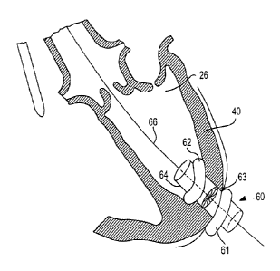
' [0080] Access device 60 may include catheter. 64 with.
distal balloon 61 and proximal balloon 62.- Balloons 61
and 62 may sandwich myocardium 4-0-to prevent bleeding
from left ventricle 26.- Access device 60 may be -
anchored in other suitable ways, as long as left . .
ventricle 26 'is appropriately sealed to prevent
bleeding, .and such that blood flow through the' coronary
arteries is not occluded. Access device 60 .also
. includes valve 63. Valve 63 allows the, passage of
guidewire 66 and the insertion.df surgical tools .while
preventing bleeding through catheter 64. . Valve 63 may
be mechanically operable as an iris diaphragm .(e.g.,
like the aperture. of a lens). Alternatively, valve 63
may be constructed of an elastic material with. a small
central opening that is dilated by_whatever.equipment
is inserted. therethrough, but always maintains a fluid-
tight seal with the inserted equipment. Valve 63 may
.,compose any- fluid,-tight valve structure..
[0081] Access device 60 can include, one or multiple
valve-like structures, like valve. 63. Multiple valves .
' in series may act as added protection against leakage
from the heart chamber. Furthermore, because of the
potential for leakage around multiple tools, .access.
device 60 may include multiple valves in parallel.
Thus, -each tool could be inserted through its own
valve. This could ensure that a proper seal is created,
around each tool being used during the operation.
[0082]' . In some embodiments of the present invention,
various endovascular, endocardiac, and/or endoluminal
visualization aids may be used. Such devices are
-illustrated in FIG. 7. Additionally, extracorporeal X-.
=
=
CA 02583591 2007-04-02
WO 2006/041505 PCT/US2004/043794
- 23 -
=
ray based. radiographic devices may be employed. .
Preferably, intracardiac ultrasound 34 is placed in the
right atrium via a femoral vein, and intravascular
ultrasound (IVUS) 70 is placed over guidewire 66 and=
= into a heart chamber = or into the diseased valve.
External fluoroscopy is also utilized to Map and.
visualize the surgical site.
. .
= .
[0083] IVUS 70. may be used to locate aortic..
valve 20, aino-tubular junction 36, and brachio-
cephalic trunk 72. In order to determine the 'precise
location of each, IVUS probe 70's location is
simultaneously tracked with AcuNavTM 34 and fluoroscopy. .
Once each landmark is located, a. radioopaque marker may
'be placed on the patient's skin or the heart's surface .
so that extracorporeal fluoroscopy can Iater be used.to
relocate these points without IVUS 70 taking up space
inside the surgical site. The. end of the native
leaflet in systole may also be marked with a ,
radioopaque marker. in order to temporarily .define. the
target zone. This technique requires that the patient
and the fluoroscopTequipment not be moved during the.
procedure, because landmarks inside the heart and. aorta.
. are being marked by radioopaque markers plaoed¨on the
patient's skin outside the body or on the beating.
heart's-surfade. .It may be desirable to place the
radioopaque markers directly on the heart and aorta.
[0084] IVUS 70, AcuNav714'34, and the fluoroscopy
equipment can also be used to take measurements of the .
diseased valve. This allows' the.surgeon.to chose a
properly sized replacement heart valve. As an
alternative to fluoroscopy, a surgeon may choose to use
standard dye visualization techniques such, as
angiography. Although it would create material .
CA 02583591 2007-04-02
WO 2006/041505 PCT/US2004/043794
- 24
limitations for manufacturing the replacement heart
valve, MRI technology could be used as an alternative
' means of visualizing the target surgical site. .
Additionally, .with the development of cameras that can
see through blood, direct.optical.technology could be -
used to create an image.of the target site. Real-time
- three-dimensional construction of -ultrasound data is_.
another visualization procedure that is currentlyunder
. development that could provide a suitable alternative.
[0085] With respect to direct optical technology/ a'
clear liquid could be introduced to the aorta or other
components of the circulatory system near the target
surgical site. Placing a clear liquid that is. capable
of carrying 'oxygen (i.e., capable of carrying on the
blood's biological function, temporarily) in the =
patient's 'circulatory system would improve the ability
.to use direct optical imaging. Furthermore; because
the heart is beating, the patient Could be transfused
with the clear oxygen-carrying fluid for. the duration
.of the procedure so that direct optical visualization.
is enabled throughout the procedure. The patient's
regular blood' would be retransfused at the conclusion
of the procedure.
[0086] Another option for a direct visualization _
technique includes placing a transparent balloon
(filled-with a transparent fluid such as. water) in .
front of the camera. , The camera and liquid-filled
balloon are pushed against the surface that the surgeon.
wishesto view. The transparent balloon displaces
blood from the camera's line of sight such that an
.image of what the camera sees through the balloon is .
- transmitted to the surgeon.
CA 02583591 2007-04-02
WO 2006/041505 PCT/US2004/043794
- 25 -
[0087] . Furthermore, the invention may include the .
placement of embolic protection device 80 in the .
ascending aorta by means of a catheter; as shown in
FIG. 8. Embolic protection device 80 is preferably
placed from the endoluminal.femoral access site in a
retrograde approach to the aortic valve site. Embolic
protection device .80 may comprise a.filtering mesh or
net. made from any. suitable material...The chosen . =
material should be able to be collapsed, expanded, and
re-collapsed multiple times.. Embolic protection . .
device 80 may alternatively be placed from the
antegrade direction. Either approach may be made using.
.guidewire 66 or additional guidewires-inserted in _
'accordance with the present invention.
.[0088] . Single embolic protection device 80. may have
unique properties to protect the outflow region of the
aortic valve which feeds aorta 28 and coronary. .
sinuses 82 and 84. Device 80 may comprise tight .
mesh 200 (see. FIG. 20) formed, in a conical shape.
Conical 'mesh 200 may terminate in perimeter 204 that
exerts .a radially outward force on the wall of
aorta 28. Device SO is operated via catheter 202.. and
is dimensioned so that it is capable of filtering the
blood supply to the aorta and the coronary arteries.
[0089] In some embodiments, embolic protection .
device 80 may be replaced with multiple embolic.
=
protection devices 90, 92, and 94, as illustrated in
FIG. 9. In FIG. 9, each of coronarysinuses.82 and 84
is protected by its own embolic protection device
(embolic protection devices 92 and 94, respectively)',
and aorta 28 is protected by embolic protection
device 90. Embolic protection devices 92 and 94 may be
placed further into the coronary arteries to keep the
CA 02583591 2007-04-02
WO 2006/041505
PCT/US2004/043794
= - 26 -
surgical site inside the aorta as clear as possible. .
Embolic protection device 80 of FIG. 8.is designed so
sthat proper placement of the single protection device -
will prevent the flow of embolic material into any of.
aorta 28 and coronary sinuses 82 and 84. -
[0090] . In certain embodiments of the present .
invention, the embolic protection device maybe placed":
in an antegrade approach. For example, FIG.' 10 shows -
=
embolic protection devices 92' and 94' having-been-
inserted in the antegrade direction.. Placing
devices 92' and 94. in the coronary sinuses from the
antegrade direction leaves guidewires 101 and 102 to _. _
exit the patient. at the thoracotomy access site. .
Coronary sinuses 82 and 84 provide useful landmarks in
placing a new aortic valve_ Thus, by placing
devices 32' and 94' in this manner, the surgeon is
provided with a guide to proper placement 'ofthe new
valve (i.e., guidewires 101 and 102 which terminate at
coronary sinuses 82 and 84). The new valve may be
.inserted in the antegrade direction along
guidewires 101 and 102 to ensure proper placement.
[0091]
Additionally, embolic filters may be.placed
in'the brachiocephalic, left common carotid, and left
subclavian arteries of the aortic arch. _
[0092] - Some embodiments of the present invention may _
employ a valve-tipped catheter or other temporary valve
device that is capable of temporarily replacing the.
native valve function during and after resection or =
removal until the new valve is deployed and functional.
Such temporary valve devices may be placed in any
number of acceptable locations. For example, when.
replacing the aortic valve's function, it .may be
preferable to place the temporary valve 'in the
CA 02583591 2007-04-02
WO 2006/041505 PCT/US2004/043794
-27-
-ascending aorta just distal to the native aortic 'valve,
However, it is possible to temporarily replace the
aortic-valve function with a device placed in the
descending aorta. Such a placement may have the
disadvantage of causing the heart to work harder,- but
such placements have been proven acceptable in previous
surgical procedures.
. [0093] Additionally, some embodiments of the.present.
invention may include the .use.of a percutaneously
placed small caliber blood pump containing an impellor
(e.g., a'VAD (Ventricular Assist Device.)).. The VAD may
be inserted in a retrograde or in- an antegrade . , _
.direction over guidewire- 66. Alternatively, the VAD.-
may be inserted Over a secondary guidewire. Because. of
,
the resection and implantation equipment that will. be'
inserted in the antegrade direction,'it may be .
desirable to place the VAD in a retrograde approach
from the percutaneous femoral access site. The VAD or =
.other temporary pump device will be used to support.the.
heart's natural function while the native valve is
being resected or repaired. The temporary assistance-.
device will remain in place until the new valve is
. deployed and functional.
= [0094] FIG. 39 shows one possible combination of an:
embolic filter, temporary valve, and VAD. The FIG. 39
embodiment shows VAD 393 passing through embolic
filter 394 and temporary valve 395. These components .
=are positioned distal to aortic valve 392 in ascending
aorta 396. Embolic filter 394 is designed to- also.
protect coronary arteries 390 and 391. Embolic.
filter 394, VAD 393, and temporary valVe 395 may all be
guided by guidewire 397. This is just one possible.
CA 02583591 2007-04-02
WO 2006/041505 PCT/US2004/043794
- 28 -
arrangement for the components that may be used in a
valve repair or replacement procedure..
[0095] In some embodiments of the present invention,
the placement of a new valve may first involve the_full-
or partial resection of the diseased valve or cardiac _
structure. To perform a resection of the diseased
valve, a surgeon may use valve removal tool 110, shown
in FIG. 11. Valve removal tool 110 incorporates outer
inflation lumen 111 and inner inflation lumen 112, -
which is placed coaxially within outer inflation
lumen 111. Outer inflation lumen 111 terminates at
proximal balloon 113. Inner inflation lumen 112
terminates at distal balloon 114. Coaxial
catheters 111 and 112 can be advanced over guidewire 66
and passed through valve 63 of access device :60.
Radially expandable proximal cutting device 115 is
mounted to the surface of distal balloon 113. Radially
expandable distal cutting device 116 is mounted to the
surface of distal balloon 114. Valve removal tool 110
is advanced with balloons 113 and 114 in the deflated
state and cutting devices 115 and 116 in the collapsed
state until distal cutting device 116 is located just
distal to diseased aortic valve 20 and proximal cutting
device 115 is positioned just proximal tip .diseased
aortic valve 20.
[0096] As shown in FIG. 12, balloons 113 and 114 are
inflated such that cutting devices 115 and 116 are
radially expanded to the approximate diameter of the
diseased valve. Next, inner inflation lumen 112, _
distal balloon 114, and distal cutting device 116 are
pulled in the retrograde direction. This causes
cutting devices 115 and 116 to cooperate with one
another to cut away diseased aortic valve leaflets 130,
CA 02583591 2007-04-02
WO 2006/041505 PCT/US2004/043794
- 29 -
as shown in FIG. 13. Balloons 113 and 114 can be
deflated and cutting devices 115 and 116 collapsed
while retaining cut away valve leaflets 130. Thus,
valve removal tool 110 and resected leaflets 130 can be
removed via access device 60.
[0097] Further, valve removal device 110 may possess
self-centering properties. Valve removal device. 110's
cutting mechanism may allow the device to cut or resect
any calcified or diseased tissue within the heart
cavities or the vasculature. The size or cut of each
bite made by the removal device, as well as the shape
of the cut may be determined by the surgeon .by
adjusting the valve removal device.
[0098] When performing surgical techniques inside a
patient's vasculature, it may be beneficial to use
ring-shaped balloons so that blood can continue to
circulate through the balloon. Also, whether using
ring-shaped balloons or more standardized balloons, it
may be beneficial to use a balloon that has more.than
one chamber, so that the balloon can be selectively
inflated. Examples of a ring-shaped balloon and .a
cylindrical balloon, both having more than one
inflation chamber are illustrated in FIGS. 37 and 38,
respectively.
[0099] FIG. 37 shows ring-shaped balloon 370.
Balloon 370 may be divided into three inflation
chambers by dividers 373', 373", and 373'1'. Each
inflation chamber may be attached to an inflation
flange (e.g., flanges 374', 374", and 174"'). Each
inflation flange is correspondingly attached to an
inflation lumen of catheter 371 {e.g., inflation
lumens 372', 372", and 372'1'). Thus, blood flow is
able to continue through the three openings left
CA 02583591 2007-04-02
WO 2006/041505 PCT/US2004/043794
- 30 -
between inflation.flangeb 374', and 374'''.
Furthermore, surgical tools (e.g., VADs, etc.) may be
passed through the openings. Balloon 370 may be guided-
,
brguidewird 375. -
[0100] FIG. 38 shows Cylindrical balloon 380 with
inflation chambers 381, 282, and 383: The inflation .
chambers may be selectively inflated by ¨inflation-. . =
' -ltmens 384, 385, and 386, respeCtivelY.of Catheter 387,
. Balloon 380 may be guided by guidewire 388. By -
providing selectively inflatable chambers in. either
. type of balloon, a Surgeon may have the ability to
manipulate tissue inside a patient's vascuiature:or- - -
properly position surgical equipment and .prostheses,
for example.
[0101] In some embodiments of the present.invention,.
a valve removal tool such as ronjeur device 210 may be .
used (see FIG. 21). Ronjeur device 210 may have spoon-
shaped heads-212 and 214 which are operably controlled- = '
by handles 216 and 218 via hinge 211. SpoOn-,shaped. -
heads 212 and 214 may have sharpened tips 213 and 215,
respectively. Ronjeur device 210 may be used-to,bite=
away the leaflets of a diseased valve and trap the
dissected tissue within spoon-shaped head8:212 and 214.
Ronjeur device 210 may be operable via access -
device 60.
. [0102] = In other embodiments Of the present
invention, valve resector 220 of FIG. 22 can'be used to
resect the diseased valve. Valve .resector 220 hat
handle 222, shaft 224, recess 226, and -reseCtor
tip 228. Resector tip 228 may be used to cut away or -
tear away the diseased leaflets of a native valve.
= Recess 226 may be used to retain the resected tissue
for removal. Resector tip 228 may also be mechanically
CA 02583591 2007-04-02
WO 2006/041505 PCT/US2004/043794
=
- 31 -.
=
operable to snip away the diseased leaflets. -
Resector 220 is also operable via access device 60.
Other suitable techniques for resecting a diseased
valve may also be used before implanting a--new valve. -
[0103] In preparation for valve resection, it may be .
beneficial to Soften or break-up the calcification of .
the diseased valve. Concentrated ultrasound waves'--
could be used to break-up the valve's calcification: .A
similar procedure is used to break down kidney stones .
" in some patients'. Calcification of the aortic valve is
often trapped in tissue pockets. Thus the broken-down '
calcification, would likely be retained by the valve -
leaflets. = However, the leaflets would-now be-more
pliable 'and easier to compress behind a new valve or to
remove. An intraluminal ultrasound device may be' used
to deliver the concentrated ultrasound waves. '
Furthermore, an intraluminal reflector' may be'. used to
' magnify the waves' intensity, and break-up the.. calcium
=
. deposits even'quickem% . .
40104] .In addition to or asarralternative to
resecting the diseased valve, plaque or calcification
of a diseased valve may be chemically dissolved. With
embolic protection devices 90,. 92, and 94 in place4 a
chemical can be introduced-to the diseased valve that .
will dissolve or release the plaque deposits. The
.target valve site may first be isolated to contain the
chemical during this process. This isolation may be -
- achieved by inflating two balloons to create a chemical
ablation chamber defined by the Wall of the aorta and
the two balloons.
" [0105] Isolation may also be achieved by a device
like ablation chamber 360 shown in FIG. 36. Ablation -
chamber 360 is positioned inside the patient's
CA 02583591 2007-04-02
WO 2006/041505 PCT/US2004/043794
- 32 -
vasculature (e.g., aorta 362). The chamber may be
placed percutaneously, by direct access, or by any
other suitable technique. Ablation chamber 360
comprises ring-shaped balloons 361 and 363.
Balloons 361 and 363 are joined by tubular member.. 367
which creates a channel for blood to by-pass the
ablation site. A ventricular assist device may be
inserted through opening 365 in tubular member 367 to
aid the patient's blood flow through the temporarily
narrowed passageway. Ablation chamber 360 may include
chemical introducer 364 and chemical evacuator 366 to
introduce a chemical to the ablation site and to clear
the chemical from the ablation site when the procedure
is completed. Thus, the chemical ablation procedure is
performed in the chamber of the isolated segment of the .
aorta while normal circulatory function takes place.
Such a technique isolates the chemical being used from .
entering the patient's circulatory system. This
treatment may be performed to repair .a diseased valve,
to decalcify a diseased valve before resection by a
valve removal tool, or to decalcify a diseased valve _
before placing a new valve within and over top of the
diseased valve. Laser ablation may also be used to
break up valve calcification or to remove and destroy
diseased valve leaflets.
[0106] As another alternative, the diseased and
calcified valve can be left as is and a new valve can
be implanted within and over top of the diseased valve.
In some embodiments of the present invention, it may be
desirable to perform a valvuloplasty to percutaneously
destroy the leaflets of the diseased valve. It_may be
easier to dilate the diseased valve with the new valve
if it has been partially destroyed first.--.
CA 02583591 2007-04-02
WO 2006/041505
PCT/US2004/043794
- 33 -
[0107] Once any manipulation of the diseased valve
is complete (e.g., marking landmark locations,
resecting the diseased leaflets, chemically dissolving
calcification, etc.), embolic protection devices 90,
92, and 94 can. removed-
(FIG. 14). The resectionr of
diseased leaflets 130 (FIG. 13) may leave behind valve
rim 141 (FIG. 14). Once the embolic protection devices-
have been removed, valve delivery device 142 may be =
inserted into left ventricle 26 via access device 60. .
Valve delivery device 142 carries new, valve 140 in a
radially compressed state. Valve 140 has been Crimped
onto delivery device 142. Alternatively, valve 140 may
be folded or collapsed in any other suitable manner.
Valve delivery device 142 is advanced along
guidewire 66.
[0108] In embodiments like that shown in FIG. 10,
valve delivery device 142 may also be guided by
guidewires 101 and 102 to ensure safe orientation of
valve 140 prior to release and deployment. Such a
delivery approach would eliminate the danger of
coronary obstruction, because guidewirss 101 and 102
terminate at coronary sinuses 82 and 84. The spaces
between the commissure supports of valve 14-0 could be
properly aligned with coronary sinuses 82 and 84 to
allow maximum blood flow to the coronary arteries.
[0109] In other embodiments of the present
invention; the placement of valve 140 may be assisted
by intracardiac ultrasound (i.e., ultrasound
equipment 34 of FIG. 7) and fluoroscopy. Positioning,
release, and deployment of valve 140 could be
simultaneously monitored by the intracardiac ultrasound
and fluoroscopy equipment. The fluoroscopy equipment
would monitor the target zone based on the radioopaque
CA 02583591 2007-04-02
WO 2006/041505 PCT/US2004/043794
- 34 -
markers, that were placed earlier in the procedure.
When the fluoroscopic (marker position) and sonographic
(intracardiac ultrasound) target sites are congruent,
the proper position for valve deployment has been
located. At that moment, valve 140 may be deployed as .
described below.
[0110] Additionally, valve delivery device 142 may.
contain two radioopaque markers. With the coronaries
being visualized with fluoroscopy, the surgeon could
visualize the alignment of the two marker bands on
delivery device 142. Thus, the surgeon would be able,
to properly orient the valve such that the commissure
posts are properly positioned upon valve deployment.
[0111] Valve delivery device 142 may terminate in
two phase balloon 150, as shown in FIG. 15.
Alternatively, the end of device 142 carrying valve 140
may have two separately operable balloons. The first
phase of balloon 150 may be inflated to provide a
positioning guide for valve 140. The first phase of
balloon 150 provides a bumper such that delivery.
device 142 is prevented from further advancement when
the proximal end of balloon 150 (i.e., the first phase
of balloon 150) reaches the region of left ventricle 26
just proximal to the aortic valve site.
[0112] Continued expansion of balloon 150 causes
base ring 154 of valve 140 to expand. As base ring 154
expands, hooks 156 may bite into remaining aortic
rim 141. Alternatively, hooks 156 may not penetrate.
-rim 141, but rather grasp the rim tightly. Commissure
support tissue 158 also begins to open up. _In some
embodiments of the present invention, valve 140
includes distal stent-like structure 152 to support a
CA 02583591 2007-04-02
WO 2006/041505 PCT/US2004/043794
- 35 -
replacement aortic valve distal to coronary sinuses 82
and 84 in sino-tubular junction 36.
[0113] During expansion, intracardiac ultrasound and
fluoroscopy can be used to monitor the orientation and
placement of valve 140. Before .valve 140 is fully
expanded, the surgeon may rotate delivery device 142
such that the spaces between commissure supports 158-
align with coronary sinuses 82 and 84.- Upon full
expansion of ring 154 (see FIG. 16), hooks 156 may ¨
fully engage rim 141, and hooks 156 and rim 141 may be .
partially embedded in aortic wall 151. Stent-ldke
structure 152 may engage aortic wall 151 in sino-
tubular junction region 36. Commissure supports 158
will be fully expanded, too. Support structure 152 may
expand in unison with base ring 154. Alternatively,
valve placement may take place in a stepped process,
wherein base ring 154 expands and secures the base of
the valve before support structure 152 expands to
secure the distal end of the valve. The location and
function of new valve,140 are identified and monitored
with IVUS, intracardiac ultrasound, and/or fluoroscopy.
Once placement and function is satisfactory to the
surgeon, balloon 150 is deflated, and valve .delivery
device 142 is removed from left ventricle 26.
[0114] The implantation process should be done
quickly, because there will be a brief total occlusion_
of the aorta. It may be desirable to block the inflow
to the heart. Thus, the heart is not straining to.-pump
blood out, and a dangerous lowering of the patient's- -
heart rate may be prevented. =
[0115] -Valve delivery device 142 may be designed to
draw the native leaflets downward when a new valve is =
being implanted over top of an existing diseased valve.
CA 02583591 2007-04-02
WO 2006/041505 PCT/US2004/043794
-36 -
The native leaflets could obstruct blood flow to the
coronary arteries. However, pulling the native
leaflets downward before compressing them against the -
aorta-wall would prevent such-occlusion.
[0116] In some embodiments of the present invention,
new valve 140 may be a self-expanding valve that can be
implanted without the use of a balloon. -Base ring 154,
hooks 156, and stent-like structure 152 may be- -
constructed of nitinol or some other shape-memory or
self-expanding material. In some embodiments,
valve 140 may be deployed by mechanical means, such as
by releasing a lasso that surrounds the exterior of
valve 140 or by operating a mechanical expansion device
within valve 140. .
[0117] In certain embodiments of the present
invention, valve 140 may not have a stent-like support
structure at the distal end (i.e., stent-like
structure 152). If commissure supports 158 are
constructed from or supported by a stiff enough support
post, valve 140 may not be fixed to the aorta at its
distal end. The mounting at base ring 154 may
sufficiently secure valve 140 in-place to function
normally and not obstruct blood flow to the -coronary
arteries.
[0118] Valve 140 may be secured in place by any - -
suitable method for anchoring tissue within the body. -
The radial expansion forces of base ring 154 may be
strong enough to secure valve 140 against dislodgment
by radial strength alone. If no native valve rim
remains, hooks 156 may be designed-to grasp -aortic
wall 151. Mechanically placed sutures or staples could
be used to secure valve 140 in place. Furthermore, -
CA 02583591 2007-04-02
WO 2006/041505 PCT/US2004/043794
- 37 -
biocompatible glue could be used to secure valve 14.0 in -
the appropriate position. -
[0119] During
a valve implantation procedure, it may
be desirable to have the ability to retract expansion
of new Valve 140. If the commissures are not properly.,.-
aligned with the corollary arteries or if the valve is
" not properly positioned within the native annulus, =
..retracting the expansion would enable repositioning or
.realignment of the valve. Such a retraction technique
is illustrated in FEG. 23 wherein valve 230 is one
. illustration of .a possible embodiment of valve 140.
[0120] .Valve 230 has
radially expandable support -
.
ring 232 and radially expandable mounting
structure 231. Mounting structure 231 maybe a
sinusoidal ring of nitinol wire.. Mounting
structure 231 is attached to wires 23,_238, and 239 at
points 234, .235, and .236,.respectively. .By advancing .
tube 233 or withdrawing wires 23.7,.238, and 239
mounting structure 231 may be drawn radially inward,.
effectively retracting the expansion of valve 230. -
Other means of retracting valve expansion could be .
employed, in accordance with the principles of the -
. present invention.
[0121] In some
embodiments of the present invention,
the dilated opening in myocardium 40 is sealed with. an
.automatic Closure device. The automatic closure -device
may be-part of access device 60. Alternatively, the
. automatic closure device may be inserted through access-
device 60 such that removal -of access device 60 leaves -
the automatic closure device behind. _
[0122] For example, FIG. 17
shows automatic. closure
device 172 being delivered with closure delivery _
device 170. Closure device 172 may include proximal
CA 02583591 2007-04-02
WO 2006/041505 PCT/US2004/043794
- 38 -
umbrella 174,, distal umbrella 178, and connecting
shaft 176 therebetween. Delivery rod 171 maybe used
to advance proximal umbrella 174 from delivery =
device 170 such that umbrella 174 opens. Balloons 6I -
and 62 of accessdevice 60 are .deflated. Then, both
access device 60 and delivery device 170 are withdrawn.' .
from heart 10. UMbrella 174 will.contact'the inner. =
-surface Of myocardium 40, as shown in FIG. 18. Upon
further withdrawal of.access device 60 and.delivery.
device 170, distal umbrella. 178 will be permitted. to .
deploy. Upon deployment of-umbrella 178, the hole
formed in myocardium 40 will. be sealed. Myocardium 40
may be sealed using any acceptable.automatic closure''
device. Alternatively, myocardium 40 may be sutured
closed. Additionally, myocardium 40. may .be'closed with
any .known closure device, such as an AmplatzerTM.
occlusion device, other double-button device; plug, or
'laser plug. =
[0123] Bleeding into the space between 'the :-. .= -
myocardium'and.thespericardium should be prevented.
The myocardium can be closed without a need to close. .
the pericardium. However, if the pericardium is to be
sealed with the automatic closure-device; the seal must
be tight enough to prevent bleeding into the void
between the two. = -
[0124] The percutaneous femoral access site will
also need to be sealed. This may be done with sutures,
or with a self-closing device such as an AngiosealTM
Hemostatic Puncture Closure. Device.
J0125] . Implantable valves in accordance with the
preferred embodiments of the present invention may take
on a-number of forms. However, the implantable valves
will likely exhibit several beneficial characteristics. .
CA 02583591 2007-04-02
WO 2006/041505 PCT/US2004/043794
- 39 -
, . .
=
Implantable valves should preferably be-constructed of -
as little material-as possible, and should be easily
-collapsible. The valve may be-radially.compre'Ssed to .6.
size significantly smaller than its_deployed diameter:
for delivery: The implantable'valve:or_support
elements of the valve may contain Gothic arch-type--
structural support elements to efficiently support and. '
maintain the valve once it is implanted.
[0126] The implantable valve may have an, outer stent
that is installed before deploying the valve structure.
.Valves manufactured in accordance with the principles
of the present invention are preferably constructedof. .%
biocompatible materials, :Some of the materials may be-
bioabsorbable, so that shortly after the.implantation-
procedure, only the anchoring device and tissue valve '
remain permanently implanted. The valve leaflets may
be composed.of homograph.valve tissue, animal-tisbue, -
valve rebuildmaterial, pericardium, synthetics, or .
alloys, Such as a thin nitinol mesh. - ,
[0127] - Implantable valves in accordance withthe .-
principles of the present invention may be drug eluding .
to prevent restenosis by inhibiting cellular division -
" or by preventing reapposition of calcium. The drug may
'act as an active. barrier that prevents the formation-of .
calcium on the valve. Additionally, the drug may .
stimulate healing of the new valve with the aorta.
Furthermore; the implantable Valves are preferably'
= treated to resist calcification. The support elements
of the implantable valve may be exterior to the valve.
(e.g., between the new valve tissue and the aorta
wall), interior to the valve (e.g., valve tissue is .,
between the support elements and the aorta wall), -or. -
may form an endoskeleton of the valve (e .g., support.
CA 02583591 2007-04-02
WO 2006/041505
PCT/US2004/043794
- 40 -
elements of the valve may be within the tissue of the
implantable valve).
[0128] FIGS. 24-26 illustrate new valves that could
be used for replacement or implantation procedures in'
accordance
accordance with the -p,rinciples of the present _
invention. Valve 240 of FIG. 24 has sinusoidal
- attachment member 241 encircling the base of commis-sure,
posts 242, 243, and 244. Attachment member 241 may be
any radially compressible and expandable member.
Member 241 of FIG. 24_has proximal peaks 245 and distal
peaks 246 which may be turned outward. Peaks 245
and 246 may be better suited to engage the wall of the
aorta when the peaks are turned outward. Peaks 245
and 246 may also be pointed or sharpened so that they.
penetrate the aorta wall. In embodiments in which a
small rim of native valve has been left behind after
resection, peaks 245 and 246 may be biased to close
outwardly, effectively biting the rim of remaining.
tissue. Commissure posts 242, 243, and 244 and the
valve's leaflets (not shown) -fold and collapse-when
member 241 is radially compressed for delivery.
[0129] Valve 240 may have distal mounting ring 248
in .some embodiments. Ring 248 may engage the distal
portion of the sino-tubular junction. Ring 248-may.
have segments 249 that are biased radially outward so.
as to more securely engage the inner wall of the aorta.
The replacement valve may be designed to mimic the
natural curvature of the sino-tubular junction. This
curvature-creates a natural bulge, in which the
replacement valve may be able, to secure itself against
dislodgement.
[0130] Valve 250 of FIG. 25 shows tissue 252 inside
stent frame 254. Tissue 252, which forms the Leaflets
CA 02583591 2007-04-02
WO 2006/041505 PCT/US2004/043794
- 41
. of the implantable valve may be engineered and/or grown
directly inside of stent frame 254. Alternatively,.
tissue 252 may be glued or sutured to stent frame 254..
Stent frame 252 may incorporate peaks that are turned'
= outward that may have pointed.or sharpened tips.-Iike, =
those described' with respect-to valve-240 ofFIG-.- 24*,
= Also, ring 256 may have hook features such as hooks 156'-
of FIG. 15. ' Stent.frame 252 may be constructed froth' a - =
shape' memory or.other self-expanding material--
' Alternatively, stent frame 252 may be constructed -from
stainless steel or other materials that .are balloon
expanded or mechanically expanded. . =-=
[0131] Valve 260 of' FIG; 26 illustrates one ,
embOdiment of a low profile valve-. 'Such -a low profile=
valve may reduce the likelihood of coronary-artery. '
obstruction. Valve 260 may comprise any-number of
leaflets. Valve 260 is illustratively shown with five
leaflets (i.e., leaflets 261, 262,'263, 264_ and 265).
'The leaflets overlap one, another in a domino-type .
-atrangement. Leaflet 265 is the top-most leaflet,
overlapping the left side of leaflet. 264.. The right
side of leaflet 264 overlaps the left side- of.
.2 leaflet 263, and so on with leaflet.261 being the
bottom-most leaflet. The leaflets Maybe arranged such
that they overlap one another in a clockwise or.a
counterclockwise fashion. Valve 260. may appear to open .
like the iris of a camera when 'viewed from the top .(as
shown in FIG. 26). The leaflets actually rise out of
the plane of the valve annulus. However,- because of
the valve's very low profile, no.commissure supports-
are needed.
. [0132] Additionally, spiral, or rolled valves may be
used in the implantation or replacement procedure.
CA 02583591 2007-04-02
WO 2006/041505 PCT/US2004/043794
- 42 -
Such valves unwind instead of being_radially expanded.
Rolled valves are reduced in diameter for percutaneous
or_minimally invasive implantation by rolling the valve
. material into a spiral. _ _
[0133] It may be beneficial to replace an
insufficient valve with a new valve that is designed so-
that it does not dilate to the size of the diseased.
valve..
valve. Insufficient valves do not fully close, .
permitting regurgitation in: the blood flow. This is
often the result of a dilated valve annulus, which does
not allow the valve leaflets to come together in the
center. Therefore, it may be desirable for the-new
valve to fill a smaller annulus. This can be achieved
by designing a valve such as valve 270 .of FIG. 27.
Valve 270 has fluid-tight membrane 276. Thus, while
support structure 272 dilates to the diameter of-the
diseased valve's annulus, leaflets 274 of the
replacement valve operate in an annulus of fixed size
determined by membrane 276.
[0134] In some embodiments of the present invention,
the new valve may be designed to be exchangeable. Many
replacement heart valves have a life expectancy of
10-20 years. Therefore, many patients will require
follow-up valve replacements. Certain structural
components of the heart valve (e.g., the base ring,
hooks, etc.) could be permanent, while the tissue
leaflets may be exchangeable. It may be preferable to
simply dilate the old valve with the new valve.
[0135] In some embodiments of the present invention,
a valve implantation procedure may take place
"off-pump," but the patient's heart may be temporarily
arrested. The patient's heart is stopped using
fibrillation. A surgeon will have just under three
CA 02583591 2007-04-02
WO 2006/041505 PCT/US2004/043794
- 43 -
minutes.tp perform the surgical procedure without.
risking harm to the patient. However, the anesthetized
patient could be cooled to provide the surgeon with
more time without increasing the risk for brain damage.
[0136] Once the patient's heart is stopped, an
incision is made to the aorta just distal to the aortic
valve. Blood is cleared from this region so that the
surgeon can visualize the valve site. Using a delivery
device like that described above (except making a
retrograde approach in this case), the new valve is
implanted directly over the diseased valve. Because
the valve is being installed in a retrograde approach,
the native leaflets will be pushed downward before.
being compressed against the aorta wall. = Therefore,
there is no concern of coronary artery occlusion,
[0137] Once the new valve is installed, the surgical
site inside the aorta is cleared of air, and a side .
bite clamp is placed on the lesion. The heart is
restarted with the electrodes that were used to stop it
previously...Once the heart is beating again, the
clamped lesion is sutured closed. An introducer device
(similar to access device 60) can be used at the
incision site to prevent the need for -clearing the
blood from the surgical site and later deairing the. .
site.
[0138] There are numerous procedures that may be
performed transapically in accordance with the
principles of the present invention. The following
describes several of the illustrative procedures that -
may be performed via a transapical access device. -
[0139] Insufficient mitral valves often result from
a dilated posterior Leaflet. FIGS. 28-31)_demonstrate a-.
toolthat could be used to repair an insufficient
CA 02583591 2007-04-02
WO 2006/041505 PCT/US2004/043794
- 44 -
mitral valve via a transapical access device. Repair
tool 280 may have U-shaped head 282 and single-pronged -
head 284. Heads 282 and 284 may be operably attached
'b hinge 288. When posterior leaflet 290 (FIG. 29) is
inserted between heads 282 'and 2'84, handles 283 and 285
can be squeezed together to cause a portion of
posterior leaflet 290 to be 'drawn downward. At this
point; attachment tool 286 can deploy connector 3-00
(FIG. 30) to retain posterior leaflet 290 in a
constrained state, repairing any excess dilation of the
mitral annulus. Connector 300 may be a surgical
staple, mechanical suture, or other suitable connector.
[0140] Aortic disseation is another defect that may
be repaired via transapical access to the heart.
Aortic dissection occurs from a tear or damage to the
inner wall of the aorta. Aortic dissection may be
caused by traumatic injury or connective tissue
diseases such as Marfan syndrome or Ehlers-Danlos
syndrome, for example-. Aortic dissection may result in
' atherosclerosis or high blood pressure. As shown in
FIG. 31, aortic dissection 318 may result in void 319.
[0141] Aortic dissection repair device 310 may be
transapically inserted into a patient via access
device 311 (substantially similar to access device 60 '
of FIG. 6). Repair device 310,may include balloon 312
and catheter 314 and may be guided by guidewire 316.
Though not shown, catheter 314 may include several
lumens (e.g., a balloon inflation lumen, a guidewire
lumen, and a glue delivery lumen).
[0142] =Once repair device 310 is properly located,
balloon 312 may be inflated as shown in FIG. 32. The
inflation of balloon 312 may cause needles 320 to
penetrate aortic dissection 318 such that the tips of
CA 02583591 2007-04-02
WO 2006/041505
PCT/US2004/043794
=
- 45 -
-needles 320 are exposed to void 319. A biologically -
compatible glue may be injected through needles 320 via .
the glue delivery lumen (not shown) of catheter 314.
=
Further inflation of, balloon 312 may ensure that'
dissectibn 318 is securely affixed to the aorta wall-by'
-the.biologically compatible glue.
[0101 .In order to make sure that the biologically
compatible glue is only injected into void 319, .and not;
the remainder of the aorta (which may introduce the
' biologically compatible glue to the circulatory
system), dye may first be injected through select
channels (i.e., needles 320). This will_allow a.
surgeon to determine if injected glue would only end up .
in the desired locations. 'Repair device 310 may then
be rotated .to align theTheedles that will inject the .
biologically compatible glue with void, 319.
Alternatively, the needles that will be.used to inject.
the glue maybe selectable so that the surgeon . .
activates, only the ,needles' alignedwith_void 319,
[0144] Because balloon 312 fully occludes the aorta, .
balloon' 312 may be doughnut-shaped to allow blood to
pass, like balloon 330 of FIG.. 33. Additionally, =
. balloon 330 may include VAD device 332 to pump blood
'from the proximal side of balloon 330 (at inlet =
ports 334) to the distal side of balloon 330 (at outlet
ports 336) The repair device may still include.
needles 338. The aortic dissection repair procedure .
may be monitored with any of the visualization
equipment discussed in more detail above. Once the
aortic dissection has been repaired, balloon 312 ..or-330
may be deflated, and repair device 310 is removed from
the patient.
. .
CA 02583591 2007-04-02
WO 2006/041505 PCT/US2004/043794
- 46 -
[0145] Left ventricular aneurysms are another
deformity of the heart that may be treated
transapically. The heart muscle in the area of a-blood
vessel blockage can die over time. .The healing-process
may form a scar that could thin and stretch to-form a
ventricular aneurysm. Such aneurysms may be repaired
as described below.
- [0146] Left ventricular aneurysm 340 may form in
left ventricle 341 of a patient, as shown in FIG.
Because aneurysm 340 can cause the heart to work harder
over time and result in eventual heart failure, the
aneurysm-should be treated. Aneurysm repair device 336
may be inserted through access device 344 . . .
(substantially like access device 60 of FIG.. 6).-
Repair device 346 may include liquid filled bolster 342
that is mounted inside left ventricular. aneurysm 340.
Bolster 342 may be mounted with a biologically .
Compatible glue, by mechanical means, or by any other_
suitable mounting technique. - -
[0147] In some embodiments of' the present invention,
aneurysm 340 may be repaired by pulling the ends of
aneurysm 340 together, as depicted by FIG. 35. In such -
embodiments, aneurysm repair device 350 may be used to
deploy hooks 352 and 354. Hooks 352 and 354 may-grasp
the interior of the heart at the extremes of the
aneurysm and then draw the aneurysm closed. Once the
aneurysm has been drawn together, any suitable
technique can be used to secure the aneurysm in the -
closed position (e.g., biologically compatible glue,
surgical staples, mechanically placed sutures, etc.) -
Once the aneurysm has been fully sealed:, repair
.device 350 may be withdrawn from the patient.
CA 02583591 2007-04-02
WO 2006/041505 PCT/US2004/043794
- 47 -
[0148] In some embodiments of the present invention, -
endoprostheses may be placed percutaneously,
transapically, or via any combination of surgical
approaches. Endoprostheses may be placed in the
ascending aorta that have arms- capable o'. extending - -
into the coronary arteries. Endoprostheses for the
ascending aorta could also include a replacement valve
or a valved 'stent.. Endoprostheses for the descending
aorta could also be pl,aced.transapically or
percutaneously, for example, to repair an abdominal
aortic aneurysm.
[0149] Additionally, endoprostheses may be placed in
the aortic arch.= One embodiment of an endoprosthesis
for the aortic arch is shown in FIG. 40. _ =
Endoprosthesis 402 may be placed in aortic arch 400.
Furthermore, endoprosthesis 402 may. include arms 403,
405, and 407 that extend into brachiocephalic .
artery 404, left common carotid artery 406, and left
subclavian artery 408, respectively.
[0150] Endoprosthesis 402 may be placed-using
guidewires 410, 412, 414, and 416, as shown in FIG. 41.
Guidewire 410 may pass through the body of
endoprosthesis 402, while guidewires 412, 414, and 416
may pass through holes 403', 405', and 407' of the ,ends -
of arms 403, 405, and 407, respectively. Once
endoprosthesis 402 is properly positioned in aortic .
arch 400, arms 403, 405, and 407 may be extended to a
position substantially perpendicular to the. body of
endoprostheses 402. In order to aid the insertion-of
the arms of endoprosthesis 402 into the respective
arterial branches, small catheters, or other pushing
devices, may be inserted over guidewires 412, 414, and
416 to manipulate .(e.g., push) the arms of the
CA 02583591 2007-04-02
WO 2006/041505 PCT/US2004/043794
- 48 -
endoprosthesis. The arms and body of
endoprosthesis 402 may be radially expanded once the. .
endoprosthesis is properly positioned.
L0151] -Currently, ventricular arrhythmias- are
percutaneously repaired with radio frequency, cold,
heat, or microwave that is applied to the offending
tissue to destroy the source of the arrhythmia.
Ventricular arrhythmias could be-repaired transapically
in accordance with theprinciples of the present - --
invention. Radio frequency, cold, heat, or microwave
devices can be introduced through an access device like
=
access device 60 of .FIG. 6.
[0152] ' Bypertrophic obstructions (i.e.-, obstructions
distal to a heart valve) and subvalvular stenosis
(i.e., an obstruction proximal to a heart valve) may
also be treated transapically. Devices such as those
described above to resect a diseased valve could be
inserted transapically to cut away the hypertrophic or
subvalvular obstruction. .The extra tissue could be
removed from the heart in the same way that the
diseased valve is resected and removed. -
[0153] Robotic technology similar to that currently -
used in operating rooms could be used to perform some
of the steps of the heart valve removal and replacement
or implantation procedure. For example, it may be- -
desirable to have a robot perform the delicate- -
resection procedure via the access device. .
Furthermore, a robot could. exercise precision in
rotating and positioning the replacement valve .with
proper alignment of the commissure posts.
[0154] - Because the heart valve operation is being
performed-inside one or more of the heart's chambers, all of the equipment
described above should be
CA 02583591 2007-04-02
WO 2006/041505 PCT/US2004/043794
- 49 -
.atraumatic to limit damage to the endothelial wall of
the heart.
[0155] It will be
understood that the foregoing is .
only illustrative of the principles of the invention,
and that various modifications can be made by those -
skilled in the art without departing from the scope and
spirit of the invention. For example, the order of
some .steps_ in the procedures that have been described.
are
are not critical and can be changed if desired. Also,
various steps may be performed with various techniques.
For example, the diseased valve may be removed
transapically, while the replacement valve is implanted
percutaneously, or vice versa. The manner in which
.visualization equipment and techniques are used for -
observation of the apparatus inside the patient may
vary. Many surgical repair procedures can be performed ,
on or near the heart in accordance with the principles
of the present invention.