Note: Descriptions are shown in the official language in which they were submitted.
CA 02584162 2016-11-23
METHOD AND APPARATUS FOR DETERMINING AN ANALYTE
CONCENTRATION IN A SAMPLE HAVING INTERFERENTS
[0001]
BACKGROUND OF THE INVENTION.
FIELD OF THE INVENTION
[0002] Certain embodiments disclosed herein relate to a method and
apparatus for
determining the concentration of an analyte in a sample, and more particularly
to a method and
system that minimize the error in determining the analyte concentration due to
the presence of sample
components that interfere with the analyte measurement.
DISCUSSION OF THE BACKGROUND
[0003] Spectroscopic analysis is a powerful technique for determining the
presence of one or
more analytes in a sample by monitoring the interaction of light with the
sample. Examples of
spectroscopic measurements include, but are not limited to, the determination
of the amount of light
transmitted, absorbed, or scattered from a sample at one or more wavelengths.
Thus, for example,
absorption analysis includes determining the decrease in the intensity of
light transmitted through a
sample at one or more wavelengths, and then comparing the change in intensity
with an absorption
model based, for example, on Beer's law.
SUMMARY
[0003A] In an aspect of the invention, there is provided a method for
estimating the
concentration of an analyte in a sample from a measurement of the sample, the
method comprising:
drawing a fluid sample into an automated monitoring system connected to a
patient; using the system
to separate a first interferent from the fluid sample, the remainder
comprising a fluid analysis sample;
performing a measurement of the fluid analysis sample; identifying, in the
fluid analysis sample,
based on the measurement, a second interferent to the estimation of the
analyte, the second interferent
located in the fluid analysis sample; calculating a calibration constant which
reduces error attributable
1
CA 02584162 2016-11-23
to the second interferent, the calibration constant based at least partly on
the measurement of the fluid
analysis sample identifying the second interferent; applying the calibration
constant to the
measurement of the sample; and estimating, based on the calibrated
measurement, the analyte
concentration in the fluid analysis sample.
[0003B] In another aspect of the invention, there is provided an analyte
detection system
comprising: a fluid network configured to be connected to a patient and
receive periodic sample
withdrawals therefrom; a separator for separating a first interferent from a
sample to provide an
analysis sample; a sensor configured to provide information relating to a
measurement of an analyte
in a sample; a processor; and a computer readable medium storing computer
executable instructions
therein that when executed by the processor perform a method for estimating an
amount of the
analyte in the sample, the method comprising: (a) identifying based on a
measurement of the analysis
sample after the first interferent has been separated, one or more possible
interferents to the
measurement of the analyte in the sample; (b) calculating, based on the
identified one or more
possible interferents, a calibration constant which reduces error attributable
to the one or more
possible interferents; (c) applying the calibration constant to the
measurement; and (d) estimating,
based on the calibrated measurement, the analyte concentration in the sample.
[0003C] In another aspect of the invention, there is provided a system for
estimating the
concentration of an analyte in a sample, the system comprising: a fluid
network configured to connect
to a patient and draw fluid samples therefrom; a separating apparatus
configured to receive the fluid
sample and separate a first interferent from the sample, the remainder
comprising an analysis sample;
an apparatus configured to perform a measurement of the analysis sample; an
identifying apparatus
configured to identify, based on the measurement of the analysis sample, a
second interferent to the
estimation of the analyte in the sample, the second interferent located in the
analysis sample; a
calibration processor configured to calculate a calibration constant which
reduces error attributable to
the second interferent, the calibration constant based at least partly on the
measurement of the fluid
analysis sample identifying the second interferent; the calibration processor
further configured to
apply the calibration constant to the measurement; and an estimating apparatus
configured to
estimate, based on the calibrated measurement, the analyte concentration in
the sample.
la
CA 02584162 2015-11-13
[0003D] In another aspect of the invention, there is provided a method for
determining a
concentration of an analyte in a portion of a fluid sample from a patient, the
method comprising:
providing a fluid handling system in fluid communication with a fluid source
in a patient, the fluid
handling system configured to draw a plurality of fluid samples while in
continuous fluid
communication with the fluid source in the patient, the plurality of fluid
samples comprising at least a
first fluid sample and a second fluid sample, the second fluid sample drawn
after the first fluid
sample; drawing, via the fluid handling system, the first fluid sample from
the patient, the first fluid
sample comprising blood or a component of blood; transporting, via the fluid
handling system, a
portion of the first fluid sample to an analysis system; removing a first
interference from the first
fluid sample; identifying based on a measurement of the first fluid sample, a
second interference to a
determination of a concentration of an analyte in the first fluid sample, the
second interference
located in the first fluid sample; correcting for the second interference by
using the measurement of
the first fluid sample to calculate a calibration constant; and applying the
calibration constant to the
measurement; wherein the fluid handling system is configured to remain in
fluid communication with
the fluid source in the patient during the drawing, the transporting, and the
correcting, and wherein
the correcting occurs before drawing the second fluid sample from the fluid
source in the patient.
lb
CA 02584162 2007-04-17
WO 2006/047182 PCT/US2005/037606
[0004] One embodiment disclosed herein diminishes the sensitivity of
analyte estimation to the
presence of interferents, so that, over their range of likely interferent
concentrations, the net effect
of the interferents on the analyte measurement is reduced below that of the
sensitivity to an analyte
of interest.
[0005] One embodiment includes a method and apparatus for determining an
analyte
concentration in a sample that may contain interferents. Possible interferents
in the sample are
determined by analysis of a sample measurement. In another embodiment, a
calibration for
estimating an analyte concentration in a sample is generated to minimize the
error in the estimation
due to possible interferents. In another embodiment, the analyte concentration
is estimated from a
sample measurement, a plurality of Sample Population spectra taken in the
absence of interferents,
and a library of interferent spectrum.
[0006] One embodiment includes a method of estimating the amount of an
analyte in a sample
from a measurement, where the sample may include one or more interferents that
affect the
measurement. The method includes determining the presence of possible
interferents to the
estimation of the analyte concentration, and determining a calibration that
reduces errors in the
calibration due to the presence of the determined possible interferents.
[0007] One embodiment includes a method of spectroscopically identifying an
interferent in a
material sample. The method includes forming a statistical model of
interferent-free spectra,
comparing combinations of material sample spectra and interferent spectra
corresponding to
varying concentrations of the interferent, and identifying the interferent as
a possible interferent if
any of said combinations are within predetermined bounds.
[0008] One embodiment includes a method for estimating the amount of an
analyte in a sample
from a measurement of the sample. The method includes identifying one or more
possible
interferents to the measurement of the analyte in the sample, and calculating
a calibration that,
when applied to the measurement, provides an estimate of the analyte
concentration in the sample.
The calculation minimizes the error of interferents on the estimated analyte
concentration.
[0009] One embodiment includes a method of generating an average
calibration vector for
estimating the amount of an analyte from the spectrum of a sample having one
or more identified
interferents. The method includes forming a plurality of spectra each
including a combination of
2
CA 02584162 2007-04-17
WO 2006/047182 PCT/US2005/037606
one of a plurality of interferent-free spectra, each having a known amount of
analyte, and the
spectrum of random combinations of possible amounts of the one or more
interferents; forming a
plurality of first subsets of spectra each including a random selection of
said plurality of spectra and
defining a corresponding second subset of spectra of the plurality of spectra
not included in said
first subset. For each first subset of spectra, the method further includes
generating a calibration
vector using the known analyte concentration corresponding to each spectrum,
estimating the
amount of analyte from each spectrum of said corresponding second subset using
the generated
calibration vector, and determining a subset-average error between the
estimated amount of analyte
and the known amount of analyte. The method further includes calculating an
average calibration
vector from the calibration vector and determined average error from each
subset of spectra to
minimize the variance of the error obtained by the use of the averaged
calibration.
[0010] One embodiment includes a method of generating a calibration vector
or estimating an
analyte where the measurement is a spectrum. In one embodiment, the spectrum
is an infrared
spectrum, such as a near infrared or a mid infrared spectrum. In another
embodiment, the
measurement is obtained on a material sample from a person.
[0011] One embodiment includes a method to determine a calibration that
minimizes errors in
the calibration due to the presence of the determined possible interferents.
[0012] One embodiment includes a carrier medium carrying one or more
computer readable
code segments to instruct a processor to implement any one or combination of
the methods
disclosed herein.
[0013] One embodiment comprises a method of estimating the concentration of
an analyte in a
sample from a measurement, where the sample may include one or more
interferents that affect the
measurement. The method comprises determining the presence in the sample of
possible
interferents to the measurement, and determining a calibration that reduces
errors in the
measurement due to the presence of the determined possible interferents. The
method can further
comprise applying the calibration to the measurement, and estimating the
analyte concentration
based on the calibrated measurement. The measurement can be from a person,
wherein the
determining the presence of possible interferents and the determining a
calibration both include
comparing the measurement with population measurements, and where the
determining does not
require the population to include the person. The measurement can further
comprise a spectrum =
3
CA 02584162 2007-04-17
WO 2006/047182 PCT/US2005/037606
obtained from a material sample, and the spectrum can be an infrared spectrum,
a near infrared
spectrum or a mid infrared spectrum. The measurement can also further comprise
a spectrum
obtained from a material sample non-invasively. The material sample can
include at least one of
the following: blood, a component of blood, interstitial fluid, or urine. The
calibration can
comprise a vector that is not required to be perpendicular to the spectra of
the determined possible
interferents. Determining a calibration can minimize errors in the calibration
due to the presence of
the determined possible interferents.
[0014] One embodiment comprises a carrier medium carrying one or more
computer readable
code segments to instruct a processor to implement a method of estimating the
amount of an
analyte in a sample from a measurement, where the sample may include one or
more interferents
that affect the measurement. The method comprises determining the presence in
the sample of
possible interferents to the measurement, and determining a calibration that
reduces errors in the
measurement due to the presence of the determined possible interferents. The
measurement can
comprise a spectrum obtained from a material sample, and the spectrum can be a
near infrared
spectrum or a mid infrared spectrum. The measurement can also comprise a
spectrum obtained
from a material sample non-invasively. The material sample can include at
least one of the
following: blood, a component of blood, interstitial fluid, or urine.
[0015] One embodiment comprises a method of spectroscopically identifying
an interferent in a
material sample. The method comprises forming a statistical model of
interferent-free spectra;
analyzing combinations of material sample spectra and interferent spectra
corresponding to varying
concentrations of the interferent; and identifying the interferent as a
possible interferent if any of
the combinations are within predetermined bounds. Identifying the interferent
can include
determining the Mahalanobis distance between the combinations of material
sample spectra and
interferent spectra corresponding to varying concentrations of the interferent
and the statistical
model of interferent-free spectra. Identifying the interferent can further
include determining
whether the minimum Mahalanobis distance as a function of interferent
concentration is
sufficiently small relative to the quantiles of a x2 random variable with L
degrees of freedom,
where L is the number of wavelengths of the spectra.
[0016] One embodiment comprises a carrier medium carrying one or more
computer readable
code segments to instruct a processor to implement a method of
spectroscopically identifying an
4
CA 02584162 2007-04-17
WO 2006/047182 PCT/US2005/037606
interferent in a material sample. The method comprises forming a statistical
model of interferent-
free spectra; analyzing combinations of material sample spectra and
interferent spectra
corresponding to varying concentrations of the interferent; and identifying
the interferent as a
possible interferent if any of the combinations are within predetermined
bounds. Identifying the
interferent can include determining the Mahalanobis distance between the
combinations of material
sample spectra and interferent spectra corresponding to varying concentrations
of the interferent
and the statistical model of interferent-free spectra. Identifying the
interferent can further include
determining whether the minimum Mahalanobis distance as a function of
interferent concentration
is sufficiently small relative to the quantiles of a x2 random variable with L
degrees of freedom,
where L is the number of wavelengths of the spectra.
[0017] One embodiment comprises a method for estimating the concentration
of an analyte in a
sample from a measurement of the sample. The method comprises identifying,
based on the
measurement, one or more possible interferents to the measurement of the
analyte in the sample;
calculating a calibration which reduces error attributable to the one or more
possible interferents;
applying the calibration to the measurement; and estimating, based on the
calibrated measurement,
the analyte concentration in the sample. The measurement can comprise a
spectrum obtained from
a material sample, and the spectrum can be a near infrared spectrum or a mid
infrared spectrum.
The measurement can also comprise a spectrum obtained from a material sample
non-invasively.
The material sample can include at least one of the following: blood, a
component of blood,
interstitial fluid, or urine. The analyte can comprise glucose.
[0018] One embodiment comprises a carrier medium carrying one or more
computer readable
code segments to instruct a processor to implement a method for estimating the
concentration of an
analyte in a sample from a measurement of the sample. The method comprises
identifying, based
on the measurement, one or more possible interferents to the measurement of
the analyte in the
sample; calculating a calibration which reduces error attributable to the one
or more possible
interferents; applying the calibration to the measurement; and estimating,
based on the calibrated
measurement, the analyte concentration in the sample. The measurement can
comprise a spectrum
obtained from a material sample, and the spectrum can be a near infrared
spectrum or a mid
infrared spectrum. The measurement can also comprise a spectrum obtained from
a material
sample non-invasively. The material sample can include at least one of the
following: blood, a
component of blood, interstitial fluid, or urine. The analyte can comprise
glucose.
CA 02584162 2007-04-17
WO 2006/047182 PCT/US2005/037606
[0019] One embodiment comprises a method of generating an average
calibration vector for
estimating the amount of an analyte from the spectrum of a sample having one
or more identified
interferents. The method comprises forming a plurality of spectra each
including a combination of
(i) one of a plurality of interferent-free spectra, each such spectrum having
an associated known
analyte concentration, and (ii) a spectrum derived from random combinations of
possible amounts
of the one or more interferents. The method further comprises forming a
plurality of first subsets of
spectra each including a random selection of the plurality of spectra and
defining a corresponding
second subset of spectra of the plurality of spectra not included in the first
subset. The method
further comprises, for each first subset of spectra: (a) generating a
calibration vector using the
known analyte concentration corresponding to each spectrum; (b) estimating the
amount of analyte
from each spectrum of the corresponding second subset using the generated
calibration vector, and
(c) determining a subset-average error between the estimated amount of analyte
and the known
amount of analyte. The method further comprises calculating an average
calibration vector from
the calibration vector and determined average error from each subset of
spectra to minimize the
variance of the error obtained by the use of the averaged calibration. In
practicing this method, the
sample can comprise a material sample, such as blood, plasma, blood
component(s), interstitial
fluid, or urine. The spectrum of the sample can be obtained non-invasively.
The spectrum of the
sample can be an infrared spectrum, a mid infrared spectrum, and/or a near
infrared spectrum. In
one embodiment, the calibration vector is not required to be perpendicular to
the spectra of the
determined possible interferents. The calibration vector can minimize errors
in the calibration due
to the presence of the determined possible interferents.
[0020] One embodiment comprises a carrier medium carrying one or more
computer readable
code segments to instruct a processor to implement a method of generating an
average calibration
vector for estimating the amount of an analyte from the spectrum of a sample
having one or more
identified interferents. The method comprises forming a plurality of spectra
each including a
combination of (i) one of a plurality of interferent-free spectra, each such
spectrum having an
associated known analyte concentration, and (ii) a spectrum derived from
random combinations of
possible amounts of the one or more interferents. The method further comprises
forming a plurality
of first subsets of spectra each including a random selection of the plurality
of spectra and defining
a corresponding second subset of spectra of the plurality of spectra not
included in the first subset.
The method further comprises, for each first subset of spectra: (a) generating
a calibration vector
6
CA 02584162 2007-04-17
WO 2006/047182 PCT/US2005/037606
using the known analyte concentration corresponding to each spectrum; (b)
estimating the amount
of analyte from each spectrum of the corresponding second subset using the
generated calibration
vector, and (c) determining a subset-average error between the estimated
amount of analyte and the
known amount of analyte. The method further comprises calculating an average
calibration vector
from the calibration vector and determined average error from each subset of
spectra to minimize
the variance of the error obtained by the use of the averaged calibration. In
practicing this method,
the sample can comprise a material sample, such as blood, plasma, blood
component(s), interstitial
fluid, or urine. The spectrum of the sample can be obtained non-invasively.
The spectrum of the
sample can be an infrared spectrum, a mid infrared spectrum, and/or a near
infrared spectrum. In
one embodiment, the calibration vector is not required to be perpendicular to
the spectra of the
determined possible interferents. The calibration vector can minimize errors
in the calibration due
to the presence of the determined possible interferents.
[0021] One embodiment comprises an apparatus for estimating the
concentration of an analyte
in a sample from a measurement, where the sample may include one or more
interferents that affect
the measurement. The apparatus comprises means for determining the presence in
the sample of
possible interferents to the measurement, and means for determining a
calibration that reduces
errors in the measurement due to the presence of the determined possible
interferents. The
apparatus can further comprise means for applying said calibration to said
measurement, and means
for estimating said analyte concentration based on said calibrated
measurement. The measurement
can be from a person, wherein the means for determining the presence of
possible interferents and
the means for determining a calibration both include means for comparing the
measurement with
population measurements, and where the determining does not require the
population to include the
person. The measurement can comprise a spectrum obtained from a material
sample, and the
spectrum can be an infrared spectrum, a near infrared spectrum or a mid
infrared spectrum. The
measurement can also comprise a spectrum obtained from a material sample non-
invasively. The
material sample can include at least one of the following: blood, plasma or
other component(s) of
blood, interstitial fluid, or urine. The calibration can be a vector that is
not required to be
perpendicular to the spectra of the determined possible interferents. The
means for determining a
calibration can minimize errors in the calibration due to the presence of the
determined possible
interferents.
[0022] One embodiment comprises an apparatus for estimating the
concentration of an analyte
7
CA 02584162 2007-04-17
WO 2006/047182 PCT/US2005/037606
in a sample from a measurement of the sample. The apparatus comprises means
for identifying,
based on the measurement, one or more possible interferents to the measurement
of the analyte in
the sample; means for calculating a calibration which reduces error
attributable to the one or more
possible interferents; means for applying the calibration to the measurement;
and means for
estimating, based on the calibrated measurement, the analyte concentration in
the sample. The
measurement can comprise a spectrum obtained from a material sample, and the
spectrum can be
an infrared spectrum, a near infrared spectrum or a mid infrared spectrum. The
measurement can
also comprise a spectrum obtained from a material sample non-invasively. The
material sample
can include at least one of the following: blood, plasma or other component(s)
of blood, interstitial
fluid, or urine. The analyte can comprise glucose.
[0023] One embodiment comprises an analyte detection system. The system
comprises a
sensor configured to provide information relating to a measurement of an
analyte in a sample; a
processor; and stored program instructions. The stored program instructions
are executable by the
processor such that the system: (a) identifies, based on the measurement, one
or more possible
interferents to the measurement of the analyte in the sample; (b) calculates a
calibration which
reduces error attributable to the one or more possible interferents; (c)
applies the calibration to the
measurement; and (d) estimates, based on the calibrated measurement, the
analyte concentration in
the sample.
[0024] One embodiment comprises an analyte detection system. The system
comprises a
sensor configured to collect information useful for making a measurement of an
analyte in a
sample; a processor; and software. The software is executable by the processor
such that the
system determines the presence in the sample of possible interferents to the
measurement; and
determines a calibration that reduces errors in the measurement due to the
presence of the
determined possible interferents.
[0025] One embodiment comprises an apparatus for analyzing a material
sample. The
apparatus comprises an analyte detection system; and a sample element
configured for operative
engagement with the analyte detection system. The sample element comprises a
sample chamber
having an internal volume for containing a material sample. The analyte
detection system includes
a processor and stored program instructions. The program instructions are
executable by the
processor such that, when the material sample is positioned in the sample
chamber and the sample
8
CA 02584162 2007-04-17
WO 2006/047182 PCT/US2005/037606
element is operatively engaged with the analyte detection system, the system
computes estimated
concentrations of the analyte in the material sample in the presence of
possible interferents to the
estimation of the analyte concentration by determining the presence of
possible interferents to the
estimation of the analyte concentration and determining a calibration that
reduces errors in the
estimation due to the presence of the determined possible interferents.
BRIEF DESCRIPTION OF THE DRAWINGS
[0026] FIG. 1 is a graph illustrating the absorption spectra of various
components that may be
present in a blood sample;
[0027] FIG. 2 is a graph illustrating the change in the absorption spectra
of blood having the
indicated additional components of FIG. 1 relative to a Sample Population
blood and glucose
concentration, where the contribution due to water has been numerically
subtracted from the
spectra;
[0028] FIG. 3 is one embodiment of an analyte measurement system;
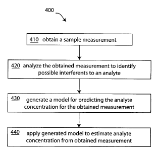
[0029] FIG. 4 is a first embodiment of an analysis method for determining
the concentration of
an analyte in the presence of possible interferents;
[0030] FIG. 5 is one embodiment of a method for identifying interferents in
a sample for use
with the first embodiment of FIG. 4;
[0031] FIG. 6A is a graph illustrating one embodiment of the method of FIG.
5, and FIG. 6B is
a graph further illustrating the method of FIG. 5;
[0032] FIG. 7 is a one embodiment of a method for generating a model for
identifying possible
interferents in a sample for use with the first embodiment of FIG. 4;
[0033] FIG. 8 is a schematic of one embodiment of a method for generating
randomly-scaled
interferent spectra;
[0034] FIG. 9 is one embodiment of a distribution of interferent
concentrations for use with the
embodiment of FIG. 8;
[0035] FIG. 10 is a schematic of one embodiment of a method for generating
combination
9
CA 02584162 2007-04-17
WO 2006/047182 PCT/US2005/037606
interferent spectra;
[0036] FIG. 11 is a schematic of one embodiment of a method for generating
an interferent-
enhanced spectral database;
[0037] FIG. 12 is a graph illustrating the effect of interferents on the
error of glucose
estimation;
[0038] FIGS. 13A, 13B, 13C, and 13D each have a graph showing a comparison
of the
absorption spectrum of glucose with different interferents taken using two
different techniques: a
Fourier Transform Infrared (FT1R) spectrometer having an interpolated
resolution of 1 cm-1 (solid
lines with triangles); and by 25 finite-bandwidth IR filters having a Gaussian
profile and full-width
half-maximum (FWHM) bandwidth of 28 cm-1 corresponding to a bandwidth that
varies from 140
nm at 7.08 in, up to 279 nm at 10 i.un (dashed lines with circles). The
Figures show a comparison
of glucose with mannitol (FIG. 13A), dextran (FIG. 13B), n-acetyl L cysteine
(FIG. 13C), and
procainamide (FIG. 13D), at a concentration level of 1 mg/dL and path length
of 1 pm;
[0039] FIG. 14 shows a graph of the blood plasma spectra for 6 blood sample
taken from three
donors in arbitrary units for a wavelength range from 7 pm to 10 pm, where the
symbols on the
curves indicate the central wavelengths of the 25 filters;
[0040] FIGS. 15A, 15B, 15C, and 15D contain spectra of the Sample
Population of 6 samples
having random amounts of mannitol (FIG. 15A), dextran (FIG. 15B), n-acetyl L
cysteine (FIG.
15C), and procainamide (FIG. 15D), at a concentration levels of 1 mg/dL and
path lengths of 1 pm;
[0041] FIGS. 16A-16D are graphs comparing calibration vectors obtained by
training in the
presence of an interferent, to the calibration vector obtained by training on
clean plasma spectra for
mannitol (FIG. 16A), dextran (FIG. 16B), n-acetyl L cysteine (FIG. 16C), and
procainamide (FIG.
16D) for water-free spectra;
[0042] FIG. 17 is a schematic of a fluid handling system;
[0043] FIG. 18 is a schematic of a first embodiment of a sampling
apparatus; and
[0044] FIG. 19 is a schematic showing details of an embodiment of a
sampling apparatus.
CA 02584162 2007-04-17
WO 2006/047182 PCT/US2005/037606
[0045] Reference symbols are used in the Figures to indicate certain
components, aspects or
features shown therein, with reference symbols common to more than one Figure
indicating like
components, aspects or features shown therein.
DETAILED DESCRIPTION OF THE PREFERRED EMBODIMENTS
[0046] Although certain embodiments and examples are disclosed below, it
will be understood
by those skilled in the art that the inventions disclosed herein extend beyond
the specifically
disclosed embodiments to other alternative embodiments and/or uses of the
inventions and obvious
modifications and equivalents thereof. Thus it is intended that the scope of
the inventions herein
disclosed should not be limited by the particular disclosed embodiments
described below. In any
method or process disclosed herein, the acts or operations making up the
method/process may be
performed in any suitable sequence, and are not necessarily limited to any
particular disclosed
sequence. For purposes of contrasting various embodiments with the prior art,
certain aspects and
advantages of these embodiments are described where appropriate herein. Of
course, it is to be
understood that not necessarily all such aspects or advantages may be achieved
in accordance with
any particular embodiment. Thus, for example, it should be recognized that the
various
embodiments may be carried out in a manner that achieves or optimizes one
advantage or group of
advantages as taught herein without necessarily achieving other aspects or
advantages as may be
taught or suggested herein.
[0047] Several disclosed embodiments are devices and methods for analyzing
material sample
measurements and for quantifying one or more analytes in the presence of
interferents. Interferents
can comprise components of a material sample being analyzed for an analyte,
where the presence
of the interferent affects the quantification of the analyte. Thus, for
example, in the spectroscopic
analysis of a sample to determine an analyte concentration, an interferent
could be a compound
having spectroscopic features that overlap with those of the analyte. The
presence of such an
interferent can introduce errors in the quantification of the analyte. More
specifically, the presence
of interferents can affect the sensitivity of a measurement technique to the
concentration of analytes
of interest in a material sample, especially when the system is calibrated in
the absence of, or with
an unknown amount of, the interferent.
[0048] Independently of or in combination with the attributes of
interferents described above,
interferents can be classified as being endogenous (i.e., originating within
the body) or exogenous
11
CA 02584162 2007-04-17
WO 2006/047182 PCT/US2005/037606
(i.e., introduced from or produced outside the body). As example of these
classes of interferents,
consider the analysis of a blood sample (or a blood component sample or a
blood plasma sample)
for the analyte glucose. Endogenous interferents include those blood
components having origins
within the body that affect the quantification of glucose, and may include
water, hemoglobin, blood
cells, and any other component that naturally occurs in blood. Exogenous
interferents include those
blood components having origins outside of the body that affect the
quantification of glucose, and
can include items administered to a person, such as medicaments, drugs, foods
or herbs, whether
administered orally, intravenously, topically, etc.
[0049] Independently of or in combination with the attributes of
interferents described above,
interferents can comprise components which are possibly but not necessarily
present in the sample
type under analysis. In the example of analyzing samples of blood or blood
plasma drawn from
patients who are receiving medical treatment, a medicament such as
acetaminophen is possibly, but
not necessarily present in this sample type. In contrast, water is necessarily
present in such blood
or plasma samples.
[0050] As used herein, the term "material sample" (or, alternatively,
"sample") is a broad term
and is used in its ordinary sense and includes, without limitation, any
material which is suitable for
analysis. For example, a material sample may comprise whole blood, blood
components (e.g.,
plasma or serum), interstitial fluid, intercellular fluid, saliva, urine,
sweat and/or other organic or
inorganic materials, or derivatives of any of these materials. As a further
example, a material
sample comprises any of the above samples as sensed non-invasively in the
body. For example,
absorption, emission, or other type of optical spectra, which may be combined
with acoustical
measurements, such as obtained using photoacoustic techniques, may be obtained
on a finger, ear,
eye, or some other body part.
[0051] As used herein, the term "analyte" is a broad term and is used in
its ordinary sense and
includes, without limitation, any chemical species the presence or
concentration of which is sought
in the material sample by an analyte detection system. For example, the
analyte(s) include, but not
are limited to, glucose, ethanol, insulin, water, carbon dioxide, blood
oxygen, cholesterol, bilirubin,
ketones, fatty acids, lipoproteins, albumin, urea, creatinine, white blood
cells, red blood cells,
hemoglobin, oxygenated hemoglobin, carboxyhemoglobin, organic molecules,
inorganic
molecules, pharmaceuticals, cytochrome, various proteins and chromophores,
microcalcifications,
12
CA 02584162 2007-04-17
WO 2006/047182 PCT/US2005/037606
electrolytes, sodium, potassium, chloride, bicarbonate, and hormones. As used
herein, the term
"measurement" is a broad term and is used in its ordinary sense and includes,
without limitation,
one or more optical, physical, chemical, electrochemical, or photoacoustic
measurements.
[0052] To facilitate an understanding of the inventions, embodiments are
discussed herein
where one or more analyte concentrations are obtained using spectroscopic
measurements of a
sample at wavelengths including one or more wavelengths that are identified
with the analyte(s).
The embodiments disclosed herein are not meant to limit, except as claimed,
the scope of certain
disclosed inventions which are directed to the analysis of measurements in
general.
[0053] As an example, certain disclosed methods are used to quantitatively
estimate the
concentration of one specific compound (an analyte) in a mixture from a
measurement, where the
mixture contains compounds (interferents) that affect the measurement. Certain
disclosed
embodiments are particularly effective if each analyte and interferent
component has a
characteristic signature in the measurement, and if the measurement is
approximately affine (i.e.,
includes a linear component and an offset) with respect to the concentration
of each analyte and
interferent. In one embodiment, a method includes a calibration process
including an algorithm for
estimating a set of coefficients and an offset value that permits the
quantitative estimation of an
analyte. In another embodiment, there is provided a method for modifying
hybrid linear algorithm
(HLA) methods to accommodate a random set of interferents, while retaining a
high degree of
sensitivity to the desired component. The data employed to accommodate the
random set of
interferents are (a) the signatures of each of the members of the family of
potential additional
components and (b) the typical quantitative level at which each additional
component, if present, is
likely to appear.
[0054] Thus various alternative embodiments include, but are not limited
to, the determination
of the presence or concentration of analytes, samples, or interferents other
than those disclosed
herein, of other spectroscopic measurements, such as Raman scattering, near
infrared spectroscopic
methods, mid infrared spectroscopic methods, of non-spectroscopic
measurements, such as
electrochemical measurement, or of combinations of different types of
measurements, to
measurements of samples that are chemically or physically altered to change
the concentration of
one or more analytes or interferents, and may include to measurements on
calibrating mixtures.
FLUID SAMPLING/HANDLING AND ANALYTE DETECTION SYSTEMS
13
CA 02584162 2007-04-17
WO 2006/047182 PCT/US2005/037606
[0055] Certain methods and devices disclosed herein are directed to the
determination of the
concentration of one or more analytes from measurements of a material sample
that may include
interferents. As an illustrative example of such measurements, a system for
obtaining optical
absorption measurements of blood or plasma samples is discussed with reference
to FIGS. 3, 17,
18, and 19, where FIG. 3 depicts one embodiment of an analyte detection
system; FIG. 17 is a
schematic of a fluid handling system which can be employed to provide material
samples to the
analyte detection system; FIG. 18 is a schematic of a first embodiment of a
sampling apparatus, and
FIG. 19 is a schematic showing details of an embodiment of a sampling
apparatus.
[0056] Figure 17 is a schematic of one embodiment of a fluid handling
system 10. Fluid
handling system 10 includes a container 15 supported by a stand 16 and having
an interior that is
finable with a fluid 14, a catheter 11, and a sampling system 100. Fluid
handling system 10
includes one or more passageways 20 that form conduits between the container,
the sampling
system, and the catheter. Generally, sampling system 100 is adapted to accept
a fluid supply, such
as fluid 14, and to be connected to a patient, including, but not limited to
catheter 11 which is used
to catheterize a patient P. Fluid 14 includes, but is not limited to, fluids
for infusing a patient such
as saline, lactated Ringer's solution, or water. Sampling system 100, when so
connected, is then
capable of providing fluid to the patient. In addition, sampling system 100 is
also capable of
drawing samples, such as blood, from the patient through catheter 11 and
passageways 20, and
analyzing at least a portion of the drawn sample. Sampling system 100 measures
characteristics of
the drawn sample including, but not limited to, one or more of the blood
plasma glucose, blood
urea nitrogen (BUN), hematocrit, hemoglobin, or lactate levels. Optionally,
sampling system 100
includes other devices or sensors to measure other patient or apparatus
related information
including, but not limited to, patient blood pressure, pressure changes within
the sampling system,
or sample draw rate. .
[0057] In some embodiments, sampling system 100 includes or is in
communication with
processors that execute or can be instructed to perform certain methods
disclosed herein. Thus, for
example, one embodiment of sampling system 100 includes one or more processors
(not shown)
that are programmed or that are provided with programs to analyze device or
sensor measurements
to determine analyte measurements from a blood sample from patient P.
[0058] More specifically, FIG. 17 shows sampling system 100 as including a
patient connector
14
CA 02584162 2007-04-17
WO 2006/047182 PCT/US2005/037606
110, a fluid handling and analysis apparatus 140, and a connector 120.
Sampling system 100 may
include combinations of passageways, fluid control and measurement devices,
and analysis devices
to direct, sample, and analyze fluid. Passageways 20 of sampling system 100
include a first
passageway 111 from connector 120 to fluid handling and analysis apparatus
140, a second
passageway 112 from the fluid handling and analysis apparatus to patient
connector 110, and a
third passageway 113 from the patient connector to the fluid handling and
analysis apparatus. The
reference of passageways 20 as including one or more passageway, for example
passageways 111,
112, and 113 are provided to facilitate discussion of the system. It is
understood that passageways
may include one or more separate components and may include other intervening
components
including, but not limited to, pumps, valves, manifolds, and analytic
equipment.
[0059] As used herein, the term "passageway" is a broad term and is used in
its ordinary sense
and includes, without limitation except as explicitly stated, as any opening
through a material
through which a fluid may pass so as to act as a conduit. Passageways include,
but are not limited
to, flexible, inflexible or partially flexible tubes, laminated structures
having openings, bores
through materials, or any other structure that can act as a conduit and any
combination or
connections thereof. The internal surfaces of passageways that provide fluid
to a patient or that are
used to transport blood are preferably biocompatible materials, including but
not limited to silicone,
polyetheretherketone (PEEK), or polyethylene (PE). One type of preferred
passageway is a flexible
tube having a fluid contacting surface formed from a biocompatible material. A
passageway, as
used herein, also includes separable portions that, when connected, form a
passageway.
[0060] The inner passageway surfaces may include coatings of various sorts
to enhance certain
properties of the conduit, such as coatings that affect the ability of blood
to clot or to reduce friction
resulting from fluid flow. Coatings include, but are not limited to, molecular
or ionic treatments.
[0061] As used herein, the term "connector" is a broad term and is used in
its ordinary sense
and includes, without limitation except as explicitly stated, as a device that
connects passageways
or electrical wires to provide communication on either side of the connector.
Some connectors
contemplated herein include a device for connecting any opening through which
a fluid may pass.
In some embodiments, a connector may also house devices for the measurement,
control, and
preparation of fluid, as described in several of the embodiments.
[0062] Fluid handling and analysis apparatus 140 may control the flow of
fluids through
CA 02584162 2007-04-17
WO 2006/047182 PCT/US2005/037606
passageways 20 and the analysis of samples drawn from a patient P, as
described subsequently.
Fluid handling and analysis apparatus 140 includes a display 141 and input
devices, such as buttons
143. Display 141 provides information on the operation or results of an
analysis performed by fluid
handling and analysis apparatus 140. In one embodiment, display 141 indicates
the function of
buttons 143, which are used to input information into fluid handling and
analysis apparatus 140.
Information that may be input into or obtained by fluid handling and analysis
apparatus 140
includes, but is not limited to, a required infusion or dosage rate, sampling
rate, or patient specific
information which may include, but is not limited to, a patient identification
number or medical
information. In an other alternative embodiment, fluid handling and analysis
apparatus 140 obtains
information on patient P over a communications network, for example an
hospital communication
network having patient specific information which may include, but is not
limited to, medical
conditions, medications being administered, laboratory blood reports, gender,
and weight. As one
example of the use of fluid handling system 10, FIG. 17 shows catheter 11
connected to patient P.
[0063] As discussed subsequently, fluid handling system 10 may catheterize
a patient's vein or
artery. Sampling system 100 is releasably connectable to container 15 and
catheter 11. Thus, for
example, FIG. 17 shows container 15 as including a tube 13 to provide for the
passage of fluid to,
or from, the container, and catheter 11 as including a tube 12 external to the
patient. Connector 120
is adapted to join tube 13 and passageway 111. Patient connector 110 is
adapted to join tube 12 and
to provide for a connection between passageways 112 and 113.
[0064] Patient connector 110 may also include devices that control, direct,
process, or
otherwise affect the flow through passageways 112 and 113. In some
embodiments, one or more
control or electrical lines 114 are provided to exchange signals between
patient connector 110 and
fluid handling and analysis apparatus 140. As shown in FIG. 17, sampling
system 100 may also
include passageways 112 and 113, and electrical lines 114, when present. The
passageways and
electrical lines between apparatus 140 and patient connector 110 are referred
to, with out limitation,
as a bundle 130.
[0065] In various embodiments, fluid handling and analysis apparatus 140
and/or patient
connector 110, includes other elements (not shown in FIG. 17) that include,
but are not limited to:
fluid control elements, including but not limited to valves and pumps; fluid
sensors, including but
not limited to pressure sensors, temperature sensors, hematocrit sensors,
hemoglobin sensors,
16
CA 02584162 2007-04-17
WO 2006/047182 PCT/US2005/037606
colorimetric sensors, and gas (or "bubble") sensors; fluid conditioning
elements, including but not
limited to gas injectors, gas filters, and blood plasma separators; and
wireless communication
devices to permit the transfer of information within the sampling system or
between sampling
system 100 and a wireless network.
[0066] In one embodiment, patient connector 110 includes devices to
determine when blood
has displaced fluid 14 at the connector end, and thus provides an indication
of when a sample is
available for being drawn through passageway 113 for sampling. The presence of
such a device at
patient connector 110 allows for the operation of fluid handling system 10 for
analyzing samples
without regard to the actual length of tube 12. Accordingly, bundle 130 may
include elements to
provide fluids, including air, or information communication between patient
connector 110 and
fluid handling and analysis apparatus 140 including, but not limited to, one
or more other
passageways and/or wires.
[0067] In one embodiment of sampling system 100, the passageways and lines
of bundle 130
are sufficiently long to permit locating patient connector 110 near patient P,
for example with tube
12 having a length of less than 0.1 to 0.5 meters, or preferably approximately
0.15 meters and with
fluid handling and analysis apparatus 140 located at a convenient distance,
for example on a nearby
stand 16. Thus, for example, bundle 130 is from 0.3 to 3 meters, or more
preferably from 1.5 to 2.0
meters in length. It is preferred, though not required, that patient connector
110 and connector 120
include removable connectors adapted for fitting to tubes 12 and 13,
respectively. Thus, in one
embodiment, container 15/tube 13 and catheter 11/tube 12 are both standard
medical components,
and sampling system 100 allows for the easy connection and disconnection of
one or both of the
container and catheter from fluid handling system 10.
[0068] In another embodiment of sampling system 100, tubes 12 and 13 and a
substantial
portion of passageways 111 and 112 have approximately the same internal cross-
sectional area. It is
preferred, though not required, that the internal cross-sectional area of
passageway 113 is less than
that of passageways 111 and 112. As described subsequently, the difference in
areas permits fluid
handling system 10 to transfer a small sample volume of blood from patient
connector 110 into
fluid handling and analysis apparatus 140.
[0069] Thus, for example, in one embodiment passageways 111 and 112 are
formed from a
tube having an inner diameter from 0.3 millimeter to 1.50 millimeter, or more
preferably having a
17
CA 02584162 2007-04-17
WO 2006/047182 PCT/US2005/037606
diameter from 0.60 millimeter to 1.2 millimeter. Passageway 113 is formed from
a tube having an
inner diameter from 0.3 millimeter to 1.5 millimeter, or more preferably
having an inner diameter
of from 0.6 millimeter to 1.2 millimeter.
[00701 While FIG. 17 shows sampling system 100 connecting a patient to a
fluid source, the
scope of the present disclosure is not meant to be limited to this embodiment.
Alternative
embodiments include, but are not limited to, a greater or fewer number of
connectors or
passageways, or the connectors may be located at different locations within
fluid handling system
10, and alternate fluid paths. Thus, for example, passageways 111 and 112 may
be formed from one
tube, or may be formed from two or more coupled tubes including, for example,
branches to other
tubes within sampling system 100, and/or there may be additional branches for
infusing or
obtaining samples from a patient. In addition, patient connector 110 and
connector 120 and
sampling system 100 alternatively include additional pumps and/or valves to
control the flow of
fluid as described below.
[00711 FIG. 18 is a schematic of a sampling system 100 configured to
analyze blood from
patient P which may be generally similar to the embodiment of the sampling
system illustrated in
FIG. 17, except as further detailed below. Where possible, similar elements
are identified with
identical reference numerals in the depiction of the embodiments of FIGS. 17
and 18. FIG. 18
shows patient connector 110 as including a sampling assembly 220 and a
connector 230, portions
of passageways 111 and 113, and electrical lines 114, and fluid handling and
analysis apparatus 140
as including a pump 203, a sampling unit 200, and a controller 210. Passageway
111 provides fluid
communication between connector 120 and pump 203 and passageway 113 provides
fluid
communication between pump 203 and connector 110. As described subsequently in
several
embodiments, sampling unit 200 may include one or more passageways, pumps
and/or valves, and
sampling assembly 220 may include passageways, sensors, valves, and/or sample
detection devices.
[0072] Controller 210 collects information from sensors and devices within
sampling assembly
220, from sensors and analytical equipment within sampling unit 200, and
provides coordinated
signals to control pump 203 and pumps and valves, if present, in sampling
assembly 220. Thus, for
example, controller 210 is in communication with pump 203, sampling unit 200,
and sampling
assembly 220 through electrical lines 114.
[0073] Controller 210 also has access to memory 212, which may contain some
or all of the
18
CA 02584162 2007-04-17
WO 2006/047182 PCT/US2005/037606
programming instructions for analyzing measurements from sensors and
analytical equipment
within sampling unit 200 according to one or more of the methods described
herein. Optionally,
controller 210 and/or memory 212 has access to a media reader 214 that accepts
a media M and/or
a communications link 216 to provide programming instructions to accomplish
one or more of the
methods described herein. Media M includes, but is not limited to, optical
media such as a DVD or
a CD-ROM. Communications link 216 includes, but is not limited to, a wired or
wireless Internet
connection.
[0074] In some embodiments, controller 210 contains or is provided with
programming
instructions through memory 212, media reader 214, and/or communications link
216, to perform
any one or combination of the methods described herein, including but not
limited to the disclosed
methods of measurement analysis, interferent determination, and/or calibration
constant generation.
Alternatively communications link 216 is used to provide measurements from
sampling unit 200
for the performance of one or more of the methods described herein.
[0075] In other embodiments, communications link 216 establishes a
connection to a computer
containing patient specific information that may be used by certain methods
described herein. Thus,
for example, information regarding the patient's medical condition or
parameters that affect the
determination of analyte concentrations may be transferred from a computer
containing patient
specific information to memory 212 to aid in the analysis. Examples of such
patient specific
information include, but are not limited to, current and/or past
concentrations of analyte(s) and/or
interferent(s) as determined by other analytical equipment.
[0076] Fluid handling and analysis apparatus 140 includes the ability to
pump in a forward
direction (towards the patient) and in a reverse direction (away from the
patient). Thus, for
example, pump 203 may direct fluid 14 into patient P or draw a sample, such as
a blood sample
from patient P, from catheter 11 to sampling assembly 220, where it is further
directed through
passageway 113 to sampling unit 200 for analysis. Preferably, pump 203
provides a forward flow
rate at least sufficient to keep the patient vascular line open. In one
embodiment, the forward flow
rate is from 1 to 5 ml/hr. When operated in a reverse direction, fluid
handling and analysis
apparatus 140 includes the ability to draw a sample from the patient to
sampling assembly 220 and
through passageway 113. In one embodiment, pump 203 provides a reverse flow to
draw blood to
sampling assembly 220, preferably by a sufficient distance past the sampling
assembly to ensure
19
CA 02584162 2007-04-17
WO 2006/047182 PCT/US2005/037606
that the sampling assembly contains an undiluted blood sample. In one
embodiment, passageway
113 has an inside diameter of from 25 to 200 microns, or more preferably from
50 to 100 microns.
Sampling unit 200 extracts a small sample, for example from 10 to 100
microliters of blood, or
more preferably approximately 40 microliters volume of blood, from sampling
assembly 220.
[0077] In one embodiment, pump 203 is a directionally controllable pump
that acts on a
flexible portion of passageway 111. Examples of a single, directionally
Controllable pump include,
but are not limited to a reversible peristaltic pump or two unidirectional
pumps that work in concert
with valves to provide flow in two directions. In an alternative embodiment,
pump 203 includes a
combination of pumps, including but not limited to displacement pumps, such as
a syringe, and/or
valve to provide bi-directional flow control through passageway 111.
[0078] Controller 210 includes one or more processors for controlling the
operation of fluid
handling system 10 and for analyzing sample measurements from fluid handling
and analysis
apparatus 140. Controller 210 also accepts input from buttons 143 and provides
information on
display 141. Optionally, controller 210 is in bi-directional communication
with a wired or wireless
communication system, for example a hospital network for patient information.
The one or more
processors comprising controller 210 may include one or more processors that
are located either
within fluid handling and analysis apparatus 140 or that are networked to the
unit.
[0079] The control of fluid handling system 10 by controller 210 may
include, but is not limited
to, controlling fluid flow to infuse a patient and to sample, prepare, and
analyze samples. The
analysis of measurements obtained by fluid handling and analysis apparatus 140
of may include,
but is not limited to, analyzing samples based on inputted patient specific
information, from
information obtained from a database regarding patient specific information,
or from information
provided over a network to controller 210 used in the analysis of measurements
by apparatus 140.
[0080] Fluid handling system 10 provides for the infusion and sampling of a
patient blood as
follows. With fluid handling system 10 connected to bag 15 having fluid 14 and
to a patient P,
controller 210 infuses a patient by operating pump 203 to direct the fluid
into the patient. Thus, for
example, in one embodiment, the controller directs that samples be obtained
from a patient by
operating pump 203 to draw a sample. In one embodiment, pump 203 draws a
predetermined
sample volume, sufficient to provide a sample to sampling assembly 220. In
another embodiment,
pump 203 draws a sample until a device within sampling assembly 220 indicates
that the sample
CA 02584162 2007-04-17
WO 2006/047182 PCT/US2005/037606
has reached the patient connector 110. As an example, one such indication is
provided by a sensor
that detects changes in the color of the sample. Another example is the use of
a device that indicates
changes in the material within passageway 111 including, but not limited to, a
decrease in the
amount of fluid 14, a change with time in the amount of fluid, a measure of
the amount of
hemoglobin, or an indication of a change from fluid to blood in the
passageway.
[0081] When the sample reaches sampling assembly 220, controller 210
provides an operating
signal to valves and/or pumps in sampling system 100 (not shown) to draw the
sample from
sampling assembly 220 into sampling unit 200. After a sample is drawn towards
sampling unit 200,
controller 210 then provides signals to pump 203 to resume infusing the
patient. In one
embodiment, controller 210 provides signals to pump 203 to resume infusing the
patient while the
sample is being drawn from sampling assembly 220. In an alternative
embodiment, controller 210
provides signals to pump 203 to stop infusing the patient while the sample is
being drawn from
sampling assembly 220. In another alternative embodiment, controller 210
provides signals to
pump 203 to slow the drawing of blood from the patient while the sample is
being drawn from
sampling assembly 220.
[0082] In another alternative embodiment, controller 210 monitors
indications of obstructions
in passageways or catheterized blood vessels during reverse pumping and
moderates the pumping
rate and/or direction of pump 203 accordingly. Thus, for example, obstructed
flow from an
obstructed or kinked passageway or of a collapsing or collapsed catheterized
blood vessel that is
being pumped will result in a lower pressure than an unobstructed flow. In one
embodiment,
obstructions are monitored using a pressure sensor in sampling assembly 220 or
along passageways
20. If the pressure begins to decrease during pumping, or reaches a value that
is lower than a
predetermined value then controller 210 directs pump 203 to decrease the
reverse pumping rate,
stop pumping, or pump in the forward direction in an effort to reestablish
unobstructed pumping.
[0083] FIG. 19 is a schematic showing details of a sampling system 300
which may be
generally similar to the embodiments of sampling system 100 as illustrated in
FIGS. 17 and 18,
except as further detailed below. Sampling system 300 includes sampling
assembly 220 having,
along passageway 112: connector 230 for connecting to tube 12, a pressure
sensor 317, a
colorimetric sensor 311, a first bubble sensor 314a, a first valve 312, a
second valve 313, and a
second bubble sensor 314b. Passageway 113 forms a "T" with passageway 111 at a
junction 318
21
CA 02584162 2007-04-17
WO 2006/047182 PCT/US2005/037606
that is positioned between the first valve 312 and second valve 313, and
includes a gas injector
manifold 315 and a third valve 316. Electrical lines 114 comprise control
and/or signal lines
extending from colorimetric sensor 311, first, second, and third valves (312,
313, 316), first and
second bubble sensors (314a, 314b), gas injector 315, and pressure sensor 317.
Sampling system
300 also includes sampling unit 200 which has a bubble sensor 321, a sample
analysis device 330, a
first valve 323a, a waste receptacle 325, a second valve 323b, and a pump 328.
Passageway 113
forms a "T" to form a waste line 324 and a pump line 327.
[0084] It is preferred, though not necessary, that the sensors of sampling
system 100 are
adapted to accept a passageway through which a sample may flow and that sense
through the walls
of the passageway. As described subsequently, this arrangement allows for the
sensors to be
reusable and for the passageways to be disposable. It is also preferred,
though not necessary, that
the passageway is smooth and without abrupt dimensional changes which may
damage blood or
prevent smooth flow of blood. In addition, is also preferred that the
passageways that deliver blood
from the patient to the analyzer not contain gaps or size changes that permit
fluid to stagnate and
not be transported through the passageway.
[0085] In one embodiment, the respective passageways on which valves 312,
313, 316, and 323
are situated along passageways that are flexible tubes, and valves 312, 313,
316, and 323 are "pinch
valves," in which one or more movable surfaces compress the tube to restrict
or stop flow
therethrough. In one embodiment, the pinch valves include one or more moving
surfaces that are
actuated to move together and "pinch" a flexible passageway to stop flow
therethrough. Examples
of a pinch valve include, for example, Model PV256 Low Power Pinch Valve
(Instech
Laboratories, Inc., Plymouth Meeting, PA). Alternatively, one or more of
valves 312, 313, 316, and
323 may be other valves for controlling the flow through their respective
passageways.
[0086] Colorimetric sensor 311 accepts or forms a portion of passageway 111
and provides an
indication of the presence or absence of blood within the passageway. In one
embodiment,
colorimetric sensor 311 permits controller 210 to differentiate between fluid
14 and blood.
Preferably, colorimetric sensor 311 is adapted to receive a tube or other
passageway for detecting
blood. This permits, for example, a disposable tube to be placed into or
through a reusable
colorimetric sensor. In an alternative embodiment, colorimetric sensor 311 is
located adjacent to
bubble sensor 314b. Examples of a colorimetric sensor include, for example, an
Optical Blood
22
CA 02584162 2007-04-17
WO 2006/047182 PCT/US2005/037606
Leak/Blood vs. Saline Detector available from Introtek International
(Edgewood, NJ).
[0087] Sampling system 300 injects a gas ¨ referred to herein and without
limitation as a
"bubble" ¨ into passageway 113. Specifically, sampling system 300 includes gas
injector manifold
315 at or near junction 318 to inject one or more bubbles, each separated by
liquid, into passageway
113. The use of bubbles is useful in preventing longitudinal mixing of liquids
as they flow through
passageways both in the delivery of a sample for analysis with dilution and
for cleaning
passageways between samples. Thus, for example the fluid in passageway 113
includes, in one
embodiment, two volumes of liquids, such as sample S or fluid 14 separated by
a bubble, or
multiple volumes of liquid each separated by a bubble therebetween.
[0088] Bubble sensors 314a, 314b and 321 each accept or form a portion of
passageway 112 or
113 and provide an indication of the presence of air, or the change between
the flow of a fluid and
the flow of air, through the passageway. Examples of bubble sensors include,
but are not limited to
ultrasonic or optical sensors, that can detect the difference between small
bubbles or foam from
liquid in the passageway. Once such bubble detector is an MEC Series Air
Bubble/ Liquid
Detection Sensor (Introtek International, Edgewood, NY). Preferably, bubble
sensor 314a, 314b,
and 321 are each adapted to receive a tube or other passageway for detecting
bubbles. This permits,
for example, a disposable tube to be placed through a reusable bubble sensor.
[0089] Pressure sensor 317 accepts or forms a portion of passageway 111 and
provides an
indication or measurement of a fluid within the passageway. When all valves
between pressure
sensor 317 and catheter 11 are open, pressure sensor 317 provides an
indication or measurement of
the pressure within the patient's catheterized blood vessel. In one embodiment
of a method, the
output of pressure sensor 317 is provided to controller 210 to regulate the
operation of pump 203.
Thus, for example, a pressure measured by pressure sensor 317 above a
predetermined value is
taken as indicative of a properly working system, and a pressure below the
predetermined value is
taken as indicative of excessive pumping due to, for example, a blocked
passageway or blood
vessel. Thus, for example, with pump 203 operating to draw blood from patient
P, if the pressure as
measured by pressure sensor 317 is within a range of normal blood pressures,
it may be assumed
that blood is being drawn from the patient and pumping continues. However, if
the pressure as
measured by pressure sensor 317 falls below some level, then controller 210
instructs pump 203 to
slow or to be operated in a forward direction to reopen the blood vessel. One
such pressure sensor
23
CA 02584162 2007-04-17
WO 2006/047182 PCT/US2005/037606
is a Deltran IV part number DPT-412 (Utah Medical Products, Midvale, UT).
[0090] Sample analysis device 330 receives a sample and performs an
analysis. In several
embodiments, device 330 is configured to prepare the sample for analysis.
Thus, for example,
device 330 may include a sample preparation unit 332 and an analyte detection
system 334, where
the sample preparation unit is located between the patient and the analyte
detection system. In
general, sample preparation occurs between sampling and analysis. Thus, for
example, sample
preparation unit 332 may take place removed from analyte detection, for
example within sampling
assembly 220, or may take place adjacent or within analyte detection system
334.
[0091] In one embodiment, sample preparation unit 332 removes separates
blood plasma from a
whole blood sample or removes contaminants from a blood sample and thus
comprises one or more
devices including, but not limited to, a filter, membrane, centrifuge, or some
combination thereof.
The preparation of blood plasma permits, for example, an optical measurement
to be made with
fewer particles, such as blood cells, that might scatter light, and/or
provides for the direct
determination of analyte concentrations in the plasma. In alternative
embodiments, analyte
detection system 334 is adapted to analyze the sample directly and sample
preparation unit 332 is
not required.
[0092] Detection system 334 is particularly suited for detecting the
concentration of one or
more analytes in a material sample S. by detecting energy transmitted through
the sample. With
reference to FIG. 3, detection system 334 comprises an energy source 20
disposed along a major
axis X of the system 334. When activated, the energy source 20 generates an
energy beam E which
advances from the energy source 20 along the major axis X. Energy beam E
passes from source 20,
through a sample element or cuvette 120, which supports or contains the
material sample S, and
then reaches a detector 145. The interaction of energy beam E with sample S
occurs over a
pathlength L along major axis X. Detector 145 responds to radiation incident
thereon by generating
an electrical signal and passing the signal to a processor 210 for analysis.
[0093] Detection system 334 provides for the measurement of sample S
according to the
wavelength of energy interacting with sample S. In general, this measurement
may be
accomplished with beam E of varying wavelengths, or optionally by providing a
beam E having a
broad range of wavelengths and providing filters between source 20 and
detector 145 for selecting a
narrower wavelength range for measurement. In one embodiment, the energy
source 20 comprises
24
CA 02584162 2007-04-17
WO 2006/047182 PCT/US2005/037606
an infrared source and the energy beam E comprises an infrared energy beam,
and energy beam E
passes through a filter 25, also situated on the major axis X. Based on the
signal(s) passed to it by
the detector 145, the processor computes the concentration of the analyte(s)
of interest in the
sample S, and/or the absorbance/transmittance characteristics of the sample S
at one or more
wavelengths or wavelength bands employed to analyze the sample.
[0094] The processor 210 computes the concentration(s), absorbance(s),
transmittance(s), etc.
by executing a data processing algorithm or program instructions residing
within memory 212
accessible by the processor 210. Any one or combination of the methods
disclosed herein
(including but not limited to the disclosed methods of measurement analysis,
interferent
determination, and/or calibration constant generation) may be provided to
memory 212 or processor
210 via communications with a computer network or by receiving computer
readable media (not
shown). In addition, any one or combination of the methods disclosed herein
may be updated,
changed, or otherwise modified by providing new or updated programming, data,
computer-
readable code, etc. to processor 210.
[0095] In one embodiment of analyte detection system 334, filter 25
comprises a varying-
passband filter, to facilitate changing, over time and/or during a measurement
taken with the
detection system 334, the wavelength or wavelength band of the energy beam E
that may pass the
filter 25 for use in analyzing the sample S. When the energy beam E is
filtered with a varying-
passband filter, the absorption/transmittance characteristics of the sample S
can be analyzed at a
number of wavelengths or wavelength bands in a separate, sequential manner. As
an example,
assume that it is desired to analyze the sample S at N separate wavelengths
(Wavelength 1 through
Wavelength N). The varying-passband filter is first operated or tuned to
permit the energy beam E
to pass at Wavelength 1, while substantially blocking the beam E at most or
all other wavelengths
to which the detector 145 is sensitive (including Wavelengths 2-N). The
absorption/transmittance
properties of the sample S are then measured at Wavelength 1, based on the
beam E that passes
through the sample S and reaches the detector 145. The varying-passband filter
is then operated or
tuned to permit the energy beam E to pass at Wavelength 2, while substantially
blocking other
wavelengths as discussed above; the sample S is then analyzed at Wavelength 2
as was done at
Wavelength 1. This process is repeated until all of the wavelengths of
interest have been employed
to analyze the sample S. The collected absorption/transmittance data can then
be analyzed by the
processor 210 to determine the concentration of the analyte(s) of interest in
the material sample S.
CA 02584162 2007-04-17
WO 2006/047182 PCT/US2005/037606
The measured spectrum of sample S is referred to herein in general as Cs(A.,),
that is, a wavelength
dependent spectrum in which Cs is, for example, a transmittance, an
absorbance, an optical density,
or some other measure of the optical properties of sample S having values
computed or measured at
or about each of a number of wavelengths where i ranges over the number of
measurements
taken. The measurement C(A1) is a linear array of measurements that is
alternatively written as Csi.
[0096] The spectral region of analyte detection system 334 depends on the
analysis technique
and the analyte and mixtures of interest. For example, one useful spectral
region for the
measurement of glucose concentration in blood or blood plasma using absorption
spectroscopy is
the mid infrared (for example, about 4 microns to about 11 microns). In an
alternative embodiment,
glucose concentration is determined using near infrared spectroscopy.
[0097] In one embodiment of system 334, energy source 20 produces a beam E
having an
output in the range of about 4 microns to about 11 microns. Although water is
the main contributor
to the total absorption across this spectral region, the peaks and other
structures present in the blood
spectrum from about 6.8 microns to 10.5 microns are due to the absorption
spectra of other blood
components. The 4 to 11 micron region has been found advantageous because
glucose has a strong=
absorption peak structure from about 8.5 to 10 microns, whereas most other
blood constituents have
a low and flat absorption spectrum in the 8.5 to 10 micron range. The main
exceptions are water
and hemoglobin, both of which are interferents in this region.
[0098] The amount of spectral detail provided by system 334 depends on the
analysis technique
and the analyte and mixture of interest. For example, the measurement of
glucose in blood by mid-
IR absorption spectroscopy can be accomplished with from 11 to 25 filters
within a spectral region.
In one embodiment of system 334, energy source 20 produces a beam E having an
output in the
range of about 4 microns to about 11 microns, and filter 25 include a number
of narrow band filters
within this range, each allowing only energy of a certain wavelength or
wavelength band to pass
therethrough. Thus, for example, one embodiment filter 25 includes a filter
wheel having 11 filters,
each having a nominal wavelength approximately equal to one of the following:
3 pm, 4.06 pm, 4.6
pm, 4.9 pm, 5.25 pm, 6.12 In, 6.47 m, 7.98 m, 8.35 m, 9.65 lam, and 12.2
m.
[0099] Blood samples may be prepared and analyzed by system 334 in a
variety of
configurations. In one embodiment, sample S is obtained by drawing blood,
either using a syringe
26
CA 02584162 2007-04-17
WO 2006/047182 PCT/US2005/037606
or as part of a blood flow system, and transferring the blood into cuvette
120. In another
embodiment, sample S is drawn into a sample container that is a cuvette 120
adapted for insertion
into system 334. In yet another embodiment, sample S is blood plasma that is
separated from whole
blood by a filter or centrifuge before being placed in cuvette 120.
MEASUREMENT ANALYSIS EMBODIMENTS
[0100] This section discusses a number of computational methods or algorithms
which may be
used to calculate the concentration of the analyte(s) of interest in the
sample S, and/or to compute
other measures that may be used in support of calculations of analyte
concentrations. Any one or
combination of the algorithms disclosed in this section may reside as program
instructions stored in
the memory 212 so as to be accessible for execution by the processor 210 of
the analyte detection
system 334 to compute the concentration of the analyte(s) of interest in the
sample, or other
relevant measures.
[0101] Certain methods disclosed herein are directed to the estimation of
analyte concentrations in
a material sample in the possible presence of an interferent. In certain
embodiments, any one or
combination of the methods disclosed herein may be accessible and executable
processor 210 of
system 334. Processor 210 may be connected to a computer network, and data
obtained from
system 334 can be transmitted over the network to one or more separate
computers that implement
the methods. The disclosed methods can include the manipulation of data
related to sample
measurements and other information supplied to the methods (including, but not
limited to,
interferent spectra, sample population models, and threshold values, as
described subsequently).
Any or all of this information, as well as specific algorithms, may be updated
or changed to
improve the method or provide additional information, such as additional
analytes or interferents.
[0102] Certain disclosed methods generate a "calibration constant" that, when
multiplied by a .
measurement, produces an estimate of an analyte concentration. Both the
calibration constant and
measurement can comprise arrays of numbers. The calibration constant is
calculated to minimize or
reduce the sensitivity of the calibration to the presence of interferents that
are identified as possibly
being present in the sample. Certain methods described herein generate a
calibration constant by:
1) identifying the presence of possible interferents; and 2) using information
related to the
identified interferents to generate the calibration constant. These certain
methods do not require that
the information related to the interferents includes an estimate of the
interferent concentration -
27
CA 02584162 2007-04-17
WO 2006/047182 PCT/US2005/037606
they merely require that the interferents be identified as possibly present.
In one embodiment, the
method uses a set of training spectra each having known analyte
concentration(s) and produces a
calibration that minimizes the variation in estimated analyte concentration
with interferent
concentration. The resulting calibration constant is proportional to analyte
concentration(s) and, on
average, is not responsive to interferent concentrations.
[0103] In one embodiment, it is not required (though not prohibited either)
that the training spectra
include any spectrum from the individual whose analyte concentration is to be
determined. That is,
the term "training" when used in reference to the disclosed methods does not
require training using
measurements from the individual whose analyte concentration will be estimated
(e.g., by
analyzing a bodily fluid sample drawn from the individual).
[0104] Several terms are used herein to describe the estimation process. As
used herein, the term
"Sample Population" is a broad term and includes, without limitation, a large
number of samples
having measurements that are used in the computation of a calibration ¨ in
other words, used to
train the method of generating a calibration. For an embodiment involving the
spectroscopic
determination of glucose concentration, the Sample Population measurements can
each include a
spectrum (analysis measurement) and a glucose concentration (analyte
measurement). In one
embodiment, the Sample Population measurements are stored in a database,
referred to herein as a
"Population Database."
[0105] The Sample Population may or may not be derived from measurements of
material samples
that contain interferents to the measurement of the analyte(s) of interest.
One distinction made
herein between different interferents is based on whether the interferent is
present in both the
Sample Population and the sample being measured, or only in the sample. As
used herein, the term
"Type-A interferent" refers to an interferent that is present in both the
Sample Population and in the
material sample being measured to determine an analyte concentration. In
certain methods it is
assumed that the Sample Population includes only interferents that are
endogenous, and does not
include any exogenous interferents, and thus Type-A interferents are
endogenous. The number of
Type-A interferents depends on the measurement and analyte(s) of interest, and
may number, in
general, from zero to a very large number. The material sample being measured,
for example
sample S, may also include interferents that are not present in the Sample
Population. As used
herein, the term "Type-B interferent" refers to an interferent that is either:
1) not found in the
28
CA 02584162 2007-04-17
WO 2006/047182 PCT/US2005/037606
Sample Population but that is found in the material sample being measured
(e.g., an exogenous
interferent), or 2) is found naturally in the Sample Population, but is at
abnormally high
concentrations in the material sample (e.g., an endogenous interferent).
Examples of a Type-B
exogenous interferent may include medications, and examples of Type-B
endogenous interferents
may include urea in persons suffering from renal failure. In the example of
mid-IR spectroscopic
absorption measurement of glucose in blood, water is found in all blood
samples, and is thus a
Type-A interferent. For a Sample Population made up of individuals who are not
taking intravenous
drugs, and a material sample taken from a hospital patient who is being
administered a selected
intravenous drug, the selected drug is a Type-B interferent.
[0106] In one embodiment, a list of one or more possible Type-B Interferents
is referred to herein
as forming a "Library of Interferents," and each interferent in the library is
referred to as a "Library
Interferent." The Library Interferents include exogenous interferents and
endogenous interferents
that may be present in a material sample due, for example, to a medical
conditon causing
abnormally high concentrations of the endogenous interferent.
[0107] In addition to components naturally found in the blood, the ingestion
or injection of some
medicines or illicit drugs can result in very high and rapidly changing
concentrations of exogenous
interferents. This results in problems in measuring analytes in blood of
hospital or emergency room
patients. An example of overlapping spectra of blood components and medicines
is illustrated in
FIG. 1 as the absorption coefficient at the same concentration and optical
pathlength of pure
glucose and three spectral interferents, specifically mannitol (chemical
formula: hexane-1,2,3,4,5,6-
hexaol), N acetyl L cysteine, dextran, and procainamide (chemical formula: 4-
amino-N-(2-
diethylaminoethyl)benzamid). FIG. 2 shows the logarithm of the change in
absorption spectra from
a Sample Population blood composition as a function of wavelength for blood
containing additional
likely concentrations of components, specifically, twice the glucose
concentration of the Sample
Population and various amounts of mannitol, N acetyl L cysteine, dextran, and
procainamide. The
presence of these components is seen to affect absorption over a wide range of
wavelengths. It can
be appreciated that the determination of the concentration of one species
without a priori
knowledge or independent measurement of the concentration of other species is
problematic.
[0108] One method for estimating the concentration of an analyte in the
presence of interferents
is presented in flowchart 400 of FIG. 4 as a first step (Block 410) where a
measurement of a sample
29
CA 02584162 2007-04-17
WO 2006/047182 PCT/US2005/037606
is obtained, a second step (Block 420), where the obtained measurement data is
analyzed to identify
possible interferents to the analyte, a third step (Block 430) where a model
is generated for
predicting the analyte concentration in the presence of the identified
possible interferents, and a
fourth step (Block 440) where the model is used to estimate the analyte
concentration in the sample
from the measurement. Preferably the step of Block 430 generates a model where
the error is
minimized for the presence of the identified interferents that are not present
in a general population
of which the sample is a member.
[0109] An embodiment of the method of flowchart 400 for the determination
of an analyte from
spectroscopic measurements will now be discussed. Further, this embodiment
will estimate the
amount of glucose concentration in blood sample S, without limit to the scope
of the inventions
disclosed herein. In one embodiment, the measurement of Block 410 is an
absorbance spectrum,
Cs(Xt), of a measurement sample S that has, in general, one analyte of
interest, glucose, and one or
more interferents. In one embodiment, the methods include generating a
calibration constant
K(A) that, when multiplied by the absorbance spectrum Cs(X), provides an
estimate, gest, of the
glucose concentration gs.
[0110] As described subsequently, one embodiment of Block 420 includes a
statistical
comparison of the absorbance spectrum of sample S with a spectrum of the
Sample Population and
combinations of individual Library Interferent spectra. After the analysis of
Block 420, a list of
Library Interferents that are possibly contained in sample S has been
identified and includes,
depending on the outcome of the analysis of Block 420, either no Library
Interferents, or one or
more Library Interferents. Block 430 then generates a large number of spectra
using the large
number of spectra of the Sample Population and their respective known analyte
concentrations and
known spectra of the identified Library Interferents. Block 430 then uses the
generated spectra to
generate a calibration constant matrix to convert a measured spectrum to an
analyte concentration
that is the least sensitive to the presence of the identified Library
Interferents. Block 440 then
applies the generated calibration constant to predict the glucose
concentration in sample S.
[0111] As indicated in Block 410, a measurement of a sample is obtained.
For illustrative
purposes, the measurement, Cs(21/44), is assumed to be a plurality of
measurements at different
wavelengths, or analyzed measurements, on a sample indicating the intensity of
light that is
absorbed by sample S. It is to be understood that spectroscopic measurements
and computations
CA 02584162 2007-04-17
WO 2006/047182
PCT/US2005/037606
may be performed in one or more domains including, but not limited to, the
transmittance,
absorbance and/or optical density domains. The measurement Cs(Xi) is an
absorption, transmittance,
optical density or other spectroscopic measurement of the sample at selected
wavelength or
wavelength bands. Such measurements may be obtained, for example, using
analyte detection
system 334. In general, sample S contains Type-A interferents, at
concentrations preferably within
the range of those found in the Sample Population.
[0112] In one embodiment, absorbance measurements are converted to
pathlength normalized
measurements. Thus, for example, the absorbance is converted to optical
density by dividing the
absorbance by the optical pathlength, L, of the measurement. In one
embodiment, the pathlength L
is measured from one or more absorption measurements on known compounds. Thus,
in one
embodiment, one or more measurements of the absorption through a sample S of
water or saline
solutions of known concentration are made and the pathlength, L, is computed
from the resulting
absorption measurement(s). In another embodiment, absorption measurements are
also obtained at
portions of the spectrum that are not appreciably affected by the analytes and
interferents, and the
analyte measurement is supplemented with an absorption measurement at those
wavelengths.
[0113] The next step of flowchart 400 is to determine which Library
Interferents are present in
the sample. In particular, Block 420 indicates that the measurements are
analyzed to identify
possible interferents. For spectroscopic measurements, it is preferred that
the determination is made
by comparing the obtained measurement to interferent spectra in the optical
density domain. The
results of this step provide a list of interferents that may, or are likely
to, be present in the sample.
In one embodiment, several input parameters are used to estimate a glucose
concentration gõt from
a measured spectrum, C. The input parameters include previously gathered
spectrum measurement
of samples that, like the measurement sample, include the analyte and
combinations of possible
interferents from the interferent library; and spectrum and concentration
ranges for each possible
interferent. More specifically, the input parameters are:
Library of lnterferent Data: Library of Interferent Data includes, for each of
"M"
interferents, the absorption spectrum of each interferent, IF = IF2,
IFml,
where m = 1, 2, ..., M; and a maximum concentration for each interferent, Tmax
=
{Tmaxi, Tmax2, Tmaxm} ; and
31
CA 02584162 2007-04-17
WO 2006/047182 PCT/US2005/037606
Sample Population Data: Sample Population Data includes individual spectra of
a
statistically large population taken over the same wavelength range as the
sample
spectrum, Csi, and an analyte concentration corresponding to each spectrum. As
an
example, if there are N Sample Population spectra, then the spectra can be
represented as C = {C1, C2, CN}, where n = 1, 2, ..., N, and the
analyte
concentration corresponding to each spectrum can be represented as g = {gi,
g2,
gN}=
Preferably, the Sample Population does not have any of the M interferents
present, and the material
sample has interferents contained in the Sample Population and none or more of
the Library
Interferents. Stated in terms of Type-A and Type-B interferents, the Sample
Population has Type-A
interferents and the material sample has Type-A and may have Type-B
interferents. The Sample
Population Data are used to statistically quantify an expected range of
spectra and analyte
concentrations. Thus, for example, for a system 10 or 334 used to determine
glucose in blood of a
person having unknown spectral characteristics, the spectral measurements are
preferably obtained
from a statistical sample of the population.
INTERFERENT DETERMINATION
[0114] One embodiment of the method of Block 420 is shown in greater detail
with reference to
the flowchart of FIG. 5. The method includes forming a statistical Sample
Population model (Block
510), assembling a library of interferent data (Block 520), comparing the
obtained measurement
and statistical Sample Population model with data for each interferent from an
interferent library
(Block 530), performing a statistical test for the presence of each
interferent from the interferent
library (Block 540), and identifying each interferent passing the statistical
test as a possible Library
Interferent (Block 550). The steps of Block 520 can be performed once or can
be updated as
necessary. The steps of Blocks 530, 540, and 550 can either be performed
sequentially for all
interferents of the library, as shown, or alternatively, be repeated
sequentially for each interferent.
[0115] One embodiment of each of the methods of Blocks 510, 520, 530, 540,
and 550 are now
described for the example of identifying Library Interferents in a sample from
a spectroscopic
measurement using Sample Population Data and a Library of Interferent Data, as
discussed
previously. Each Sample Population spectrum includes measurements (e.g., of
optical density)
taken on a sample in the absence of any Library Interferents and has an
associated known analyte
32
CA 02584162 2007-04-17
WO 2006/047182 PCT/US2005/037606
concentration. A statistical Sample Population model is formed (Block 510) for
the range of analyte
concentrations by combining all Sample Population spectra to obtain a mean
matrix and a
covariance matrix for the Sample Population. Thus, for example, if each
spectrum at n different
wavelengths is represented by an n x 1 matrix, C, then the mean spectrum, 1.1,
is a n x 1 matrix with
the (e.g., optical density) value at each wavelength averaged over the range
of spectra, and the
covariance matrix, V, is the expected value of the deviation between C and p.
as V = ERC- ) (C-
)T). The matrices and V are one model that describes the statistical
distribution of the Sample
Population spectra.
[0116] In another step, Library Interferent information is assembled (Block
520). A number of
possible interferents are identified, for example as a list of possible
medications or foods that might
be ingested by the population of patients at issue or measured by system 10 or
334, and their
spectra (in the absorbance, optical density, or transmission domains) are
obtained. In addition, a
range of expected interferent concentrations in the blood, or other expected
sample material, are
estimated. Thus, each of M interferents has spectrum IF and maximum
concentration Tmax. This
information is preferably assembled once and is accessed as needed.
[0117] The obtained measurement data and statistical Sample Population
model are next
compared with data for each interferent from the interferent library (Block
530) to perform a
statistical test (Block 540) to determine the identity of any interferent in
the mixture (Block 550).
This interferent test will first be shown in a rigorous mathematical
formulation, followed by a
discussion of FIGS. 6A and 6B which illustrates the method.
[0118] Mathematically, the test of the presence of an interferent in a
measurement proceeds as
follows. The measured optical density spectrum, Cõ is modified for each
interferent of the library
by analytically subtracting the effect of the interferent, if present, on the
measured spectrum. More
specifically, the measured optical density spectrum, Cõ is modified,
wavelength-by-wavelength, by
subtracting an interferent optical density spectrum. For an interferent, M,
having an absorption
spectrum per unit of interferent concentration, [FM, a modified spectrum is
given by C's(T) = Cs
T, where T is the interferent concentration, which ranges from a minimum
value, Tmin, to a
maximum value Tmax. The value of Tmin may be zero or, alternatively, be a
value between zero
and Tmax, such as some fraction of Tmax.
33
CA 02584162 2007-04-17
WO 2006/047182 PCT/US2005/037606
[0119] Next, the Mahalanobis distance (MD) between the modified spectrum
C's (T) and the
statistical model (1, V) of the Sample Population spectra is calculated as:
MD2 (C-(T t), ; ) = (Cs - (T IF,,) ¨ jT V -' (C5- (T IF,,) ¨
Eq. (1)
The test for the presence of interferent IF is to vary T from Tmin to Tmax
(i.e., evaluate C's (T)
over a range of values of T) and determine whether the minimum MD in this
interval is in a
predetermined range. Thus for example, one could determine whether the minimum
MD in,the
interval is sufficiently small relative to the quantiles of a x2 random
variable with L degrees of
freedom (L = number of wavelengths).
[0120] FIG. 6A is a graph 600 illustrating the steps of Blocks 530 and 540.
The axes of graph
600, OD; and OD, are used to plot optical densities at two of the many
wavelengths at which
measurements are obtained. The points 601 are the measurements in the Sample
Population
distribution. Points 601 are clustered within an ellipse that has been drawn
to encircle the majority
of points. Points 601 inside ellipse 602 represent measurements in the absence
of Library
Interferents. Point 603 is the sample measurement. Presumably, point 603 is
outside of the spread
of points 601 due the presence of one or more Library Interferents. Lines 604,
607, and 609
indicate the measurement of point 603 as corrected for increasing
concentration, T, of three
different Library Interferents over the range from Tmin to Tmax. The three
interferents of this
example are referred to as interferent #1, interferent #2, and interferent #3.
Specifically, lines 604,
607, and 609 are obtained by subtracting from the sample measurement an amount
T of a Library
Interferent (interferent #1, interferent #2, and interferent #3,
respectively), and plotting the
corrected sample measurement for increasing T.
[0121] FIG. 6B is a graph further illustrating the method of FIG. 5. In the
graph of FIG. 6B, the
squared Mahalanobis distance, MD2 has been calculated and plotted as a
function oft for lines 604,
607, and 609. Referring to FIG. 6A, line 604 reflects decreasing
concentrations of interferent #1
and only slightly approaches points 601. The value of MD2 of line 604, as
shown in FIG. 6B,
decreases slightly and then increases with decreasing interferent #1
concentration.
[0122] Referring to FIG. 6A, line 607 reflects decreasing concentrations of
interferent #2 and
approaches or passes through many points 601. The value of MD2 of line 607, as
shown in FIG. 6B,
shows a large decrease at some interferent #2 concentration, then increases.
Referring to FIG. 6A,
34
CA 02584162 2007-04-17
WO 2006/047182 PCT/US2005/037606
line 609 has decreasing concentrations of interferent #3 and approaches or
passes through even
more points 3303. The value of MD2 of line 609, as shown in FIG. 6B, shows a
still larger decrease
at some interferent #3 concentration.
[0123] In one embodiment, a threshold level of MD2 is set as an indication
of the presence of a
particular interferent. Thus, for example, FIG. 6B shows a line labeled
"original spectrum"
indicating MD2 when no interferents are subtracted from the spectrum, and a
line labeled "95%
Threshold", indicating the 95% quantile for the chi2 distribution with L
degrees of freedom (where
L is the number of wavelengths represented in the spectra). This level is the
value which should
exceed 95% of the values of the MD2 metric; in other words, values at this
level are uncommon,
and those far above it should be quite rare. Of the three interferents
represented in FIGS. 6A and
6B, only interferent #3 has a value of MD2 below the threshold. Thus, this
analysis of the sample
indicates that interferent #3 is the most likely interferent present in the
sample. Interferent #1 has its
minimum far above the threshold level and is extremely unlikely to be present;
interferent #2 barely
crosses the threshold, making its presence more likely than interferent #1,
but still far less likely to
be present than interferent #1.
[0124] As described subsequently, information related to the identified
interferents is used in
generating a calibration constant that is relatively insensitive to a likely
range of concentration of
the identified interferents. In addition to being used in certain methods
described subsequently, the
identification of the interferents may be of interest and may be provided in a
manner that would be
useful. Thus, for example, for a hospital based glucose monitor, identified
interferents may be
reported on display 141 or be transmitted to a hospital computer via
communications link 216.
CALIBRATION CONSTANT GENERATION EMBODIMENTS
[0125] Once Library Interferents are identified as being possibly present
in the sample under
analysis, a calibration constant for estimating the concentration of analytes
in the presence of the
identified interferents is generated (Block 430). More specifically, after
Block 420, a list of possible
Library Interferents is identified as being present. One embodiment of the
steps of Block 420 are
shown in the flowchart of FIG. 7 as Block 710, where synthesized Sample
Population
measurements are generated, Block 720, where the synthesized Sample Population
measurements
are partitioned in to calibration and test sets, Block 730, where the
calibration are is used to
generate a calibration constant, Block 740, where the calibration set is used
to estimate the analyte
CA 02584162 2007-04-17
WO 2006/047182 PCT/US2005/037606
concentration of the test set, Block 750 where the errors in the estimated
analyte concentration of
the test set is calculated, and Block 760 where an average calibration
constant is calculated.
[0126] One embodiment of each of the methods of Blocks 710, 720, 730, 740,
750, and 760 are
now described for the example of using identifying interferents in a sample
for generating an
average calibration constant. As indicated in Block 710, one step is to
generate synthesized Sample
Population spectra, by adding a random concentration of possible Library
Interferents to each
Sample Population spectrum. The spectra generated by the method of Block 710
are referred to
herein as an Interferent-Enhanced Spectral Database, or IESD. The IESD can be
formed by the
steps illustrated in FIGS. 8-12, where FIG. 8 is a schematic diagram 800
illustrating the generation
of Randomly-Scaled Single Interferent Spectra, or RSIS; FIG. 9 is a graph 900
of the interferent
scaling; FIG. 10 is a schematic diagram illustrating the combination of RSIS
into Combination
Interferent Spectra, or CIS; and FIG. 11 is a schematic diagram illustrating
the combination of CIS
and the Sample Population spectra into an IESD.
[0127] The first step in Block 710 is shown in FIGS. 8 and 9. As shown
schematically in
flowchart 800 in FIG. 8, and in graph 900 in FIG. 9, a plurality of RSIS
(Block 840) are formed by
combinations of each previously identified Library Interferent having spectrum
IF,, (Block 810),
multiplied by the maximum concentration Tmaxff, (Block 820) that is scaled by
a random factor
between zero and one (Block 830), as indicated by the distribution of the
random number indicated
in graph 900. In one embodiment, the scaling places the maximum concentration
at the 95th
percentile of a log-normal distribution to produce a wide range of
concentrations with the
distribution having a standard deviation equal to half of its mean value. The
distribution of the
random numbers in graph 900 are a log-normal distribution of =100, a=50.
[0128] Once the individual Library Interferent spectra have been multiplied
by the random
concentrations to produce the RSIS, the RSIS are combined to produce a large
population of
interferent-only spectra, the CIS, as illustrated in FIG. 10. The individual
RSIS are combined
independently and in random combinations, to produce a large family of CIS,
with each spectrum
within the CIS consisting of a random combination of RSIS, selected from the
full set of identified
Library Interferents. The method illustrated in FIG. 10 produces adequate
variability with respect to
each interferent, independently across separate interferents.
[0129] The next step combines the CIS and replicates of the Sample
Population spectra to form
36
CA 02584162 2007-04-17
WO 2006/047182 PCT/US2005/037606
the IESD, as illustrated in FIG. 11. Since the Interferent Data and Sample
Population spectra may
have been obtained at different pathlengths, the CIS are first scaled (i.e.,
multiplied) to the same
pathlength. The Sample Population database is then replicated M times, where M
depends on the
size of the database, as well as the number of interferents to be treated. The
IESD includes M
copies of each of the Sample Population spectra, where one copy is the
original Sample Population
Data, and the remaining M-1 copies each have an added random one of the CIS
spectra. Each of the
IESD spectra has an associated analyte concentration from the Sample
Population spectra used to
form the particular IESD spectrum.
[0130] In one embodiment, a 10-fold replication of the Sample Population
database is used for
130 Sample Population spectra obtained from 58 different individuals and 18
Library Interferents.
Greater spectral variety among the Library Interferent spectra requires a
smaller replication factor,
and a greater number of Library Interferents requires a larger replication
factor.
[0131] The steps of Blocks 720, 730, 740, and 750 are executed to
repeatedly combine different
ones of the spectra of the IESD to statistically average out the effect of the
identified Library
Interferents. First, as noted in Block 720, the IESD is partitioned into two
subsets: a calibration set
and a test set. As described subsequently, the repeated partitioning of the
IESD into different
calibration and test sets improves the statistical significance of the
calibration constant. In one
embodiment, the calibration set is a random selection of some of the IESD
spectra and the test set
are the unselected IESD spectra. In a preferred embodiment, the calibration
set includes
approximately two-thirds of the IESD spectra.
[0132] In an alternative embodiment, the steps of Blocks 720, 730, 740, and
750 are replaced
with a single calculation of an average calibration constant using all
available data.
[0133] Next, as indicted in Block 730, the calibration set is used to
generate a calibration
constant for predicting the analyte concentration from a sample measurement.
First an analyte
spectrum is obtained. For the embodiment of glucose determined from absorption
measurements, a
glucose absorption spectrum is indicated as cLG. The calibration constant is
then generated as
follows. Using the calibration set having calibration spectra C = {C1, C25 = =
= 5 cn} and corresponding
glucose concentration values q = {gi, g 2, . . . , gn }, then glucose-free
spectra C'= {C'1, C'2, = = = ,
C'n} can be calculated as: C'j =C ¨ et.G gj . Next, the calibration constant,
K, is calculated from C'
37
CA 02584162 2007-04-17
WO 2006/047182 PCT/US2005/037606
and a.G, according to the following 5 steps:
1) C' is decomposed into C' = Ae Ae Be, that is, a singular value
decomposition, where
the A-factor is an orthonormal basis of column space, or span, of C';
2) Ae is truncated to avoid overfitting to a particular column rank r, based
on the sizes of
the diagonal entries of z (the singular values of C'). The selection of r
involves a trade-
off between the precision and stability of the calibration, with a larger r
resulting in a
more precise but less stable solution. In one embodiment, each spectrum C
includes 25
wavelengths, and r ranges from 15 to 19;
3) The first r columns of Ae are taken as an orthonormal basis of span( C');
4) The projection from the background is found as the product Pe = AeAT , that
is the
orthogonal projection onto the span of C, and the complementary, or nulling
projection
Pe-L = 1 ¨ Pe, which forms the projection onto the complementary subspace CI,
is
calculated; and
5) The calibration vector lc is then found by applying the nulling projection
to the
absorption spectrum of the analyte of interest: KRAW =
,and normalizing: tc. = KRAW
(KR
9 gi-G ), where the angle brackets (,) denote the standard inner (or dqt)
product of
vectors. The normalized calibration constant produces a unit response for a
unit a_G
spectral input for one particular calibration set.
[0134] Next, the calibration constant is used to estimate the analyte
concentration in the test set
(Block 740). Specifically, each spectrum of the test set (each spectrum having
an associated
glucose concentration from the Sample Population spectra used to generate the
test set) is
multiplied by the calibration vector lc from Block 730 to calculate an
estimated glucose
concentration. The error between the calculated and known glucose
concentration is then
calculated (Block 750). Specifically, the measure of the error can include a
weighted value
averaged over the entire test set according to 1/rms2.
[0135] Blocks 720, 730, 740, and 750 are repeated for many different random
combinations of
calibration sets. Preferably, Blocks 720, 730, 740, and 750 are repeated are
repeated hundreds to
thousands of times. Finally, an average calibration constant is calculated
from the calibration and
error from the many calibration and test sets (Block 760). Specifically, the
average calibration is
38
CA 02584162 2007-04-17
WO 2006/047182 PCT/US2005/037606
computed as weighted average calibration vector. In one embodiment the
weighting is in proportion
to a normalized rms, such as the Kaye = K * rms2/E(rms2) for all tests.
[0136] With the last of Block 430 executed according to FIG. 7, the average
calibration
constant icave is applied to the obtained spectrum (Block 440).
[0137] Accordingly, one emdobiment of a method of computing a calibration
constant based on
identified interferents can be summarized as follows:
1. Generate synthesized Sample Population spectra by adding the RSIS to raw
(interferent-
free) Sample Population spectra, thus forming an Interferent Enhanced Spectral
Database
(IESD) -- each spectrum of the IESD is synthesized from one spectrum of the
Sample
Population, and thus each spectrum of the IESD has at least one associated
known analyte
concentration
=
2. Separate the spectra of the IESD into a calibration set of spectra and a
test set of spectra
3. Generate a calibration constant for the calibration set based on the
calibration set spectra
and their associated known correct analyte concentrations (e.g., using the
matrix
manipulation outlined in five steps above)
4. Use the calibration constant generated in step 3 to calculate the error in
the corresponding
test set as follows (repeat for each spectrum in the test set):
a. Multiply (the selected test set spectrum) x (average calibration constant
generated in
step 3) to generate an estimated glucose concentration
b. Evaluate the difference between this estimated glucose concentration and
the known,
correct glucose concentration associated with the selected test spectrum to
generate
an error associated with the selected test spectrum
5. Average the errors calculated in step 4 to arrive at a weighted or average
error for the
current calibration set - test set pair
6. Repeat steps 2 through 5 n times, resulting in n calibration constants and
n average errors
7. Compute a "grand average" error from the n average errors and an average
calibration
constant from the n calibration constants (preferably weighted averages
wherein the largest
average errors and calibration constants are discounted), to arrive at a
calibration constant
which is minimally sensitive to the effect of the identified interferents
EXAMPLE 1
[0138] One example of certain methods disclosed herein is illustrated with
reference to the
39
CA 02584162 2007-04-17
WO 2006/047182 PCT/US2005/037606
detection of glucose in blood using mid-IR absorption spectroscopy. Table 1
lists 10 Library
Interferents (each having absorption features that overlap with glucose) and
the corresponding
maximum concentration of each Library Interferent. Table 1 also lists a
Glucose Sensitivity to
Interferent without and with training. The Glucose Sensitivity to Interferent
is the calculated change
in estimated glucose concentration for a unit change in interferent
concentration. For a highly
glucose selective analyte detection technique, this value is zero. The Glucose
Sensitivity to
Interferent without training is the Glucose Sensitivity to Interferent where
the calibration has been
determined using the methods above without any identified interferents. The
Glucose Sensitivity to
Interferent with training is the Glucose Sensitivity to Interferent where the
calibration has been
determined using the methods above with the appropriately identified
interferents. In this case, least
improvement (in terms of reduction in sensitivity to an interferent) occurs
for urea, seeing a factor
of 6.4 lower sensitivity, followed by three with ratios from 60 to 80 in
improvement. The remaining
six all have seen sensitivity factors reduced by over 100, up to over 1600.
The decreased Glucose
Sensitivity to Interferent with training indicates that the methods are
effective at producing a
calibration constant that is selective to glucose in the presence of
interferents.
Glucose Glucose
Library Maximum Sensitivity to Sensitivity to
Interferent
Concentration Interferent Interferent
w/o training w/
training
Sodium Bicarbonate 103 0.330 0.0002
Urea 100 -0.132 0.0206
Magnesium Sulfate 0.7 1.056 -0.0016
Naproxen 10 0.600 -0.0091
Uric Acid 12 -0.557 0.0108
Salicylate 10 0.411 -0.0050
Glutathione 100 0.041 0.0003
Niacin 1.8 1.594 -0.0086
Nicotinamide 12.2 0.452 -0.0026
Chlorpropamide 18.3 0.334 0.0012
Table 1. Rejection of 10 interfering substances
EXAMPLE 2
[0139] Another example illustrates the effect of the methods for 18
interferents. Table 2 lists of
18 interferents and maximum concentrations that were modeled for this example,
and the glucose
sensitivity to the interferent without and with training. The table summarizes
the results of a series
of 1000 calibration and test simulations that were performed both in the
absence of the interferents,
CA 02584162 2007-04-17
WO 2006/047182 PCT/US2005/037606
and with all interferents present. Figure 12 shows the distribution of the
R.M.S. error in the glucose
concentration estimation for 1000 trials. While a number of substances show
significantly less
sensitivity (sodium bicarbonate, magnesium sulfate, tolbutamide), others show
increased sensitivity
(ethanol, acetoacetate), as listed in Table 2. The curves in FIG. 12 are for
calibration set and the test
set both without any interferents and with all 18 interferents. The
interferent produces a degradation
of performance, as can be seen by comparing the calibration or test curves of
FIG. 12. Thus, for
example, the peaks appear to be shifted by about 2 mg/dL, and the width of the
distributions is
increased slightly. The reduction in height of the peaks is due to the
spreading of the distributions,
resulting in a modest degradation in performance.
Glucose Sensitivity Glucose Sensitivity to
Library Conc.
to Interferent w/o Interferent w/
Interferent (mg/dL)
training training
1 Urea 300 -0.167 -0.100
2 Ethanol 400.15 -0.007 -0.044
3 Sodium Bicarbonate 489 0.157 -0.093
4 Acetoacetate Li 96 0.387 0.601
Hydroxybutyric Acid 465 -0.252 -0.101
6 Magnesium Sulfate 29.1 2.479 0.023
7 Naproxen 49.91 0.442 0.564
8 Salicylate 59.94 0.252 0.283
9 Ticarcillin Disodium 102 -0.038 -0.086
Cefazolin 119.99 -0.087 -0.006
11 Chlorpropamide 27.7 0.387 0.231
12 Nicotinamide 36.6 0.265 0.366
13 Uric Acid 36 -0.641 -0.712
14 Ibuprofen 49.96 -0.172 -0.125
Tolbutamide 63.99 0.132 0.004
16 Tolazamide 9.9 0.196 0.091
17 Bilirubin 3 -0.391 -0.266
_
18 Acetaminophen 25.07 0.169 0.126 _
Table 2. List of 18 Interfering Substances with maximum concentrations and
Sensitivity with
respect to interferents, with/without training
41
CA 02584162 2007-04-17
WO 2006/047182 PCT/US2005/037606
EXAMPLE 3
[0140] In a third example, certain methods disclosed herein were tested for
measuring glucose
in blood using mid-IR absorption spectroscopy in the presence of four
interferents not normally
found in blood (Type-B interferents) and that may be common for patients in
hospital intensive care
units (ICUs). The four Type-B interferents are mannitol, dextran, n-acetyl L
cysteine, and
procainamide.
[0141] Of the four Type-B interferents, mannitol and dextran have the
potential to interfere
substantially with the estimation of glucose: both are spectrally similar to
glucose (see Figure 1),
and the dosages employed in ICUs are very large in comparison to typical
glucose levels. Mannitol,
for example, may be present in the blood at concentrations of 2500 mg/dL, and
dextran may be
present at concentrations in excess of 5000 mg/dL. For comparison, typical
plasma glucose levels
are on the order of 100 ¨ 200 mg/dL. The other Type-B interferents, n-acetyl L
cysteine and
procainamide, have spectra that are quite unlike the glucose spectrum.
[0142] Figures 13A, 13B, 13C, and 13D each have a graph showing a
comparison of the
absorption spectrum of glucose with different interferents taken using two
different techniques: a
Fourier Transform Infrared (FTIR) spectrometer having an interpolated
resolution of 1 cm-I (solid
lines with triangles); and by 25 finite-bandwidth IR filters having a Gaussian
profile and full-width
half-maximum (FWHM) bandwidth of 28 cm' corresponding to a bandwidth that
varies from 140
nm at 7.08 m, up to 279 nm at 10 pm (dashed lines with circles).
Specifically, the figures show a
comparison of glucose with mannitol (FIG. 13A), with dextran (FIG. 13B), with
n-acetyl L cysteine
(FIG. 13C), and with procainamide (FIG. 13D), at a concentration level of 1
mg/dL and path length
of 1 m. The horizontal axis in FIGS. 13A-13D has units of wavelength in
microns ( m), ranging
from 7 pm to 10 p.m, and the vertical axis has arbitrary units.
[0143] The central wavelength of the data obtained using filter is
indicated in FIGS. 13A, 13B,
13C, and 13D by the circles along each dashed curve, and corresponds to the
following
wavelengths, in microns: 7.082, 7.158, 7.241, 7.331, 7.424, 7.513, 7.605,
7.704, 7.800, 7.905,
8.019, 8.150, 8.271, 8.598, 8.718, 8.834, 8.969, 9.099, 9.217, 9.346, 9.461,
9.579, 9.718, 9.862, and
9.990. The effect of the bandwidth of the filters on the spectral features can
be seen in FIGS. 13A-
13D as the decrease in the sharpness of spectral features on the solid curves
and the relative
absence of sharp features on the dashed curves.
42
CA 02584162 2007-04-17
WO 2006/047182 PCT/US2005/037606
[0144] FIG. 14 shows a graph of the blood plasma spectra for 6 blood
samples taken from three
donors in arbitrary units for a wavelength range from 7 1.11ri to 10 pm, where
the symbols on the
curves indicate the central wavelengths of the 25 filters. The 6 blood samples
do not contain any
mannitol, dextran, n-acetyl L cysteine, and procainamide ¨ the Type-B
interferents of this Example,
and are thus a Sample Population. Three donors (indicated as donor A, B, and
C) provided blood at
different times, resulting in different blood glucose levels, shown in the
graph legend in mg/dL as
measured using a YSI Biochemistry Analyzer (YSI Incorporated, Yellow Springs,
OH). The path
length of these samples, estimated at 36.3 by analysis of the spectrum of a
reference scan of
saline in the same cell immediately prior to each sample spectrum, was used to
normalize these
measurements. This quantity was taken into account in the computation of the
calibration vectors
provided, and the application of these vectors to spectra obtained from other
equipment would
require a similar pathlength estimation and normalization process to obtain
valid results.
[0145] Next, random amounts of each Type-B interferent of this Example are
added to the
spectra to produce mixtures that, for example could make up an Interferent
Enhanced Spectral.
Each of the Sample Population spectra was combined with a random amount of a
single interferent
added, as indicated in Table 3, which lists an index number N, the Donor, the
glucose concentration
(GLU), interferent concentration (conc(IF)), and the interferent for each of
54 spectra. The
conditions of Table 3 were used to form combined spectra including each of the
6 plasma spectra
was combined with 2 levels of each of the 4 interferents.
N Donor GLU conc(IF) IF
1 A 157.7 N/A
2 A 382 N/A
3 B 122 N/A
4 B 477.3 N/A
199.7 N/A
6 C 399 N/A
7 A 157.7 1001.2 Mannitol___
8 A 382 2716.5 Mannitol
9 A 157.7 1107.7 Mannitol
A 382 1394.2 Mannitol
11 B 122 2280.6 Mannitol
12 B 477.3 1669.3 Mannitol
43
CA 02584162 2007-04-17
WO 2006/047182
PCT/US2005/037606
13 B 122 1710.2 Mannitol
14 B 477.3 1113.0 Mannitol
15 C 199.7 1316.4 Mannitol
16 C 399 399.1 Mannitol
17 C 199.7 969.8 Mannitol
18 C 399 2607.7 Mannitol
19 A 157.7 8.8 N Acetyl L Cysteine
20 A 382 2.3 N Acetyl L Cysteine
21 A 157.7 3.7 N Acetyl L Cysteine
22 A 382 8.0 N Acetyl L Cysteine
23 B 122 3.0 N Acetyl L Cysteine
24 B 477.3 4.3 N Acetyl L -Cysteine
25 B 122 8:4 N Acetyl L Cysteine
26 B 477.3 5.8 -N. Acetyl L Cysteine
27 C 199.7 7.1 N Acetyl L Cysteine
28 C 399 8.5 N Acetyl L Cysteine
29 C 199.7 4.4 N Acetyl L Cysteine
30 C 399 4.3 N Acetyl L Cysteine
31 A 157.7 4089.2 Dextran
32 A 382 1023.7 Dextran
33 A 157.7 1171.8 Dextran
34 A 382 4436.9 Dextran
35 B 122 2050.6 Dextran
36 B 477.3 2093.3 Dextran
37 B 122 2183.3 Dextran
38 B 477.3 -3-750.4 Dextran
39 C 199.7 2598.1 Dextran
40 C 399 2226.3 Dextran
41 C 199.7 2793.0 Dextran
42 C 399 2941.8 Dextran
43 A 157.7 22.5 Procainamide
44 A 382 35.3 Procainamide
45 A 157.7 5.5 Procainamide
46 A 382 7.7 Procainamide
47 B 122 18.5 Procainamide
48 B 477.3 5.6 Procainamide
49 B 122 31.8 Procainamide
50 B 477.3 8.2 Procainamide
44
CA 02584162 2007-04-17
WO 2006/047182 PCT/US2005/037606
51 C 199.7 22.0 Procainamide
52 C 399 9.3 Procainamide
53 199.7 19.7 Procainamide
54 C 399 12.5 Procainamide
Table 3. Interferent Enhanced Spectral Database for Example 3.
[0146] FIGS. 15A, 15B, 15C, and 15D contain spectra formed from the
conditions of Table 3.
Specifically, the figures show spectra of the Sample Population of 6 samples
having random
amounts of marmitol (FIG. 15A), dextran (FIG. 15B), n-acetyl L cysteine (FIG.
15C), and
procainamide (FIG. 15D), at a concentration levels of 1 mg/dL and path lengths
of 1 pm.
[0147] Next, calibration vectors were generated using the spectra of FIGS.
15A-15D, in effect
reproducing the steps of Block 420. The next step of this Example is the
spectral subtraction of
water that is present in the sample to produce water-free spectra. As
discussed above, certain
methods disclosed herein provide for the estimation of an analyte
concentration in the presence of
interferents that are present in both a sample population and the measurement
sample (Type-A
interferents), and it is not necessary to remove the spectra for interferents
present in Sample
Population and sample being measured. The step of removing water from the
spectrum is thus an
alternative embodiment of the disclosed methods.
[0148] The calibration vectors are shown in FIGS. 16A-16D for mannitol
(FIG. 16A), dextran
(FIG. 16B), n-acetyl L cysteine (FIG. 16C), and procainamide (FIG. 16D) for
water-free spectra.
Specifically each one of FIGS. 16A-16D compares calibration vectors obtained
by training in the
presence of an interferent, to the calibration vector obtained by training on
clean plasma spectra
alone. The calibration vector is used by computing its dot-product with the
vector representing
(pathlength-normalized) spectral absorption values for the filters used in
processing the reference
spectra. Large values (whether positive or negative) typically represent
wavelengths for which the
corresponding spectral absorbance is sensitive to the presence of glucose,
while small values
generally represent wavelengths for which the spectral absorbance is
insensitive to the presence of
glucose. In the presence of an interfering substance, this correspondence is
somewhat less
transparent, being modified by the tendency of interfering substances to mask
the presence of
glucose.
CA 02584162 2007-04-17
WO 2006/047182 PCT/US2005/037606
[0149] The similarity of the calibration vectors obtained for minimizing
the effects of the two
interferents n-acetyl L cysteine and procainamide, to that obtained for pure
plasma, is a reflection
of the fact that these two interferents are spectrally quite distinct from the
glucose spectrum; the
large differences seen between the calibration vectors for minimizing the
effects of dextran and
mannitol, and the calibration obtained for pure plasma, are conversely
representative of the large
degree of similarity between the spectra of these substances and that of
glucose. For those cases in
which the interfering spectrum is similar to the glucose spectrum (that is,
marmitol and dextran), the
greatest change in the calibration vector. For those cases in which the
interfering spectrum is
different from the glucose spectrum (that is, n-acetyl L cysteine and
procainamide), it is difficult to
detect the difference between the calibration vectors obtained with and
without the interferent.
[0150] It will be understood that the steps of methods discussed are
performed in one
embodiment by an appropriate processor (or processors) of a processing (i.e.,
computer) system
executing instructions (code segments) stored in appropriate storage. It will
also be understood that
the disclosed methods and apparatus are not limited to any particular
implementation or
programming technique and that the methods and apparatus may be implemented
using any
appropriate techniques for implementing the functionality described herein.
The methods and
apparatus are not limited to any particular programming language or operating
system. In addition,
the various components of the apparatus may be included in a single housing or
in multiple
housings that communication by wire or wireless communication.
[0151] Further, the interferent, analyte, or population data used in the
method may be updated,
changed, added, removed, or otherwise modified as needed. Thus, for example,
spectral
information and/or concentrations of interferents that are accessible to the
methods may be updated
or changed by updating or changing a database of a program implementing the
method. The
updating may occur by providing new computer readable media or over a computer
network. Other
changes that may be made to the methods or apparatus include, but are not
limited to, the adding of
additional analytes or the changing of population spectral information.
[0152] One embodiment of each of the methods described herein may include a
computer
program accessible to and/or executable by a processing system, e.g., a one or
more processors and
memories that are part of an embedded system. Thus, as will be appreciated by
those skilled in the
art, embodiments of the disclosed inventions may be embodied as a method, an
apparatus such as a
46
CA 02584162 2007-04-17
WO 2006/047182 PCT/US2005/037606
special purpose apparatus, an apparatus such as a data processing system, or a
carrier medium, e.g.,
a computer program product. The carrier medium carries one or more computer
readable code
segments for controlling a processing system to implement a method.
Accordingly, various ones of
the disclosed inventions may take the form of a method, an entirely hardware
embodiment, an
entirely software embodiment or an embodiment combining software and hardware
aspects.
Furthermore, any one or more of the disclosed methods (including but not
limited to the disclosed
methods of measurement analysis, interferent determination, and/or calibration
constant generation)
may be stored as one or more computer readable code segments or data
compilations on a carrier
medium. Any suitable computer readable carrier medium may be used including a
magnetic storage
device such as a diskette or a hard disk; a memory cartridge, module, card or
chip (either alone or
installed within a larger device); or an optical storage device such as a CD
or DVD.
[0153] Reference throughout this specification to "one embodiment" or "an
embodiment"
means that a particular feature, structure or characteristic described in
connection with the
embodiment is included in at least one embodiment. Thus, appearances of the
phrases "in one
embodiment" or "in an embodiment" in various places throughout this
specification are not
necessarily all referring to the same embodiment. Furthermore, the particular
features, structures or
characteristics may be combined in any suitable manner, as would be apparent
to one of ordinary
skill in the art from this disclosure, in one or more embodiments.
[0154] Similarly, it should be appreciated that in the above description of
exemplary
embodiments, various features of the inventions are sometimes grouped together
in a single
embodiment, figure, or description thereof for the purpose of streamlining the
disclosure and aiding
in the understanding of one or more of the various inventive aspects. This
method of disclosure,
however, is not to be interpreted as reflecting an intention that any claim
require more features than
are expressly recited in that claim. Rather, as the following claims reflect,
inventive aspects lie in a
combination of fewer than all features of any single foregoing disclosed
embodiment. Thus, the
claims following the Detailed Description are hereby expressly incorporated
into this Detailed
Description, with each claim standing on its own as a separate embodiment.
[0155] Further information on analyte detection systems, sample elements,
algorithms and methods
for computing analyte concentrations, and other related apparatus and methods
can be found in U.S.
Patent Application Publication No. 2003/0090649, published May 15, 2003,
titled REAGENT-
47
CA 02584162 2014-09-12
LESS WHOLE BLOOD GLUCOSE METER; U.S. Patent Application Publication No.
2003/0178569, published September 25, 2003, titled PATHLENGTH-
INDEPENDENT METHODS FOR OPTICALLY DETERMINING MATERIAL
COMPOSITION; U.S. Patent Application Publication No. 2004/0019431, published
January 29,
2004, titled METHOD OF DETERMINING AN ANALYTE CONCENTRATION IN A
SAMPLE FROM AN ABSORPTION SPECTRUM; U.S. Patent Application Publication No.
2005/0036147, published February 17, 2005, titled METHOD OF DETERMINING
ANALYTE
CONCENTRATION IN A SAMPLE USING INFRARED TRANSMISSION DATA; and U.S.
Patent Application Publication No. 2005/0038357, published on February 17,
2005, titled
SAMPLE ELEMENT WITH BARRIER MATERIAL.
[0156]
48