Note: Descriptions are shown in the official language in which they were submitted.
CA 02586115 2012-07-09
MEDICAL CONNECTOR HAVING HIGH FLOW RATE CHARACTERITICS
CROSS-REFERENCE TO RELATED APPLICATIONS
This application claims the benefit of U.S. Provisional Application No.
60/625,644,
filed on November 5, 2004, and U.S. Provisional Application No. 60/654,250,
filed on
February 18, 2005.
BACKGROUND OF THE INVENTIONS
Field of the Invention
The inventions disclosed herein relate in general to the field of medical
connectors, and
in particular to needle-less medical connectors.
Description of the Related Art
The manipulation of fluids for parenteral administration in hospitals and
medical
settings routinely involves the use of connectors for selectively facilitating
the movement of
fluids to or from patients. For example, a connector may be attached to a
catheter that leads to
a tip positioned within a patient, and various connectors may be attached to
one or more tubes
and medical implements to control the fluid flow to or from the patient.
Needle-less connectors are typically structured so that a medical implement
without a
needle can be selectively connected to such a connector for providing fluid
flow between a
patient and a fluid source or receptacle. When the medical implement is
removed, the
connector closes, effectively sealing the catheter connected to the patient
without requiring
multiple injections to the patient and without exposing health care
professionals to the risk of
inadvertent needle sticks. The medical implement used with the connector may
be a tube or
other medical device such as a conduit, syringe, IV set (both peripheral and
central lines),
piggyback line, or similar component which is adapted for connection to the
medical valve.
-1-
CA 02586115 2012-07-09
Many existing medical connectors can be relatively difficult to grasp by
health care
professionals during use. In most applications, medical connectors are
designed to be
relatively small to minimize the cost of manufacturing and to minimize the
amount of fluid
"dead space" inside the connectors. Moreover, most medical connectors include
a housing
with a hard, smooth outer surface. As a result, it is sometimes uncomfortable
for health care
professionals to tightly pinch their fingers around the connectors and firmly
grasp
-1 a-
CA 02586115 2007-05-01
WO 2006/052655 PCT/US2005/039791
them during medical procedures in a repetitious manner. Because health care
professionals
use such connectors very frequently during patient care, enhancements in their
ability to
effectively grasp the connectors can result in significant improvement in the
time and effort
required to use them. Additionally, the existing hard-surface medical
connectors can be
uncomfortable against a patient's skin. This discomfort can become especially
pronounced
when a patient requires frequent medical attention involving the use of
medical connectors,
such as hemodialysis.
Additionally, many existing medical connectors at least partially
obstruct fluid flow with complex flow passageways including various turns,
bends, and
corners. These obstructions can result in a fairly low flow rate. The
obstructions can also
damage blood platelets.
Further, many existing connectors permit some degree of retrograde fluid flow
upon
the disconnection of these medical devices from the valve. These connectors
typically
include an internal space through which a fluid may flow from the medical
implement to
the catheter attached to the connector. When the medical implement is attached
to the
connector, it typically occupies a portion of this internal valve space,
displacing a certain
amount of fluid within the connector. When the medical implement is
disconnected, a
vacuum is created by the removal of the portion of the medical implement from
the internal
space of the connector, which tends to draw fluid up through the line from the
patient
toward the connector to fill the space left by the removal of the implement.
This regression of fluid has certain disadvantages. When the connector is
attached
to a fluid line leading to a patient, retrograde movement of fluid through the
line towards
the space in the connector has the effect of drawing a small amount of blood
away from the
patient in the direction of the connector. The blood thus drawn into the
catheter may, over
time, result in a clog in the catheter near its tip, potentially limiting the
effectiveness of the
catheter tip.
The likelihood of blood clogging the tip of a catheter is heightened when the
inner
diameter of the catheter is small. In parenteral applications, such smaller-
diameter
catheters are used frequently due to their numerous advantages. For example,
smaller
catheters reduce the trauma and discomfort caused by insertion into a patient.
Because
these catheters have small lumens, even a small suction force may draw fluid
back a
comparatively large distance through the catheter toward the connector.
-2-
= CA 02586115 2010-07-26 I
Further, in some existing medical connectors, there are gaps between an
internal
sealing member and the outer housing of the connector. These gaps may allow
bacteria,
debris, or disinfectant solution to enter through the opening into the
interior of the
connector and potentially reach the flow of fluid to or from the patient.
SUMMARY OF THE INVENTIONS
In one aspect of the invention, there is provided a medical connector for
selectively
permitting fluid to flow at high flow rates between a first medical device and
a second
medical device having a standard medical Luer tip at the first end thereof,
the medical
connector comprising: a housing comprising an upstream end adapted to receive
the
second medical device, a downstream end with an interface configured to
receive the first
medical device, an outer surface, a base member defining a generally annular
surface, an
upper housing cavity extending from the upstream end of the housing to the
generally
annular surface of the base member, and an interior cannula extending from the
generally
annular surface toward the upstream end of the housing, the interior cannula
having a
height defined by the distance between the generally annular surface and an
upstream end
of the interior cannula, the upstream end of the housing having a
substantially circular,
substantially rigid, and substantially continuous periphery; and a valve
member at least
partially positioned within the housing and the valve member configured to
control a flow
of fluid through the housing, the valve member comprising a seal element made
of a
flexible material, the seal element having a downstream end, an upstream end
configured
to receive at least a portion of the second medical device, and a normally
substantially
closed passage in fluid communication with the downstream end of the seal
element and
the upstream end of the seal element, the seal element comprising a first
sidewall and a
second sidewall, the second sidewall generally opposite the first sidewall;
wherein the
medical connector is configured so that upon full insertion of a portion of
the first end of
the second medical device into an upstream end of the passage, the distance
between the
lead surface of the Luer tip at the first end of the second medical device and
the upstream
end of the interior cannula is approximately the same size as an inner
diameter of the
interior cannula; wherein the valve member further comprises a lead lumen at
the
downstream end of the seal element configured to fit over an outside surface
of the interior
cannula such that the downstream end of the seal element is separated from the
generally
-3-
CA 02586115 2012-09-14
annular surface of the base member by a first distance when the valve member
is in a
substantially closed state, and wherein upon insertion of the portion of the
first end of the
second medical device into the passage, the downstream end of the seal element
slides in a
downsteam direction along the outer surface of the interior cannula such that
the
downstream end of the seal element is separated from the generally annular
surface of the
base member by a second distance, the first distance being greater than the
second distance;
wherein the valve member further comprises a transverse flange having at least
one slit
opening therethrough in fluid communication with the passage; wherein at least
one of the
first sidewall and the second sidewall comprises at least a portion having a
lateral thickness
that is greater than a thickness of the transverse flange.
In another aspect of the present invention, there is provided a medical
connector for
selectively permitting fluid to flow between a first medical device and a
second medical
device, the medical connector comprising: a housing comprising an upstream end
adapted
to receive the second medical device, a downstream end with an interface
configured to
receive the first medical device, an outer surface, a base member with an
annular surface, an
upper housing cavity extending from the upstream end of the housing to the
annular surface
of the base member, and an interior cannula extending from the annular surface
toward the
upstream end of the housing; and a valve member at least partially positioned
within the
housing and the valve member configured to control a flow of fluid through the
housing, the
valve member comprising a seal body made of a flexible material, the seal body
having a
downstream end, an upstream end configured to receive at least a portion of
the second
medical device, and a normally closed passage in fluid communication with the
downstream
end of the seal body and the upstream end of the seal body, the seal body
comprising a first
sidewall and a second sidewall, the second sidewall opposing the first
sidewall, the first and
second sidewalls diverging in a downstream direction such that the passage has
a non-zero
volume when in an undisturbed state in which a first end of the second medical
device is not
in contact with the upstream end of the seal body; wherein the valve member
comprises a
lead lumen at the downstream end of the seal body configured to fit over an
outside surface
- 3a
CA 02586115 2012-09-14
of the interior cannula such that the downstream end of the seal body is
separated from the
annular surface of the base member by a first distance when the valve member
is in a closed
state, and wherein, upon insertion of the portion of the second medical device
into the
passage, the downstream end of the seal body slides in the downstream
direction along the
outer surface of the interior cannula such that the downstream end of the seal
body is
separated from the annular surface of the base member by a second distance,
the first
distance being greater than the second distance; wherein the valve member
comprises a
transverse flange having a slit opening therethrough in fluid communication
with the
passage; and wherein the valve member comprises shoulders configured to engage
corresponding edges of the housing, wherein the distance between the upstream
end of the
housing and the edges is greater than the distance between the transverse
flange and the
shoulders, such that the valve member is under a preload when the shoulders
engage the
edges causing the transverse flange to bear on the upstream end of the
housing.
Methods of forming a gripping and/or sealing portion of a medical device are
also
provided. In some embodiments, a method comprises injecting an uncured
material into a
mold, thereby molding a first preform from a substantially flexible material.
The preform is
removed from the preform mold, and a second preform is molded (though not
necessarily in
the same mold as the first). The first preform and the second preform are then
inserted into a
final mold, and an uncured material is injected into the final mold in order
to over-mold the
first and second pre¨forms into a final structure having a valve member and a
sleeve portion
extending from the valve member.
Methods of making a medical fluid connector are also provided. In some
embodiments, the methods comprise the steps of forming a valve member with a
sleeve
extending therefrom, the valve and sleeve being integrally formed of a
substantially flexible
material and forming a relatively rigid housing. A portion of the valve member
is inserted
into a cavity of the housing such that the sleeve extends from the housing
member.
- 3b
CA 02586115 2012-07-09
The sleeve is then inverted to cover or surround at least a portion of an
outer surface of the
housing member.
In embodiments of a method of using a soft-grip connector, the downstream end
is
connected to a first medical implement such as a catheter. A second medical
implement is
inserted into an opening in the upstream end of the connector. Upon
introduction of the second
medical implement into the connector, in certain embodiments, the valve member
expands,
creating a larger internal volume. Fluid from the second medical implement is
permitted to
flow into the valve member. In some embodiments, this introduction of fluid
causes further
expansion of the volume inside the valve member, and as the fluid flow
diminishes or stops,
the inside volume of the valve member contracts.
As the second medical implement is withdrawn from the connector, the internal
volume of the valve member also decreases. In some embodiments, the valve
member can
rapidly return to its original state (i.e., before insertion of the second
medical implement). A
region inside of the valve member near the upstream end is narrower than a
region near the
downstream end to impede the flow of fluid in the upstream direction and
encourage the flow
of fluid in the downstream direction. In this way, fluid inside the connector
is forced toward
the downstream end of the connector in the direction of the patient, creating
a positive flow
effect and minimizing regression of fluid back into the valve. Various
configurations of
positive-flow valves are disclosed in U.S. Patent No. 6,695,817 and U.S.
Patent Application
Publication No. 2004/0006330, owned by ICU Medical, Inc.
In many embodiments, the connector is small yet easily grippable. The outer
sleeve
can be made, for example, of silicone rubber, which creates a desirable degree
of anti-slip
friction against standard rubber gloves worn by health care professionals. In
some
embodiments, the contours of the connector in the region near the upstream end
are generally
smooth and seamless due to the integral formation of the flexible outer sleeve
and the valve
member. In this configuration, it is less likely that bacteria or other debris
will gather in areas
-4-
CA 02586115 2012-07-09
where fluid flow passes through to the patient and it is easier and more
effective to swab such
areas with antiseptic. The integral formation of the valve member and outer
sleeve also
simplifies, and increases the cost-effectiveness, of the manufacturing
processes
-4a-
CA 02586115 2007-05-01
WO 2006/052655 PCT/US2005/039791
BRIEF DESCRIPTION OF DRAWINGS
Having thus summarized the general nature of the invention, certain preferred
embodiments and modifications thereof will become apparent to those skilled in
the art
from the detailed description herein having reference to the figures that
follow, of which:
FIG. 1 is a perspective view of one embodiment of a soft-grip medical
connector
including an outer sleeve surrounding a housing member;
FIG. 2 is a perspective view of one embodiment of a housing member of a soft-
grip
medical connector;
FIG. 3 is a top plan view of the housing member of FIG. 2;
FIG. 4 is a bottom plan view of the housing member of FIG. 2
FIG. 5 is a transverse cross-sectional view of the housing member of FIG. 2
taken
through line 5-5 (shown in FIG. 3);
FIG. 6 is a transverse cross-sectional view of the housing member of FIG. 2
taken
through line 6-6 (shown in FIG. 3);
FIG. 7 is an exploded perspective view of another embodiment of housing member
of a soft-grip medical connector;
FIG. 8A is a perspective view of a first housing portion of the housing member
of
the housing member of FIG. 7;
FIG. 8B is a perspective view of the first housing portion of FIG. 8A from a
reverse
angle;
FIG. 9A is a perspective view of a second housing portion of FIG. 7;
FIG. 9B is a perspective view of the second housing portion of FIG. 9A from a
reverse angle;
FIG. 10 is a transverse cross-sectional view of the housing member of FIG. 7
taken
through line 10-10;
FIG. 11 is a transverse cross-sectional view of the housing member of FIG. 7
taken
through line 11-11;
FIG. 12 is a perspective view of a flexible member including a valve member
and a
sleeve connected to the valve member;
FIG. 13 is a cross-sectional view of the connector of FIG. 12, taken through
line 13-
13;
FIG. 14 is a cross-sectional view of the flexible member of FIG. 12, taken
through
line 14-14;
-5-
CA 02586115 2007-05-01
WO 2006/052655 PCT/US2005/039791
FIG. 15 is a perspective view of one embodiment of a preform for use in
manufacturing some embodiments of a flexible member;
FIG. 16 is a perspective view of another embodiment of a flexible member
including a valve member and a sleeve connected to the valve member;
FIG. 17 is a cross-sectional view of the flexible member of FIG. 16, taken
through
line 17-17;
FIG. 18 is a cross-sectional view of the flexible member of FIG. 16, taken
through
line 18-18;
FIG. 19 is a perspective view of a third embodiment of a flexible member
having a
valve member and a sleeve connected to the valve member;
FIG. 20 is a cross-sectional view of the flexible member of FIG. 19, taken
through
line 20-20;
FIG. 21 is a cross-sectional view of the flexible member of FIG. 19, taken
through
line 21-21;
FIG. 22 is a perspective view illustrating an assembly of a flexible member
with a
housing member;
FIG. 23 is a perspective view illustrating the sleeve of the flexible member
adjacent
to the housing member, with the valve member of the flexible member inserted
into the
housing member.
FIG. 24 is a cross-sectional view of an assembled soft-grip medical connector;
FIG. 25 is a cross-sectional view of a soft¨grip medical connector taken at
about 90
relative to the cross-section of FIG. 24.
FIG. 26 is a cross-sectional view of the connector of FIG. 24 with a syringe
connected thereto; and
FIG. 27 is a cross-sectional view of the connector of FIG. 24 taken at about
90
relative to the cross-section of FIG. 26.
DETAILED DESCRIPTION OF THE PREFERRED EMBODIMENTS
With reference to the attached figures, certain embodiments and examples of
soft-
grip medical connectors will now be described. Although certain embodiments
and
examples of a soft-grip connector are shown and described as including
positive-flow
valves, certain aspects and advantages of the systems and methods described
herein can be
advantageously applied to numerous other fluid connector designs including
those without
positive-flow characteristics.
-6-
CA 02586115 2007-05-01
WO 2006/052655 PCT/US2005/039791
Referring now to Figure 1, the illustrated embodiment of a medical connector
10
comprises a substantially rigid housing 12 with a flexible member 80 that has
been
stretched over the outer surface of the housing 12 to provide a soft,
grippable outer surface
22. A slit opening 100 is formed at an upstream end 16 of the flexible member
80.
The upstream end of the flexible member 80 surrounding the housing 12 provides
a surface
that is easily cleaned, and is substantially free from cavities or recesses in
which
contaminants may collect. While as illustrated, the upstream end of the
flexible member 80
surrounds the entire circumference of the housing 12, it is contemplated that
in other
embodiments, the upstream end of the flexible member may circumferentially
surround
substantially all of the housing 12, or can circumferentially surround a
portion of the
housing 12 such as approximately three-quarters, approximately one-half, or
less. In other
embodiments, the flexible member 80 can be segmented to surround multiple
portions of
the housing 12. For example, the flexible member 80 can have one or more
openings or
perforations that expose a portion of the underlying housing 12 beneath the
flexible
member 80, and/or the portions of the flexible member 80 on the outside of the
housing 12
can be made of strips or bands that contact the housing 12. The outer surface
of the
flexible member 80 can cover internal portions of the flexible member 80, such
as lateral
extensions 84 (discussed in further detail below), to prevent interference
with those
portions during use, thereby providing for more consistent functionality of
the flexible
member 80.
Referring now to Figures 2-11, embodiments of a housing 12 are described.
Figures 2-6 depict one embodiment of a housing 12 for use in a soft-grip
medical
connector. Figures 7-11 depict another embodiment of a housing for use in a
soft-grip
medical connector. Many other embodiments can also be formed by using or
combining
one or more features of the disclosed embodiments.
With reference to the housing depicted in Figures 2-6, the housing 12
comprises an
upper cavity 42 for receiving a flexible member 80, and interfaces 16, 30 for
joining the
connector to a variety of medical devices. An upper housing 40 generally
comprises a
cylindrical wall 44 having longitudinal slots 46 positioned on opposite sides,
e.g., oriented
at about 180 relative to one another. At a lower end, the upper housing 40
joins a base
member 48 which comprises a lower Luer connector 30 (see, e.g., Figures 5 and
6). During
storage and shipping of a sterilized connector 10, a protective cap (not
shown) can be
attached to the lower Luer connector 30 to maintain its sterility before use.
The cap is
-7-
CA 02586115 2007-05-01
WO 2006/052655 PCT/US2005/039791
generally removed by a health care professional immediately before connecting
the lower
Luer connector 30 to a medical implement.
As illustrated, embodiments of a housing member 12 can also include a
plurality of
ring sections 60 extending radially outwards from the outer surface of the
cylindrical wall
44 of the upper housing 40. In some embodiments, the rings 60 are
progressively smaller
in diameter from top 60a to bottom 60c. In still other embodiments, the
number, size, and
configuration of the rings 60 can be modified in many other ways.
Flanges 62 can also be provided at the intersections between the rings 60 and
the
slots 46. The flanges 62 prevent lateral extensions 84 of the flexible member
80
(see, e.g., Figure 23), when inserted into the upper housing 40, from snagging
or catching
on the edges of the rings 60 at the points where such rings 60 are bisected by
the
longitudinal slots 46. The rings 60 and flanges 62 are generally configured to
retain
portions of a sleeve 20 on the flexible member 80, as will be discussed in
further detail
below.
As illustrated in Figures 1, 5 and 6, the progressively smaller diameter rings
60
coupled with a frustoconically shaped skirt 52 generally result in an
"hourglass"
shaped housing. This advantageously assists in providing an easily grippable
connector.
The smaller-diameter region near the lower end of the upper housing 40 can be
grasped
between the thumb and index finger of a health care professional. In the
region of the
rings 60, the progressively larger diameter regions above and below the
smaller-diameter
region make it less likely that the person's grip will slide along the outside
surface of the
connector 10 when other medical implements are attached to it or detached from
it.
In addition, other gripping surfaces such as bumps, ridges, and other types of
indentations
or protrusions can be provided on the outside surface of the sleeve 20 in the
region where
the health care provider's fingers are expected to grasp the connector 10.
The dimensions of the housing 12 preferably allow for a compact connector.
Advantageously, a compact connector is relatively low cost as it requires a
relatively small
amount of material to manufacture. Further, the compactness typically results
in a
lightweight connector, thus reducing irritation to a patient when a connector
is rested on or
hanging from the patient for a relatively long duration use. For example, in
some
embodiments, the housing 12 has a height from an upstream end 16 to a
downstream end of
a Luer cannula 32 of between about 0.400" and 1.200". In other embodiments,
the height
of the housing 12 can be between about 0.500" and 1.000". In still other
embodiments, the
-8-
CA 02586115 2007-05-01
WO 2006/052655 PCT/US2005/039791
height is less than 1.000". The height of the upper housing 40 from an
upstream end 16 to
the lower Luer connector 30 is between about 0.500" and 0.750". Preferably,
the upper
housing 40 comprises approximately three-fourths to four-fifths of the overall
height of the
housing 12. A Luer cavity 74 has a height extending from the lower end 36 of
the housing
12 to a lower surface of the base member 48. In certain embodiments, the
height of the
Luer cavity is between approximately 0.150" and 0.350". In other embodiments,
the height
of the Luer cavity is less than approximately 0.400". In a certain embodiment,
the height
of the Luer cavity is approximately 0.220". Preferably, the height of the Luer
cavity 74
corresponds to a length of a Luer connector to be inserted in the Luer cavity
74 such that
the Luer connector can be flushly inserted into the Luer cavity 74.
Preferably, the height of
the Luer cavity 74 comprises from between approximately one-eighth to
approximately
one-third of the height of the housing 12. In certain embodiments, a Luer
cannula 32
extends past the lower end 36 of the housing 12 approximately .050" to 0.150".
In other
embodiments, the Luer cannula 32 extends past the lower end 36 approximately
0.80" to
0.120". In a certain embodiment, the Luer cannula 32 extends past the lower
end 36
approximately 0.093". Preferably, the Luer cannula is sized and configured to
couple with
a Luer connector to be inserted into the Luer cavity 74
The dimensions of the rings 60 and other housing structures correspond to
features
of the sleeve 20 as will be further described below. For example, in some
embodiments,
the cylindrical wall 44 has an outer diameter of between about 0.200" and
about 0.300",
preferably between about 0.250 and about 0.275, and in one particular
embodiment, a
diameter of about 0.265. In such embodiments, the upper ring 60a has a height
'h' (i.e. the
difference between the outer diameter of the ring and the outer diameter of
the cylindrical
upper housing) of about 0.110" ( Ø02"), the middle ring 60b has a height of
about 0.093"
( Ø02"), and the lower ring 60c has a height of about 0.073" ( Ø02").
Thus, in certain
embodiments, the housing 12 includes a generally hourglass-shaped body defined
by the
cylindrical wall 44 and the rings 60a, 60b, 60c and having a maximum diameter
of between
about 0.310" and 0.410", preferably between about 0.360" and 0.385", and in
one
particular embodiment, about 0.375". Other dimensions within and outside of
the above
ranges can also be used depending on the particular application desired.
As shown, for example, in Figures 1, 2 and 5, the housing 12 can also
include protrusions 70 such as lugs for receiving a threaded medical connector
such as a
Luer connector of a medical device such as a syringe. In the illustrated
embodiment, the
-9-
CA 02586115 2007-05-01
WO 2006/052655 PCT/US2005/039791
protrusions 70 lugs are generally rectangular in shape. The lugs can also have
substantially
rounded or beveled edges so as to prevent damage to the sleeve 20 of the
flexible member
80 after it is stretched over the outside of the housing 12, as described in
greater detail
below.
The sleeve 20 can include windows 126 configured to allow the protrusions 70
to
protrude through the flexible member 80, while preferably tightly engaging the
periphery
of the protrusions 70, when the sleeve 12 is inverted (as will be discussed in
further detail
below). In other embodiments, the protrusions 70 can comprise other shapes and
configurations as desired. In some embodiments without windows 126, the
protrusions 70
are sized to cooperate with a thickness of the sleeve, such that the
protrusions 70 form a
lump in the sleeve sufficient to engage a female thread of a Luer connector to
be attached
to the upstream end 16 of the connector 10.
In some embodiments, the lower housing interface comprises a Luer connector 30
to facilitate joining the connector 10 to medical devices with female Luer
connectors. The
Luer connector 30 of the housing 12 can comprise a hard cannula 32 extending
downwardly from the lower end 36 of the housing 12 to provide a connection
with another
medical device, such as a catheter hub. Other interfaces and connections can
also be used
in place of the Luer connector 30, such as Luer slip connections, barbed hose
fittings, etc.
As shown in Figures 5 and 6, the housing also includes an interior
cannula 50 extending into the upper housing cavity 42. The interior cannula 50
comprises
a lumen 45 extending through the base member 48 and through the Luer cannula
32 of the
lower Luer connector 30. The lower Luer connector 30 also includes a skirt 52
which
extends downwards from the base member 48 and typically comprises internal
threads 56
or other features for securing the connector 10 to another medical device. The
skirt 52 can
comprise a taper from a narrower upper portion to a larger-diameter lower
portion. In some
embodiments, the skirt 52 also includes an incut annular groove 54 around the
perimeter of
the skirt 52 at a lower portion thereof. This annular groove 54 can be used to
retain a
portion of the sleeve as will be described in further detail below.
In certain embodiments, it is desirable to provide vents 72 (see Fig. 4)
between the
upper housing cavity 40 and the cavity 74 defined by the lower Luer skirt 52.
Since the
outer surfaces of the housing 12 are generally in contact with the sleeve 20
in the final
assembly (and, as discussed below in connection with assembly of the medical
connector
-10-
CA 02586115 2007-05-01
WO 2006/052655 PCT/US2005/039791
10, in certain embodiments, the sleeve 20 can cover the entire outer surface,
or nearly the
entire outer surface, of the housing 12), such ventilation between the upper
housing 40 and
the cavity 74 is helpful in allowing air, gaseous sterilizing agents or other
gases to flow
freely into and/or out of the upper housing cavity. This ventilation can be
particularly
helpful when and as a medical implement is inserted into the slit opening 100
of the
connector 10 and the flexible member 80 expands, diminishing the volume
between the
outer surface of the flexible member 80 and the inner wall of the upper
housing 40. The
vents 72 may also allow moisture and other liquids to flow freely into and/or
out of the
upper housing cavity, thus reducing the risk that a volume of liquid could
become trapped
in the upper housing 40 and restrict expansion of the flexible member 80,
provide a
hospitable environment for the growth of unwanted bacteria, or otherwise
adversely affect
the operation of the medical connector 10. Without venting, such insertion of
the medical
implement could be met with resistance, creating undue wear on the flexible
member 80
and requiring additional effort to use the connector 10. Similarly, recessed
vents 76 can be
provided in the lower end 36 of the Luer skirt 52 to allow air or other gases
to escape from
the interior of the Luer cavity 74 while the connector 10 is attached to
another medical
device. Additionally, the recessed vents 76 allow air or other ambient gases
to enter the
Luer cavity 74 while the other medical device is removed from the medical
connector 10
such that the medical device does not become vacuum locked to the medical
connector 10.
The recessed vents 76 also allow water, cleaning or disinfecting solutions, or
other liquids
to escape the Luer cavity 74 while the medical connector 10 is connected to
another
medical device. In some embodiments, it can be desirable to provide
ventilation holes in
the sleeve 20 itself.
With reference to Figures 7-11, in certain embodiments, the soft-grip medical
connector comprises a housing formed of more than one housing portion. In the
illustrated
embodiments, the housing is formed of a first housing portion 41 and a second
housing
portion 51. Figure 7 illustrates an exploded perspective view of a two-piece
housing.
Figures 8A and 8B are perspective views of the first housing portion 41, and
Figures 9A
and 9B are perspective views of the second housing portion 51.
In some embodiments, a two-piece housing may include many or all
of the structural features of the housing illustrated in Figures 2-6 and
described above.
In other embodiments, the housing may include more than two pieces. The two-
piece
housing illustrated in Figures 7-11 includes protruding lugs 71 for receiving
a threaded
-11-
CA 02586115 2007-05-01
WO 2006/052655 PCT/US2005/039791
medical connector such as a Luer connector of a medical device such as a
syringe. The
first housing portion 41 also includes longitudinal slots 49 oriented at
approximately 1800
relative to each other. In some embodiments, a different number of slots or
ridges can be
provided and the slots or ridges can be of sizes or positions. The first
housing portion 41
defines an upper cavity 43 for receiving a flexible member 80. The second
housing portion
51 includes a threaded Luer cavity 59. Additionally, the second housing
portion may
include recessed vents 77 in the lower surface of the Luer cavity 59. The
second housing
portion includes an interior cannula 53 comprising a lumen 55 extending
through the
second housing portion 51. Moreover, the second housing portion may include
vents 57
between the first housing portion 41 and the second housing portion 51.
Further, it is
contemplated that a two-piece housing can have dimensions corresponding to the
ranges
discussed above with respect to the embodiments of one-piece housing 12
illustrated in
Figures 2-6. Therefore, in certain embodiments of medical connector, a two-
piece housing
could be used interchangeably with a one-piece housing
The two piece housing illustrated in Figures 7-11 also can also include
additional
features. For example, the two-piece housing can include various alignment and
coupling
features to ease assembly of the first housing portion 41 with the second
housing portion 51
into a complete housing. For alignment, the second housing portion may include
at least
one ridge 65, and the first housing portion at least one corresponding recess
63.
As illustrated in Figure 7, the ridge 65 and sidewall 63 are configured to
align the first
housing portion 41 in a desired orientation with the second housing portion 51
during
assembly of the housing. To retain the housing in a coupled orientation, the
first housing
portion 41 includes at least one tab 89, and the second housing portion 51
includes at least
one recess 85 configured to receive the tab 89. As illustrated, the tab 89 has
a wedge-
shaped profile including a lead-in surface and an interference surface such
that the lead in
surface facilitates insertion of the tab 89 into the recess and the
interference surface
prevents withdrawal of the tab 89 from the recess 85. While described herein
and
illustrated in terms of certain structures, it is contemplated that other
alignment and
coupling features can be used to couple the two housing portions 41, 51.
In the housing illustrated in Figures 7-11, the assembly of first and second
housing
portions 41, 51 results in a space 61 between the housing portions 41, 51.
Advantageously,
the space 61 may be sized and configured to retain an end of a flexible member
81. Thus,
in such a configuration, the rings 60 used in one-piece housing 12 (Figures 2-
6) need not be
-12-
CA 02586115 2007-05-01
WO 2006/052655 PCT/US2005/039791
present on a two-piece housing to reduce slippage of the housing relative to a
flexible
member 80 disposed thereon. In order to further reduce slippage of a flexible
member 80
relative to the housing, an area of the first housing portion adjacent the
lugs 71 may include
a recess 73 to receive an adhesive such that the flexible member 80 may be
adhered to the
housing. The adhesive and housing materials should be chosen to be compatible.
For
example, a silicone-based adhesive may be applied to adhere a glass-reinforced
thermoplastic polyester resin housing to a silicone rubber sleeve 20. In
addition to the
slippage reduction noted above, the two-piece housing depicted in Figures 7-11
may be
manufactured quickly and inexpensively in two separate one-step molding
processes as
opposed to a two-step molding process required to manufacture a more complex
single-
piece housing.
As illustrated in Figures 12-14, in some embodiments, the valve member 14 and
sleeve 20 are unitarily formed in a flexible member 80. The flexible member 80
is shown
removed from the housing 12 to emphasize details. Some embodiments of valve
member
14 have a seal body 82 which may take the form of a slab-like structure that
is relatively
thin in one dimension and relatively wide in another. The valve member 14 is
configured
to selectively seal the connector. The term "seal" is used herein for
convenience to refer to
structures capable of impeding fluid flow but does not necessarily denote that
such
structures, either alone or in combination with other structures, form a
barrier that is
completely impermeable to fluid flow. In some embodiments, the body 82
comprises
lateral extensions 84 extending laterally from the body 82. The body 82 can
also comprise
a flat, generally rectangular neck 86 and a transverse flange 90. In some
embodiments, the
sleeve 20 is integrally formed with the flange 90 and extends axially away
from the seal
body 82.
The neck 86 is positioned between first and second lateral extensions 84,
which
each have shoulders 92 comprising those portions of the lateral extensions
nearest the
flange 90. The body 82, neck 86, flange 90, and sleeve 20 can thus form an
integral unit.
The body 82 is generally configured to include a narrow passageway or slit 94
extending
through the body 82. The slit 94 generally extends through the body 82
including the neck
86 and the flange 90. In Figure 14, the vertical cross-sectional plane of the
drawings
coincides with the vertical plane of the slit 94, revealing the wide
horizontal width of the
slit 94 on the downstream end in this dimension. The slit 94 also includes
tapering sides
-13-
CA 02586115 2012-07-09
95, and a narrower neck 97. Figure 13 demonstrates the narrowness of the slit
94 in a cross-
sectional plan orthogonal to the cross-sectional plane of Figure 14.
As will be described more fully below, the valve member 14 is inserted into
the cavity
42 of the housing 12. The slit 94 is generally sized and shaped to permit
insertion of a cannula
of a syringe or other medical device therein. The connector can be adapted to
receive an ANSI
standard syringe Luer tip. In some embodiments, the slit 94 is configured to
assist in
producing a valve that exhibits positive flow characteristics.
The slit 94 extends from the slit opening 100 in the flange 90 to a lead lumen
102
formed in the downstream end of the body 82 opposite the flange 90. In some
embodiments,
the lead lumen 102 can be substantially cylindrical and centered about an axis
that is
substantially parallel to or collinear with the longitudinal axis of the valve
member 14. The
lead lumen 102 can also be provided with an enlarged external diameter section
104 (e.g. see
Figure 14) configured to aid in positioning the lead lumen 102 over the
interior cannula 50 of
the housing 12 and to avoid unduly diminishing the cross-sectional area for
fluid flow after the
flexible member 80 is so positioned.
As illustrated in Figure 13, some embodiments of the slit 94 can be
substantially planar
and have a very small thickness in the undisturbed state (i.e. when a syringe
cannula is not
inserted into the valve member 14). The slit 94 thus forms a selectively
restricted fluid flow
path from the slit opening 100 to the lead lumen 102. Preferably, the flow
path permits either
no fluid, or a clinically negligible amount of fluid, to pass through the
flexible member 80
under the various standard fluid pressure conditions of patient treatment.
The slit 94 is generally configured to provide a sealable fluid pathway
between the slit
opening 100 and the lead lumen 102. In some embodiments, the slit 94 can be
configured as
shown and described herein or as shown and described in any of the patents and
applications.
The slit 94 is typically made to have essentially no space between adjacent
faces of the slit.
Examples of methods for making a suitable seal are described in further detail
below.
-14-
CA 02586115 2012-07-09
In the embodiment illustrated in Figure 12, the lateral extensions 84
generally
comprise polygonal, angular shapes, although other suitable shapes can be used
in view of
particular design objectives. The lateral extensions 84 are generally
configured to provide
structures that interact with portions of the housing 12 in order to retain
the valve member
-14a-
CA 02586115 2007-05-01
WO 2006/052655 PCT/US2005/039791
14 in the housing 12 at a desired orientation. As illustrated in Figure 12,
dimples 110 can
be formed in the flat surfaces of the lateral extensions 84. In other
embodiments, dimples
110 can be formed on another surface of the valve member 14, and, in still
other
embodiments, the valve member 14 does not include dimples 110. The dimples 110
can be
used for retaining and positioning the valve member 14 and lateral extensions
84 during
molding and assembly of the connector as will be further described below.
In the embodiments of Figures 13 and 14, a sleeve 20 extends axially from the
transverse flange 90 of the valve member 14 to the opposite end of the
flexible
element 80. The sleeve 20 can comprise a first section 112 with a first
diameter D1
substantially corresponding to the diameter of the transverse flange 90, and a
second
section 114 with a second diameter D2 that is slightly larger. In some
embodiments, the
length of the first section 112 having the first diameter DI is approximately
equal to a
distance between the upstream end 16 of the housing 12, and the upper ring 60a
of the
housing 12. The second section 114 of the sleeve 20 is typically sized to be
approximately
the same diameter as, or slightly smaller than, the narrowest portion of the
hourglass-
shaped housing. Thus, when the sleeve 20 is inverted and stretched to surround
the
housing 12, the sleeve 20 will preferably cling tightly to the exterior
surface of the housing
along substantially the entire length of the housing 12.
To retain the sleeve 20 in an inverted position surrounding the housing 12,
the
sleeve 20 can be provided with retaining structures to engage portions of the
housing 12.
Such retaining structures can include any of a variety of structures, such as
protrusions,
ribs, ridges, and constrictions. In the embodiments illustrated in Figures 12-
14, the sleeve
20 comprises a plurality of protrusions 120. In other embodiments, continuous
annular ribs
can be used in place of the protrusions. Such annular ribs may tend to buckle
when the
sleeve is turned inside-out, thus causing ripples and irregularities in the
outer surface of the
finally assembled device. Thus, rows of protrusions 120 such as those
illustrated in Figure
12 are used in many embodiments to allow the sleeve 20 to lie more smoothly on
the outer
surface of the housing 12. The rows are generally configured such that
adjacent
protrusions abut one another without deforming the sleeve 20 when the sleeve
20 is
inverted. Each of the protrusions 120 can have many shapes including
rectangular,
circular, and/or elliptical shapes.
The protrusions 120 can be provided in annular rows generally configured to
correspond to the spaces between the rings 60 of the housing 12. The length of
each row is
-15-
CA 02586115 2007-05-01
WO 2006/052655 PCT/US2005/039791
generally also sized to allow the protrusions to lie between the linear
flanges 62 adjacent
the slots 46. In other embodiments, the sleeve protrusions 120 and/or the
rings 60 and
flanges 62 of the housing 12 can be provided in any pattern of cooperating
structures to
allow the sleeve 20 to be retained against axial and/or rotational movement
relative to the
housing 12. For example, in some embodiments, the sleeve 20 further comprises
recesses
or windows 126 for receiving and surrounding portions of the housing, such as
the Luer
protrusions 70 (see Figure 1). In other embodiments, as discussed above with
reference to
the two-piece housing of Figures 7-11, the housing does not have rings 60, so
the flexible
member need not have protrusions (see Figures 16-18).
In the illustrated embodiment, the sleeve 20 comprises a constriction 122
surrounding the opening 124 of the sleeve 20. The constriction 122 generally
comprises a
section of the sleeve with a reduced diameter as compared to the second
section 114. The
constriction 122 can be configured to engage a feature on the housing 12 such
as the
annular groove 54 (see e.g. Figures 24 and 25) when the sleeve 20 is inverted
over the
housing 12. In other embodiments, the constriction 122 can be configured to
engage and
be retained by a space 61 between a first housing portion 41 and a second
housing portion
51 (see Figures 10 and 11).
As described previously, some embodiments of a sleeve 20 can be provided with
one or more windows 126 to accommodate and surround one or more structures on
the
housing such as protrusions 70 (also referred to as Luer lugs) or sized to
receive a standard
Luer connector. In such embodiments, the windows 126 can be molded to include
thicker
edges to prevent undesirable tearing of the sleeve material during assembly or
use.
Moreover, as previously described, in some embodiments the sleeve 20 is not
formed integrally with the valve member 14. The sleeve 20 can also be formed
by
adhering, coating, or otherwise providing an outside surface on the housing 12
with a
suitable gripping region (instead of mechanically stretching a separately
formed sleeve
member over the outside surface of the housing 12). The sleeve 20 can also be
formed as a
band or clip that extends around only the portion of the housing 12 where the
fingers of the
health care provider are expected to grip the connector 10. Also, in certain
embodiments,
the connector 10 may be constructed without a sleeve 20.
In the embodiments depicted in Figures 16-18, a flexible member 81 includes at
least one stiffening rib 87 oriented substantially along a longitudinal axis
of the valve
-16-
CA 02586115 2007-05-01
WO 2006/052655 PCT/US2005/039791
member 14 and protruding transversely to the flat surfaces of the lateral
extensions 84.
Figure 16 illustrates a perspective view of various embodiments of flexible
member 81
including two stiffening ribs 87, and Figures 17 and 18 illustrate cut-away
views of the
flexible member 81 of Figure 16. In the illustrated embodiments, the flexible
member 81 is
configured to be assembled with a housing lacking rings 60 as the flexible
member 81 does
not include any protrusions 120 (see Figures 12-14). In other embodiments, a
flexible
member can include both a stiffening rib 87 and protrusions 120 for
application to a
housing having rings 60 such as is illustrated in Figure 2.
The stiffening ribs 87 can provide resiliency and durability to the valve
member 14.
In some embodiments, the ribs 87 can help the valve member 14 to resist
crumpling in a substantially longitudinal direction upon insertion of a
medical implement
into the slit opening 100. Such crumpling could block or restrict fluid flow,
prevent the
connector from closing, or otherwise result in some degree of inconsistent
performance.
Since the crumpling tendency could be exacerbated by aging of a medical
connector and
repeated usage cycles, the stiffening ribs can greatly extend the lifespan of
a valve member
14 in a medical connector. In some embodiments, additional structures and/or
materials
can be used in the medical connector 10, either in combination with or absent
stiffening
ribs 87, to resist crumpling of the valve member 14. For example, the valve
member 14
may be constructed of a material selected to be flexible enough to permit
insertion of a
medical implement into the slit opening 100, but stiff enough to resist
crumpling over
repeated usage cycles. Likewise, a desired balance between flexibility and
valve longevity
and resistance to crumpling may be achieved by selecting a desired thickness
of the valve
member 14 (with relatively thicker material used in the valve member 14
increasing the
valve longevity and crumple resistance at the expense of flexibility and ease
of insertion of
medical implements into the slit opening 100). For example, in some
embodiments, the
thickness of the wall of the valve member 14 across most, nearly all, or all
of its outside
surface area can be about as thick as the wall of the valve member 14 plus a
stiffening rib
87. In some embodiments, the thickness of the wall of the valve member 14, in
at least
some regions, is at least as large as, or at least about 1 1/2 - 2 times as
large as, the diameter
of the lead lumen 102.
Another embodiment of flexible member 83 for use in a soft-grip medical
connector
that is configured to extend the usage lifespan of a valve member is
illustrated in Figures
19-21. Figure 19 illustrates a perspective view of the flexible member 83. As
illustrated in
-17-
CA 02586115 2007-05-01
WO 2006/052655 PCT/US2005/039791
Figure 19, the flexible member 83 may share many external features with other
embodiments of flexible member 80, 81 as previously discussed (including but
not limited
to those that are illustrated in Figures 19-21). For example, the flexible
member 83
includes a valve member 153 and a sleeve 165. In certain embodiments, the
sleeve 165
includes protrusions 157 for coupling with corresponding flanges on a housing.
The sleeve
165 includes a constriction 161 surrounding an opening 163. The sleeve can
include one or
more windows 159 to accommodate and surround one or more protrusions 70 or
other
structures on the housing. The flexible member includes a transverse flange
155, a neck
167, and lateral extensions 169. As illustrated in Figures 20 and 21, the
flexible member
83 includes a lead lumen 173 having a downstream opening 151.
As illustrated in Figures 20 and 21, which present cut-away views of the
flexible
member 83 of Figure 19, the internal structure of the embodiments of flexible
member 83
illustrated in Figures 19-21 can include features absent from other
embodiments of flexible
member 80, 81 illustrated herein. The valve member 153 of the flexible member
comprises
a pair of opposing sidewalls 177, 179 that intersect at an upstream end of the
valve member
153 to form a slit 171 configured for insertion of a medical implement. In an
undisturbed
, state, the slit 171 provides a sealed closure of the medical device to
prevent the passage of
fluid therethrough. In the downstream direction, the sidewalls 177, 179
diverge such that
in an undisturbed state, a passage 175 defined by the valve member has a non-
zero volume.
Thus, unlike the previously-described flexible member 80, 81 embodiments,
this flexible member 83 does not have a passage that is substantially planar
in an
undisturbed state.
In some embodiments, this non-zero volume of the passage 175 in an undisturbed
state can prevent the illustrated embodiment of flexible member 83 from
exhibiting positive
flow characteristics when a medical implement inserted completely into the
slit 171 is
removed under certain circumstances. This passage 175 configuration has
certain other
advantages. As previously noted, the flexible member 83 resists crumpling. The
divergence of the sidewalls 177, 179 enhances the durability of the valve
member 153 as
compared with planar sidewalls of other flexible member 80, 81 embodiments.
Additionally, the slit 171 of the flexible member 83 has a relatively small
region of
contact between the sidewalls 177, 179. The small region of contact results in
a
corresponding small resistance to flow in an undisturbed state. Thus, the flow
through the
valve member can be quickly initiated by inserting a medical implement only
partially into
-18-
CA 02586115 2007-05-01
WO 2006/052655 PCT/US2005/039791
the passage, or even merely positioning the medical implement adjacent to, but
not within,
the slit 171. Thus, either the tip of the implement or the pressure of the
fluid flow breaks
contact of the sidewalls 177, 179 at the slit 171 to open the valve.
Advantageously, where
partial insertion of, or merely adjacent contact with, a medical implement is
performed, the
valve member 153 may exhibit positive flow characteristics as the interior
volume of the
passage 175 in the undisturbed state is smaller than the interior volume of
the passage 175
in the partially inserted state.
Furthermore, if, as in the illustrated flexible member 83, the passage 175
does not
configured to provide positive flow characteristics on complete insertion of a
medical
implement, the passage 175 of the flexible member 83 need not include a region
of
relatively larger width. Thus, the passage 175 and the lateral extensions 169
of the flexible
member 83 can be relatively narrow. Correspondingly, the housing can have a
relatively
smaller diameter as compared with a positive flow medical connector. Thus, a
reduction in
materials costs and connector weight could be achieved with a non-positive
flow
embodiment of flexible member 83.
Embodiments of methods for making the valve member 14 of the flexible
connectors 80, 81 will now be discussed with reference to Figures 12-18. In
general, a
valve member 14 for use in the present system can be made according to any
suitable
process available to those of skill in this field. In some advantageous
embodiments, the
valve member 14 is built by molding first and second "pre-forms" 130 which are
then
placed face to face within a second mold. The pre-forms 130 are then over-
molded in a
separate molding process to form an integral flexible member 80 with valve
member 14
and sleeve 20 portions such as those shown and described herein.
In one embodiment, a valve member 14 can be molded according to the general
process described in U.S. Patent Application Publication No. 2004/0006330. A
pair of
preforms are molded between first and second mold pairs. After this initial
molding step,
the mold halves with the preforms still positioned therein, are pressed
together with an
overmold plate positioned between the mold halves. The overmold plate is
generally
configured to produce the final shape of the valve member 14. With the mold
apparatus
(including the preform mold halves and overmold plate) fully assembled,
additional
uncured material is then injected into the mold apparatus to fill the
additional space in the
mold cavity created by the overmold plate, thereby forming the remainder of
the valve
member 14.
-19-
CA 02586115 2007-05-01
WO 2006/052655 PCT/US2005/039791
In some embodiments, the overmolding method described in the '330 publication
can be
adapted to form a valve member 14 with an integral sleeve as described herein.
Alternatively, a valve member 14 can be molded according to the method of the
'330
patent, and a sleeve 20 can be subsequently joined to the valve member 14 by
any suitable
process such as molding, welding, or adhesives.
Another embodiment of an overmolding method is provided with reference to
Figure 15. According to this method, preforms 130 are molded and completely
removed
from their molds prior to performing an overmolding or joining step. Figure 15
illustrates
one embodiment of a preform 130 for use in forming a valve member 14.
Each preform 130 has a generally planar face 132 that, in the completed valve
member 14,
forms a wall of the slit 94. A flange portion 134 is also integrally molded
with each
preform 132. The sides of the flange portion 134 can be set back from the face
132 of the
planar portion in order to provide a space 136 for overmold material to flow
between and
connect the flange portions 134 of two preforms 130. The molding of the
preforms 130 is
typically accomplished by injecting a thermoset material into the cavity
formed between
the mold pairs and heating the molds and/or material to the set temperature of
the specific
material used. Pressure may be applied as needed to prevent material from
leaking
between the halves of the preform mold (not shown). In some embodiments, the
preforms
130 can be provided with dimples 110 on a back side 138 opposite the face 132.
After each preform 130 is molded, it can be removed from the preform mold and
placed into an over-mold. The over-mold is generally configured to form a
final desired
valve member/sleeve structure 80. In some embodiments, an overmold comprises
first and
second halves. Each half can comprise pins configured to locate the preforms
130 in the
overmold by aligning the pins with dimples 110 in the preforms 130.
Once the preforms are properly located in the overmold halves, the overmold
halves
can be brought together and an uncured overmolding material can be injected
into the mold
cavity. In some embodiments, the additional (overmolding) material is injected
soon (i.e., a
few seconds) after the preforms 130 are molded and while they are still
somewhat hot from
their initial molding. The additional material injected into the mold cavity
bonds to the
edges of the preforms 130 and forms the edges of the slit 94 in the completed
valve
member 14 and sleeve 20. In this way, the remainder of the valve
member 14 and the sleeve 20 are overmolded and integrally formed with one
another and
with a pair of preforms during the over-molding step.
-20-
CA 02586115 2007-05-01
WO 2006/052655 PCT/US2005/039791
In some embodiments, the preforms 130 are pressed together with sufficient
force
during the overmolding process to prevent the overmolding material from
migrating
between the contacting surfaces of the preforms 130. This preserves the
patency of the slit
94 by preventing the contacting faces of the preforms 130 from bonding to each
other
during the overmold step.
In other embodiments of this method, additional material is allowed to flow
between and bond the contacting faces of the preforms to one another.
Subsequently, the
valve member 14 can be re-opened by inserting a blade between the preforms,
thereby
cutting open the slit 94. In still another embodiment, the entire valve
member/sleeve
structure can be molded in a single process (i.e. without a pre-formed slit),
and a slit 94 can
be subsequently formed by inserting a blade into a solid valve member section.
In another
alternative embodiment, a sleeve 20 and valve member 14 can be individually
pre-formed
and subsequently attached to one another, such as by overmolding, welding or
with
adhesives.
In some embodiments, the material added in the overmold step is similar to
that
utilized in molding the preforms 130. However, in other embodiments the
preform
material and the overmold material may comprise different but nonetheless
suitable
materials for manufacturing the valve member 14 and sleeve 20.
In general, the sleeve 20 is typically made of a material with sufficient
flexibility to
allow the sleeve 20 to be inverted and stretched around the housing 12, and
sufficient
resilience to tightly grip the housing 12 in the inverted orientation.
Similarly, the valve
member 14 is typically made of a material that is sufficiently flexible to
allow a cannula to
be inserted therein to open the slit, and also has sufficient resilience to re-
close the valve
member 14 once the cannula is withdrawn. In some embodiments, the valve
member 14 and the sleeve 20 are unitarily formed of an elastomeric material
such as
silicone rubber. In one preferred embodiment, the valve member 14 and sleeve
20 are
integrally molded from 50 durometer silicone rubber. Alternatively, the valve
member 14
and sleeve 20 can be made of synthetic polyisoprene, other silicone rubber
and/or urethane
formulations, or other materials acceptable for medical use. In some
embodiments, the
sleeve 20 can be molded from a first material, and the valve member 14 can be
molded
from a second, different material.
Some embodiments of a flexible member 83 (Figures 19-21) not including
positive
flow characteristics can be more efficiently manufactured. The manufacture of
a flexible
-21-
CA 02586115 2007-05-01
WO 2006/052655 PCT/US2005/039791
member 83 as illustrated in Figures 19-21 can be accomplished with fewer steps
and,
accordingly, lower costs than other embodiments featuring positive flow
functionality.
The relatively small region of contact between the sidewalls 177, 179
facilitates
manufacture of the flexible member 83 embodiments illustrated in Figures 19-
21.
With reference now to Figures 22-25, embodiments of a method of assembling a
soft grip medical connector 10 will be described. The valve member 14 can be
inserted
into the upper housing cavity 42 portion of the housing 12 by partially
folding or
compressing the lateral extensions 84 inwards and pushing the valve member 14
into the
upper housing cavity 42 until the compressed or folded lateral extensions 84
reach the
slots 46 and are permitted to uncompress or unfold and extend through the
slots 46 to the
outside of the housing 12. In some embodiments, tooling can be employed to
grasp the
lateral extensions 84 and pull the valve member 14 into the upper housing
cavity 42. In
some of these embodiments, the tool can be configured to engage the dimples
110 in the
lateral extensions 84 to grasp and pull the valve member 14. As the lateral
extensions 84
are aligned and pulled or pushed through the slots 46, an additional downward
force can be
applied to slightly stretch the valve member 14 and allow the shoulders 92 to
engage the
top edges 140 of the slots 46. In this way, a preload (discussed in further
detail below) can
be applied to the valve member 14. This downward force also allows the lead
lumen to
more securely engage the interior carmula 50 within the housing 12.
Once the valve member 14 is fully inserted into the upper housing 40
(e.g. as shown in Figure 25), the sleeve portion 20 can be inverted and
stretched over the
housing 12. This can be accomplished using any suitable tooling. The sleeve 20
can also
be grasped by a person's fingers and pulled outwards and downwards in the
direction of the
arrows 146 in Figure 23. As the sleeve 20 is inverted, the protrusions 120
will generally
align with the spaces between the rings 60 of the housing 12. If provided, the
windows 126
will also be aligned with the protrusions 70 so that the protrusions 70 pass
through and
extend beyond the flexible member 80.
When a cleaning solution or other liquid is applied to the medical connector
10, the
liquid may seep around the protrusions 70 between the sleeve 20 and the
housing 12, thus
causing the sleeve to slip relative to the housing 12 and making it more
difficult for a health
care professional to grip the outside surface of the medical connector 10. To
reduce the
risk that the sleeve 20 will slide or separate from the housing 12, the sleeve
20 can be
adhered to the housing 12. Additionally, in various embodiments, the sleeve 20
may be
-22-
CA 02586115 2007-05-01
WO 2006/052655 PCT/US2005/039791
stretched over an annular groove 54 (Figure 24) or sandwiched in a space 61
between
housing portions 41, 51 (Figures 10, 11) to reduce the risk of slippage.
Before the sleeve
20 is inverted and stretched over the housing 12, an adhesive can be applied
to the housing
12 or the sleeve 20 in a location of contact between the sleeve 20 and the
housing 12 of an
assembled connector 10. For example, in certain embodiments, the housing may
include a
recess 73 (Figure 11) adjacent the Luer lugs 71 to which adhesive may be
applied.
Alternatively, adhesive may be spread over an outer surface of the housing 12.
Preferably, the housing 12, sleeve 20, and adhesive are chosen of compatible
materials to reduce the risk of material degradation due to the application of
adhesive. For
example, the sleeve 20 can be constructed of a silicone rubber, to be bonded
with the
housing 12 with a silicone-based adhesive such as an adhesive comprising
dimethylpolysiloxane. In certain embodiments, the adhesive may require the
mixture of
two components, at least one of which includes a catalyst such as a platinum-
based catalyst.
In certain embodiments, the adhesive may require curing such as, for example,
by heating
the adhesive to a predetermined temperature for a predetermined time. For
material
compatibility with a silicone-based adhesive, the housing 12 can be
constructed of a glass-
reinforced thermoplastic polyester resin, such as, for example, glass-filled
Valox
including approximately 30% glass fill, produced by General Electric Company.
In some
embodiments, the housing 12 can be constructed of a polycarbonate material,
although in
some situations the polycarbonate may not be compatible with a silicone-based
adhesive.
Figures 24 and 25 illustrate cross-sectional views of embodiments of a fully
assembled soft grip medical connector 10. In the illustrated embodiment, the
sleeve 20
fully surrounds the housing 12 including the upper housing 40, the rings 60,
and a
substantial portion of the Luer skirt 52. But, it is contemplated that in
other embodiments,
the sleeve 20 may extend over a portion of the housing 12. For example, in
certain
embodiments, the sleeve may extend from the upstream end 16 of the housing 12
downward over between approximately one-half a height of the upper housing 40
and the
entire upper housing 40. In other embodiments, the sleeve 20 may extend from
the
upstream end 16 of the housing 12 downward over between approximately one-
fourth the
height of the upper housing 40 to one-half the height of the upper housing 40.
Likewise, in
embodiments of medical connector 10 including a two-piece housing, as
illustrated in
Figures 7-11, in various embodiments, the sleeve 20 can surround a portion of
the first
housing portion 41, substantially all of the first housing portion 41, all of
the first housing
-23-
CA 02586115 2007-05-01
WO 2006/052655 PCT/US2005/039791
portion and a portion of the second housing portion 51, or all of the first
housing portion
and substantially all of the second housing portion 51. The sleeve 20 can also
surround the
lateral extensions 84 extending through the slots 46 of the housing 12.
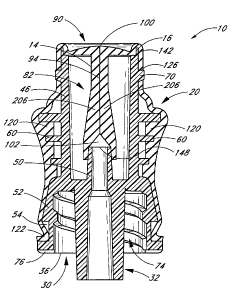
Figures 24 and 25 illustrate an example of an assembled connector in a sealed
state
(i.e., in which fluid flow through the connector is impeded). The valve
member 14 is positioned within the upper housing cavity 42 of the housing 12,
with the
first and second lateral extensions 84 of the valve member 14 protruding from
the first and
second slots 46 in the housing 12. The lead lumen 102 of the valve member 14
is
positioned so that the interior cannula 50 extends at least partway into the
lead lumen 102
of the valve member 14, facilitating fluid communication between the valve
member 14
and the Luer cannula 32 when the connector is in the open state (as
illustrated in Figures 15
and 16). The flange 90 covers the axial opening at the upstream end 16 of the
housing 12.
The sleeve 20 on the outside surface of the housing 12 allows health care
providers
to more comfortably and effectively grasp the connector 10. The flexible
material of the
sleeve 20 provides a softer surface for the fingers. There is preferably a
high-friction
interface between the flexible material of the sleeve 20 and the rubber gloves
typically
worn by health care providers, requiring less finger-pinching effort to screw
the connector
10 onto a catheter or other medical implement and to maintain the connector 10
in a desired
position and orientation during the connection and fluid-administration
processes.
In addition to providing a soft, easily grippable outer surface, the sleeve 20
surrounding the exterior of the housing 12 protects the lateral extensions
from being
pinched or otherwise undesirably manipulated during handling and use of the
connector. In
one embodiment, the valve member 14 and housing 12 are constructed such that
the
distance between the upstream end 16 and the top edges 140 of the slots 46 of
the housing
12 is slightly larger than the distance between the flange 90 and the
shoulders 92 of the
lateral extensions 84 of the valve member 14. This arrangement results in the
application
of a tensile force or preload to the valve member 14 between the flange 90 and
the lateral
extensions 84.
The preload arises as the shoulders 92 bear against the top edges 140 of the
housing
and the seal flange 90 bears against the upstream end 16 and/or the shoulder
142 of the
axial opening at the upstream end of the housing. In some embodiments, the
preload
causes the flange 90 to assume a slightly bowl-shaped or concave configuration
as the
edges of the upstream housing end 16 bear against the underside of the flange
90. The
-24-
WO 2006/052655 CA 02586115 2007-05-01 PCT/US2005/039791
bowl-shaped flange 90 tends to more tightly pinch closed the slit opening 100
and thus
enhances the ability of the valve member 14 to prevent fluid flow. The preload
also
prevents buckling of the valve member 14 along its longitudinal axis and
maintains the
sides of the slit 94 in close proximity to each other along their entire
length. The preload
thus promotes a relatively thin slit 94 below the flange 90, which enhances
the sealing
performance of the slit 94. In some embodiments, a distance between the
shoulders 92 and
the opening 148 of the interior cannula 50 is sized such that the lead lumen
102 of the valve
member 14 will be engaged with and sealed to the interior cannula 50 of the
housing 12.
Referring now to Figures 26 and 27, during use of the connector 10, a cannula
200
of a medical device 202, such a syringe , can be inserted into the valve
member 14 of the connector 10, thereby opening the valve member 14 to fluid
flow 204
between the medical device 202 and the Luer cannula 32 of the connector 10.
Before the cannula 200 is inserted, the connector 10 is in a sealed state
(see, e.g.,
Figures 24 and 25). In this state, the slit 94 defines a substantially closed
or highly
restricted flow path through the valve member 14. As illustrated in Figure 16,
when the
cannula 200 is inserted through the slit 94, the valve member 14 opens a fluid
flow path
within the connector 10 while exerting an inwardly directed force against the
cannula 200
of the medical device 202, preferably forming a tight seal around the
circumference of the
cannula 200 to prevent leakage of fluid through the upstream end of the
connector 10. The
insertion of the cannula 200 into the valve member 14 also causes the valve
member 14 to
stretch in the downstream direction over the interior cannula 50.
As fluid is injected from the medical device 202, through the cannula 200, and
into
the interior space within the valve member 14, the space between the slits
walls 206
increases further and the slit walls 206 expand further and lengthen further
in the
downstream direction. The valve member 14 thus selectively permits fluid 204
to flow
between a medical device 202 on the upstream end of the connector 10 and a
medical
implement
(not shown) to which the lower Luer connector 30 is attached.
As shown in Figures 26 and 27, when in an open state, the connector 10 permits
fluid flow 204 that is preferably substantially unobstructed and linear. This
generally
allows the connector to achieve higher flow rates. In some embodiments, the
fluid flow
rates through the connector 10 can exceed 600 cubic centimeters per minute. In
addition,
the unobstructed and linear fluid flow 204 interferes less with the inherent
qualities of the
-25-
CA 02586115 2007-05-01
WO 2006/052655 PCT/US2005/039791
flowing fluid 204. For example, if the fluid flow 204 is blood, the various
blood cells and
other constituents are less likely to break down within the illustrated
connector 10 as
compared to a connector in which there is a circuitous fluid flow path with
fluid turbulently
strikes against hard and/or angular internal surfaces.
As the fluid flow 204 diminishes and/or the cannula 200 of the medical device
202
is withdrawn from the valve member 14, the slit walls 206 retract and return
to their
original configuration to once again define a narrow, restrictive path width
between them
(as illustrated, for example, in Figures 24 and 25). This retraction of the
slit walls 206
causes the volume within the slit 94 to decrease to a certain minimum. The
retracting
action of the slit walls 206 also forces out the remaining fluid in the area
between the walls
206. As the syringe cannula 200 is being withdrawn, the displaced fluid cannot
flow out of
the slit 94 through the upstream end of the valve member 14 because this space
is occupied
by the syringe cannula 200. The resilient narrow neck 97 of the slit 94
preferably blocks
any significant flow of fluid between the outer surface of the cannula 200 and
the inner
surface of the flexible member 80 by forming a tight seal around the
circumference of the
cannula 200. Thus, the displaced fluid is instead forced downwardly from the
slit 94,
through the interior cannula 50 and downwardly directed cannula 32, and
ultimately out of
the housing 12.
This advantageously results in automatic positive flow from the connector 10
toward the
patient upon withdrawal of the medical device 202 from the upstream end of the
connector
10, and avoids or minimizes retrograde fluid flow toward the connector 10 and
away from
the patient.
Although the foregoing description refers to a syringe, it is contemplated
that any
type of suitable medical devices may be joined to either end of the connector
10, such as IV
bags, other connectors, and tubing, for the purposes of fluid transfer or for
any other
desired purpose. An auxiliary connector also may be connected to the soft grip
connector,
and both connectors can be placed in fluid communication with a catheter with
an end
positioned within a patient. This arrangement can provide several advantages
in situations
which call for the use of a unique auxiliary connector. For example, when it
is necessary to
replace or reconfigure fluid lines connected to auxiliary connectors, such
lines may be
removed from fluid communication with the catheter without creating a backflow
in the
catheter, and replaced with a similar connector or any other medical
implement. In some
embodiments, one such auxiliary connector may be the CLAVE connector sold by
ICU
-26-
WO 2006/052655 CA 02586115 2007-05-01 PCT/US2005/039791
Medical, Inc. However, any connector or other medical implement or device may
be
placed in fluid communication with the soft grip connector 10 to introduce
fluid to the
patient or to withdraw blood from the patient including, but not limited to,
pierceable
connectors, needle-less connectors, medical tubing, syringes or any other
medical
implement or device.
Although certain embodiments and examples have been described herein, it will
be
understood by those skilled in the art that many aspects of the methods and
devices shown
and described in the present disclosure may be differently combined and/or
modified to
form still further embodiments. For example, the various embodiments of
housing may be
interchangeable applied to the various embodiments of flexible member to
achieve multiple
embodiments of soft-grip medical connector. Additionally, it will be
recognized that the
methods described herein may be practiced using any device suitable for
performing the
recited steps. Such alternative embodiments and/or uses of the methods and
devices
described above and obvious modifications and equivalents thereof are intended
to be
within the scope of the present disclosure. Thus, it is intended that the
scope of the present
invention should not be limited by the particular embodiments described above,
but should
be determined only by a fair reading of the claims that follow.
-27-