Note: Descriptions are shown in the official language in which they were submitted.
CA 02593167 2007-07-05
WO 2006/076428 PCT/US2006/000973
SYSTEMS AND METHODS FOR THREE DIMENSIONAL
IMAGING WITH AN ORIENTATION ADJUSTABLE ARRA.Y
FIELD OF THE INVENTION
The systems and methods relate generally to medical ultrasound imaging, and
more
particularly to three dimensional ultrasound imaging with an orientation
adjustable array.
BACKGROUND INFORMATION
The ability to perform three-dimensional (3D) ultrasound imaging of the
interior of a
living being provides numerous diagnostic and therapeutic advantages. However,
3D imaging
with intravascular or otller internally inserted imaging systems, such as
intravascular ultrasound
or intracardiac echocardiography (ICE) imaging systems, is difficult. This is
mainly because of
the size constraints inherent in the use of internal imaging devices.
For instance, conventional 3D imaging systems require a two-dimensional (2D)
phased
array having numerous transducer elements. This 2D array provides a steerable
imaging beam
which images in one direction and can be steered in two additional directions,
thus providing
3D capability. However, 2D arrays are very costly and typically too large for
insertion into
most regions of a living being, such as narrow blood vessels. Furthermore,
each eleinent is
typically coupled with a separate communication line, e.g., a cable, in order
to communicate
with an external imaging system. These communication lines add undesirable
cross-sectional
area to the insertable device (such as a catheter) being used to deploy and
navigate the array
within the body. This added cross-sectional area, or width, can also prevent
use of the array
within narrow regions of the body. Finally, 2D arrays are susceptible to cross-
talk between
elements, which can Significantly degrade perfonnance.
Other conventional 3D imaging systems use a single element transducer mounted
on the
distal end of a rotating drive shaft. This single element transducer images
one dimensionally in
a radial direction perpendicular or transverse to the central axis of the
drive shaft. When the
transducer is rotated in a second direction, the image data collected can be
used to generate a
2D cross-sectional image of the body tissue. The driveshaft is typically
located within an outer
sheath and can be slid proximally and distally within the sheath along the
central axis of the
drive sliaft. Multiple 2D cross-sectional images can be obtained at different
positions along the
central axis. An image processing system can then be used to assemble, or
reconstnict these
images into a 3D image of the body tissue. However, this process cannot be
perforn-ied in real-
time since it requires the reconstnxction of previously obtained 2D images.
CA 02593167 2007-07-05
WO 2006/076428 2 PCT/US2006/000973
improved systems and methods for 3D imaging wliich
overcome the shortcomings of conventional 3D imaging systems.
SUMMARY
The systems and methods described herein provide for a medical ultrasound
imaging
system configured for 3D imaging of a living being with an orientation
adjustable imaging
device insertable into a living being and configured to image the interior of
the living being. In
one example embodiment as described below, the imaging device includes an
ultrasound array
having an imaging field and an orientation adjustment unit coupled with tlie
array and
configured to adjust the orientation of the array. The array can include
inultiple transducer
elements configured as a linear array arranged along a one dimensional axis.
The array can
preferably image a two-dimensional imaging field such that when the
orientation of the array is
adjusted in a third dimension, image data from a three-dimensional region can
be collected.
The orientation adjustment unit can be configured to adjust the orientation of
the array
in any manner. In one embodiment, orientation adjustment unit adjusts the
pitch of the array
about an axis. The orientation adjustment unit can include an orientatioii
control unit
configured to control the orientation of the array, control the rate of
adjustment of the array aiid
optionally determine the orientation of the array. The orientation control
unit can control the
orientation of the airay in any manner, such as electrically, mechanically,
magnetically and the
lilce. The orientation adjustinent unit can also include an adjustable
mounting for mounting the
array thereon. In one embodiment, the adjustable mounting is a flexible
circuit having a
multiplexer for multiplexing signals coinmunicated to and from the array.
The imaging system can also include an image processing system communicatively
coupled with the array. In an example embodiment as described below, the image
processing
system can be configured to control the imaging direction of the array and can
be configured to
receive an output signal from each element in the array, where one or more of
the output
signals are representative of an echo received in the imaging direction. This
image processing
system can also be configured to process the received output signals and
generate a three-
dimensional image therefrom. In one example embodiment, the image processing
system can
be configured to process the one or more output signals into echo data and
store the echo data
in an echogenic record, wllere one echogenic record is generated for each
imaging direction
imaged by the aiTay. The image processing system can be configured to store
the echogenic
records generated at each orientation of the array as a separate image data
set and can also be
CA 02593167 2007-07-05
WO 2006/076428 PCT/US2006/000973
3
ff . EG Ir E-c~s~ ijix}i, ~r~ei~s ', ni
configured .,6 genera e a hr~onal image from the image data sets corresponding
to
multiple orientations of the array.
Otlier systems, methods, features and advantages of the invention will be or
will
become apparent to one with skill in the art upon exainination of the
following figures and
detailed description. It is intended that all sucli additional systems,
methods, features and
advantages be included within this description, be within the scope of the
invention, and be
protected by the accompanying claims. It is also intended that the invention
is not limited to
require the details of the example embodiments.
BRIEF DESCRIPTION OF THE FIGURES
The details of the invention, including fabrication, structure and operation,
may be
gleaned in part by study of the accompanying figures, in which like reference
numerals refer to
like segments.
FIGs. lA-C are block diagrams depicting an example embodiment of an medical
imaging system with an orientation adjustable iinaging device.
FIG. 2A is a perspective view depicting an example embodiment of an
orientation
adjustable imaging device.
FIGs. 2B-C are top down views depicting additional example embodiments of an
orientation adjustable imaging device.
FIG. 3 is a block diagram depicting another example einbodiment of a medical
imaging
system with an orientation adjustable imaging device.
FIG. 4 is a schematic view depicting an example embodiment of an orientation
adjustable imaging device.
FIG. 5 is a block diagram depicting another example embodiment of a medical
imaging
system with a multiplexer.
FIG. 6 is a perspective view depicting another exainple enzbodiment of a
medical
imaging system with an orientation adjustable inzaging device.
FIG. 7 is a block diagram depicting another exaniple embodiment of a medical
imaging
system with an orientation adjustable imaging device.
DETAILED DESCRIPTION
The systems and methods described herein provide for 3D imaging with a medical
ultrasound inlaging system using an orientation adjustable imaging device.
FIGs. 1 A-C depict
CA 02593167 2007-07-05
WO 2006/076428 4 PCT/US2006/000973
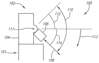
bne'exampl''e''e'moc~'i'irferii""oP''a"uftrBound imaging system 100 having an
orientation
adjustable imaging device 102. Imaging device 102 is preferably a component of
a flexible
elongate medical device 101, such as a catheter, endoscope and the like, which
is insertable
into a living being and configured to allow imaging of the interior of the
living being with
imaging device 102. Imaging system 100 can be any type of ultrasound imaging
system having
an insertable imaging device 102, such as an IVUS imaging system, an ICE
imaging system or
other imaging systems. Imaging device 102 preferably includes an orientation
adjustment unit
104 and an ultrasound transducer device 106 configured to image an imaging
field 108, which
is preferably 2D. Ultrasound transducer device 106 is preferably a transducer
array, but can
also be inultiple transducer elements in a non-array configuration or a single
element
transducer. Orientation adjustment unit 104 is preferably configured to adjust
the orientation of
array 106 in a third dimension, indicated by directions 111 and 113, to allow
array 106 to image
a 3D region of the body.
In the enlbodiments depicted in FIGs. lA-C, array 106 is adjustable over a
range of
motion 116. In this embodiment, array 106 is rotatable about axis 117. FIGs.
1A-C each
depict array 106 at a separate orientation with motion range 116. FIG. lA
depicts array 106
positioned at a first orientation located approximately in the center of
motion range 116. FIG.
1B depicts array 106 positioned at a second orientation where the pitch of
airay 106 has been
adjusted in direction 111 by an angle 112, while FIG. 1 C depicts array 106
positioned at a third
orientation where the pitch of array 106 has been adjusted in direction 113 by
an angle 114.
Here, motion range 116 is approximately 120 degrees; however, the limits of
motion range 116
are entirely dependent upon the needs of the application and can be set to auy
appropriate range
or ranges.
At each orientation within range 116, array 106 can be used to image field
108.
Preferably, array 106 sweeps back and forth across motion range 116 while at
the same time
collecting image data across 2D imaging field 108 that can be used to generate
a 3D image. It
should be noted that motion range 116 is not limited to motion only in
directions 111 and 113.
The orientation of array 106 can be adjusted in any manner and through any
range of motion.
For instance, motion range 116 can include up/down movement, left/right
movement,
forward/backward movement, rotational inovement, tilting movement, pivoting
movemcnt,
wobbling movement, oscillating movement and other types of movement.
FIG. 2A depicts a perspective view of one example embodiment of an=ay 106
configured as a linear, curved linear or one-dimensional (1D) phased array
including a series of
individual transducer elements 202 arranged along a cominon axis 204. In this
embodiment,
CA 02593167 2007-07-05
WO 2006/076428 5 PCT/US2006/000973
Eray' 1'06 is'"cori~'igure tognerVe itil imaging beam 205 in a variable
direction 206.
Specifically, array 106 can be configured to transinit an ultrasound signal
beam 205 along
direction 206 and receive echoes propagating back towards array 102 along
direction 206, the
echoes generally resulting from the collision of the transniitted ultrasound
signal with body
tissue. Direction 206 is variable, or steerable, and array 106 is preferably
configured to image
the body tissue in multiple different directions 206. In other embodiments of
imaging device
102 that image only in one dimension, such as a single element transducer,
orientation
adjustment unit 104 is preferably configured to move the imaging device in two
diinensions to
allow for 3D imaging.
FIG. 2B depicts a top down view of an exainple einbodiment of array 106 with a
steerable imaging beain 205. Iniaging beain 205 can be generated in multiple
different
directions 206, each at a different angular location 208 with respect to array
106. Here, the
ultrasound beams 205 generated at each angular location 208 define the imaging
field 108 of
the array 106. Preferably, during an imaging procedure, the beam 205 images in
direction 206
at one angular location 208 and then is adjusted, or steered, to a second
adjacent angular -
location 208 and images again. In this manner, beam 205 can be swept across
imaging field
108. Because imaging field 108 extends substantially in two directions, X and
Y, the data
collected from each sweep of imaging field 108 can be used to collect 2D image
data of the
body tissue.
In practice, beam 205 will have a finite cross-sectional area and imaging
field 108 will
extend into the Z direction by a small amount. However, this amount is
generally negligible for
3D imaging purposes, so imaging field 108 is referred to herein as being
substantially 2D. One
of skill in the art will readily recognize that the shape of beam 205 can be
adjusted to provide
greater resolution in the Z direction as required by the needs of the
application.
FIG. 2C depicts a top down view of another exainple embodiment of array 106.
Here,
array 106 is configured to image in multiple directions 206, each direction
206 being
substantially perpendicular to the face 212 of array 106 and located at a
different position along
the face 212. By adjusting the position along face 212, beam 205 can be swept
across imaging
field 108 to collect 2D image data of the body tissue.
After collecting 2D image data over the imaging field 108 at a first
orientation of array
106, the orientation adjustment unit 104 preferably adjusts the array 106 to a
second orientation
to collect 2D image data over the inlaging field 108 at that orientation. This
process repeats
until 2D image data has been collected for a desired number of different
orientations of array
106. This collected 2D image data can then be assembled, or reconstructed, by
an image
CA 02593167 2007-07-05
WO 2006/076428 6 PCT/US2006/000973
4C., qõr It..r qs ,,Jr iLdF nl r= .,,!r il,.d+.ir, r,.. it
processing system 3 6( escribed below) to generate a 3D image of the body
tissue. Thus, in
this embodiment a 1D array 106 caii be used to generate a 3D image with
superior quality than
conventional systems, due in part to the reduced potential for cross-talk
resulting from the use
of a 1D array 106.
However, any type of transducer array 106 can be used including 2D arrays and
other
appropriate transducer configurations. Array 106 can be a linear or phased
array. Array 106
can also be fabricated in any inanner desired. For instance, array 106 can
include piezoelectric
transducer elements, micromachined ultrasound transducer (MUT) elements such
as capacitive
micromachined ultrasound transducers (CMUTs) or piezoelectric microinachined
ultrasound
transducers (PMUTs), or other known transducer array structures.
The rate at which the orientation of imaging device 102 is adjusted is
dependent upon
the needs of the application and can be as rapid or as slow as desired. Also,
the orientation
adjustment can be continuous or can proceed in a stepped fashion. The
adjustlnent rate can
also be related to the imaging fraine rate of imaging system 100, for
instance, to allow for real-
time 3D imaging. In one example, a video frame may include image data
collected from 100
separate imaging fields 108, each located at a different pitch within motion
range 116. If the
imaging frame rate is 30 frames per second, then each sweep of array 106
across motion range
116 can talce no longer than 0.0333 seconds. If the pitch is adjusted in a
stepped fashion and it
takes 20 microseconds to image one imaging field 108, then the time to adjust
array 106 from
one pitch to the next can be no longer than 133 microseconds. It should be
noted that these
values serve only as an example and in no way limit the systems and methods
described herein.
FIG. 3 depicts a block diagrain of another example embodiment of imaging
systein 100.
Here, array 106 is located at or near the distal end 304 of medical device 101
and is
communicatively coupled with the image processing system 306 via one or more
communication lines 308. Image processing systein 306 is preferably located
extenially to the
living being at the proximal end 310 of medical device 101. Image processing
system 306 is
preferably configured to control the imaging direction 206 of beam 205. hiiage
processing
system 306 is also preferably configured to receive an output signal from each
element 202 in
array 206 and process the output signal into echo data representative of an
echo received by
array 106 in direction 206.
In one embodiment, image processing system 306 is configured to store the echo
data in
an echogenic record, where each echogenic record includes the echo data
received in direction
206 at one angular location 208 in the imaging field 108. One echogenic record
can be
generated for each angular location 208 in an imaging field 108 for one
orientation of array
CA 02593167 2007-07-05
WO 2006/076428 7 PCT/US2006/000973
1"0~':" ~llro~ tlie e'c~ogenic"records froin a given imaging field 108 can
then be grouped together
by image processing system 306 into an image data set. Image processing system
306 is
preferably configured to assemble each of the image data sets and generate a
3D image of the
body tissue. Image processing system 306 preferably includes the processing
hardware and/or
software to generate the 3D images in real-time, or near real-time, for the
benefit of the
physician or technician operating system 100.
FIG. 4 depicts a scheinatic view of another example embodiment of imaging
system
100 showing imaging device 102 in closer detail. Here, imaging device 102
includes a housing
402 coupled with a base structure 404. Base structure 404, in turn, is coupled
to the distal end
406 of an elongate shaft 408. Elongate shaft 408 can be used to position
imaging device 102
into proximity with the desired region for imaging, by moving the imaging
device 102 along its
longitudinal axis, for exainple. Array 106 and orientation adjustment unit 104
are preferably
housed within a housing 402. Housing 402 can optionally include an imaging
window 410
composed of a material that does not substantially interfere with the
transmission or reception
of the ultrasound signals, including known sonulucent materials. Window 410
can also be an
aperture in housing 402. Preferably, window 410 is large enough to
accoininodate imaging
across the entire motion range 116 of array 106. In another embodiment, an
elongate tubular
outer sheath (not shown) having an inner lumen is provided. The inner lumen
can be
configured to slidably receive imaging device 102 and shaft 408.
The term "orientation" is defined herein as the position of array 106 with
respect to the
structure or device used to move, navigate or guide array 106 within the
living being. In this
embodiment, although shaft 408 can be used to move the imaging device 102
within the living
being, for instance to position imaging device 102 in proximity with the
desired region for
imaging, the orientation of array 106 remains adjustable even when shaft 408
is stationary.
In this embodiment, orientation adjustment unit 104 is configi.tred to control
the
orientation of the array 106 and determine the orientation of array 106 at any
given time, for
instance, in order to allow tracking of array 106. Orientation adjustment unit
104 can include
an orientation control unit 412 for controlling and determining the
orientation of array 106.
Orientation control unit 412 can be configured in any manner in accordance
with the needs of
the application.
For instance, orientation control unit 412 can be configured to electrically,
mechanically
or magnetically operate or control the orientation of airay 106, or any
combination thereof. In
one exainple enlbodiment, orientation control unit 412 includes one or more
actuators for
adjusting the orientation of array 106. One example actuator that can be used
is a piezo-filni
CA 02593167 2007-07-05
WO 2006/076428 8 PCT/US2006/000973
~ctuator;' altnoug~i"t~ie"'sys't'em's and methods described herein are not
limited to such. In another
einbodiment, orientation control unit 412 includes a piezoelectric drive for
orientation control
of array 106. In yet another einbodiment, orientation control unit 412
inch.ides a rolling wheel
and an electrical servo motor for powering the wheel, which is in turn coupled
with array 106
by a wire or tether. Adjustment of the rolling wheel applies tension to the
array via the wire or
tether and can be used to control and adjust the orientation of array 106.
Orientation
adjustment unit 104 can also optionally include one or more sensors 418 for
detennining the
orientation of array 106 at any given time. Sensors 418 can use any type of
sensing technique
such as electrical, optical, inagnetic, capacitive, inductive etc.
Orientation control unit 412 can be adjustably coupled witli array 106. For
instance in
one embodiment, orientation control unit 412 is a flexible circuit physically
coupled witll aixay
106. Alternatively, orientation adjustment unit 104 can also include a
position adjustable
mounting 414 for adjustably coupling airay 106 with orientation control unit
412. Any type of
position adjustable mounting 414 can be used in accordance with the needs of
the application.
For instance, in one einbodiment, cominunication lines 308 are flexible and
function as
position adjustable mounting 414. In another embodiment, position adjustable
mounting 414 is
a hinge-type structure configured to limit the motion of array 106 to movement
solely within
motion range 116. It should be understood that these embodiments are only
examples and in
no way limit the systems and inethods described herein.
Orientation adjustment unit 104 can also include a multiplexer 416. FIG. 5 is
a block
diagram depicting an example embodiment of imaging device 102 with multiplexer
416. In
this embodinient, each array element 202-1 through 202-N (where 'N' indicates
that any
number of elements 202 can be present) is coupled with a separate
cominunication line 502-1
through 502-N. Multiplexer 416 includes coinmunication ports 504-1 through 504-
N coupled
with each element 202-1 through 202-N by way of communication lines 502-1
through 502-N.
Multiplexer 416 also includes communication ports 506-1 tlirough 506-M (where
'M"
indicates that any number of ports 506 can be present, unless otherwise
noted). Each
coinmunication port 506-1 through 506-M is preferably coupled with a
communication line
308-1 tlirough 308-M and routed to image processing system 306 with shaft 408.
Preferably,
multiplexer 416 is an N:M multiplexer configured to multiplex the signals
input to ports 504-1
through 504-N and output the multiplexed signals from ports 506-1 through 506-
M, where M is
less then N. Multiplexer 416 also preferably includes corresponding M:N
demultiplexer
circuitiy to demultiplex the signals input to ports 506-1 through 506-M and
output the
demultiplexed signals from ports 504-1 tluough 504-N to array 106. Also, image
processing
CA 02593167 2007-07-05
WO 2006/076428 PCT/US2006/000973
9
system'3'66 prdeiabT"y'incluaes' compl'ementary multiplexing and
demultiplexing hardware
and/or software for communication with array 106.
The use of a multiplexer 416, with the value of M less than N, reduces the
number of
communication lines 308 necessary to transmit signals between array 106 and
image processing
system 306. A reduction in the number of communication lines 308 can decrease
the potential
for cross-talk and can also allow the radial cross-sectional area of device
101, or width, to be
minimized, which in turn can allow the introduction of device 101 into smaller
regions of the
body.
Also, inultiplexer 416 can also be used as, or in conjunction with, position
adjustable
mounting 414 to provide adjustable support for array 106. For instailce, in
one embodim.ent,
multiplexer 416 is a flexible circuit coupled with array 106. Furthermore, in
embodiments
where the elements 202 of array 106 are MUTs, multiplexer 416 and array 106
can be
monolithically integrated together on a common semiconductor substrate. The
integration of
multiplexer 416 and array 106 on the saine substrate can reduce the size of
imaging device 102
and improve the interface performance between array 106 and multiplexer 416.
FIG. 6 depicts a perspective view of another example embodiment of imaging
system
100 further illustrating the imaging capability of orientation adjustable
imaging device 102. In
this embodiment, 3D spatial region 602 represents the area that imaging device
102 can image
by adjusting the orientation, or pitch, of imaging device 102 in the Z
direction and collecting
image data from multiple 2D imaging fields 108. Here, imaging device 102 is
positioned in a
side-looking configuration with respect to medical device 101. Imaging device
102 can also be
moved as desired to adjust the overall position of imaging device 102 within
the body. For
instance, shaft 108 can be moved proximally and distally along central axis
604 and rotated
about central axis 604 in direction 606.
FIG. 7 depicts a block diagram of anotlier example embodiment of imaging
system 100.
Here, imaging device 102 is positioned in a forward-looking configuration with
respect to
medical device 101. Here, the orientation of array 106 can adjusted across
motion range 116 to
allow imaging of body tissue located distal to the distal end 304 of medical
device 101. One of
skill in the art will readily recognize that imaging device can be positioned
in any manner
within medical device 101 and at any location on medical device 101. In this
embodiment,
forward-looking array 106 can be an annular array with a symmetric or non-
syininetric beam
pattern, a non-diffraction array and the like.
CA 02593167 2007-07-05
WO 2006/076428 10 PCT/US2006/000973
"'" "Ifi t1Y6'f'dr8g8f1!g 9VeMft6a'tioW 'the invention has been described with
reference to
specific embodinlents thereof. It will, however, be evident that various
modifications and
changes may be made thereto without departing from the broader spirit and
scope of the
invention. For example, each feature of one embodiment can be mixed and
matched with other
features shown in other einbodiments. Features and processes lcnown to those
of ordinary skill
may similarly be incorporated as desired. Additionally and obviously, features
may be added
or subtracted as desired. Accordingly, the invention is not to be restricted
except in liglit of the
attached claims and their equivalents.