Note: Descriptions are shown in the official language in which they were submitted.
CA 02597050 2007-08-15
1
CROSS-REFERENCE TO RELATED APPLICATION
This application is a divisional of Canadian Patent Application No. 2,423,878
which is based on international application No. PCT/US01/42311 filed on
September
26, 2001, which claims the benefit of priority of U.S. Patent Application No.
09/680,202 filed on October 5, 2000, the disclosures of which are incorporated
herein
by reference.
MINIMALLY-INVASIVE ANNULOPLASTY REPAIR SEGMENT
DELIVERY TEMPLATE, SYSTEM AND METHOD OF USE
Field of the Invention
The present invention relates generally to medical devices and particularly to
heart valve prostheses having a low profile sewing ring that enables larger
valve
orifices to be used.
Background of the Invention
Prosthetic heart valves are used to replace damaged or diseased heart
valves. In vertebrate animals, the heart is a hollow muscular organ having
four
pumping chambers: the left and right atria and the left and right ventricles,
each
provided with its own one-way valve. The natural heart valves are identified
as the
aortic, mitral (or bicuspid), tricuspid and pulmonary valves. Prosthetic heart
valves
can be used to replace any of these naturally occurring valves, although
repair or
replacement of the aortic or mitral valves is most common because they reside
in the
left side of the heart where pressures are the greatest.
Two primary types of heart valve replacements or prostheses are known. One
is a mechanical-type heart valve that uses a ball and cage arrangement or a
pivoting
mechanical closure to provide unidirectional blood flow. The other is a tissue-
type or
"bioprosthetic" valve which is constructed with natural-tissue valve leaflets
which
function much like a natural human heart valve's, imitating the natural action
of the
flexible heart valve leaflets which seal against each other to ensure the one-
way
blood flow. In both types of prosthetic valves, a biocompatible fabric-covered
suture
CA 02597050 2007-08-15
la
or sewing ring or cuff on the valve body (mechanical) or stent (tissue-type)
provides a
platform for attaching the valve to the annulus of the particular valve being
replaced.
The valves of the heart separate chambers therein, and are each mounted in
an annulus therebetween. The annuluses comprise dense fibrous rings attached
CA 02597050 2007-08-15
2
either directly or indirectly to the atrial and ventricular muscle fibers. In
a valve
replacement operation, the damaged leaflets are excised and the annulus
sculpted
to receive a replacement valve. Ideally the annulus presents relatively
healthy
tissue that can be formed by the surgeon irito a uniform ledge projecting into
the
orifice left by the removed valve. The time and spacial constraints imposed by
surgery, however, often dictate that the shape of the resulting annulus is
less than
perfect for attachment of a sewing ring. Moreover, the annulus may be
calcified
as well as the leaflets and complete annular debridement, or removal of the
hardened tissue, results in a larger orifice and less defined annulus ledge to
which
lo to attach the sewing ring. In short, the contours of the resulting annulus
vary
widely after the natural valve has been excised.
Conventional placement of the valve is intra-annular, with the valve body
deep within the narrowest portion of the annulus to enhance any seal effected
by
the sewing ring/suture combination and reduce the chance of perivalvular
leakage.
Surgeons report using at least 30 siinple sutures or 20 mattress-type sutures
to
prevent leakage. Mattress sutures are more time consuming and essentially
comprise double passes of the needle through the tissue with one knot.
Naturally, the implantation of a prosthetic heart valve, either a
mechanical valve or a bioprosthetic valve (i.e., "tissue" valve), requires a
great
2o deal of skill and concentration given the delicate nature of the native
heart
tissue, the spatial constraints of the surgical field and the criticality of
achieving
a secure and reliable implantation. It is of equal importance that the valve
itself
has characteristics that promote a long valve life and that have minimal
impact
on the physiological makeup of the heart environment.
In view of the foregoing, it is evident that an improved sewing ring that
addresses the apparent deficiencies in existing sewing rings is necessary and
desired. That is, there is a need for a sewing ring that increases the orifice
area
of the valve while at the same time sixnplifying the fabrication and
implantation
steps.
CA 02597050 2007-08-15
3
Summary of the Invention
The present invention provides an improved sewing ring and sewing
ring/stent assembly that facilitates manufacture and implantation of heart
valves. The sewing ring is adapted to pivot or move outward from the stent,
thus enabling a surgeon during the implantation procedure to more easily
isolate
the sewing ring against the native tissue and away from the stent and tissue
leaflets. Thus, there is less chance of the surgeon puncturing the leaflets.
Furthermore, the compliance of the sewing ring, or ability to pivot the ring
away from the stent, enables the sewing ring to be made smaller in the radial
dimension, and thus the overall valve orifice size can be increased.
Additionally, the manufacturing process is facilitated because various regions
around the stent can be more easily visualized and accessed by virtue of the
movable sewing ring.
In one aspect, the piresent invention provides a sewing ring attached to a
generally annular periphery of a heart valve. The sewing ring includes a
suture=
permeable ring attached to the heart valve periphery and configured to pivot
from a first position substantially adjacent the periphery to a second
position
outward from the first position. The sewing ring desirably comprises a sutara
permeable insert ring and a fabric cover. The insert ring may be substantially
planar. The fabric covering the insert ring also desirably covers a portion of
the
heart valve. Moreover, the fabric covering both the insert ring and a portion
of
heart valve also preferably connects the ring to the heart valve periphery. A
seani may be provided wherein the sewing ring pivots between the first and
second positions about the seam. In one embodiment, the first and second
positions are stable such that the sewing ring is bi-stable.
In a further aspect, a heart valve having an inflow end and an outflow
end is provided, comprising a generally annular stent, and a suture penneable
sewing ring attached to a periphery thereof. The sewing ring is movable
between two positions, wherein in the first position the sewing ring extends
generally toward the outflow end of the valve and in the second position the
CA 02597050 2007-08-15
4
sewing ring extends generally toward the inflow end of the valve. The sewing
ring may comprise an insert ring and a fabric cover, and the fabric covering
the
insert ring may also cover a portion of the stent. In a preferred embodiment,
the
sewing ring attaches to the stent exclusively with a portion of a fabric that
also
covers a portion of the sewing ring. A seam is desirably provided in the
fabric
at the line of attachment between the sewing ring and the stent, wherein the
sewing ring pivots about the seam between the first and second positions. The
first and second positions may be stable, and the insert ring may be
frustoconical in shape such that in the first posifion the ring extends toward
the
outflow end and in the second position the ring extends toward the inflow end.
Furthermore, the insert ring may be provided with alternating radially thick
and
thin regions, or it may have a radially unulating shape, to fadlitate
moveinent
between the first and second positions.
In another aspect, the present invention provides a heart valve including
a generally annular stent having a periphery, a tubular fabric, and a
generally
annular suture-permeable insert sized at least as large as the stent
periphery.
The stent and insert are connected together exclusively by a portion of the
fabric
that permits relative outward pivoting of the insert with respect to the
stent. In a
preferred embodiment, the fabric at least partly covers both the stent and
insert.
2o A seam may be provided in the fabric at the line of attachment between the
insert and the stent to provide a discrete pivot line. In a preferred
embod'unent,
the tubular fabric is a single piece prior to assembly of heart valve, and
desirably encompasses both the stent and insert. The stent may have an
undulating outflow edge comprising alternating commissures and cusps,
wherein the fabric covers the outflow edge. The insert is desirably disposed
around stent to pivot about the outer surface thereof, and a sewing tab along
the
undulating outflow edge is desirably sewn directly to the stent to prevent
relative movement of the fabric upon pivoting of the insert.
In a further embodiment, a method of implanting a heart valve in host
tissue (e.g., an aortic annulus) is provided. The heart valve has an inflow
end
CA 02597050 2007-08-15
and an outflow end, and a sewing ring attached to a periphery thereof. The
method includes positioning the sewing ring to extend generally toward the
inflow end of the valve, attaching the sewing ring to the host tissue, and ro
positioning the valve with respect to the attached sewing ring so that the
sewing
5 ring extends generally toward the outflow end of the valve. The method of
attachment preferably comprises suturing. The method also may include
providing the heart valve having a stent and a plurality of leaflets supported
thereby, the sewing ring being located substantially adjacent the valve when
extending generally toward the inflow end of the valve. The method of re-
1o positioning may thus include inverting the sewing ring by pivoting it
outward
from the position substantially adjacent the valve. In one embodiment, the
sewing ring is configured and attached to the stent so as to be bi-stable
between
the two positions.
Further, the present invention provides a method of assembling a heart
valve, including providing a generally annular stent having a periphery, a
tubular fabric, and a generally annular suturo-permeable insert ring sized at
least
as large as the stent periphery. The method includes connecting the stent and
insert ring with the fabric to permit relative outward pivoting of the fa.briG
covered insert ring with respect to the stent. The method may include
completely covering the stent with the tabular fabric prior to connecting the
insert ring with the fabric. Furthermore, the tubular fabric preferably
consists of
a single piece, wherein the method includes covering both the stent and the
insert ring with the single piece. The method further may include holding a
portion of tubular fabric against the annular stent using an assembly fixture.
The assembly fixture desirably comprises an annular member and is mounted
for rotation about an assembly handle. The handle has an elongated grip,
wherein the axis of rotation of the assembly fixture is angled with respect to
the
grip.
CA 02597050 2007-08-15
6
A further understanding of the nature and advantages of the invention
will become apparent by reference to the remaining portions of the
specification
and drawings.
Brief Description of the Drawings
Figure 1 is a perspective view of a stent assembly used in an exemplary
mitral or pulmonary position heart valve of the present invention;
Figure 2 is a perspective view of a suture-permeable insert for an
exemplary mitral or puhnonary position heart valve sewing ring of the present
invention;
Figures 3A and 3B are perspective views of initial steps in an assembly
process of a heart valve of the present invention wherein a tubular fabric
covering is wrapped around the stent assembly of Figure 1; .
Figure 3C is a cross-sectional view taken along line 3C-3C of Figure 3B;
Figures 4A and 4B are perspective views of further steps in the heart
valve assembly process in which the fabric covering is attached along the
outflow edge of the stent assembly;
Figure 5A is a perspective view of a further step in the heart valve
assembly process in which free edges of the tubular fabric covering are
created
in preparation for addition of the insert shown in Figure 2;
Figure 5B is a cross-sectional view taken along line 5B-5B of Figure
5A;
Figure 6A is a perspective view of a further step in the heart valve
assembly process wherein the insert of Figure 2 is positioned around the stent
assembly of Figure 1, with the fabric covering therebetween, and with the help
of an assembly fixture;
Figure 6B is a cross-sectional view taken along line 6B-6B of Figure
6A;
Figure 7A is a perspective view of a further step in a heart valve
assembly process wherein an outflovv portion of the suture-permeable insert is
CA 02597050 2007-08-15
7
1. providing a holder having a flexible template adapted to attach to an
annuloplasty repair segment, the template being convertible from a
generally linear shape to a curved shape;
2. attaching an annuloplasty repair segment to the flexible template;
3. delivering the repair segment attached to the template to a heart valve
annulus;
4. causing the template and repair segment to simultaneously undergo a
shape change; and
5. attaching the annuloplasty repair segment to the annulus.
The method may also include a step of delivering the annuloplasty repair
segment attached to the template through a minimally-invasive tube. The
minimally invasive tube may be inserted through an access incision in the
chest
wall, or through an access incision in the peripheral vasculature and through
vascular system, both into proximity within the annulus. The method may
include
releasing the template from the end of the tube, and maintaining a tether
connection
between the template and an anchor mandrel from within the tube.
A further understanding of the nature and advantages of the invention will
become apparent by reference to the remaining portions of the specification
and
drawings.
Brief Description of the Drawings
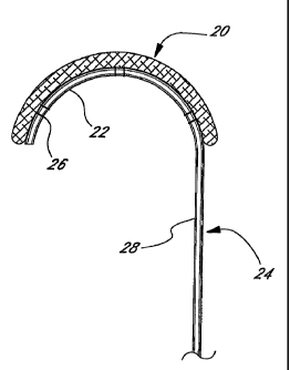
Figure 1 is an elevational view of a holder of the present invention
having an annuloplasty repair segment attached to a flexible distal template;
Figure 2 is an elevational view of an alternative holder of the present
invention having an annuloplasty repair segment attached to a flexible distal
template;
Figures 3A-D are elevational views of the deployment of the holder of
Figure 1 from within a delivery tube;
CA 02597050 2007-08-15
8
Figure 4 is an elevational view of a still further holder of the present
invention having an annuloplasty repair segment attached to a distal template
having markers;
Figure 5 is an elevational view of another holder of the present invention
having an annuloplasty repair segment attached to a flexible distal template
that
can pivot with respect to a proximal handle;
Figure 6A and 6B are elevational views of the deployment of the holder
of Figure 5;
Figures 7A and 7B are elevational views of another holder of the present
invention having an annuloplasty repair segment attached to a distal multi-
segmented template that can curl with respect to a proximal handle upon
actuation of a pull string;
Figures 8A-8C are perspective views of a further holder of the present
invention having an annuloplasty repair segment attached to a distal template
that is biased to curl in three-dimensions with respect to a proximal handle;
Figures 9A and 9B are perspective views of an annuloplasty delivery
system of the present invention having an annuloplasty repair segment attached
to a template that is biased to curl when ejected from a proximal delivery
tube;
Figure 10 is a perspective exploded view of the annuloplasty delivery
system of Figures 9A and 9B;
Figure 11 is an enlarged perspective view of the distal end ofthe
annuloplasty delivery system of Figures 9A and 9B;
Figures 12 and 12A are schematic illustrations depicting a human chest and
the disposition of a right parasternal incision in connection with an aortic
surgery
procedure in accordance with the present invention;
Figure 13 is a pictorial illustration depicting the right para.sternal
incision of
Figure 12 showing respective costal cartilages;
Figure 14 is a pictorial illustration depicting the right parasternal incision
of
CA 02597050 2007-08-15
9
Figure 12 after respective costal cartilage units are excised and incision
retracted;
Figure 15 is a pictorial illustration depicting the right parastemal incision
of
Figure 12 after the aortic valve is removed, with traction sutures placed at
the
commissures;
Figure 16 is a pictorial illustration depicting the right parasternal incision
of
Figure 12 after the aorta is opened to expose the aortic valve, and injection
of
cardioplegia into the coronary ostia;
Figure 17 is a pictorial illustration of the implantation of an annuloplasty
ring of the present invention to repair the aortic valve;
Figure 18 is a pictorial illustration depicting the surgery field of Figure 17
after an incision of the right atrium;
Figure 19 is a pictorial illustration depicting an alternative way of exposing
the surgical field of Figure 17;
Figure 20 is a pictorial illustration of the performance of an annuloplasty in
the surgical field of Figure 17;
Figure 21 is a pictorial illustration of the performance of an annuloplasty in
the surgical field of Figure 17; and
Figure 22 is a pictorial illustration of the completion of an annuloplasty in
the surgical field of Figure 17.
Description of the Preferred Embodiments
The present invention provides a number of different templates for
delivering and facilitating implantation of annuloplasty rings or repair
segments.
It should be understood that the term annuloplasty ring or repair segments
refers to any generally elongated structure used in annulus repair, whether
straight or curved. For example, an annuloplasty ring is conventionally
understood to provide either a complete or substantially complete loop sized
to
correct a misshapen and or dilated native annulus. In many instances, a
partial
CA 02597050 2007-08-15
ring or even a straight repair segment may be used around just a portion of
the
annulus, such as around the posterior edge. Consequently, the term
"annulopla.sty repair segment" as used herein is intended to encompass all of
such structures. Additionally, although annuloplasty repair devices are
typically
5 suture-permeable, the use of the invention to implant other structures which
are
attached to the annulus without passage of sutures therethrough is also
contemplated.
A first embodiment of the present invention is illustrated in Figure 1 in
which an annuloplasty repair segment 20 is attached to a curved portion 22 of
a
10 delivery template 24. The annuloplasty repair segment 20 is flexible and
conforms to the curved portion 22 by virtue of a plurality of attaching
sutures
26, or other similar expedient.
The template 24 comprises the curved portion 22 defining a distal end,
and a generally straight, elongated shaft portion 28 defining a proximal end.
Depending on the implantation technique, the shaft 28 may be flexible or
rigid.
The curved portion 22, on the other hand, is highly flexible, preferably
elastic.
Specifically, curved portion 22 may be formed of a biocompatible metal such as
stainless-steel or Elgiloy, or from a super-elastic material such as Nitinol.
The
material used for the curved portion 22 may be the same as that used for the
shaft portion 28, or the two portions may be formed of different material and
connected using conventional means. The usage of the template 24 will be
described below with respect to Figures 3A-3C.
Figure 2 illustrates an alternative embodiment of the present invention
similar to that shown in Figure 1, with an annuloplasty repair segment 20
supported on a curved wire-like portion 30 of a template 32. Again, the
template 32 comprises the wire-like portion 30 on the distal end, and a shaft
portion 34 on the proximal end.
CA 02597050 2007-08-15
11
In contrast to the suture attachment means shown in Figure 1, the curved
wire-like portion 30 passes through the body of the annuloplasty repair
segment
20 to secure it thereto. In this regard, therefore, the annuloplasty repair
segment
20 must be sufficiently permeable for the wire-like portion 30 to pass
therethrough. In one embodiment, the annuloplasty repair segment 20
comprises an elastic inner core (not shown) surrounded by a tubular fabric
covering 36. The wire-like portion 30 may therefore be passed between the
inner core and the fabric covering 36, or may even be embedded within the
inner core for a more secure coupling. The inner core may take a number of
1o forms, including a solid metal rod such as titanium, a mdal rod in
combination
with a silicone sleeve, or a silicone rod. Various other annuloplasty repair
segment constractions are well-known in the art, and are incorporated herein.
Figures 3A-3C illustrate a series of positions of the combined
annuloplasty repair segment 20 and template 24 of Figure 1 being delivered
through a delivery tube 40, such as a cannula or catheter. It should be
understood that the same operation applies to the combined ring 20 and
template 34 shown in Figure 2.
The delivery tube 40 comprises a proximal end (not shown) and an open
distal end 42. In use, the combined annuloplasty repair segment 20 and
template 24 are located as shown adjacent the distal end 42, or are advanced
into that positioned through the tube 40. It should be noted that the curved
portion 22 on the distal end of the template 24 (and the attached ring 20)
assumes a straightened or elongate configuration when located within the tube
40.
As will be explained in greater detail below, the distal end 42 is
advanced into proximity with the site at which the annuloplasty repair segment
20 will be implanted; namely, a distended or otherwise damaged heart valve
annulus. Subsequeirtly, as seen Figures 3B-3D, the combined annuloplasty
CA 02597050 2007-08-15
12
repair segment 20 and template 24 are advanced from the distal end 42 in the
direction of arrow 44. By virtue of the elasticity of the curved portion 22,
the
annuloplasty repair segment 20 ultimately undergoes a shape change to the
curved shape as seen in Figure 3D. As the curved portion 22 passes from the
distal end 42 of the tube 40, its own spring-bias causes it to revert to its
original
shape. It should be noted that the spring bias might be in more than one
plane.
That is, the resulting curved configuration may be a three-dimensional shape
as
desired.
The template 24 may be advanced from the open mouth 42 by either
distal displacement of the template 24 with respect to the fixed tube 40, or
by
proximal displacement of the tube 40 with respect to the fixed template 24.
That is, the template 24 can be pushed from within the tube 40, or the tube
can
be retracted to expose the ring 20 and curved portion 22. In an exemplary
embodiment, the shaft 28 extends a sufficient distance in the proximal
direction
to emerge from within the proximal end (not shown) of the tube 40, and is
manipulated by a handle, or other such means.
Figure 4 illustrates an alternative embodiment of the present invention in
which an annuloplasty repair segment 50 is removably attached to an elongate,
preferably straight template 52. In this embodiment, the combined ring 50 and
template 52 are sized to be advanced into implantation position through a
minimally invasive access tube or catheter, with a distal portion of the
template
52 remaining straight so that the annuloplasty repair segment 50 also remains
straight. The straight ring 50 may be attached to a short section of annulus
that
has been plicated or otherwise tightened where the need to repair the entire
annulus is absent. In this regard, the template 52 need not be flexible, the
advantage being the reduced profile or cross-sectional size of the template
and
repair segment combination that enables minimally-invasive passage through a
tube such as a cannula or catheter. In a preferred embodiYiment, the maximum
CA 02597050 2007-08-15
13
cross-sectional dimension of the teinplate and repair segment combination is
sufficiently small, for example 5-10 mm, so as to pass through known
minimally invasive cannulas or catheters.
Alternatively, the material of the template 52 may be such that it
changes shape and forms a curve upon reaching body temperature. That is,
certain shape memory metals (e.g., Nitinol) may be used that undergo a shape
change upon crystalline transformation between two temperatures.
A plurality of markers 54 are also provided on the distal portion of the
template 52 to indicate suture placement. Such markers 54 may be, for
lo example, colored or contrasting lines or dots, or may be radiopaque or
otherwise
highly visible, such as fluorescent. Location and spacing of the individual
markers 54 may correspond to particular anatomical landmarks, as previously
measured using an endoscope, for example.
Figure 5 illustrates a still further embodiment of the present invention in
which an annuloplasty repair segment 60 is fastened to a flexible template 62
connected to the distal end of the insertion handle 64 at a hinge 66. The ring
60
attaches to the flexible template 62 using one or more mounting sutures 68.
The
mounting suture(s) 68 desirably pass through the sutur&permeable ring 60, or
may be looped therearound, and are threaded through apertures or guides
provided in the template 62 and secure thereto, such as with knots. A
plurality
of cutting guides or prompts 70 are also provided at spaced intervals on the
flexible template 62 across which the mounting sutures 68 extend. The cutting
prompts 70 may take the form of a pair of raised notches across which a suture
68 extends such that a scalpel blade may be inserted between the notches to .
sever the suture. Examples of such cutting prompts 70 are seen in USPN
5,683,402, hereby expressly incorporated by reference.
Figures 6A and 6B schematically illustrate several steps in implantation
of the annuloplasty repair segment 60 and operation of the template 62. The
CA 02597050 2007-08-15
14
assembly of the ring 60, template 62, and handle 64 is first inserted through
an
access incision 72 in the wall of the chest (schematically shown at 74). After
locating the annuloplasty repair segment 60 in proximity with the damaged
annulus, the flexible template 62 pivots with respect to the handle 64 at the
hinge 66. Such pivoting may be accomplished using a push or pull mechanism,
such as a suture 76 connected at the extreme distal most tip of the template
62
and passing through a series of guides or pulleys (not shown) within the
handle
64. In a preferred embodiment, the hinge 66 permits the flexible template 62
to
pivot an angle of less than 90 with respect to the handle 64, after which
point
fiuther pulling on the suture 76 causes the template 62 to bend, as seen in
Figure
6B. For example, hinge 66 may permit the template 62 to pivot an angle of
between about 70-85 , more preferably about 80 . In this manner, stress
imposed on a flexible template 62 is reduced in contrast to simply bending the
template through the entire angular rotation.
Figures 7A-7C illustrate a still further embodiment of present invention
in which an annuloplasty repair segment 80 is secured to a multi-segmented
template 82 provided on the distal end of a handle 84. The template 82
comprises a series of segments 86 linked together at pivot points 88. By
forming the segments 86 with cutouts 90, for example, the segmented template
82 can form the curvature seen Figures 7B, but is structurally prevented from
curling in the opposite direction.
An exemplary cross-section of a segment 86 is seen in Figure 7C and
comprises a generally rectilinear shape having a groove or depression 92 on
one
end for receiving the annuloplasty repair segment 80, and a through bore 94.
The through bores 94 in each of the segments 86 are aligned to receive a pra-
biased bend wire 96. Figure 7A is an exploded view, while Figure 7B shows
the components assembled with the bend wire 96 causing the segmented
template 82 to form the aforementioned curvilinear shape. In addition, the
CA 02597050 2007-08-15
annuloplasty repair segment 80 conforms to the shape of the bend wire 96 and
template 82.
In use, the assembled components, including the bend wire 96, may be
advanced through a minimally invasive introducer tube, such as a cannula or a
5 catheter. Depending on the rigidity of the introducer tube, the assembly
seen in
Figure 7B may be partially or completely straight. Further advancement of the
assembly from the open distal end of the introducer tube permits the bend wire
96 to curl the template 82 and annuloplasty repair segment 80 into the
configuration shown. This technique is much like that shown in Figures 3A3C
10 for the first two embodiment illustrated.
Alternatively, the assembly minus the bend wire 96 may be advanced
into proximity with the damaged annulus tbrough an access incision, or through
a minimally invasive introducer tube. Subsequently, and after projection of
the
annuloplasty repair segment 80 from the introducer tube, if used, the bend
wire
is 96 may be introduced into the proximal end of the handle 84, as indicated
by the
arrow 98 in Figure 7B. As the bend wire 96 advances through the aligned
through bores 94, the resulting curvilinear sbape as seen in Figure 7B is
attained.
Figures 8A-8C illustrate a further holder 100 of the present invention
having an annuloplasty repair segment 102 attached to a distal template 104
that
is biased to curl in three-dimensions with respect to a proximal handle 106.
The
annuloplasty repair segment 102 may be attached to one side of the template
104, as in the earlier embodiments, or the template may be sized to insert
within
the repair segment. In the latter instance, the template 102 may be a wire
that
fits within a receiving bore of the annuloplasty repair segment 102, or the
wire
may simply slide between an outer fabric cover and inner structure of the
repair
segment 102.
CA 02597050 2007-08-15
16
In use, the holder 100 may be disposed within and ejected from a
delivery tube, such as with the earlier embodiment seen in Figures 3A-3B.
Once the distal end of the holder 100 emerges from within the tube, the pre-
biased template 104 assumes its particular three-dimensional shape, and so
does
the attached annuloplasty repair segment 102. Ideally, the shape of the
template
104 re-orients the annuloplasty repair segment 102 from being aligned with the
tube axis, to defining a ring or ring segment that lies in a plane angled with
respect to the tube axis. As best seen in Figure 8A, the ring or ring segment
desirably lies in a plane that is nearly perpendicular to the tube axis, which
is
typical as the native valve annulus lies at a similar orientation with respect
to
the direction of insertion of the delivery tube. The surgeon then attaches the
segment 102 in a manner to correct the affected valve annulus, and disconnecis
the template 104. If the template 104 is attached via sutures, it is
disconnected
with a scalpel. If the template 104 is inserted within the body of the segment
102, the surgeon braces the segment with forceps, or otherwise, and retracts
the
template from within. The template may be made of a suitable metal or
polymer. A lubricious polymer, such as silicon, may be desirable if the
template inserts within the segment 102 to facilitate removal therefrom.
Figures 9A-9B, 10 and 11 illustrate an annuloplasty delivery system 120
of the present invention having an annuloplasty repair segment 122 attached to
a
template 124 that is biased to curl when ejected from a proximal delivery
sheath
126. The teinplate 124 includes a proximal handle section 128 and a distal
forming section 130. The forming section attaches to or inserts within the
annuloplasty repair segment 122, and causes the segment to assume the same
shape. The handle section 128 is enlarged relative to the forming section 130
and includes a plurality of through holes 132 to which a tether 134 attaches.
The tether 134, in turn, initially coils around and attaches to a post 136
provided
on an anchor mandrel 138. The anchor mandrel 138 is sized to fit and slide
CA 02597050 2007-08-15
17
within a delivery tube 140 concentrically disposed within the delivery sheath
126. The anchor mandrel 138 farther includes a rectangular pin 142 on its
distal
end that mates with a similarly-sized cavity 144 in the proximal end of the
handle section 128 of the template 124.
In use, the template 124 mates with the anchor mandrel 138, and the two
as well as the annuloplasty repair segment 122 are housed within the delivery
tube 140. The delivery tube 140 is initially retracted within the delivery
sheath
126 that is typically rigid and inserted though a chest incision or so-called
stab
wound. As before, however, the delivery sheath 126 may take the form of an
1o elongated, flexible catheter for percutaneous, vascular insertion.
After the distal end of the delivery sheath 126 is positioned near the
valve annulus site, the delivery tube 140 is advanced from within the delivery
sheath, as seen in Figures 9A and 9B. Using a pusher rod (not shown), the
anchor mandrel 138 is at least partially advanced out of the end of the
delivery
tube 140. The anchor mandrel 138 may include an enlarged cylindrical
proximal end that is stopped at the end of the delivery tube 140 by a flange
or
tab. At least the post 136 extends from the tube 140, as shown. The
rectangular
pin 142 and cavity 144 may engage with an interference fit, or a more positive
coupling may be provided. In either case, the surgeon disengages the two
elements to release the template 124. The tether 134 maintains a connection
between the anchor mandrel 138 and teniplate 124, and thus between fie sheath
126 and template.
By manipulating the handle portion 128, the surgeon can maneuver the
curled annuloplasty repair segment 122 into the proper position, and attach it
to
correct the affected annulus. At this stage, the template 124 may be detached
from the annuloplasty repair segment 122 by severing connecting sutures, if
the
template is attached to the side of the segment. Alternatively, if the forming
portion 130 inserts within the repair segment 122, it may be retracted by
bracing
CA 02597050 2007-08-15
18
the segment and pulling the template 124 free, such as by pulling the tether
134.
The advantage of such a system as shown in Figures 9-11 is the ability
of the surgeon to freely maneuver the annuloplasty repair segment 122 into
position, within the constraint of an attached handle. Moreover, the template
124 maintains the proper repair segment shape while the attachment procedure
is done. The annuloplasty repair segment 122 is typically relatively flexible,
and the reinforcement of the forming portion 130 greatly reduces the surgeon's
task, especially in the small spaces of minimally-invasive surgeries. Finally,
although a semi-circular, planar shape of the forming portion 130 is shown,
other shapes such as a three-dimensional shape may be utilized, or the shape
may be customized based on patient need.
Methods of Use
Figures 12-22 illustrate two exemplary minimally invasive techniques for
repairing a heart valve annulus using the present invention. Figures 13-16
pertain
to an aortic valve repair, while Figures 17-22 pertain to a mitral valve
repair.
These procedures involve creation of an access channel from the outside of the
body through the patient's chest cavity, with the heart being stopped and the
patient
put on bypass. The repair is done with the affectedheart valve being exposed
through the channel. Other procedures are contemplated, however, including a
wholly vascular approach with elongated, flexible catheters inserted through
the
femoral artery, for example, eliminating the chest incision. Therefore, the
following methods should be considered exemplary only, and illustrative of the
ultimate delivery and implantation of the annuloplasty devices described
herein.
Aortic Procedure
Referring now to Figure 12, in a typical human, a stemum 150, a planary
bone structure centrally disposed in the cbest, is connected to a plurality
ofribs 152
by respective costal cartilages Rl, R2, R3, R4, R5, and L1, L2, L3, L4, L5.
The
CA 02597050 2007-08-15
19
heart and great vessels are located within a tissue sack (pericardium),
located
beneath the stemum, extending laterally under the costal cartilages and ribs,
with
the aorta disposed in part underlying the second and third right costal
cartilages R2
and R3 and a portion of the right coronary artery located generally underlying
the
vicinity of the fourth and fifth right costal cartilages R4 and R5.
In accordance with one aspect of the present invention, it has been
determined that a surgery on portions of the heart and great vessels located
between
a point approximately three centimeters above supra annular ridge and the mid-
ventricular cavity, can be effected with minimal invasion, without a median
1o sternotomy, or other gross thoracotomy, by, as illustrated in Figure 12,
making a
relatively short parasternal incision 154 extending across a predetermined
number
of costal cartilage, e.g., a right parastemal incision extending from the
lower edge
of the second costal cartilage R2 to the superior edge of the fifth costal
cartilage R5
and removing one or more costal cartilages, e.g., the third and fourth costal
cartilages, R3 and R4. It has been determined that over a period of time the
chest
wall in the area of the resected cartilages becomes stable secondary to
scarring of
the remaining tissue. In effect, scar tissue resulting from the procedure
functionally replaces the excised cartilage, providing a relatively rigid
chest wall.
This procedure can be readily employed to perform operations on structures
located on portions of the heart and great vessels located between a point
approximately three centimeters above supra annular ridge and the mid-
ventricular
cavity. As will be more fully described, the procedure is of particular
utility with
respect to surgery to repair or replace the aortic valve. Specifically, in the
context
of exemplary surgery to replace an aortic valve, the patient is anesthetized
and
intubated, and placed supine on the operating room table. Preferably,
defibrillator
pads are placed on the patient's back and anterior left chest, and a
transesophageal
echocardiography probe is placed to access the etiology of the aortic valve
disease
and to assist in removing air from the heart after completiori of the
operation.
CA 02597050 2007-08-15
Referring to Figures 12 and 12A, a right parasternal incision is made
extending from the lower edge of the second costal cartilage R2 to the
superior
edge of the fifth costal cartilage. The pectoral major muscle is divided,
exposing
the second, third, and fourth intercostal spaces, and the third and fourth
costal
5 cartilages R3 and R4 as shown in Figure 13. The third and fourth costal
cartilages
R3 and R4 are totally excised (Figure 12). The right internal thoracic artery
is
ligated just below the second costal cartilage R2 and just above the fifth
costal
cartilage R5. Intercostal muscles and pleura are incised lateral to the edge
of the
sternum, entering the right pleural cavity. As shown in Figure 14, the
pericardium
10 156 is then incised, exposing the ascending aorta 158, and is stitched
back. The
incision is held open using a conventional chest retractor 160.
A cardiopulmonary by-pass is then established. Typically, a conunon
femoral artery and vein are exposed and, after infusion of an anti-coagulant,
e.g.,
heparinization, are cannulated. Catheters are placed in the femoral artery and
in
is femoral vein, respectively. Adequate venous drainage may be obtained by
utilizing
a long venous cannula disposed so that the tip of the cannula passes through
the
right atrium and preferably into the superior vena cava 162 (Figure 14).
Alternativeiy, venous return can be affected by introducing an appropriate
catheter
into the right atrial appendage. Catheters direct the blood to a conventional
hear~
20 lung machine (not shown) that oxygenates the blood and pumps it back under
pressure to the patient.
After catheters are placed, the heart is excluded from circulation. For
example, the aorta 158 is suitably encircled with umbilical tape 170 and the
ascending aorta cross clamped with a right angle clamp 172. The aorta is then
incised along line 174 in Figure 14 to expose the coronary ostia 166 and the
aortic
valve 178, as seen in Figure 15. Aortic valve 178 includes a plurality,
typically
three, of leaflets (valve cusps) 180, joined at respective commissures 182,
and
surrounded by a relatively fibrous aortic annulus 184. Cardiac furr;tion is
arrested,
CA 02597050 2007-08-15
21
by e.g., by administering cardioplegia into the ascending aorta. Typically,
after
performing the aortatomy, a suitable cardioplegia is introduced into the left
coronary artery. Preferably, a suitable cardioplegia fluid, such as a
coldpotassium
solution is infused through a catheter 186 inserted in coronary ostia 176.
Sutures
188 are the suitably placed just above each commissure 182, and clamped under
tension to a drape (not shown) surrounding the operating site. This elevates
the
aortic root (e.g., aortic annulus 184) into the operative field.
Aortic valve 178 is then repaired. For example, referring to Figure 16, the
annuloplasty delivery system 120 of Figures 9-11 is introduced into the
surgical
field and the annuloplasty repair segment 122 attached to the template 124 is
released into proximity of the annulus 184 from the delivery sheath 126. The
tether
134 maintains a connection between the template 124 and delivery sheath 126 as
the repair segment 122 is maneuvered and securedinto a corrective position in
the
annulus 184. Various implements are known for manipulating and suturing
surgical devices in tight spaces, including robotically-assisted forceps and
suture
needles or stapling mechanisms, and will not be described or shown here.
Finally,
the template 124 is disengaged from the repair segment 122, and the
annuloplasty
delivery system 120 removed from the surgical site.
At the completion of the repair, the aortatomy is closed with sutures. Air is
then removed from the heart through the aorta with the assistance of the
transesophageal echocardiography probe; all air bubbles are preferably removed
from the heart by removing clamp 74 to restore blood flow, and inflating the
lungs,
until blood flows through the closure sutures, then tightening the sutures.
Mitral Procedure
In another aspect of the present invention, a similar incision as that
described above with reference to Figures 12 and 12A, can be used in
performing
surgery to repair or replace a mitral valve. More specifically, referring to
Figures
CA 02597050 2007-08-15
22
12A, a parasterna.l incision approximately 10cm in length is made over the
third
and fourth intercostal cartilages R3 and R4. The pectoralis major muscle is
then
divided longitudinally, exposing the third and fourth cartilages R3, R4. The
cartilages R3, R4 are completely resected and the internal thoracic artery
(not
shown) is then ligated and divided. The pericardium is opened and suspended
under tension to the drapes of the patient.
Referring to Figure 17, the resulting wound provides access into the chest
cavity and particularly exposes the first portion of the ascending aorta 196,
the
superior vena cava 198 and the right atrium 200. The wound also provides
access
for making a planned incision 202 into the right atrium 200.
Referring to Figure 18, prior to making the incision 202 into the right
atrium 200, the patient must be cannulated so that the heart may be bypassed
from
blood flow during the surgery on the heart. In that connection, a first
cannula (not
shown) is inserted directly into the superior vena cava 198. A second cannula
may
be inserted into the inferior vena cava, either via the right atrium 200 or
via a
venous cannula introduced through a femoral vein as known in the art. Arterial
return is established by a third cannula that may be inserted either directly
into the
ascending aorta 196 or through a femoral artery.
Once cannulation is complete, a cross clamp 204 is applied to the ascending
2o aorta 196 as shown in Figure 18 to occlude blood flow. Antegrade
cardioplegia is
then applied directly into the ascending aorta proximal of the clamp via a
cardioplegia catheter 206. Bypass is established and then the heart
progressively
dimi.nishes its beating activity until it ceases beating altogether. The
incision 202
into the right atrium 200 is made and the tissue draped back to expose the
coronary
sinus 208 and intra-arterial septum 210 (Figure 18). Additional cardioplegia
is
introduced, as necessary, in a retrograde fashion into the coronary sinus 208
with a
retrograde cardioplegia catheter 212. The retrograde cardioplegia catheter 212
can
be either a conventional retrograde catheter or an occluding balloon catheter
to
CA 02597050 2007-08-15
23
ensure proper introduction of the cardioplegia without leakage. The stage is
then
set to cut the intra-atnal septum 210 along an incision line 214 and thereby
expose
the dome of the left atrium. The incision 214 is made in the intra-atrial
septum 210
starting at the foramen ovale and extending inferiorly and superiorly into the
dome
of the left atrium.
With reference to Figure 19, hand-held refractors 220, 222 are then inserted
into the superior and inferior portions of the left atrium, respectively, and
used to
pull the atrial tissue back and expose the mitral valve 224. Additionally,
downward traction may be applied on the posterior lateral left atrial wall 225
to
provide better exposure to the mitral valve 224. A deformable retractor 226,
which
may be manipulated into a shape that grasps the tissue but does not obstruct
the
surgical field, may be used to provide the downward traction on the posterior
lateral left atrial wall 224. In addition, to further expose the surgical
field, a
flexible and resilient ring member 228 may be inserted into the field between
the
valve 224 and the left atrial wall. Aiter the ring member is inserted, the
ring 228
expands to facilitate lifting the tissue away from the valve area requiring
surgeiy.
The mitral valve 224 being fully exposed after achieving the above=described
configuration, repair of the valve 224 may then be achieved using the devices
of
the present invention. By way of example only, the procedure for completing
the
surgical method after repair of a mitral valve is hereinafter described.
Referring to Figures 20-22, after exposure of the mitral valve 224, an
annuloplasty is performed. For example, the annuloplasty delivery system 120
of
Figures 9-11 is introduced into the surgical field and the annuloplasty repair
segment 122 attached to the template 124 is released into proximity ofthe
annulus
230 from the delivery sheath 126. The tether 134 maintains a connection
between
the template 124 and delivery sheath 126 as the repair segment 122 is
maneuvered
and secured by sutures 232 into a corrective position in the annulus 230.
Again,
various implements are known for manipulating and saturing surgical devices in
CA 02597050 2007-08-15
24
tight spaces, including robotically-assisted forceps and suture needles or
stapling
mechanisms, and will not be described or shown here. Finally, the template 124
is
disengaged from the repair segment 122, and the annuloplasty delivery system
120
removed from the surgical site, as in Figure 22.
The present invention thus provides an improved annuloplasty delivery
system and/or holder that is especially suitable for miniunally-invasive
surgeries.
The system enables delivery of an annuloplasty repair segment to the valve
annulus
through a tube, such as a catheter or cannulaThe system/holder includes a
template
to which the repair segment attaches that is capable ofundergoing a shape
change,
either actively via a deflection mechanism or passively by virtue of
instrinsic
properties, such as a spring bias or material memory. The shape may be two- or
three-dimensions, and typically fonns a curve along at least a portion to
conform
around the annulus. The template is desirably an elongate member that assumes
a
generally linear shape for passing through the delivery tube, and then is
actively or
passively converted to the changed shape upon exiting from the distal end of
the
tube. The repair segment inay be various lengths, from relatively short to
almost a
complete ring shape, and is flexible to assume the respective shapes of the
template. The template may remain rigidly attached to a handle that extends
from
the proximal end of the tube, or may be released to enable free manipulation
by the
surgeon at the implantation site. A tether may be provided to maintain
connection
between the delivery tube and template while permitting maximum access and
visibility around the repair segment during the attachment procedure. The
template
remains attached to the repair segment during the attachment procedure to
support
and maintain a desired shape of the repair segment. Once the repair segment is
implanted, the template is detached, such as by severing conneding sutures, or
by
pulling it longitudinally from within the repair segment.
While the foregoing is a complete description of the preferred embodiments
of the invention, various alternatives, modifications, and equivalents may be
used.
CA 02597050 2007-08-15
Moreover, it will be obvious that certain other modifications may be practiced
within the scope of the appended claims.