Note: Descriptions are shown in the official language in which they were submitted.
CA 02609144 2013-03-18
PATELLO-FEMORAL JOINT IMPLANT AND INSTRUMENTATION
Background of the Invention
1. Field of the Invention
[0001]
This invention relates generally to orthopaedic devices and more particularly
to
patello-femoral joint implants and instrumentation.
2. Related Art
[0002]
The knee joint is a frequent place for joint damage, and the loss of normal
(i.e., relatively pain-free) ambulatory function is a frequent result of such
damage. Many
different causes, or combination of causes, result in knee joint damage. For
example, a
modest overextension of a knee weakened by osteoporosis can result in damage.
Moreover,
the extent of the damage to the knee joint can vary greatly depending on the
cause, age of
the patient, pre-existing conditions and other factors.
[0003]
The knee is a common source of problems because the joint has an unusually
large range of motion and bears nearly half of the weight of the entire body.
A primary knee
movement, known as flexion-extension movement, includes bending (flexion) and
straightening (extension) of the leg in which a lower part of the leg (tibia
and fibula bones)
flexes in relation to an upper part of the leg (femur bone). Ideally, the knee
joint is capable
of almost 180 degrees of flexion-extension movement. The knee joint can also
accommodate a certain amount of rotational motion in which the lower leg
rotates a few
2 0
degrees in relation to the upper leg. This wide range of motion requires
extensive contact
surface between the femur and the tibia. Further, the knee joint is rather
loosely held
together by tendons and ligaments to permit such a wide range of motion.
CA 02609144 2007-11-20
WO 2006/127486 PCT/US2006/019512
[00051 The front, or anterior side, of the knee joint is protected by the
knee cap or
patella. The patella is held in place by ligaments and slides over a femoral
joint surface
during flexion-extension movement. The patella and its ligaments are
mechanically
involved in joint extension. If any of the joint surfaces (e.g., femoral
surface, patellar
surface, or tibial surface) becomes damaged or roughened, the knee joint will
not operate
properly and the patient is likely to experience significant pain.
[0006] A common problem is damage to the patello-femoral joint that
causes free
motion of the patella to be inhibited and/or painful. Such damage is sometimes
referred to
as "runner's knee." Patello-femoral joint (PFJ) damage can make normal joint
movement
almost impossible.
[0007] A variety of prosthetic replacements have been developed for
different joint
surfaces of the knee joint. In extreme cases, the entire joint can be replaced
with a
prosthetic device. Such a prosthetic replacement is referred to as a total
knee replacement.
However, total knee replacement requires a considerable time for recovery, and
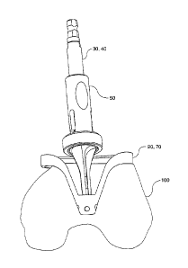
it may be
advantageous to replace only the damaged part of the joint in less extreme
cases.
[0008] In some cases, PFJ damage may be adequately addressed with a PFJ
artlu-oplasty, as opposed to a total knee replacement system. This type of
knee surgery is
less drastic than total knee replacement. It is designed for patients whose
main problems
involve only the patello-femoral part of the knee and is directed to providing
a smooth
sliding relationship between the femur and the patella. The surface of the
femur on which
the patella slides is referred to as the trochlear groove. The trochlear
groove is the
indentation or groove located between the medial and lateral condylar surfaces
at the distal
end of the femur.
[0009] In prior art PFJ prosthetic systems, a prosthetic patellar bearing
surface is
introduced. The prosthetic bearing surface typically includes an anchoring
portion for
2
CA 02609144 2007-11-20
WO 2006/127486 PCT/US2006/019512
receiving natural patellar remnants. As a result, the final patellar structure
includes a
posterior prosthetic bearing surface and an anterior natural patella surface.
The anterior
natural patella surface typically retains the connective tissue that connects
the patella to the
quadriceps and tibia.
[0010] In order to achieve adequate translational movement of the
prosthetic patellar
bearing surface, particularly in the presence of damage to the trochlear
groove, a cooperating
prosthetic femur implant is typically affixed onto the end of the femur. The
prosthetic
femur implant in most cases includes a bearing surface that is specially
adapted to receive
the prosthetic patellar bearing surface to ensure reliable travel during
flexion movement.
[0011] Such prior art systems, however, are typically highly artificial
systems that
employ unnatural patello-femoral tracking or movement of the patella. One
drawback of
such systems is that they are not compatible with total knee replacement
systems. In many
cases, the PFJ system requires so significant an amount of bone removal as to
render
subsequent total knee replacement almost impossible.
[0012] More natural patellar devices employ a saddle-shaped design. The
saddle-
shaped design may be used with or without a femoral implant and is intended to
track the
within the natural trochlear groove.
[0013] There is a need, therefore, for a patello-femoral prosthesis
having the
advantages of more naturally tracking designs. There is a further need for a
femoral implant
that requires less bone removal for implantation.
Summary of the Invention
[0014] It is in view of the above problems that the present invention was
developed.
The invention is a patello-femoral joint implant and associated
instrumentation. The implant
resurfaces only the patello-femoral compartment of the knee and leaves the
rest of the knee
intact. The implant utilizes asymmetric components and a lateralized patellar
groove to
3
CA 02609144 2007-11-20
WO 2006/127486 PCT/US2006/019512
improve patellar tracking. The instrumentation allows the device to be
implanted through a
minimally invasive approach without extensive damage to the quadriceps
mechanism. A key
feature of the instrumentation system is the reaming system which allows for
reproducible
preparation of the trochlear region of the femur.
[0015] In one aspect of the invention, there is a system for preparing a
trochlear
region of a resected femur, the system comprising: a reamer guide, the reamer
guide having
a first arcuate portion, a second arcuate portion, a wall extending
therebetween, a protrusion
connected to the wall, at least one leg connected to one of the first arcuate
portion or the
second arcuate portion, and a distal tip portion connected to the leg; and a
reamer adapted to
rotatably connect to the distal tip portion of the reamer guide, the reamer
having a first end
portion, a second end portion, and at least one flute.
[0016] In another aspect of the invention, there is a patello-femoral
joint implant, the
implant comprising: an intracondylar notch portion, a proximal portion, and a
distal portion;
an upper surface extending from the intracondylar notch portion to the
proximal portion, the
upper surface having a medial portion and a lateral portion; a lateralized
groove forming a
curved outer surface in between the medial portion and the lateral portion; a
substantially
planar undersurface connected to the intracondylar notch portion and opposite
the upper
surface; at least one anterior peg connected to the substantially planar
undersurface; and at
least one distal peg (19) connected to the substantially planar undersurface
(16).
[0017] The invention has several advantages over prior devices and
techniques.
First, the implant has an asymmetric patellar track to provide better coverage
of the anterior
femur. The patellar track is lateralized to improve patellar tracking. The
natural patella
tracks lateral to medial as the knee flexes. Other devices generate this
tracking by rotating
the component and angling a straight patellar track. This can lead to the
patella moving too
4
CA 02609144 2007-11-20
WO 2006/127486 PCT/US2006/019512
far medial and lead to unfavorable tracking. The implant assures proper
central placement
of the patella in flexion.
[0018] Second, the instrumentation is designed for a minimally invasive
approach.
A minimally invasive approach provides several advantages to the patient,
including, among
other things, a shorter recovery period and reduced pain. The instruments for
all prior art
systems are not designed for a minimally invasive approach.
[0019] Third, the reaming system allows for reproducible trochlear
preparation and
aids in proper alignment of the implant. Most prior art systems are not
precise and rely on
some kind of free-hand preparation of the trochlear and/or anterior region of
the femur.
This leads to inaccurate preparation and mal-rotation of the components, which
is the
second leading cause of failure in these devices. The reaming system also
allows for a more
uniform cement mantle than hand preparation would allow which may help prevent
cement
fatigue and loosening.
[0020] Further features, aspects, and advantages of the present
invention, as well as
the structure and operation of various embodiments of the present invention,
are described in
detail below with reference to the accompanying drawings.
Brief Description of the Drawings
[0021] The accompanying drawings, which are incorporated in and form a
part of the
specification, illustrate the embodiments of the present invention and
together with the
description, serve to explain the principles of the invention. In the
drawings:
[0022] FIG. 1 is a posterior view of a patello-femoral implant;
[0023] FIG. 2 is a distal view of the patello-femoral implant shown in
FIG. 1;
[0024] FIG. 3 is a proximal view of the patello-femoral implant shown in
FIG. 1;
[0025] FIG. 4 is a anterior view of the patello-femoral implant shown in
FIG. 1;
CA 02609144 2007-11-20
WO 2006/127486 PCT/US2006/019512
[0026] FIG. 5 is a side view of a first embodiment of a reamer guide and a
first
embodiment of a reamer;
[0027] FIG. 6 is a perspective top view of the reamer guide and the reamer
in a first
position;
[0028] FIG. 7 is a perspective top view of the reamer guide and the reamer
in a
second position;
[0029] FIG. 8 is a perspective top view of the reamer guide and the reamer
in a third
position;
[0030] FIG. 9 is an exploded view of a first embodiment of a reamer sleeve
and a
second embodiment of the reamer;
[0031] FIG. 10 is a front view of a reamer sleeve in a second embodiment;
[0032] FIG. 11 is a front view of the first embodiment of the reamer
sleeve;
[0033] FIG. 12 is a front view of a reamer sleeve in a third embodiment;
[0034] FIG. 13 is a perspective front view of a second embodiment of the
reamer
guide;
[0035] FIG. 14 is a perspective top view of the reamer guide shown in FIG.
13;
[0036] FIG. 15 is a front perspective view of a femur and a femoral
extramedullary
alignment rod;
[0037] FIG. 16 is a front perspective view of the femur and a drill guide;
[0038] FIG. 17 is a front perspective view of the femur and a drill;
[0039] FIG. 18 is a front perspective view of the femur and an
intramedullary rod;
[0040] FIG. 19 is a front perspective view of the femur and an anterior
cutting guide;
[0041] FIG. 20 is a front view illustrating the femur, a tibia, and an
extramedullary
up rod;
6
CA 02609144 2007-11-20
WO 2006/127486 PCT/US2006/019512
[0042] FIG. 21 is a side view of the femur, the tibia, and an
extramedullary guide
assembly;
[0043] FIG. 22 is a front view illustrating the femur and a handle;
[0044] FIG. 23 is top view of the femur and an alignment bar assembly;
[0045] FIG. 24 is a front view of the femur and a first punch;
[0046] FIG. 25 is a front view of the femur and an extramedullary
alignment device;
[0047] FIG. 26 is side view of the femur and a locking handle;
[0048] FIG. 27 is a top perspective view of the femur and a drill guide;
[0049] FIG. 28 is a front view of the femur, drill guide, bone pins, and
bone pin
insertion tool;
[0050] FIG. 29 is a front view of the femur, drill guide, and a second pin
punch;
[0051] FIG. 30 is a front perspective view of the femur, the drill guide,
and
temporary fixation pins;
[0052] FIG. 31 is a top perspective view of the femur and a second
embodiment of a
reamer guide;
[0053] FIG. 32 is a front perspective view of the femur, the second
embodiment of
the reamer guide, and a plurality of fixation devices;
[0054] FIG. 33 is a side view of the femur, the second embodiment of the
reamer
guide, and the second embodiment of the reamer;
[0055] FIG. 34 is a front view of the components shown in FIG. 33;
[0056] FIG. 35 is a top perspective view of the femur, a depth gauge, and
a handle;
[0057] FIG. 36 is a front view of the femur, the drill guide, peg drill,
and fixation
pegs;
[0058] FIG. 37 is a side perspective illustrating the femur and a trial;
and
[0059] FIG. 38 is a side view of the femur, the implant, and an impactor.
7
CA 02609144 2007-11-20
WO 2006/127486 PCT/US2006/019512
Detailed Description of the Preferred Embodiments
[0060] Referring to the accompanying drawings in which like reference
numbers
indicate like elements, FIGS. 1-4 illustrate a patello-femoral implant 10. The
implant 10 may
be made from any biocompatible material. As examples, the implant 10 may be
made from
cobalt chromium, stainless steel, titanium, oxidized zirconium, other metal
alloys, standard
polyethylene, cross-linked polyethylene, ultra high molecular weight plastic,
other plastics, or
a composite material. The implant 10 resurfaces the patello-femoral region of
the knee to
alleviate the pain from patello-femoral arthritis. The implant 10 may be used
with an
unresurfaced patella (i.e., natural patella) or with any resurfaced patella
implant, such as a
dome-shaped patella implant or an oval patella implant.
[0061] The implant 10 includes an intracondylar notch portion 11, a medial
portion
13, an upper surface 14, and a lateral portion 17. The implant 10 also
includes a proximal
portion or region 96 and a distal portion or region 98. The upper surface, or
implant anterior
surface, 14 extends from the intracondylar notch portion 11 to the proximal
portion 96. The
intracondylar notch portion 11 is constructed and arranged to provide a smooth
transition to
the femoral condyles. In some embodiments, the medial portion 13 and the
lateral portion
17 are shaped to provide maximum bone coverage of the anterior femur. The
lateral portion
17 has an increased thickness in the proximal region 96 to prevent patellar
subluxation. In
some embodiments, the medial portion 13 has less material than the lateral
portion 17 in the
proximal region 96. This may be done for several reasons. For example, the
medial portion
13 may have less material in order to decrease the overall size of the implant
10, to reduce
the weight of the implant 10, or to allow the implant 10 to achieve a better
fit.
[0062] As best seen in FIG. 4, the implant 10 has a lateralized patello-
femoral
groove 12. In the embodiment depicted in FIG. 4, the black line represents a
central area or
average location of the groove 12, but those of ordinary skill in the art
would understand
8
CA 02609144 2007-11-20
WO 2006/127486 PCT/US2006/019512
that the black line is merely representative of location and the actual shape
of the groove is
such that an unresurfaced or resurfaced patella may track within it. The
groove 12 forms a
curved outer surface, or bearing surface, in which the patella tracks. The
groove 12 is
lateralized in the proximal region 96 to allow the patella to track normally,
regardless of
whether the patella has been resurfaced or not. The patella is lateralized in
extension and
transitions to the intracondylar notch portion 11 in flexion as it moves in
the groove 12.
[0063] The implant 10 includes a substantially planar anterior under-
surface 16 for
placement on a generally flat or planar anterior cut 190 (best seen in FIG.
31) on a femur
100. The substantially planar anterior under-surface, or implant posterior
surface, 16 is
located opposite the upper surface 14. The substantially planar anterior under-
surface 16
may be parallel to the coronal plane, or the anterior under-surface 16 may be
sloped to
prevent stress shielding. In other words, some embodiments of the implant 10
are sloped, or
shaped, such that a force upon the groove 12 tends to push the implant 10
towards the
anatomic axis of the femur and not along the anatomic axis. The anterior under-
surface 16
may be sloped in the range from about one degree to about ten degrees relative
to the
coronal plane in order to prevent stress shielding. In the embodiment depicted
in FIG. 1, the
anterior under-surface 16 slopes at about three degrees.
[0064] In some embodiments, the anterior under-surface 16 includes
features to
enhance its use with bone cement. For example, the anterior under-surface 16
may be grit
blasted to roughen the surface or may include indentations, pockets,
depressions, or dimples
15. The dimples 15 may be elongated cavities, circular depressions,
rectangular voids,
triangular cavities, or any other shape of indentation.
[0065] The implant 10 includes anterior anchors or pegs 18 and, in some
embodiments, distal peg or pegs 19. In the embodiment depicted in FIGS. 1 and
3, the
implant 10 has three anterior pegs 18 and one distal peg 19, but those of
ordinary skill in the
9
CA 02609144 2007-11-20
WO 2006/127486 PCT/US2006/019512
art would understand that a greater or lesser number of pegs may be used. As
examples, in
some embodiments, the distal peg 19 may be omitted entirely or the implant 10
may include
a plurality of distal pegs 19. The distal peg 19 may be angled relative to the
anterior pegs 18
to enhance cement fixation. In the embodiment depicted in FIG. 1, the distal
peg 19 is
oblique relative to the anterior pegs 18. The oblique angle of the distal peg
19 allows for a
snap fit of the implant 10.
[0066] FIGS. 5-8 illustrate a first embodiment of a reamer guide 20 and a
first
embodiment of a reamer 30. The reamer guide 20 and the reamer 30 reproducibly
ream the
trochlear region of the femur 100 to allow for installation of the implant 10.
The reamer
guide 20 is small and medially biased to allow for insertion into a small
incision, such as is
used in minimally invasive surgery (MIS). The reamer guide 20 may be adapted
for use on
a left knee, a right knee, or either knee. In the depicted embodiments, the
reamer guide 20 is
adapted for use on either knee. The reamer guide 20 is adapted to attach to
the resected
anterior surface 190. The reamer guide 20 further includes a first arcuate
portion 21, a
second arcuate portion 23, a wall 25, a protrusion 27, at least one locating
member 24, a leg
28, a distal tip portion 26, and a nearly-spherical indentation or a more-than-
hemispherical
depression 22. The wall 25 is connected to the first arcuate portion 21, the
second arcuate
portion 23, and the protrusion 27. The shape of the wall 25 is constructed and
arranged such
that it follows an outline of the underside of the implant 10. The locating
member 24
extends from an underside of the protrusion 27. The leg 28 is connected to the
first arcuate
portion 21 and the second arcuate portion 23. In the depicted embodiments, the
leg 28 is V-
shaped but other shapes may be used. The distal tip portion 26 is connected to
the leg 28,
and the more-than-hemispherical depression 22 is located in the distal tip
portion 26.
[00671 In some embodiments, the first and second arcuate portions 21, 23
are sized
and located to limit the amount of medial and lateral resection. As such, the
first arcuate
CA 02609144 2007-11-20
WO 2006/127486 PCT/US2006/019512
portion 21, the second arcuate portion 23, and the wall 25 control the shape
and depth of
resection of the trochlear region. In other embodiments, the first and second
arcuate
portions 21, 23 are merely structural members that connect the leg 28 to the
other
components of the reamer guide 20, and, therefore, the user must exercise
caution to ensure
that the trochlear region is not over-resected medially or laterally.
[0068] The reamer 30, alternatively termed a mill, is a somewhat
hourglass shaped
cutting instrument. In some embodiments, the reamer 30 is adapted for use with
a standard
drill. The reamer 30 has a connector 31 that is housed by a more-than-
hemispherical
depression or over-hemispherical depression 22 in the reamer guide 20. In the
depicted
embodiments, the connector 31 is spherical or has a ball nose shape. The
distal part of the
reamer 30 is housed in the hemispherical indentation, and the proximal part is
leaned against
the reamer guide 20 and slid medio-laterally to ream the trochlear region. The
reamer 30
also includes a first end portion 32, a bearing 34, a second end portion 36,
and at least one
tooth or flute 38. As the bearing 34 decreases in size, the deeper the reamer
30 will ream the
trochlear region, and as the bearing increases in size, the shallower the
reamer 30 will ream
the trochlear region. Alternatively, the teeth 38 may increase in size such
that additional
bone is reamed. Thus, the reamer 30 may be available in different versions
with a
correspondingly sized bearing 34 or teeth 38 such that a particular size of
reamer is chosen
according to the desired amount of reaming. A kit of differently sized reamers
and a reamer
guide may be provided.
[0069] In use, the reamer guide 20 is mounted to the distal end of a
resected femur.
A user inserts the distal tip 31 into the over-hemispherical depression 22.
The user levers or
pivots the reamer 30 downward onto the reamer guide 20, resecting bone as the
reamer 30 is
pivoted. The reamer 30 is pivoted until the bearing 34 rides on or rotates
against the wall
11
CA 02609144 2007-11-20
WO 2006/127486 PCT/US2006/019512
25. The reamer 30 is then moved medial-to-lateral, or vice versa, to prepare
the trochlear
region.
[0070] FIG. 9 illustrates a second embodiment of the reamer, generally
indicated by
reference numeral 40, and a first embodiment of a reamer sleeve 50. The reamer
40 and the
sleeve 50 are assembled together to form a reamer assembly 45. The reamer 40
may also be
referred to as a mill. The reamer 40 has a shaft 42, at least one tooth or
flute 44, a first end
portion 47, and a second end portion 48. Optionally, the reamer 40 may also
include a
groove or channel 46 located on the shaft 42. In some embodiments, a profile
of the flute 44
is shaped to match the underside of the implant 10. In the embodiment depicted
in FIG. 9,
the reamer 40 has four flutes 44, but those of ordinary skill in the art would
understand that
a greater or lesser number of flutes may be used. The flute 44 has a relief
angle A which
ranges from about five degrees to about thirty-five degrees. In the embodiment
depicted in
FIG. 9, the relief angle A is about twenty degrees. The second end portion 48
may have any
number of shapes and is adapted to rotatably connect to the reamer guide. In
the
embodiment depicted in FIG. 9, the second end portion 48 terminates in a
connector 49
which has a substantially hemispherical shape. The first end portion 47 also
may have any
number of shapes but in the depicted embodiment has three circumferentially
spaced, planar
surfaces 43 adapted for use or engagement with a drill chuck (not shown).
[0071] In some embodiments, the reamer guide 20 and the reamer 30, 40 may
be one
piece. For example, the reamer guide 20 may include a rotatable bearing, such
as a spherical
bearing, and the reamer 30, 40 may rotate within this bearing.
[0072] The reamer 40 is adapted to receive the sleeve 50. The sleeve 50
includes a
main body 52, at least one arm 54, and a bearing or platform 56. The sleeve 50
may be
made from a metal, such as stainless steel, a plastic, such as an acetal
copolymer, or a
composite material. Optionally, the main body 52 may include a grip portion
58. The grip
12
CA 02609144 2007-11-20
WO 2006/127486 PCT/US2006/019512
portion 58 provides a convenient place for the user to place his or her thumb
and forefinger.
The reamer sleeve 50 is used to control the depth that the reamer 40 engages
the trochlear
region. In other words, the sleeve 50 controls the amount of resection. This
is
accomplished by appropriately sizing the platform 56. As the platform 56
decreases in size,
the more material is resected. The arm 54 is adapted to engage the groove 46
such that the
sleeve 50 is removably attached or temporarily affixed to the shaft 42.
Although the arm 54
engages the groove 46, the sleeve 50 is still free to rotate relative to the
reamer 40.
[0073] While the embodiment depicted in FIG. 9 includes the arm 54, those
skilled
in the art would understand that other methods of removably attaching the
sleeve 50 to the
reamer 40 may be used. For example, a C-clip may be used to engage the groove
46 and
connect the sleeve 50 to the reamer 40.
[0074] FIGS. 10 -12 illustrate alternative embodiments of the reamer
sleeve. FIG.
illustrates an undersized reamer sleeve 62. As used herein, the term
"undersized" refers
to the degree or volume of bone resection, and the undersized reamer sleeve 62
has a
platform 63 with a diameter larger than that of the standard size platform 56.
FIG. 11
illustrates the standard reamer sleeve 50. The standard reamer sleeve 50
includes the
standard size platform 56. FIG. 12 illustrates an oversized reamer sleeve 64.
As used
herein, the term "oversized" refers to the degree or volume of bone resection,
and the
oversized reamer sleeve 64 has a platform 65 with a diameter smaller than that
of the
standard size platform 56.
[0075] FIGS. 13 and 14 illustrate a second embodiment of the reamer
guide,
generally indicated by reference numeral 70. The reamer guide 70 may be used
with either
the first embodiment of the reamer 30 or the second embodiment of the reamer
40. The
reamer guide 70 includes a first arcuate portion 71, a second arcuate portion
73, a wall 75, a
protrusion 77, at least one slot 79, a first leg 74, a second leg 78, a distal
tip portion 76, and
13
CA 02609144 2007-11-20
WO 2006/127486 PCT/US2006/019512
a more-than-hemispherical depression or cup 72. In some embodiments, the
reamer guide
70 includes a lip 80. The lip 80 and the wall 75 are shaped to match the
curvature of the
implant 10. The cup 72 is adapted to receive the connector 49 of the reamer
40. The cup 72
is located at a fixed depth and is over hemispherical so the tip portion 48 of
the reamer 40
cannot pop out or easily slide out when the trochlear region is being reamed.
In some
embodiments, the reamer guide 70 includes a first hole 82, a second hole 84, a
third hole 86,
a fourth hole 88, a fifth hole 90, and a sixth hole 92.
[0076] The reamer guide 70 may be adapted for use with a left knee or a
right knee.
As such, the reamer guide 70 would limit the movement of the reamer 30, 40 so
that the
appropriate amount of the trochlear region is removed. In the case of the
universal reamer
guide, the user must be careful not to overly resect the trochlear region and
caution must be
exercised to limit the amount of resected bone.
[0077] In use, the reamer guide 70 is mounted to a distal end of a
resected femur. A
user inserts the connector 49 into the cup 72. The user levers or pivots the
reamer 40
downward onto the reamer guide 70, resecting bone as the reamer 40 is pivoted.
The reamer
40 is pivoted until the bearing or platform 56, 61, 63, 65 rides on or rotates
against the lip 80
of the wall 75. The reamer 40 is then moved medial-to-lateral, or vice versa,
to prepare the
trochlear region.
[0078] FIGS. 15-38 illustrate preparation of the femur 100 and
installation of the
implant 10. FIG. 15 illustrates the femur 100 and a femoral extramedullary
alignment rod
102. As best seen in FIG. 16, an intramedullary drill guide 104 is connected
to the femoral
extramedullary alignment rod 102. In the depicted embodiment, the center-to-
center
distance between the holes is about twenty-six millimeters. FIG. 17
illustrates a drill 106
being inserted into the drill guide 104 in order to drill through cortical
bone to the
intramedullary canal. After a hole is drilled into the intramedullary canal,
FIG. 18 illustrates
14
CA 02609144 2007-11-20
WO 2006/127486 PCT/US2006/019512
an intramedullary rod 108 being inserted into the canal. The rod 108 may be
thin to reduce
damage to the intramedullary canal during insertion. As best seen in FIG. 19,
an anterior
cutting guide 110 is connected to the intramedullary rod 108 and placed next
to the end of
the femur 100. The cutting guide 110 fits over the thin intramedullary rod 108
affixed
through the intramedullary canal of the femur 100. The cutting guide 110 is
medially biased
to fit into a minimally invasive incision. The anterior cutting guide 110 is
designed with
adjustable height so that a user can achieve the proper resection level.
[0079] After the cutting guide 110 is placed over the rod 108, there are
two methods
which may be used to orient the anterior cutting guide 110. The methods may be
used
separately or in combination. For example, both methods may be performed to
confirm the
results of whichever method was performed first. In a first method, best seen
in FIGS. 20
and 21, an extramedullary guide assembly 130 is attached to a tibia 112. The
extramedullary guide assembly 130 includes an extramedullary up rod 114, a
tibial
extramedullary guide platform 132, an extramedullary tibial down rod 134, and
an ankle
clamp 136. The ankle clamp 136 is attached to the distal portion of the tibia
112, and the
extramedullary up rod 114 is extended upwardly until the extramedullary guide
platform
132 contacts a bottom portion of the anterior cutting guide 110. The platform
132 properly
orients the cutting guide 110, and thereafter the cutting guide 110 can be
adjusted to achieve
the proper height for the resection plane.
[0080] In a second method to orient the cutting guide 110, best seen in
FIGS. 22-24,
a handle 116 is attached to the anterior cutting guide 110 and an alignment
bar assembly 120
is attached to the handle 116. As an example only, the handle 116 may be a
quick connect
handle that includes a quick release mechanism. The alignment bar assembly 120
includes a
grip 122, a first alignment bar rod 124, a second alignment bar rod 126, and
an alignment
bar clip 128. The first alignment bar rod 124 is positioned such that it is
parallel to the
CA 02609144 2007-11-20
WO 2006/127486 PCT/US2006/019512
epicondyles. The second alignment bar rod 126 is positioned such that it is
parallel to the
mechanical axis of the femur. The grip 122 may be connected to the handle 116
through the
use of the alignment bar clip 128. Once the first alignment bar rod 124 and
the second
alignment bar rod 126, and thereafter the cutting guide 110 can be adjusted to
achieve the
proper height for the resection plane.
[0081] Once the anterior cutting guide 110 is properly positioned, the
anterior
cutting guide 110 is pinned into place. FIG. 24 illustrates the anterior guide
110 being
pinned into place using a first punch 140 and a first pin 142. One or more
first pins 142
may be used to pin in place the anterior guide 110.
[0082] After the anterior cutting guide 110 is pinned, an anterior
stylus 144 is
connected to the anterior cutting guide 110, as best seen in FIG. 25, to set
the height of the
resection plane. A knob 111 of the anterior cutting guide 110 is rotated until
a tip portion
148 of the anterior stylus 144 rests upon the femur 100. Once the proper
resection level is
set, the height of a saw guide 118 is locked into place. FIG. 26 illustrates
an exemplary
method of temporarily fixating the saw guide 118 wherein a locking handle or
screwdriver
146 is used to rotate a set screw (not shown) in the anterior cutting guide
110. Thereafter,
the anterior portion of the femur 100 is resected to achieve the generally
planar surface 190
(best seen in FIG. 31). In the depicted embodiment, the anterior cut is made
in three degrees
of flexion to allow proper orientation of the implant 10.
1 [0083] After the anterior cut is made, a patello-femoral drill guide
150 mounted to
the femur 100. FIGS. 27-30 illustrate the patello-femoral drill guide 150. The
patello-
femoral drill guide 150 includes a first pin hole 152, a second pin hole 154,
a third pin hole
156, a fourth pin hole 158, a first drill guide hole 160, a second drill guide
hole 162, a third
drill guide hole 164, a fourth drill guide hole 166, and a receptacle or
receiver 168.
16
CA 02609144 2007-11-20
WO 2006/127486 PCT/US2006/019512
10084] Once the patello-femoral drill guide 150 is placed on the femur
100, it is
pinned into place. In the embodiment depicted in FIG. 28, one or more second
pins 170 are
driven into place through the use of a first pin driver 172. Second pins 170
may be headed
or non-headed. In the depicted embodiments, second pins 170 are placed in
second pin hole
154 and third pin hole 156. Optionally, additional fixation pins may be used
to temporarily
affix the patello-femoral drill guide 150 to the femur 100. For example, as
best seen in FIG.
29, a second pin driver 176 may be used to install a third fixation pin 178.
Thereafter, an
outline of the trochlear region of the implant 10 is traced on the cartilage
and/or bone from
the patello-femoral drill guide 150. The outline may be achieved through the
use of a
cauterizer or methylene blue. For example, a user may trace the edges of the
patello-femoral
drill guide 150 with the cauterizer to mark the cartilage and/or bone.
Additionally, the
patello-femoral drill guide 150 has an indicator 151, such as a line or a
triangle. A mark
153, such as a line or "X," must be placed on the femur 100 in order to
reinstall the patello-
femoral drill guide 150 at a later time.
[0085] Alternatively, the patello-femoral drill guide 150 is not fixed to
the femur
100 when outlining or marking the trochlear region. Instead, the patello-
femoral drill guide
150 is merely held in place, such as through the use of the handle 116, and
the cartilage
and/or bone is marked by tracing the outline of the patello-femoral drill
guide 150.
[0086] After the trochlear region is outlined or marked, the patello-
femoral drill
guide 150 is removed and a reamer guide, such as the first embodiment 20 or
the second
embodiment 70, is temporarily affixed to the anterior cut surface of the femur
100. In the
embodiment depicted in FIGS. 31 and 32, the non-headed pins 170 have not been
removed.
As such, the reamer guide 20 is placed over the pins 170, or the reamer guide
70 is slid into
place such that the non-head pins 170 enter the slots 79. In some embodiments,
the second
pins 170 are headed, the reamer guide 70 is slid into place such that the pins
170 enter the
17
CA 02609144 2007-11-20
WO 2006/127486 PCT/US2006/019512
slots 79, and the headed pins are tapped downwardly to lock the reamer guide
70 in place.
The reamer guide 20, 70 contacts the resection plane 190 and is slid back
until a leg 28, 74,
78 contacts the intracondylar notch. Thereafter, the reamer guide 20, 70 is
pinned to the
femur 100. In the embodiment depicted in FIG. 32, a third pin 208 is inserted
into the hole
82 and a fourth pin 210 is inserted into the hole 92, but other pin and hole
combinations may
be used. Although three pins are shown near the distal tip portion 26, 76,
only one pin may
be used as the axis of each hole 88, 90, 92 is coplanar with the other holes.
[0087] After the reamer guide 20, 70 is pinned, the trochlear region is
reamed. In
the embodiment depicted in FIGS. 33 and 34, the reamer 40 and the sleeve 50
are used to
resect the trochlear region. The reamer 40, or alternatively the reamer 30, is
moved from
side-to-side to ream the trochlear region. If the reamer guide is universal,
the user must
exercise care to only ream up until the outline or mark on the cartilage
and/or bone.
However, if the reamer guide is constructed and arranged for use only on a
single side, the
reamer 30, 40 may be moved from side-to-side until the reamer contacts one of
the arcuate
portions 21, 23, 71, 73.
[0088] In some methods, the trochlear region is not reamed with a reamer
but is
merely prepared with a rasp, osteotome, or other sharp tool.
[0089] After the trochlear region is reamed, it is necessary to verify
the depth of the
reaming. As best seen in FIG. 35, a depth gauge 242 is used to check the depth
of the
resection. The depth gauge 242 may be left-hand, right-hand, or universal. In
the depicted
embodiment, the depth gauge 242 is designed for use only a single side (e.g.,
left-hand).
The depth gauge 242 may be adapted for a particular size of implant or it may
be designed
for use across an entire series of implants. In the depicted embodiments, the
implants are all
of the same thickness, regardless of size. In other words, implants may grow
in width and
length as they increase in size but do not increase in thickness, and,
therefore, it is possible
18
CA 02609144 2007-11-20
WO 2006/127486 PCT/US2006/019512
to have one depth gauge per side for a series of implants. When checking the
depth of the
ream, it is important that there is a smooth transition between the cartilage
in the
intracondylar notch and the depth gauge 242. If the depth gauge 242 is raised
above the
remainder of the trochlear region, it will be necessary to ream to an
additional depth. This is
accomplished by reaming with a reamer having a smaller bearing than previously
used, by
reaming with a reamer having a larger flute than previously used, or by
reaming with a
sleeve that has a smaller diameter platform than was previously used. However,
if the depth
gauge is at or below the remainder of the trochlear region, the depth of the
ream is
sufficient. In some embodiments, the handle 116 is attached to the depth gauge
242 for ease
of use and manipulation.
[0090] After the depth of the ream is verified, the pins and the reamer
guide are
removed, and the patello-femoral drill guide 150 is reinstalled for drilling
of the anterior
and/or distal peg holes. FIG. 36 illustrates reattachment of the drill guide
150. The
intracondylar notch portion of the patello-femoral drill guide 150 is placed
in the reamed
trochlear region, and the mark 153 on the femur 100 is aligned with the
indicator 151 on the
patello-femoral drill guide 150. Thereafter, the patello-femoral drill guide
150 is pinned
into place through the use of one or more fixation pins. After the patello-
femoral drill guide
150 is located and pinned, one of the holes is drilled. A drill guide
alignment post 252 is
placed in the drilled hole to stabilize the construct. Then, a second hole is
drilled, and
another drill guide alignment post 254 is placed in the second drilled hole
for additional
stability. Optionally, additional alignment posts may be used for stability.
Thereafter, the
remaining holes are drilled. The patello-femoral drill guide 150 and the
fixation pins are
then removed.
[0091] FIG. 37 illustrates a trial 300 being placed on the femur 100. A
user utilizes
the trial 300 to test patellar tracking as well as the fit and articulation of
the revised knee.
19
CA 02609144 2007-11-20
WO 2006/127486 PCT/US2006/019512
The user may need to adjust the size of the trial 300 or its location to
perfect the revised
knee. The trial 300 generally snaps into place, but an impactor may be used to
positively
locate the trial 300.
[0092] FIG. 38 illustrates the implant 10 being installed with the use of
an impactor
310. The impactor 310 includes an impactor handle 312 and a bumper 314. In
some
embodiments, bone cement may be applied to the underside of the implant 10
prior to
installation.
[0093] The invention also includes a method of installing an asymmetric
patello-
femoral implant. The method includes the step of: (1) resecting a femur; (2)
reaming a
distal portion of the femur; (3) attaching a patello-femoral drill guide to
the femur; and (4)
installing the implant. The step of resecting the femur may include the step
of attaching an
anterior cutting guide to the femur. The step of reaming the distal portion of
the femur may
include the steps of attaching a reamer guide to the femur, rotatably
connecting a reamer to
the reamer guide, and moving the reamer relative to the reamer guide in order
to ream the
distal end of the femur. Further, the step of reaming the distal portion of
the femur may
include attaching a reamer sleeve to a reamer. Additionally, the step of
reaming the distal
portion of the femur may include verifying the depth of the ream with a depth
gauge. The
step of attaching a patello-femoral drill guide to the femur may include the
steps of marking
an outline of the patello-femoral drill guide on cartilage and/or bone and the
step of placing
a mark on the femur. Further, the step of attaching a patello-femoral drill
guide to the femur
may include the step of drilling a plurality of holes in the femur. The step
of installing the
implant may include the steps of placing bone cement on the implant or the
femur, placing
pegs of the implant into drilled holes in the femur, and striking an impactor
to seat the
implant on the femur.
CA 02609144 2007-11-20
WO 2006/127486 PCT/US2006/019512
[0094] A kit may be provided. The kit may include one or more of the
following
items: a reamer, a reamer guide, a depth gauge, a trial, and an implant.
Optionally, the kit
may also include one or more standard reamer sleeves, oversized reamer
sleeves, or
undersized reamer sleeves.
[0095] In view of the foregoing, it will be seen that the several
advantages of the
invention are achieved and attained.
[0096] The embodiments were chosen and described in order to best explain
the
principles of the invention and its practical application to thereby enable
others skilled in the
art to best utilize the invention in various embodiments and with various
modifications as are
suited to the particular use contemplated.
[0097] As various modifications could be made in the constructions and
methods
herein described and illustrated without departing from the scope of the
invention, it is
intended that all matter contained in the foregoing description or shown in
the accompanying
drawings shall be interpreted as illustrative rather than limiting. For
example, while FIGS. 33
and 34 illustrate the use of a reamer in combination with a reamer sleeve, it
should be
understood that reamers without sleeves are equally acceptable. Thus, the
breadth and scope
of the present invention should not be limited by any of the above-described
exemplary
embodiments, but should be defined only in accordance with the following
claims appended
hereto and their equivalents.
21