Note: Descriptions are shown in the official language in which they were submitted.
CA 02614259 2007-12-13
BIOPSY SAMPLE STORAGE
BACKGROUND
[001] Some embodiments of the present invention relate in general to biopsy
devices,
and more particularly to biopsy devices having the capability to store
multiple
tissue samples, such as in a spaced-apart, sequenced manner, within a portion
of
the biopsy device.
[002] When a suspicious tissue mass is discovered in a patient's breast or in
another
area through examination, ultrasound, MRI, X-ray imaging or the like, it may
be
necessary to perform a biopsy procedure to remove one or more samples of that
tissue in order to determine whether the mass contains cancerous cells. A
biopsy
may be performed using an open or percutaneous method. Medical devices for
obtaining tissue samples for subsequent sampling and/or testing are known in
the
biopsy art. For instance, a biopsy instrument now marketed under the tradename
MAMMOTOME is commercially available from Ethicon Endo-Surgery, Inc. for
use in obtaining breast biopsy samples.
[003] An open biopsy may be performed by making a large incision in the breast
and
removing either the entire mass, called an excisional biopsy, or a substantial
portion of it, known as an incisional biopsy. An open biopsy is a surgical
procedure that may be done as an outpatient procedure in a hospital or a
surgical
center, and may involve a high cost and a high level of trauma to the patient.
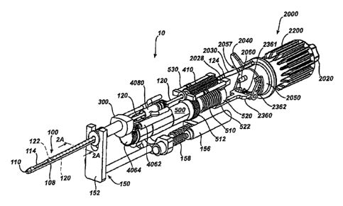
Open biopsy may carry relatively higher risk of infection and bleeding than
does
percutaneous biopsy, and the disfigurement that may result from an open biopsy
may make it difficult to read future mammograms. Further, the aesthetic
considerations of the patient might make open biopsy even less appealing due
to
the potential risk of disfigurement. Given that some biopsies show that the
- 1 -
CA 02614259 2015-03-25
suspicious tissue mass is not cancerous, the potential downsides of the open
biopsy procedure might render this method inappropriate in some cases.
[004] Percutaneous biopsy may be less invasive than open biopsy.
Percutaneous
biopsy may be performed using fine needle aspiration (FNA), core needle
biopsy, or otherwise. In FNA, a very thin needle may be used to withdraw fluid
and cells from the suspicious tissue mass. This method may be low-pain, so
low-pain that local anesthetic is not necessarily always used because the
application of it may be more painful than the FNA itself. However, in some
FNA procedures, only a small number of cells might be obtained through the
procedure, rendering it relatively less useful in some situations in analyzing
the
suspicious tissue and making an assessment of the progression of the cancer
less simple if the sample is found to be malignant.
[005] During some core needle biopsy procedures, a small tissue sample may
be
removed allowing for a pathological assessment of the tissue, including an
assessment of the progression of any cancerous cells that are found.
[006] The biopsy instrument marketed under the trade name MAMMOTOME
generally retrieves multiple core biopsy samples from one insertion into
breast
tissue with vacuum assistance. In particular, a cutter tube is extended into a
probe to cut tissue prolapsed into a side aperture under vacuum assistance,
and
then the cutter tube is fully retracted between cuts to extract the sample.
[007] With a device having a relatively long cutter travel, the rate of
sample taking
may be limited not only by the time required to rotate or reposition the probe
but also by the time needed to translate the cutter. As an alternative to
relatively "long stroke" biopsy devices, a "short stroke" biopsy device is
described in the following commonly assigned patent applications: US Patent
Application 10/676,944, entitled "Biopsy Instrument with Internal Specimen
- 2 -
CA 02614259 2017-02-10
Collection Mechanism," filed September 30, 2003 in the name of Hibner et al.,
published as U.S. Pub. No. 2005/0215921; and US Patent Application
10/732,843, entitled "Biopsy Device with Sample Tube," filed December 10,
2003 in the name of Cicenas et al, published as U.S. Pub. No. 2004/0153003.
The cutter can be cycled through a distance substantially equal to or slightly
greater than the distance across the side aperture, reducing the sample time.
[008] The following patent documents disclose various biopsy devices:
US 6,273,862 issued Aug.
14, 2001; US 6,231,522 issued May 15, 2001; US 6,228,055 issued May 8,
2001; US 6,120,462 issued September 19, 2000; US 6,086,544 issued July 11,
2000; US 6,077,230 issued June 20, 2000; US 6,017,316 issued Jan. 25, 2000;
US 6,007,497 issued Dec. 28, 1999; US 5,980,469 issued Nov. 9, 1999; US
5,964,716 issued Oct. 12, 1999; US 5,928,164 issued July 27, 1999; US
5,775,333 issued July 7, 1998; US 5,769,086 issued June 23, 1998; US
5,649,547 issued July 22, 1997; US 5,526,822 issued June 18, 1996; and US
Patent Application 2003/0199753 published Oct. 23, 2003 to Hibner et al. US
Patent 5,526,822, discloses a tissue sample cassette, including a rotary
sample
cassette that is belt driven. Other tissue sample storage devices are
disclosed in
US Patent Application Serial Number 10/953,395, entitled "Biopsy Device with
Sample Storage," filed September 29, 2004, published as U.S. Pub. No.
2006/0074343; and US Patent Application Serial Number 11/198,558 filed
August 8, 2005, entitled "Biopsy Device with Replaceable Probe and
Incorporating Vibration Insertion Assist and Static Vacuum Source Sample
Stacking Retrieval," published as U.S. Pub. No. 2007/0032741.
[009] While a variety of biopsy devices have been made and used, and a
variety of
tissue sample storage devices and techniques have been devised, it is believed
- 3 -
CA 02614259 2016-05-12
, ,
that no one prior to the inventors has made or used a biopsy system as
described
in the appended claims.
BRIEF DESCRIPTION OF THE DRAWINGS
[0010] While the specification concludes with claims particularly
pointing out and
distinctly claiming the present invention, it is believed the same will be
better
understood by reference to the following description, taken in conjunction
with
the accompanying drawings in which:
[0011] Figure 1 is a schematic illustration of a biopsy device
having a tissue sample
storage assembly according to one embodiment of the present invention;
[0012] Figure 2 is a schematic illustration of a biopsy device
having a tissue sample
storage assembly according to one embodiment of the present invention, with
portions of the biopsy device removed to illustrate internal components of the
device, and with the cutter in a retracted position.
[0013] Figure 2A is a schematic cross-section taken along section
2A-2A in Figure 2.
[0014] Figure 3 is a schematic illustration of a biopsy device
having a tissue sample
storage assembly according to one embodiment of the present invention, with
portions of the biopsy device removed to illustrate internal components of the
device, with the cutter advanced across a tissue receiving port.
- 4 -
CA 02614259 2007-12-13
[0015] Figure 4 is a schematic illustration of a biopsy device having a tissue
sample
storage assembly according to one embodiment of the present invention, with
portions of the biopsy device removed to illustrate internal components of the
device, and showing a tissue sample being deposited on the tissue sample
holder.
[0016] Figure 5 is a schematic illustration of a biopsy device having a tissue
sample
storage assembly according to one embodiment of the present invention, with
portions of the biopsy device removed to illustrate internal components of the
device, and showing retraction of the cutter and indexing of the tissue sample
holder.
[0017] Figure 6 provides an enlarged illustration of an exemplary tissue
storage
assembly.
[0018] Figure 7 provides an exploded view of components of the tissue storage
assembly of Figure 6.
[0019] Figure 8 provides a cross-sectional illustration of components of the
tissue
storage assembly of Figure 6.
[0020] Figure 9 is a schematic illustration of a manifold having radially
extending fins
and vacuum ports for conveying vacuum between adjacent fins.
[0021] Figure 10 illustrates an alternative embodiment of a tissue holder
[0022] Figure 11 illustrates another alternative embodiment of a tissue
holder.
[0023] Figure 11A illustrates a tissue holder of Figure 11 in a relatively
flat
configuration.
- 5 -
I
CA 02614259 2015-03-25
[0024] Figure 12 illustrates a tissue holder for holding tissue samples in
an end-to-end
configuration.
[0025] Figure 12A is a cross-sectional schematic illustration taken along
lines 12A-12A
in Figure 12.
[0026] Figure 12B is a sectional view taken along a portion of the length
of the tissue
holder shown in Figure 12.
[0027] Figure 13 is a schematic illustration of the distal portion of an
exemplary
cannula.
[0028] Figure 14 is a schematic illustration of the vacuum level provided
in a vacuum
lumen as a cutter is advanced and retracted in a cutter lumen relative to a
tissue
receiving aperture.
[0029] Figure 15 is a schematic illustration of a pneumatic control
configuration that
may be used with a biopsy device.
[0030] Figure 16 illustrates multiple control states that can be employed
in controlling a
biopsy device.
[0030a] The present disclosure relates to a biopsy device comprising: (a) a
cannula
extending distally from the biopsy device; (b) a tubular cutter translatable
with
respect to the cannula, the tubular cutter having a proximal end and a distal
end,
the cutter distal end for severing tissue received in the cannula; and (c) a
tissue
storage assembly disposed proximal of the tubular cutter, the tissue storage
assembly for receiving tissue samples severed by the tubular cutter, the
tissue
storage assembly comprising: (i) a generally transparent cover disposed at the
proximal end of the biopsy device, (ii) a rotatable member disposed at least
partially within the generally transparent cover wherein the rotatable member
- 6 -
CA 02614259 2015-03-25
comprises a manifold for directing vacuum to a portion of the tissue holder,
and
(iii) at least one tissue holder releasably carried by the rotatable member,
the
tissue holder adapted to receive a plurality of tissue samples.
[0030b] Also described is a tissue sample storage assembly for use with a
biopsy device,
the tissue sample storage assembly comprising: (a) a removable cover; (b) a
rotatable manifold disposed at least partially within the cover, the manifold
having at least one vacuum passageway; and (c) at least one tissue sample
strip
disposed on an outer surface of the manifold and extending about at least a
portion of the outer surface of the manifold.
[0030c] The
disclosure also relates to a biopsy device comprising: (a) a cannula
extending distally from the biopsy device; (b) a cutter movable with respect
to
the cannula, wherein the cutter is operable to sever tissue received in the
cannula;
and (c) a tissue storage assembly supported by the biopsy device for receiving
tissue samples severed by the cutter, the tissue storage assembly comprising:
(i) a
rotatable member disposed proximally of the cannula, and (ii) a plurality of
tissue
sample holders releasably carried by the rotatable member, wherein each tissue
sample holder is configured to receive at least one tissue sample severed by
the
cutter, wherein each tissue sample holder is removable from the rotatable
member while the rotatable member is supported on the biopsy device.
[0030d] Another aspect of the disclosure relates to a biopsy device
comprising: (a) a
cannula having a lateral tissue receiving opening; (b) a cutter movable with
respect to the tissue receiving opening to sever tissue received therein; (c)
a
tissue storage assembly comprising: (i) a manifold supported by the biopsy
device proximal of the cannula, and (ii) a plurality of removable tissue
sample
holders releasably carried by the manifold, each tissue sample holder being
independently removable from the manifold; and (d) a source of vacuum,
- 6a -
CA 02614259 2015-03-25
wherein the source of vacuum provides vacuum to the tissue sample holders
through the manifold.
[0030e] Another aspect relates to a biopsy device comprising: (a) a probe
having a body,
a cannula extending distally from the body, a tubular cutter movable with
respect
to the cannula for severing tissue received in the cannula, and a tissue
storage
assembly; and (b) a reusable portion, wherein the reusable portion is operable
to
provide motion of the cutter; and wherein the probe and reusable portion are
configured to be releasably joined together to provide a handpiece; and
wherein
the tissue storage assembly is rotatable with respect to a relatively
stationary
portion of the handpiece.
[0030f] A
further aspect relates to a tissue storage assembly for use with a biopsy
device,
the tissue storage assembly comprising: (a) a rotatable member comprising at
least one fluid passageway; and (b) at least two tissue sample holders carried
by
the rotatable member; wherein each tissue sample holder is removable from the
rotatable member without removing the other tissue sample holder, and wherein
each tissue sample holder has vacuum openings communicating with a fluid
passageway in the rotatable member.
[0030g] Another biopsy device is one having a tissue sample storage assembly
for use
with a biopsy device, the biopsy device comprising: (a) a cannula extending
distally from the biopsy device; (b) a tubular cutter translatable with
respect to
the cannula, wherein the tubular cutter has a proximal end and a distal end,
wherein the distal end is configured to sever tissue received by the cannula;
and
(c) a tissue sample storage assembly comprising: (i) a rotatable member,
wherein
the rotatable member comprises a plurality of recesses, (ii) a sample holder,
wherein the sample holder comprises a plurality of individual tissue receiving
compartments, wherein the plurality of individual tissue receiving
compartments
- 6b -
CA 02614259 2015-03-25
fit within a corresponding recess of the plurality of recesses, (iii) a cover,
wherein the rotatable member and the sample holder are at least partially
disposed within the cover.
[0030h] Another form of the device comprises a biopsy device having (a) a
cannula
extending distally from the biopsy device; (b) a tubular cutter translatable
with
respect to the cannula, the tubular cutter having a proximal end and a distal
end,
the cutter distal end for severing tissue received in the cannula; and (c) a
tissue
storage assembly disposed proximal of tubular cutter, the tissue storage
assembly
for receiving tissue samples severed by the tubular cutter, the tissue storage
assembly comprising: (i) a rotatable member disposed proximally of the
cannula,
and (ii) at least one tissue holder releasably carried by the rotatable
member, the
tissue holder adapted to receive a plurality of tissue samples severed by the
cutter, wherein the tissue sample holder is removable from the rotatable
member
while the rotatable member is supported on the biopsy device.
[0030i] A further tissue sample storage assembly is a tissue sample storage
assembly for
use with a biopsy device, the tissue sample storage assembly comprising: (a) a
rotatable manifold comprising at least one vacuum passageway; and (b) at least
two tissue sample holders carried by the rotatable manifold wherein each
tissue
sample holder is removable from the rotatable member without removing the
other tissue sample holder, and wherein each tissue sample holder has vacuum
openings communicating with the vacuum passageway in the rotatable member.
[0030j] Finally there is described a biopsy device comprising: (a) a probe
having a body,
a cannula extending distally from the body, a tubular cutter movable with
respect
to the cannula for severing tissue received in the cannula, and a tissue
storage
assembly; and (b) a reusable holster, wherein the holster is operable to
provide
control of at least one component of the probe; and wherein the probe and
holster
- 6c -
CA 02614259 2015-03-25
are configured to be releasably joined together to provide a handpiece such
that
the tissue storage assembly is disposed at a proximal end of the handpiece;
and
wherein the tissue storage assembly is rotatable with respect to a relatively
stationary portion of the handpiece.
DETAILED DESCRIPTION
[0031]
Figure 1 shows a biopsy device 10 according to one embodiment of the present
invention. The biopsy device 10 of this example comprises a handpiece
identified generally as numeral 30. Handpiece 30 can be held comfortably in a
single hand, and can be manipulated with a single hand. Biopsy device 10 can
include a tissue piercing portion, such as carmula 100, extending distally
from the
handpiece 30. The cannula 100 can include a distal tissue piercing tip 110 and
a
transverse tissue receiving aperture 114 spaced proximally of the tip 110. The
- 6d -
CA 02614259 2007-12-13
cannula 100 can be inserted into a tissue mass to be sampled. Tissue drawn
into
the aperture 114 can then be severed by the distal end 122 of a tubular cutter
120
(Figure 2) translating within the cannula 100.
[0032] The biopsy device 10 of the present example also includes a tissue
storage
assembly 2000, which can be disposed at a proximal end of the handpiece 30,
proximal to the cutter 120. The tissue storage assembly 2000 can include a
generally transparent cover 2010, which can be releasably joined to biopsy
device 10, such as at the proximal end of the handpiece 30. Of course, the
cover
2010 may be substantially translucent, opaque, combinations of transparent and
opaque, etc., or have any other suitable properties. The cover 2010 can be
releasably joined to the proximal end of the handpiece 30 by any suitable
attachment mechanism or feature, including but not limited to by snap fit,
bayonet fitting, threaded style fitting, etc. In Figure 1, the cover 2010 has
a
flange 2012 that provides a releasable snap fit engagement with proximal end
of
handpiece 30.
[0033] The tissue storage assembly 2000 shown also includes at least one
tissue holder
2200 disposed within a removable cover 2010, the holder 2200 being releasably
carried on a rotatable member. In the present example, the rotatable member is
in the form of a manifold 2300 (Figure 7), though other structures or
configurations may be used. The manifold 2300 can be disposed at least
partially
within the cover 2010. The tissue sample holder 2200 can be shaped or
otherwise configured to hold a plurality of tissue samples (designated
generally
by numeral 42), such as those severed by the cutter 120, in a sequenced,
spaced
apart order on the holder 2200.
[0034] The rotatable manifold 2300 and the tissue holder 2200 of the present
example
can be rotated automatically, such as by being rotationally indexed through a
- 7 -
I
CA 02614259 2007-12-13
predetermined angular increment each time a tissue sample 42 is severed, as
described below. A manual rotation knob 2020 can be provided to permit
manual rotation of the manifold 2300 and the tissue holder 2200, in case the
user
desires to "override" the automatic indexing. The tissue storage assembly 2000
can also include an actuator, such as a control lever 2040 for selecting the
direction of rotational indexing (clockwise or counterclockwise) of the
rotatable
manifold 2300 and the tissue holder 2200 within the transparent cover 2010. Of
course, as with other components described herein, knob 2020 and lever 2040,
are merely optional, and may be modified, substituted, supplemented, or
omitted
as desired.
[0035] A sequence of operation of a biopsy device employing a tissue storage
assembly
2000 according to one embodiment of the present invention is shown in Figures
2-5. In Figures 2-5, the tissue storage assembly 2000 is shown with the cover
2010 removed. Figures 6, 7, and 8 illustrate the tissue storage assembly 2000
of
the present example in more detail.
[0036] Referring to Figure 2, the internal components of an exemplary biopsy
device 10
suitable for use in a stereotactic application are shown. In Figure 2, the
cannula
100 is shown in a "fired" position, corresponding to the cannula 100 having
been
directed into tissue by a firing assembly 150. The firing assembly 150 of the
present example includes a firing fork 152, which can be cocked (such as by a
manual cocking apparatus or by a cocking motor 156), with the energy for
firing
being stored in a firing spring 158. Alternatively, firing assembly 150 may
have
any other suitable configuration or may be omitted altogether.
[0037] A tubular cutter 120 is disposed in a cutter lumen 104 of cannula 100
in the
present example. In Figure 2, the cutter 120 is shown in a retracted position,
with a distal cutting end 122 (shown in phantom) of the cutter 120 positioned
just
- 8 -
I
CA 02614259 2007-12-13
proximal of the tissue receiving aperture 114 in cannula 100. After firing the
cannula 100 into tissue, the cutter 120 retracts to the position shown in
Figure 2
in an exemplary mode of operation.
[0038] Referring to Figure 2 and Figure 2A, the cannula 100 can also include a
vacuum
lumen 108 disposed beneath cutter lumen 104. Vacuum lumen 108 of the
present example communicates vacuum to cutter lumen 104 just below port
aperture 114 through a plurality of passageways 107 (Figure 13) to assist in
drawing tissue into port 114 when the cannula 100 is disposed in tissue and
the
cutter 120 is in the retracted position shown in Figure 2. The cutter 120
extends
from the distal cutting end 122 to a cutter proximal end 124. Intermediate the
distal cutting end 122 and the proximal end 124, the cutter 120 passes through
the cutter lumen 104 of cannula 100, a vacuum manifold 300, saline/vacuum
valve assembly 4080, and a cutter gear 410.
[0039] In the present example, the proximal end 124 of the cutter 120
communicates
with a distal end of a tissue sample transfer tube 2030. Sample transfer tube
2030 can be a flexible tube joined to the proximal end 124 of cutter 120 by a
slip
joint 2028, or other suitable connection for permitting relative axial motion
and
rotation of the cutter end 124 relative to the tube 2030. The proximal end of
the
sample transfer tube 2030 communicates with a tissue sample port 2057 of
proximal cover 2050 of the tissue storage assembly 2000. The proximal cover
2050 encloses the proximal end of the tissue storage assembly 2000. The
proximal cover 2050 and the generally transparent cover 2010, together,
provide
a tissue storage chamber within which the manifold 2300 and the tissue holder
2200 are at least partially disposed. The port 2057 and a vacuum port 2060 for
communicating vacuum to the manifold 2300 can be formed integral with the
cover 2050. It will be appreciated in view of the teachings herein, however,
that
sample transfer tube 2030 may be varied, substituted, supplemented, or omitted
- 9 -
I
CA 02614259 2007-12-13
as desired; and that a variety of other components or features may be used to
provide fluid communication between cutter 120 and tissue storage assembly
2000.
[0040] Tissue samples 42 cut by cutter 120 can be transported through cutter
120, then
through sample transfer tube 2030, to be deposited into the tissue storage
chamber of the tissue storage assembly 2000. Vacuum can be provided from a
vacuum source to the tissue storage assembly 2000 through vacuum port 2060 in
cover 2050 or otherwise. The tissue samples 42 entering the tissue storage
chamber through the port 2057 in cover 2050 are deposited on the tissue holder
2200 in the present example.
[0041] The proximal cover 2050 can also include an access opening 2052, as
shown in
Figure 6. Teeth 2354 of a manifold rotation gear 2350 can be engaged by an
indexing pawl 2362 through opening 2052. Manifold rotation gear 2350 can be
coupled to the manifold 2300 by a spline connection or other suitable
connection,
such that rotation of gear 2350 causes manifold 2300 to rotate.
[0042] In the present example, the proximal cover 2050 includes a flange 2056
that can
be captured between mating halves (not shown) of the body of the biopsy device
10, such that the cover 2050 can be rotated about a longitudinal axis 2055
(Figure
6) of the tissue storage assembly 2000. Lever 2040 can be employed to rotate
the
cover 2050 (and access opening 2052) circumferentially from a first o'clock
position to a second o'clock position. When lever 2040 is in the position
shown
in the Figures (e.g., Figures 1 and 6), pawl 2362 engages the teeth 2354
through
opening 2052. When lever 2040 is repositioned to the other end of the slot 62
in
the outer cover of the biopsy device (Figure 1), the cover 2050 and access
- 10 -
I
CA 02614259 2007-12-13
opening 2052 are rotated so that pawl 2364 engages the teeth 2354. Pawl 2362
provides indexed rotation of the manifold 2300 in one direction about axis
2055,
while pawl 2364 provides indexed rotation of the manifold 2300 in the opposite
direction about axis 2055.
[0043] By changing the position of lever 2040, the entry point of the tissue
samples 42
into the tissue storage assembly 2000 is also changed in the present example.
The position of port 2057 moves circumferentially when the position of lever
2040 is changed, with the transfer tube 2030 accommodating this motion. The
entry point of the tissue samples is offset from a twelve o'clock position
(straight
up) in the embodiment shown. The lever 2040 can be employed to select the
tissue entry point to be at either about a 2 o'clock or about a 10 o'clock
position,
so that the entry of the samples into the assembly 2000 can be seen through
clear
cover 2010 of the tissue storage assembly 2000 by an operator using the biopsy
device 10 in a stereotactic environment, where the top of the biopsy device 10
may be positioned close to the surface of the underside of the stereotactic
table.
[0044] It will be appreciated that any other suitable structure(s) or
device(s) may be used
in addition to or in lieu of lever 2040 to reposition port 2057. For instance,
one
or more motors or transmissions may be used to selectively reposition port
2057.
Alternatively, the circumferential or angular position port 2057 may be
substantially fixed (e.g., at 12 o'clock, etc.). Similarly, pawls 2362, 2364
may be
varied, substituted, supplemented, or omitted as desired.
[0045] Referring again to Figure 2, a cutter lead screw 510 is supported on
the biopsy
device 10, to be rotated by a cutter motor 500. Rotation of screw 510 causes
cutter carriage nut 520 to advance or retract on screw 510, depending on the
direction of rotation of the screw 510. Nut 520 can be attached to, or
integral
with, a cutter carriage 530. Cutter 120 is rotatably supported on the carriage
530
- 11 -
CA 02614259 2007-12-13
in this example, so that cutter 120 can be rotated by gear 410 as the cutter
carriage translates the cutter 120. Movement of nut 520 on screw 510 causes
carriage 530 and cutter 120 to move axially, either proximally (retract) or
distally
(advance), depending on the direction of rotation of motor 500 and screw 510.
The cutter 120 is rotated about its axis by cutter rotation gear 410 as gear
410
engages drive gear 512. Rotation of drive gear 512 can also be powered by
motor 500. Cutter 120 may thus be rotated and translated concomitantly by
activation of motor 500. In Figure 2, the cutter carriage nut 520 is shown in
its
proximal (retracted) position corresponding to distal end 122 of cutter 120
being
positioned just proximal of the aperture 114.
[0046] Still referring to Figure 2, pawl 2362 is pivotably mounted on pawl
sled 2360. In
Figure 2, the pawl sled 2360 is shown in its proximal most position. The sled
2360 has been pushed proximally by a tab extension 522 on the carriage nut
520.
Proximal movement of sled 2360 compresses sled spring 2361. The compressed
sled spring 2361 can be employed to urge sled 2360 distally (advance sled)
when -
nut 520 and tab extension 522 advance distally on the lead screw 510. Of
course,
spring 2361 may be omitted, varied, etc., if desired.
[0047] In the position shown in Figure 2, the biopsy device 10 is ready to
obtain a tissue
sample 42. A vacuum control module 5000 (Figure 15) can direct vacuum to the
vacuum lumen 108 to draw tissue into the aperture 114. Referring to Figure 3,
the cutter 120 has been advanced to its distal most position to sever a tissue
sample 42, and the cutter 120 is shown to extend across and closes the
aperture
114 in cannula 100. As the cutter 120 advances from the position of Figure 2
to
the position shown in Figure 3, the cutter 120 also rotates under the action
of
cutter rotation gear 410, which is rotated by drive gear 512. In Figure 3,
carriage
530 and cutter 120 have been advanced by translation of nut 520 on screw 510.
As nut 520 moves distally, tab extension 522 also moves distally, and pawl
sled
- 12 -
CA 02614259 2007-12-13
2360 is pushed distally by expansion of spring 2361. The pawl 2362 is
disengaged and "rides over" teeth 2354 of the manifold gear 2350 as the cutter
120 advances distally from the position shown in Figure 2 to the position
shown
in Figure 3.
[0048] Referring to Figure 4, a severed tissue sample 42 is shown exiting the
sample
transfer tube 2030 and being deposited on the tissue holder 2200. To provide
transfer of the severed tissue sample 42 from the cannula 100 to the tissue
storage assembly 2000, the vacuum in vacuum lumen 108 is turned off and a
vacuum control valve is employed to provide atmospheric pressure to the
vacuum lumen 108, while cutter 120 is still in its distal most position
covering
the tissue receiving aperture 114. Additionally, or alternatively, a supply of
saline can be provided. Valving can be employed to direct the saline to flow
distally through the vacuum lumen 108 of cannula 100 to one or more holes or
passageways 107 providing fluid communication between cutter lumen 104 and
vacuum lumen 108 at the distal end of cannula 100. The saline flow and/or the
atmospheric pressure provide a proximally directed force on the tissue sample
severed by cutter 120, which provides a proximally directed "push" on the
severed tissue sample disposed at the distal portion of cutter 120.
[0049] Additionally, a tissue storage vacuum source can be provided that
communicates
a vacuum to the vacuum port 2060. The vacuum provided at port 2060 is
communicated through tissue storage assembly 2000 to the cutter 120 via sample
transfer tube 2030. Accordingly, the vacuum provided at port 2060 provides a
proximally directed "pull" on the severed tissue sample in the cutter 120. The
combined proximally directed "push" and "pull" on the severed tissue sample
serve to convey the tissue sample 42 through the cutter 120 (from the cutter
distal
end 122 to the cutter proximal end 124), through the sample transfer tube
2030,
(which can extend through an opening in cover 2050 as shown in Figure 8), and
- 13 -
CA 02614259 2017-02-10
into the tissue storage assembly 2000, to be deposited on a portion of the
tissue
holder 2200 (such as a recess or compartment) aligned with the sample transfer
tube 2030. Tissue sample 42 is shown being deposited on the tissue holder 2200
in Figure 4. The vacuum provided to vacuum port 2060 can be kept "on"
throughout the sample cutting cycle, so that vacuum pressure is present inside
the
hollow cutter 120 at all times. Alternatively, the vacuum provided to vacuum
port 2060 can be cycled on and off according to a predetermined schedule.
Other
ways in which a tissue sample 42 may be conveyed from the cutter distal end
122
to the tissue holder 2200, including structures and devices that may be used,
as
well as fluid communication parameters, will be apparent to those of ordinary
skill in the art in view of the teachings herein.
[0050] Referring to Figure 5, the manifold and 2300 and the tissue holder
.2200 can be
rotationally indexed (as indicated by arrow labeled R) during retraction of
the
cutter 120. Motor 500 powers rotation of lead screw 510 to translate nut 520,
cutter carriage 530, and cutter 120 proximally, thereby opening tissue
aperture
114 in cannula 100. As nut 520 travels proximally, the tab extension 522
pushes
the pawl sled 2360 in a proximal direction, causing pawl 2362 to engage teeth
2354 and push "upward" on teeth 2354, causing manifold gear 2350 (and so
manifold 2300) to rotate in the direction "R" shown in Figure 5. The tissue
holder 2200 rotates with the manifold 2300, and is positioned to have an empty
recess or compartment aligned with port 2057 and sample transfer tube 2030.
[0051] In an alternative embodiment, rather than employing a pawl mechanism to
index
the manifold 2300, the manifold 2300 and tissue holder 2200 can be rotated
based on a rotational position of the tissue receiving aperture 114. For
instance,
the cannula 100 may be rotatable with respect to the biopsy device to position
the
aperture 114 at a desired o'clock position, such as with a thumbwheel. US
Patent
6,602,203 discloses a
thumbwheel for rotating
- 14-
=
CA 02614259 2007-12-13
a tissue receiving aperture. If desired, the manifold 2300 and tissue holder
2200
can configured to be rotated concomitantly with rotation of the aperture 114,
so
that each tissue holding recess or compartment on the tissue holder 2200
corresponds to a specific o'clock position of the aperture 114 during tissue
sampling. In another alternative embodiment, motor 500 or a separate motor can
be provided to rotate the tissue holder 2200 and manifold 2300. Alternatively,
manifold 2300 may be rotated using any other suitable components, features,
devices, or techniques.
[0052] Tissue holder 2200 may additionally or alternatively rotate a
predetermined
amount upon translation of the cutter 120, such as when the cutter 120 is
advanced or retracted, so that with each cutter 120 stroke, the rotatable
manifold
2300 repositions the tissue holder(s) 2200 supported on the manifold 2300,
such
that the tissue holders 2200 are "clocked" to receive the tissue sample 42
severed
during that cutter 120 stroke. In one embodiment, a switch or lever (e.g.,
lever
2040) can be employed to allow selection of the direction of rotation of the
rotatable manifold in either the clockwise or counter clockwise direction. By
switching the lever, the entry point of the tissue sample into the tissue
storage
assembly can also be changed. For
instance, the entry point of the tissue
samples may be offset from a twelve o'clock position (straight up). In one
embodiment, the lever can be employed to select the tissue entry point to be
at
either about a 2 o'clock or about a 10 o'clock position, so that the entry of
the
samples into the assembly can be seen through a clear cover 2010 of the tissue
storage assembly 2000 by an operator using the biopsy device 10 in a
stereotactic
environment where the biopsy device 10 may be positioned close to the surface
of the underside of the stereotactic table or elsewhere.
[0053] Figure 6 provides an enlarged illustration of the tissue storage
assembly 2000 of
the present example. Figure 7 provides an exploded view of components of the
- 15 -
CA 02614259 2007-12-13
tissue storage assembly 2000. Figure 8 provides a cross-sectional illustration
of
components of the tissue storage assembly 2000. Figure 9 is a schematic
illustration of a manifold 2300 having radially extending fins 2320 and vacuum
ports 2324 for conveying vacuum between adjacent fins.
[0054] Referring to Figures 6-8, the tissue sample assembly 2000 of the
present example
can comprise a plurality of tissue holders 2200. In Figure 7, the tissue
sample
assembly comprises three sample holders designated 2200A, 2200B, and 2200C.
The tissue holders 2200A-C are releasably carried by the manifold 2300. Each
tissue holder 2200 can be removed from the biopsy device 10 by removing the
cover 2010 and lifting or sliding the holder 2200 off of the manifold 2300
with
the tissue samples 42 held in place on the tissue holder 2200.
[0055] In the embodiment shown in Figures 6-8, each of the tissue holders
2200A-C can
extend about one hundred and twenty degrees around the circumference of the
manifold 2300. Alternatively, tissue holders 2200AA-C may extend to any other
suitable range (e.g., approximately 90 degrees, approximately 180 degrees,
approximately 360 degrees, etc.). Each of the tissue holders 2200A-C of the
present example is shaped or otherwise formed to receive and hold a plurality
of
tissue samples 42 in spaced apart, sequenced order. In Figure 7, each tissue
holder 2200A-C is in the form of a flexible tissue sample strip, and each
tissue
sample strip includes a plurality of recesses 2204, each recess 2204 for
receiving
a respective tissue sample 42.
[0056] Referring to Figures 6-9, the manifold 2300 can include a plurality of
radially
extending projections, such as radially extending fins 2320. The tissue
holders
2200A-C can be shaped or otherwise formed to engage the fins 2320, such that
the fins 2320 prevent circumferential motion of the holders 2200A-C relative
to
the manifold 2300, and such that the fins 2320 maintain the three tissue
holders
- 16 -
CA 02614259 2007-12-13
2200A-C in position on the manifold 2300. When the tissue holders 2200A-C
are seated on the manifold 2300, each recess 2204 can be positioned between a
pair of adjacent fins 2320 of the manifold 2300. Alternatively, any suitable
structure or feature other than fins 2320, radially extending or otherwise,
may be
provided by manifold 2300.
[0057] The tissue sample holders 2200A-C can be formed from thin polymeric
sheet
stock or using other materials, structures, and techniques. If desired, the
tissue
sample holders 2200A-C can be formed of a material that is generally
radiotransparent (generally transparent to X-rays). In one embodiment, the
sample holders 2200A-C can be formed of polypropylene having a thickness of
about 0.015 inch or any other suitable thickness. The sample holders 2200A-C
can have a radially inward facing surface 2203 and a radially outward facing
surface 2205 (Figure 8). The sample holders 2200A-C can be formed (such as by
molding, vacuum forming, or pressing, etc.) to have tissue receiving recesses
2204 on the radially outward facing surface 2205 of the strips, each recess
2204
having a floor 2205, radially outward extending sidewalls 2206, and a proximal
back wall 2208. Each recess 2204 can be positioned between a pair of adjacent
manifold fins 2320 when the sample holder 2200A-C is seated on the manifold
2300. Each sample holder 2200A-C can also include a distally extending lip
2260.
[0058] Alternatively, sample holders 2200A-C can be formed to receive tissue
samples
on a radially inward facing surface 2203 of the sample holders 2200A-C or in
some other suitable fashion.
[0059] Vacuum openings 2212, 2214, such as in the form of holes or slots, can
be
provided in the sample holders 2200A-C to communicate vacuum from the
manifold 2300 through the sample holder 2200A-C, to convey tissue samples 42
- 17 -
CA 02614259 2007-12-13
from sample transfer tube 2030 to recesses 2204. For instance, vacuum openings
2214 can extend through the sidewalls 2206, and vacuum openings 2212 can
extend through the floor 2205. Alternatively, any other structures in any
suitable
location may be used to permit fluid communication through sample holders
2200A-C.
[0060] Referring to Figures 8 and 9, the manifold 2300 can include fins 2320
extending
from a generally disc shaped distal end plate 2310. The distal face of the end
plate 2310 includes a spline feature 2314 for mating with a complementary
recess (not shown) in the manifold gear 2350. The end plate 2310 also includes
a
plurality of vacuum ports 2324 extending through the end plate 2310, each port
2324 positioned to direct vacuum between an adjacent pair of fins 2320.
[0061] Vacuum provided from a vacuum source (e.g., from vacuum pump 4010,
described below) and communicated to vacuum port 2060 in proximal cover
2050 is directed through cover 2050 to one of a plurality of passageways 2352
extending through the gear 2350. Each passageway 2352 extending through the
manifold gear 2350 can be aligned with an associated vacuum port 2324 in the
manifold end plate 2310 when the spline feature 2314 mates with the manifold
gear 2350. Accordingly, vacuum provided at port 2060 is directed through cover
2050 to a passageway in the gear 2350, and then through a vacuum port 2324 in
manifold 2300, to be directed between a pair of adjacent fins 2320. The
direction
of airflow provided by the sample storage vacuum supply is illustrated by
arrows
in Figure 8.
[0062] In the above-described embodiment, the tissue holder 2200 comprises a
plurality
of sample holders 2200A-C. Alternatively, the tissue holder 2200 can comprise
a
single piece, such as in the form of a continuous ring extending three hundred
and sixty degrees around the manifold 2300, or as a single strip extending
- 18 -
CA 02614259 2007-12-13
substantially three hundred sixty degrees around the manifold. Alternatively,
any
other suitable structures, features, or devices may be used to hold tissue
samples
42.
[0063] Figure 10 illustrates an alternative embodiment of a tissue sample
holder 2200.
In Figure 10, a sample holder 2200 comprises a single strip comprising four
segments 2201A-D, with adjacent pairs of the segments 2201A-D being joined
together at a flexible hinges 2203. Each segment 2201A-D can have a generally
arcuate shape encompassing about 90 degrees, with a generally convex outer
surface having a plurality of recesses, each recess for holding a tissue
sample.
[0064] The holder 2200 of this example, which can be radiotransparent or have
other
properties, can be removed from the tissue storage assembly 2000 with the
tissue
samples 42 held in spaced apart, sequenced order on the tissue holder 2200.
The
samples 42 can be tested and/or prepared for testing without removing the
samples 42 from the holder 2200, and without disturbing the order of the
samples
42 and/or without touching the individual samples 42, such as by placing the
samples 42 and holder 2200 in any suitable test equipment or in a suitable
test
preparation fluid.
[0065] For example, if a patient is being examined for microcalcifications, a
specimen
radiograph can be performed on the samples 42 while the samples 42 are
positioned on the tissue holder 2200. The specimen radiograph obtained is
checked for microcalcifications. Those samples 42 on the tissue holder 2200
that
exhibit microcalcifications can be removed from the tissue holder 2200, if
desired. The tissue holder 2200, any tissue samples 42 remaining on the tissue
holder 2200, and any tissue samples 42that contain microcalcifications, can be
immersed together in FORMALIN liquid to prepare the samples for pathology.
Alternatively, the tissue holder 2200 together with the tissue samples 42 held
on
- 19 -
CA 02614259 2007-12-13
the tissue holder 2200 may be immersed together in saline or a suitable liquid
preparation agent. Of course, tissue samples 42 may be subject to any other
suitable processing, either while still on tissue holder 2200 or otherwise.
[0066] Figures 11 and 11A illustrate another embodiment of a tissue sample
holder
2200. In Figure 11, the sample holder 2200 comprises a plurality of arms 2272
extending radially from a hub 2270. The hub 2270 can have a generally
cylindrical shape and can be sized to fit over an end of a manifold 2300. The
arms 2272 can be sized and spaced to fit between adjacent fins 2320 of the
manifold 2300. The arms 2272 can each include a tissue receiving compartment
2274, which can be integrally formed in the arm 2272 itself, or alternatively,
be a
separate piece joined to the arm 2272. Tissue receiving compartments 2274
provide a cup-like or recessed configuration in the present example, though
other
configurations may be provided. Each arm 2272 can include one or more
vacuum openings for communicating vacuum from the manifold 2300 to the
tissue receiving compartment 2274.
[0067] Each arm 2272 can be joined to the hub at a hinge 2275. Each hinge 2275
can be
a flexible "living" hinge, formed for instance, from a thin flexible strip of
the
material from which the arms 2272 are formed.
Alternatively, other
configurations may be used. The flexible hinge 2275 of the present example
permits the arms 2272 to folded radially inward to extend generally parallel
to
the longitudinal axis 2271 of the hub 2270 when the holder 2200 is disposed in
the tissue storage assembly 2000. When the holder 2200 is removed from the
tissue storage assembly 2000, the flexible hinge 2275 of the present example
permits the arms 2272 to folded radially outward to a flatter configuration,
such
as is shown in Figure 11A.
-20-
CA 02614259 2007-12-13
[0068] The tissue holder 2200 can include indicia (e.g. letters, numbers,
symbols, etc.)
that are visible and/or radiopaque. For instance, the indicia can be used to
identify each individual tissue holding recess or compartment 2274 on the
tissue
holder 2200. Each recess or compartment 2274 can have a unique number or
symbol associated with it, where the number or symbol is formed or printed on
the holder 2200 to be visible to the naked eye and/or radiopaque. Accordingly,
when the tissue holder 2200 is withdrawn from the biopsy device 10, the
individual tissue samples 42 can be uniquely identified on the holder 2200
both
visibly and/or under x-ray.
[0069] If desired, the biopsy device 10 can include a display (not shown) for
indicating
the number of tissue samples 42 cut as the biopsy device 10 is operated. For
instance, the biopsy device 10 or the control unit 5000 can include a display,
such as an LED display, that indicates the number of tissue samples 42 that
have
been severed and stored. Additionally, the LED display can include a
diagrammatic display that indicates the location from which each sample 42 is
taken relative to a reference. For instance, the display can indicate the
"o'clock"
position at which each sample 42 is taken relative to an initial 12 o'clock
(straight up) position of the tissue aperture 114. Alternatively, the LED
display
can indicate a position for each compartment or recess 2274 on the tissue
holder
2200, with the LED display indicating if a tissue sample 42 has been deposited
in
a particular compartment or recess 2274, or at a particular location around
the
circumference of the tissue holder 2200. Alternatively, a display may be
incorporated with biopsy device 10 in any other suitable way and location, and
may display any desired information.
[0070] Figure 12 illustrates an alternative tissue storage device 2700 that
can be used to
hold tissue samples 42 in an end-to-end configuration. Figure 12A is a cross-
sectional view taken along lines 12A-12A. Figure 12B is a sectional view taken
- 21 -
CA 02614259 2007-12-13
along a length of a portion of the storage device 2700. The storage device
2700
of this example includes a flexible, coiled tissue sample holder 2710, which
can
be formed of thin-walled, flexible tubing, such as an extruded tubing. The
tissue
sample holder 2710 includes three lumens 2712, 2714, 2716 extending alongside
each other along the length of the coiled tissue sample holder 2710.
[0071] The sample holder 2710 of this example includes a tissue sample lumen
2712 for
receiving tissue samples 42 in end-to-end sequence, a vacuum supply lumen
2714, and a vacuum control lumen 2716 positioned between and extending
alongside of lumen 2712 and lumen 2714. A flexible, elongated control member
2718 is positioned in control lumen 2716. When the elongated control member
2718 is fully inserted in the control lumen 2716, the control member 2718
serves
to block a series of cross flow ports 2719, which are disposed at spaced
intervals
along the length of the vacuum control lumen 2716. The cross flow ports 2719
can be spaced apart along the length of the vacuum control lumen 2716 a
distance generally equal to or slightly larger than the maximum length of a
tissue
sample 42 severed by the cutter 120 of the biopsy device 10. Control member
2718 can be withdrawn from lumen 2716 in the direction shown, in incremental
steps, to permit vacuum in vacuum supply lumen 2714 to be communicated to
sample lumen 2712. Each time the control member 2718 is incrementally
withdrawn, a pair of cross flow ports 2719 in vacuum control lumen 2716 are
uncovered to provide vacuum in sample lumen 2712. Alternatively, a vacuum
may be selectively communicated to a sample lumen 2712 using any other
suitable structures, features, devices, or techniques.
[0072] In the present example, tissue sample lumen 2712 can be connected
directly or
indirectly to the sample transfer tube 2030. Lumen 2714 can be connected to a
source of vacuum (e.g., vacuum control module 5000). The control member
2718 can be withdrawn manually, or alternatively, can be automatically
-22 -
CA 02614259 2007-12-13
withdrawn by a winding mechanism (not shown) associated with the biopsy
device 10.
[0073] Figure 13 provides a schematic illustration of the distal portion of
cannula 100,
and Figure 14 provides a schematic illustration of the vacuum level that can
be
provided in vacuum lumen 108 as cutter 120 is advanced and retracted in cutter
lumen 104 relative to tissue receiving aperture 114. Figure 13 illustrates
passageways that can be provided in the internal structure of cannula 100 to
provide flow communication between vacuum lumen 108 and cutter lumen 104.
Passageways 107 are disposed generally beneath aperture 114, and passageway
109 is disposed distal of aperture 114.
[0074] Figure 15 is a schematic illustration of a pneumatic configuration that
can be
used with the biopsy device 10 of the present example. The pneumatic system
can include a vacuum pump 4010, a regulator 4020, a vacuum canister/reservoir
4030, a master control valve 4060, and a saline/vacuum valve 4080.
[0075] The vacuum provided by the vacuum pump 4010 can be directed through
vacuum line 4012, through tissue storage assembly 2000, to cutter 120 and
cutter
lumen 104 without valving, so that the vacuum provided to the interior of
cutter
120 and to cutter lumen 104 of cannula 100 is always "on" when vacuum pump
4010 is operating. Alternatively, one or more valves or other features or
mechanisms may be provided along such a fluid path. The pneumatic circuit for
vacuum lumen 108 includes two valves 4060, 4080. Valve 4080 can comprise a
3- port/2-position valve, with two input ports. One input port can be
connected
to vacuum line 4012, and the other input port can be connect to a source of
saline
- 23 -
CA 02614259 2007-12-13
4016 (or alternatively vented to atmospheric pressure through filter 4018).
The
output port of valve 4080 communicates with an inlet port of master valve
4060.
[0076] The position of the valve 4080 is configured to correspond to the
position of the
cutter 120. When the cutter 120 is retracted proximally (e.g., such that
tissue
aperture 114 is open), the valve 4080 communicates vacuum to the master
control valve 4060. When the cutter 120 is advanced distally (e.g., such that
tissue receiving aperture 114 closed), the valve 4080 communicates saline to
the
master control valve 4060. Alternatively, if saline is not available, or not
desired,
then valve 4080 communicates atmospheric air via filter 4018 to the master
control valve 4060. The valve 4080 can be actuated in any suitable manner,
including with a solenoid, a motor, by a mechanical link to the cutter 120, or
otherwise. The valve 4080 can be spring loaded in one position, and movement
of the cutter 120 (such as movement of the cutter 120 to the distal position)
can
be employed to change the valve 4080 position.
[0077] The master control valve 4060 can comprise a 3-port/3-position valve.
One input
port can be connected to the output port of the valve 4080. The second input
port
can be vented to filtered atmospheric air. The output port of the valve 4060
can
be connected to the proximal end of vacuum lumen 108 of cannula 100. The
position of valve 4060 can be controlled by the operator of the biopsy device
10
using one or more user control interfaces, such as the control buttons listed
in
Figure 16. The control buttons (not shown on device 10), can be located at any
convenient position on the body of the biopsy device 10, including for
instance
on handpiece 30, or elsewhere (e.g., on vacuum control module 5000). The
valve 4060 can be actuated by a solenoid, motor, via a link to the cutter 120,
or
otherwise.
-24-
CA 02614259 2007-12-13
[0078] Of course, the pneumatic circuit shown in Figure 15 is merely
illustrative, and
any other suitable circuit, including different components, arrangements of
components, and operation of components, may be used.
[0079] Figure 16 illustrates multiple control states that can be employed in
controlling
the biopsy device 10. With reference to Figure 16, With reference to Figure 8,
the "Ready State" of biopsy device 10 corresponds to the cutter 120 being
advanced to its distal most position and tissue aperture 114 being closed. In
the
Ready State, the valve 4080 communicates saline to the master control valve
4060 and the master control valve is positioned to seal off (close) its other
ports,
including the output port communicating with vacuum lumen 108. By closing
the port to the lateral lumen 108 while in the Ready State, airflow through
the
device may be minimized, which may allow the pump 4010 to more easily
maintain the desired vacuum level at the vacuum canister 4030.
[0080] When the operator depresses the "Sample" button 170 in the present
example, the
cutter motor is activated to cause the cutter 120 to retract proximally. As
the
cutter retracts, the valve 4080 changes position to communicate vacuum to the
master control valve 4060. At the same time, the master control valve changes
position to communicate a vacuum to the vacuum lumen 108. With the tissue
aperture 114 open, vacuum from vacuum pump 4010 is applied to the cutter 120
(such as via the tissue storage assembly 2000) and cutter lumen 104 (via the
cutter 120), as well as to the vacuum lumen 108 (via the valves 4080 and
4060).
Vacuum applied to both cutter lumen 104 and vacuum lumen 108 assists in
prolapsing tissue into aperture 114 of cannula 100.
[0081] After maintaining this vacuum state for a second or more to ensure
tissue has
prolapsed into aperture 114, the cutter 120 is advanced distally (and
simultaneously rotated) to close the aperture 114 and sever a tissue sample 42
in
- 25 -
CA 02614259 2007-12-13
the distal portion of the hollow cutter 120. As the cutter 120 advances
distally,
the cutter 120 can contact or otherwise actuate the valve 4080 to change the
valve position to communicate saline to the master control valve 4060. Also,
as
the cutter 120 advances, a microprocessor can be employed to change the master
control valve 4060 position to communicate filtered atmospheric air to vacuum
lumen 108, which in turn is communicated via passageways 107, 109 to the
distal
face of the severed tissue sample 42 positioned in the distal portion of
hollow
cutter 120. The atmospheric air on the distal face of the tissue sample
provides a
proximal pushing force on the tissue sample 42, while the vacuum provided in
cutter 120 (via the tissue storage assembly 2000) provides a proximally
directed
pulling force on the severed tissue sample 42. The resulting proximally
directed
force on the tissue sample 42 conveys the tissue sample 42 through the hollow
cutter 120 into tissue storage assembly 2000. Of course, any other suitable
structures or techniques may be used to capture a tissue sample 42 and
communicate it to a tissue storage assembly 2000.
[0082] In an alternative embodiment, the microprocessor can be employed to
change the
position of master control valve 4060 to first communicate saline to vacuum
lumen 108 for a predetermined period of time, and then change the valve's
position to communicate atmospheric air to the lumen 108. Accordingly, a fixed
volume of saline can be delivered via passageways 107,109 to the distal end of
hollow cutter 120, thereby assisting in moving the severed tissue sample
proximally through hollow cutter 120 to tissue storage assembly 2000. The
control system can be programmed to return to the Ready State after a
predetermined period of time (e.g., one or more seconds).
[0083] The biopsy device operator can depress the "Clear Probe" button 172
while in the
Ready State (e.g., after having operated the "Sample" button 170 to sever
tissue)
in order to direct a microprocessor control to cause the cutter 120 to
reciprocate
- 26 -
CA 02614259 2007-12-13
slightly to open and close aperture 114 a fraction of an inch (e.g. 0.2
inches), or
to any suitable degree. This reciprocation of cutter 120 can be effective to
dislodge the tissue sample 42 or otherwise free the sample 42 so that the
sample
42 can travel freely through hollow cutter 120. While the cutter 120 is
reciprocating, the vacuum control valve 4060 can be repositioned to
communicate saline to the vacuum lumen 108 and through passageways 107, 109
to provide a pushing force on the distal face of the tissue sample 42. After a
predetermined period of time, the microprocessor can return the pneumatic
system to the Ready State.
[0084] The operator can depress and release the "Aspirate/Insert" button 174
when the
device is in the Ready State to insert medication or a tissue marker into the
tissue
being sampled or into the site from which a tissue sample has been or will be
taken. When the button 174 is depressed in this example, the cutter 120 moves
proximally to open aperture 114. The position of the master control valve 4060
is changed to communicate atmospheric air to the vacuum lumen 108.
Depressing the "Aspirate/Insert" button 174 also turns off the vacuum (such as
by either turning off the pump 4010 or opening regulator 4020 to vent pump
4010 outlet to atmosphere, etc.). The tissue marker applier (or medication)
can
be fed into the proximal end of the cannula 100 through hollow cutter 120,
such
as via the tissue storage assembly 2000 or otherwise, with the marker (or
medication) being then deployed through the open aperture 114 in cannula 100.
After the marker or medication has been deployed, the user may press any
button, which may advance the cutter 120 to return to the Ready State with the
master control valve 4060 positioned up.
[0085] The operator can depress and hold the "Aspirate/Insert" button 174 to
aspirate
fluid in the vicinity of aperture 114. When the operator depresses the button
174
in this example, the cutter 120 moves proximally to open aperture 114. With
the
- 27 -
CA 02614259 2007-12-13
cutter positioned proximally, the valve 4080 communicates vacuum to the master
control valve 4060, and the master control valve 4060 is positioned to
communicate vacuum to the vacuum lumen 108. Accordingly, vacuum is
applied to both the lumen 108 and the cutter lumen 104 (because vacuum is
provided continuously through cutter 120 to lumen 104 while the pump 4010
operates in this example). The vacuum provided to lumen 104 and lumen 108
aspirates any liquid near the aperture 114. When the Aspirate/Insert button
174
is released, the pneumatic system is controlled to return the Ready State. The
cutter 120 is advanced to close aperture 114. As the cutter 120 is advanced in
this example, the master control valve 4060 is positioned to communicate
filtered
atmospheric air to the vacuum lumen 108. Once the aperture 114 is closed, the
master control valve 4060 is positioned to close all its ports to attain the
Ready
State. As with other operational sequences described herein, the foregoing
operational sequence is merely illustrative, and any other suitable
operational
sequences may be used in addition to or in lieu of those explicitly described
herein.
[0086] The length of the vacuum line 4012 from the control module 5000 housing
the
vacuum pump 4010 to the biopsy device 10 can be relatively long (as much as 20
feet or more in some cases) in order to accommodate movement of the biopsy
device 10 in the operating room, or due to limitations of the position of the
control module 5000 in magnetic resonance imaging environments. Of course,
vacuum line 4012 may be of any desired length. In the present example, the
vacuum line 4012 can account for a considerable portion of the flow volume
that
needs to be supplied or maintained by the vacuum pump 4010 and vacuum
canister 4030 when the tissue aperture 114 is open. Placing the saline vacuum
valve 4080 and the master control valve 4080 at the distal end of the vacuum
line
4012 (the end associated with the biopsy device 10) instead of in the biopsy
-28-
CA 02614259 2007-12-13
vacuum control unit 5000 may mean that a smaller vacuum pump 4010 and a
smaller vacuum canister 4030 can be used. In some conventional biopsy devices,
valving may be placed in the control unit that includes the vacuum pump, and
the
control unit is may be mounted on wheels due to its weight and size. In Figure
16 the valves 4060, 4080 are shown disposed in the biopsy device 10. The
valves 4060, 4080 can be disposed in a disposable probe portion that includes
the
cannula 100 and cutter 120 (e.g. handpiece 30) and/or is a non-disposable
(e.g.,
holster) portion of the biopsy device 10. Such a valve placement may allow a
relatively low weight diaphragm vacuum pump 4010 having a flow rate of about
18 liters per minute to be used, as compared to a conventional pump and valve
arrangement requiring more than 80 liters per minute. Of course, any desired
vacuum pump having any desired properties may be used.
[0087] Similarly, the vacuum canister 4030 can be relatively small, with a
volume of
less than about 300 cubic centimeters, as compared to a conventional vacuum
canister having a volume storage capacity of 1200 cc's or more. As a result, a
relatively lightweight, hand-portable vacuum control module 5000 can be
employed. The vacuum control module 5000 (Figure 15) can weigh less than 25
pounds, can be carried by one hand, and can have height, width, and length
dimensions each less than about 1.5 feet. Alternatively, vacuum canister 4030
and control module 5000 may have any other suitable capacity, size, weight, or
other properties.
[0088] If desired, a foot pedal (not shown) or remote keypad (not shown) can
be
employed to provide control input or instructions to the biopsy device 10
directly
and/or to the vacuum control module 5000. The foot pedal and remote keypad
can be tethered (e.g., with one or more wires extending from the food pedal/
keypad to the vacuum control module 5000, etc.). Alternatively, "wireless"
communication between the foot pedal/keypad and the control module 5000
- 29 -
CA 02614259 2007-12-13
=
and/or the biopsy device 10 can be employed. For instance, wireless
"Bluetooth"
communication and associated hardware and software can be employed to
provide wireless control signals to the vacuum control module 5000 and/or the
biopsy device 10 without requiring a "line of sight" for signal transmission
and
reception. Alternatively, an infrared transmitter and receiver can be employed
to
send and receive control signals. Other ways in which communication may be
provided between components of a biopsy system (e.g., between a pedal/keypad
and control module 5000), whether wired, wireless, or otherwise, will be
apparent to those of ordinary skill in the art in view of the teachings
herein.
[0089] While preferred embodiments of the present invention have been shown
and
described herein, it will be obvious to those skilled in the art that such
embodiments are provided by way of example only. Numerous variations,
changes, and substitutions will now occur to those skilled in the art without
departing from the spirit and scope of the appended claims. Additionally, each
element described in relation to the invention can be alternatively described
as a
means for performing that element's function.
-30-