Note: Descriptions are shown in the official language in which they were submitted.
CA 02618126 2008-02-07
WO 2007/019536 PCT/US2006/030968
-1-
EYE-SAFE PHOTOCOSMETIC DEVICE
BACKGROUND OF THE INVENTION
Reference to Related Application
This application claims priority to U.S. Provisional Application No.
60/706,505,
filed August 8, 2005.
Technical Field.
The invention relates to the photocosmetic treatment of skin. In particular,
the
invention relates to eye safe, efficacious, devices for treating skin.
Background Art
There exists a variety of conditions treatable using photocosmetic procedures
(also referred to herein as photocosmetic treatments), including light-based
(e.g., using a
laser, lamp or other light source) hair growth management, treatment of
pseudofolliculitis barbae, treatment of acne, treatment of various skin
lesions (including
pigmented and vascular lesions), leg vein removal, tattoo removal, facial
resurfacing,
treatment of fat, including cellulite, removal of warts and scars, and skin
rejuvenation,
including treatment of wrinkles and improving skin tone and texture, and
various other
dermatology treatments.
Currently, various photocosmetic procedures are performed using professional-
grade devices that cause destructive heating of target structures located in
the
epidermis/dermis of a patient's skin. These procedures are typically performed
in a
physician's office or the office of another licensed practitioner, partially
because of the
expense of the devices used to perform the procedures, partially because of
safety
concerns related to the devices, and partially because of the need to care for
optically
induced wounds on the patient's skin. Such wounds may arise from damage to a
patient's epidermis caused by the high-power radiation and may result in
significant pain
and/or risk of infection.
CA 02618126 2008-02-07
WO 2007/019536 PCT/US2006/030968
-2-
While certain photocosmetic procedures, such as CO2 laser facial resurfacing,
will continue to be performed in the dermatologist's office for medical
reasons (e.g., the
need for post-operative wound care), there are a large number of photocosmetic
procedures that could be performed in either a medical or in a non-medical
environment
(e.g., home, barber shop, or spa), if the consumer could perform the procedure
in a safe
and effective manner. Even for procedures performed in a medical environment,
less
expensive, safer and easier to use devices would be advantageous and reduced
skin
damage would reduce recovery time.
Photocosmetic devices for use in medical or non-medical environments
preferably should be designed to be safe for use on the skin or other tissues,
and, for
example, to prevent eye and skin injuries, including damage to a patient's
iris even when
an eye lid is closed. Such devices also preferably should be designed to be
easy to use,
thus allowing an operator to achieve acceptable cosmetic results with only
simple
instructions and potentially to enhance the overall safety of the device. The
safety of
currently available photocosmetic devices, including those used in the
professional
- - --- -- - -
setting, could be improved in these areas.
For example, eye-safe consumer devices would prevent accidental injuries to
users of those devices. Prior art solutions to provide eye safety generally
have been
directed to protecting the retina and may not protect a-patient's iris. The
iris often
includes a high concentration of melanin which may absorb treatment energy
even when
the eye lid is closed. Often eye protection techniques (e.g. frosted glass,
defocused
optics, low power) negatively impact the efficacy of treatment. Furthermore,
existing
devices sold to consumers are generally of very low power, and the safety
measures on
such devices may not adequately protect the retina, iris or any other part of
the eye or
other tissue when used in conjunction with a consumer device designed to
irradiate
tissue using higher power densities and fluences.
It would be desirable to provide a skin treatment device which provides
increased eye safety and efficacy.
SUMMARY OF THE INVENTION
One aspect of the invention is a method for treating tissue of a subject with
radiation in an eye-safe manner. The tissue may be irradiated with eye-safe
radiation
CA 02618126 2008-02-07
WO 2007/019536 PCT/US2006/030968
-3-
having a wavelength and intensity that cause an aversive response by the
subject when
the eye-safe radiation irradiates the subject's eye. After the eye-safe
radiation is
transmitted, there is a pause for a predetermined period of time to see if any
aversive
reaction occurs. If no aversive reaction occurs, the tissue is irradiated with
an
appropriate treatment radiation. If an aversive reaction does occur, the
tissue is not
irradiated with the treatment radiation.
Preferred embodiments of this aspect may include some of the following
additional features. The eye-safe radiation may have a wavelength in the range
of 600-
680 nm, and may have a wavelength that is predominately red. The eye-safe
radiation
may have an intensity in the range of 1-10 mW/cm2. The period of time may be
in a
range of approximately 0.1 to 3.0 seconds, or more particularly may be in a
range of
approximately 1.0 to 2.0 seconds.
The method may further include determining whether the aversive response has
occurred and inhibiting the transmission of the treatment radiation when the
aversive
response has occurred. Alternatively, the method may rely on the aversive
response to
____ ____----
---
erisure that no treatment radiation -is -applied to the -eye. The method may
further include
contacting the tissue with an applicator to transmit the eye-safe radiation,
and irradiating
with the eye-safe radiation only if the applicator is in contact with the
tissue. The
method may further include irradiating the tissue with the treatment radiation
only if the
applicator is in contact with the tissue. The method may also include
orienting an
applicator to irradiate the tissue with the eye-safe radiation, and
irradiating the tissue
only if the applicator is in proximity of the tissue.
Another aspect of the invention is an apparatus for treating tissue with
radiation
in an eye-safe manner, which includes a controller for controlling the
production of
radiation and configured to provide first and second control signals; a first
radiation
source configured to produce in response to the first control signal an eye-
safe radiation
at an intensity that irritates a subject's eye; a second radiation source
configured to
produce in response to the second control signal a treatment radiation; a
radiation
transmission path configured to transmit radiation from the first radiation
source to the
tissue through a radiation transmission surface; a sensor in electrical
communication
with the controller and configured to provide a sensor signal when the
radiation
transmission surface is in proximity to the tissue. The controller may be
configured to
CA 02618126 2008-02-07
WO 2007/019536 PCT/US2006/030968
-4-
provide the second control signal after a predetermined time interval
following the first
control signal and when the sensor signal indicates that the radiation
transmission
surface remains in proximity to the tissue.
Preferred embodiments of this aspect of the invention may include some of the
following additional features. The first radiation source may be a diode. The
first
radiation source may be configured to produce radiation in the range of 600-
680 nm.
The first radiation source may be configured to produce radiation having a
wavelength
that is predominately red. The first radiation source may be configured to
produce
radiation having an intensity in the range of 1-10 mW/cm2.
The predetermined time interval may be in the range of approximately 0.1 to
3.0
seconds, or, more particularly, may be in the range of approximately 1.0 to
2.0 seconds.
The controller may be configured to provide the second control signal when the
radiation transmission surface is in contact with the tissue, and may be
configured to
provide the first control signal when the radiation transmission surface is in
contact with
the tissue. The sensor may be configured to detect an aversive response from
the subject
in response to the eye-safe radiation. The aversive response may be any
movement that
causes the subject to move the device from the tissue, or any movement that
indicates to
a person treating the subject that the eye may be irradiated, including,
without limitation,
squinting, pupil dilation, eye movement, head movement, and arm movement.
The first radiation source may be further configured to provide sensor
radiation.
The sensor may be a detector configured to detect the sensor radiation. The
sensor
radiation may have a wavelength in the near infrared range. The detector may
be
configured to provide the sensor signal when the sensor radiation exceeds a
first
predetermined threshold.
The radiation transmission path may be configured to substantially totally
internally reflect the sensor radiation when the radiation transmission
surface is not in
contact with the tissue. The radiation transmission path may be configured to
substantially totally internally reflect the eye-safe radiation when the
radiation
transmission surface is not in contact with the tissue.
The radiation transmission path may be configured to substantially totally
internally reflect the treatment radiation when the radiation transmission
surface is not in
contact with the tissue. The radiation transmission path may further include a
first
CA 02618126 2008-02-07
WO 2007/019536 PCT/US2006/030968
-5-
waveguide section; a second waveguide section; and a diffuser. The first
waveguide
section may be located between the first source and the diffuser and the
second
waveguide section may be located between the diffuser and the radiation
transmission
surface. The diffuser may extend across substantially the entire the radiation
transniission path, and may be made of plastic, glass, sapphire or other
suitable material.
The second waveguide section may be sapphire or other suitable material, and
may
include a cooling mechanism configured to cool the tissue.
Another aspect of the invention is an apparatus for treating tissue with
radiation
in an eye-safe manner. The apparatus may include a radiation source assembly
in
electrical communication with a controller; a waveguide configured to transmit
radiation
from the radiation source assembly to the tissue; and a sensor in electrical
communication with the controller and configured to provide a sensor signal
when the
radiation transmission surface may be in proximity to the tissue. The
radiation source
assembly may be configured to provide in response to signals from the
controller a first
radiation that may be eye-safe and of an intensity capable of causing an
aversive
reaction from a subject when irradiating the subject's eye. The radiation
source
assembly may also be configured to provide, a predetermined time after the
first
radiation, a second radiation capable of treating the tissue. The second
radiation may
only be provided when the sensor indicates that the waveguide remains in
proxirnity of
the tissue.
Preferred embodiments of this aspect of the invention may include some of the
following additional features. The radiation source assembly may be further
configured
to provide a third radiation, and the sensor may be configured to detect the
third
radiation and issue a sensor signal based on the level of radiation detected.
The waveguide may include a diffuser extending across a portion of the
waveguide and oriented to diffuse radiation produced by the radiation source
assembly.
The waveguide may include a cooling mechanism configured to cool the tissue.
The
sensor may be configured to provide a sensor signal only when the radiation
transmission surface is in contact with the tissue.
Another aspect of the invention is an apparatus for photocosmetic treatment of
a
subject's tissue, which may include a pressure source; a cavity having an open
end, the
cavity in fluid communication with the pressure source, and the open end
configured to
CA 02618126 2008-02-07
WO 2007/019536 PCT/US2006/030968
-6-
receive the tissue when the pressure source applies pressure; at least one
radiation source
configured to transmit radiation into the cavity; and a sensor configured to
issue a sensor
signal. The sensor signal may prevent the transmission of radiation from the
radiation
transmission source when the sensor detects tissue that may be not suitable
for
treatment.
Preferred embodiments of this aspect of the invention may include some of the
following additional features. The radiation source may be configured to
transmit
radiation from at least two different directions within the cavity. The
radiation source
may be configured to treat a set of two or more volumes of tissue each
separated by
untreated tissue. The radiation source may be configured to provide radiation
to an array
of independent treatment sites within the cavity, wherein each such treatment
site may
be separated by untreated tissue within the cavity.
The sensor may be a pressure sensor. The sensor may be a depth sensor
configured to sense a depth of the tissue within the cavity, and may be
configured to
provide a control signal inhibiting the transmission of radiation by the
radiation source ______ _--
wheri the tissue extends beyond_ a predetermined depth into the cavity. The
sensor may
be a radiation intensity sensor, and may be configured to provide a control
signal
inhibiting the transmission of radiation by the radiation source when the
radiation
exceeds a predetermined threshold. The sensor may be configured to provide a
control
signal inhibiting the transmission of radiation by the radiation source when
the radiation
is substantially totally internally reflected.
The apparatus may be configured to operate within a predetermined safety
ratio.
The cavity may have a depth that is greater than the depth of a target in the
tissue to be
treated, when measured from the target to the surface of the tissue, and may
have a side
that is less than four times the depth of a target in the tissue when measured
from the
target to the surface of the tissue.
The radiation source may be configured to irradiate the tissue at a fluence of
about 0.1 to about 100 J/cm2. The radiation source may be configured to
irradiate the
tissue at a pulse width of about 1 ms to about 500 ms. The radiation source
may be
configured to irradiate the tissue at a wavelength range between approximately
400 -
1350 nm, and, more particularly, at a wavelength range between approximately
600 -
1200 nm.
CA 02618126 2008-02-07
WO 2007/019536 PCT/US2006/030968
-7-
Another aspect of the invention is a method for photocosmetic treatment of a
subject's tissue that may include drawing a volume of the tissue into a
cavity;
determining whether the volume of tissue may be safe to treat using radiation;
and
treating the volume of tissue with radiation based on the determination. The
volume of
tissue is not treated, if it is determined that the tissue may be unsafe to
treat, and is
treated if it is determined that the tissue is safe to treat.
Preferred embodiments of this aspect of the invention may include some of the
following additional features. Treating may include transmitting radiation
from at least
two different directions, and the radiation from at least two different
directions may
overlap at one or more targets on the skin. The radiation from at least two
different
directions may treat a set of two or more volumes of tissue each surrounded by
untreated
tissue.
The method may also include providing an array of independent treatment sites
within the volume of tissue, in which each such treatment site is separated by
untreated
tissue within the volume. A pressure applied to the tissue may be sensed to
determine
whether it is safe to treat the tissue. The depth of the volume of the tissue
within the
cavity may be sensed so that it can be determined whether the tissue is safe
to treat. The
determ'vnation may also be made by sensing radiation using a radiation
intensity sensor.
The treatment may be at a fluence of about 0.1 to about 100 J/cm2, a pulse
width
of about 1 ms to about 500 ms, and within a wavelength range of approximately
400 -
1350 nm, or, more particularly, approximately 600 - 1200 nm.
BRIEF DESCRIPTION OF THE DRAWINGS
Illustrative, non-limiting embodiments of the present invention will be
described
by way of example with reference to the accompanying drawings, in which the
same
reference numeral is for the common elements in the various figures, and in
which:
FIG. I is a cross-sectional perspective view of a photocosmetic device
according
to some aspects of the present invention;
FIG. 2 is a cross-sectional perspective side view of the treatment head of the
device of FIG. 1;
CA 02618126 2008-02-07
WO 2007/019536 PCT/US2006/030968
-8-
FIG. 3 is a cut away side view of the device of FIG. 1;
FIGs. 4A and 4B are schematic views showing the optical properties and
additional safety features of the device of FIG. 1;
FIG. 5A is a light distribution chart for a prior art direct light treatment
device;
FIG. 5B is a light distribution chart of one example of closed loop light
coupling
according to the invention;
FIG. 6 is a graph of skin fold height vs. pressure for a cavity similar to the
cavity
of FIG. 1;
FIG. 7A is a light distribution chart for closed loop light coupling without a
reflector for a treatment head similar to the treatment head of FIG. 1;
FIG. 7B is a light distribution chart for closed loop light coupling with a
reflector
for a treatment head similar to the treatment head of FIG. 3;
FIG. 8A is a light intensity distribution chart along a width axis for a
cavity
similar to the cavity of FIG. 1 and having a width of 5 mm;
FIG. 8B is a light intensity distribution chart along a height axis for a
cavity
similar to the cavity of FIG. 1 and having a width of 5 mm;
FIG. 9A is a light intensity distribution chart along a width axis for a
cavity
similar to the cavity of FIG. 1 and having a width of 4 mm;
FIG. 9B is a light intensity distribution chart along a height axis for a
cavity
similar to the cavity of FIG. 1 and having a width of 4 mm;
CA 02618126 2008-02-07
WO 2007/019536 PCT/US2006/030968
-9-
FIG. l0A is a side view of another embodiment of the invention including a
flashlamp optical radiation source;
FIG. lOB is a side view of yet another embodiment of the invention including a
flashlamp integrated with radiation directing elements;
FIG. 11 is a side view of a pressure controlled firing mechanism to protect a
person's eye according to another aspect of the invention;
FIG. 12 is a perspective view of a hair growth management device including a
self contained power supply according to another embodiment of the invention;
FIG. 13 is a perspective view of a conical shaped prism for use in yet another
embodiment of the invention; and
FIG. 14 is a schematic view of an alternate embodiment of an eye-safe
treatment
device.
DETAILED DESCRIPTION
The embodiments described below provide improved optical radiation delivery
and safety. For example, with reference to FIG. 1, a device 10 includes a
cavity into
which skin is drawn, and light delivery mechanisms to direct light to the skin
within the
cavity from multiple directions. The skin preferably is placed in the cavity
by applying
negative pressure, but other methods are possible, such as positive pressure
or crimping
the tissue within the cavity or a channel. By optimizing the dimensions of the
cavity, the
optical radiation from two or more different directions may be overlapped or
combined
at the location of one or more targets within the skin to be treated. This
combined
treatment energy within the skin increases the efficacy of treatment while
also
improving the safety ratio to better protect the epidermis. The safety ratio
is the ratio of
the temperature change of the treatment target over the temperature change of
the
epidermis. Generally, it is preferable to have a high temperature at the
target without
damaging the epidermis (i.e., excessive temperature at the epidermis).
Combining light
CA 02618126 2008-02-07
WO 2007/019536 PCT/US2006/030968
-10-
from multiple directions means that targets within the tissue receive light
from more
than one direction while the skin surface receives light substantially from
only one
direction. As a result, the target receives more light than the skin surface.
In addition, drawing the skin into the cavity compresses the skin thereby
removing blood and thinning the skin within the cavity. Since blood often
absorbs
optical radiation at the wavelengths used for treatment of the target,
removing blood
improves efficacy by avoiding energy loss to blood absorption which increases
the
energy available for absorption by the real target. Further, removal of blood
from the
skin within the cavity also increases safety by avoiding bulk heating due to
blood
absorption of optical radiation. Thinning the skin decreases scattering of the
optical
radiation and the distance to the target both of which improve efficacy.
Drawing the skin into the cavity also stretches the skin, which stretches the
basal
membrane. Stretching the basal membrane can decrease the melanin optical
density
(MOD). Like blood, the melanin in the basal membrane often absorbs optical
radiation
at the wavelengths used for treatment. Consequently, like removing blood,
decreasing
-
the MOD provides more-energy for absorption by the target thereby increasing
efficacy_
and also reduces bulk heating that can lead to skin damage thereby increasing
safety.
Directing light to skin within the cavity can also provide eye safety. In one
embodiment, light traveling substantially parallel to the opening of the
cavity is
delivered to the skin within the cavity such that there is little or no direct
light emission
from the cavity. Direct light presents a potential risk of eye injury because
such direct
light can be focused onto the retina or absorbed by melanin in the iris
thereby (if the
intensity is sufficient) damaging the eye. Accordingly, reducing or
eliminating direct
light emission from the cavity improves eye safety. The iris can also be
damaged by
absorption of light propagating through a closed eye lid. Thus, reducing or
eliminating
direct light emission from the cavity also reduces the amount of light that
can propagate
through the eye lid and be absorbed by the iris.
To further improve eye safety, the device may only direct light within the
cavity
when it is determined that skin is within the cavity. Many mechanisms for
making such
a determin.ation are possible. In one embodiment, the light delivery
mechanisms may
have total internal reflection such that light will not pass into the cavity
until skin comes
into contact with the internal walls of the cavity. In another embodiment, one
or more
CA 02618126 2008-02-07
WO 2007/019536 PCT/US2006/030968
-11-
sensors are located near the opening of the cavity and will not allow light to
be delivered
to the cavity until they detect the presence of skin drawn within the cavity.
As another
eye safety measure, the cavity may be blocked, for example, by a shutter, when
negative
pressure is not being applied to the cavity and such shutter may only open
when skin is
drawn within the cavity.
Another safety mechanism involves detecting skin which is not sufficiently
firm,
for example, skin around the eye area, including the eye lid, and not
permitting the
device to emit light into the cavity when such skin is detected. Because the
skin around
the eye, especially the eye lid, is very thin, treating around the eye area
can lead to light
propagation through the skin, absorption by the iris and potential eye damage.
To
prevent such injury, one or more sensors can be located within the cavity at a
height -
that is, distance from the cavity opening - beyond which other skin of typical
firmness
can be drawn for the given dimensions of the cavity. As a result, if these
sensors detect
the presence of skin, it is an indication that the skin is of a type that
should not be treated
(e.g., skin around the eye), and the sensors prevent the device from emitting
light into_
the cavity.
Eye safety can also be improved by requiring that a certain level of pressure
be
applied by the device to the skin before the device will emit light into the
cavity. That
is, the device will not emit light into the cavity until it is pushed
sufficiently hard against
the skin. One mechanism for this is a pressure controlled firing mechanism.
This can
provide eye safety because such pressure may not be applied to the eye, or at
least not
without significant pain, such that the device cannot emit light into the
cavity if it is
directed toward the eye.
Eye safety can also be improved by utilizing light to create an aversive
reflex in
the subject being treated. In yet another embodiment, light having a
wavelength that is
generally eye-safe but that is particularly irritating to the eye is
transmitted at a level that
does not cause damage, but that is intense enough to cause an aversive
reaction or reflex,
such as the closing of the eye, turning of the head, or movement of the device
away from
the face.
Treatment may provide a result that is either permanent or temporary (e.g.,
permanent hair reduction or temporary hair removal) and the result may be
immediate
(e.g., vaporization of all or a portion of a hair shaft or change in the
structure of a hair
CA 02618126 2008-02-07
WO 2007/019536 PCT/US2006/030968
-12-
shaft) or take time to manifest (e.g., a hair shaft eventually falls out). In
addition,
multiple treatments may be required to provide a result, for example, a result
may
require the accumulated effects of radiation treatment, and again such result
might be
permanent or temporary. If temporary, periodic treatments may be required to
maintain
the result. For example, several treatments might be required to remove hair,
and to
maintain hair-free skin, periodic re-treatment may be required. Although hair
removal
was given as an example of treatment, it is to be understood that many
dermatologic and
other treatments are possible. The devices disclosed herein may be used to
treat various
targets within skin, including but not limited to hair follicles, sebaceous
glands,
wrinkles, scars, deep dermis, dermis/hypodermis junction, subcutaneous fat and
superficial muscle, by modifying the dimensions of the cavity and other
parameters.
Certain embodiments may also be useful for other treatments or devices in
which eye
safety may be a concern. For example, in a consumer device to treat dental
tissue, it
may be appropriate or beneficial to ensure that the light cannot be shined
accidentally or
otherwise in a subject's eyes, even though the device is not intended_for use
near or_
around the eyes.
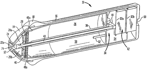
Now referring to FIG. 1, an exemplary device 10 can be used to treat tissues
in a
person's skin including, for example, hair follicles for hair growth
management,
including hair removal. The device 10 includes a housing 16 having a curved
section 18
for easier handling. Device 10 further includes a treatment head 14 disposed
adjacent
the housing 16 includes two radiation directing elements 20a and 20b
(generally referred
to as radiation directing elements 20) and sidewalls 22a and 22b (not shown)
which form
a cavity 12. The treatment head 14 further includes an optical radiation
source 28
optically coupled to the radiation directing elements 20 as described below in
more
detail in conjunction with FIGs. 2 and 3.
The treatment head 14 includes an outer edge 24, which may be contoured to
form a more efficient pressure seal with the skin. In order to provide cooling
to the
radiation source 28, device 10 includes a chamber 26 that includes a volume of
liquid or
other material 50. Liquid or other material 50 is thermally coupled to the
radiation
source 28 and optionally the radiation directing elements 20. The device 10
optionally
includes a dispenser (not shown) which can dispense, for example, a cooling
lotion, to
the skin. Device 10 also includes a skin gathering implement 34 having a
cylinder 30
CA 02618126 2008-02-07
WO 2007/019536 PCT/US2006/030968
-13-
enclosing a piston rod 40 coupled to a piston 74 to provide a manually
generated source
of differential pressure (e.g. low or negative pressure or vacuum) within the
cavity 12 to
draw skin within the cavity. The piston forms a moveable pneumatic seal with
the
radiation directing elements 20 and sidewalls 22a and 22b. The skin gathering
implement 34 further includes a reversing mechanism 44 coupled to rod 40.
In alternative embodiments, each radiation directing element 20a and 20b can
include multiple radiation directing elements and sidewalls 22a and 22b can
also include
one or more radiation directing elements. In an alternative embodiment, the
optical
radiation source 28 may be located remotely in a console and optically coupled
to the
treatment head 14, for example, through a fiber optic cable. In another
alternative
embodiment, the optical radiation source 28 may be located elsewhere within
device 10.
In all embodiments, optical radiation source 28 may include one or more
optical
radiation sources, which may be any of a number of different types, including,
without
limitation, both broad and narrow band light sources such as lasers, diode
lasers, tunable
lasers, diodes, halogen lamps, flashlamps, and/or other types of lamps.
Further, several
different sources and/or types of sources can be used to provide radiation at
various
wavelengths. In one embodiment the optical radiation source 28 can include
multiple
sources, for example, light emitting diodes (LEDs), lamps, laser diode bars,
lasers, and
other sources. One optical radiation source can provide radiation to one or
more
radiation directing elements, or multiple radiation sources can provide
radiation to one
or more radiation directing elements. For example, a beam splitter or other
means
known in the art may be used to direct light from one source to multiple
radiation
directing elements. In one embodiment the radiation directing elements 20 are
provided
as prisms, for example triangular equilateral prisms or right angle prisms.
However,
various other shapes are possible.
A cooling system of device 10 includes a heat sink 46 having fins 48a - 48n
that
are in thermal contact with cooling material 50 disposed within chaYnber 26.
Cooling
material 50 may be a phase transfer material which changes phases as it
absorbs heat
from heat sink 46 or cooling material 50 may be a liquid which is circulated
within
chamber 26 through conductor pipe 52 coupled to pump 54. Optionally, device 10
may
include batteries (not shown) or a connection port 60 disposed in the housing
16 can
provide a connection (not shown) to an external power source, such as a wall
outlet.
CA 02618126 2008-02-07
WO 2007/019536 PCT/US2006/030968
-14-
Optionally, device 10 may include a mechanism for chilling material 50 when it
is
circulated within chamber 26 or connection port 60 may be used to connect
chamber 26
through pipe 52 with an external source (not shown) of cooling material 50,
for example,
water. Device 10 includes a controller 42, e.g., electronic circuit boards 32a
and 32b
disposed within the housing 16, which may include control circuitry coupled to
optical
source 28, sensors, and other components as described below.
Gathering or drawing skin into the cavity 12 changes the optical properties of
skin to be treated. Drawing the skin within cavity 12 compresses the skin and
causes
both a stretching of the skin and also a restriction of the blood flow within
the skin in the
cavity. Advantageously the irradiance of a target within the skin in the
cavity, for
example, a follicle including a hair bulb or a sebaceous gland, can be
increased, for
example, by a factor of 1.2-2.5, because of decreased scattering, amount of
blood, and
skin thickness of the skin within the cavity.
In operation, device 10 is operated with a stamping motion or sliding motion
to
treat skin. A stamping motion is accomplished, for example, by placing the
device 10 in
contact with-the skin and treating the skin, then removing the device from the
skin and
placing the device in contact with another area of the skin. A sliding motion
is
accomplished by simultaneously moving the device over the skin surface as the
device
treats the skin. The treatment of the skin can be coordinated with the
velocity of the
movement of the device over the skin surface.
As the treatment head 14 is placed in contact with the surface of the skin, a
portion of the skin to be treated is drawn into the cavity 12 by activating
skin gathering
implement 34 to lower pressure within the cavity 12. The skin within the
cavity is then
exposed to treatment radiation from optical source 28 through radiation
directing
elements 20a and 20b.
In one embodiment, as device 10 is placed in contact with skin, pressure of
skin
against piston 74 is sensed by the reversing mechanism 44, which then pulls
the rod 40
within the cylinder 30 away from the entrance to the cavity 12 such that
piston 74 is also
moved away from the entrance. The movement of piston 74 generates the pressure
differential within the cavity 12, which pulls the skin within the cavity.
Piston 74, thus,
acts as a shutter for cavity 12 when device 10 is not in contact with skin.
CA 02618126 2008-02-07
WO 2007/019536 PCT/US2006/030968
-15-
As an alternative to skin gathering implement 34, device 10 could include an
external vacuum source (not shown) coupled to the cavity 12. This vacuum
source, as
well as skin gathering implement 34 may be triggered by a button (not shown)
pressed
by an operator on device 10 or, as explained above, pressure of skin against
piston 74 or
another type of shutter might be used to trigger the activation of the
external vacuum
source or skin gathering implement.
The dimensions of cavity 12 are selected such that targets in the skin drawn
into
cavity 12 receive optimal treatment from light passing into cavity 12 from
different
directions through radiation directing elements 20a and 20b. For example, hair
follicles
or sebaceous glands within the skin may be substantially centered between
radiation
directing elements 20a and 20b, and the optical radiation applied to the skin
can be
selected to overlap in the central portion of the cavity such that the targets
receive light
from both radiation directing elements while the skin surfaces against the
radiation
directing elements receive light only from the radiation directing element
with which
they are in contact.
In device 10, the radiation directing elements are located on opposite sides
of
cavity 12. However, in alternative embodiments the radiation elements may be
located
on adjacent sides such that they are at a ninety degree angle from each other.
As
mentioned above, one or more radiation directing elements may be located on
each side
of cavity 12. In addition, cavity 12 may be circular instead of square or
rectangle or any
other shape. For example, referring to Fig. 13, instead of radiation directing
elements
20a and 20b, device 10 can include a conical shaped prism 1301 having a
cylindrical
hole 1305 that serves as the cavity into which skin 1303 is drawn with
negative pressure.
In addition, a plurality of light sources 1302 (e.g., laser diodes, LEDs,
lamps) may be
coupled to conical prism 1301 to direct light beams 1304 into the portion of
the skin
1303. Conical prism 1301 can provide axial symmetry allowing for higher
amplification
of light inside skin than the planar symmetry provided by radiation directing
elements
20.
The dimensions of cavity 12 as well as the amount of negative pressure (also
referred to as pressure differential) that may be applied are selected in
accordance with
the amount of skin to be treated and desired effect on both mechanical and
optical
properties of skin while being treated. For most applications, it is desirable
to treat a
CA 02618126 2008-02-07
WO 2007/019536 PCT/US2006/030968
-16-
large amount of tissue with each application of light such that less time is
required to
complete the overall treatment of a larger area of skin. However, this is
contrasted with
also desiring smaller, less expensive devices and other factors. For example,
the amount
of skin that can be treated at one time by device 10 is limited by the amount
of skin that
can be drawn into cavity 12. Although the width (shown as W 62 in FIG. 2) of
cavity 12
can be made quite large to treat more skin, doing so can prevent radiation
from radiation
directing elements 20a and 20b from overlapping and combining within the skin
in
cavity 12. If device 10 further includes radiation directing elements in side
walls 22a
and 22b, then the same is true for the length(shown as L 64 in FIG. 2) of
cavity 12.
Similarly, making these dimensions too small can cause overlap of the
radiation and the
skin surface. Thus, the length and width of cavity 12 are limited by the
desire to
combine radiation from different radiation directing elements delivering
radiation from
different directions into cavity 12.
In addition, the height (shown as H 66 in FIG. 2) of cavity 12 is also
limited. In
theory, a very long or deep height could be used to draw more skin into cavity
12 for
treatment. However, for any given dimensions at the entrance to the cavity,
only a
certain amount of skin may be drawn into the cavity without bruising the skin.
Thus, the
height dimension is also limited.
In the embodiment shown in FIG. 1, the cavity 12 of device 10 has a length L
64
of approximately 10 mm which matches the optical sources 28 having lengths of
approximately 10 mm. In this embodiment, the cavity 12 has a width W 66 of
about 2
mm to about 6 mm, and preferably about 4 mm. This width and length allows firm
treatable skin (e.g., not skin around the eye area) to be treated with
combined uniform
radiation from multiple radiation directing elements 20 in the predetermined
volume V
68. In this embodiment, the height of the cavity is 13 mm and a pressure
differential of
approximately 20 cm of Hg (8 inches of Hg) is applied. This is described
further in
conjunction with FIG. 6.
In one embodiment, the targets of the treatment are hair follicles. Typically
hair
follicles are found at a skin depth of 1-4mm. As a result, gathering skin
within cavity 12
such that the height (H9kin 66 shown in FIG. 2) of the skin within the chamber
is about 2
mm to about 6 mm. Such a skin height hA;n 66 locates the person's hair
follicles within
CA 02618126 2008-02-07
WO 2007/019536 PCT/US2006/030968
-17-
the predetermined volume V 68 (FIG. 2) and subjects the follicles to combined
radiation
from the radiation directing elements 20 coupled to the optical radiation
source 28.
In another embodiment for treating acne, the targets of the treatment are
sebaceous glands. Typically sebaceous glands are found at a skin depth of 1-3
mm. As
a result, gathering skin within cavity 12 such that the height of the skin
within the
chamber is about 1;mm to about 3mm locates sebaceous glands within the
predetermined volume and subjects the sebaceous glands to combined radiation
from the
radiation directing elements 20 coupled to the optical radiation source 28.
Optionally a lotion may be applied to the skin to allow the skin to be more
easily
drawn within the cavity. Such a lotion can also improve optical and thermal
coupling
between the skin and the internal walls of the cavity.
As described above, as the skin is drawn into cavity 12, blood is removed.
This
allows the use of wavelengths that are normally absorbed by blood to be used
more
effectively. For example, optical sources 28 may generate optical radiation
from 380-
1350 nm. While drawing skin within cavity 12 may remove most of the blood
within _
the skin, it may concentrate the remaining blood in the sldn at the top of the
cavity - that
is, in the tip of the fold of the skin that is deepest or at the greatest
height within cavity
12. Such concentration of blood may be the focus of treatment for removal of
superficial targets such as vascular lesions.
Furthermore, the temperature of epidermis can be decreased by a factor of
about
1.1 to about 1.5 times because the basal membrane is stretched thereby
decreasing the
melanin optical density (MOD) which, as described above, can absorb part of
the
treatment energy.
Consequently, the tissue (e.g. hair follicle) can be treated more effectively
as a result of
the optical and mechanical property changes created in the skin as it is drawn
into cavity
12 and from the combined optical radiation from multiple radiation directing
elements
20 which.
Now referring to FIG 2, further details of the device 10 are shown. The cavity
12 includes a length L 64 and a width W 62. It will be appreciated, that in
other
embodiments, device 10 could include a cavity having a different geometry, for
example, circular, square, hexagonal, asymmetric, triangular, domed, and
instead of
straight internal walls, such walls could be, for example, slanted (inwardly
or
CA 02618126 2008-02-07
WO 2007/019536 PCT/US2006/030968
-18-
outwardly), curved, or made of flexible or soft material. A skin height hk;,,
66 within the
chamber is measured from the entrance of the cavity 12 as shown. The cavity 12
includes a volume 68 in which radiation from radiation directing elements 20a
and 20b
is combined. In this embodiment, the skin gathering implement 34 includes the
piston
74 coupled to the rod 40. The optical source 28, in this embodiment includes a
pair of
laser diode bars 70a and 70b (collectively referred to as laser diode bars
70). The laser
diode bars 70a and 70b include emitter surfaces 72a and 72b, respectively. The
optical
source 28 optionally includes optical elements 76a and 76b which are located
between
the emitter surfaces 72a and 72b and the radiation directing elements 20a and
20b,
respectively and extend the length L 64 of the cavity 12. If a longer cavity
12 is desired,
multiple diode bars can be combined. Heat sink 46 includes cooling fins 48
arranged in
an array.
In operation, laser diode bars 72, provide continuous or pulsed optical
radiation
to skin drawn into cavity 12. It will be appreciated that other sources of
optical radiation
including but not limited to incandescent lamps, flashlamps, halogen lamps,
light
---- -- -
emittirig diodes-or any other suitable Iight source presently available or yet-
to-be
developed can be used to provide treatment radiation. These sources can be
optionally
combined with filters to provide one or more selected wavelengths or separate
bands of
wavelengths from about 380 nm to about 1350 nm. The optical radiation source
or
sources may also provide a fluence of between 0.1-100 J/cm2, pulse widths of
between
1-1000 ms, spot sizes of 0.5-10 cm2, and rep rates of 0.2 Hz or continuous
wave. It is to
be understood that pulse widths can include individual pulses or groups of
pulses applied
to each section of skin treated within cavity 12 in stamping mode, or pulse
width can be
the effective pulse width seen by each section of skin treated within cavity
12 as the
device is moved over the surface of the skin and different sections of skin
are moved
into and out of the cavity.
Optional optical elements 76a and 76b can focus, concentrate, diverge or
collimate the radiation from optical radiation sources 72. The optical
radiation sources
28 and optional optical elements 76a and 76b are aligned with radiation
directing
elements 20 such that optical radiation from each of the optical sources is
coupled into
radiation directing elements that then direct the light into cavity 12. As
described above,
the dimensions of cavity 12 are preferably chosen to allow the light from the
different
CA 02618126 2008-02-07
WO 2007/019536 PCT/US2006/030968
-19-
radiation directing elements to combine within the cavity in the area of the
targets to be
treated to provide improved efficacy while also providing an improved safety
ratio to
protect the epidermis. This is described more fully below in conjunction with
FIGs. 8A,
8B, 9A and 9B.
Optionally, the radiation directing elements can deliver a pattern of
treatment
energy to the tissue within cavity 12 such that separate volumes of tissue
within the
cavity 12 are treated while surrounding tissue is untreated. That is, instead
of uniformly
treating all the tissue within cavity 12, only certain small volumes within
the cavity may
be treated. The healthy tissue in between these treated portions can improve
healing
time and tissue response to treatment. Such patterns of treatment may be
provided by,
for example, including focusing elements within the radiation directing
elements or
coating the internal walls of cavity 12 with a mask having openings such that
light only
passes through the opening. As another example, the internal walls of cavity
12 may be
textured to provide such a pattern of treatment.
In one embodiment, the operator triggers the negative pressure within cavity -
12
by pushing device 10 (Fig. 1) against skin. For example, housing 16 (Fig. 1)
of device
10 can be slidable with respect to treatment head 14, such that when the
operator places
the treatment head 14 against skin and continues to push against curved
section 18,
housing 16 slides further towards the skin. When the operator stops pushing on
curved
section 18, the action of the housing 16 sliding away from the skin can work
in
conjunction with the cylinder 30, rod 40, piston 74 and reversing mechanism 44
to lower
the pressure within the cavity 12 and, hence, gather skin into the cavity. As
described
above, sensors (not shown) may be included within cavity 12, such that when
skin is
detected within the cavity 12, the laser diode bars 70 are activated to
provide treatment
radiation to the skin within the cavity.
In order to provide cooling for the optical source and optical components, the
cooling material 50, for example, chilled water may be circulated in the
chamber 26 by
means of the pump 54 and the conductor pipe 52. It will be appreciated that
other
cooling means may be used. For example, chamber 26 may house a phase transfer
material (e.g., ice, wax) that changes phase as it absorbs heat from fins 48.
Instead,
device 10 may include a small fan to force air past fins 48. As another
example, the heat
CA 02618126 2008-02-07
WO 2007/019536 PCT/US2006/030968
-20-
sink may be thermally coupled to housing 16 such that heat is passed to the
operator's
hand during use and/or to the air.
In an alternative embodiment, parameters of device 10 may be changed to
provide different treatment to skin drawn within cavity 12 or to provide
different
treatment to different types of skin (e.g., facial skin might be treated
differently than
back or underarm skin). For example, the dimensions of the cavity (e.g.,
cavity width,
length and/or height), the pressure differential in the cavity, the position
of an optical
source, filters, fluence, pulse width and other parameters are adjustable. In
yet another
embodiment, the skin gathering implement 34 uses an adhesive force or pinching
force
applied in conjunction with piston 74 to gather skin into the cavity. In still
another
embodiment, ultrasound energy is used instead of optical radiation.
As described above, device 10 can include safety sensors. In one embodiment,
such sensors are used to detect the presence of skin within cavity 12 and only
then allow
the eniission of light within the cavity. In addition, safety sensors can be
used to detect
when skin is drawn too deeply within cavity 12 and prevent emission of light
within
cavity 12. This would prevent less firm skin and any anatomy located nearby
from
being exposed to the light from optical source 28. For example, the skin
around a
person's eye, including the eye lid, is generally very pliable. The light that
is generated
from optical source 28 may be such as to be not safe for use around the eye,
as it might
potentially injure the eye. For example, the light may be such that it can
pass through
the eye lid, be absorbed by melanin in the iris, and damage the eye.
Consequently, one
or more safety sensors can be located to detect skin within the cavity 12 at a
height /
depth which indicates that it may be skin around the eye thereby preventing
device 10
from operating the light source.
In one embodiment, safety sensors include one or more pairs of emitters and
detectors. Referring to FIG. 3, treatment head 14 is shown to include emitter
84 and
detector 86 which are aligned with radiation directing elements 20a and 20b,
such that
emitter 84 directs light along light path 88 which is then received by
detector 86 when
no skin is within cavity 12. When skin is drawn into cavity 12, light path 88
is
interrupted and detector 86 sends a signal to control circuitry within
electronic circuit
boards 32a and 32b to allow the control circuitry to drive optical source 28
to emit light
into cavity 12 to treat the skin. As shown, light path 88 is located close to
the opening of
CA 02618126 2008-02-07
WO 2007/019536 PCT/US2006/030968
-21-
cavity 12. However, light path 88 may be located deeper within (or at a
greater height)
within cavity 12 (though not as deep as light path 90) to indicate not only
that the skin
has been drawn within cavity 12 but that it has been drawn to a sufficient
depth to permit
treatment. This height is more fully described below with respect to Figs. 4A
and 4B.
In Fig. 3, treatment head 14 is also shown to include emitter 80 and detector
82
aligned to provide light path 90 which is deeper (or at a greater height)
within cavity 12.
Emitter 80 and detector 82 can be used to detect when skin is drawn too deeply
within
cavity 12, which as described above can indicate that this is skin that should
not be
treated. In this case, when light path 90 is interrupted, detector 82 sends a
signal to the
control circuitry to prevent the driving of optical source 28 such that the
skin within the
cavity is not treated.
In one embodiment photodiodes are used as detectors 82 and 86 and light
emitting diodes (LEDs) are used as emitters 80 and 84. In another embodiment,
treatment head 14 could include one or more reflectometer sensors (not shown)
to detect
the melanin content or other characteristics of the skin to be_ treated.
Optionally
treatment head 14 includes reflective surfaces 78 which allow optical
radiation to enter
the prisms 20a and 20b from optical source 28 but do not allow optical
radiation
scattered and reflected from the skin within cavity 12 back towards the
optical radiation
source 28 to escape from prisms 20a and 20b. Instead, reflective surfaces 78
return this
scattered and reflected light back toward the skin in cavity 12 to improve the
efficacy of
treatment. This is referred to as "photon recycling".
FIG. 4A shows a portion of skin to be treated 100 gathered into cavity 12 to a
height sufficient to interrupt light path 88. In this example, the portion of
the skin to be
treated 100 includes one or more hair follicles and the treatment may be for
hair
removal. For simplicity, only hair shaft 102 and hair bulb 104 are shown. As
described
above, the height of skin within cavity 12 is a function of the width and
shape of the
cavity 12, the pressure differential applied to cavity 12, and the firmness of
the skin: In
this example, the hair follicles are treated by optical radiation from laser
diode bars 70a
and 70b delivered from opposite sides of cavity 12 by radiation directing
elements 20a
and 20b along path 92. As described above, the dimensions of cavity 12 and the
optical
radiation parameters are chosen such that the radiation from both radiation
elements 20a
CA 02618126 2008-02-07
WO 2007/019536 PCT/US2006/030968
-22-
and 20b is combined in hair follicles within the skin to improve the efficacy
of
treatment.
With regard to avoiding treating more pliable skin around the eye area, it has
been discovered that an approximate relationship exists between the maximum
height of
the skin gathered into the cavity hskin-max and the width of the cavity w.vity
as follows:
hskin-n,ax ~ Wcavity for treatable skin (e.g., skin of sufficient firmness)
and a nominal
pressure differential of 20 cm of Hg (8 inches of Hg); and Generally, hskin-
max > 2 Wcavity
for non-treatable skin (e.g. less firm skin such as an eye lid) and a nominal
pressure
differential of 20 cm of Hg.
It has further been discovered that the range of skin height h9kin is in an
approximate range of
= 5 Wcavity > hskin > 2 Wcavity
The determination of hskin-max and hk;n allows for the determination of the
location of
sensor light paths 88 and 90.
FIG. 4B shows that the skin 108 drawn within cavity 12 has been drawn in so
deeply that it has interrupted light path 90, indicating that this is skin
that is not to be
treated, for example an eye lid. The structure of the iris 112 is such that
the iris is not
pulled into the cavity 12. As described above, less firm skin is gathered a
much further
distance (to a greater height) into the cavity 12. In one embodiment, the
light path 90 is
set to be interrupted when the skin height hskin is greater than 10 mm thereby
causing
detector 82 to send a signal to the control circuitry to prevent optical
source 28 from
being triggered.
FIG. 5A is a diagram showing direct optical radiation being applied to a
volume
of skin 114' by a prior art device. This diagram illustrates that significant
radiation
penetrates into volume 114' and, if volume 114' were an eye lid, such
radiation would
reach iris 112 through the eye lid. If this radiation is absorbed by melanin
in the iris, it
may damage the eye. In contrast, FIG. 5B is a diagrarn showing optical
radiation being
applied through radiation directing elements 20a and 20b to skin which has
been drawn
CA 02618126 2008-02-07
WO 2007/019536 PCT/US2006/030968
- 23 -
into cavity 12 and the significantly smaller amount of indirect light that may
reach skin
volume 114 outside of cavity 12. This diagram demonstrates, that even if
device 10 did
not have the safety sensors (e.g., 80, 82) described above for detecting the
drawing into
the cavity of skin around the eye, it would still provide significant eye
safety as
compared to prior art devices because it reduces the amount of light that
could reach the
iris.
FIG. 6 is a graph 130 of skin fold height vs. pressure for a cavity similar to
the
cavity 12 of FIG. 1. It has been determined that the dimensions of cavity 12
limit the
volume of skin that can be gathered into cavity 12 such that increasing the
pressure
differential beyond the pressure necessary to draw that volume of skin into
cavity 12
will not draw in more skin. Here point 134 on curve 132 represents an initial
pressure of
about 8 inches of Hg (20 cm of Hg) (200 Torr), which results in a skin height
of about
5 mm in the cavity. Due to skin elasticity, the skin pulls back to a slightly
lesser height,
and as shown, increasing the pressure differential beyond point 134 does not
increase the
height of the gathered skin. Here, pressure differential refers to the
pressure gradient _
between the volume in a cavity and the ambient (e.g. atmospheric pressure)
outside the
cavity.
It is advantageous to provide the minimum pressure differential to achieve a
consistent skin height, preferably about 2 mm to about 6 mm, in the cavity for
hair
treatment and 1-3 mm for acne treatment. Using contoured edges at the entrance
to the
cavity and/or applying a lotion or oil to the skin surface to be treated.
FIG. 7A represents treatment head 14 without reflector 78 (see also FIG. 3)
and
without reflective surfaces on the external surfaces of radiation directing
elements 20a
and 20b. In contrast, FIG. 7B represents treatment head 14 including reflector
78 and
also reflective surfaces on the external surfaces of radiation directing
elements 20a and
20b. That is, in FIG. 7B, radiation directing elements 20a and 20b have
reflective
surfaces except on the surfaces that form cavity 12. As shown, the device of
FIG. 7B
directs significantly more light to the skin drawn into cavity 12 than does
the device of
FIG. 7A. In one embodiment, the reflective surfaces, including reflector 78,
are coatings
applied to all the surfaces of radiation directing elements 20a and 20b except
those
surfaces that form or are coupled to cavity 12. Using reflective surfaces, the
efficiency
of the delivery of optical radiation to targets within cavity 12 may be
increased 1.2 - 4
CA 02618126 2008-02-07
WO 2007/019536 PCT/US2006/030968
-24-
times as compared with not using such reflective surfaces. As shown in FIGs.
7A and
7B, the targets within the skin volume 140 drawn into cavity 12 may be
follicles 144a-
144n.
FIG 8A, 8B, 9A and 9B are light distribution graphs for two experimental
treatment heads similar to the treatment head 14 of device 10. In this device,
the optical
source or sources (e.g., diode lasers generating light of wavelength 800nm)
are coupled
to cavity 12 through optical fibers. The light distribution graphs illustrate
the
importance of the cavity dimensions (e.g., width) and how proper selection of
dimensions with alignment of the optical components allows light from
different
radiation directing elements to be combined or overlapped within cavity 12 for
better
efficacy and higher safety ratio.
FIG. 8A is a graph of light intensity versus cavity width, and each of curves
164
and 166 represent light emitted into a cavity from only one radiation
directing element,
in this case a fiber, at one side of the cavity (e.g., 5 mm). In this example,
the width of
the cavity is 5 mm and curves 164 and 166 show that the maximum light
intensity is at
the skin surface adjacent the cavity wall (e.g., at 5 mm). This results in a
low safety
ratio which can lead to epidermal injury. Curve 166 has reduced light
intensity as
compared to curve 164 because the reflector used for curve 166 was brown paper
which
absorbed more light than the more reflective surface used for curve 164. In-
contrast,
curve 162 represents light being emitted from two radiation directing elements
on
opposite sides of the cavity and combining within the volume of the cavity
such that the
light intensity within the volume is substantially the same as the light at
each of the
surfaces of the cavity (0 mm and 5 mm). This results in a higher safety ratio
such that
targets may more easily be treated while protecting the epidermis. It is also
possible to
configure the cavity and/or radiation directing elements such that the amount
of light
received within the volume of skin within the cavity is higher than the amount
of light
received at the skin surface in contact with the cavity walls. In addition,
the cavity walls
can be cooled to cool the skin surface and provide additional epidermal
safety.
FIG. 8B is a graph of light intensity versus cavity height, and again each of
curves 174 and 176 represent light emitted into a cavity from only one
radiation
directing element and curve 172 represents light emitted from two radiation
directing
elements on opposite sides of a cavity. Curve 176 has reduced light intensity
as
CA 02618126 2008-02-07
WO 2007/019536 PCT/US2006/030968
-25-
compared to curve 174 again because the reflector used for curve 176 was brown
paper
which absorbed more light than the more reflective surface used for curve 174.
In this
example, the height to which skin is drawn within the cavity is 5mm. As shown
in curve
172, combining light from multiple radiation directing elements on different
sides of the
cavity provides increased light intensity at the same height as provided by
light from
only one radiation directing element (curves 174 and 176).
The graphs of FIGs. 9A and 9B are similar to the graphs of FIGs. 8A and 8B
except that the cavity width and height are 4mm.
It has been determined that the optimum cavity dimensions include a height
which is larger than the depth of the target from the skin surface and a width
that is less
than four times the depth of the target from the skin surface, preferably less
than 2 times
the depth of the target from the skin surface.
Referring to FIGs. 10A and l OB two alternative lamp based optical sources 230
and 240 are shown. Optical source 230 includes lamps 232a and 232b
(collectively
referred to as lamp 232) disposed adjacent reflectors 234a and
234(collectively_
referred to as reflector 234), respectively. Each lamp 232 and reflector 234
combination
operates similarly to the laser diode bars 70 of FIG. 3. In one embodiment,
the lamp is a
high efficiency Xe flashlamp. In this embodiment, lamp 232 operates with a
fluence of
about 0.1 to about 100 J/cm2, a pulse width of aboiut -1 ms to about 500 ms, a
wavelength range of between 400 -1350 nm and preferably between 600 - 1200 nm.
The reflectors 232 include a reflective coating and external surfaces of
prisms 246 may
also have a reflective coating. Here the overall efficiency of the treatment
head is
approximately 10- 40 percent. Optionally, a spectral filter can be
incorporated in device
230. In one embodiment, such spectral filter can be a dielectric coating on
the surfaces
of prisms 246 that receive light from the lamps or a coating on the lamps
themselves.
The lamps can be cooled by air or liquid flow.
In the embodiment of FIG. lOB, lamps 242a and 242b are integrated within
prisms 246. That is, cavities are made within the prisms such that the
internal walls of
these cavities provide the envelope for the lamps eliminating the need for a
separate
glass envelope for each lamp. In this embodiment, the overall efficiency of
the
treatment head is increased by approximately 150 -250%. As described above a
reflective coating can be provided on the external surfaces of the prisms 246.
CA 02618126 2008-02-07
WO 2007/019536 PCT/US2006/030968
- 26 -
Referring to FIG. 11, a pressure controlled firing mechanism may be used to
provide eye protection. Device 260 includes a housing 262 that is slideable
over internal
component 264, and housing 262 and internal component 264 are forced apart by
a
spring 266. Housing 262 includes a sensor 268, which may, for example, be a
microswitch. In order for control circuitry to cause the optical radiation
sources to emit
light, a signal must be received from sensor 268. Sensor 268 is triggered when
it is
pressed against internal component 264. In operation, this occurs when device
260 is
placed against skin (specifically the skin contacting surface of internal
component 264 is
placed against the skin) and sufficient force is applied to housing 262 to
compress spring
266 and allow the housing to slide toward the skin. As the housing is moved
toward the
skin, it will bring sensor 268 into contact with internal component 264 and
sensor 268
will send a signal to the control circuitry allowing it to trigger the
radiation source(s).
Because most treatable facial skin has bone behind, the treatable areas of
facial skin can
tolerate the pressure necessary to enable activation. However, because there
is no bone
behind the eye, it would be difficult and painful to place enough force on
device 260 to
enable activation, thereby providing eye protection. Spring 266 can be
provided with an
adjustable spring tension mechanism (not shown).
The pressure-controlled firing mechanism shown in device 260 can be combined
with other safety features. Irn addition, the pressure controlled firing
mechanism can be
combined into device such as device 10 of FIG. 1 having a cavity within which
skin may
be drawn.
Referring to FIG. 12, a skin treatment device 10" similar to the device 10 of
FIG.
1 is shown. Device 10" includes treatment head 14", including a cavity 12",
power
supply 282, cooling cartridge 284 thermally coupled to the treatment head 14",
and skin
gathering implement 286 (partially shown).
Device 10" operates in a manner similar to device 10. In one embodiment, the
power supply 282 is one or more high capacity rechargeable batteries or
capacitors. In a
home use application, the power supply 282 could provide power for about two
to about
five minutes of operation, or longer depending on the desired duration of the
treatment.
The removable cooling cartridge 284 provides cooling for the optical sources
and other
optical components, for example, radiation directing elements in treatment
head 14".
The removable cooling cartridge 284 may act as chamber 26 including material
50 to
CA 02618126 2008-02-07
WO 2007/019536 PCT/US2006/030968
-27-
provide cooling a heat sink, such as heat sink 46 of FIG. 1. In one
embodiment, the
removable cooling cartridge 284 can be placed in a household freezer before
being used.
Referring to FIG. 14, in another embodiment, a derrnatological treatment
device
utilizes a treatment radiation delivery system 400 that is configured to treat
tissue, such
as the skin, while ensuring that the radiation transmitted to the tissue is
both eye and skin
safe. Thus, system 400 will not damage the eye, tissues in the eye, or other
tissues.
System 400 can be designed or integrated as part of photocosmetic devices for
home or
professional or other uses.
System 400 includes a treatment radiation source 402, an eye-safety radiation
source 404, a waveguide 406, a diffuser 408 and a contact element 410.
Treatment
radiation source 402 is a laser diode bar having two laser diodes 412 and 414.
However,
many other possible configurations of treatment radiation source 402 are
possible, such
as solid state lasers, incoherent sources (i.e. lamps of various types), etc.
Additionally,
different configurations of laser diode bars in particular are possible and
potentially
preferable depending on the application and the_ design specifications. For
example, the
number of radiation sources can be varied and positioned to provide the
required
radiation power at the skin and to provide a homogenized distribution of
treatment
radiation throughout the waveguide and at the transition from the device to
the tissue
being treated. The optimal design configuration(s) will depend on a number of
variables, including the type of treatment, the size of the device, the spot
size of the
treatment, the materials being used, the wavelength(s) of radiation selected
for the
treatment, etc.
In treatment radiation delivery system 400, laser diodes 412 and 414 are
mounted
in a substrate 416. Substrate 416 is made of copper, but could alternatively
be made of
silicon carbide, copper tungsten, or other suitable materials. Substrate 416
provides
mechanical stability and removes waste heat during operation. Preferably, the
surface
418 of substrate 416 is coated with a material that is highly reflective of
the treatment-
radiation wavelength to recycle photons scattered/reflected from the skin.
Recycling
photons both reduces the heat load on substrate 416 (and system 400
generally), and it
improves treatment efficacy.
CA 02618126 2008-02-07
WO 2007/019536 PCT/US2006/030968
- 28 -
Waveguide 406 channels the treatment radiation and homogenizes the spatial
profile of the treatment radiation to more evenly distribute the treatment
radiation that is
transmitted to the tissue. Treatment radiation from treatment radiation source
402 is
coupled into waveguide 406. The input surface of waveguide 406 can be, for
example,
anti-reflection coated or bonded to the radiation source 402 to prevent
radiation loss.
Alternatively, radiation source 402 may be protected by a window between
surface 418 and waveguide 406. Such a window may be configured, for example,
to
reduce the exposure of radiation source 204 to reflected radiation. The inner
surface of
the window (i.e. the portion facing radiation source 402) can be anti-
reflection coated,
while the outer surface (i.e., the portion facing waveguide 406) can either be
anti-
reflection coated or bonded to the input surface of waveguide 406.
Diffuser 408 is located on the side of the waveguide opposite treatment
radiation
source 402. Diffuser 408 is typically made of glass, plastic, or other optical
material.
Diffuser 408 increases the angular spectrum of the treatment radiation at each
point
within the radiation beam. For a fixed treatment having radiation output power
that is
_ _ ___ - -
-
_ _can be _ desi-
-leve, th diffuser_
- gned to prevent
-limited to a predeterm _ined maximum-
retinal damage to the subject being treated by increasing the angular spectrum
to the
point where the output beam meets the ANSI eye safety standards. Many
different
radiation diffusers can be used including, -but not limited to, holographic,
diffractive,
photolithographic, fiber bundle, milk glass, sandblasted glass, or other
suitable material.
Some diffusers that are suitable for use in embodiments similar to system 400
may have
a textured output surface. For some of those diffusers, a space (e.g., filled
with air or a
fluid) may be required between the diffuser and the contact element, if the
device
includes a contact element 410. Volume diffusers may be used with or without
an air
gap.
In system 400, the radiation exiting diffuser 408 is coupled directly into
contact
element 410. Contact element 410 serves several functions. Contact element 410
acts
as a waveguide to couple the radiation to the skin. The treatment radiation
exits device
400 through contact surface 420. The length of contact element 410 preferably
is chosen
so as to create a uniform radiation distribution at the skin surface. In
system 400, the
length of contact element 410 can be adjusted based on the design parameters
to
optimize the device, but typically would be in the range of 0.5-100 mm.
Contact
CA 02618126 2008-02-07
WO 2007/019536 PCT/US2006/030968
-29-
element 410 also provides contact cooling during treatment. Contact element
410 is
made of a thermally conductive transparent material, in this case sapphire.
However,
other substances can be used. Heat can be removed from contact element 410 by,
for
example, attaching (with glue or other suitable means) a metal heat exchanger
to the
exterior surface of contact element 410. The metal heat exchanger can be
coated with a
highly reflective coating, so that any treatment radiation that is not totally
internally
reflected at the sapphire/glue interface is not absorbed.
Eye safety radiation source 404 is an LED located at the top surface of
waveguide 404. Source 404 provides radiation at a wavelength that is chosen to
maximize the perceived brightness by the user after the radiation propagates
through the
eyelid and the anterior portion of the eye. In other words, the wavelength is
preferably a
wavelength that is irritating to the subject being treated but is generally
safe even at
intensities perceived by the subject being treated to be painful or harmful.
Source 400
emits light at wavelengths in the red range (600-680 nm), and at a power
density of 1-10
mW/cm2. Other wavelengths and intensities are possible, however, depending on
the__
design and specifications.
After the source 404 has been engaged and contact surface 420 has been in
contact with the tissue being treated for approximately 1.0 - 2.0 seconds,
treatment
source 402 is engaged and the tissue is irradiated. The 1.0 - 2.0 second time
period is
chosen so the user has sufficient time to remove the device from her eye prior
to
irradiation by the treatment source 402. However, other embodiments are
possible. For
example, a shorter time or longer time could be used, such as 0.1 to 3.0
seconds. A
shorter time period could be used, for example, to allow time for a quicker
aversive
response to occur, such as the twitch or squint of an eye, that would indicate
to a person
treating the subject that the eye has been irradiated. The aversive response
may be any
movement that causes the subject to move the device from the tissue, or any
movement
that indicates to a person treating the subject that the eye may be
irradiated, including,
without limitation, squinting, pupil dilation, eye movement, head movement,
and arm
movement.
The existence of contact with the tissue being treated can be determined by a
number of different contact sensors. System 400 also utilizes source 404 as
part of a
contact sensing mechanism in addition to the "pre-pulse" safety mechanism
described in
CA 02618126 2008-02-07
WO 2007/019536 PCT/US2006/030968
=30-
the paragraph above. The contact sensing mechanism includes source 404, prism
422
and detector 424. Prism 422 is mounted at an angle along on outer side surface
of
contact element 410. Prism 422 is optically coupled to contact element 410 to
allow
light to pass from contact element 410 and through prism 422. Detector 424 is
attached
to an end surface of prism 422 and is optically coupled to prism 422 to
receive light
passing from contact element 410 and through prism 422.
System 400 determines whether surface 420 is in contact with tissue by
transmitting radiation from source 404 through treatment radiation delivery
system 400
to contact surface 420. When contact surface 420 is in contact with the skin,
there is
only a very small background signal at detector 424 due to total internal
reflection at
coupling prism interface 426. When contact surface 420 is in contact with
tissue, the
amount of radiation coupled out of contact element 410 via prism 422 and into
detector
424 increases significantly due to the scattering of light from the skin to
the coupling
prism interface 426 at angles that are not internally reflected within contact
element 410.
The output of detector 424_is monitored by control el_ectronics ofthe device
(not shown),
and, when the voltage exceeds pre-determined thresholds, the device determines
that
contact surface 420 is in contact with the tissue being treated. Thus,
detector 424 can
serve as an aversive sensor by detecting aversive motion of the patient
relative to the
device.
To facilitate the dual purposes of source 404, source 404 is a bicolor LED
with
one wavelength for contact sensing (in system 400, in the near infrared range)
and one
wavelength for the "pre-pulse" safety mechanism (in system 400, in the red
range as
discussed above). Preferably, the wavelengths used for the contact sensing
mechanism
and the "pre-pulse" safety mechanism will be different than the primary
wavelength(s)
used for treatment, although this is not essential. The first wavelength of
source 404 is
applied to sense contact. After contact with the tissue has been detected for
a certain
minimum time (typically 50 ms), the second wavelength is applied to warn the
subject
that the laser is about to fire. If the device is aimed at the eye, the light
from the second
wavelength will severely irritate (but not damage) the eye. Even if the system
is in
contact with a closed eyelid, the second wavelength is at such an intensity
that the
subject will still react to the light by turning her head or pulling the
device away. At that
CA 02618126 2008-02-07
WO 2007/019536 PCT/US2006/030968
-31-
point, the contact sensing mechanism determines that contact surface 420 is no
longer in
contact with the tissue and the device will not irradiate the tissue.
Although system 400 is designed for use while in contact with the tissue to be
treated other embodiments are possible. For example, an alternate embodiment
could
utilize proximity sensors to operate near, but not in contact with, the
tissue. The device
could also eliminate all such sensors and could be designed to operate at some
distance
from the tissue (or to operate while in contact with the tissue without
utilizing a contact
sensor). Additionally, the cooling provided by contact element 410 could be
provided
by other mechanisms (such as a cryogenic spray, a separate cooling plate, pre-
cooling
the tissue,~ or by other mechanisms). Furthermore, although system 400 is
primarily
designed for use with optical wavelengths of light, many other wavelengths or
combinations of wavelengths (both optical and otherwise) are possible.
While this invention has been particularly shown and described with references
to preferred embodiments thereof, it will be understood by those skilled in
the art that
various changes in form and details may be made therein without departing from
the
__ .
__
_---
- spirit- and- scope of the -irivention as defined by the appended claaims.
Those skilled in the
art will recognize, or be able to ascertain using no more than routine
experimentation,
many equivalents to the specific embodiments of the invention described
specifically
herein. Such equivalents are intended to be encompassed in the scope of the
appended
claims.