Note: Descriptions are shown in the official language in which they were submitted.
CA 02623756 2008-03-19
WO 2007/035804 PCT/US2006/036600
SYSTEMS, METHODS, AND APPARATUS hUK
RESPIRATORY SUPPORT OF A PATIENT
PRIORITY CLAIM
[0001] This patent application claims priorityto U.S. Serial No. 60/718,318,
"Systems,
Methods and Apparatus for Respiratory Support for a Patient", filed September
20, 2005,
which is incorporated herein by reference in its entirety.
FIELD OF INVENTION
[0002] The present invention relates generallyto respiratory systems and more
particularlyto
specialized systems, methods, and devices for enhanced ventilation of a
patient.
BACIiGROUND OF THE INVENTION
[0003] In order for the body to take in oxygen and give off carbon dioxide,
two components
of the respiratory bronchial system must function - the lungs as a gas-
exchanging organ and
the respiratory pump as a ventilation organ that transports air into the lungs
and back out
again. The breathing center in the brain, central and peripheral nerves, the
osseous thorax
and the breatlung musculature as well as free, stable respiratory paths are
necessary for a
correct functioning of the respiratory pump.
[0004] In certain diseases there is a constant overload on or exhaustion of
the respiratory
pump. A typical syndrome is pulmonary emphysema with flat-standing diaphragms.
Flat-
standing diaphragms do not have the abilit,yto contract. In the case of
pulmonary
emphysema, respiratorypaths are usuallyextremelyslack and tend to collapse. As
a
consequence of the flattened, over-extended diaphragms, the patient cannot
inhale deeply
enough. In addition, the patient cannot exhale sufficiently due to collapsing
respiratory paths.
This results in an insufficient respiration with an undersupply of oxygen and
a rise of carbon
dioxide in the blood, i.e. a ventilatory insufficiency.
[0005] The treatment for inhalation difficulty often involves a breathing
device. A home
ventilator is an artificial respirator for supporting or completely relieving
the respiratory
pump. Artificial respiration can be applied non-invasively via a nose or mouth
mask that the
patient can put on and take off as needed. However, the nose or mouth mask
prevents the
and speaking freely, and is veryinvasive.
1
CA 02623756 2008-03-19
WO 2007/035804 PCT/US2006/036600
[0006] Another treatment option is invasive ventilation. Invasive ventilation
is usually
applied via a cuffed endotracheal tube that is passed through the mouth and
the larynx and
into the windpipe, or is applied via a tracheostomy. The tracheostomy involves
an opening
placed in the trachea by an operation. A catheter about the diameter of a
finger with a
blocking balloon or cuff is inserted via the opening into the trachea and
connected to a
ventilator that applies cyclic positive pressure. This procedure makes
sufficiently deep
respiration possible, but prevents the patient from speaking.
[0007] In addition to home ventilation with a mask and invasive ventilation,
there is also
transtracheal admuiistration of oxygen via thinner catheters. U.S. Patents No.
5,181,509 or
5,279,288 disclose corresponding embodiments. In this manner, a highly dosed
administration of oxygen is administered to the patient in a continuous stream
with a
perinanently adjusted frequency. The flow rate of oxygen is regulated manually
by a
regulator. However, simulation of the natural breathing process of a patient
is not aclueved
because the depth of breathing is not enhanced. Some common problems
associated with
these transtracheal catheters are irritations and traumas of the sensitive
inner skin of the
windpipe (tracheal mucosa). It is a common observation that the tip of the
small catheter
strikes against the inner wall of trachea as a consequence of the respiratory
movement. In
addition to this mechanical trauma, the surrounding tissue is dried out by the
high flow
oxygen stream.
[0008] Furtherlnore, so-called "Montgomery T-tubes" can be inserted into the
trachea and a
patient can obtain oxygen via a shank of the T-piece external to the patient.
In needed, the
patient can draw off secretions using a suction catheter and a vacuum pump.
The patient can
breathe freely and speak when the front shank is closed; however, normal
artificial positive
pressure ventilation is not possible via the Montgomery 7' tube since the
introduced air
escapes upward into the oral cavity or the pharyngeal area. An additional
lina.itation of the
above-referenced therapies is the impaired mobility of the patient because of
inadequate
ventilation or because of the bulk of the apparatuses.
[0009] Jet ventilators are state of the art, but these devices are not
synchronized with a
patient's breathing. On the other hand, invasive ventilators with cuffed tubes
are
synchronized because there is a direct feedback of the pressure inside the
inflated lung to the
sensors inside the respirator. However, there are no respiratory systems that
use feedback
r iy to properly synchronize and control the ventilator.
2
CA 02623756 2008-03-19
WO 2007/035804 PCT/US2006/036600
[0010] Whether the breathing disorder is COPll/ emphysema, tibrosis, sleep
apnea, or
otherwise, difficult breathing is a serious, often life-threatening problem.
Therefore, there is
an existing need for a respiratory system that provides a more efficient
method for supporting
the respiration of a patient that can be used to treat many disorders, are
inin.i.tnally invasive,
mobile and taken along by the patient, and/or reliable in use. Moreover, there
is a need for
respiratory support systems that simulate the patient's spontaneous
respiration without
adversely affecting the patient's ability to speak. Additionally, there is a
need for a respiratory
support system capable of using pressure or flow signals from inside the body
to properly
synchronize and control a ventilator.
SUMMARY OF EXEMPLARY EMBODIMENTS
[0011] The invention includes systems, methods, and apparatuses that improve
the qualityof
life for patients that require respiratory support. These respiratory systems,
methods, and
apparatuses can provide a more efficient way of supporting the respiration of
a patient by
providing additional oxygen when needed in accordance with the principles of
the invention.
[0012] In one embodiment, a tracheal prosthesis and a catheter in accordance
with the
principles of the invention can provide for respiratory support that can be
synchronized with
the spontaneous respiration of the patient and still. allow the patient to
speak.
[0013] Additional features, advantages, and embodiments of the invention maybe
set forth
or apparent from consideration of the following detailed description,
drawings, and claims.
Moreover, it is to be understood that both the foregoing summary of the
invention and the
following detailed description are exemplary and intended to provide further
explanation
without limiting the scope of the invention as claimed.
BRIEF DESCRIPTION OF THE FIGURES
[0014] The accompanying drawings, which are included to provide a further
understanding
of the invention, are incorporated in and constitute a part of this
specification, illustra.te
preferred embodiments of the invention and together with the detailed
description serve to
explain the principles of the invention.
[0015] In the drawings:
3
CA 02623756 2008-03-19
WO 2007/035804 PCT/US2006/036600
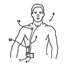
[0016] FIG. 1 shows the upper body of a patient carrying an embodunent ot a
system tor
respiration support in accordance with the principles of the invention.
[0017] FIG. 2 shows a diagram with a view of the respiration flow of an
emphysema patient
without respiration support and with respiration support in accordance with
the principles of
the invention.
[0018] FIG. 3 shows a technically simplified view of an embodiment of a
tracheal prosthesis
in accordance with the principles of the invention.
[0019] FIG. 4 shows another embodiment of a tracheal prosthesis in accordance
with the
principles of the invention.
[0020] FIG. 5 shows a schematic of an embodiment of an oxygen-bearing gas tank
and
pump showing the conduction of air and a control unit in accordance with the
principles of
the invention.
[0021] FIG. 6 shows an embodiment of the end section of a catheter in
accordance with the
principles of the invention.
[0022] FIG. 7 shows the catheter according to FIG. 6 inserted in a support
body in
accordance with the principles of the invention.
[0023] FIGS. 8A and 8B show graphs of breathing data generated from a bench
model test
in accordance with the principles of the invention.
[0024] FIG. 9 shows an embodiment of a catheter and sensors in accordance with
the
invention.
[0025] FIG. 10 shows a schematic of an embodiment of a circuit in accordance
with the
invention.
[0026] FIG. 11 shows another embodiment of a catheter and sensors in
accordance with the
invention.
[0027] FIG. 12 shows a schematic of another circuit in accordance with the
invention.
4
CA 02623756 2008-03-19
WO 2007/035804 PCT/US2006/036600
[0028] FIG. 13 shows a system in accordance with an embodiment of the
invention where
the pump and control unit are integrated with the oxygen tank,
[0029] FIG. 14 shows an embodiment of a distal end of a catheter in accordance
with the
invention.
[0030] FIG. 15 shows another embodiment of a distal end of a catheter in
accordance with
the invention.
[0031] FIGS. 16 A- 16E shows embodiments of a catheter in accordance with the
invention.
[0032] FIG. 17 shows an embodiment of a dual lumen catheter in accordance with
the
invention.
[0033] FIG. 18 shows an embodiment of the flow through the catheter of FIG. 17
during
inspiration in accordance with the principles of the invention.
[0034] FIG. 19 shows an embodiunent of the flow through the catheter of FIG.
17 during
expiration in accordance with the principles of the invention.
[0035] FIG. 20 shows an embodiment of a dual lumen catheter having a gliding
wall in
accordance with the invention.
[0036] FIG. 21 shows the catheter of FIG. 20 with the gliding wall in a
different position.
[0037] FIG. 22 shows an expanded view of an air outlet of the catheter in FIG.
20.
[0038] FIG. 23 shows an expanded view of an air outlet of the catheter in FIG.
21.
[0039] FIG. 24 is a flow diagram illustrating the operation of an embodiment
of the
invention.
[0040] FIG. 25 is a diagram of the overall system.
DETAILED DESCRIPTION OF PREFERRED EMBODIMENTS
[0041] The present invention, in a preferred embodiment, provides systems,
methods, and
apparatus for supporting the respiration of a patient. This can be
accomplished by providing
CA 02623756 2008-03-19
WO 2007/035804 PCT/US2006/036600
controlled synchronized ventilation with a directed flow of an oxygen-bearing
gas. The
oxygen-bearing gas maybe substantially pure oxygen, mixtures of oxygen and
nitrogen,
mixtures of oxygen and inert gases, ambient air, or various combinations
thereof. In addition,
the oxygen-bearing gas may include fragrances, aerosolized drugs,
humidification or heating.
The oxygen-bearing gas can be provided as needed upon inhalation and/or
expiration,
preferably, based upon sensing of the patient's spontaneous breathing.
[0042] Byproviding a jet boost of an oxygen-bearing gas upon inspiration, as
needed, the
patient can inhale more oxygen. Preferably, the additional oxygen is
administered at the end
of inhalation, in particular, after the peak of inspiratory flow is detected.
The administration
of additional oxygen can improve the depth of ventilation during inhalation.
However, the
additional oxygen maybe administered at any point during inhalation.
Additionally, a
countercurrent or counter pulse during expiration can be delivered, which
creates a back-
pressure in the airways similar to the pursed lips breathing strategy applied
by physiotherapists
in order to avoid a collapse of the respiration paths. Byproviding an oxygen-
bearing gas
upon expiration through counter pulses (e.g. bursts or pulses of oxygen-
bearing gas directed
against the direction of the flow during expiration), a dynamic collapse of
the airways can be
minimized or prevented, over inflation of the lung can be minimized, and
clearance of carbon
dioxide from the lungs can be improved. Therefore, in accordance with the
principles of the
invention, whether used for inhalation and/or exhalation, breathing requires
less energy and
the patient's pain, dyspnea and exhaustion are relieved. Moreover, the systems
and methods
of the invention can be used for treatment of many breathing disorders,
including, but not
limited to, COPD, emphqsema, fibrosis, and sleep apnea.
[0043] Referring to FIG. 1, in accordance with one embodiment of the
invention, patient P
designates a patient suffering from a breathing disorder, for example,
pulmonaryemphysema,
with overloading and exhaustion of the respiratorymuscles. As a consequence,
the patient
cannot inhale enough oxygen because the lungs are compromised. In addition,
the patient
cannot exhale enough carbon dioxide because the patient has slack and
collapsing respiratory
paths. The system of FIG. 1 generally includes the abilityto detect the
patient's spontaneous
respiration and the ability to provide oxygen to the lungs of the patient
during spontaneous
inspiration and/or exhalation.
[0044] As shown, the respiration support of patient P in accordance with the
principles of
r~, ;~<> ~r;~r ~~ t--plemented in a system, method, or apparatus that maybe
compact
6
CA 02623756 2008-03-19
WO 2007/035804 PCT/US2006/036600
and/or portable. Other systems are contemplated including, for example,
providing tor use
with a ventilator or oxygen source as shown in FIG. 13. The overall system of
the invention
is described in FIG. 25, indicating the gas source 02, the pump apparatus 1
and control
system 12, the catheter 5 and internal sensors 8, 9 and the patient P. The gas
source 02,
pump apparatus 1 and control system 12 can be separate or integrated
components of the
system. The control unit 12 maybe connected I to internal sensors 8, 9 and/or
external
sensors 13, 14.
[0045] In accordance with the embodiment of FIG. 1, in general, patient P's
spontaneous
breathing can be detected byway of sensors. A catheter 5 can be used to
introduce oxygen
into the lungs as needed. The sensors and catheter can be associated with the
patient in a
variety of ways. As illustrated in FIG. 1, a catheter 5 is introduced in the
trachea. Also, a
catheter 5 could be introduced at other points into a patient P, including,
for example,
through the mouth or nose of the patient P, or accessed into the trachea by an
artificially
created entrypoin.t somewhere on the body and tunneled internallyto and into
the trachea.
The catheter 5 can be secured in the trachea in a variety of ways. In one
embodiment, the
catheter 5 can be associated with a tracheal prosthesis as discussed later or
using a positioning
catheter as also discussed later with reference to FIGS. 3 and 4, for example.
[0046] The system of FIG. 1 generallyincludes an oxygen-bearing gas source
(not shown),
gas pump 1, mobile respiratorydevice 4, a set of exterior sensors 13, 14, and
a set of interior
sensors (not shown) disposed inside the trachea of the patient P. The oxygen-
bearing gas
pump 1 can be connected to a gas source (see FIG. 5) and catheter 5 to
introduce an oxygen-
bearing gas into the patient's lungs bywayof the trachea, as shown, although
other entry
points can be used in accordance with the principles of the invention as
discussed above.
According to FIG. 1, the oxygen-bearing gas pump 1 is shown as a component of
a compact,
easilyportable respiration device 4. The device 4 could alternativelybe housed
in a
component with a gas tank or oxygen-bearing gas source as illustrated in FIG.
13. With the
sensors in accordance with the principles of the invention, the spontaneous
respiration of the
patient can be detected. Typically, the information from the sensors is
communicated to the
gas pump 1. However, the information from the sensors may also be communicated
to a
cellular telephone or other wireless systems that can communicate information
to a healthcare
provider/ hospital, etc., for 24-hour monitoring and response from the
healthcare
provider/hospital, etc. The patient then can receive a pressure boost of
oxygen-bearing gas
7
CA 02623756 2008-03-19
WO 2007/035804 PCT/US2006/036600
as needed in accordance with the principles of the invention. FIG. 2
illustrates both
spontaneous respiration of the patient P without the invention (right) and
respiration
supported in accordance with the principles of the invention (left). The x
axis in this diagram
represents time and the y axis represents the amount of flow (change in volume
over time) of
oxygen-bearing gas, which can be liters per second or any other appropriate
measurements.
The spontaneous respiration process with inspiratory flow and expiratory flow
without
respiratory support for patient P is shown in the left half of FIG. 2. The
curve for inhalation
is designated by E 1 and the curve for exhalation byAl. As illustrated by
curve E 1, during
inhalation the tidal volume inhaled is reduced from that of a normal patient.
For example, a
patient with emphysema with flattened diaphragms or a patient with stiff lungs
suffering from
fibrosis cannot breathe in enough air (oxygen) in one breath. Both patients
typically
experience shallow breathing. Therefore, the patient requires more breathing
cycles to get the
requisite amount of oxygen and clear carbon dioxide. During exhalation, as
illustrated by
curve Al, the expiratoryflow of the emphysema patient is reduced because the
respiratory
paths can be slack and tend to collapse before an adequate amount of carbon
dioxide is
expelled from the lungs.
[0047] The sensors allow the patient P's breathing to be monitored
contuzuously so that a jet
flow of oxygen-bearing gas can be supplied in accordance with the principles
of the invention,
that is, when a deeper breath is needed. In particular, at the end of an
inhalation process of
the lungs, an additional volume (oxygen) can be administered to patient P, as
discussed in
more detail below. This respiratory flow is illustrated in the right half of
FIG. 2. As
illustrated, an additional amount of oxygen-bearing gas provided to patient P
increases the
respiratory volume during inhalation according to curve E2 by the volume
difference shown
darkened in the upper curve and designated byE3. The additional amount of
oxygen-bearing
gas can have an extra space tidal volume between 25 ml and 150 inl.
[0048] In addition, the exhalation process of the patient can be braked or
slowed by a
countercurrent. As a consequence thereof, the respiratory flow shifts during
exhalation along
the curve designated byA2. This purposeful resistance acting opposite to the
exhalation
prevents a collapsing of the respiratory paths during exhalation. In this
manner, the
exhalation volume can be increased bythe volume also shown darkened and
designated by
A3. The amount of carbon dioxide that is exhaled can be increased by a
statistically
significant amount. The amount of carbon dioxide that is exhaled can be
increased byat least
8
CA 02623756 2008-03-19
WO 2007/035804 PCT/US2006/036600
5%. Preferably, the amount of carbon dioxide exhaled rs increased trom 5% to
:SU%. More
preferably, the amount of carbon dioxide exhaled is increased about 20% to
30%.
[0049] As a consequence, the invention may avoid insufficient respiration from
an
undersupply of oxygen and an increase of carbon dioxide in the blood. The
patient P may be
significantly less stressed and more mobile, and mayperceive less or no
shortage of air.
[0050] The sensors for detecting and monitoring respiration will now be
discussed in more
detail. To detect spontaneous respiration of the patient P, sensors can be
associated with an
end of the catheter that is disposed in the trachea of the patient P. In one
embodiment, the
inventioii can include connecting the catheter to a tracheal prosthesis (e.g.
FIGS. 3, 4, and 7)
or can include a catheter-positioning device (e.g. FIGS. 14, 15, and 16A -
16E) to more
reliably and accurately direct the oxygen flow into the patient's airways and
away from a
tracheal wall. Preferably, in accordance with the principles of the invention,
oxygen is
introduced into the patient P in such a manner that the patient P can freely
breathe and speak
without restriction.
[0051] In one embodiment, as shown in FIGS. 3 and 4, the sensors can be
disposed on a
tracheal prosthesis 2, 3. Each tracheal prosthesis 2, 3 is shown having a
tubular support body
6 with a connection 7 for a catheter 5. As shown, two sensors 8, 9 detect
spontaneous
respiration of the patient P, and can be associated with a support body 6. The
sensors 8, 9
can be thermistors, that is, temperature dependent resistors. The sensors 8, 9
can detect
tracheal flow of the patient because inspired air and expired air have
different temperatures.
The thermistors 8, 9 can be connected together in a bridge circuit in the
apparatus to
compensate for changes in ambient air temperature. Other types of sensors can
be used in
accordance with the principles of the invention including, for example, a
pressure sensor as
discussed later. Both sensors 8, 9 can be located on an inner wall 10 of the
support body 6
(FIG. 3), or one sensor 8 can be fixed on the inner wall 10 of the support
body 6 and the
other sensor 9 can be located on an outer wa.ll 11 of the support body 6 (FIG.
4). The
sensors 8, 9 communicate with a control unit 12 for activating an oxygen jet
pump 1. The
sensors 8, 9 can be connected by wires or by wireless communication. The
control unit 12
can be anytype of microprocessor that is capable of processing the collected
data in
accordance with the invention. The control unit 12 is schematicallyshown in
FIG. 5 with its
inputs (I) and outputs (0). The inputs (I) represent information coming from
the sensors.
.___.___ent infonnation that is used to control the pump 1.
9
CA 02623756 2008-03-19
WO 2007/035804 PCT/US2006/036600
L0052J In the tracheal prosthesis 2 according to FIG. 3, the jet catheter 5
can be inserted via
connection 7 into the support body 6. An end 15 of jet catheter 5, located in
support body 6,
is preferably guided or deflected approximately parallel to its longitudinal
axis L. The data
lines from sensors 8, 9 to the control unit 12 run inside the catheter 5. The
invention is not
limited to data lines; transmission from sensors can be anytype of
transmission, including
wireless. On the discharge side, the end 15 of the jet catheter 5 is
preferablydesigned as a jet
nozzle 25. The jet nozzle 25 increases the speed of an oxygen current being
discharged from
the catheter 5, and the current is conducted in the direction of the bronchial
tract. The
diameter of the support body 6 is dimensioned with a sufficiently free lumen
in such a
manner that the patient P can freely breathe and speak even with the
integrated catheter 5.
[0053] In another embodiment, a separate coupling 18 is provided on the
connection 7 in the
tracheal prosthesis 3 according to FIG. 4. The catheter 5 can be connected to
the tracheal
prosthesis 3 with the separate coupling 18. In this instance, a fixed
longitudinal section 19
aligned parallel to the longitudinal axis L can serve as the catheter end in
the support body 6,
and the oxygen current is conducted via a jet nozzle 26 in the direction of
the bronchial tract.
[0054] The tracheal prosthesis, when used, can comprise various
configurations, shapes and
dimensions. For example, the tube could be T-shaped or L-shaped or otherwise.
The size,
shape, and/or cross-section can vary, for example, to accommodate removal or
to direct the
catheter. The tracheal prosthesis could be a portion of a tube having, for
example, a semi
circular cross-section. Furthermore, expandable and self-expandable prongs or
petals can be
used at the tracheal opening to secure the prosthesis in place. In one
embodiment, the
prosthesis can include a tubular member with a tracheal side opening including
prongs or
petals surrounding, in whole or in part, the access hole. The prongs or petals
may function
like a rivet in the neck opening. The tracheal prosthesis can also be coated
to avoid mucus
retention, prevent the formation of granulation tissue, or can act as a drug-
releasing device.
The tracheal prosthesis may also include other coatings, such as lubricious
coatings and
hydrogel anesthetics. Thus, the tracheal prosthesis can serve as a guide for
the catheter, to
hold sensing devices, serve as a drug delivery device, and/or to minimize
mucus plugs that
can form on the catheter tip.
[0055] In addition to internal sensors, external sensors can be provided. FIG.
1 also shows
respiration sensors 13, 14, preferably, impedance electrodes or respibands.
Signals from the
cancnrc 1A I e_ ..i..- for detecting the spontaneous respiratory efforts of
the patient P. An
CA 02623756 2008-03-19
WO 2007/035804 PCT/US2006/036600
exact image of the respiration process of patient P can be obtained
byprocessing the
measured values received via sensors 8, 9 and 13, 14. In addition, the safety
against false
measurements or the failure of one of sensors 8, 9 and/or 13, 14 can be
increased due to
redundancy. Although the sensors are shown in certain locations on the patient
P, other
locations that would allow the sensor to sense the patient's respiration,
directly or indirectly,
can be used. For example, sensors can be provided on the catheter as discussed
later.
Alternatively, a pill-type sensor or nano device can be used and/or implanted
to communicate
by, for example, wireless transmission to communicate with the control unit to
operate the
oxygen flow through the catheter in accordance with the principles of the
uivention.
[0056] One embodiment where sensors are provided on the catheter is shown in
FIG. 6.
FIG. 6 shows a catheter 28 with a long, flexible tube 29 and an end 31 on the
discharge side
bent in a curvature 30. The catheter 28 can be pre-formed to provide a desired
curvature 30.
With the appropriate curvature 30, the catheter 28 can be entered into the
trachea with or
without use of a prosthesis. In this embodiment, two sensors 32, 33 for
detecting the
spontaneous respiration of the patient P can be fastened on the end of the
catheter 28. The
sensors 32, 33 are preferablythermistors, but as in all embodiments herein,
could be other
types of sensors. Furthermore, in other embodiments of the invention,
additional seiisors
may be used. In still other embodiments of the invention, fewer sensors may be
used. Data
lines are not shown in the drawing for the sake of simplicity and could
include any form of
data transmission. In a hard-wired embodiment, data lines can run through the
catheter 28.
A catheter flange 34 designates a stop for use with a support body 36, as
shown in FIG. 7. It
can also be seen that an end 31 of the catheter 28 is provided with a jet
nozzle 35. The cross-
section of gas flow is reduced relative to the cross-section of the catheter
28 in the jet nozzle
35 so that the discharge rate of the supplied oxygen is increased.
[0057] The catheter 28 can be introduced into the support body 36, as shown in
FIG. 7. The
support body 36 is located in the trachea of the patient P. A connection to
the outside is
established via a connection 37. In the body, the tip or jet nozzle end 35 of
the catheter 28
can be disposed in the trachea. Preferably, the tip of the catheter 28 does
not touch the
trachealwall. The support body 36 can be a traditional Montgomery T-stent.
[0058] FIGS. 8A and 8B show measurements in a lung model emulating respiratory
diseases.
FIGS. 8A and 8B graphically illustrate an increased tidal volume with the
invention. FIG. 8A
' ' --' -~ 'le volume (ml) of breath comparing a pathologically low breath of
a
11
CA 02623756 2008-03-19
WO 2007/035804 PCT/US2006/036600
patient with emphysema at about 90 ml; the volume with jet oxygen in
accordance wrth the
principles of the invention upon inhalation at about 260 ml; and the volume
with the jet
oxygen in accordance with the principles of the invention upon inhalation and
with the flow
brake (oxygen jet) upon exhalation at about 300 ml. FIG. 8B shows a graph of
the flow of
breath (liters per second) over time for a breath of an emphysema patient; the
flow with jet
oxygen in accordance with the principles of the invention upon inhalation; and
the flow with
jet oxygen in accordance with the principles of the invention upon inhalation
and with the
flow brake (oxygen jet) upon exhalation.
[0059] In another embodiment shown in FIGS. 9 and 10, thermistors 81 and 82
can be
provided on a catheter tip inside the trachea. The thermistor 81 is more
exposed to the gas
stream than thermistor 82, which is protected against fast temperature changes
because it is
inside the catheter wall (or under a protection film). Alternatively, multiple
thermistors with
different response times could be used. Over a longer period (e.g. 10
seconds), both mean
temperatures will be the same (equilibrium) and the bridge (FIG. 10) will be
readjusted. This
compensates for changes in ambient temperature, fever, etc. Rapid changes
based upon
breathing in colder air and breathing out warmer air is detected by the
thermistor 81. The
output signal is sent through a differentiator. The peaks of the thermistor
signal match the
highest flow rates. The min;mum in the differentiated signal matches the peak
of the
inspiratory flow and the peak of the expiratory flow. Undifferentiated and
differentiated
signals are fed into the microprocessor. One way to determine peak inspiratory
flow (trigger
for beginning introduction of oxygen) would be to look for minimum in absolute
temperature
(cold air comes in) and zero change of temperature (differentiated signal is
zero). The
advantage of using the above multiple thermistor approach is that the
difference between the
signals from the two thermistors cancels out flow artifacts found in the
measured respiratory
flow pattern, such as would be caused by vibration or other anticipated
events, and to
compensate for drift in the thermistor signal such as would be caused by
changing external or
internal temperature or humidity conditions.
[0060] In another embodiment, as shown in FIGS. 11 and 12, FIG. 11 shows a
pressure
transducer that is a modified silicone wire strain gauge element 90. Instead
of a typical
silicone membrane, the wall of the catheter is used. If the wall of the
catheter deforms under
the pressure swings inside the trachea (breathing effort), then an electrical
signal from the
bridge amplifier is fed into a microprocessor. This embodiment can be used
alternatively to
12
CA 02623756 2008-03-19
WO 2007/035804 PCT/US2006/036600
the thermistors, as a redundant signal or as a back-up signal. Other sensors
could be
semiconductor flow sensors or pressure sensors. FIG. 12 shows a circuit
diagram of a bridge
amplifier.
[0061] Other sensors can be used in accordance with the invention. For
example, sensors
and/or secondary control sensors could be: respibands (chest wall strain
gages), respitrace
signals (conductance plethysmographs), pressure sensors inside or outside the
body,
transthoracic electrical impedance measuring devices, flow sensors at the
mouth or nose
(pneumotachographs), and/or capnometers (carbon-dioxide sensors). Moreover,
the sensors
in accordance with the invention can communicate data or information to the
control unit by
any devices, mechanisms, or methods. For example, communication can occur
byway of
wire, wireless, or remote transmission. The advantage of using non-thermistor
sensors is that
the thermistor approach may have the disadvantage of the thermistor head
collecting airway
mucus, which could be corrected for in a variety of ways such as with
cleaning. However,
other non-thermistor sensors maybe less susceptible to annoyances like mucus
collection.
Further, with thermistor sensors, inevitable changes in ambient temperature,
while
compensatable in the thermistor signal processing algorithms, are
potentiallyproblematic to
system reliability. Therefore, the other types of sensors stated above may be
advantageous
over thermistor sensors, or in addition to the thermistor sensors.
[0062] In addition to measuring the respiration pattern, it is often desirable
to measure
airwaypressure for safety reasons, for which thermistor sensors may not be the
best
approach. Therefore, some of the sensors mentioned above can also be used as a
safety
control device. For example, pressure sensors can be used to sense the
inspiration of the
patient (like the thermistors), but they can also be used to sense a high
pressure in the trachea
and shut off the jet machine in order to prevent baro-trauma (damage from high
pressure).
[0063] An oxygen-bearing gas is provided on demand bythe gas pump 1. The gas
pump 1 is
schematically shown in FIG. 5. The gas pump 1 can be a piston pump with a
double-acting
piston 20 arranged in a cylinder 27. The piston pump of the present embodiment
comprises
four valves V1 to V4. Other piston pumps (not shown) may have greater than or
fewer than
four valves. The supply of oxygen emanates from an external oxygen reservoir
via a
connection 21. The switching states of valves V1 to V4 and the supply lines
and removal
lines are designated by letters a to g. Other types of pumps can be used in
accordance with
t'llP Y~Yfnr~tT~lac n~ t~o ->ntion.
13
CA 02623756 2008-03-19
WO 2007/035804 PCT/US2006/036600
[0064] The gas pump 1 functions in the apparatus during the support of
respiration as
follows. When valve V1 is open from c to a (b to c closed) and valve V2 is
open from b to e
(e to d closed), piston 20 moves to the left in the plane of the figure and
the oxygen flows via
outlet 22 and jet catheter 5 to the patient P. An additional amount of oxygen
E3 is
administered during the inhalation process of the patient P.
[0065] When valve V1 is open from b to c (c to a closed) and valve V2 is open
from e to d (b
to e closed), piston 20 moves to the right in the plane of the figure and the
flow of oxygen
takes place in the direction of valve V3. Valve V3 is connected to the ambient
air via an
outlet 23. In the instance in which valve V3 is open from d to g, the oxygen
flows off
without expiration brake. That means that the exhalation process is not braked
by a
countercurrent.
[0066] If valve V3 is closed from d to g and open from d to f, the oxygen
flows via access
path 24 in the direction of the outlet 22 and the catheter 5 in order to be
administered to the
patient P during the exhalation process and in order to break the respiratory
flow. The
countercurrent prevents a collapsing of the respiratory paths and keeps them
open, making a
deeper exhalation possible.
[0067] Furthermore, valve V4 is located in access path 24 of the apparatus,
via which the
flow through (f to a) can be variably adjusted. This advantageously can be a
proportional
valve with pulse-width modulation.
[0068] As discussed above, the catheter preferablyin.cludes a jet nozzle. Any
type of jet
nozzle can be used to achieve the necessary jet flow. The jet flow speed in
accordance with
the invention can be significantly higher than 100 m/s. By comparison, the
speed through a
conventional ventilator tube or mask is significantly lower than 100 rn/s.
When the jet flow
rate is high enough, there is enough speed so that directed flow is
accomplished and no
sealing tube cuff would be necessary. Under norinal ventilation, the
volumetric inspiratory
flow rate is in the range of about 500 m3 to 1000 cm3 in 2 seconds. A peak
inspiratory flow
maximum can be 1000 cm3/second. In the case of normal invasive ventilation,
the flow of
1000 cm3/s (peak) goes through a tube of approximately 8 mm diameter. The
speed of this
gas stream, determined by dividing the volumetric inspiratory f low rate by
the area of the
tube, is 1000 cm3/(0.4)2 cm2 " Pi = 2000 cm/s = 20 m/s. During jet
ventilation,
approxinzately half of this flow goes through a jet cannula of 1.5 mm
diameter. As the flow
14
CA 02623756 2008-03-19
WO 2007/035804 PCT/US2006/036600
profile is rectangular, the peak flow rate is 500 cm' /s. Therefore, the speed
of the jet gas
stream is 500 cm3/ (0.075)2 cm2 1 Pi = 28313 cm/s = 283 m/s. In accordance
with a
preferred embodiment of the invention, 100 ml (cm3) are pressed through a
catheter of
approx 1.5 mm diameter in half a second. Preferably, the peak flow for this
embodiment is
100 cm3 in 0.25 seconds = 400 cm3 /s. The speed of this gas stream is 400 cm3
/(0.075)2 cm2
Pi = 22650 cin/ s = 226 m/s. In other preferred embodiments, the speed of the
gas
stream is from approximately 100 m/s to approximately 300 m/s. Preferably, the
speed of
the gas stream is from approximately 200 m/s to approximately 300 m/s.
Preferably, the
speed of the gas stream is from approximately 250 m/s to approximately 300
m/s.
[0069] When the tip of the catheter touches the wall of the trachea, there is
a potential risk of
tissue damage. The catheter tip or the high flow gas stream can harm the
mucosa. To
efficiently and effectively direct the air inside the body, the catheter can
be configured to
provide a directed flow of oxygen. In particular, the catheter is preferably
configured so that
the exit of air from the catheter output end can expel and direct air down the
center of the
trachea to avoid directing the jet flow of oxygen against the tracheal wall.
Also, the catheter
tips are preferably configured to min;m~e venturi and the mucus formation
proxiunal to the
venturi on the outer wall of the catheter. A shielding Montgomery T-tube as
described above
can be used to overcome that problem. In FIGS. 14 and 15, the catheters are
configured
such that the catheter tip or jet nozzle avoids contact with the wall of the
airway, the tip is
substantially centered in the trachea. This can be accomplished by configuring
the catheter so
that the catheter will contact the tracheal wall at several locations to
distribute the local
pressure, and the tip where the jet flow of oxygen exits the catheter is
substantially centered in
the trachea. Accordingly, the use of a tracheal prosthesis is not necessary.
One wayto avoid
the contact between the tip (jet nozzle) and the airway wall is to bend the
catheter like a
zigzag in two planes as illustrated in FIG. 14. Another embodirnent is a
corkscrew as
illustrated in FIG. 15.
[0070] FIGS. 16A - 16E show alternate embodiments for centering the catheter
where
balloons (FIGS. 16A and 16B) or clips (FIGS.16C - 16E) can be used to center
the catheter
tip. FIG. 16A shows a balloon for centering the catheter tip where the balloon
has a roughly
circular cross section through line J-J. Openings in the balloon may be
located in the
longitudinal direction of the catheter. FIG. 16B shows a balloon for centering
the catheter tip
where the balloon can have multiple extensions. The extensions may appear as
cone-shaped
CA 02623756 2008-03-19
WO 2007/035804 PCT/US2006/036600
projections in cross section K-K along the longitudinal direction of the
catheter. FIG. 16C
shows clips extending radially out from the catheter. The clips in this
embodiment are
relatively flat and extend outward in opposing pairs. FIG. 16D shows another
embodiment
of clips with extensions on the end of the clips. The clips and extensions may
extend at
multiple angles relative to the catheter for centering the catheter tip within
the trachea. FIG.
16E shows another embodiment of clips having shaped protrusions at various
locations along
the length of the catheter. The protrusions may have flat tops with rounded
edges and
undercuts. Preferably, the clips of the various embodiments are made of a
resilient material.
[0071] Referring now to FIGS. 17 - 23, a dual lumen catheter will be
described. The
invention can also include the abilityto better distribute the directed flow
(FIGS. 17 - 19)
and/or change the direction of the flow (FIGS. 20 - 23). FIGS. 17 - 19 show a
dual lumen
catheter 172. The catheter tip, shown generally at 170, is disposed in a
trachea 174. The
catheter 172 has two lumens, formed byinner cannula 176 and outer cannula 178.
Inner
cannula 176 directs flow to a catheter nozzle 180, as discussed above. As
shown in FIG. 18,
upon inspiration, inspired flow is enhanced by air entrainment from the jet
flow through the
inner cannula plus by the additional jet flow itself 176. Upon expiration
(FIG. 19), exhaled
flow is enhanced by turbulence from counter flow through ports 182 by means of
propping
the respiratory paths open. The ports 182 need not be of any particular shape
and may be,
for example, circular, hexagonal, oval, or slits. Although not shown,
turbulent flow could also
be provided through inner cannula 176 during exhalation to enhance exhaled
flow depending
upon the desired effect.
[0072] Referring to FIGS. 20 - 23, another embodiment of a catheter is shown.
A catheter
200 is shown with a distal tip 202 in a trachea 204. The catheter tip 202
includes a cannula
configuration with an inner lumen 206, an outer lumen 208 concentric to the
inner lumen,
and a gliding sheath 210. In this embodiment, the gliding sheath 210 moves
relative to the
cannula to allow ports 210 to change the direction of oxygen flow as
illustrated in FIG. 20
verses FIG. 21, and in close-up in FIG. 22 verses FIG. 23. As shown in FIG.
22, upon
expiration, the flow braking turbulence caused by movement of the gliding
sheath 210 may
create a resistance such as in pursed-lip breathing, which can prop the
respiratorypaths open
to enhance the amount of exhaled volume. Or, as shown in FIG. 23, the addition
of venturi
flow towards the mouth caused by movement of the gliding sheath 210 can
entrain exhaled
flow to enhance the overall exhaled volume. Although the gliding sheath 210 is
shown to
16
CA 02623756 2008-03-19
WO 2007/035804 PCT/US2006/036600
move, more or other parts can be made to move to accomplish the directed flow
of this
embodiment. For example, flow braking turbulence or venturi flow toward the
mouth may
be produced by the use of shutters, louvers, or slats.
[0073] Regardless, the flow can be directed towards the mouth or back into the
lungs as
desired. The flow brake for the expira.toryflow of the patient can be adjusted
from
disturbance (pursed lips effect) or to augmentation (venturi principle). The
whole catheter
preferably does not have more than 4 mm outer diameter, but can be very
versatile. This
embodiment, like the other embodiments of the invention, can also be used to
applyvibratory
flow to the respiratory paths to improve mucus clearance.
[0074] The system in accordance with the principles of the invention can be
implantable. In
one embodiment, the system including the jet catheter and system sensors can
be implanted
inside the body. Although it is possible to implant the pump, it is
contemplated that tubing
attached to the pump can be connected to a connector exposed from the body.
The pump
tubing can be attached to the connector in a conventional manner so that the
oxygen-bearing
gas flows through the implanted jet catheters into the patient in accordance
with the
principles of the invention. The system can be tailored to the needs of the
patient. The jet
pressure and timing and duration of the pulses can be monitored and controlled
and adjusted
as necessary based on the patient's respiratory condition and general status.
As shown in
FIG. 1, the catheter can extend along the outside of the body. Alternatively,
the catheter
could be irnplanted inside the patient's body. For example, the catheter could
have one
exposed end for connection with the pump and some or all of the remainder of
the catheter
could be irnplanted inside the patient and/or under the skin of the patient.
The output end of
the catheter could, for example, be exposed for connection to the tracheal
prosthesis or
positioned in the nose or mouth. Furthermore, the portion of the catheter
disposed in the
patient can be treated. For example, it can be treated with an antibacterial,
a drug, a
lubricious coating, a treatment to prevent mucous f ormation, or otherwise.
[0075] FIG. 24 is a flow diagram illustrating an embodiment of a method of the
invention.
In accordance with this embodiment of the invention, the patient is provided
with the system
in accordance with the invention. The system is used to detect the spontaneous
respiration of
the patient. At or near the peak of inspiration flow, the system determines
whether additional
oxygen is needed by the patient. If yes, the system provides a jet boost of
oxygen to the
r the peak of expiration flow, the system determines whether more
17
CA 02623756 2008-03-19
WO 2007/035804 PCT/US2006/036600
carbon dioxide must be exhaled by the patient. If more must be exhaled, then
the system
provides a counter current of oxygen to the patient. The process is repeated
as needed. The
advantage of this embodiment is to allow the therapyto match the needs of the
patient.
Other ventilator systems tend to apply a predetermined therapy regardless of
the changing
condition of the patient, until a clinician changes a setting on the
ventilator. Other ventilator
systems are therapeuticallysuboptirnal for a wide range of patient situations,
often leading to
over treatment, making the patient too dependent on artificial ventilation, or
leading to under
treatment, and thus worsening the patient's clinical condition. Therefore, in
accordance with
this invention the ventilator will adjust an output to the patient based on
the patient's need.
The ventilator can make a determination by using patient inf ormation already
obtained by the
sensors, such as breath rate, depth of respiration, length of inspiration or
exhalation, agitation,
or gas concentration levels. For example, if a patient is exercising and an
unusuallylow
exhalation flow rate is detected by the sensors, indicating that airways are
collapsing too much
during exhalation, then, exhalation counter flow could be switched on or
increased to prop
the airways open and enhance exhaled flow. Or, for example, if the patient's
breathing
becomes unusuallyfast as measured by the breath sensors, indicating the
patient is
compensating for shortness of breath, the inspiratory augmentation pulse could
be switched
on or increased to relieve the patient's dyspnea. Or as another example, gas
composition
sensors detecting COZ and OZ levels in the airway can determine if the therapy
is adequate and
increase or lower the therapy as needed.
[0076] As mentioned above, the principles of the invention can be used in
treating and/or
assisting in the treatment of a variety of breathing disorders and/or
breathing difficulties. In
such treatments, the invention can provide an oxygen-bearing gas into any of
the airways of
the patient. In one such embodiment, instead of directing the oxygen-bearing
gas into the
lungs, the oxygen-bearing gas can be directed into the upper airways,
including, for example,
using a catheter and, more particularly, a tracheal or coated catheter.
[0077] In one embodiment, an oxygen-bearing gas can be directed into the upper
airways to
treat or assist in the treatment of sleep apnea. Sleep apnea is a serious
sleep disorder that
occurs when a person's breathing is interrupted repeatedly during their sleep.
People with
untreated sleep apnea stop breathing repeatedlyduri.ng their sleep, sometimes
hundreds of
times during the night. One type of sleep apnea can be referred to as
obstructive sleep apnea
(OSA). OSA is caused by a blockage of the airway, usuallywhen the soft tissue
in the rear of
18
CA 02623756 2008-03-19
WO 2007/035804 PCT/US2006/036600
the throat collapses during sleep. Currently, sleep apnea can be treated by
continuous positive
airwaypressure (CPAP) treatment in which a patient wears a mask over the nose
and/or
mouth. An air blower forces air through the upper airway. The air pressure is
adjusted so
that it is just enough to prevent the upper airway tissue from collapsing
during sleep. The
pressure is constant and continuous, and the flow rate is sometimes adjusted
by bilevel
positive airways pressure (BiPAP) machines, depending on need. CQ'AP can
prevent airway
closure wliile in use, but apnea episodes return when CI'AP is stopped or it
is used
improperly. The use of the nasal mask and oral delivery of gas/oxygen/ambient
air is
cumbersome and inhibits the patient. In contrast, in accordance with the
principles of the
invention, the oxygen-bearing gas can be provided to the patient bywayof a
catheter,
including a tracheal catheter. The oxygen-bearing gas can be provided to the
patient based
upon the breathing monitored by sensors in accordance with the invention. This
includes
sensors placed in the upper airwaytissues that sense tissue movement or
collapse. These
sensors could communicate to the pump via wireless or hard wire. The sensors
can detect the
breathing cycles and based upon that information the oxygen flow and volume
can be
controlled. The oxygen-bearing gas can be provided continuously,
intermittently, or pulsed as
needed. Alternatively, as discussed above, the oxygen-bearing gas can be
provided in a jet
flow. Further, the portable respiration device can be programmed such that a
continuous
flow of oxygen-bearing gas is delivered and a jet boost is activated only if
necessary. As a
result, the oxygen can be tailored to the patient's needs.
[0078] The invention can be used to treat any kind of disease where alveolar
ventilation and
oxygen uptake are iinpaired. This includes chronic obstructive
airwaypulmonarydiseases
including lung emphysema, as well as restrictive diseases such as
pulmonaryfibrosis,
sarcoidosis, pleural adhesions, chest-wall diseases, neuromuscular diseases,
and phrenic neive
paralysis. Basically, whenever a patient has a problem breathing deeply
enough, the invention
can be helpful.
[0079] In contrast to the present invention, typical invasive ventilation is
provided all the
time, but a patient cannot exercise at all (walk, carry something, etc). The
patient has a tube
in the throat and is fixed to a bed (usually in intensive care). Non-invasive
ventilation with a
mask is sometimes provided in order to help the patient's weak breathing
muscles recover.
For example, if the patient is ventilated overnight, the diaphragm and
auxiliary muscles can
rest, and the patient can perform better at daytime. However, whenever the
patient would
19
CA 02623756 2008-03-19
WO 2007/035804 PCT/US2006/036600
need help most (dunng exercise), the patient has to breathe on their own. With
the minimally
invasive or percutaneous ventilation and the synchronized jet from the system
in accordance
with the invention, support is given when needed (e.g., during exercise).
[0080] Although the foregoing description is directed to the preferred
embodiments of the
invention, it is noted that other variations and modifications will be
apparent to those skilled
in the art, and may be made departing from the spirit or scope of the
invention. Moreover,
features described in connection with one embodiment of the invention maybe
used in
conjunction with other embodiments, even if not explicitly stated above. The
present
invention may be embodied in other specific forms without departing from its
spirit or
essential characteristics. The described embodiments are to be considered in
all respects only
as illustrative and not restrictive. The scope of the invention is, therefore,
indicated by the
appended claims, rather than by the foregoing description. All changes, which
come within
the meaning and range of equivalency of the claims, are to be embraced within
their scope.