Note: Descriptions are shown in the official language in which they were submitted.
CA 02638744 2009-05-29
HIGH FLOW RATE NEEDLELESS MEDICAL CONNECTOR
Background of the Invention
Field of the Invention
This invention relates generally to a medical valve, and in particular to a
valve which, when connected between a
first medical device, such as a fluid source, and a second medical device,
such as a catheter, facilitates fluid flow
therebetween, and when the first medical device is disconnected therefrom,
induces a positive flow of fluid through the
valve in the direction of the second medical device.
Description of the Related Art
The manipulation of fluids for parenteral administration in hospitals and
medical settings routinely involves the
use of connectors and valves for selectively facilitating the movement of
fluids between two points. These valves are
typically placed along a fluid flow line leading to a patient or other
destination. For example, the tube may lead to a
catheter having its tip positioned within a patient.
The valve is arranged so that a fluid source or other line may be connected
thereto for providing a fluid flow from
the source to the patient. When the fluid source or line is removed, the valve
closes, sealing the line leading to the patient.
The element which is connected to the valve may comprise a tube or other
medical device such as a conduit,
syringe, IV set (both peripheral and central lines), piggyback line, or
similar component which is adapted for connection to
the medical valve. Unfortunately, prior art valves suffer from a problem
arising from the disconnection of these medical
devices from the valve.
These valves define a space within them through which a fluid or other
material may flow from the device to the
line on which the valve is mounted. When the medical device is connected to
the valve, it typically occupies a portion of
this internal valve space, displacing the fluid (whether it be a liquid or
air) within the valve.
A problem arises when the medical device is disconnected from the valve. When
the device is disconnected, it no
longer occupies a portion of the space in the valve. The increase in space
within the valve causes the fluid in the valve and
line to which the valve is connected, to move to fill the space. In effect,
the removal of the device creates a suction force
which draws fluid into the valve.
In the medical setting, this movement of fluid is very undesirable. When the
valve is connected to a fluid line
leading to a patient, the movement of fluid through the line towards the space
in the valve has the effect of drawing blood
from the patient in the direction of the valve. A serious problem may result
in that this blood may clot and clog the
catheter near its tip, rendering it inoperable, and may even result in a clot
of blood in the patient, which may prove fatal.
One attempt at overcoming this clogging problem has been to coat the inner
surface of the catheter near its tip
in order to prevent blood from sticking to its interior surfaces. This method
has generally been unsuccessful in preventing
clogging of the catheter.
-1-
CA 02638744 2008-09-15
WO 02/04065 PCT/US01/21904
The risk of blood clogging of the catheter is significantly heightened where
the inner diameter of the catheter is
small (e.g., 27 gauge). These small catheters have the advantage, however, in
that they reduce the trauma and discomfort
caused by insertion into a patient. Because these catheters have a very small
passage therethrough, even a small suction
force may draw sufficient amount of fluid back through a catheter toward the
valve to introduce blood into the catheter
tip, which blood may clog the catheter's passage.
Overcoming the above-stated problem is made more difficult when considering
other criteria which the valve
must satisfy. For example, the valve should be arranged to so that it does not
have any fluid stagnation points. If the fluid
is allowed to stagnate in one or more areas of the valve, bacteria growth and
other problems may occur.
In addition, the valve should have an internal flow path which is smooth.
Sharp edges and corners may damage
blood cells and cause hemolysis.
A valve that overcomes the above-stated problems is desired.
Summary of the Invention
In accordance with one preferred embodiment, a medical valve for selectively
permitting fluid to flow
between a first medical device and a second medical device comprises a housing
that has an interface suitable for
receiving a connector portion of the first medical device, and a seal. The
seal is made of a flexible material and has a
downstream and in fluid communication with the interface, an upstream end
suitable for receiving the second medical
device, and a normally substantially closed passage in fluid communication
with the downstream end and the upstream
end. The passage has a relatively small interior volume when in an undisturbed
state and a larger interior volume upon
the introduction of the second medical instrument into the upstream end of the
passage. The passage retracts to
define a restricted flow path and a relatively small interior volume upon the
withdrawal of the second medical device
from the seal (the upstream end initially being sealed as the second medical
device is withdrawn) so that a fluid
occupying the interior volume is forced toward the downstream and as the
passage walls collapse.
In accordance with another preferred embodiment there is provided a valve seal
for use in a medical valve
having an interface for fluid communication with a first medical device. The
seal comprises a first end in fluid
communication with the interface, a second end suitable for receiving a second
medical device, and at least one slit in
fluid communication with the first end and the second end. The slit defines a
restricted fluid flow path and a relatively
small interior volume when in an undisturbed state, and defines an expanded
fluid flow path and a larger interior
volume upon the introduction of the second medical device into the slit. The
slit retracts to define a restricted flow
path and a relatively small interior volume upon the withdrawal of the second
medical device from the seal.
In accordance with another preferred embodiment a method is provided for
causing a positive flow in the
direction, of a first medical device from a valve that connects the first
medical device to a second medical device and
has an associated seal. The seal is adapted to receive at least a portion of
the second medical device and provide fluid
communication between the first and second medical devices. The method
comprises the steps of withdrawing the
second medical device from the seal and
-2-
CA 02638744 2008-09-15
WO 02/04065 PCT/US01121904
permitting the seal to retract from a large interior volume to a relatively
small interior volume so as to displace any
fluid within the seal in the direction of the first medical device.
In accordance with another preferred embodiment there is provided a method of
preventing blood from
flowing out of a patient into a catheter when a syringe is withdrawn from a
valve between the syringe and the
catheter. The method comprises the steps, of connecting the downstream end of
the valve to the catheter and
inserting the end of the syringe into a slit forming the upstream end of a
normally substantially closed seal passage
that is located in a resilient seal and is in fluid communication with the
downstream end of the valve. This causes the
seal passage to open while providing sealing contact between the syringe and
the upstream end of the seal passage.
The method further comprises the steps of injecting fluid from the syringe
through the seal passage to the catheter
and into the patient, and withdrawing the syringe, allowing the walls of the
seal passage to return to their
substantially closed position while initially maintaining sealing contact
between the upstream end and the syringe.
This provides a force urging fluid in the passage toward the catheter.
In accordance with another preferred embodiment there is provided a medical
valve for selectively permitting
fluid to flow between a first medical device and a second medical device
through an associated seal. The valve
comprises an interface suitable for receiving a connector portion of the first
medical device, and a seal holder in fluid
communication with the interface.
In accordance with another preferred embodiment a system for administering
fluid to a blood vessel of a
patient comprises a catheter having an upstream and and a downstream end that
is suitable for placement in fluid
communication with the blood vessel, and a syringe suitable for expelling
fluid into the catheter. The system further
comprises a valve having a fitting suitable for connection to the upstream end
of the catheter and providing selective
fluid communication between the syringe and the catheter. The valve further..
comprises a seal made of a flexible
material. The seal has a downstream end in fluid communication with the
fitting, an upstream end suitable for
receiving the syringe, and a normally substantially closed passage in fluid
communication with the downstream end
and the upstream end. The passage has a relatively small interior volume when
in an undisturbed state and a larger
interior volume upon the introduction of the syringe into the upstream end of
the passage. The passage retracts to
define a restricted flow path and a relatively small interior volume upon the
withdrawal of the second medical device
from the seal (the upstream end initially being sealed as the syringe is
withdrawn), so that a fluid occupying the
interior volume is forced toward the downstream end as the passage walls
collapse.
In accordance with another preferred embodiment there is provided a method of
making a medical valve seal
of the type having a body made of a flexible material and at least one slit
formed within the body between adjacent
first and second slit walls. The method comprises molding first and second
preforms, each preform comprising one of
the first and second slit walls and a perimeter edge portion, and pressing the
first and second preforms together so
that the first and second slit walls face each other. The method further
comprises molding an additional amount of a
flexible material to at least part of the perimeter edge portions of the first
and second preforms so that the first and
second preforms and the additional material form a unitary mass with the slit
formed therein.
-3-
CA 02638744 2008-09-15
WO 02/04065 PCT/US01/2190 4
In accordance with yet another preferred embodiment a catheter for
establishing fluid communication
between a medical device and the blood stream of a patient comprises an
elongated catheter or cannula having a
proximal end, a distal end, and at least one axial lumen extending through the
cannula. The catheter further comprises
a valve for selectively opening and closing the proximal end of the cannula.
The valve comprises a housing having an
interface suitable for connection to the proximal end of the cannula, and a
seal. The seal is made of a flexible material
and has a distal end in fluid communication with the interface, a proximal end
suitable for receiving a medical device,
and a normally substantially closed passage in fluid communication with the
distal end and the proximal end. The
passage has a relatively small interior volume when in an undisturbed state
and a larger interior volume upon the
introduction of the medical device into the proximal end of the passage. The
passage retracts to define a restricted
flow path and a relatively small interior volume upon the withdrawal of the
medical device from the seal, the proximal
end initially being sealed as the medical device is withdrawn, so that a fluid
occupying the interior volume is forced
toward the distal end as the passage walls collapse.
In accordance with yet another preferred embodiment a catheter comprises
an elongated cannula having a proximal and, a distal end, and at least one
internal lumen. The distal and is suitable for
insertion into the vasculature of a patient. The catheter further comprises a
valve connected to the proximal end of
the cannula. The valve has a seal which defines a restricted flow path in its
undisturbed state and which is capable of
expanding to define an enlarged flow path to permit fluid communication past
the proximal end of the cannula. The
seal is further capable of retracting to define the restricted flow path,
while simultaneously urging any fluid within the
enlarged flow path into the cannula.
In accordance with still another preferred embodiment, a method of introducing
a fluid into the vasculature of
a patient comprises inserting a distal end of a cannula into the vasculature
of the patient. The cannula has a valve
connected to its proximal end, and the valve comprises a housing and a seal.
The method further comprises inserting a
medical device into the seal, operating the medical device so as to force
fluid through the cannula and into the
vasculature of the patient, and withdrawing the medical device from the seal.
The seal is made of a flexible material,
and has a distal end in fluid communication with the cannula, a proximal end
suitable for receiving the medical device,
and a normally substantially closed passage in fluid communication with the
distal end and the proximal end. The
passage has a relatively small interior volume when in an undisturbed state
and a larger interior volume upon the
introduction of the medical device into the proximal end of the passage. The
passage retracts to define a restricted
flow path and a relatively small interior volume upon the withdrawal of the
medical device from the seal. The proximal
end of the seal is initially sealed as the second medical device is withdrawn,
so that any of the fluid occupying the
interior volume is forced toward the distal end as the passage walls collapse.
In accordance with still another preferred embodiment a method of facilitating
replacement of a first
pierceable-seal connector which is in fluid communication with a cannula in
fluid communication with a patient's
vasculature, with a second pierceable-seal connector, comprises interposing a
valve between the first pierceable-seal
connector and a proximal end of the cannula. The valve comprises a housing,
and a seal disposed within the housing.
-4-
CA 02638744 2008-09-15
WO 02/04065 PCT/US41/21904
The seal defines a restricted flow path in its undisturbed state and is
capable of expanding to define an enlarged flow
path to permit fluid communication past the proximal end of the cannula. The
seal is further capable of retracting to
define the restricted flow path, while simultaneously urging any fluid within
the enlarged flow path into the cannula.
The method further comprises removing the first pierceable-seal connector from
a proximal end of the valve so as to
permit the seal to retract to define the restricted flow path, and connecting
the second pierceable-seal connector to
the valve, thereby causing the valve to expand to define the enlarged flow
path.
For purposes of summarizing the invention and the advantages achieved over the
prior an, certain objects and
advantages of the invention have been described herein above. Of course, it is
to be understood that not necessarily all
such objects or advantages may be achieved in accordance with any particular
embodiment of the invention. Thus, for
example, those skilled in the art will recognize that the invention may be
embodied or carried out in a manner that achieves
or optimizes one advantage or group of advantages as taught herein without
necessarily achieving other objects or
advantages as may be taught or suggested herein.
All of these embodiments are intended to be within the scope of the invention
herein disclosed. These and other
embodiments of the present invention will become readily apparent to those
skilled in the art from the following detailed
description of the preferred embodiments having reference to the attached
figures, the invention not being limited to any
particular preferred embodiment(s) disclosed.
Brief Description of the Drawings -
Having thus summarized the general nature of the invention and its essential
features and advantages, certain
preferred embodiments and modifications thereof will become apparent to those
skilled in the art from the detailed
description herein having reference to the figures that follow, of which:
Figure 1 is a schematic view -of the use of a valve in accordance with the
invention to interconnect a catheter
with a fluid source such a syringe;
Figure 2 is a perspective view of the valve;
Figure 3 is a front elevation view of the valve;
Figure 4 is a side elevation view of the valve;
Figure 5 is a perspective view of a seal for use in the valve;
Figure 6A is a front elevation view of the seal;
Figure 6B is a front cross-sectional view of the seal;
Figure 7A is a side elevation view of the seal;
Figure 7B is a side cross-sectional view of the seal;
Figure 8 is a front elevation view of the seal with a series of cross-
sectional schematic views of the insertion of
a medical device into the seal;
Figure 9 is a front cross-sectional view of a housing for use in the valve;
Figure 10 is a side cross-sectional view of the valve and the syringe before
insertion of the syringe into the valve;
-5-
CA 02638744 2008-09-15
WO 02/04065 PCT/USOI/21904
Figure 11 is a side cross-sectional view of the valve with the syringe fully
inserted;
Figure 12 is a front cross-sectional view of the valve with the syringe fully
inserted;
Figure 13 is a side cross-sectional view of the valve with the syringe partly
withdrawn;
Figure 14 a side cross-sectional view of the valve with the syringe further
withdrawn in comparison to Figure
13;
Figure 15 is a side elevation view of an alternative embodiment of the valve,
with the syringe partly inserted;
Figure 16 is a side elevation view of an alternative embodiment of the valve,
with the syringe fully inserted;
Figure 17 is a front elevation view of the valve as used with a syringe having
a Luer lock;
Figure 18 is a side elevation view of an alternative embodiment of the valve
housing;
Figures 19A-19E are schematic views of a process of making the seal;
Figure 20 is a plan view of an overmold plate used in making the seal;
Figure 21 is a partial cross-sectional view of a catheter incorporating a
valve of the present invention and a
guidewire;
Figure 22 is a perspective view of the catheter of Figure 21 inserted into the
arm of a patient; and
Figure 23 is a partial cross-sectional view of a pierceable-seal connector
connected to the valve of the catheter.
Detailed Description of the Preferred Embodiments
Figures 1-9 depict a valve 20 in accordance with a preferred embodiment of the
invention. Figure 1
illustrates a particular use of the valve 20 to which it is well suited. Of
course, the valve 20 may be used in a variety
of other manners.
As illustrated in Figure 1, the valve 20 may advantageously be used to
selectively control the flow of fluid to
a first medical device (such as a catheter 22 shown here) from a second
medical device (generally comprising a fluid
source such as an ISO standard syringe 24). In this arrangement, the catheter
22 is connected to one end of the valve
20 and has a tip 26 inserted into the arm of a patient. The syringe 24 has a
cannula tip or Luer 28 that is inserted into
the other end of the valve 20, which is designed to accept the Luer 28 of the
syringe 24 without a needle installed on
the Luer.
When so connected, the valve 20 permits fluid to flow from the syringe 24 to
the catheter 22 and into the
patient. The valve 20 is also arranged so that when the syringe 24 is
disconnected, fluid flow through the valve 20 is
prevented. In addition, when the syringe 24 is disconnected, the valve 20
generates a "positive" fluid flow, i.e. flow of
fluid in the direction of the patient, thereby preventing blood from entering
the catheter 22 and causing the associated
adverse effects.
Figures 2-4 depict one preferred embodiment of a valve 20 in accordance with
the invention. The valve 20
comprises a relatively rigid housing 30 and a relatively flexible and
resilient seal 32 disposed on or within the housing
30. The housing 30 has a Luer lock interface 34 at its lower and to facilitate
connecting the valve 20 to a variety of
-6-
CA 02638744 2008-09-15
WO 02/04065 PCT/US01/21904
medical devices. One skilled in the art will readily appreciate that a number
of other interface or connection types are
suitable for use in place of the Luer lock 34, such as a Luer slip connection
or a barbed hose fitting.
The seal 32 has a slit opening 36 (best seen in Figure 2) which is configured
to permit the Luer 28 of a
syringe 24 (see Figure 1) to enter the seal 32 upon application of moderate
pressure by the user. The syringe Luer 28
thus enters a slit 38 (see Figure 3) formed in the interior of the seal 32.
With the syringe Luer 28 thus inserted, the
seal permits fluid ejected from the syringe 24 through the Luer 28 to flow
through the slit 38 and Luer lock 34 and into
the catheter 22 or other medical device attached to the Luer lock 34.
Figures 5.78 show the seal 32 removed from the housing for purposes of
clarity. The seal 32 has a body 40
which may take the form of a slab having a flat, generally rectangular shape.
Like the entirety of the seal 32, the body
40 is preferably formed of molded, 50 durometer silicone rubber, or is
alternatively formed of synthetic polyisoprene.
At one end of the body 40 is formed a flat, generally rectangular neck 42 and
a generally circular transverse flange 44.
The neck 42 is situated between first and second lateral extensions 43a, 43b
which have shoulders 43c, 43d
comprising those portions of the lateral extensions nearest the flange 44. The
body 40, neck 42 and flange 44 thus
form an integral unit, inside of which is formed the (preferably substantially
planar) slit 38. The slit 38 extends from
the slit opening 36 (best seen in Figure 2) in the flange 44 to a lead lumen
46 formed in an end of the body 40 opposite
the flange 44. The lead lumen 46 is preferably substantially cylindrical and
centered about an axis that is substantially
parallel to or collinear with the longitudinal axis of the seal. The slit 38
is preferably substantially planar and of
virtually no thickness unless a Luer is connected. The slit 38 thus forms (in
its undisturbed state, i.e. when the syringe
Luer 28 has not been inserted into the seal 32) a highly restricted fluid flow
path from the slit opening 36 to the lead
lumen 46. As used herein in reference to a flow path, "restricted" means a
flow path that permits either no fluid, or a
clinically negligible amount of fluid, to pass.
The preferred configuration of the slit 38 and lead lumen 46 is best seen in
Figures 6A-7B. The slit 38 has a
body portion 48 within the body 40 of the seal 32. Advantageously, the body
portion 48 is a region of maximum
width, preferably about .228", of the slit 38. The slit 38 tapers to a point
or region 50 of minimum width, which is
preferably located within the neck 42. Advantageously, at the region 50 of
minimum width the slit 38 is preferably
about .120" wide. In other words, the width of the slit 38 in the body portion
48 is almost twice that of the region 50
of minimum width. From the region 50 of minimum width the slit 38 tapers
outward to the slit opening 36, where it
attains a preferred width of about .200". This tapered configuration acts as
lead-in for insertion of the syringe Luer
28 into the slit 38. The slit 38 may also have beveled corners 52 at its lower
end, opposite the neck 42. At its lower
end the slit 38 connects to the lead lumen 46 to facilitate fluid
communication between the slit 38 and the lead lumen
46. The lead lumen 46 preferably has a lead-in chamfer 54 and a beveled
transition 56 to the slit 38. The preferred
inside diameter of the lead lumen 46 is about .040".
In the side views of Figures 7A and 78, it may be seen that the seal 32 has a
T-shaped cross section before
installation in the housing 30, with the flange 44 forming the cross portion
of the "T". Viewed from the side, the slit
38 is uniformly thin, i.e. of no or virtually no thickness, as it runs from
the top of the seal 32 to the lead lumen 46.
-7-
CA 02638744 2008-09-15
WO 02/0406-5 PCT/USOI/21904
However, upon installation in the housing 30, the thickness of the slit 38
(when viewed from the side) will vary
somewhat as will be explained in greater detail below.
Figures 8A-8D show the effects, in terms of sealing performance, of the
varying width of the slit 38 after
introduction of a syringe Luer 28 into the slit 38. (The syringe Luer 28 is
not shown in Figure BA for purposes of
clarity.) Figure 8B shows the arrangement of the slit 38 and the syringe Luer
28 at the region 50 of minimum width,
when the Luer 28 has been fully inserted into the slit 38. Due to the relative
narrowness of the slit 38 at the region
50, the slit 38 draws up against substantially the entire perimeter of the
syringe Luer 28 at that location, creating a
relatively tight perimeter seal between the slit 38 and the Luer 28. In other
words, the perimeter of the open slit 38 at
the region 50 is less than the circumference of the Luer 28.
Figures 8C and 8D show that where the slit 38 is wider (i.e., in the body
portion 48 of the slit and the
transition from the region 50) the slit no longer contacts the entire
perimeter of the syringe Luer 28, leaving gaps 57
on one or both sides and the end of the Luer 28. In other words, the perimeter
of the open slit in the body portion 48 is
greater than the circumference of the Luer 28. As will be discussed in greater
detail below, this arrangement of a slit-
Luer seal near the top of the slit 38 and a fluid-occupiable volume (in the
form of the gaps 57) below the slit-Luer seal,
promotes a positive-flow function for the valve 20 when the syringe Luer 28 is
withdrawn.
Figures 3, 4, and 9 show a preferred configuration of the housing 30 and the
installation of the seal 32
therein. The housing 30 is preferably formed of molded polycarbonate, or
alternatively formed from any suitable
thermoplastic. The housing 30 has a seal holder 58 attached to the Luer lock
34; the seal holder preferably has a
cylindrical configuration, but may comprise any shape or construction
sufficient to hold the seal 32 on or in the
housing 30 without interfering with operation of the valve 20. The seal holder
has an axial opening 60 opposite the
Luer lock 34, and first and second side openings 62a, 62b which have first and
second top edges 63a, 63b that
comprise the edges of the side openings nearest the axial opening 60. A lead
cannula 64 (best seen in Figure 9)
extends from the Luer lock 34 toward the axial opening 60 and contains an
internal lumen 66 which is in fluid
communication with a lumen 68 in the Luer lock 34. The lead cannula 64 is
preferably substantially cylindrical or
frusto-conical in shape and centered about an axis that is substantially
parallel to or collinear with the longitudinal axis
of the housing 30. A pair of lugs 70 are positioned on the end of the seal
holder 58 near the axial opening 60, to
permit a Luer lock or other threaded connection (not shown) to threadably
engage the housing 30 at the axial opening
60.
As best seen in Figures 3 and 4, most of the seal 32 is situated within the
seal holder 58, with the first and
second lateral extensions 43a, 43b of the seal 32 protruding from the first
and second side openings 62a, 62b. The
lead lumen 46 of the seal 32 is situated so that the lead cannula 64 extends
at least partway into the lead lumen,
facilitating fluid communication between the seal 32 and the Luer lock 34. The
flange 44 covers the axial opening 60
and contacts the adjacent edges of the opening. Preferably, the distance
between the axial opening 60 and the top
edges 63a, 63b of the side openings 62a, 62b is slightly larger than the
distance between the flange 44 and the
shoulders 43c, 43d of the lateral extensions 43a, 43b. This arrangement
results in the application of a tensile force
-8-
CA 02638744 2008-09-15
WO 02/04065 PCT/US01/21904
or preload to the seal 32 between the flange 44 and the lateral extensions
43a, 43b. The preload arises as the
shoulders 43c, 43d bear against the top edges 63a, 63b and the flange 44 bears
against the edges of the axial opening
60. The preload causes the flange 44 to assume a slightly bowl-shaped or
concave configuration as the edges of the
axial opening 60 bear against the underside of the flange 44. The bowl-shaped
flange 44 thus serves as a lead-in for
the insertion of the syringe Luer 28 into the slit opening 36 (best seen in
Figure 2), and tends to pinch closed the slit
opening 36 and thus enhances the ability of the seal 32 to prevent fluid flow.
The preload also prevents buckling of
the seal along its longitudinal axis and maintains the sides of the slit 38 in
close proximity along their entire length.
The preload thus promotes a relatively thin slit below the flange 44, which
enhances the sealing performance of the
slit 38.
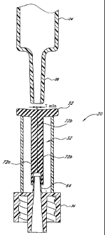
Figures 10-14 illustrate the function of the valve 20 as a syringe Luer 28 is
inserted into and withdrawn from
the slit 38. Figure 10 shows the valve 20 prior to insertion of the syringe
Luer 28; at this point the slit 38 defines a
substantially closed or highly restricted flow path through the seal 32,
marked by a very thin (or substantially
nonexistent) path thickness Tmin between slit walls 72a, 72b. This thin or
nonexistent path thickness Tmin prevails
along most or substantially all of the length of the slit 38 below the flange
44. This condition restricts fluid flow
through the seal 32 so as to seal off the catheter 22 (see Figure 1) or other
medical device connected to the Luer lock
34. At this point the slit 38 also defines a relatively small interior volume
Vmi, within the seal 32, between the slit
walls 72a, 72b. (As used herein in reference to an interior volume of the
seal, "relatively small" means a volume that
is either nonexistent or clinically negligible in size.) In this initial
state, the seal 32 is situated upon the lead cannula 64
such that substantially none of the lead cannula 64 extends into the slit 38.
Figures 11 and 12 show the valve 20 after the syringe Luer 28 has been
completely inserted into the slit 38.
The seal 32 has also been stretched or forced downward onto the lead cannula
64, at least part of which penetrates
into the slit 38 itself. At this point the slit 38 defines an expanded flow
path through the seal 32, in that the slit walls
72a, 72b have spread to a path width Tmax. The seal 32 thus permits fluid to
flow between the syringe 24 and the
catheter 22. In addition, the slit 38 now defines a larger or maximum interior
volume Vmax= Vmax comprises the entire
space between the slit walls 72a, 72b less the volume taken up by the cannula
(but not the internal lumen) of the
syringe Luer 28 and less that portion of the lead cannula 64 which has
penetrated into the slit 38. Accordingly, under
pressure exerted via the syringe 24 an amount of fluid substantially
equivalent to Vmax now fills the slit 38 between the
slit walls 72a, 72b. This is also shown as gaps 57 in Figures 8C and 8D.
Figures 13 and 14 show the function of the slit 38 as the syringe Luer 28 is
withdrawn from the valve 20.
As the syringe Luer 28 and lead cannula 64 exit the slit, the slit walls 72a,
72b retract to substantially their original
configuration to once again define a narrow path width (approaching Tmin)
between them. This retraction of the slit
walls 72a, 72b reduces the volume between the walls; that is, the internal
volume within the slit 38 is decreasing from
Vmax= Thus the amount of fluid within the slit must also decrease from Vmax.
Accordingly, the retracting slit walls 72a,
72b displace the fluid from the slit 38 as the syringe Luer 28 is withdrawn.
-9-
CA 02638744 2008-09-15
WO 02/04065 PCT/US01/21904
The fluid thus displaced cannot flow out of the slit 38 through the top of the
seal 32. As detailed above with
regard to Figures 8A-8B, the slit 38 maintains a tight seal against the
syringe Luer 28 at the region 50 of minimum
width as the syringe Luer 28 is withdrawn. In addition, the displaced fluid
cannot flow into the interior of the syringe
24 at all times relevant to the use of the valve 20. Therefore, substantially
all of the displaced fluid must exit the slit
38 through the lead cannula 64 and Luer lock 34, resulting in positive flow
from the valve 20 upon withdrawal of the
syringe Luer 28.
Figures 15-18 show variations on the valve 20 disclosed above, which
variations may be desirable under
certain operating conditions. For example, as seen in Figures 15 and 16 the
housing 30 may have a break 74 running
vertically between the axial opening 60 and one or both of the side openings
62a, 62b. The break 74 permits the seal
holder 58 to spread open as a Luer slip 28 (as opposed to a Luer lock 76 shown
in Figure 17) is inserted into the seal
32. This spreading action has been found to be advantageous for using the
valve 20 with a Luer slip 28, as the valve
becomes less likely to squeeze or pinch the Luer 28 out of the seal 32.
Figure 18 shows an alternative configuration of the housing 30, with a curved
or streamlined appearance in
comparison to the housing disclosed above. Both this type of housing or the
type disclosed above, may have an
15 external coating or layer of a relatively soft, pliant material such as a
thermoplastic elastomer to enhance operator
comfort and to promote the theme of a valve 20 that provides a connection
without the use of sharp, puncturing
elements such as needles or blades.
Figures 19A-21 depict a preferred method of making the seal 32. First, a pair
of preforms 202a, 202b are
molded between first and second mold pairs 204a, 204b and 206a, 206b
respectively. Each preform 202 has a
20 generally planar portion 208 that, in the completed seal 32, forms a wall
of the slit 38 (see Figures 6A-7B). A flange
portion 210 is also integrally molded into both preforms 202. The sides of the
flange portion 210 are preferably set
back from the upper face of the planar portion 208, to provide a space for
overmold material (discussed in further
detail below) to flow between and connect the flange portions 210. The molding
of the preforms 202 is accomplished
using conventional techniques and equipment, preferably by injecting a
thermoset material into the cavity formed
between the mold pairs 204a, 204b and 206a, 206b and heating the molds andlor
material to the set temperature of
the specific material used. Pressure may be applied as needed to prevent
material from leaking between the halves of
the mold.
After this initial molding step, the mold halves 204a, 206a, with the preforms
202a, 202b still positioned in
them, are pressed together with an overmold plate 212 positioned between the
mold halves, as depicted in Figures
19B-19C. The overmold plate 212, best seen in Figure 20 (with the outline of
the preforms 202 also shown in
phantom), comprises a generally planar plate body 214 with an overmold opening
216 cut into the body 214. The
overmold opening 216 has a plan perimeter that conforms to the outer edges of
the completed seal 32, and may
include a mandrel 218 that projects from the lower portion of the opening 216
and forms the lead lumen 46 (see
Figures 6A-7B) during the overmold process, as will be discussed in greater
detail below. The contacting faces of the
mold halves 204a, 206a and the overmold plate 212 are advantageously
substantially planar. Thus the mold halves
-10-
CA 02638744 2008-09-15
WO 02/04065 PCT/USO1/21904
204a, 206a, plate 212, and preforms 202a, 202b define a mold cavity or volume
220 between the walls of the
overmold opening 216 and the outer edges of the preforms 202a, 202b, and
between the faces of the mold halves
204a, 206a.
With the mold apparatus (mold halves 204a, 206a and overmold plate 212)
arranged as shown in Figure 19C,
additional thermoset material is injected into the mold apparatus to fill the
mold cavity 220 and form the remainder of
the seal 32. Preferably, the additional material is injected soon (i.e., a few
seconds) after the preforms 202 are molded
and while they are still somewhat, hot from the initial molding. The
additional material injected into the mold cavity
220 bonds to the edges of the preforms 202 and forms the edges of the slit 38
in the completed seal 32. In other
words, the remainder of the seal is overmolded onto the "sandwich" of preforms
202. Preferably, the preforms 202
are pressed together with sufficient force during the overmolding process to
prevent the additional material from
migrating between the contacting surfaces of the preforms 202. This preserves
the patency of the slit 38 by
preventing the contacting faces of the preforms 202 from bonding to each other
during the overmold step.
The overmold plate 212 may be made with a thickness approximately the same as
that of the "sandwich" of
preforms 202a, 202b to define a mold cavity 220 that, as described above,
comprises the open space between the
walls of the overmold opening 216 and the outer edges of the preforms 202a,
202b, and between the faces of the
mold halves 204a, 206a. This overmold opening thus also has a thickness
approximately equal to that of the preform
sandwich, and all or nearly all of the overmold material injected therein
bonds only to the edges of the preforms 202a,
202b. In an alternative embodiment, the overmold plate 212 may have a
thickness greater than the preform sandwich.
This thicker, alternative overmold plate thereby defines a mold cavity that
also includes open space that is created
between the mold halves 204a, 206a and the outer (i.e., facing away from the
slit in the completed seal) faces of the
preforms 202a, 202b. The mold halves 204a, 206a are preferably configured with
projections, ridges, channels, gaps
or the like to create such space during this alternative overmold step while
pressing the preforms together as may be
needed during the overmold. Accordingly, in this embodiment the overmold
material bonds to both the edges and to the
outer faces of the preforms 202a, 202b. In other words this alternative
overmold step involves injecting the overmold
material into a mold cavity that surrounds most or all of the preform
sandwich, rather than overmolding to the only the
edges of the preforms.
It is preferred that the material added in the overmold step is similar to
that utilized in molding the preforms
202; however, in other embodiments the preform material and the overmold
material may comprise different but
nonetheless suitable materials for manufacturing the seal, as discussed above.
Therefore as used herein "a flexible
material" refers to any material selected from the class of suitable seal
materials as disclosed.
After the overmolding is complete, the mold halves 204a, 206a are removed from
the seal plate 212, which
now contains a substantially completed seal 32, as seen in Figures 19D-19E.
The completed seal 32 is easily removed
from the seal plate 212, and the seal thus formed comprises, as discussed
above, a unitary mass of molded material
with the slit arranged within it.
.l 1-
CA 02638744 2008-09-15
WO 02/04065 PCT/USOI/21904
Referring to Figures 21 and 22, a catheter and valve assembly 300 is depicted
that may be used to deliver
fluids to the vasculature of a patient. The catheter and valve assembly 300
comprises an elongated cannula 302 and a
valve 304 connected to the cannula at its proximal end. In one embodiment, the
cannula 302 comprises a PICC
cannula. It is intended that the valve 304 generally resembles the valve 20
disclosed in detail above; however, as
shown in Figure 21 the valve 304 may be connected to the cannula 302 via a
barbed fitting 306 integrally formed with
the valve 304. Of course, other types of connection may be employed,
including, but not limited to, an adhesive,
chemical bonding, threads, andlor an ultrasonic-welded connection. In order to
facilitate insertion of the catheter 302
into a patient's vasculature a guidewire 308 may be disposed within the lumen
of the cannula 302, extending through
the seal 32 of the valve 304. In the event that the cannula 302 includes an
opening at the distal end thereof, the
guidewire can be threaded into the bloodstream of a patient and, thereafter,
the cannula 302 and valve 304 assembly
may be slid distally on the guidewire 308 until the distal end of the cannula
302 is also within the bloodstream of a
patient. Thereafter, the guidewire 308 may be removed leaving the cannula 302
in place in the bloodstream as will be
understood by those of skill in the art. Alternatively, the guidewire 308 and
cannula 302 may be simultaneously
placed in fluid communication with the bloodstream of a patient and,
thereafter, the guidewire may he removed.
In the event that the catheter 302 does not include either a guidewire lumen
therein or an opening in the
distal end of the cannula 302, a guidewire 308 may not be necessary. If such a
cannula 302 is used, an introducer
needle known to those of skill in the art may be used to introduce the
catheter and valve assembly 300. The
introducer needle may be a split type needle so that the introducer needle may
be withdrawn from the patient once the
cannula 302 is properly placed with a distal end thereof in the bloodstream of
a patient. The catheter and valve
assembly 300 may be introduced into the bloodstream of a patient using many
methods known in the art for
introduction of catheters into a patient. Use of the catheter and valve
assembly 300 with any insertion method is
within the scope of the present invention. Moreover, the valve 304 may be used
with any catheter 302 known to
those of skill in the art.
Figure 22 shows the catheter 302 in fluid communication with the vasculature
of a patient, via an insertion
site in the patient's arm. However, any other suitable insertion site may be
used for the catheter 302. As discussed
above, various insertion techniques may be used. For example, a conventional
introducer sheath or needle (not shown)
may be first'placed in the insertion site, and the catheter 302, with or
without the guidewire 308 positioned therein,
advanced through the introducer sheath until the distal portion of the cannula
302 lies within the patient's vasculature.
Alternatively, the guidewire 308 alone may be first inserted through the
sheath and into the target vessel, and the
cannula 302 subsequently advanced over the guidewire, through the sheath and
into the vessel. With any of these
insertion techniques, the introducer sheath may advantageously be of the peel-
away type, so as to promote easy
removal of the sheath after the guidewire and/or cannula has been advanced
through it and into the patient. As a
further alternative, the catheter 302 may be inserted without the assistance
of an introducer needle or sheath. Where
the guidewire 308 is used, it is advantageously withdrawn after the assembly
300 has been inserted, freeing the
catheter 300 for use as a fluid-delivery or fluid withdrawal device.
-12-
CA 02638744 2008-09-15
WO 02/04065 PCT/US01/21904
Upon insertion of a distal portion of the cannula 302 into the patient's
vasculature, the valve 304 functions
as a catheter hub to facilitate connection andlor exchange of various medical
devices to the cannula, as well as the
delivery of fluids to the patient through the cannula. All of these functions
may be performed while preserving the
advantages of the valve 20 discussed at length above, i.e. positive-flow
characteristics, fluid-tight sealing of the
cannula, etc. As an example, a Luer-type syringe tip (see Figures 1, 10-17)
may be inserted into the valve 304 and the
syringe operated to introduce fluid through the valve 304 and cannula 302, and
into the patient's vasculature. Upon
withdrawal of the syringe tip, the valve 304 re-seals the proximal and of the
cannula 302 and creates positive flow as
discussed above. Alternatively, blood may be withdrawn through the cannula 302
and valve 304.
It is contemplated that any suitable medical device may be connected to the
valve 304, such as IV bags,
additional cannulae, etc., for the purposes of fluid transfer or for any other
desired purpose. As seen in Figure 23, a
connector 400 may be connected to the valve 304 and placed in fluid
communication with the cannula 302 and the
patient. This arrangement can provide several advantages in situations which
call for the use of a unique connector.
For example, when it is necessary to replace the connector 400, it may be
removed from fluid communication with the
cannula 302 without exposing the cannula (or the patient's vasculature) to the
open air, and replaced with a similar
connector or any other medical implement. As discussed previously, the valve
304 re-seals the cannula 302 while the
connector 400 is being replaced, which also prevents blood from flowing from
the patient and out the proximal end of
the cannula 302 when the connector 400 is absent. Thus, the catheter 300
advantageously prevents both infection
and blood loss when used in common clinical applications. As shown in Figure
23, one such connector 400 may be the
CLAW connector sold by ICU Medical, inc. However, any connector or other
medical implement or device may be
placed in fluid communication with the valve 304 to introduce fluid to the
patient or to withdraw blood from the
patient including, but not limited to, pierceable connectors, needleless
connectors, medical tubing, syringes or any
other medical implement or device. Thus, advantageously the catheter 302 and
valve 304 assembly 300 creates a
closed, swabable catheter hub which prevents patient infections and
inadvertent loss of blood among other
advantages.
The valve 304 may also be used with a standard hub of a catheter. Thus, the
valve 304 and catheter 302
may be an integral unit or removably secured by luer threads as shown in
Figure 23 or other attachment mechanisms
known to those of skill in 'the art. In Figure 23, the catheter.302 includes
an integral hub 303 at the proximal end
thereof. A distal end of valve 304 is threadably engaged with the hub 303 to
place the valve 304 in fluid
communication with the catheter 302 without leakage. Upon replacement of a
connector 400 in this embodiment, the
withdrawal of the connector 400 causes the valve 304 to create a positive
displacement and prevents the distal end of
the catheter 302 from occluding. The seal 32 of the valve 304 may be cleaned
as discussed herein and a new
connector of the same type or a different type may be placed in fluid
communication with the valve 304 causing the
seal 32 to open and establish fluid flow between the connector 400 and the
patient through valve 304 and catheter
302.
-13-
CA 02638744 2008-09-15
WO 02/04065 PCT/US01/21904
Although this invention has been disclosed in the context of certain preferred
embodiments and examples, it will
be understood by those skilled in the art that the present invention extends
beyond the specifically disclosed embodiments
to other alternative embodiments and/or uses of the invention and obvious
modifications and equivalents thereof. Thus, it
is intended that the scope of the present invention herein disclosed should
not be limited by the particular disclosed
embodiments described above, but should be determined only by a fair reading
of the claims that follow.
-14-