Note: Descriptions are shown in the official language in which they were submitted.
CA 02639954 2015-07-15
DROPLET-BASED CELL CULTURE AND CELL ASSAYS USING DIGITAL
MICROFLUIDICS
FIELD OF THE INVENTION
The present invention relates to droplet-based cell assays and/or cell
culture using digital microfluidics, and more particularly, the present
invention
relates to devices and methods used with those devices for performing cell
assays and/or cell culture.
BACKGROUND OF THE INVENTION
The cell is the irreducible element of life and is often studied as a living
model of complex biological systems. Cell-based assays are conventionally
performed in well plates that enable simultaneous analysis of multiple cell
types
or stimuli. For such multiplexed analyses, cells in well plates are often
evaluated
using microplate readers, which can be integrated with fluid handling and
other
miscellaneous equipment in a robotic analysis platform. A major drawback of
such systems is the expense of the instrumentation and the experimental
1
CA 02639954 2008-09-26
consumables (e.g., plates, pipette tips, reagents, and cells). The latter is a
particular disadvantage for cell-based assays as they are generally more
complex and require larger amounts of reagents than cell-free assays.1
Recently, microfluidics has been touted as a solution for the challenges
inherent in conducting multiplexed cell-based assays.2 The conventional format
for microfluidics, which is characterized by devices containing networks of
micron-dimension channels, allows integration of multiple processes on a
single
platform while reducing reagent consumption and analysis time. There are
numerous advantages of using microfluidic based systems for cell assays, some
of which are self-similarity in dimensions of cells and microchannels (10 -
100 pm
widths and depths), laminar flow dominance and formation of highly resolved
chemical gradients, subcellular delivery of stimuli, reduced dilution of
analytes,
and favorable scaling of electrical and magnetic fields. For the last ten
years,
researchers have used microchannels to manipulate and sort cells, to analyze
cell lysates, to assay intact-cell biochemistry, and to evaluate cell
mechanical
and electrical responses. In most of these studies, cells were exposed to one
stimulus or to a limited number of stimuli. There have been just a few
attempts to
conduct multiplexed assays as it is difficult to control many reagents
simultaneously in a complex network of connected channels, even when using
microvalve architectures developed for microfluidic devices.3 Finally, we note
that there have been only a few microfluidic devices integrated to multiplexed
detection instruments such as microplate readers;4 we believe this will be a
2
CA 02639954 2008-09-26
necessary step for the technology to become competitive with robotic screening
systems.
A potential solution to the limitations of the channel-microfluidic format is
the use of "digital" or droplet-based microfluidics. In digital microfluidics
(DMF),
discrete droplets containing reagents are manipulated by sequentially applying
potentials to adjacent electrodes in an array.5-14 Droplets can be manipulated
independently or in parallel on a reconfigurable path defined by the electrode
actuation sequence, which allows for precise spatial and temporal control over
reagents. As with all microscale techniques, cross-contamination is a concern
for DMF, but this phenomenon can be avoided by dedicating separate paths for
each reagent. DMF has been used to actuate a wide range of volumes (nL to pL)
and, unlike channel devices, there is no sample wasted in creating small plugs
for analysis. In addition, each droplet is isolated from its surroundings
rather
than being embedded in a stream of fluid ¨ a simple method of forming a
microreactor in which there is no possibility that products will diffuse away.
The
preservation of products in a droplet is of great importance in cell assays
targeting molecules secreted from cells into extracellular space. In addition,
droplets provide mostly static fluid conditions without unwanted shear stress
that
is inevitable in continuous flow microfluidics. A further advantage of DMF is
its
capacity to generate nanoliter samples by translating droplets through
selective
wettability areas on an electrowetting-based platform.15
There is currently much enthusiasm for using DMF to implement
multiplexed assays; however, it has only been applied to a few non-cell
assays.
3
CA 02639954 2008-09-26
To the inventors' knowledge, there are no reports of the use of DMF to analyze
cells. There are a few studies demonstrating only dispensing and manipulation
of
droplets containing cells, cell sorting, and cell concentration on a DMF
platform.
WO 2007/120241 A2 entitled "Droplet-Based Biochemistry"18 discloses
dispensing and dividing droplets containing cells, generating droplets with
single
cells, detecting a type of cell, and sorting cells. US20070148763 Al entitled
"Quantitative cell dispensing apparatus using liquid drop manipulation"17
describes cell droplet handling, to achieve a predetermined number of cells.
In a
journal paper by Fan et a1,18 dielectrophoresis was used to concentrate
neuroblastoma cells within droplets on a DMF platform.
It would be very advantageous to provide droplet-based cell culture and/or
assays using digital microfluidics in order to enable automated cell micro
culture
and high-throughput screening ability for cell analysis. DMF would also
address
some problems associated with standard culture and assaying in well-plates or
in
continuous-flow microfluidic devices.
SUMMARY OF INVENTION
The present invention provides embodiments of devices and methods for
droplet-based cell culture and assays using digital microfluidic devices
designed
to manipulate, operate, and analyze cell-containing droplets. Cells in a
suspension and cell-assay and/or cell-culture reagents are deposited in the
device by either dispensing them from device reservoirs or dispensing them
into
the device using external means (e.g., pipette, robotic dispenser, etc.). In
order
to perform an assay with cells in suspension, cell-containing droplets and
4
CA 02639954 2008-09-26
reagent-containing droplets are moved between adjacent electrodes by applying
voltages to electrodes. General assay protocol comprises dispensing and
translating droplets, merging and mixing droplets with cells and reagents at
least
once, possible splitting of droplets, incubating cells with reagents in
merged/mixed (and split) droplets at least once, and detecting signal from
cells in
merged/mixed (and split) droplets in the device after final incubation. Using
the
same DMF techniques, suspended cells are also long-term cultured and split at
regular time intervals.
Additionally, DMF devices are designed to culture and assay adherent
cells. After being introduced in a device in suspension, adherent cells are
seeded
on cell culture sites (patterned DMF device surface for cell attachment),
where
they can be long-term cultured in droplets, subcultured using standard
subculture
protocols, and assayed. Media exchange and regent delivery on cell culture
sites
(CSSs) is performed using standard DMF operations: translating, merging,
mixing and splitting droplets. In addition, a new technique, passive
dispensing, is
developed for more efficient delivery of reagents/media from big source
droplet
translating over CCSs. By means of DMF and passive dispensing, a first
multigenerational cell culture in a microscale is realized.
Culture and assay reagents comprise chemical, biochemical and
biological reagents. Droplets contain additives including pluronics and
various
hydrophilic polymers to facilitate cell-containing droplet actuation by
preventing
non-specific adsorption of cells and proteins to a device surface.
5
CA 02639954 2008-09-26
In a multiplexed assay, multiple cell-containing droplets (which may
include one kind or multiple kinds of cells) are manipulated and assayed
simultaneously or in a certain sequence with one or multiple reagents.
Thus, in an embodiment of the present there is provided a digital
microfluidic based method of performing any one or both of cell assays and
cell
cultures, comprising the steps of:
a) providing a digital microfluidic device having an array of actuating
electrodes formed on a substrate surface, a coating having a working surface
coating the substrate surface and array of actuating electrodes, an actuating
electrode controller for exciting or de-exciting said actuating electrodes for
translating liquid droplets over said working surface;
b) dispensing one or more first droplets containing a suspension of at least
one kind of cells onto one or more first positions on a working surface of the
digital microfluidic device above the array of actuating electrodes and
substrate
surface, and dispensing one or more second droplets containing any one of at
least one chemical reagent, at least one biochemical reagent, at least one
biological reagent, and any combination thereof onto one or more second
positions on the working surface;
c) translating each of the one or more first and second droplets to a
corresponding third position on the working surface such that they
substantially
mix to form one or more secondary droplets;
d) incubating the one or more secondary droplets; and
6
i
CA 02639954 2008-09-26
e) analyzing the one or more secondary droplets to identify products
produced by incubation of the one or more secondary droplets.
In another aspect of the present invention there is provided a digital
microfluidic device for conducting one or both of cell assays and cell
culture,
comprising:
a first substrate having a first substrate surface;
an array of actuating electrodes formed on the first substrate
surface;
at least one dielectric layer formed on the first substrate surface
covering each actuating electrode such that the actuating electrodes are
electrically insulated from one another; and
at least one reference electrode, wherein each actuating electrode
is proximal to at least one of the reference electrodes;
an electrode controller capable of selectively exciting or de-exciting
actuating electrodes for translating liquid droplets across a surface of the
dielectric layer;
one or more first reservoirs in flow communication with the surface
of said dielectric layer for holding at least one suspension of cells and one
or more reagent reservoirs in flow communication with the surface of said
dielectric layer for holding one or more cell assay reagents, cell culture
reagents; and
7
CA 02639954 2008-09-26
dispensing means for dispensing droplets of said at least one
suspension of cells and droplets of said at least one cell assay reagents,
cell culture reagents onto said surface of said dielectric layer; and
a computer controller interfaced to said dispensing means and said
electrode controller and being programmed to dispense droplets of the
suspension of cells and droplets of said one or more cell assay reagents,
cell culture reagents onto said surface of said dielectric layer and
translating them over said array of actuating electrodes for mixing and
optionally splitting said droplets in selected positions on said array of
actuating electrodes to form one or more secondary droplets in a selected
order defined by a selected cell assay protocol or cell culture protocol for
which said computer controller is programmed.
A further understanding of the functional and advantageous aspects of the
invention can be realized by reference to the following detailed description
and
drawings.
BRIEF DESCRIPTION OF THE DRAWINGS
Preferred embodiments of the invention will now be described, by way of
example only, with reference to the drawings, in which:
Figure 1 is a top view of a complete digital-microfluidic device showing
three droplet sources: cells, reagent, and dye;
Figure 2(a) shows a cross-sectional view of the device of Figure 1;
8
,
CA 02639954 2008-09-26
Figure 2(b) shows a cross sectional view of an alternative embodiment of
the device of Figure 1 which uses a one-plate design;
Figures 3(a) to (c) show three frames from a movie wherein a droplet with
cells is dispensed from a reservoir;
Figure 4 is a plot of numerically simulated potential drops across a droplet
and a dielectric layer;
Figure 5 is a graph of viability and proliferation tests for cells actuated by
digital microfluidics showing no significant differences between the actuated
and
non-actuated cells;
Figures 6(a) and (b) are graphs of vitality tests wherein cells in droplets
were actuated, lysed, and analyzed by Matrix Assisted Laser Desorption
Ionization Mass Spectrometry (MALDI-MS) showing no major qualitative
differences between the (a) actuated and (b) non-actuated cells;
Figures 7(a) to (f) show sequential images from a movie depicting a
digital microfluidic cell-based assay;
Figures 8(a) and (b) show fluorescent images of droplets with cells
treated with (a) 0% and (b) 0.5% Tween 20 and stained with viability dyes
(calcein AM and ethidium homodimer-1); in the droplet (a), almost all cells
were
live (dead cells in (a) are marked with small circles), and in the droplet
(b), all
cells were dead;
Figures 9(a) and (b) show two dose-response curves for Jurkat T-cells
exposed to Tween 20 (0.002% to 0.5% (v/v)) using (a) a digital microfluidics
assay and (b) a well-plate assay;
9
1
CA 02639954 2008-09-26
Figure 10 shows a top view of an embodiment of a DMF device for
multiplexed cell assays which comprises reservoirs for four different cell
suspensions and nine different assay reagents, and a waste reservoir;
Figures 11(a) to (d) are diagrammatic representations of seeding
adherent cells in a DMF device where (a) shows actively dispensed droplet of
cell suspension translating to a cell culture site (CCS), (b) shows passively
dispensing a droplet of cell suspension onto the CCS from a source droplet,
(c)
shows cells in suspension seeded on the CCS, and (d) shows cell monolayer
formed on the ECM substrate on the CCS;
Figure 12 is a diagrammatic representation showing passive dispensing
of a droplet where a source droplet provides a smaller liquid droplet located
on
the CCS;
Figure 13 shows several examples of the hydrophilic area positions
relative to actuating electrodes and to the source droplet path;
Figure 14 shows a diagrammatic representation showing a passive
washing/exchange process whereby a droplet on a CCS is replaced by a new
droplet;
Figure 15 shows a graph of fluorescein fluorescence signal intensity
versus washing cycle to show washing efficiency;
Figure 16 shows a digital image of ¨130 mouse fibroblast cells (NIH-3T3)
cultured in a DMF device for 72h; media was replenished using passive
dispensing/exchange technique every 24h; after 72 h cells were stained with
calcein AM for viability;
CA 02639954 2008-09-26
Figures 17(a) to (f) are diagrammatic representations of subculturing
adherent cells in a DMF device in which (a) shows monolayer of adherent cells
cultured on a CCS, (b) washing cells via passive exchange, (c) delivering a
dissociation agent to cells via passive exchange, (d) detachment of cells
after
incubation with a dissociation agent, (e) blocking of a dissociation agent and
resuspending cells via passive exchange, and (f) seeding of cells resuspended
in
fresh media on a new CCS;
Figures 18(a) to (d) show diagrammatic representations of assaying
adherent cells in a DMF device where, (a) shows a monolayer of adherent cells
cultured on a CCS in cell culture media, (b) washing cells and delivering
assay
reagents to cells via passive exchange, (c) incubating cells with assay
reagents,
and (d) detecting and analyzing cell response to assay stimuli; and
Figure 19 shows a DMF device for multiplexed cell assays with adherent
cells using passive dispensing and passive reagent exchange.
DETAILED DESCRIPTION OF THE INVENTION
Without limitation, the majority of the systems described herein are
directed to methods and devices for droplet-based cell assays using digital
microfluidics. As required, embodiments of the present invention are disclosed
herein. However, the disclosed embodiments are merely exemplary, and it
should be understood that the invention may be embodied in many various and
alternative forms.
11
,
CA 02639954 2008-09-26
The figures are not to scale and some features may be exaggerated or
minimized to show details of particular elements while related elements may
have been eliminated to prevent obscuring novel aspects. Therefore, specific
structural and functional details disclosed herein are not to be interpreted
as
limiting but merely as a basis for the claims and as a representative basis
for
teaching one skilled in the art to variously employ the present invention. For
purposes of teaching and not limitation, the illustrated embodiments are
directed
to droplet-based cell assays and culture using digital microfluidics (DMF).
As used herein, the term "about" and the symbol " ", when used in
conjunction with ranges of dimensions, temperatures or other physical and/or
chemical properties and/or characteristics is meant to cover slight variations
that
may exist in the upper and lower limits of the ranges of dimensions as to not
exclude embodiments where on average most of the dimensions are satisfied but
where statistically dimensions may exist outside this region. For example, in
embodiments of the present invention dimensions of a digital microfluidic
device
are given but it will be understood that these are not meant to be limiting.
Figure 1 shows a top view of a microfluidic device shown generally at 10
which may be used for droplet-based cell culture and cell assays using digital
digital microfiuidics in accordance with the present invention. Reservoir
electrodes 32, 34, and 36 store droplets 42, 44, 46 containing cells, reagent,
and
dye, respectively, and are capable of dispensing the liquids onto the center
region 38 of the device. Small volumes of liquids are dispensed as droplets
and
translated by applying voltages to actuating electrodes 14. There is also
another
12
CA 02639954 2008-09-26
reservoir electrode 30 shown in the device in Figure 1 which may be used as a
reservoir as well.
Figure 2(a) is a cross-sectional view of a portion of the microfluidic device
of Figure 1 showing two adjacent electrodes 14 of the electrode array.
5 Electrodes 14(10 nm Cr+, 100 nm Au) rest on a substrate layer 12 and are
separated from each other by a dielectric material 16 (for example 2 pm
Parylene-C). The device can have more than one dielectric layer16. Located on
top of dielectric material 16 is a hydrophobic layer 18 (for example Teflon
AF, 50
nm). The array of actuating electrodes and exposed areas of substrate surface
10 are thus covered by a working surface. Spaced above electrodes
14/dielectric
layer 16 is a continuous reference electrode 22 coated on a substrate layer
24,
and a hydrophobic layer 20 (for example Teflon AF, 50 nm) is coated on
reference electrode 22. Alternatively, another dielectric layer can be
deposited
between layers 20, 22. Liquid droplets 42 rest in-between two hydrophobic
layers 18 and 20. Electrodes 14, voltage source 26, and the continuous
reference electrode 22 together form an electric field, digitally manipulated
by
controller 28. For droplet manipulation, reference electrodes 22 are biased to
a
potential different from the actuating potential. Commonly used reference
potential is ground.
In a preferred embodiment of the present invention, the upper hydrophobic
layer 20, reference electrode 22, and substrate layer 24 are substantially
transparent to allow optical analysis of the assays. Furthermore, layers 20,
22,
and 24 are not necessary to translate droplets.
13
CA 02639954 2008-09-26
While the present invention discusses the two-plate design of Figure 2(a),
a one-plate design is also possible, as shown in Figure 2(b). In Figure 2(b),
layers 20, 22, and 24 are removed. Rather than have a dedicated reference
electrode layer 22, the reference electrode is patterned adjacent to
electrodes
14, forming a continuous grid 52 separated from electrodes 14 by dielectric
material 16. The continuous grid 52 extends in both directions defining the
plane
in which electrodes 14 are located.
Reference electrodes can also be coplanar with the top surface of the
dielectric layer. In a device with multiple dielectric layers, reference
electrodes
can be coplanar with the top surface of any dielectric layer, while being
insulated
from actuating electrodes 14. The design of reference electrodes is not
limited to
a grid, e.g. they can be in a form of a wire or an array similarly to
electrodes 14.
Figure 3 shows three frames from a movie wherein a 150 nL droplet 42
containing ¨260 cells is dispensed from a reservoir of a microfluidic device
with
identical dimensions but fewer electrodes than the microfluidic device 10
shown
in Figure 1, wherein cells were labeled with a viability dye, calcein AM,
which
fluoresces green.
Figures 7(a) to (f) show sequential images from a movie depicting a
digital microfluidic cell-based assay, wherein a 150 nL droplet 42 containing
¨525
cells was dispensed (a, 402), translated (b, 404), and merged (c, 406) with a
150 nL droplet 44 of Tween 20 dispensed (b, 402) from a second reservoir. The
merged droplet was actively mixed (408) on four neighboring electrodes (d);
after
20 min incubation in a humidified environment, the combined droplet was merged
14
CA 02639954 2008-09-26
(e, 406) and mixed (e, 408) with a 150 nL droplet 46 containing viability
dyes.
The final droplet was incubated (f, 410) for 20 minutes in a humidified
environment.
A sample result of the microfluidic cell-based assay of Figure 7(f) is
shown in Figures 8(a) and (b), wherein fluorescent images of droplets treated
with (a) 0% and (b) 0.5% Tween 20. Calcein AM (green) was used to stain live
cells, and ethidium homodimer-1 (red) for dead cells. In the former droplet
(a),
almost all cells were live (dead cells in (a) are marked with small circles),
and in
the latter (b), all cells were dead.
While digital microfluidics has been used previously to manipulate and
evaluate a wide range of liquids and reagents, we report herein the first
application of digital microfluidics to transport, analyze and culture
biological
cells. Using the parameters reported in the experimental section (elaborated
below), cell suspensions representing a wide range of concentrations
(including
very dense solutions of 1 x 10⁸ cells/mL) were found to be feasible to be
actuated
by DMF, with no differences observed in velocity or reliability relative to
liquids
not containing cells.
For example, Figures 3(a) to (c) depict a routine operation irrour
experiments: dispensing of a 150 nL droplet containing ¨260 Jurkat T-cells.
However, in initial work (with un-optimized parameters), droplets containing
cells
were difficult to manipulate, as cells tended to stick to the surface of the
devices,
causing contact line pinning. This problem was overcome by the use of the non-
ionic surfactant, pluronic F68, which when used as a solution additive,
facilitated
CA 02639954 2008-09-26
actuation of suspensions of cells in all liquids tested (including PBS and
complete
media containing 10% fetal bovine serum).
Pluronics are block copolymers formed from poly(propylene oxide) (PPO)
and poly(ethylene oxide) (PEO), and are commonly used as surface coatings for
preventing non-specific protein adsorption. In our work, we used pluronics in
solution, rather than as a surface coating; we hypothesize that in this
configuration, the polymer coats cells and proteins in a manner such that
their
functionality is retained, but adsorption to hydrophobic surfaces is
minimized.
We note that pluronic F68 has been used extensively in cell-based assays with
no evidence for detrimental effects on cell vitality,1920 and it is even used
as a
constituent in commercial cell growth media.21 Our experiments support this
trend ¨ Jurkat T-cells incubated in medium containing 0.2% (wt/vol) F68 for 4
days (humidified incubator, 5% CO2, 37 C) had identical growth rates and
morphology as cells grown in media without pluronics. In on-going work, the
optimal conditions (concentration and type of pluronic, etc.) for reducing
unwanted adsorption in DMF are being evaluated; we used F68 for all of the
results reported here.
A second challenge for using DMF for actuation of cells is droplet
evaporation, which raises the concentration of salts and other buffer
constituents,
making the solution hypertonic. In the work described here, we controlled
evaporation by positioning devices in a humidified atmosphere when not
actively
manipulating droplets by DMF. For the duration of the assay experiments (up to
a
few hours), such measures prevented significant evaporation, and no negative
16
1
CA 02639954 2008-09-26
effects on cell viability were observed. For culturing cells, devices were
placed in
cell culture incubators at 37 C and 5 % CO2. The DMF devices may be
contained in a sterile, humidified chamber for the full duration of the assay
or cell
culture process (including actuation, incubation, and analysis) or culture
which
facilitates long-term cell culture and examination.
Effects of DMF Manipulation on Cell Vitality.
Digital microfluidic devices use electrical fields to actuate droplets, which
led us to investigate the effects of droplet actuation on cell vitality. As
described
above, droplets are translated by an energized actuating electrode 14 on a
bottom plate and a reference electrode 22 on a top plate (Figure 2(a)). It
should
be noted that the reference electrode may also be placed on the bottom plate,
as
in reference electrode 52 (Figure 2(b)). Because of the high conductivity of a
droplet 42 of phosphate buffered saline (PBS) relative to the insulating
dielectric
layer 16 formed from Parylene-C, the inventors believe that cells would
experience negligible electrical field upon application of driving potentials.
This
hypothesis was supported by a numerical simulation using the COMSOL
Multiphysics 3.3a analysis package. In a simulation, shown in Figure 4, in
which
100 V was applied between top and bottom electrodes, the potential drop in the
droplet was found to be only 3.73 x 10⁻⁸ V, or 0.00000004% of the applied
potential. Thus, it is contemplated that one would expect to observe modest
effects (if any) on the vitality of suspensions of cells, upon application of
electrical
field. These effects were evaluated by three tests, measuring cell viability,
proliferation, and biochemistry.
17
CA 02639954 2008-09-26
As shown in Figure 5, the viability of actuated and non-actuated cells was
compared immediately after actuation, and proliferation was measured after 48-
h
incubation in a humidified incubator. There was no significant difference
between actuated and non-actuated cells (P = 0.11 for the viability assay,
P = 0.43 for the proliferation assay).
Cell biochemistry was evaluated qualitatively by analyzing lysates with
MALDI mass spectrometry. Figure 6 (a) and (b) show spectra of lysates of
actuated cells and non-actuated cells, respectively. From previous studies of
protein content in Jurkat T-cells,22 we tentatively assigned several peaks,
including heat shock protein (HSP10) 302, macrophage migration inhibitory
factor 304, epidermal fatty-acid binding protein (E-FABP) 306, and peptidyl-
prolyl
cis-trans isomerase A 308. As shown, there are no major qualitative
differences
between the two spectra, which suggests that actuation by DMF does not cause
catastrophic effects on cell biochemistry. We note that MALDI-MS is not a
quantitative analysis technique (i.e., peak heights can vary considerably
within
multiple spectra of a single sample) The gene expression of T-cells and other
cell
types using quantitative PCR or gene microarray would be more appropriate
quantitative techniques.
Cell Phenotype Assays by DMF.
To illustrate that DMF is compatible with phenotypic assays, a dose-
response toxicology screen was performed using Jurkat T-cells, shown in
Figures 7 and 8. Cells were exposed to varying concentrations of the
surfactant,
Tween 20 (0.002% to 0.5% (v/v)) (Figure 7) and then stained with viability
dyes
18
CA 02639954 2008-09-26
(Figure 8). The complete assay, from droplet dispensing to the final
incubation
with dyes was performed on-chip. 150 nL droplets (-1 mm in diameter) were
dispensed via DMF, and after merging and incubation, resulted in a final -450
nL
droplet (-1.8 mm diameter, 150 pm height). An equivalent assay was
implemented in a 384-well plate with the same number of cells (-525 cells/well
or
droplet) but different sample volume. In the well-plate assays, 5 pL aliquots
of
each reagent were pipetted into conical wells (3.3 mm top-, 2 mm bottom
diameter) resulting in a final volume of 15 pL (-5 mm height) which is in the
recommended range for 384-well plates. Hence, well plates required -30-fold
greater reagent use than DMF, leading to a much lower cell concentration in
the
wells. As described below, this had significant effects on assay sensitivity.
A fluorescence microplate reader was used to generate dose response
curves for DMF and well plate assays using identical settings (Figure 9, error
bars are 1 standard deviation). As shown in Figure 9, the DMF assays (a) had
much lower background signals than the well-plate assay (b), resulting in a
much
larger signal-to-noise ratio than the well-based assays (b). As a consequence,
the lowest detectable number of live cells in droplets was -10 (a), compared
to
-200 cells in wells (b). The latter value matches the general limits of
detection
listed by the manufacturer for such assays. One consequence of this difference
was the determination of different 100% - lethal concentrations of Tween 20:
-0.5% (v/v) from the DMF assay and -0.03% (v/v) from the well plate assay. The
true 100%-lethal concentration was determined empirically by staining cells
exposed to varying concentrations of Tween-20 and counting them using a
19
CA 02639954 2008-09-26
hemacytometer. At the concentrations evaluated here, the fluorescence
microplate reader results generated by the digital microfluidic method (a)
were
found to be a much better approximation of the empirical value than the
conventional method (b). Thus, in this assay, the conventional method over-
estimates the toxicity of Tween 20 by more than 15-fold; this is important, as
cytotoxicity is widely used by regulatory agencies in initial screens for
determining acceptable exposure limits, and by the pharmaceutical industry in
early drug discovery.
Another cause of the improved sensitivity in droplet-based assays is the
high cell concentration in -nL droplets. The same number of cells in pL
aliquots
results in a much lower concentration and therefore, lower signal-to-noise
ratio.
In this experiment, 525 cells yielded 1.2 x 10⁶cells/mL in droplets, but only
3.5 x 10⁴ cells/mL in wells. In addition, the cross-sectional density of cells
in
droplets was higher because of the slightly smaller droplet diameter (-1.8 mm)
relative to that of the conical wells (2 mm bottom, 3.3 mm top). If it is
assumed
that all cells settled to the bottom of each well or droplet, then the same
number
of cells was distributed over an area that was -20% smaller in droplets
relative to
wells, resulting in a higher signal. It is possible that all cells sedimented
in
droplets (150 pm height), while not all cells sedimented in wells (-5 mm
height).
If this were the case, it would obviously contribute to the observed
differences in
detection limits.
It should be noted that while the assay described above involved
dispensing, translating, merging and mixing of droplets, other embodiments of
CA 02639954 2008-09-26
cell assays and cell culture in DMF devices can include droplet splitting.
Droplet
splitting is implemented to reduce a droplet size, number of cells in a
droplet, etc.
Some cell assays target molecules that cells secrete into their
microenvironment, such as growth factors, signaling molecules, and metabolic
products. Since DMF droplets of cell suspension are precise, confined volumes
where all cell products are preserved, they are ideal microenvironment for
extracellular biochemistry assays. In these assays, signal is detected from a
suspension medium rather than cells. Suspension medium can be analyzed by
immunoassays or other means. Droplets of cell suspension can alternatively be
removed from a DMF device and analyzed externally.
The results presented above demonstrate assaying population of cells of
one kind; nevertheless, it is also possible to assay droplets containing
multiple
kinds of cells (e.g., different cell types, or different phenotypes of the
same cell
type). Droplets with multiple kinds of cells can be generated by either
dispensing
them from reservoirs containing the same mixed population of cells, or by
combining droplets containing one or several kinds of cells. Combining
droplets,
merging and mixing, results in larger droplets which can be split in droplets
of
desired size.
Concentration of cells in a droplet can be controlled by the concentration
of cells in a source (a device reservoir or an external reservoir) or by
combining
droplets of suspended cells with droplets of cell suspension medium. In this
way,
concentration of cells is reduced by the ratio of the combined volumes.
Combined droplet can be split in smaller droplets which can be further merged
21
1
CA 02639954 2008-09-26
with cell suspension medium for additional cell concentration reduction. By
repeating the procedure above, droplets with single cells can be generated and
used in single-cell assays.
The results described above demonstrate that DMF can be used to
implement cell-based assays with very high performance. With reduced reagent
and cell consumption, and automated liquid manipulation, DMF devices
outperformed standard well plate assays, and resulted in significant
improvements in assay sensitivity. The above results clearly demonstrate the
efficacy of c DMF cell-based assays for phenotypic screening.
CELLS IN SUSPENSION CULTURE
Cell culture entails growing cells in a growth medium under controlled
temperature and atmosphere conditions. For example, mammalian cells are
grown in humidified atmosphere at 37 C and 5 % CO2, in cell culture
incubators.
Growth medium supplies nutrients and growth factors to cells; its ingredients
are
cell type dependant. In standard cell culture, cells grow suspended in
milliliter
volumes in cell culture flasks; they are split/subcultured every 2-3 days and
resuspended in a fresh growth medium.
In one embodiment of this invention we demonstrate: (1) growing cells in
nanoliter-microliter droplets in DMF devices (in a cell culture incubator),
(2)
changing media daily, and 3) splitting cells every 2-3 days. Media change
involves adding one or more droplets of fresh media to a droplet of incubated
cells and thereby partially replenishing growth media. Cells are further
incubated
in the combined droplet or in smaller droplets generated by splitting the
22
,
CA 02639954 2008-09-26
combined droplet. Cell subculture or splitting is achieved similarly to media
change by combining (merging and mixing) a droplet of incubated cells and a
droplet of fresh media, splitting the combined droplet, and repeating this
procedure using the split droplet(s) until a desired cell concentration is
reached.
Final droplets are then incubated, while other droplets of suspended cells
generated in the subculturing process are discarded.
MULTIPLEXED CELL CULTURE /CELLS ASSAYS
In a multiplexed assay 100 (shown in Figure 10), multiple droplets 106
containing one kind or multiple kinds of cells are exposed to droplets 108
containing one or multiple reagents 104 and are assayed similarly to the
assays
described above. Cells in a suspension and cell-assay reagents can be
deposited in the device either by dispensing them from device reservoirs 102
(cells) and 104 (reagents) or by dispensing them using external means (e.g.,
pipette, robotic dispenser, etc.), not shown herein. A multiplex device, an
example of which is shown in Figure 10, can also be used for multiplex cell
culture, where cells can be grown and maintained in multiple droplets.
There are several ways of configuring the reservoirs. In one configuration
of the method and system the reservoirs may be external to digital
microfluidic
device and include for example arrays of pipettes, robotic dispensers,
microprinters and microstamps. Alternatively, the reservoirs could be
integrated
as part of the digital microfluidic device, which are in flow communication
with the
hydrophobic/dielectric surface above the array of actuating electrodes. The
reservoirs can be containers integrated as part of the digital microfluidic
device.
23
CA 02639954 2008-09-26
Alternatively they may include actuating electrodes from said array of
actuating
electrodes modified to act as the liquid reservoirs as shown in Figure 1 where
reservoir electrodes 32, 34, and 36 store droplets 42, 44, 46 containing
cells,
reagent, and dye, respectively.
The reservoirs could be part of a cartridge assembled with the digital
microfluidic device which is in flow communication with the
hydrophobic/dielectric
surface above the array of actuating electrodes.
The droplets are then translated to pre-selected sites on the top surface of
the substrate 114 on which the array of actuating electrodes 116 is located.
Assays in multiple droplets are performed simultaneously or sequentially in a
certain order defined by the cell assay protocol. For example, a computer
controller interfaced to the device reservoirs and associated dispensing
devices
is programmed to dispense droplets of the suspension of cells and droplets of
one or more cell assay reagents onto the top surface of the dielectric layer
covering the electrode array 116 and surface of the substrate 114, and
translating them over said array of actuating electrodes for mixing the
droplets in
selected positions on the array of actuating electrodes to form one or more
secondary droplets in a selected order defined by a selected cell assay
protocol
for which said computer controller is programmed.
Signals from secondary droplets are detected using multiplexed detection
instruments such as optical sensors, optical detectors comprising a light
source
and a photodetector, optical detectors that measure absorbance, fluorescence,
epifluorescence, chemiluminescence, UV light detector, radiometric detector,
24
CA 02639954 2008-09-26
scanning, imaging, and confocal microscopy detectors, CCD cameras, and
microplate readers. The detection step is to detect or identify any reaction
products formed by the cell assay, or to identify, monitor and count the cells
if a
cell culture is being performed to mention just a few.
The detection step may be conducted by first translating the secondary
droplet(s) to one or more selected positions on the substrate surface for
analysis
or the secondary droplet(s) may be removed from the device and analyzed
externally.
All waste liquid droplets generated during the assay are translated to the
waste container 120. Reservoirs 122 may contain wash solutions for cleaning
the
surface of the device between assays.
EXPERIMENTAL
The use of the digital microfluidics for conducting droplet-based cell
assays using digital microfluidics will now be illustrated with the following
non-
limiting examples/studies. More particularly, herebelow, it is shown
experimentally that the effects of actuation by digital microfluidics on cell
vitality
are minimal, and in addition, it is shown that a cytotoxicity assay
implemented by
DMF has much better sensitivity than macroscale methods, which suggests
applications in regulatory policy and in drug discovery. It is also
demonstrate
compatibility of DMF cell assays with fluorescence microplate reader
detection.
This technique has great potential as a simple yet versatile analytical tool
for
implementing cell-based assays on the microscale.
Reagents and Materials.
CA 02639954 2015-07-15
Unless otherwise indicated, reagents used outside of the clean room were
purchased from Sigma-Aldrich (Oakville, ON), and cells and cell culture
reagents
were from American Type Culture Collection (ATCC, Manassas, VA).
Fluorescent dyes were from Invitrogen-Molecular Probes (Eugene, OR),
Parylene-C dimer was from Specialty Coating Systems (Indianapolis, IN), and
Teflon -AF was purchased from DuPont (Wilmington, DE). Clean room reagents
and supplies included Shipley S1811 photoresist and MF-321 developer from
Rohm and Haas (Marlborough, MA), solid chromium and gold from Kurt J. Lesker
Canada (Toronto, ON), standard gold etchant from Sigma-Aldrich, CR-4
chromium etchant from Cyantek (Fremont, CA), AZ-300T photoresist striper from
AZ Electronic Materials (Somerville, NJ), and hexamethyldisilazane (HMDS) from
Shin-Etsu MicroSi (Phoenix, AZ). Concentrated sulfuric acid and hydrogen
peroxide (30%) were from Fisher Scientific Canada (Ottawa, ON), and piranha
solution was prepared as a 3:1 (v/v) mixture of sulfuric acid and hydrogen
peroxide.
Cell Culture.
Jurkat T-cells (human leukemia lymphocytes) were maintained in a
humidified atmosphere (5% CO2, 37 C) in RPM, 1640 medium supplemented
with 10% fetal bovine serum (Invitrogen Canada, Burlington, ON), penicillin
(100
IU/mL), and streptomycin (100 pg/mL). Cells were subcultured every 3-4 days at
-1 x 10⁶ cells/mL. A working buffer of 0.2% (wt/v) pluronic F68 (Sigma-
Aldrich)
in Dulbecco's phosphate buffered saline (PBS) (Invitrogen Canada) was used for
most cell-based assays. Prior to experiments, cells were washed three times in
26
CA 02639954 2008-09-26
PBS, suspended in 0.2% F68 (wt/v) in PBS at 3.5 x 10⁶ cells/mL, and then
incubated at room temperature (1 h). Cell numbers and viability were
quantified
using a hemocytometer and trypan blue exclusion (Invitrogen Canada)
immediately prior to all experiments. Prior to cell viability/proliferation
assays and
analysis by mass spectrometry, cells were incubated for 1 h in 3% (wt/v) F68
in
PBS at 7.2 x 10⁶ cells/mL and at 6 x 10⁷ cells/mL, respectively.
Device Fabrication and Use.
Digital microfluidic devices were fabricated using conventional
microfabrication methods. 100 nm thick gold electrodes were patterned on the
bottom plate of a device (glass wafer) and coated with 2 pm of Parylene-C and
50 nm of Teflon-AF. Unpatterned indium-tin oxide (ITO) coated glass substrates
were coated with 50 nm of Teflon-AF. Devices were assembled with an
unpatterned ITO-glass top plate and a patterned bottom plate and separated by
a
-150 pm thick spacer. .Driving potentials (100-140 VRms) were generated by
amplifying the output of a function generator operating at 15 kHz. Droplets
were
sandwiched between the two plates and actuated by applying driving potentials
between the top reference electrode 22 and sequential electrodes 14 on the
bottom plate (Figure 2(a)) via the exposed contact pads. Droplet actuation was
monitored and recorded by a CCD camera mated to a stereomicroscope with
fluorescence imaging capability. Most devices used here had a geometry
identical to that shown in Figure 2(a) (or Figure 1), with 1 mm x 1 mm
actuation
electrodes (suitable for manipulating 150 nL droplets), and inter-electrode
gaps
of 5 -40 pm. The reservoirs were 2 mm x 2 mm electrodes. Some devices had
27
CA 02639954 2008-09-26
7 mm x 7 mm actuation electrodes which were used to manipulate much larger
droplets (11 pL).
Electrical Field Modeling.
Electrical fields in digital microfluidic devices were modeled with COMSOL
Multiphysics 3.3a (COMSOL, Burlington, MA) using the conductive media direct
current module and the electrostatics module, shown in Figure 4. The two-
dimensional geometry of the model was nearly identical to the device
illustrated
in Figure 2, including three patterned electrodes (1 mm length) on the bottom
plate, a layer of Parylene-C (2 pm thick), a layer of PBS and air (150 pm
thick),
and a continuous electrode on the top plate. The hydrophobic Teflon AF layer
18
was omitted from the model because of its porosity and insignificant
thickness.
Dielectric constants, E, and conductivities, a, used in the model included
Eparyiene = 2.65, Epbs = 70, Eair = 1, aparylene = 0 SIM, aair = 0 S/m, and
apbs = 4.7 S/m
(measured using a conductivity meter). With a 100 V potential applied between
the bottom-right electrode and the top electrode (ground), a mesh with 233,831
triangular elements was used to simulate electrical field, using the linear
solver
UMFPACK.
Vitality Assays.
The effects of the electric field driven droplet actuation on cell vitality
were
evaluated by three assays, measuring cell viability (Figure 5 day 0),
proliferation
(Figure 5 day 2), and biochemistry (Figure 6). In these vitality assays, large
droplets (> 1 pL) were used because the more conventional sub-microliter
droplets (used in the cell phenotype assays) were difficult to handle off-chip
and
28
CA 02639954 2015-07-15
did not contain enough cells for analysis. In the cell viability and
proliferation assays,
ten 11 pL droplets of cells suspended in PBS/F68 (each containing -79,200
cells)
were actuated on devices with 7 x 7 mm electrodes. Each droplet was moved
across 10 electrodes (approximately 15 s of actuation per droplet) and was
then
removed from the device and suspended in 300 pL of cell medium at
2.5 x 10⁵ cells/mL. For viability assays, immediately after suspension in
media, live
and dead cells were counted on a hemacytometer with trypan blue exclusion. For
proliferation assays, live and dead cells were counted after 48 h of
incubation off-
chip (humidified incubator, 5% CO2, 37 C). A second group of ten 11 pL
droplets of
the original cell solution (in PBS/F68) were treated identically, but were not
actuated, and served as a control. The data was analyzed with two-tailed t-
test
assuming unequal variances.
In the cell biochemistry assay, four 11 pL droplets of cell suspension
(-6.6 x 10⁵ cells/droplet) were actuated over ten electrodes as above, and
were then
pooled and suspended in lysing medium at 3 x 10⁷ cells/mL. Lysing medium was
PBS
with 3% (wt/v) F68, 1% TritonTm X-100, and 1 mM phenylmethylsulphonyl fluoride
(PMSF). After incubation on ice (30 min), the lysate was centrifuged (12,000
rpm,
5 min) and the supernatant was collected and stored in a -85 C freezer.
Immediately
prior to analysis, the supernatant (100 pL) was thawed and desalted using a
microspin G-25 column (Amersham BioSciences, Piscataway, NJ) at 2800 rpm for
2 min. Proteins were eluted in distilled water with 0.05% (v/v) Kathon (1.5
pL), and
the eluent was spotted onto MALDI (matrix assisted laser
desorption/ionization)
target plate. A 1.5 pL aliquot of MALDI matrix solution
29
CA 02639954 2015-07-15
(10 mg/mL sinapinic acid in 80% (v/v) acetonitrile/water) was added and the
combined droplet was allowed to dry. Non-actuated droplets of the original
cell
suspension were lysed and processed identically, and served as a control.
Samples were analyzed using a MALDI-TOF Micro MX mass spectrometer
(Waters, Milford, MA) in linear positive mode for the mass range of 4,000 to
25,000 m/z. One hundred shots were collected per spectrum, with laser power
tuned to optimize the signal over noise ratio. Data were then processed by
normalization to the largest analyte peak, baseline subtraction, smoothed with
a 15-
point running average.
Cell Phenotype Assays.
For phenotypic assays, cells were exposed to the surfactant, Tween 20
(lethal to mammalian cells at high concentrations), diluted in working buffer
in a
range of concentrations (0.002% to 0.5% (wt/vol)). Each Tween 20
concentration
was evaluated in 4 - 6 replicates. In each experiment, a 150 nL droplet
containing
-525 cells was dispensed and merged with a 150 nL droplet containing Tween
20.
The merged droplets were then actively mixed by moving them on four
neighboring
electrodes in a circle. After 20 min of incubation in a humidified environment
(a
closed petri dish half-filled with water), the combined droplet containing
cells and
Tween 20 was merged and mixed with a 150-nL probe droplet containing
viability
dye(s), and then incubated for a second time in a humidified environment (20
min).
In all experiments, the probe droplet contained calcein AM (1 pM in the
working
buffer), and in some experiments, the droplet also contained ethidium
homodimer-1
(2 pM in the working buffer).
CA 02639954 2015-07-15
For quantitative experiments, a digital microfluidic device was positioned on
the top of a well plate and inserted into a fluorescence microplate reader
(Pherastar,
BMG Labtech, Durham, NC) equipped with a module for 480 nm excitation and 520
nm emission. Each droplet was evaluated using a multipoint scanning program,
in
which the average fluorescence was recorded from each of 9 excitation flashes
illuminated onto a 1-mm square 3 x 3 array with 0.5 mm resolution. The array
was
located in the centre of each droplet, and the focal height was set for each
analysis
at the highest-signal intensity, with gain = 376. This multipoint program,
designed by
BMG Labtech for standard assays in well plates, was found empirically to have
lower variance between runs than comparable single point analyses. Samples
containing only Tween 20, pluronic F68, and calcein AM in PBS were evaluated
to
determine the background signal. Each analysis was repeated 4-6 times to
determine standard deviations. All data were normalized to the average
fluorescence intensity of cell samples exposed to control droplets (containing
no
Tween 20), and were plotted as a function of Tweene 20 concentration.
For comparison, each assay implemented by digital microfluidics was
duplicated in standard 384-well plates by pipetting reagents, cells, and dyes.
In
these experiments, all parameters were identical to those described above,
except
that the -525 cells, reagents, and dyes were suspended in a final volume of 15
pL.
31
CA 02639954 2008-09-26
CULTURING AND ASSAYING ADHERENT CELLS
The majority of mammalian cells are adherent, i.e. anchorage dependent.
In a further embodiment of the present invention, we demonstrate that DMF can
also be used to culture and assay adherent cells. In in vitro conditions,
adherent
cells grow in layers attached to a substrate that is typically hydrophilic and
negatively charged, such as tissue culture treated polystyrene. Cells are
maintained/grown in cell culture (growth) media in incubators with humidified
atmosphere at 37 C and with 5 % CO2.
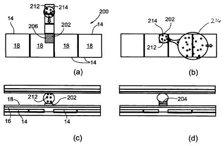
As shown in Figures 11a, 11b, 11c, and 11d, the surface of a DMF
device 200 (specifically the hydrophobic surface 18 that covers the dielectric
material 16 on the lower electrode 14 (see Figure 2(a)) is modified in
specific
areas, cell culture sites (CCS) 202, to facilitate cell adhesion and
proliferation
(cell growth and division). The surface modification procedure reported here
makes use of standard techniques, such as depositing (microprinting,
micorstamping) a bio-substrate (typically extracellular matrix proteins 206),
rendering a hydrophilic and charged surface via microfabrication, or any other
surface modification procedure that can also be cell specific.
In addition to using standard techniques, a bio-substrate can be formed by
dispensing a droplet of a bio-substrate solution in a DMF device and
translating it
to the cell culture site 202, where after incubation and drying, it forms a
bio-
substrate layer for cell attachment. In this case, a device has an extra
reservoir
holding the bio-substrate solution. After the cell culture site 202 is formed,
cells
are seeded by generating a droplet 214 of growth media with suspended cells
32
CA 02639954 2008-09-26
212 on the cell culture site CCS 202 (Figure 11c). Cells are allowed to adhere
to
the surface forming a cell monolayer 204 (Figure 11d).
There are two ways of generating a droplet 214 on the cell culture sites
202: (1) by actively dispensing a droplet from a device reservoir or via
external
means (e.g. pipetting) and translating the droplet to the cell culture sites
202
(Figure 11(a)), and (2) by actuating a droplet 216 (source droplet) larger
than the
cell culture sites 202 over the cell culture sites 202 and thereby passively
dispensing the desired droplet on the hydrophilic cell culture sites 202
(Figure
11(b)). Passive dispensing will be described in more details in the following
section.
Passive Dipensing, Passive Washing, Passive Media/Reagent Exchange
Referring to Figure 12, when a source droplet 210 is actuated in a DMF
device over a patterned hydrophilic area 201 smaller than the base area of the
source droplet 210, it leaves behind a smaller droplet 230 on the hydrophilic
area
201 and the rest of source droplet 210 is translated away from droplet 230.
This
method of generating droplets is termed passive dispensing. Methods for
producing the hydrophilic areas 201 include but are not limited to
microfabrication
techniques (e.g. exposing hydrophilic layers of a device, such as glass or
electrodes, in specific areas), hydrophobic surface plasma treatment, or
deposition of a thin, patterned, hydrophilic layer onto a device surface.
Hydrophilic areas can be formed on either the top plate, the bottom plate, or
both
the top and bottom plate of a two plate device. In the applications disclosed
33
CA 02639954 2008-09-26
herein of adherent cell culture and assaying, hydrophilic areas 201 are used
as
the cell culturing sites (indicated by reference numeral 202 in Figure 11)
which
preferably patterned by depositing bio-substrates, made from cell specific
constituents, such as, but not limited to, extracellular matrix (ECM)
proteins.
ECMs are more favorable substrate for cell attachment than bare glass,
electrodes, or a dielectric layer.
Examples of extracellular matrix proteins include, but are not limited to
fibronectin, laminin, collagen, elastin. The cell specific constituents may
also
comprise synthetic molecules comprised of one of poly-L-lysine, poly-D-lysine
and any combination thereof for example.
Typically, there are no electrodes underneath hydrophilic areas, as these
areas (inherently hydrophilic) do not need to be electrically addressed to
attract
droplets; however, they have to be at least in the vicinity of electrodes. It
will be
appreciated that the hydrophilic arrays can also be formed on the top surface
of
the layer coating electrodes right above electrodes themselves. In most cell-
based applications, it is desirable to have transparent attachment substrate
to
enable facile cell visualization.
Referring to Figure 13, the size and position of a hydrophilic area can vary
relative to size and position of electrodes 14 for source droplets actuation.
Two
relative sizes of hydrophilic areas - 1/4 and 1/9 of the electrode size were
studied, and several positions relative to electrodes 14 and to a source
droplet
path. It should be noted that size and position of hydrophilic areas 201 is
not
34
CA 02639954 2008-09-26
limited by the examples in Figure 13, and that the shape of hydrophilic areas
201
and actuating electrodes 14 is not limited to the square shape.
Referring to Figure 14, when a hydrophilic area 201 is already occupied
by a droplet 230, a source droplet 210 will remove the smaller droplet 230 and
replace it with a new droplet 232 of the source solution while removing
droplet
230 in droplet 210'. This process is termed passive washing or passive
exchange
of liquid solutions on hydrophilic areas 201 (e.g., on CCSs) in a DMF device.
We
report passive exchange efficiency of 95% with a single source droplet, or
99% with two or more consecutive source droplets. Figure 15 shows efficency of
0.5 nM fluorescein passive exchange with phosphate buffered saline. These
results were obtained with fibronectin hydrophilic areas 201, ¨1/9 of the
electrode
size, having two different positions relative to actuating electrodes 14.
Culturing and Passaging Adherent Cells
For adherent cell culture, a DMF device with seeded cells is placed in a
cell culture incubator and a droplet of culture media on top of the cell layer
204 is
regularly replenished with fresh media via DMF passive exchange every 24 h.
We report culturing cells on cell culture sites 202 for 72 h; growth
characteristics
and morphology of the cells are comparable to cells grown in standard tissue
culture flasks (Figure 16). No detachment of cells was observed during media
droplet actuation over the cell culture sites 202. Cells are subcultured at
regular
intervals using standard subculturing protocols adapted to DMF system: (1)
washing cells as shown in Figure 17(b) in which washing droplet 213 has been
dispensed and translated over cell culture site 202, (2) harvesting cells by
CA 02639954 2008-09-26
dispensing and translating a droplet 215 containing a dissociation agent (e.g.
trypsin, collagenase) over cell culture site 202 as shown in Figure 17(c) and
incubating to detach the adhered cells and resuspend them as shown in Figure
17(d), (3) a droplet 240 containing a blocking agent (typically serum in cell
culture media) for blocking the dissociation agent is dispensed and translated
over cell culture site 202, while removing the detached cells away from the
cell
culture sites 202 as shown in Figure 17(e), (4) splitting the resulting cell
suspension as necessary and resuspending in fresh media in droplet 242 and (5)
seeding resuspended cells on a new cell culture site 202 as shown in Figure
17(f) . Blocked dissociation agent and cell suspension are diluted in a big
source
droplet 240 of a blocking agent (cell culture media with serum) by the ratio
of the
volumes of the two droplets, cell culture site 202 droplet and the source
droplet.
In step (4), the resulting cell suspension can be split in smaller droplets
and
resuspended in droplets of fresh media for further reduction of cell
concentration.
When a desired cell concentration is achieved, new generation of cells is
seeded
on new cell culture sites 202 by either translating actively dispensed
droplets of
the cell suspension to new cell culture sites, or by passively dispensing
droplets
with cells on cell culture sites 202 from droplet 242 (Figure 17f). The
inventors
have demonstrated subculturing several generations of mammalian cells in the
same DMF device following the procedure outlined above.
Assaying Adherent Cells
Adherent cell assays in DMF devices are executed in droplets on cell
culture sites 202 where adherent cells are seeded. Devices with seeded cells
are
36
1
CA 02639954 2008-09-26
placed in incubators for few hours or overnight to allow cell attachment and
adjustment to a new DMF device environment (Figure 18a). When adherent cell
deposits 204 are ready for assaying, droplets of reagents and washing
solutions
are deposited on cell culture sites 202 either by translating a droplet
actively
dispensed from a device reservoir or externally, or by passive
dispensing/exchange from source droplets 250 (Figure 18b). Source droplets
250 are either dispensed via DMF from reservoirs or externally deposited on a
device. Washing solutions and reagents are incubated with cells following cell
assay protocols (Figure 18c). Upon assay completion, cell response to a
stimulus (e.g. a lead drug compound) can be detected and measured by
apparatus 260 which may be any standard means (e.g. fluorescence microscopy,
microplate reader to give a few examples) (Figure 18d).
In assays targeting extracellular biochemistry (growth factors, signaling
molecules, metabolic products, etc.), cell response to stimulus is detected in
medium where cells are grown and stimulated with reagents, rather than in
cells.
Medium can be analyzed by immunoassays or other means. Droplets of cell
suspension can alternatively be removed from the cell culture sites 202 (e.g.
with
a bigger source droplet) and its signal can be detected on another spot or its
contents can be analyzed externally.
MULTIPLEXED ADHERENT CELL CULTURE/CELL ASSAYS
Referring to Figure 19, multiple cell culturing sites 202 in a DMF device
300 which is similar to device 100 in Figure 10 but device 300 includes a
plurality
of cell culture sites 202. Device 300 may be used in multiplexed assays where
37
,
CA 02639954 2008-09-26
cells of one kind or multiple kinds are assayed with one or multiple reagents
simultaneously in which cell culturing may be involved as well. In addition, a
single cell culture site 202 can be seeded with multiple cell lines (cell co-
culture).
Assay reagents and/or culture media can be delivered to cell culture sites 202
via
passive dispensing/exchange or in actively dispensed droplets.
In a multiplexed assay, a single source droplet can deliver reagents to
multiple cell culture sites 202 (serial passive dispensing/exchange), or to
only
one cell culture site 202 (parallel passive dispensing/exchange). Signals from
assayed cells or suspension media is detected using multiplexed detection
instruments such as microplate readers.
EXPERIMENTAL
The following non-limiting examples demonstrates the efficacy of the
present invention for conducting adherent cell assays and culture.
Device Design and Fabrication.
Digital microfluidic devices were fabricated using conventional
microfabrication methods. 100 nm thick gold electrodes were patterned on the
bottom plate of a device (glass wafer) and coated with 2 pm of Parylene-C and
50 nm of Teflon-AF. Unpattemed indium-tin oxide (ITO) coated glass substrates
were coated with 50 nm of Teflon-AF. Devices were assembled with an
unpatterned ITO-glass top plate and a patterned bottom plate and separated by
a
¨150 pm thick spacer. Driving potentials (100-140 VRms) were generated by
amplifying the output of a function generator operating at 15 kHz. Droplets
were
sandwiched between the two plates and actuated by applying driving potentials
38
1
CA 02639954 2008-09-26
between the top reference electrode 22 and sequential electrodes 14 on the
bottom plate (Figure 2(a)) via the exposed contact pads. Most devices had a
basic geometry identical to that shown in Figure 11 with the addition of
reservoirs. Source droplets (-800 nL) were actuated on 2.5 mm x 2.5 mm
actuation electrodes, and smaller droplets were actuated on 0.8 mm x 0.8 mm
actuation electrodes. Cell culture site (CCS) areas were patterned either as
transparent, non-conductive fields in 2.5 mm x 2.5 mm electrodes or as smaller
(0.8 mm x 0.8 mm) electrodes within the area of larger 2.5 mm x 2.5 mm
electrodes. Devices were sterilized in 70% ethanol prior to use.
Cell Culture
NIH-3T3 cells (mouse fibroblasts) were maintained in a humidified
atmosphere (5% CO2, 37 C) in DMEM supplemented with 10% fetal bovine
serum, penicillin (100 IU mL-1), and streptomycin (100 pg mL-1). Cells were
subcultured every 2-3 days at 5 x 10³ cells cm.-2 Prior to each DMF
experiment,
cells were suspended in DMEM with the addition of 0.05% (wt/v) pluronic F68
(Sigma-Aldrich) at -7 x 10⁵ cells mL.-1Cell number and viability were
quantified
using a hemocytometer and trypan blue exclusion (Invitrogen Canada)
immediately prior to all experiments.
DMF Cell Seeding
CCSs were formed by depositing 500 nL droplets of fibronectin (100 pg
mL-1 in ddH20) on designated areas in DMF devices. Fibronectin solution was
air-dried resulting in - 1mm2 bio-substrates with -5pg/cm2of fibronectin. Cell
suspension was delivered to CCSs by either passive dispensing from a source
39
CA 02639954 2008-09-26
droplet or by translating actively dispensed droplets from a device reservoir
to
CCSs. CCS droplets were ¨ 200 nL in volume and contained ¨140 cells. Cells
were allowed to attach to the substrate and adapt overnight in a cell culture
incubator (5% CO2, 37 C).
DMF Cell Culture
NIH-3T3 cells were maintained on CCSs by changing media via passive
dispensing every 24 hours. Complete DMEM containing 0.05% (wt/v) pluronic
F68 was dispensed in ¨800 nL droplets and translated over CCSs while
replenishing CCS droplet of media. Complete media exchange was
accomplished with two consecutive source droplets and cells were returned to
the incubator. No cell detachment was observed during passive media exchange.
DMF Cell Subculture
Upon reaching confluency on CCSs, cells were subcultured following
standard subculturing protocols adapted to the DMF format. All reagents and
media containing 0.05% (wt/v) pluronic F68 were delivered to cells using
passive
dispensing/exchange from two consecutive source droplets. Cells were first
washed with PBS without Ca2+/ Mg2+ and then supplied and incubated with
GIBCO Trypsin-EDTA dissociation agent (0.25% Trypsin, 1 mM EDTA 4Na) for
5-10 min at 37 C. DMEM source droplet was then translated to the CCS to block
the dissociation agent with the serum present in media, whereby harvested
cells
were resuspended in DMEM droplet at the 1:4 ratio. DMEM droplet with
suspended cells was actuated away from the CCS and used either as a source
droplet or a reservoir droplet to seed the new generation of cells on a new
CCS
CA 02639954 2008-09-26
in the same device. Seeded cells were placed in a cell culture incubator
overnight followed by media change. Cells were grown on the new CCS for 2
days and further subcultured on the same device.
DMF Cell Viability Assay
Cells cultured on CCSs were assayed on a device for viability. Source
droplets of 0.05% (wt/v) pluronic F68 (Sigma-Aldrich) in phosphate buffered
saline containing viability dyes, calcein AM (1 pM) and ethidium homodimer-1
(2 pM) (lnvitrogen Canada), were dispensed in a device and translated over the
CCS. With two consecutive source droplets, growth media was removed from the
CCS and replaced with viability dyes. Cells were incubated with dyes at room
temperature and visualized using stereomicroscope. Viability of cells was
higher
than 95% and there was no significant difference in morphology between cells
grown on CCSs and cells grown in cell culture flasks.
It will be understood that when doing cell culturing or cell assaying, the
suspension of cells may contain a combination of cells, a suspension medium,
and a non-ionic surfactant. The suspension medium may be selected to
facilitate
cell-containing droplet actuation by preventing non-specific adsorption of
cells
and proteins to device surfaces. The suspension of cells may be a combination
of cells and a suspension medium comprised of block copolymers formed from
poly(propylene oxide) and poly(ethylene oxide), pluronic F68, pluronic F127,
hydrophilic polymers, sodium bicarbonate, phosphate buffered saline (PBS),
HEPES, and other biological buffers, and any combination thereof, which may be
combined or mixed with cell culture medium which in turn may include balanced
41
1
CA 02639954 2008-09-26
salt solutions, nutrient mixtures, basal media, complex media, serum free
media,
insect cell media, virus production media, serum, fetal bovine serum, serum
replacements, antibiotics, antimycotics, and any combination thereof.
In an embodiment the suspension of cells may be a combination of cells,
phosphate buffered saline, and pluronic F68. The droplets including a cell
assay
reagent may include chemicals, biochemicals, drugs, drug lead compounds,
toxins, surfactants, transfection reagents, supplements, cell culture media,
anti-
clumping agents, streptavidin, biotin, antibody production enhancers,
antibodies,
antibody ligands, nucleic acids, nucleic acid binding molecules, enzymes,
proteins, viruses, cell process agonists or antagonists, labeling agents,
fluorescent dyes, fluorogenic dyes, viability dyes, calcein AM, quantum dots,
nano particles, Tween 20, and ethidium homodimer-1, block copolymers formed
from poly(propylene oxide) and poly(ethylene oxide), pluronic F68, pluronic
F127,
hydrophilic polymers, sodium bicarbonate, phosphate buffered saline (PBS),
HEPES, and other biological buffers, and any combination thereof, which may be
combined or mixed with cell culture medium which in turn may include balanced
salt solutions, nutrient mixtures, basal media, complex media, serum free
media,
insect cell media, virus production media, serum, fetal bovine serum, serum
replacements, antibiotics, antimycotics, and any combination thereof.
The cells in the suspension of cells may include primary/isolated or
transformed/cultured cells selected from the group consisting of various
eukaryotic and prokaryotic cells, including animal cells (blood cells, human
leukemia cells, lymphocytes, beta cells, oocytes, eggs, primary cells, primary
42
1
CA 02639954 2008-09-26
bone marrow cells, stem cells, neuronal cells, endothelial cells, epithelial
cells,
fibroblasts), insect cells, plant cells, bacterial cells, archebacterial
cells.
As used herein the word "incubation" can mean allowing a reaction to take
place over a period of time under specified conditions. For cell assays
involving
mixing of cells with one or more cell assay reagents, the incubation period
may
be very short or almost instantaneous upon mixing the droplets wherein the
reaction or response of the cells to the reagent occurs quickly. For cell
culture,
"incubation" can mean maintaining the cells growing or alive under specific
conditions and the period of time of the "incubation" may be arbitrary, after
which
point the cells may be subcultured, assayed or subject to further culturing.
The results disclose herein demonstrate the utility of the present invention
for its application of digital microfluidics to multiplexed, high throughput,
phenotypic cell-based assays, an important tool used in drug discovery and
environmental monitoring. To facilitate high-throughput screening, arrays of
DMF
cell culture sites (Figure 19) can be addressed with compounds from chemical
libraries, and the potential drugs evaluated on the basis of observed
phenotypic
changes. The proposed method will enable high-throughput phenotypic
screening with 100-1000x lower reagent consumption than conventional
methods; in addition, the devices are inexpensive (relative to robotic
dispensers),
have small laboratory footprint and no moving parts. This method could
transform
high-throughput screening, making it attractive to pharmaceutical companies
and
accessible for basic and applied scientists, world-wide.
43
CA 02639954 2008-09-26
In addition to cell assaying the inventors disclose herein the first
multigenerational lab-on-a-chip cell culture using DMF devices. Cells are
grown,
maintained and subcultured in nanoliter volumes. DMF devices are inherently
easily automated and as such have a high potential to be used as tool for a
completely automated microscale cell culture system.
As used herein, the terms "comprises", "comprising", "includes" and
"including" are to be construed as being inclusive and open ended, and not
exclusive. Specifically, when used in this specification including claims, the
terms
"comprises", "comprising", "includes" and "including" and variations thereof
mean
the specified features, steps or components are included. These terms are not
to
be interpreted to exclude the presence of other features, steps or components.
The foregoing description of the preferred embodiments of the invention
has been presented to illustrate the principles of the invention and not to
limit the
invention to the particular embodiment illustrated. It is intended that the
scope of
the invention be defined by all of the embodiments encompassed within the
following claims and their equivalents.
44
CA 02639954 2008-09-26
REFERENCES
(1) Verkman, A. S., "Drug discovery in academia," American Journal of
Physiology-Cell Physiology 2004, 286, C465-C474.
(2) El-Ali, J., Sorger, P. K., Jensen, K. F., "Cells on chips," Nature 2006,
442,
403-411.
(3) Unger, M. A., Chou, H. P., Thorsen, T., Scherer, A., Quake, S. R.,
"Monolithic
Microfabricated Valves and Pumps by Multilayer Soft Lithography," Science
2000, 288, 113-116.
(4) Yu, H. M., Alexander, C. M., Beebe, D. J., "A plate reader-compatible
microchannel array for cell biology assays," Lab on a Chip 2007, 7, 388-391.
(5) Le Pesant, J.-P., 1987, US 4636785.
(6) Ohkawa, T., 1996, US 5486337.
(7) Washizu, M., Kurosawa, 0., 1998, Japan 10267801.
(8) Washizu, M., "Electrostatic Actuation of Liquid Droplets for Microreactor
Applications," IEEE Transactions on Industry Applications 1998, 34, 732-737.
(9) Lee, J., Moon, H., Fowler, J., Schoellhammer, T., Kim, C.-J.,
"Electrowetting
and electrowetting-on-dielectric for microscale liquid handling," Sensors &
Actuators A 2002, 95, 259-268.
(10) Pollack, M. G., Fair, R. B., Shenderov, A. D., "Electrowetting-based
actuation of liquid droplets for microfluidic applications," Applied Physics
Letters 2000, 77, 1725-1726.
(11) Shenderov, A. D., 2003, US 6565727.
(12) Shenderov, A. D., 2007, US 7255780.
(13) Elrod, S. A., Peeters, E. T., Biegelsen, D. K., Dunec, J. L., 2006, US
7,147,763.
(14) Pamula, V. K., Pollack, M. G., Paik, P., H., R., Fair, R., 2005, US
6911132.
(15) Chen, T.-H., Su, C.-M., Shih, H.-C., Yang, C.-T., "Selective Wettability
Assisted Nanoliter Sample Generation via Electro wetting-Based
Transportation," Proceedings of the Fifth International Conference on
Nanochannels, Microchannels and Minichannels (ICNMM2007), Puebla,
Mexico, June 18-20 2007.
(16) Pollack, M., G., Pamula, V., K., Srinivasan, V., Paik, P., Y., Eckhardt,
A., E.,
Fair, R., B., 2007 WO/2007/120241.
(17) Huh, N., Lee, J.-g., 2007, US 20070148763
(18) Fan, S.-K., Huang, P.-W., Wang, T.-T., Peng, Y.-H., "Cross-scale electric
manipulations of cells and droplets by frequency-modulated dielectrophoresis
and electrowetting," Lab on a Chip 2008, 10.1039/b803204a.
(19) Smith, C. M., Hebbel, R. P., Tukey, D. P., Clawson, C. C., White, J. G.,
Vercellotti, G. M., "Pluronic F-68 Reduces the Endothelial Adherence and
Improves the Rheology of Liganded Sickle Erythrocytes," Blood 1987, 69,
1631-1636.
(20) Mizrahi, A., "Pluronic Polyols in Human Lymphocyte Cell Line Cultures,"
Journal of Clinical Microbiology 1975, 2, 11-13.
CA 02639954 2015-07-15
(21) Thiede, B., Siejak, F., Dimmler, C., Jungblut, P. R., Rude!, T., "A two
dimensional electrophoresis database of a human Jurkat T-cell line,"
Electrophoresis 2000, 21, 2713-2720.
46