Note: Descriptions are shown in the official language in which they were submitted.
CA 02651804 2008-11-10
WO 2007/133699 PCT/US2007/011441
Immobilized Biologically Active Entities Having A High Degree of Biological
Activity Following Mechanical Manipulation or Sterilization
FIELD OF THE INVENTION
The present invention relates to substrate materials having immobilized
biologically active entities that maintain their biological activity following
exposure to
conditions of elevated heat, high humidity, antibiotic agents, and/or
mechanical
stress. The present invention is particularly useful in the field of medical
devices.
BACKGROUND OF THE INVENTION
In the field of medical devices, glass, polymeric, and/or metallic materials
are
common substrate materials. These materials can be used for diagnostic devices
or
extracorporeal devices. With the exception of glass, many of the materials can
be
used for implantable devices.
Immobilization of biologically active entities on substrate materials in a
biologically active form involves an appreciation of the respective
chemistries of the
entity and the substrate material. Modification of the chemical composition of
a
substrate material is often required to immobilize a biologically active
entity thereon.
This is usually accomplished by treating surfaces of the substrate material to
generate a population of chemically reactive elements or groups, followed by
immobilization of the biologically active entity with an appropriate protocol.
With
other substrate materials, surfaces of a substrate material are covered, or
coated,
with a material having reactive chemical groups incorporated therein.
Biologically
active entities are then immobilized on the substrate material through the
reactive
chemical groups of the covering material. A variety of schemes for covering,
or
coating, substrate materials have been described. Representative examples of
biologically active entities immobilized to a substrate material with a
covering, or
coating, material are described in U.S. Patent Nos.: 4,810,784; 5,213,898;
5,897,955; 5,914,182; 5,916,585; and 6,461,665.
When biologically active compounds, compositions, or entities are
immobilized, the biological activity of these "biologics" can be negatively
impacted by
the process of immobilization. The biological activity of many of biologics is
CA 02651804 2008-11-10
WO 2007/133699 PCT/US2007/011441
dependent on the conformation (i.e., primary, secondary, tertiary, etc.) of
the biologic
in its immobilized state. In addition to a carefully selected immobilization
process,
chemical alterations to the biologic may be required for the biologic to be
incorporated into the covering material with a conformation that renders the
biologic
sufficiently active to perform its intended function.
Despite an optimized covering and immobilization scheme, additional
processing, such as sterilization, can degrade the biological activity of the
immobilized biologic. For implantable medical devices, sterilization is
required prior
to use. Sterilization may also be required for in vitro diagnostic devices
having
sensitivity to contaminants. Sterilization of such devices usually requires
exposure
of the devices to elevated temperature, pressure, and humidity, often for
several
cycles. In some instances, antibiotic agents, such as ethylene oxide gas (EtO)
or
vapor hydrogen peroxide are included in the sterilization process. In addition
to
sterilization, mechanical compaction and expansion, or long-term storage of an
immobilized biologic can degrade the activity of the biologic.
There exists a need for medical devices having biologically active entities
immobilized thereon that can be subjected to sterilization, mechanical
compaction
and expansion, and/or storage without significant loss of biological activity.
Such a
medical device would have biologically compatible compositions or compounds
included with the immobilized biological entities that serve to minimize
degradation of
the biological activity of the entities during sterilization, mechanical
compaction and
expansion, and/or storage. In some instances, the additional biologically
compatible
compositions or compounds would increase the biological activity of some
biologically active entities following a sterilization procedure.
SUMMARY OF THE INVENTION
The present invention relates to medical devices having substrate materials
with biologically active entities immobilized thereon in combination with
additional
biologically compatible organic chemical compositions that enable the
biologically
active entities to retain significant biological activity following exposure
of the
immobilized entities to processing and storage conditions that would otherwise
degrade the biological activity of the entities.
2
CA 02651804 2008-11-10
WO 2007/133699 PCT/US2007/011441
A suitable substrate material can be any material with a surface having
reactive chemical groups that are capable of attaching, confining, or
otherwise
immobilizing a biologically active entity in a biologically active form to one
or more
surfaces of the substrate material. Substrate materials can also have a
multiplicity of
reactive chemical groups added to surfaces of the materials through the
application
of one or more covering compositions, or materials, to the surfaces. At least
a
portion of a covering material has chemical elements, groups, compounds, or
components that are reactive to biologically active entities and serve to
attach,
confine, or otherwise immobilize a biologically active entity in a
biologically active
form to the covering material. In some embodiments, the biologically active
entity
can be reversibly immobilized.
At least one type of biologically active entity is chemically attached,
confined,
or otherwise immobilized to suitable reactive chemical groups on the substrate
material and/or covering material. Following immobilization of a plurality of
biologically active entities to at least a portion of a multiplicity of
reactive chemical
groups present on a substrate material and/or covering material, an additional
biologically compatible organic composition is covalently or non-covalently
combined
with the biologically active entities, substrate, and/or polymeric covering
material.
The biologically compatible organic composition interacts with the
biologically active
entities and reactive chemical groups of the substrate material and/or
covering
material to prevent the biologically active entities from loosing biological
activity
under conditions that would otherwise significantly degrade the biological
activity of
the entities. These conditions include sterilization and storage. With
expandable
endoluminal medical devices, for example, mechanical compaction and expansion
of
such devices can also significantly degrade the biological activity of the
entities.
In some cases, the additional biologically compatible organic composition
seems to maintain the biological activity of the entities during
sterilization, storage,
and/or mechanical manipulation by limiting undesirable alterations to the
entities
often induced by sterilization, storage, and/or a mechanical manipulation
process.
The activity-diminishing alterations could include conformational changes to a
biologically active entity obscuring an active site on the entity. The
activity-
diminishing alterations could also include interactions between neighboring
biologically active entities. Rearrangements of biologically active entities
with
respect to a polymeric covering material are other possible activity-
diminishing
3
CA 02651804 2008-11-10
WO 2007/133699 PCT/US2007/011441
alterations to the entities. Simple denaturation, or other degradation, of the
biologically active entities could be another means by which the entities
loose
biological activity. As described in greater detail herein, immobilized
biologically
active entities sterilized, stored, and/or mechanically manipulated in the
presence of
the additional biologically compatible organic composition may retain a degree
of
biological activity significantly greater than a similar immobilized
biologically active
entity processed under the same conditions in the absence of the additional
biologically compatible organic composition.
The additional biologically compatible organic composition can be removed
from a sterilized medical device during post-sterilization processing or the
composition can be removed by physiological processes of an implant recipient
following deployment of the sterilized medical device at an implantation site.
Preferred biologically active entities reduce or inhibit thrombus formation on
surfaces of a substrate and/or covering material. Glycosaminoglycans are
preferred
anti-thrombotic agents for use in the present invention, with heparin, heparin
analogs, and derivatives being particularly preferred. Other preferred
biologically
active substances reduce undesirable cellular growth from tissue in which the
present invention is implanted. Preferred anti-proliferative agents for use in
the
present invention include, but are not limited to, dexamethasone, rapamycin,
and
paclitaxel.
Accordingly, one embodiment of the present invention relates to a medical
device comprising a substrate material, a polymeric covering material attached
to at
least a portion of a surface of said substrate material, a plurality of
biologically active
entities having anti-thrombin III binding activity covalently attached to at
least a
portion of said polymeric covering material, and a biologically compatible
composition combined with said polymeric covering material, wherein said
biologically active entities have an anti-thrombin III binding activity of at
least 5
picomoles anti-thrombin III per square centimeter (pmol/cm2) substrate
material
following sterilization or compaction and expansion of said substrate
material. In
other embodiments, the anti-thrombin binding activity is at least 6 picomoles
anti-
thrombin III per square centimeter (pmol/cm2) substrate material, at least 7
picomoles anti-thrombin III per square centimeter (pmol/cm2) substrate
material, at
least 8 picomoles anti-thrombin III per square centimeter (pmol/cm2) substrate
material, at least 9 picomoles anti-thrombin III per square centimeter
(pmol/cm2)
4
CA 02651804 2008-11-10
WO 2007/133699 PCT/US2007/011441
substrate material, or at least 10 picomoles anti-thrombin III per square
centimeter
(pmol/cm2) substrate material. In some embodiments, the anti-thrombin III
binding
activity is at least 100 pmoVcm2 picomoles anti-thrombin III per square
centimeter
(pmoVcm2) substrate material.
In another embodiment, the invention relates to a medical device comprising a
substrate material, a polymeric covering material attached to at least a
portion of a
surface of said substrate material, a first plurality of heparin molecules
having anti-
thrombin III binding activity end point attached to at least a portion of said
polymeric
covering material, and a biologically compatible composition combined with
said
polymeric covering material, wherein said first plurality of heparin molecules
have an
anti-thrombin III binding activity of at least 10 picomoles anti-thrombin III
per square
centimeter (pmol/cm2) substrate material following compaction and expansion of
said
substrate material.
In another embodiment, the present invention relates to a sterilized medical
device comprising a polymeric substrate material, a polymeric covering
material
attached to at least a portion of a surface of said substrate material, a
plurality of
heparin molecules having anti-thrombin III binding activity end point attached
to at
least a portion of said polymeric covering material, and a composition
comprising a
plurality of polyethylene glycol molecules combined with said polymeric
covering
material, wherein said first plurality of heparin molecules have an anti-
thrombin III
binding activity of at least 50 picomoles anti-thrombin III per square
centimeter
(pmoVcm2) substrate material following compaction and expansion of said
substrate
material.
In yet another embodiment; the present invention relates to a medical device
comprising a substrate material, a plurality of chemical entities having anti-
thrombin
III binding activity present on at least a portion of said substrate material,
a first
biologically compatible composition combined with said substrate material, and
a
second biologically compatible composition admixed therewith.
A further embodiment of the present invention relates to a medical device
comprising a substrate material, a polymeric covering material attached to at
least a
portion of a surface of said substrate material, a plurality of chemical
entities having
anti-thrombin III binding activity present on at least a portion of said
polymeric
covering material, a first biologically compatible composition combined with
said
5
CA 02651804 2008-11-10
WO 2007/133699 PCT/US2007/011441
substrate material, and a second biologically compatible composition admixed
therewith.
In embodiments relating to non-covalently combined biologically compatible
organic compositions, at least a portion of the organic composition or the
second
plurality of heparin molecules is often released from the sterilized or
mechanically
manipulated medical device within several hours when placed in a 0.15 M
phosphate
buffer solution having a temperature of about thirty-seven degrees centigrade
and a
substantially neutral pH. Presence of the released compounds can be detected
in
the buffer solution with routine assay techniques.
In embodiments relating to covalently combined biologically compatible
organic compositions, the organic composition or the second plurality of
heparin
molecules is substantially retained on the sterilized or mechanically
manipulated
medical device following sterilization or mechanical manipulation.
In yet other embodiments, the covalently combined biologically compatible
organic composition may be released from the polymeric covering material
through
reversal of a covalent bond. Presence of the compounds released by reversal of
a
covalent bond can be detected in a buffer solution with routine assay
techniques.
In some embodiments, the biologically compatible organic composition may
be admixed prior to mechanical manipulation and/or sterilization. In other
embodiments, the biologically compatible organic composition may be admixed
following mechanical manipulation or sterilization (i.e., in an operating
room). This is
particularly useful when the organic composition may be degraded through
mechanical manipulation or sterilization of a substrate or device utilizing
the
composition. This also permits the organic composition to be placed at
particular
locations on a substrate or device and at varying dosages.
6
CA 02651804 2008-11-10
WO 2007/133699 PCT/US2007/011441
BRIEF DESCRIPTION OF THE DRAWINGS
Figure 1 is a schematic representation of a polymeric substrate material
having a multiplicity of reactive chemical groups thereon.
Figure 1 A is a schematic representation of a metallic substrate material.
Figure 2 is a schematic representation of a polymeric substrate material
having a plurality of biologically active entities immobilized thereto.
Figure 3 is a schematic representation of a polymeric substrate material
having a polymeric covering material with a multiplicity of reactive chemical
groups
thereon.
Figure 3A is a schematic representation of a metallic substrate material
having a polymeric covering material with a multiplicity of reactive chemical
groups
thereon.
Figure 4 is a schematic representation of a polymeric substrate material
having a polymeric covering material with a plurality of biologically active
entities
immobilized thereto.
Figure 4A is a schematic representation of a metallic substrate material
having a polymeric covering material with a plurality of biologically active
entities
immobilized thereto.
Figure 5 is a schematic representation of a polymeric substrate material
having a plurality of biologically active entities immobilized thereto and an
additional
biologically compatible composition combined therewith.
Figure 6 is a schematic representation of a polymeric substrate material
having a polymeric covering material with a plurality of biologically active
entities
immobilized thereto and an additional biologically compatible composition
combined
therewith.
Figure 6A is a schematic representation of a metallic substrate material
having a polymeric covering material with a plurality of biologically active
entities
immobilized thereto and an additional biologically compatible composition
combined
therewith.
Figure 6B is a schematic representation of a polymeric substrate material
having a polymeric covering material with a plurality of biologically active
entities
immobilized thereto showing some of the biologically compatible composition
7
CA 02651804 2008-11-10
WO 2007/133699 PCT/US2007/011441
illustrated in Figure 6 having been released from the substrate material and
polymeric covering material.
Figure 6C is a schematic representation of a metallic substrate material
having a polymeric covering material with a plurality of biologically active
entities
immobilized thereto showing some of the biologically compatible composition
illustrated in Figure 6A having been released from the substrate material and
polymeric covering material.
Figure 7 is a schematic representation of a polymeric substrate material
having three layers of polymeric covering material applied thereto with a
plurality of
biologically active entities immobilized thereto and an additional
biologically
compatible composition combined therewith.
Figure 7A is a schematic representation of a metallic substrate material
having three layers of polymeric covering material applied thereto with a
plurality of
biologically active entities immobilized thereto and an additional
biologically
compatible composition combined therewith.
Figure 7B is a schematic representation of a polymeric substrate material
having three layers of polymeric covering material applied thereto with a
plurality of
biologically active entities immobilized thereto showing some of the
biologically
compatible composition illustrated in Figure 7 having been released from the
substrate material and polymeric covering material.
Figure 7C is a schematic representation of a metallic substrate material
having three layers of polymeric covering material applied thereto with a
plurality of
biologically active entities immobilized thereto showing some of the
biologically
compatible composition illustrated in Figure 7A having been released from the
substrate material and polymeric covering material.
Figure 8 is a bar graph illustrating how sterilization of unbound heparin does
not significantly reduce the biological activity of the heparin.
Figure 9 is a bar graph illustrating the effect of a variety of biologically
compatible organic compositions on the biological activity of end-point
attached
heparin immobilized to reactive chemical groups on a polymeric covering
material
during and after exposure of the immobilized heparin to an ethylene oxide
sterilization regimen.
Figure 10 is a bar graph illustrating the ability of added heparin or dextran
sulfate biologically compatible organic compositions to result in high levels
of ATIII
1 8
CA 02651804 2008-11-10
WO 2007/133699 PCT/US2007/011441
binding activity of heparin immobilized to a polymeric covering material on a
substrate during and after exposure of the immobilized heparin to an ethylene
oxide
sterilization regimen.
Figure 11 is a bar graph illustrating the ability of added dextran sulfate to
maintain the biological activity of end-point attached heparin immobilized on
a
polyvinyl alcohol coated substrate during and after exposure of the
immobilized
heparin to an ethylene oxide sterilization regimen.
Figure 12 is a bar graph illustrating the ability of added glycerol to
maintain
the biological activity of end-point attached heparin immobilized on a
polymeric
covering material of a substrate following compaction and expansion of the
substrate
material.
Figure 13 is a bar graph illustrating the ability of added glycerol and
heparin to
maintain the biological activity of end-point attached heparin immobilized on
a
polymeric covering material of a substrate following mechanical compaction,
exposure to an ethylene oxide sterilization regimen, and mechanical expansion
of
the substrate material.
Figure 14 is a schematic representation of a polymeric substrate material
having a polymeric covering material with a plurality of biologically active
entities
immobilized thereto and reactive chemical groups thereon.
Figure 15 is a schematic representation of a metallic substrate material
having
a polymeric covering material with a plurality of biologically active entities
immobilized thereto and reactive chemical groups thereon.
Figure 16 is a schematic representation of a polymeric substrate material
having a polymeric covering material with a plurality of biologically active
entities and
an additional biologically compatible composition covalently combined thereto.
Figure 17 is a schematic representation of a metallic substrate material
having
a polymeric covering material with a plurality of biologically active entities
and an
additional biologically compatible composition covalently combined thereto.
. Figure 18 is a schematic representation of embodiments of the present
invention having a second biologically compatible composition combined
therewith.
Figure 19 is a schematic representation of embodiments of the present
invention having a second biologically compatible composition combined
therewith.
Figure 20 is a schematic representation of a polymeric substrate material
having a polymeric covering material with a plurality of biologically active
entities
9
CA 02651804 2008-11-10
WO 2007/133699 PCT/US2007/011441
immobilized thereto, a first biologically compatible composition, and a second
biologically compatible composition combined therewith.
Figure 21 is a schematic representation of a metallic substrate material
having
a polymeric covering material with a plurality of biologically active entities
immobilized thereto, a first biologically compatible composition, and a second
biologically compatible composition combined therewith.
DETAILED DESCRIPTION OF THE INVENTION
The present invention relates to materials and devices having biologically
active entities immobilized thereto that retain significant biological
activity following
sterilization, mechanical compaction and expansion, and/or storage conditions
that
would otherwise significantly decrease the biological activity of the
entities. The
biological activity of an immobilized biological entity subjected to such
conditions
may be positively influenced by the presence of at least one additional
biologically
compatible composition covalently or non-covalently combined with the
biologically
active entities. In most embodiments, the additional composition is an organic
compound. In some embodiments, however, the biologically compatible
composition
is an inorganic compound. In preferred embodiments, the additional composition
is
a carbohydrate in the form of a polysaccharide. Preferred polysaccharides are
glycosaminoglycans. Preferred glycosaminoglycans are heparin compositions,
heparin analogs, and heparin derivatives.
Referring to Figures 1 and 2, some polymeric substrate materials (12) have
multiplicities of reactive chemical groups (16) populating at least a portion
of the
surfaces of the substrate materials to which a plurality of biologically
active entities
(17) are attached, confined, or otherwise immobilized. Most biologically
active
entities (17) are covalently attached, or bound, to the substrate materials
(12)
through the reactive chemical groups (16). Surfaces of the polymeric substrate
material (12) can be smooth, rough, porous, curved, planar, angular,
irregular, or
combinations thereof. In some embodiments, substrate materials with surface
pores
have internal void spaces extending from the porous surface of the material
into the
body of the material. These porous substrate materials have internal substrate
material bounding the pores that often provides surfaces amenable to
immobilizing
CA 02651804 2008-11-10
WO 2007/133699 PCT/US2007/011441
biologically active entities. Whether porous or non-porous, substrate
materials can
be in the form of filaments, films, sheets, tubes, meshworks, wovens, non-
wovens,
and combinations thereof.
Suitable substrate materials (12) for immobilizing biologically active
entities
(17) include biocompatible polymeric materials such as polyethylene,
polyurethane,
silicone, polyamide-containing polymers, and polypropylene. Full density or
porous
polytetrafluoroethylene is a suitable polymeric substrate material (12) if
reactive
chemical groups (16) are introduced in constituents of the polymeric material.
Substrate materials with a multiplicity of reactive chemical groups that are
part of the
substrate material are referred to herein as "functionalizable materials."
Following
reaction of a biologically active entity with a functionalizable substrate
material, the
substrate material is considered functionalized and the biologically active
entity
immobilized. In order to maintain the biological activity of the immobilized
entity
during subsequent processing conditions, such as sterilization, mechanical
compaction and expansion, or storage, an additional biologically compatible
organic
chemical composition is non-covalently combined with the functionalized
material
and immobilized entity.
Substrate materials can also have a multiplicity of reactive chemical groups
added to surfaces of the materials through the application of one or more
covering
compositions, or materials, to the surfaces. At least a portion of a covering
material
has chemical elements, groups, compounds, or components that are reactive to
biologically active entities and serve to attach, confine, or otherwise
immobilize a
biologically active entity in a biologically active form to the covering
material. The
covering material can be applied in the form of a solute, particle,
dispersion, coating,
or overlay and attached to the substrate material in a variety of ways
including, but
not limited to, covalent bonding, adsorption, such as, physisorption or
chemisorption,
and non-covalent bonding, such as hydrogen bonding or ionic bonding. In
preferred
embodiments, the covering material is applied in a solution and forms a
continuous
or discontinuous film layer on one or more surfaces of the substrate material
upon
removal of the solvent. The covering material can be applied in one or more
layers.
The chemical constituents of the covering material in each layer can be the
same or
different. In some embodiments, the covering material is cross-linked to
itself or
other covering materials in other layers. The cross-linking bonds can be
covalent or
ionic.
11
CA 02651804 2008-11-10
WO 2007/133699 PCT/US2007/011441
Substrate materials (12, 14) lacking reactive chemical groups on their
surfaces (Figure 1A) (or lacking appropriately reactive chemical groups) are
covered,
at least in part, with a polymeric covering material (18) having a
multiplicity of
reactive chemical groups (16) thereon (Figures 3 and 3A) to which biologically
active
entities (17) can be attached, confined, or otherwise immobilized (Figures 4
and 4A).
Most biologically active entities (17) are covalently attached, or bound, to
the
polymeric covering material (18) through the reactive chemical groups (16) of
the
covering material (18). The polymeric covering material (18) forms at least
one layer
on at least a portion of a substrate material (12, 14). In some embodiments,
the
polymeric covering material (18) is cross-linked (19) to itself or other
layers (18a,
18b) of polymeric covering material (Figure 7 and 7a). The cross-linking can
be
covalent, ionic, or both. Substrate materials amenable to covering are glass,
metals
(14), ceramics, polymeric materials (12), particularly chemically inert
polymeric
materials such as polytetrafluoroethylene.
At least one type of biologically active entity having anti-thrombin III
binding
capability (17) is chemically attached, confined, or otherwise immobilized to
suitable
reactive chemical groups (16) on the substrate material (12, 14) and/or
covering
material (18).
Biologically compatible compositions (11, 15, 100) include, but are not
limited
to, antithrombotics, anticoagulants, fibrinolytic or thrombolytic agents,
antibiotics,
antimicrobial/antiseptic compounds, anti-viral compounds, antiproliferatives,
cell
adhesive compounds, cell anti-adhesive compounds, and anti-inflammatories.
Antithrombotics of particular interest are glycosaminoglycans, particularly
heparin,
including derivatives and analogs thereof. Other anticoagulant agents include,
but
are not limited to, hirudin, activated protein C, and prostaglandins.
Fibrinolytic or
thrombolytic agents include, but are not limited to, streptokinase, urokinase,
and
tissue plasminogen activator (tPA). Examples of antibiotics include, but are
not
limited to, penicillin, tetracycline, chloramphenicol, minocycline,
doxycycline,
vancomycin, bacitracin, kanamycin, neomycin, gentamycin, erythromycin and
cephalosporins. Examples of cephalosporins include cephalothin, cephapirin,
cefazolin, cephalexin, cephradine, cefadroxil, cefamandole, cefoxitin,
cefaclor,
cefuroxime, cefonicid, ceforanide, cefotaxime, moxalactam, ceftrizoxime,
ceftriaxone,
and cefoperazone. Examples of antimicrobiaVantiseptics include, but are not
limited
to, silver sulfadiazine, chlorhexidine, peracetic acid, sodium hypochlorite,
triclosan,
12
CA 02651804 2011-09-14
WO 2007/133699 PCT/US2007/011441
phenols, phenolic compounds, iodophor compounds, quaternary ammonium
compounds, chlorine compounds; heparin and combinations thereof. Examples of
anti-viral agents include, but are not limited to, alpha.-methyl-1-
adamantanemethylamine, hydroxy-ethoxymethylguanine, adamantanamine, 5-iodo-
2'-deoxyuridine, trifluorothymidine, interferon, and adenine arabinoside. Cell
adhesive compounds include, but are not limited to, fibronectin, laminin,
collagen,
vitronectin, osteopontin, RGD peptides, RGDS peptides, YIGSR peptides, and
antibodies targeting cell surface antigens. Compounds that may resist cellular
attachment include Poly HEMA, poly ethylene glycol, polysaccharides,
polyvinylpyrrolidone, and phospholipids. Other biologically active entities
include,
but are not limited to, enzymes, organic catalysts, ribozymes,
organometallics,
proteins, glycoproteins, peptides, polyamino acids, antibodies, nucleosides,
nucleotides, nucleic acids, steroidal molecules, antibiotics, antimicrobial
compounds,
antimycotics, cytokines, carbohydrates, oleophobics, lipids, pharmaceuticals,
and
therapeutics.
While a variety of biologically active entities (17) can be used in the
present
invention, as described above, entities capable of interacting with components
of
mammalian blood to prevent coagulation or thrombus formation on surfaces of a
substrate material (12, 14) or covering material (18) by the blood components
are
most preferred. Many of these biologically active entities are
oligosaccharides or
polysaccharides. Some of the polysaccharides are glycosaminoglycans including
glucosamine or galactosamine compositions. Preferred glycosaminoglycans are
heparin compositions, heparin analogs, and heparin derivatives. Heparin is a
complex glycosaminoglycan with many biological functions mediated by its
binding to
growth factors, enzymes, morphogens, cell adhesions molecules, and cytokines.
The biological activity of heparin to function as an anticoagulant is based on
the
ability of heparin to act as a catalyst for thrombin and anti-thrombin III (AT
111)
binding. Most of the anti-coagulant activity of heparin is associated with a
pentasaccharide sequence that facilitates this binding.
The most preferred heparin composition for immobilization in the present
invention is a heparin composition having a free terminal aldehyde group made
according the teachings of U.S. Patent No. 4,613,665, issued to Larm.
In the process of making heparin with a free
terminal aldehyde group, the heparin is subjected to degradation by
diazotation to
13
CA 02651804 2011-09-14
WO 2007/133699 PCTIUS2007/011441
form a heparin fragment having a free terminal aldehyde group. The free
terminal
aldehyde group allows the heparin composition to be "end point attached" to
primary
amino groups of a substrate or polymeric covering material to form an imine
which,
by reduction, is converted to a secondary amine. End point attachment of the
heparin composition permits the heparin to be immobilized in a conformation
that
most advantageously exposes the biologically active portion of the heparin
composition to components of the blood responsible for coagulation and
thrombus
formation. When exposed to the blood components responsible for thrombus
formation and coagulation, the optimally immobilized heparin interacts with
the blood
components to reduce or prevent thrombus formation or other coagulation events
on
surfaces of the substrate and/or covering material.
Other desirable biologically active entities (17) for use in the present
invention
include synthetic heparin compositions referred to as "fondaparinuxTM,"
compositions
involving antithrombin III-mediated inhibition of factor Xa,
antiproliferatives, and anti-
inflammatories.
Despite an optimized immobilization scheme, the biological activity of a
heparin-based biological entity is significantly decreased during
sterilization,
mechanical compaction and expansion, and/or storage of the entities (Figures
9, 11,
12, and 13). As discussed above, the decrease in biological activity of an
immobilized biologically active entity may be caused by a variety of factors.
Regardless of the mechanism by which the biological activity of an immobilized
entity
is decreased, addition of a biologically compatible organic composition
covalently
and/or non-covalently combined with the immobilized biologically active entity
maintains the biological activity of the entity during and after
sterilization, mechanical
manipulation - such as mechanical compaction and expansion, and/or storage of
the
entities.
The additional biologically compatible organic composition can have biological
activity or no biological activity. The additional biologically compatible
organic
composition can be a carbohydrate in the form of polyhydroxy aldehydes or
ketones
and their derivatives. These carbohydrates include monosaccharides,
disaccharides, oligosaccharides, and polysaccharides, including
glycosaminoglycans, glycosaminomannans, and storage polysaccharides such as
dextran and its derivatives. Other biologically compatible organic
compositions
suitable for use in the present invention include acid mucopolysaccharides,
amino
14
CA 02651804 2008-11-10
WO 2007/133699 PCT/US2007/011441
acids, polypeptides, proteins, glycoproteins, nucleosides, nucleotides,
polynucleotides, or other biologically compatible aliphatic or aromatic
compound,
charged or uncharged, having a molecular weight less than about 100,000 MW.
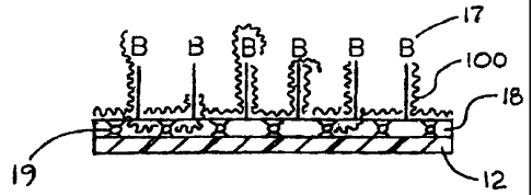
Referring to Figures 5 - 6A, covered or uncovered substrate materials (14, 12,
respectively) having biologically active entities (17) immobilized thereon
have an
additional biologically compatible composition (100) combined with the
biologically
active entities (17), the substrate material (12, 14) and/or the covering
material (18).
The biologically compatible composition is preferably organic. The
biologically
compatible organic composition can be applied to the immobilized biologically
active
entities, substrate, and/or covering material in a variety of ways. In a
preferred
embodiment, a suitable carbohydrate-based biologically compatible composition
is
dissolved in an aqueous solvent and the solution applied to the immobilized
biologically active entities, substrate, and/or polymeric covering material by
spraying,
dip coating, immersing, rolling, spreading, or other deposition means. In
appropriate
systems, biologically compatible compositions can be dissolved in organic
solvents
and similarly applied.
A preferred embodiment of the present invention relates to a sterilized
medical device for implantation, or other placement, at an anatomical site.
Also
preferred are sterilized medical devices for placement inside an anatomical
structure
delimiting a void space, or lumen, to reinforce the anatomical structure or
maintain
the void space delimited thereby. When these sterilized devices are used
within a
vascular structure, immobilized biologically active entities in the form of
end point
attached heparin interact with blood flowing through, or around, the devices
to
minimize or prevent formation of thrombus or other coagulation products on
blood-
contacting surfaces of the devices. In a preferred embodiment, the additional
biologically compatible organic composition is a polyethylene glycol compound
covalently combined with the substrate material and/or covering material. The
covalently bound heparin is allowed to remain with the sterilized devices. The
preferred sterilization method includes ethylene oxide gas.
The manufacturing of medical devices may require mechanical manipulation
that often reduces the biological activity of an immobilized biologically
active entity.
The additional biologically compatible composition combined with the
immobilized
biologically active entities, substrate material, and/or covering material as
described
above, may also maintain the biological activity of the immobilized
biologically active
CA 02651804 2008-11-10
WO 2007/133699 PCT/US2007/011441
entities following mechanical compaction and expansion of a medical device
(Figures 12 and 13). Expandable stents and stent-grafts are medical devices
for
which improved biological activity of immobilized biologically active entities
is
particularly significant.
The present invention, therefore, provides sterilized devices having
biologically active entities immobilized thereto where the biological activity
of the
immobilized entities is significantly retained during and after a
sterilization process
(Figures 9 - 11, and 13). Prior to sterilization, the devices can be
mechanically
manipulated, through compaction and expansion, for example, and retain
significant
biological activity (Figures 12 and 13).
Figure 14 schematically illustrates embodiments of the present invention (50)
having a polymeric substrate (12) having a polymeric covering, or coating,
material
(18) cross-linked (19) thereon. Covering (18) has a plurality of immobilized
biologically active entities "B" (17) attached thereto. Covering (18) also has
a
plurality of chemically reactive groups "R" (13) thereon to which a
biologically
compatible composition "S" (15) can be covalently attached (Figures 16 and
17). In
some embodiments, the covalent bonds are reversible thereby rendering the
biologically compatible composition "S" (15) releasable from the invention
under
appropriate conditions. Figures 15 and 17 schematically illustrate similar
constructions (50) using a metallic substrate (14).
Figures 18 and 19 schematically illustrate embodiments of the present
invention (70) having a polymeric substrate (12) or metallic substrate (14)
having a
polymeric covering, or coating, material (18) cross-linked (19) thereon.
Covering
(18) has a plurality of immobilized biologically active entities "B" (17) and
a first
biologically compatible composition "S" (15) covalently attached thereto. In
some
embodiments, the covalent bonds are reversible thereby rendering the
biologically
compatible composition "S" (15) releasable from the invention under
appropriate
conditions. In addition, this embodiment has a second biologically compatible
composition "A" (11) admixed therewith.
Figures 20 and 21 schematically illustrate embodiments of the present
invention (80) having a polymeric substrate (12) or metallic substrate (14)
having a
polymeric covering, or coating, material (18) cross-linked (19) thereon.
Covering
(18) has a plurality of biologically active entities "B" (17) immobilized
thereto. A first
biologically compatible composition (100) is combined with the biologically
active
16
CA 02651804 2008-11-10
WO 2007/133699 PCT/US2007/011441
entities (17). In addition, this embodiment has second biologically compatible
composition "A" (11) admixed therewith.
EXAMPLES
Except-for Example 1, calculations of heparin activity on surfaces in the
present invention were conducted using the surface area of only one side of
the
sample material, although the entire sample, including interstices, may have
heparin
immobilized thereon. The heparin activity was assayed by measuring the
ability, or
capacity, of the end-point attached heparin to bind a known quantity of anti-
thrombin
III (ATIII). The results were expressed as picomoles anti-thrombin III (ATIII)
bound
per square centimeter of substrate material (pmol ATIII/cm2 substrate
material). This
assay is described by Larsen M.L., et al. in "Assay of plasma heparin using
thrombin
and the chromogenic substrate H-D-Phe-Pip-Arg-pNA" (S-2238) (Thromb. Res.
1978; 13:285-288) and Pasche, et al. in "A binding of antithrombin to
immobilized
heparin under varying flow conditions" (Artif. Organs 1991; 15:281-491).
ATIII binding activity per surface area of substrate material is defined as
the
number of picomoles of ATIII bound per apparent surface area of covered or
uncovered substrate material. The apparent substrate surface area does not
take
into account multiple covered surfaces nor porosity considerations of a porous
substrate material. If the substrate material is porous, the effect of
porosity on
surface area is not considered for these calculations. For example, the
apparent
surface area of a cylindrical tubular ePTFE vascular graft (which is made of a
porous
material) with end-point attached heparin immobilized on substrate material
comprising the inner surface of the tubular graft is calculated as it is for
any
cylindrical geometry as 2TTrL: where r is the graft inner radius; L is the
axial length;
and it is the number pi. It is important to note that the porous nature of
ePTFE and
its effect on surface area is not accounted for herein. Accordingly, non-
porous
substrate materials that are cut into squares for analysis are taken to have a
surface
area of the length multiplied by the width.
17
CA 02651804 2011-09-14
WO 2007/133699 PCT/US2007/011441
Example 1
This example demonstrates retention of biological activity of unbound "neat"
heparin following exposure of the heparin to an ethylene oxide (EtO)
sterilization
process.
In this example, unsterilized USP grade heparin-sodium in lyophilized powder
form was obtained from Celsus Laboratories (Cincinnati, OH). Measured
quantities
of heparin were placed into CHEX-ALL sterilization pouches (Long Island City,
N.Y.) for testing. One group of heparin-containing pouches was exposed to EtO
sterilization. Ethylene oxide sterilization was carried out under conditions
of
conditioning for one hour (1 hr), an EtO gas dwell time of one hour (1 hr), a
set point
temperature of fifty-five degrees centigrade (55 C), and an aeration time of
twelve
hours (12 hr). Another group was subjected to the sterilization procedure in
the
absence of EtO. A third group was not exposed to the sterilization procedure.
Following the sterilization procedure, known quantities of heparin were
removed from each pouch and tested for bio-activity with an ACTICHROMET""
Heparin
(anti-FXa) assay kit available from American Diagnostica Inc. (Stamford, CT).
Bioactivity values for each heparin sample were expressed as international
units of
heparin per mass of heparin (IU/mg). International units of heparin are
calculated
based on Factor Xa inactivation by ATIII that is catalyzed by heparin.
International
units are therefore a measure of the ATIII binding activity of heparin. Any
reduction
in heparin activity is expressed simply as a reduction in the lU/mg for
comparable
heparin controls from the ACTICHROME test. Heparin exhibiting a reduction in
activity is considered to have been deactivated to a degree by the
sterilization
process.
Figure 8 is a bar graph illustrating the effect of EtO sterilization on the
anti-
thrombin III (ATIII).binding activity of dry powdered heparin in an unbound
state.
Figure 8 shows the mean activity levels, expressed as IU/mg, for the heparin
samples (n=3) in each group. Control heparin samples that did not undergo
sterilization had a mean value of 1381U/mg. Control heparin samples that
underwent the sterilization process in the absence of EtO (i.e., high
humidity, high
temperatures, etc.) had a mean value of 119 IU/mg. The heparin samples that
underwent the sterilization process in the presence of EtO had a mean value of
123
IU/mg. The heparin samples exposed to the sterilization process in the absence
of
EtO had an fourteen percent (14%) decrease in activity compared to the
unsterilized
18 ,
CA 02651804 2011-09-14
WO 2007/133699 PCT/US2007/011441
control samples, while the samples exposed to the sterilization process in the
presence of EtO had only an eleven percent (11 %) decrease in activity. As
seen
from Figure 8, sterilization of unbound, neat, heparin powder in the presence
or
absence of EtO does not significantly reduce ATIII binding to the heparin when
compared to unsterilized control samples. The anti-thrombin III binding
activity of
unbound, unsterilized, heparin is not significantly diminished by
sterilization without
EtO or sterilization with EtO. Therefore, degradation of the anti-thrombin III
binding
activity of immobilized heparin subjected to similar EtO sterilization
conditions must
be caused by a mechanism other than simple exposure to sterilization with or
without
EtO.
Example 2
This example describes the construction of an embodiment of the present
invention in which heparin anti-thrombin III (ATIII) binding is not
significantly
diminished by exposure to EtO sterilization.
In accordance with U.S. Patent No. 6,653,457
an aldehyde modified heparin composition made according to U.S.
Patent No. 4,613,665, which is incorporated herein by reference, was end-point
attached to a covering material, or coating layer, placed on an expanded
polytetrafluoroethylene (ePTFE) material. An additional biologically
compatible
organic composition was incorporated within the covering material and bound
heparin to enable the immobilized heparin to undergo EtO sterilization without
significant loss in biological activity.
An ePTFE material in sheet form was obtained from W.L. Gore &
Associates, Inc., Flagstaff, AZ under the tradename GORETM Microfiltration
Media
(GMM-406). A covering material in the form of a base coating was applied to
the
ePTFE material by mounting the material on a ten centimeter (10 cm) diameter
plastic embroidery hoop and immersing the supported ePTFE material first in
100%
isopropyl alcohol (IPA) for about five minutes (5 min) and then in a solution
of
LUPASOL polyethylene imine (PEI) and IPA in a one to one ratio (1:1).
LUPASOL water-free PEI was obtained from BASF and diluted to a concentration
of about four percent (4%) and adjusted to pH 9.6. Following immersion of the
ePTFE material in the solution for about fifteen minutes (15 min), the
material was
removed from the solution and rinsed in deionized (Di) water at pH 9.6 for
fifteen
19
CA 02651804 2008-11-10
WO 2007/133699 PCT/US2007/011441
minutes (15 min). PEI remaining on the ePTFE material was cross-linked with a
0.05% aqueous solution of glutaraldehyde (obtained from Amresco) at pH 9.6 for
fifteen minutes (15 min). Additional PEI was added to the construction by
placing the
construction in a 0.5% aqueous solution of PEI at pH 9.6 for fifteen minutes
(15 min)
and rinsing again in DI water at pH 9.6 for fifteen minutes (15 min). The
imine
formed as a result of the reaction between glutaraldehyde and the PEI layer is
reduced with a sodium cyanborohydride (NaCNBH3) solution (5 g dissolved in 1 L
DI
water, pH 9.6) for fifteen minutes (15 min) and rinsed in DI water for thirty
minutes
(30 min).
An additional layer of PEI was added to the construction by immersing the
construction in 0.05% aqueous glutaraldehyde solution at pH 9.6 for fifteen
minutes
(15 min), followed by immersion in a 0.5% aqueous solution of PEI at pH 9.6
for
fifteen minutes (15 min). The construction was then rinsed in DI water at pH
9.6 for
fifteen minutes (15 min). The resultant imines were reduced by immersing the
construction in a solution of NaCNBH3 (5 g dissolved.in 1 L DI water, pH 9.6)
for
fifteen minutes (15 min) followed by a rinse in DI water for thirty minutes
(30 min). A
third layer was applied to the construction by repeating these steps. The
result was
a porous hydrophobic fluoropolymeric base material having a hydrophilic cross-
linked polymer base coat on substantially all of the exposed and interstitial
surfaces
of the base material.
An intermediate chemical layer was attached to the polymer base coat in
preparation for placement of another layer of PEI on the construction. The
intermediate ionic charge layer was made by incubating the construction in a
solution
of dextran sulfate (Amersham Pharmacia Biotech) and sodium chloride (0.15 g
dextran sulfate and 100 g NaCl dissolved in 1 L DI water, pH 3) at 60 C for
ninety
minutes (90 min) followed by rinsing in DI water for fifteen minutes (15 min).
A layer of PEI, referred to herein as a "capping layer" was attached to the
intermediate layer by placing the construction in a 0.3% aqueous solution of
PEI (pH
9) for about forty-five minutes (45 min). followed by a rinse in a sodium
chloride
solution (50 g NaCl dissolved in 1 L DI water) for twenty minutes (20 min). A
final DI
water rinse was conducted for twenty minutes (20 min).
Aldehyde modified heparin was end point attached, or conjugated, to the PEI
layer(s) by placing the construction in a heparin-containing sodium chloride
salt
solution (1.5 g heparin, 29.3 g NaCl dissolved in 1 L DI water, pH 3.9) for
one
CA 02651804 2008-11-10
WO 2007/133699 PCT/US2007/011441
hundred twenty minutes (120 min) at sixty degrees centigrade (60 C). A 2.86
mL
volume of a 2.5% (w/v) aqueous NaCNBH3 solution was added to the one liter (1
L)
heparin solution prior to adding the samples. The samples were then rinsed in
DI
water for fifteen minutes (15 min), borate buffer solution (10.6 g boric acid,
2.7 g
NaOH and 0.7 g NaCl dissolved in 1 L DI water, pH 9.0) for twenty minutes (20
min),
and finally in DI water for fifteen minutes (15 min) followed by
lyophilization of the
entire construction to produce dry heparin bound to the ePTFE material. The
presence and uniformity of the heparin was determined by staining samples of
the
construction on both sides with toluidine blue. The staining produced an
evenly
purpled surface indicating heparin was present and uniformly bound to the
ePTFE
material.
By adding particular compounds or compositions to the heparin-bound
construction, the biological activity of the heparin can be maintained
following
exposure to conditions that would otherwise decrease the biological activity
of the
heparin. The conditions include, but are not limited to, EtO sterilization,
mechanical
compaction and expansion, and storage.
The above-described constructions coated with a covering material were
exposed to solutions of the following compounds to evaluate their stabilizing
effect
on the biological activity of the heparin bound to parts of the coating: USP
grade
calcium chloride (Fisher Scientific), USP grade heparin sodium (Celsus),
polyethylene glycol (20,000 molecular weight, Sigma), DEAE dextran (500,000
molecular weight, PK chemical), dextran sulfate sodium salt (8,000 molecular
weight, Sigma), and dextran (9,500 molecular weight, Sigma) at concentrations
of
0.5 g per 100 ml DI water adjusted to pH 9.6. Dexamethasone was also utilized
at
0.5 g per 100 ml ethanol with no pH adjustment. Each of these solutions is
referred
to herein as a "treatment solution." The effect of these various compounds on
binding activity of heparin to anti-thrombin III
(ATIII) following EtO sterilization was expressed as picomoles anti-thrombin
III bound
per square centimeter (cm2) substrate material. These data are summarized in
Figure 9.
To expose a particular heparin-containing construction to a particular
treatment solution, the construction was placed into a two liter (2 L) beaker
and one
hundred milliliters (100 ml) of treatment solution was added, sufficient to
completely
immerse the construction in the treatment solution. Each construction was
exposed
21
CA 02651804 2008-11-10
WO 2007/133699 PCT/US2007/011441
to the treatment solution for one hour (1 hr) at sixty degrees centigrade (60
C). The
construction was removed from the solution and lyophilized prior to exposure
to a
sterilization procedure.
In preparation for EtO sterilization, each lyophilized construction was placed
and sealed in a Tower DUALPEEL Self-Seal Pouch (Allegiance Healthcare Corp.,
McGaw Park, IL). Ethylene oxide sterilization was carried out under conditions
of
conditioning for one hour (1 hr), an EtO gas dwell time of one hour (1 hr), a
set point
temperature of fifty-five degree centigrade (55 C), and an aeration time of
twelve
hours (12 hr).
After EtO sterilization, each construction (including controls) was removed
from its pouch and washed in DI water for fifteen minutes (15 min), borate
buffer
solution (10.6 g boric acid, 2.7 g NaOH and 0.7 g NaCl dissolved in 1 L DI
water, pH
9.0) for twenty minutes (20 min), and finally a rinse in DI water for fifteen
minutes (15
min).
Samples approximately one square centimeter (1 cm2) in size were cut from
the construction and assayed for heparin activity by measuring the capacity of
the
end point attached heparin to bind ATIII. The assay is described by Larsen
M.L., et
al., in "Assay of plasma heparin using thrombin and the chromogenic substrate
H-D-
Phe-Pip-Arg-pNA (S-2238)." Thromb Res 13:285-288 (1978) and Pasche B., et al.,
in "A binding of antithrombin to immobilized heparin under varying flow
conditions."
Artif. Organs 15:281-491 (1991). The results were expressed as amount of ATIII
bound per unit surface area substrate material in picomoles per square
centimeter
(pmoVcm2). All samples were maintained in a wet condition throughout the
assay. It
is important to note that while the approximately one square centimeter (1
cm2)
samples each have a total surface area of two square centimeters (2 cm2) if
both
sides of the material are considered, only one surface on the sample (i.e., 1
cm2)
was used for calculating ATIll heparin-binding activity in pmol/cm2.
Figure 9 is a bar graph illustrating the effects various biologically
compatible
organic compositions non-covalently combined with heparin immobilized on a
covered substrate material on the anti-thrombin III binding activity of the
immobilized
heparin following exposure of the immobilized heparin to EtO sterilization.
The anti-thrombin III binding activity to the immobilized heparin was
expressed in picomoles ATIII bound per square centimeter of substrate material
(pmoVcm2). One set of control samples was not sterilized. Another set of
control
22
CA 02651804 2008-11-10
WO 2007/133699 PCT/US2007/011441
samples was subjected to EtO sterilization in the absence of a biologically
compatible organic composition non-covalently combined with the immobilized
heparin and covering material. Each remaining bar represents the anti-thrombin
III
binding. activity of immobilized heparin in the presence of the indicated
biologically
compatible organic composition non-covalently combined with the immobilized
heparin and covering material. All bars represent mean values of n=3 samples,
except for dextran sulfate with n=6 samples.
As can be seen from the bar graph, sterilized control samples showed a
dramatic reduction in anti-thrombin III binding activity compared to
unsterilized
control samples. The anti-thrombin III binding activity of the unsterilized
control
samples was 103 pmoVcm2 substrate material. The anti-thrombin III binding
activity
of the sterilized control samples was 66 pmoVcm2 substrate material. EtO
sterilization caused a thirty-six percent (36%) reduction in anti-thrombin III
binding
activity compared to the unsterilized samples.
The influence of the above described biologically compatible organic
compositions non-covalently combined with the immobilized heparin and covering
material on the anti-thrombin III binding activity following sterilization is
summarized
in the following paragraph.. Each biologically compatible organic composition
was
rinsed, as described earlier, from each construction before the anti-thrombin
III
binding activity was determined.
When heparin was added to the construction, the mean anti-thrombin III
binding activity was 108 pmoVcm2. Addition of dextran to the construction
resulted in
a mean anti-thrombin III binding activity of 98 pmoVcm2 substrate material.
When
dextran sulfate was added to the construction, the mean anti-thrombin III
binding
activity was 134 pmoVcm2 substrate material. Additionally, polyethylene glycol
resulted in a mean anti-thrombin III binding activity of 129 pmol/cm2
substrate
material. Interestingly, these values are greater than the mean values for the
unsterilized control samples at 103 pmoVcm2 substrate material.
When inorganic calcium chloride (CaCl2) was added to the construction, the
mean anti-thrombin III binding activity of the immobilized heparin was 75
pmol/cm2
substrate material. Addition of dexamethasone to the construction resulted in
a
mean anti-thrombin III binding activity of 42 pmoVcm2 substrate material. DEAE
dextran seemed to diminish the anti-thrombin III binding activity of the
immobilized
heparin with a mean activity of 5 pmol/cm2 substrate material.
23
CA 02651804 2011-09-14
WO 2007/133699 PCT/US2007/011441
These results demonstrate the ability to maintain, or increase, the anti-
thrombin III binding activity of end point attached heparin following EtO
sterilization
with an appropriate biologically compatible composition non-covalently
combined
with the immobilized heparin and covering material.
Example 3
This example describes the ability of an additional biologically compatible
organic composition to produce a high anti-thrombin III (ATIII) binding
activity of
heparin end point attached to a polymeric covering material on a substrate
material
that is a component of an implantable medical device.
The implantable medical device used in this example was in the form of a
nitinol wire reinforced tube made of a porous, expanded,
polytetrafluoroethylene
(ePTFE) material obtained from W.L. Gore & Associates, Inc., Flagstaff, AZ
under
the tradename VIABAHN Endoprosthesis. The tubular device was fifteen
centimeters (15 cm) in length and six millimeters (6 mm) in diameter.
The VIABAHN Endoprosthesis was constrained within a delivery catheter
and required removal from the catheter before immobilizing heparin thereon.
Each
catheter-constrained device was removed for processing by pulling a release
cord
attached to a constraining sheath and releasing the sheath from around the
device.
Once unconstrained, each device was expanded and used as a separate substrate
material. Each substrate material (endoprosthetic device) was immersed in a
PEI
solution (5% in DI water) and IPA (USP grade) in a volume percent ratio of
30:70,
respectively, for about twelve hours (12 hr) to place a polymeric covering
material
(18) on the substrate material (12). The polymeric covering material (18) had
a
multiplicity of reactive chemical groups (16) to which a plurality of aldehyde-
modified
heparin molecules (17) were eventually end point attached.
At least one additional layer of covering material (e.g. 18A, 18B of Figs. 7-
7C) was
placed on the first PEI layer (18). This was performed by placing each
endoprosthetic
device within a separate silicone tube and the tube connected to a peristaltic
pump and
solution-reservoir. This allowed an additional solution containing a covering
material to be
repeatedly passed through the center of the tubular medical device to coat
primarily the
inside surfaces of the device.
With each endoprosthesis contained within one of these dynamic flow
systems, a covering material (18) in the form of an aqueous solution of 0.10%
(pH
24
CA 02651804 2008-11-10
WO 2007/133699 PCT/US2007/011441
9.0) PEI and IPA in a volume percent ratio of 45:55, respectively, was passed
through the device for about twenty minutes (20 min). -Each device was then
rinsed
in DI water (pH 9.0) for five minutes (5 min) and the PEI layers cross-linked
(19) by
exposure to a 0.05% aqueous glutaraldehyde solution (pH 9.0) for twenty
minutes
(20 min). The devices were then rinsed again with an aqueous solution of PEI
(0.10%, pH 9.0) for five minutes (5 min). The resultant imines were reduced
with a
sodium cyanborohydride solution (5 g in 1 L DI water, pH 9.0) for fifteen
minutes (15
min) and rinsed in DI water for thirty minutes (30 min).
An intermediate ionic charge layer was placed on the cross-linked PEI layer(s)
of each device by flowing a solution of dextran sulfate (0.15 g dextran
sulfate and
one hundred grams sodium chloride (100 g NaCI) dissolved in one liter (1 L) of
DI
water, pH 3) through the dynamic flow system and over the PEI layer at sixty
degrees centigrade (60 C) for about ninety minutes (90 min). This was
followed by
rinsing the system with DI water for fifteen minutes (15 min).
A "capping" layer (1 8b) of PEI was added to the ionically charged dextran
sulfate layer (18a) by flowing an aqueous solution of PEI (0.075%, pH 9.0)
through
the dynamic flow system for about forty-five minutes (45 min) followed by a
rinse in a
sodium chloride solution (50 g NaCl dissolved in 1 L DI water) for fifteen
minutes (15
min). The rinse was followed by a brief DI water flush for about two and a
half
minutes (2.5 min).
Aldehyde modified heparin was end point attached, or conjugated, to the PEI
layer(s) by placing the construction in a heparin-containing sodium chloride
salt
solution (1.5 g heparin, 29.3 g NaCl dissolved in 1 L DI water, pH 3.9) for
one
hundred twenty minutes (120 min) at sixty degrees centigrade (60 C). A 2.86
mL
volume of a 2.5% (w/v) aqueous NaCNBH3 solution was added to the one liter (1
L)
heparin solution ten minutes (10 min) after beginning the step. A first rinse
in DI
water for fifteen minutes (15 min), was followed by a rinse in a boric acid
solution
(0.7 g NaCl, 10.6 g boric acid and 2.7 g NaOH dissolved in 1 L DI water, pH
9.0) for
about twenty minutes (20 min), and a final rinse in DI water for fifteen
minutes (15
min). The construction was then subjected to a lyophilization process.
Staining of
selected samples with toluidine blue produced a consistent purple surface
indicating
uniformly bound heparin.
Based on the results obtained in the studies described in Example 2, supra,
USP grade heparin (sodium salt) and 8,000 MW dextran sulfate (sodium salt) at
a
CA 02651804 2008-11-10
WO 2007/133699 PCT/US2007/011441
concentration of 0.5 g/100 ml DI water (pH9.6), were chosen as the preferred
biologically compatible organic compositions to maintain, or stabilize, the
anti-
thrombin III binding activity of the immobilized heparin during and after EtO-
sterilization.
For each preferred biologically compatible organic composition, sections of
the endoprostheses having heparin end-point attached to a polymeric covering
material were placed in plastic tubes containing a solution of said
biologically
compatible organic compositions (each at a concentration of 0.5 g/100 mL DI
water,
pH 9.6) and incubated at sixty degrees centigrade (60 C) for one hour (1 hr).
Each
treated sample was removed from the plastic tube and exposed to a
Iyophilization
process.
Each lyophilized sample was placed in an individual Tower DUALPEELO Self
Sealing Pouch (Allegiance Healthcare Corp., McGraw Park, IL) and sealed for
EtO
sterilization. Ethylene oxide sterilization was carried out under conditions
of
conditioning for one hour (1 hr), an EtO gas dwell time of one hour (1 hr), a
set point
temperature of fifty-five degrees centigrade (55 C), and an aeration time of
twelve
hours (12 hr).
After EtO sterilization, each construction was removed from its pouch and
washed in DI water for fifteen minutes (15 min), borate buffer solution (10.6
g boric
acid 2.7 g NaOH and 0.7 g NaCI dissolved in 1 L DI water, pH 9.0) for twenty
minutes (20 min), and finally a rinse in DI water for fifteen minutes (15
min).
Samples of substrate material from each EtO-sterilized device (approx. 0.5
cm long) were cut from each device and the immobilized heparin measured for
biological activity using the above-described ATIII binding assay (Example 2).
Samples were kept wet throughout the assay process. The results were expressed
as picomoles of anti-thrombin III bound per area unit of substrate material
(pmoVcm2)
as measured on the luminal surface of each device and not the entire surface
area of
the device (i.e., both abluminal and luminal surfaces).
Figure 10 is a bar graph illustrating the effect of two separate biologically
compatible organic compositions in the form of heparin and dextran sulfate on
anti-
thrombin Ill binding activity of heparin immobilized on a covered substrate
material
during and after exposure to an EtO sterilization regimen. Anti-thrombin III
binding
activity is expressed as picomoles of bound anti-thrombin III per square
centimeter of
substrate material. As seen from the results, the use of heparin and dextran
sulfate
26
CA 02651804 2011-09-14
WO 2007/133699 PCT/US2007/011441
biologically compatible organic compositions resulted in high anti-thrombin
III binding
activity to immobilized heparin following EtO sterilization, with activities
of 97
pmoVcm2 substrate material and 91 pmoVcm2 substrate material, respectively.
All
bars represent mean values of n=6 samples.
Example 4
This example describes construction of an embodiment of the present
invention having an aldehyde modified heparin compound end point attached to a
polymeric covering material that includes an ionically neutral first covering
layer. The
construction had heparin ATIII binding that was not significantly diminished
by
exposure to EtO sterilization.
The covering material used as a base coat in this construction was chosen to
render a heparin-containing covering material, or coating, that had
essentially no
ionic charge. Polyvinyl alcohol and PEI were used as the covering materials.
In accordance with U.S. Patent No. 6,653,457,
an aldehyde modified heparin composition was bound to a covered
substrate material. The substrate material (12) was expanded
polytetrafluoroethylene (ePTFE) material. An additional biocompatible organic
chemical composition (100) was incorporated into the heparin-containing
covering
material (18) of the construction to enable the heparin to undergo EtO
sterilization
without significant loss in biological activity.
An ePTFE substrate material in sheet form was obtained from W.L. -Gore &
Associates, Inc., Flagstaff, AZ under the tradename GORETM Microfiltration
Media
(GMM-406). A layer of covering material, or base coat, was applied to the
ePTFE
substrate material by mounting the material on a 10 cm diameter plastic
embroidery
hoop and immersing the supported ePTFE material in a solution of 100% IPA for
about five minutes (5 min). This was followed by immersion of the ePTFE
material in
an aqueous two percent (2%) solution of USP grade polyvinyl alcohol (PVA)
(Spectrum) for fifteen minutes (15 min). After a fifteen minute (15 min) rinse
in DI
water, the PVA layer was exposed to a solution of two percent (2%) aqueous
glutaraldehyde and one percent (1%) hydrochloric acid (HCL) for fifteen
minutes (15
min) to cross-link (19) the PVA (18), in situ. The construction was rinsed in
DI water
for fifteen minutes (15 min) followed by a second fifteen minute (15 min) Dl
water
rinse. The resulting cross-linked PVA base coating had no net ionic charge.
27
CA 02651804 2008-11-10
WO 2007/133699 PCT/US2007/011441
Another layer of polymeric covering material (1 8a) was added to the
construction by immersing the construction in an aqueous 0.15% solution of PEI
(pH
10.5) solution for thirty minutes (30 min). The resultant imines were reduced
by
immersing the construction in an aqueous solution of sodium cyanborohydride
solution (5 g/L in DI water, pH 10.5) for fifteen minutes (15 min). The
construction
was rinsed in DI water for fifteen minutes (15 min) followed by a second
fifteen
minute (15 min) DI water rinse.
A covered ePTFE substrate material having a multiplicity of reactive chemical
groups thereon was immersed in the heparin solution (1.0 g heparin, 29.3 g
NaCl
dissolved in 1 L DI water, pH 3.9) for ninety minutes (90 min) at 60 C. A
2.86 mL
volume of a 2.5% (w/v) aqueous NaCNBH3 solution was added to the 1 L heparin
solution prior to beginning this step. A first fifteen minute (15 min) rinse
in DI water,
was followed by a rinse in an aqueous boric acid solution (0.7 g NaCl, 10.6 g
boric
acid, 2.7 g NaOH dissolved in 1 L DI water, pH 9.0) for about twenty minutes
(20
min), and a final rinse in DI water rinse for fifteen minutes (15 min). The
construction
was then subjected to a lyophilization process. Samples of the construction
were
then stained with toluidine blue. The staining produced a consistent purple
surface
indicating uniformly bound heparin on the covered ePTFE material.
The construction was exposed to an aqueous treatment solution containing a
biologically compatible organic composition (100) in the form of 8,000 MW USP
grade dextran sulfate (sodium salt) (Sigma) by immersing the construction in
100 ml
treatment solution (0.5 g of dextran sulfate/100 mL DI water, pH 9.6) at sixty
degrees
centigrade (60 C) for one hour (1 hr). Following removal of the construction
from
the treatment solution, the construction was lyophilized.
Each lyophilized construction was placed in a Tower DUALPEELO Self Seal
Pouch (Alligiance Healthcare Corp., McGaw Park, IL) for EtO sterilization.
Ethylene
oxide sterilization was carried out under conditions of conditioning for one
hour (1
hr), an EtO gas dwell time of one hour (1 hr), a set point temperature of
fifty-five
degrees centigrade (55 C), and an aeration time of twelve hours (12 hr).
After EtO sterilization, each construction (including controls) was removed
from its pouch and washed in DI water for fifteen minutes (15 min), a borate
buffer
solution (10.6 g boric acid, 2.7 g NaOH, 0.7 g NaCl dissolved in 1 L DI water,
pH 9.0)
for twenty minutes (20 min), and finally a rinse in DI water for fifteen
minutes (15
min).
28
CA 02651804 2008-11-10
WO 2007/133699 PCT/US2007/011441
Samples of the membrane (approx. 1 cm2) with end-point attached heparin
were cut and the immobilized heparin measured for anti-thrombin III binding
activity
using the above-described ATIII binding assay (Example 2). Samples were kept
wet
throughout the assay process. The results were expressed as picomoles of anti-
thrombin III bound per unit of substrate surface area (pmol/cm2 substrate
material).
Figure 11 is a bar graph illustrating the effect of a biologically compatible
organic composition in the form dextran sulfate on anti-thrombin III binding
activity of
end-point attached heparin immobilized on a porous expanded
polytetrafluoroethylene substrate material and a covering material of
polyvinyl
alcohol and PEI, following EtO sterilization. The biological activity of the
immobilized
heparin was expressed as picomoles of anti-thrombin III bound per square
centimeter of substrate material.
Unsterilized control samples had an anti-thrombin III binding activity of 150
pmol/cm2 substrate material. The sterilized control samples had an anti-
thrombin III
binding activity of 93 pmoUcm2 substrate material. Ethylene oxide sterilized
samples
treated with dextran sulfate had an anti-thrombin III binding activity of 115
pmol/cm2
substrate material. This value was greater than the control values for EtO-
sterilized
devices which were not exposed to a dextran sulfate treatment solution (i.e.,
93
pmoUcm2 substrate material), indicating the added dextran sulfate increased
the
biological activity of the immobilized heparin following EtO sterilization.
Both of
these constructions had anti-thrombin III binding activity values that were
significantly lower than the non-treated, non-EtO-sterilized, controls (150
pmol/cm2
substrate material).
As seen from the results, dextran sulfate significantly impacted the anti-
thrombin III binding activity of the immobilized heparin attached to a
construction with
a polymeric covering material that includes an ionically neutral first
covering layer,
following EtO sterilization. All bars represent mean values of n=3 samples.
Example 5
This example describes the ability of an additional biologically compatible
organic composition to maintain or increase the biological activity of
biologically
active heparin immobilized to a covered substrate material during and after
imposition of a mechanical stress of sufficient magnitude to otherwise
significantly
reduce the biological activity of the entity.
29
CA 02651804 2008-11-10
WO 2007/133699 PCT/US2007/011441
In this example, implantable medical devices in the form of endoluminal
prostheses were provided with a heparin-containing coating as described in
Example
3, supra. Each prosthesis was in the form of a nitinol wire reinforced tube
made of a
porous, expanded, polytetrafluoroethylene (ePTFE) material obtained from W.L.
Gore & Associates, Inc., Flagstaff, AZ under the tradename VIABAHNO
Endoprosthesis. The tubular device was fifteen centimeters (15 cm) in length
and
six millimeters (6 mm) in diameter. The same process was utilized as detailed
in
Example 3 for forming a heparin-containing coating on the device.
For treatment with the biologically compatible organic composition (100),
substrate material (12) of the endoluminal device was prepared with a
polymeric
covering material (18) having aldehyde modified heparin (17) end point
attached to
at least a portion thereof. Sections of the prepared device were placed in
plastic
tubes and incubated with a glycerol solution (5 mL Sigma-Aldrich SigmaUltra
glycerol in 100 mL of DI water, pH 9.6) at sixty degrees centigrade (60 C)
for one
hour (1 hr). Each treated device was removed from the plastic tube and exposed
to
a lyophilization process.
Each cylindrical endoprosthesis was placed over an intravascular delivery
system and mechanically compressed until it was sufficiently compacted on the
delivery system to be restrained with a constraining sheath. Devices made
according to Example 3 can withstand the mechanical stresses associated with
compaction of the endoprosthesis on the delivery system without significant
loss in
the activity of the heparin incorporated in the coating.
Glycerol was chosen as the non-covalently bound biologically compatible
organic composition (100) to maintain the biological activity of the end point
attached
heparin (17) during diametrical compaction and expansion of each test
endoprosthesis. Each control endoprosthesis device section did not have the
non-
covalently bound biologically compatible glycerol composition (100) included
with the
end point attached (i.e., covalently bound) heparin (17) and polymeric
covering
material (18). Each device was subjected to a lyophilization process.
To compress and compact the endoluminal devices on a delivery system,
each endoprosthesis was pulled through a tapered funnel with a fixed diameter.
Each endoprosthesis had six (6) sutures (Gore-Tex(D CV-0, ON05) sewn through
one
end to pull the devices through the funnel. Each device was pulled through the
opening of a twenty-five milliliter (25 ml) pipet tip (Falcon , product
#357525) with a
CA 02651804 2008-11-10
WO 2007/133699 PCT/US2007/011441
diameter of about three millimeters (3 mm) and into a glass tube with a
diameter of
about 3.1 mm to hold it in the compacted state.
After compaction, each endoprosthesis was deployed in a 0.9% aqueous
saline. solution at thirty-seven degree centigrade (37 C), rinsed and tested
for anti-
thrombin III binding activity as described herein. The results are shown in
Figure 12.
Each endoprosthesis was prepared for testing by washing in DI water for
fifteen
minutes (15 min), followed by a rinse in borate buffer solution (10.6 g boric
acid 2.7 g
NaOH, 0.7 g NaCl, dissolved in 1 L of DI water, pH 9.0) for twenty minutes (20
min)
and a final fifteen minute (15 min) DI water rinse.
Samples of heparin-containing material from each endoprosthesis (approx.
0.5 cm long) were cut and the bound heparin measured for biological activity
using
the above-described anti-thrombin III (ATIII) binding assay (Example 2).
Samples
were kept wet throughout the assay process. The results were expressed as anti-
thrombin III binding per unit of substrate surface area (pmoVcm2 substrate
material).
Figure 12 is a bar graph illustrating the effect of a glycerol composition
with
immobilized heparin on a covered substrate material following compaction and
expansion. Results show that the addition of glycerol to immobilized heparin
significantly improves the anti-thrombin III binding activity of the bound
heparin
following compaction and expansion of the immobilized heparin compared to
similarly treated control samples not having the added glycerol. All vertical
bars
represent mean values of n=3 samples.
Heparin-immobilized to a polymeric covering material that did not receive the
additional glycerol biologically compatible organic composition, and was
diametrically
compacted and expanded, showed a significant reduction in anti-thrombin III
binding
activity (85 pmoVcm2) compared to similarly constructed and treated control
materials not diametrically compacted and expanded (137 pmoVcm2). When
heparin-immobilized covered substrate materials were treated with a
biologically
compatible organic glycerol composition and exposed to the same mechanical
manipulations as the untreated construction, the anti-thrombin III binding
activity of
the immobilized heparin remained similar to the control materials (129
pmol/cm2).
Example 6
This example describes the effect of the addition of a biologically compatible
organic composition on the ATIII binding activity of the coated medical device
31
CA 02651804 2008-11-10
WO 2007/133699 PCT/US2007/011441
described in Examples 3 and 5, subjected to compaction, expansion and EtO
sterilization.
The implantable medical device used in this example was constructed in the
same way as described in Example 3. The device was in the form of a nitinol
wire
reinforced tube made of a porous, expanded, polytetrafluoroethylene (ePTFE)
material obtained from W.L. Gore & Associates, Inc., Flagstaff, AZ under the
tradename VIABAHNO Endoprosthesis. The tubular device was fifteen centimeters
(15 cm) in length and six millimeters (6 mm) in diameter. The same process was
utilized as detailed in Examples 3 for forming a heparin-containing coating on
the
device.
For treatment with the biologically compatible organic composition (100),
substrate material (12) of the endoluminal device was prepared with a
polymeric
covering material (18) having aldehyde modified heparin (17) end point
attached to
at least a portion thereof. The prepared device was placed in a plastic tube
and
incubated with a heparin and glycerol solution (0.5 g USP heparin and 5 mL
glycerol
dissolved in 100 mL of DI water, pH 9.6) at sixty degrees centigrade (60 C)
for one
hour (1 hr). The choice of these compounds is a result of the outcome of
Examples
2, 3 and 5. Each treated device was removed from the heparin and glycerol
solution
and exposed to a Iyophilization process. Further processing and analysis of
devices
was identical to Example 5, supra.
Figure 13 is a bar graph illustrating the ability of a biologically compatible
organic composition in the form of glycerol and heparin to maintain the
biological
activity of heparin immobilized to a polymeric covering material on a
substrate
material both during and after exposure to an EtO sterilization regimen and
mechanic manipulation in the form of compaction and expansion of the substrate
and polymeric covering material to which the heparin was immobilized. All
vertical
bars represent mean values of n=3 samples.
Heparin-immobilized covered substrate materials that did not receive the
additional glycerol and heparin biologically compatible organic compositions
and
were exposed to EtO sterilization and diametrically compacted and expanded
showed a significant reduction in anti-thrombin III binding activity (63
pmol/cm2)
compared to similarly constructed and treated control materials not subjected
to EtO
sterilization and diametrical compaction and expansion (158 pmol/cm2). When
heparin-immobilized covered substrate materials were treated with a
biologically
32
CA 02651804 2008-11-10
WO 2007/133699 PCT/US2007/011441
compatible organic glycerol and heparin composition and exposed to the same
EtO
sterilization conditions and mechanical manipulations as the untreated
construction,
the anti-thrombin III binding activity of the immobilized heparin remained
similar to
the control materials (147 pmol/cm2).
Example 7
This example demonstrates a relatively low anti-thrombin III binding activity
of
a commercially available heparin-coated medical device. The device was a fifty
centimeter (50cm) long, six millimeter (6mm) diameter, sterilized, and
packaged
heparin-coated vascular graft available under the tradename FLOWLINE BIPORE
Heparin Coated Vascular Graft (Catalog Number 15TW5006N) from JOTEC GmbH
(Hechingen, Germany). According to the manufacturer, the tubular vascular
graft is
made of an expanded polytetrafluoroethylene (ePTFE) material with heparin
covalently and ionically attached to the luminal surface of the graft. The
manufacturer states that the heparin is stably and permanently attached to the
ePTFE. Surfaces of the heparin-containing graft are said to be anti-
thrombotic.
Samples (0.5 cm long) of the heparin-containing vascular graft were obtained
and tested as described Example 2, supra. As with the inventive materials, the
anti-
thrombin III binding activity of the vascular graft were expressed as
picomoles anti-
thrombin III binding activity per square centimeter of substrate material
(pmol/cm2).
As in previous examples, only the luminal surface area of each device was
measured, not the entire surface area of the device. The results of the ATIII
binding.
assay showed that there was no anti-thrombin III binding activity despite the
claims
by the manufacturer that biologically active heparin was present on luminal
surface
of the vascular graft. It should be noted that the anti-thrombin Ili binding
activity
assay is capable of detecting anti-thrombin III binding activity at a level of
approximately five picomoles per square centimeter substrate material (5
pmol/cm2
substrate material) and above.
Example 8
This example describes the use of a peptide antibiotic agent as a biologically
compatible organic composition in conjunction with biologically active heparin
33
CA 02651804 2008-11-10
WO 2007/133699 PCT/US2007/011441
immobilized to a covered, or coated, substrate material. The construction
exhibited
significant ATIII binding after exposure to EtO sterilization.
In this example, an ePTFE material in sheet form was obtained from W.L.
Gore & Associates, Inc.,. Flagstaff, AZ under the tradename GORE'""
Microfiltration
Media (GMM-406) and provided with a heparin-containing coating using a process
substantially equivalent to Example 2.
The above-described construction was exposed to a solution of bacitracin
(72,000 units/gram) at a concentration of 0.5 g per 100 ml deionized water (DI
water)
by immersing the construction in one hundred milliliters (100 ml) of the
bacitracin
solution for three hours (3 hr) at room temperature. The construction was
removed
from the solution and lyophilized prior to exposure to a sterilization
procedure.
In preparation for EtO sterilization, each lyophilized construction was placed
and sealed in a Tower DUALPEEL Self-Seal Pouch (Allegiance Healthcare Corp.,
McGaw Park, IL). Ethylene oxide sterilization was carried out under conditions
of
conditioning for one hour (1 hr), an EtO gas dwell time of one hour (1 hr), a
set point
temperature of fifty-five degree centigrade (55 C), and an aeration time of
twelve
hours (12 hr).
After EtO sterilization, the construction was removed from its pouch and
washed in DI water for fifteen minutes (15 min), borate buffer solution (10.6
g boric
acid, 2.7 g NaOH and 0.7 g NaCl dissolved in 1000 ml of DI water, pH 9.0) for
twenty
minutes (20 min), and finally rinsed in DI water for fifteen minutes (15 min).
Samples of the membrane (approx. 1 cm2) with end-point attached heparin
were cut from the sterilized construction and the immobilized heparin measured
for
anti-thrombin III binding activity using the above-described ATIII binding
assay
(Example 2). Samples were kept wet throughout the assay process. The results
were expressed as picomoles of anti-thrombin III bound per unit of substrate
surface
area (pmol/cm2).
The sample treated with bacitracin and subsequently sterilized with ethylene
oxide had an anti-thrombin III binding activity of 9 pmol/cm2 (n=3).
Example 9
This example describes the addition of a biologically compatible organic
composition to biologically active heparin immobilized to a covered, or
coated,
substrate material and previously exposed to EtO sterilization. A peptide
antibiotic
34
CA 02651804 2011-09-14
WO 2007/133699 PCT/US2007/011441
agent was selected as the biologically compatible organic composition in"this
example. A construction treated in this way had significant heparin ATIII
binding
after exposure to EtO sterilization.
In this example, an ePTFE material in sheet form was obtained from W.L.
Gore & Associates, Inc_, Flagstaff, AZ under the tradename GORETM
Microfiltration
Media (GMM-406) and provided with a heparin-containing coating using a process
substantially equivalent to Example 2.
In preparation for EtO sterilization, each lyophilized construction was placed
and sealed in a Tower DUALPEEL Self-Seal Pouch (Allegiance Healthcare Corp.,
McGaw Park, IL). Ethylene oxide sterilization was carried out under conditions
of
conditioning for one hour (1 hr), an EtO gas dwell time of one hour (1 hr), a
set point
temperature of fifty-five degree centigrade (55 C), and an aeration time of
twelve,
hours (12 hr).
After EtO sterilization, the construction was aseptically handled in a
NUAIRETM
Biological Safety Cabinets, class II, type A/B3, model NU-425-600 (Plymouth,
MN).
Sterilized samples approximately one square centimeter (1 cm2) in size were
cut from the construction and submerged in a filter-sterilized bacitracin
solution
(649.4 mg at 77000 units/g dissolved in 10 ml of 0.9% sodium chloride
irrigation
solution purchased from Hospira, Inc.) with a resultant concentration of
approximately five thousand (5000) units per ml of USP grade 0.9% sodium
chloride
irrigation solution. Samples were exposed to the bacitracin solution for two
minutes
(2 min) at room temperature.
Samples were removed from the solution and washed in DI water for fifteen
minutes (15 min), borate buffer solution (10.6 g boric acid, 2.7 g NaOH and
0.7 g
NaCl dissolved in 1000 ml of DI water, pH 9.0) for twenty minutes (20 min),
and
finally a rinse in DI water for fifteen minutes (15 min).
Samples of the sheet material (approx. 1 cm2) with end-point attached heparin
were cut and the immobilized heparin measured for anti-thrombin III binding
activity
using the above-described ATIII binding assay (Example 2). Samples were kept
wet
throughout the assay process. The results were expressed as picomoles of anti.-
thrombin III bound per unit of substrate surface area (pmoVcm2).
The sample that was initially sterilized with ethylene oxide and subsequently
treated with bacitracin had an anti-thrombin III binding activity of 185
pmol/cm2
pmoVcm2 (n=3). As these results indicate, a therapeutic agent can be admixed
with
CA 02651804 2008-11-10
WO 2007/133699 PCT/US2007/011441
biologically active heparin immobilized to a covered substrate material after
the entity
has been sterilized without significantly reducing its biological activity.
Example 10
This example demonstrates biologically active heparin immobilized to a
covered substrate material that was admixed with a biologically compatible
organic
composition, EtO sterilized, and finally treated with a peptide antibiotic
agent. A
construction treated in this way has significant heparin ATIII binding.
In this example, an ePTFE material in sheet form was obtained from W.L.
Gore & Associates, Inc., Flagstaff, AZ under the tradename GORET""
Microfiltration
Media (GMM-406) and provided with a heparin-containing coating using a process
substantially equivalent to Example 2.
The above-described construction was exposed to a solution of polyethylene
glycol (20,000 molecular weight, Sigma) at a concentration of 0.5 g per 100 ml
DI
water adjusted to pH 9.6. The construction was placed into a beaker and one
hundred milliliters (100 ml) was added to completely immerse the construction
in the
polyethylene glycol solution. The construction was exposed to the polyethylene
glycol solution for one hour (1 hr) at sixty degrees centigrade (60 C). The
construction was removed from the solution and lyophilized prior to exposure
to a
sterilization procedure.
In preparation for EtO sterilization, the lyophilized construction was placed
and sealed in a Convertors Self-Seal Pouch (Cardinal Health, McGaw Park, IL).
Ethylene oxide sterilization was carried out under conditions of conditioning
for one
hour (1 hr), an EtO gas dwell time of one hour (1 hr), a set point temperature
of fifty-
five degree centigrade (55 C), and an aeration time of twelve hours (12 hr).
After EtO sterilization, the construction was aseptically handled in a NUAIRE
Biological Safety Cabinet, class II, type A/B3, model NU-425-600 (Plymouth,
MN).
Sterilized samples approximately one square centimeter (1 cm2) in size were
cut from the construction and submerged in a filter-sterilized bacitracin
solution
(649.4 mg at 77,000 units/g dissolved in 10 ml of 0.9% sodium chloride
irrigation
solution) with a resultant concentration of approximately five thousand
(5,000) units
per ml of USP grade 0.9% sodium chloride irrigation solution. Samples were
exposed to the bacitracin solution for two minutes (2 min) at room
temperature.
36
CA 02651804 2008-11-10
WO 2007/133699 PCT/US2007/011441
Samples were removed from the solution and washed in DI water for fifteen
minutes (15 min), borate buffer solution (10.6 g boric acid, 2.7 g NaOH and
0.7 g
NaCl dissolved in 1,000 ml of DI water, pH 9.0) for twenty minutes (20 min),
and
finally a rinse in DI water for fifteen minutes (15 min).
Samples of the membrane (approx. 1 cm2) with end-point attached heparin
were cut and the immobilized heparin measured for anti-thrombin III binding
activity
using the above-described ATIII binding assay (Example 2). Samples were kept
wet
throughout the assay process. The results were expressed as picomoles of anti-
thrombin III bound per unit of substrate surface area (pmol/cm2).
The sample that was initially treated with polyethylene glycol, sterilized
with
ethylene oxide, and then treated with bacitracin had an anti-thrombin III
binding
activity of 195 pmol/cm2 (n=3). Hence, a sterilized covered substrate material
with a
biologically active heparin immobilized thereto and a first biologically
compatible
organic composition (PEG) admixed therewith can be further treated with a
second
biologically compatible organic composition (bacitracin) following EtO
sterilization
and retain significant ATIII binding activity.
Example 11
This example demonstrates the covalent attachment of biologically active
heparin to a covering material, or coating layer, placed on an expanded
polytetrafluoroethylene (ePTFE) material followed by the subsequent covalent
attachment of a biologically compatible organic composition (aldehyde
activated
dextran) to the covering material. This composition was exposed to EtO
sterilization
and thereafter demonstrated significant biological heparin activity.
In this example, an ePTFE material in sheet form was obtained from W.L.
Gore & Associates, Inc., Flagstaff, AZ under the tradename GORE'"
Microfiltration
Media (GMM-406). This ePTFE material was provided with a heparin-containing
coating using a process substantially equivalent to Example 2, however, the
construction was stored in DI water after being coated rather than
lyophilized.
The above-described construction coated with a covering material was
exposed to an aldehyde-activated dextran (40,000 molecular weight, Pierce)
solution
(0.050 g aldehyde-activated dextran, 2.93 g NaCl dissolved in 100 ml DI water,
pH
5.5) for one hundred twenty minutes (120 min) at sixty degrees centigrade (60
C).
A 0.286 mL volume of a 2.5% (w/v) aqueous NaCNBH3 solution was added to the
37
CA 02651804 2008-11-10
WO 2007/133699 PCT/US2007/011441
one hundred milliliters (100 ml) aldehyde-activated dextran solution prior to
adding
the sample.
The construction was removed from the aldehyde-activated dextran solution
and washed in DI water for fifteen minutes (15 min), borate buffer solution
(10.6 g
boric acid, 2.7 g NaOH and 0.7 g NaCl dissolved in 1,000 ml of DI water, pH
9.0) for
twenty minutes (20 min), and finally a rinse in DI water for fifteen minutes
(15 min)
followed by lyophilization of the entire construction to produce dry heparin
bound to
the ePTFE material.
In preparation for EtO sterilization, the lyophilized construction was placed
and sealed in a Convertors Self-Seal Pouch (Cardinal Health, McGaw Park, IL).
Ethylene oxide sterilization was carried out under conditions of conditioning
for one
hour (1 hr), an EtO gas dwell time of one hour (1 hr), a set point temperature
of fifty-
five degree centigrade (55 C), and an aeration time of twelve hours (12 hr).
Samples of the sterilized membrane (approx. 1 cm2) with end-point attached
heparin were cut and the immobilized heparin measured for anti-thrombin III
binding
activity using the above-described ATIII binding assay (Example 2). The
results
were expressed as picomoles of anti-thrombin Ill bound per unit of substrate
surface
area (pmol/cm2).
The sample prepared as described in this example had a mean anti-thrombin
III binding activity of 65 pmoVcm2 (n=3). This example demonstrates that a
biocompatible organic composition can, in addition to the covalently bound end-
point
attached heparin, be covalently attached to the coating layer while
maintaining
significant heparin activity following EtO sterilization.
Example 12
This example demonstrates the covalent attachment of biologically active
heparin to a covering material, or coating layer, placed on an expanded
polytetrafluoroethylene (ePTFE) material followed by the subsequent covalent
attachment of a biologically compatible organic composition (aldehyde
activated
polyethylene glycol, 1,000 molecular weight) to the covering material. This
composition was exposed to EtO sterilization and thereafter demonstrated
significant
biological heparin activity.
In this example, an ePTFE material in sheet form was obtained from W.L.
Gore & Associates, Inc., Flagstaff, AZ under the tradename GORETM
Microfiltration
38
CA 02651804 2008-11-10
WO 2007/133699 PCT/US2007/011441
Media (GMM-406). This ePTFE material was provided with a heparin-containing
coating using a process substantially equivalent to Example 2, however, the
construction was stored in DI water after being coated rather than
lyophilized.
. The above-described construction coated with a covering material was
exposed to an aldehyde activated PEG (1,000 molecular weight, Nanocs) solution
(0.20 g PEG, 3.90 g NaCl dissolved in 133 ml DI water, pH 5.5) for one hundred
twenty minutes (120 min) at sixty degrees centigrade (60 C). A 0.380 mL
volume of
a 2.5% (w/v) aqueous NaCNBH3 solution was added to the one hundred milliliters
(100 ml) PEG solution prior to adding the sample.
The construction was removed from the PEG solution and washed in DI water
for fifteen minutes (15 min), borate buffer solution (10.6 g boric acid, 2.7 g
NaOH and
0.7 g NaCl dissolved in 1,000 ml of DI water, pH 9.0) for twenty minutes (20
min),
and finally a rinse in DI water for fifteen minutes (15 min) followed by
Iyophilization of
the entire construction to produce dry heparin bound to the ePTFE material.
In preparation for EtO sterilization, the lyophilized construction was placed
and sealed in a Convertors Self-Seal Pouch (Cardinal Health, McGaw Park, IL).
Ethylene oxide sterilization was carried out under conditions of conditioning
for one
hour (1 hr), an EtO gas dwell time of one hour (1 hr), a set point temperature
of fifty-
five degree centigrade (55 C), and an aeration time of twelve hours (12 hr).
Samples of the sterilized membrane (approx. 1 cm2) with end-point attached
heparin were cut and the immobilized heparin measured for anti-thrombin III
binding
activity using the above-described ATIII binding assay.(Example 2). The
results
were expressed as picomoles of anti-thrombin III bound per unit of substrate
surface
area (pmol/cm).
The sample prepared as described in this example had a mean anti-thrombin
III binding activity of 96 pmoVcm2 (n=3). This example demonstrates a
biocompatible organic composition can, in addition to the covalently bound end-
point
attached heparin, be covalently attached to the coating layer while
maintaining
significant heparin activity.
Example 13
This example demonstrates the covalent attachment of biologically active
heparin to a covering material, or coating layer, placed on an expanded
polytetrafluoroethylene (ePTFE) material followed by the subsequent covalent
39
CA 02651804 2008-11-10
WO 2007/133699 PCT/US2007/011441
attachment of a biologically compatible organic composition (aldehyde
activated
polyethylene glycol, 5,000 molecular weight) to the covering, or coating,
material.
This composition was exposed to EtO sterilization and thereafter demonstrated
significant biological heparin activity.
In this example, an ePTFE material in sheet form was obtained from W.L.
Gore & Associates, Inc., Flagstaff, AZ under the tradename GORET",
Microfiltration
Media (GMM-406). This ePTFE material was provided with a heparin-containing
coating using a process substantially equivalent to Example 2, however, the
construction was stored in DI water after being coated rather than
lyophilized.
The above-described construction coated with a covering material was
exposed to an aldehyde activated PEG (5,000 molecular weight, Nanocs) solution
(0.20 g PEG, 3.90 g NaCl dissolved in 133 ml DI water, pH 5.5) for one hundred
twenty minutes (120 min) at sixty degrees centigrade (60 C). A 0.380 mL
volume of
a 2.5% (w/v) aqueous NaCNBH3 solution was added to the one hundred milliliters
(100 ml) PEG solution prior to adding the sample.
The construction was removed from the PEG solution and washed in DI water
for fifteen minutes (15 min), borate buffer solution (10.6 g boric acid, 2.7 g
NaOH and
0.7 g NaCl dissolved in 1,000 ml of DI water, pH 9.0) for twenty minutes (20
min),
and finally a rinse in DI water for fifteen minutes (15 min) followed by
lyophilization of
the entire construction to produce dry heparin bound to the ePTFE material.
In preparation for EtO sterilization, the lyophilized construction was placed
and sealed in a Convertors Self-Seal Pouch (Cardinal Health, McGaw Park, IL).
Ethylene oxide sterilization was carried out under conditions of conditioning
for one
hour (1 hr), an EtO gas dwell time of one hour (1 hr), a set point temperature
of fifty-
five degree centigrade (55 C), and an aeration time of twelve hours (12 hr).
Samples of the sterilized membrane (approx. 1 cm2) with end-point attached
heparin were cut and the immobilized heparin measured for anti-thrombin III
binding
activity using the above-described ATIII binding assay (Example 2). The
results
were expressed as picomoles of anti-thrombin III bound per unit of substrate
surface
area (pmol/cm).
The sample prepared as described in this example had a mean anti-thrombin
III binding activity of 64 pmol/cm2 (n=3). This example demonstrates that a
biocompatible organic composition can, in addition to the covalently bound end-
point
CA 02651804 2008-11-10
WO 2007/133699 PCT/US2007/011441
attached heparin, be covalently attached to the coating layer while
maintaining
significant heparin activity.
Example 14
This example demonstrates the covalent attachment of biologically active
heparin to a covering material, or coating layer, placed on an expanded
polytetrafluoroethylene (ePTFE) material followed by the subsequent covalent
attachment of a biologically compatible organic composition (EDC activated USP
heparin) to the covering material. This composition was exposed to EtO
sterilization
and thereafter demonstrated significant biological heparin activity.
In this example, an ePTFE material in sheet form was obtained from W.L.
Gore & Associates, Inc., Flagstaff, AZ under the tradename GORETM
Microfiltration
Media (GMM-406). This ePTFE material was provided with a heparin-containing
coating using a process substantially equivalent to Example 2, however, the
construction was stored in DI water after being coated rather than
lyophilized.
USP grade heparin was attached, or conjugated, to the PEI layer(s) already.
containing end point attached heparin by placing the construction in a USP
grade
heparin-containing sodium chloride salt solution (1.5 g USP heparin, 29.3 g
NaCl
dissolved in 1 L DI water, pH 3.9) for one hundred twenty minutes (120 min) at
sixty
degrees centigrade (60 C). The constructions were transferred to a solution
of 0.1
M MES [ 2-(N-morpholino)ethanesulfonic acid] BupHt" MES buffered saline
(Pierce), 1.5 g USP heparin, 29.3 g NaCl, 0.20 g N-(3-Dimethylaminopropyl)-N-
ethylcarbodiimide hydrochloride (EDC), and 0.13 g N-hydroxysulfosuccinimide
(NHS) dissolved in 1 L DI water, at pH 5.5 for 4 hours (4 hr) at room
temperature.
The construction was removed from the above-described solution and
washed in DI water for fifteen minutes (15 min), borate buffer solution (10.6
g boric
acid, 2.7 g NaOH and 0.7 g NaCl dissolved in 1000 ml of DI water, pH 9.0) for
twenty
minutes (20 min), and finally a rinse in DI water for fifteen minutes (15 min)
followed
by lyophilization of the entire construction to produce dry heparin bound to
the
ePTFE material.
In preparation for EtO sterilization, the lyophilized construction was placed
and sealed in a Convertors Self-Seal Pouch (Cardinal Health, McGaw Park, IL).
Ethylene oxide sterilization was carried out under conditions of conditioning
for one
41
CA 02651804 2008-11-10
WO 2007/133699 PCT/US2007/011441
hour (1 hr), an E10 gas dwell time of one hour (1 hr), a set point temperature
of fifty-
five degree centigrade (55 C), and an aeration time of twelve hours (12 hr).
Samples of the sterilized membrane (approx. 1 cm2) with end-point attached
heparin were cut and the immobilized heparin measured for anti-thrombin III
binding
activity using the above-described ATIII binding assay (Example 2). The
results
were expressed as picomoles of anti-thrombin I I I bound per unit of substrate
surface
area (pmol/cm).
The sample prepared as described in this example had a mean anti-thrombin
III binding activity of 31 pmol/cm2 (n=3). This example demonstrates that a
biocompatible organic composition can, in addition to the covalently bound end-
point
attached heparin, be covalently attached to the coating layer while
maintaining
significant heparin activity.
Example 15
This example demonstrates covalent attachment of biologically active heparin
to a covering material, or coating layer, placed on an expanded
polytetrafluoroethylene (ePTFE) material followed by a secondary covalent
attachment of the heparin to the coating layer. To achieve the secondary
covalent
attachment of the end-point attached heparin, the carboxylic acid groups are
activated with EDC and reacted with the remaining primary amine groups present
in
the coating layer. This composition was exposed to EtO sterilization and
thereafter
demonstrated significant biological heparin activity.
In this example, an ePTFE material in sheet form was obtained from W.L:
Gore & Associates, Inc., Flagstaff, AZ under the tradename GORE TM
Microfiltration
Media (GMM-406). This ePTFE material was provided with a heparin-containing
coating using a process substantially equivalent to Example 2, however, the
construction was stored in DI water after being coated rather than
lyophilized.
Membranes were transferred to a solution of 0.1 M MES [ 2-(N-
morpholino)ethanesulfonic acid] BupH" MES buffered saline (Pierce), 29.3 g
NaCl, 0.20 g N-(3-Dimethylaminopropyl)-N-ethylcarbodiimide hydrochloride
(EDC),
and 0.13 g N-hydroxysulfosuccinimide (NHS) dissolved in 1 L DI water, at pH
5.5
for 4 hours (4 hr) at room temperature.
The construction was removed from the above-described solution and
washed in DI water for fifteen minutes (15 min), borate buffer solution (10.6
g boric
42
CA 02651804 2008-11-10
WO 2007/133699 PCT/US2007/011441
acid, 2.7 g NaOH and 0.7 g NaCl dissolved in 1,000 ml of DI water, pH 9.0) for
twenty minutes (20 min), and finally a rinse in DI water for fifteen minutes
(15 min)
followed by Iyophilization of the entire construction to produce dry heparin
bound to
the ePTFE material.
In preparation for EtO sterilization, the lyophilized construction was placed
and sealed in a Convertors@ Self-Seal Pouch (Cardinal Health, McGaw Park, IL).
Ethylene oxide sterilization was carried out under conditions of conditioning
for one
hour (1 hr), an EtO gas dwell time of one hour (1 hr), a set point temperature
of fifty-
five degree centigrade (55 C), and an aeration time of twelve hours (12 hr).
Samples of the sterilized membrane (approx. 1 cm2) with end-point attached
heparin were cut and the immobilized heparin measured for anti-thrombin III
binding
activity using the above-described ATIII binding assay (Example 2). The
results
were expressed as picomoles of anti-thrombin III bound per unit of substrate
surface
area (pmoVcm2).
The sample prepared as described in this example had a mean anti-thrombin
III binding activity of 20 pmoVcm2 (n=3). This example demonstrates that
heparin
can be further covalently attached to a coating layer, in addition to the
covalent end-
point attachment, while maintaining significant heparin activity following EtO
sterilization.
Example 16
This example describes use of a peptide antibiotic agent as a biologically
compatible organic composition in conjunction with biologically active heparin
that is
immobilized to a covered substrate material. The construction has greater than
5
pmol/cm2 ATIII binding activity after mechanical compaction and expansion.
In this example, implantable medical devices in the form of endoluminal
prostheses are heparinized as described in Example 3. Bacitracin is then
applied
using the conditions described in Example 8. The heparinized endoluminal
prostheses are then mechanically compacted, mechanically expanded, rinsed, cut
for testing, and assayed for ATIII binding as described in Example 5.
The sample treated with bacitracin and subsequently compacted and
expanded has an anti-thrombin III binding activity of greater than 5 pmol/cm2.
43
CA 02651804 2008-11-10
WO 2007/133699 PCT/US2007/011441
Example 17
This example describes the addition of a biologically compatible organic
composition to biologically active heparin that is immobilized to a covered
substrate
material and previously compacted and expanded. A peptide antibiotic agent is
selected as the biologically compatible organic composition. A construction
treated
in this way has significant heparin ATIII binding after compaction and
expansion.
In this example, implantable medical devices in the form of endoluminal
prostheses are heparinized and as described in Example 3 and mechanically
compacted and expanded as described in Example 5. The endoluminal prosthesis
is
then treated with. bacitracin and rinsed as described in Example 9. The
heparinized
endoluminal prostheses are then cut for testing and assayed for ATIII binding
as
described in Example 5.
A heparinized sample that is compacted and expanded, followed by the
addition of bacitracin, has an anti-thrombin III binding activity greater than
5
pmoVcm2.
Example 18
This example describes biologically active heparin immobilized to a covered
substrate material. The immobilized biologically active heparin is admixed
with a
biologically compatible organic composition, mechanically compacted,
mechanically
.expanded, and treated with a peptide antibiotic agent. A construction treated
in this
way has significant heparin ATIII binding after exposure to mechanical
manipulation.
Endoluminal prosthesis are treated and tested as described in Example 17
with one exception; polyethylene glycol is admixed with the heparinized
endoluminal
prosthesis, as described in Example 10, prior to compaction and expansion.
A heparinized sample with an admixed biologically compatible organic
composition that is compacted and expanded, followed by the addition of
bacitracin,
has an anti-thrombin III binding activity greater than 5 pmoVcm2.
Example 19
This example demonstrates covalent attachment of biologically active heparin
to a covering material, or coating layer, placed on an expanded
polytetrafluoroethylene (ePTFE) material followed by covalent attachment of a
biologically compatible organic composition (aldehyde activated dextran) to
the
44
CA 02651804 2008-11-10
WO 2007/133699 PCT/US2007/011441
covering material. This composition is exposed to mechanical compaction and
expansion and thereafter demonstrates significant biological heparin activity.
In this example, implantable medical devices in the form of endoluminal
prostheses are heparinized as described in Example 3. Aldehyde activated
dextran
is immobilized to the covering layer as described in Example 11. The
heparinized
endoluminal prostheses are then mechanically compacted, mechanically expanded,
cut for testing, and assayed for ATIII binding as described in Example 5.
The sample prepared as described in this example has anti-thrombin III
binding activity greater than 5 pmoVcm2.
Example 20
This example demonstrates the covalent attachment of biologically active
heparin to a covering material, or coating layer, placed on an expanded
polytetrafluoroethylene (ePTFE) material followed by the subsequent covalent
attachment of a biologically compatible organic composition (aldehyde
activated
polyethylene glycol, 1,000 molecular weight) to the covering material. This
composition is exposed to mechanical compaction and expansion and thereafter
demonstrates significant biological heparin activity.
In this example, implantable medical devices in the form of endoluminal
prostheses are heparinized as described in Example 3. Aldehyde activated
polyethylene glycol is immobilized to the covering layer as described in
Example 12.
The heparinized endoluminal prostheses are then compacted, expanded, cut for
testing, and assayed for ATIII binding as described in Example 5.
The sample prepared as described in this example has anti-thrombin III
binding activity greater than 5 pmol/cm2.
Example 21
This example demonstrates the covalent attachment of biologically active
heparin to a covering material, or coating layer, placed on an expanded
polytetrafluoroethylene (ePTFE) material followed by the subsequent covalent
attachment of a biologically compatible organic composition (aldehyde
activated
polyethylene glycol, 5,000 molecular weight) to the covering material. This
composition is exposed to mechanical compaction and expansion and thereafter
demonstrates significant biological heparin activity.
CA 02651804 2008-11-10
WO 2007/133699 PCT/US2007/011441
In this example, implantable medical devices in the form of endoluminal
prostheses are heparinized as described in Example 3. Aldehyde activated
polyethylene glycol is immobilized to the covering layer as described in
Example 13.
The heparinized endoluminal prostheses are then compacted, expanded, cut for
testing, and assayed for ATIII binding as described in Example 5.
The sample prepared as described in this example has anti-thrombin III
binding activity greater than 5 pmol/cm2.
Example 22
This example demonstrates covalent attachment of biologically active heparin
to a covering material, or coating layer, placed on an expanded
polytetrafluoroethylene (ePTFE) material followed by the subsequent covalent
attachment of a biologically compatible organic composition (EDC activated USP
heparin) to the covering material. This composition is exposed to mechanical
compaction and expansion and thereafter demonstrates significant biological
heparin
activity.
In this example, implantable medical devices in the form of endoluminal
prostheses are heparinized as described in Example 3. USP Heparin is
immobilized
to the covering layer as described in Example 14. The heparinized endoluminal
prostheses are then compacted, expanded, cut for testing, and assayed for
ATIII
binding as described in Example 5.
The sample prepared as described in this example has anti-thrombin III
binding activity greater than 5 pmol/cm2.
Example 23
This example demonstrates covalent attachment of biologically active heparin
to a covering material, or coating layer, placed on an expanded
polytetrafluoroethylene (ePTFE) material followed by a secondary covalent
attachment of the heparin to reactive groups of the coating layer. To achieve
the
secondary covalent attachment of the end-point attached heparin, the
carboxylic acid
groups are activated with EDC and reacted with the remaining primary amine
groups
present in the coating layer. This composition is exposed to mechanical
compaction
and expansion. The construction thereafter demonstrates significant biological
heparin activity.
46
CA 02651804 2008-11-10
WO 2007/133699 PCT/US2007/011441
In this example, implantable medical devices in the form of endoluminal
prostheses are heparinized as described in Example 3. Further covalent
attachment
of the immobilized heparin to the covering layer is conducted as in Example
15. The
heparinized endoluminal prostheses are then mechanically compacted,
mechanically
expanded, cut for testing, and assayed for ATIII binding as described in
Example 5.
The samples prepared as described in this example have anti-thrombin III
binding activity greater than 5 pmoVcm2.
Example 24
This example demonstrates the covalent attachment of biologically active
heparin to a covering material, or coating layer, placed on an expanded
polytetrafluoroethylene (ePTFE) material followed by the additional attachment
of a
biologically compatible organic composition via a labile bond to the covering
.
material. The labile bond allows for local delivery of a therapeutic compound
while
the stably bound heparin retains significant ATIII binding activity following
sterilization and mechanical compaction and expansion
In this example, implantable medical devices in the form of endoluminal
prostheses are heparinized as described in Example 3. Additional aldehyde
modified heparin is end point attached to the coating layer via a labile
covalent bond
by placing the construction in a heparin-containing sodium chloride salt
solution (1.5
g aldehyde modified heparin, 29.3 g NaCI dissolved in 1 L DI water, pH 3.9)
for one
hundred twenty minutes (120 min) at sixty degrees centigrade (60 C). It is
important to note that the reducing agent, NaCNBH3, is not added during this
second
conjugation of aldehyde modified heparin. The bond formed between primary
amines and aldehydes, when left in the un-reduced state, is labile. The
samples are
then rinsed in DI water for fifteen minutes (15 min), borate buffer solution
(10.6 g
boric acid, 2.7 g NaOH and 0.7 g NaCl dissolved in 1 L DI water, pH 9.0) for
twenty
minutes (20 min), and finally in DI water for fifteen minutes (15 min)
followed by .
lyophilization of the entire construction to produce dry heparin bound to the
ePTFE
material.
The heparinized endoluminal prostheses are then compacted as described in
Example 5 and sterilized as described in Example 3. They are then expanded,
cut
for testing, and assayed for ATIII binding as described in Example 5.
47
CA 02651804 2008-11-10
WO 2007/133699 PCT/US2007/011441
The samples prepared as described in this example have anti-thrombin III
binding activity greater than 5 pmoVcm2.
Example 25
This example demonstrates the covalent attachment of biologically active
heparin to a covering material, or coating layer, placed on an expanded
polytetrafluoroethylene (ePTFE) material followed by the additional attachment
of a
biologically compatible organic composition via a labile bond to the covering
material. The labile bond allows for local delivery of a therapeutic compound
while
the stably bound heparin retains significant ATIII binding activity following
mechanical compaction and expansion.
In this example, implantable medical devices in the form of endoluminal
prostheses are heparinized as described in Example 3. Additional aldehyde
modified heparin is end point attached to the coating layer via a labile
covalent bond
by placing the construction in a heparin-containing sodium chloride salt
solution (1.5
g aldehyde modified heparin, 29.3 g NaCl dissolved in 1 L DI water, pH 3.9)
for one
hundred twenty minutes (120 min) at sixty degrees centigrade (60 C). It is
important to note that the reducing agent, NaCNBH3, is not added during this
second
conjugation of aldehyde modified heparin. The bond formed between primary
amines and aldehydes, when left in the un-reduced state, is labile. The
samples are
then rinsed in DI water for fifteen minutes (15 min), borate buffer solution
(10.6 g
boric acid, 2.7 g NaOH and 0.7 g NaCI dissolved in 1 L DI water, pH 9.0) for
twenty
minutes (20 min), and finally in DI water for fifteen minutes (15 min)
followed by
lyophilization of the entire construction to produce dry heparin bound to the
ePTFE
material.
The heparinized endoluminal prostheses are then compacted, expanded, cut
for testing, and assayed for ATIII binding as described in Example 5.
The samples prepared as described in this example have anti-thrombin III
binding activity greater than 5 pmol/cm2.
Example 26
This example demonstrates the covalent attachment of biologically active
heparin to a covering material, or coating layer, placed on an expanded
polytetrafluoroethylene (ePTFE) material followed by the additional attachment
of a
48
CA 02651804 2008-11-10
WO 2007/133699 PCT/US2007/011441
biologically compatible organic composition via a labile bond to the covering
material. The labile bond allows for local delivery of a therapeutic compound
while
the stably bound heparin retains significant ATIII binding activity following
EtO
sterilization.
In this example, an ePTFE material in sheet form is obtained from W.L. Gore
& Associates, Inc., Flagstaff, AZ under the tradename GORE' Microfiltration
Media
(GMM-406) and provided with a heparin-containing coating using a process
substantially equivalent to Example 2. Additional aldehyde modified heparin is
end
point attached to the coating layer via a labile covalent bond by placing the
construction in a heparin-containing sodium chloride salt solution (1.5 g
aldehyde
modified heparin, 29.3 g NaCl dissolved in 1 L DI water, pH 3.9) for one
hundred
twenty minutes (120 min) at sixty degrees centigrade (60 C). It is important
to note
that the reducing agent, NaCNBH3, is not added during this second conjugation
of
aldehyde modified heparin. The bond formed between primary amines and
aldehydes, when left in the un-reduced state, is labile. The samples are then
rinsed
in DI water for fifteen minutes (15 min), borate buffer solution (10.6 g boric
acid, 2.7
g NaOH and 0.7 g NaCl dissolved in 1 L DI water, pH 9.0) for twenty minutes
(20
min), and finally in DI water for fifteen minutes (15 min) followed by
lyophilization of
the entire construction to produce dry heparin bound to the ePTFE material.
The heparinized material is then sterilized, cut for testing, and assayed for
ATIII binding as described in Example 2.
The samples prepared as described in this example have anti-thrombin III
binding activity greater than 5 pmol/cm2.
Example 27
This example demonstrates the covalent attachment of biologically active
heparin to a covering material placed on an expanded polytetrafluoroethylene
(ePTFE) material followed by the subsequent covalent attachment of a
biologically
compatible organic composition (aldehyde activated polyethylene glycol) to the
covering material, with the final addition of a non-covalently admixed
biologically
compatible organic composition (Bacitracin). This composition is exposed to
EtO
sterilization and thereafter demonstrates significant biological heparin
activity.
In this example, an ePTFE material in sheet form is obtained from W.L. Gore
& Associates, Inc., Flagstaff, AZ under the tradename GORE TM Microfiltration
Media
49
CA 02651804 2008-11-10
WO 2007/133699 PCT/US2007/011441
(GMM-406) and provided with a heparin-containing coating using a process
substantially equivalent to Example 2. Aldehyde activated polyethylene glycol
(1,000
molecular weight) is covalently attached to the covering material as described
in
Example 12 and Bacitracin is then non-covalently admixed with this composition
as
described in Example 8. The composition is then sterilized and sampled as
described in Example 8 and the immobilized heparin is measured for anti-
thrombin III
binding activity using the ATIII binding assay described in Example 2.
The samples prepared as described in this example have anti-thrombin III
binding activity greater than 5 pmol/cm2.
Example 28
This example demonstrates the covalent attachment of biologically active
heparin to a covering material placed on an expanded polytetrafluoroethylene
(ePTFE) material followed by the subsequent covalent attachment of a
biologically
compatible organic composition (aldehyde activated polyethylene glycol) to the
covering material, with the final addition of a non-covalently admixed
biologically
compatible organic composition (Bacitracin). This composition is exposed to
mechanical compaction and expansion and thereafter demonstrates significant
biological heparin activity.
In this example, implantable medical devices in the form of endoluminal
prostheses are heparinized as described in Example 3. Aldehyde activated
polyethylene glycol (1,000 molecular weight) is covalently attached to the
covering
material as described in Example 12 and Bacitracin is then non-covalently
admixed
with this composition as described in Example 8. The heparinized endoluminal
prostheses are then mechanically compacted, mechanically expanded, cut for
testing, rinsed, and assayed for ATIII binding as described in Example 5.
The sample prepared as described in this example has anti-thrombin III
binding activity greater than 5 pmoVcm2.
Example 29
This example demonstrates the covalent attachment of biologically active
heparin to a covering material placed on an expanded polytetrafluoroethylene
(ePTFE) material followed by the subsequent covalent attachment of a
biologically
compatible organic composition (aldehyde activated dextran) to the covering
CA 02651804 2008-11-10
WO 2007/133699 PCT/US2007/011441
material, with the final addition of a non-covalently admixed biologically
compatible
organic composition (dexamethasone). This composition is exposed to EtO
sterilization and thereafter demonstrates significant biological heparin
activity.
In this example, an ePTFE material in sheet form is obtained from W.L. Gore
& Associates, Inc., Flagstaff, AZ under the tradename GORE TM Microfiltration
Media
(GMM-406) and provided with a heparin-containing coating using a process
substantially equivalent to Example 2. Aldehyde activated dextran is
covalently
attached to the covering material as described in Example 11 and dexamethasone
is
then non-covalently admixed with this composition which is then sterilized and
sampled as described in Example 2. The immobilized heparin is measured for
anti-
thrombin III binding activity using the ATIII binding assay described in
Example 2.
The samples prepared as described in this example have anti-thrombin III
binding activity greater than 5 pmol/cm2.
Example 30
This example demonstrates the covalent attachment of biologically active
heparin to a covering material placed on an expanded polytetrafluoroethylene
(ePTFE) material followed by the subsequent covalent attachment of a
biologically
compatible organic composition (aldehyde activated dextran) to the covering
material, with the final addition of a non-covalently admixed biologically
compatible
organic composition (dexamethasone). This composition is exposed to mechanical
compaction and expansion and thereafter demonstrates significant biological
heparin
activity.
In this example, implantable medical devices in the form of endoluminal
prostheses are heparinized as described in Example 3. Aldehyde activated
dextran
is covalently attached to the covering material as described in Example 11 and
dexamethasone is then non-covalently admixed with this composition as
described
in Example 2. The heparinized endoluminal prostheses are then mechanically
compacted, mechanically expanded, cut for testing, rinsed, and assayed for
ATIII
binding as described in Example 5.
The sample prepared as described in this example has anti-thrombin III
binding activity greater than 5 pmol/cm2.
51
CA 02651804 2008-11-10
WO 2007/133699 PCT/US2007/011441
Example 31
This example demonstrates the covalent attachment of biologically active
heparin to a covering material, or coating layer, placed on an expanded
polytetrafluoroethylene (ePTFE) material followed by the additional attachment
of a
biologically compatible organic composition (polyethylene glycol) via a labile
bond to
the covering material, with the final addition of a non-covalently admixed
biologically
compatible organic composition (dexamethasone). This composition is exposed to
EtO sterilization and thereafter demonstrates significant biological heparin
activity.
In this example, an ePTFE material in sheet form is obtained from W.L. Gore
& Associates, Inc., Flagstaff, AZ under the tradename GORETM Microfiltration
Media
(GMM-406) and provided with a heparin-containing coating using a process
substantially equivalent to Example 2. Aldehyde modified polyethylene glycol
(1,000
MW) is attached to the coating layer via a labile covalent bond using the
process
described in Example 12 excluding the addition of NaCNBH3. It is important to
note
that the reducing agent, NaCNBH3, is not added during the conjugation of
aldehyde
modified polyethylene glycol. The bond formed between primary amines and
aldehydes, when left in the un-reduced state, is labile. The samples are then
rinsed
in DI water for fifteen minutes (15 min), borate buffer solution (10.6 g boric
acid, 2.7
g NaOH and 0.7 g NaCl dissolved in 1 L DI water, pH 9.0) for twenty minutes
(20
min), and finally in DI water for fifteen minutes (15 min) followed by
lyophilization of
the entire construction to produce dry heparin and polyethylene glycol bound
to the
ePTFE material. Dexamethasone is then non-covalently admixed with this
composition as described in Example 2. The heparinized material is then
sterilized,
sampled, and measured for anti-thrombin III binding activity using the ATIII
binding
assay described in Example 2.
The samples prepared as described in this example have anti-thrombin III
binding activity greater than 5 pmol/cm2.
Example 32
This example demonstrates the covalent attachment of biologically active
heparin to a covering material, or coating layer, placed on an expanded
polytetrafluoroethylene (ePTFE) material followed by the additional attachment
of a
biologically compatible organic composition (polyethylene glycol) via a labile
bond to
the covering material, with the final addition of a non-covalently admixed
biologically
52
CA 02651804 2008-11-10
WO 2007/133699 PCT/US2007/011441
compatible organic composition (dexamethasone). This composition is exposed to
mechanical compaction and expansion and thereafter demonstrates significant
biological heparin activity.
In this example, implantable medical devices in the form of endoluminal
prostheses are heparinized as described in Example 3. Aldehyde modified
polyethylene glycol (1,000 MW) is attached to the coating layer via a labile
covalent
bond using the process described in Example 12 excluding the addition of
NaCNBH3. It is important to note that the reducing agent, NaCNBH3, is not
added
during the conjugation of aldehyde modified polyethylene glycol. The bond
formed
between primary amines and aldehydes, when left in the un-reduced state, is
labile.
The samples are then rinsed in DI water for fifteen minutes (15 min), borate
buffer
solution (10.6 g boric acid, 2.7 g NaOH and 0.7 g NaCl dissolved in 1 L DI
water, pH
9.0) for twenty minutes (20 min), and finally in DI water for fifteen minutes
(15 min)
followed by Iyophilization of the entire construction to produce dry heparin
and
polyethylene glycol bound to the ePTFE material. Dexamethasone is then non-
covalently admixed with this composition as described in Example 2. The
heparinized endoluminal prostheses are then mechanically compacted,
mechanically
expanded, cut for testing, rinsed, and assayed for ATIII binding as described
in
Example 5.
The sample prepared as described in this example has anti-thrombin III
binding activity greater than 5 pmoVcm2.
53