Note: Descriptions are shown in the official language in which they were submitted.
CA 02659518 2000-03-27
WO 02/41786 PCT/US01/43403
TISSUE SITE MARKERS FOR IN VIVO IMAGING
BACKGROUND OF THE INVENTION
In diagnosing and treating certain medical conditions, it is often
desirable to perform a biopsy, in which a specimen or sample of the
suspicious tissue is removed for pathological examination, tests and
analysis. As is known, obtaining a tissue sample by biopsy and the
subsequent examination are typically employed in the diagnosis of
cancers and other malignant tumors, or to confirm that a suspected
lesion or tumor is not malignant. The information obtained from these
diagnostic tests and/or examinations is frequently used to devise a
therapeutic plan for the appropriate surgical procedure or other course
of treatment.
In many instances, the suspicious tissue to be sampled is located
in a subcutaneous site, such as inside a human breast. Such removal
of tissue samples may be accomplished by open surgical technique, or
through the use of a specialized biopsy instrument and techniques. To
minimize surgical intrusion into patient's body, it is often desirable to
insert a small instrument, such as a biopsy needle, into the body for
extracting the biopsy specimen while imaging the procedure using
fluoroscopy, ultrasonic imaging, x-rays, MRI or any other suitable form
of imaging technique. Examination of tissue samples taken by biopsy is
of particular significance in the diagnosis and treatment of breast
1
CA 02659518 2000-03-27
WO 02/41786 PCT/USO1/43403
cancer. In the ensuing discussion, the biopsy and treatment site
described will generally be the human breast, although the invention is
suitable for marking biopsy sites in other parts of the human and other
mammalian body as well.
Periodic physical examination of the breasts and mammography
are important for early detection of potentially cancerous lesions. In
mammography, the breast is compressed between two plates while
specialized x-ray images are taken. If an abnormal mass in the breast is
found by physical examination or mammography, ultrasound may be
used to determine whether the mass is a solid tumor or a fluid-filled
cyst. Soiid masses are usually subjected to some type of tissue biopsy
to determine if the mass is cancerous.
If a solid mass or lesion is large enough to be palpable, a tissue
specimen can be removed from the mass by a variety of techniques,
including but not limited to open surgical biopsy, a technique known as
Fine Needle Aspiration Biopsy (FNAB) and instruments characterized as
"vacuum assisted large core biopsy devices".
If a solid mass of the breast is small and non palpable (e.g., the
type typically discovered through mammography), a relatively new
biopsy procedure known as stereotactic needle biopsy may be used. In
performing a stereotactic needle biopsy of a breast, the patient lies on a
special biopsy table with her breast compressed between the plates of
a mammography apparatus and two separate x-rays or digital video
2
CA 02659518 2000-03-27
WO 02/41786 PCT/USO1/43403
views are taken from two different points of view. A computer
calculates the exact position of the lesion as well as depth of the lesion
within the breast. Thereafter, a mechanical stereotactic apparatus is
programmed with the coordinates and depth information calculated by
the computer, and such apparatus is used to precisely advance the
biopsy needle into the small lesion. Depending on the type of biopsy
needle(s) used, this stereotactic technique may be used to obtain
cytologic specimens, e.g., obtained through FNAB or it may be used to
obtain histologic specimens. e.g., obtained through -coring needle
biopsy. Usually at least five separate biopsy specimens are obtained
from locations around the small lesion as weli as one from the center of
the lesion.
The available treatment options for cancerous lesions of the
breast include various degrees of mastectomy or lumpectomy and
radiation therapy, as well as chemotherapy and combinations of these
treatments. However, radiographically visible tissue features, originally
observed in a mammogram, may be removed, altered or obscured by
the biopsy procedure. In order for the surgeon or radiation oncologist
to direct surgical or radiation treatment to the precise location of the
breast lesion several days or weeks after the biopsy procedure was
performed, it is desirable that a biopsy site marker be placed in or on
the patient's body to serve as a landmark for subsequent location of
the lesion site. While current radiographic type markers may persist at
3
CA 02659518 2000-03-27
WO 02/41786 PCT/USO1/43403
the biopsy site, an additional mammography generally must be
performed at the time of follow up treatment or surgery in order to
locate the site of the previous surgery or biopsy. In addition, once the
site of the previous procedure is located using mammography, the site
must usually be marked with a location wire which has a barb on the
end which is advanced into site of the previous procedure. The barb is
meant to fix the tip of the location wire with respect to the site of the
previous procedure so that the patient can then be removed from the
confinement of the mammography apparatus and the follow-up
procedure performed. However, as the patient is removed from the
mammography apparatus, or otherwise transported the position of the
location wire can change or shift in relation to the site of the previous
procedure. This, in turn, can result in follow-up treatments being
misdirected to an undesired portion of the patient's tissue.
As an alternative or adjunct to radiographic imaging, ultrasonic
imaging and visualization techniques (herein abbreviated as "USI") can
be used to image the tissue of interest at the site of interest during a
surgical or biopsy procedure or follow-up procedure. USI is capable of
providing precise location and imaging of suspicious tissue, surrounding
tissue and biopsy instruments within the patient's body during a
procedure. Such imaging facilitates accurate and controllable removal
or sampling of the suspicious tissue so as to minimize trauma to
surrounding healthy tissue.
4
CA 02659518 2000-03-27
WO 02/41786 PCT/US01/43403
For example, during a breast biopsy procedure, the biopsy device is
often imaged with USI while the device is being inserted into the patient's
breast and activated to remove a sample of suspicious breast tissue. As
USI is often used to image tissue during follow-up treatment, it may be
desirable to have a marker, similar to the radiographic markers discussed
above, which can be placed in a patient's body at the site of a surgical
procedure and which are visible using USI. Such a marker enables a
follow-up procedure to be performed without the need for traditional
radiographic mammography imaging which, as discussed above, can be
subject to inaccuracies as a result of shifting of the location wire as well
as
being tedious and uncomfortable for the patient.
SUMMARY OF THE INVENTION
The invention is directed generally to devices and methods of
marking a biopsy site, so that the location of the biopsy cavity is readily
visible by ultrasonic imaging, as well as by conventional imaging
methods, such as x-rays. The biopsy site marker of the invention is a
persistent marker which may be identified and located by ultrasound
visualization.
The biopsy site markers of the invention have a body
conformation to enhance acoustical reflective signature or signal. The
body conformation may include boundaries of high contrast of acoustic
impedance to enhance ultrasound reflection. The markers are readily
detected by USI and present a substantial acoustic signature from a
5
CA 02659518 2000-03-27
WO 02/41786 PCT/US01/43403
marker with small physical dimensions or size. Because of the high
acoustic reflectivity of the markers of the invention, the marker size
may be reduced to dimensions determined by the physical limits of the
imaging system itself, e.g., the ultrasound (US) beam width, without
requiring a larger or excessive marker size to reflect sufficient US
energy to be noticeable.
In one embodiment, the biopsy site markers of the invention have
a characteristic body shape which is recognizably artificial during
medical imaging, so as to be readily distinguishable from biological
features within the marked tissue. In particuiar, the markers are readily
distinguishable in the various imaging procedures from diagnostically
important tissue features, such as lines of calcifications which
frequently are signs for a developing malignancy. The marker body
shape may have one or more distinct features which may be visualized
in different marker orientations. The shape may correspond to a
generally known symbol, so a to enhance recognition.
In another embodiment, the markers of the invention have a body
conformation to enhance the acoustic signature or signal, so that the
body has high acoustic reflectivity when situated in tissue. The
acoustic reflective signature of the markers depends on a number of
factors. The marker may comprise a composition which presents at
least one boundary of high contrast in acoustic impedance to incident
US energy, effectively reflecting the US energy to be received by the
6
CA 02659518 2000-03-27
WO 02/41786 PCT/US01/43403
imaging system. Acoustic impedance (AI) of a material is equal to the
product of the characteristic density (p) of the material and the acoustic
velocity (c) in the material, (i.e., Al = p x c). As an incident US beam
encounters a boundary with a large change in acoustic impedance (e.g.,
at the marker surface or internal to the marker), much of the US energy
is effectively reflected.
Different types of tissue have a wide range of acoustical
impedance, for example lung tissue with high air content having low
acoustical impedance as compared to bone tissue having high mineral
content. However, for uses such as biopsy site marking in typical
mammalian soft tissue of high aqueous content, the typical range of
tissue acoustical impedance is intermediate these extremes. The
composition and body conformation of the markers of the invention
may be selected so as to provide boundaries of high contrast of
acoustic impedance with respect to the particular tissue site of use.
In an embodiment of the invention, the marker may have a
composition in which a base or matrix substance of the marker body
(e.g., stainless steel) has an acoustic impedance substantially higher
than the tissue at the marked body site. For example, typical bio-
compatible metal materials, such as stainless steel, titanium, platinum
and the like, generally have acoustic impedance values in the range of
15 to more than 30 times that of typical soft tissue of high aqueous or
fatty content. The high acoustic impedance of the marker body base
7
CA 02659518 2000-03-27
WO 02/41786 PCT/USO1/43403
material relative to the surrounding tissue presents a reflective interface
to an incident US beam.
A suitable marker body composition with acoustic impedance
substantially higher than the tissue at the marked body site is 31 6L
stainless steel. Other alternative compositions, such as compositions of
bio-compatible metals, ceramics, metal oxides or polymers, or
composites or mixtures of these materials, may be suitable. The
marker body may also be radio-opaque.
In another embodiment of the invention, the marker may have a
composition in which marker body includes one or more (preferably a
large plurality) of internal bounded spaces, such as voids, pores,
discontinuities, inclusions, bubbles and the like. These internal spaces
preferably contain or entrain air or other gases.
Air has an extremely low acoustic impedance relative to the
marker body base or matrix substance. This is true even for matrix
materials which themselves have acoustic impedance close to that of
the surrounding tissue (e.g,, some bio-compatible polymers). The
marker body presents internal boundaries of high contrast in acoustic
impedance, i.e., at the boundary between the matrix and each internal
air-filled space. The marker body thus presents plurality of reflective
interfaces to an incident US beam.
Alternatively or in combination with to the materials of high
acoustic impedance described above, a marker body with internal voids
8
CA 02659518 2000-03-27
WO 02/41786 PCT/US01/43403
or air spaces may, if desired, comprise a matrix or base composition
which has an acoustic impedance close to that of the tissue at the
marked body site, since the air or other gas within the internal spaces
provides a dramatic contrast to the matrix material. Suitable bio-
compatible materials include polyethylene, polytetrafluoroethylene,
*
PEBAX (made by Autochem Corp.), and the like.
The body matrix material can have a hydrophobic composition or
be treated to be hydrophobic. The surface area bounding Internal open-
cell pores should be hydrophobic so as to resist the displacement of air
or other gases in the pores by aqueous fluid from the surrounding
tissue, particularly in the case of relatively large pore or space size.
In some embodiments of the invention, the markers can include
surface characteristics which enhance the acoustic signature and
Improve visibility under US imaging, as opposed to a smooth, rounded
15, body surface. In order to provide enhanced ultrasound imaging
visibiiity from all directions of US impingement, =the biopsy marker can
have a plurality of reflective external surfaces. By making the surface
of an object lobulate or faceted or otherwise Irregular, more refiective
surfaces are created, and a brighter acoustic signature is =achieved.
For example, a smooth solid sphere provides at least some
reflective surface oriented in each direction, but the refiection is
achieved over a small portion to the area of the sphere, thus producing
an unremarkable acoustic signature. In contrast, an object of the same
*-trademark
9
CA 02659518 2000-03-27
WO 02/41786 PCT/US01/43403
composition and average diameter as the sphere, but with a highly
irregular surface texture, a much brighter acoustic signature or signal is
achieved. Thus, the by providing more reflective surfaces of differing
or random orientation, the markers appears brighter in US imaging.
The signal-enhancing body conformation may include non-smooth
surface texture, such as a porous, frosted, matte, pitted, peened, or
scratched surface texture, and the like. The body conformation may
also include a multi-element surface contour, such as a faceted, multi-
planar, lobulate, coiled, grooved, folded, or inlet surface contour, and
the like. Such external body conformations may be used in
combination with one another and in combination with the internal
discontinuities or air spaces described above.
The body length, diameter * or other characteristic scale
dimensions of some embodiments of the biopsy marker of the invention
may be of a range of sizes. The optimum dimensions of the body will
depend upon the specific selected factors which influence acoustic
signature as described herein, such as material impedance, surface
contours, surface texture, and internal conformation. In addition, the
optimum size may depend upon such factors as the type of ultrasound
imaging/visualization system used, its imaging resolution, the operating
ultrasound frequency, and the biophysical nature of the tissue of
interest.
CA 02659518 2000-03-27
WO 02/41786 PCT/US01/43403
The body dimensions may be selected so as to be large enough
to provide a distinct, recognizable marker image within the tissue
biopsy site, when visualized * under the particular imaging system and
operating conditions of use. The body dimensions may also be selected
to be small enough to avoid masking or obscuring diagnostically
important tissue features. Thus different marker dimensions may be
selected to suit particular biopsy site tissue types, and to suit particular
known and future medical imaging equipment.
In terms of over-all size, it is desirable that the marker have at
least one dimension which is about as large as or greater than the beam
width of the USI system with which it is to be visualized. Typically, for
current USI systems, the marker will have at least one dimension of
about 1 mm or greater, and preferably of at least about 1.5 mm.
In addition, for convenience in applying the marker to the tissue
site, the specific marker dimensions and shape may be selected so as
to accommodate the dimensions of a particular known or novel biopsy
needle device or sampling apparatus, while still achieving a distinct and
recognizable marker image under medical imaging as placed at the
tissue site. By selecting a marker size and shape to fit within the
internal diameter of a biopsy needle or sampling device, the marker may
be implanted or applied to the biopsy cavity during the course of the
biopsy procedure, following sample recovery but prior to removal of the
biopsy device. For example, the marker of the invention may have a
11
CA 02659518 2000-03-27
WO 02/41786 PCT/US01/43403
size and shape selected to permit application of the marker through the
hollow interior space of a vacuum assisted large core biopsy device,
such as is commercially available from Johnson and Johnson, Ethicon
Endosurgery Division. The small physical size of the markers of the
invention relative to their acoustic reflectivity permits fitting the markers
to a wide variety of biopsy devices.
In terms of the size of features, including external or internal
pores, texture features, facets and the like, it is preferable that these
features have a characteristic dimension approximately equal to or
exceeding the wavelength of the US beam of the imaging system. For.
example, with current imaging systems, for a marker with internal air-
filled pores, the pore size is typically from about 1 m to 100 m and
preferably from about 5 m to 40 m, to provide high reflectivity of the
incident US energy.
Optionally, some embodiments of the biopsy site marker of the
invention may have elements which assist in accurately fixing the
marker to the biopsy site so as to resist migration from the biopsy
cavity. Such migration can occur when a placement instrument is
withdrawn, and when the marked tissue is subsequently moved or
manipulated, as for example when a breast is decompressed and
removed from the mammography apparatus. In one embodiment, one
or more tissue engaging structures or haptic elements are mounted or
affixed to the main marker body, so as to resist movement or migration
12
CA 02659518 2000-03-27
WO 02/41786 PCT/USO1/43403
of the marker from the biopsy site in which it has been implanted
during use.
In another embodiment, the biopsy site marker may comprise a
pellet-shaped element which encapsuiates the high impedance marker
body, and assists in resisting migration. The encapsulating pellet may
be of a composition, such as gelatin, which is absorbed or dissipated
over time, leaving the persistent marker body at the tissue site. In yet
another embodiment, the marker body (and/or the optional
encapsulating element) may include an adhesive component to cause
the marker body (or encapsulating element) to adhere to adjacent tissue
within the biopsy site.
A method of the invention for marking a tissue site of interest
can include implanting one or more of the markers of the invention,
such as one of the exemplary marker embodiments described herein, in
or adjacent to a tissue site of interest, e.g., within a biopsy cavity. The
marker may then be visualized in situ, such as for purposes of
subsequent medical and surgical procedures. The visualization may be
by various known medical imaging systems and methods, and in
particular may be visualized by known USI systems.
Biopsy markers of the invention can be deposited in accordance
with the various methods and techniques utilized in the state of the art.
One technique of applying the biopsy markers of the invention is to
place or deposit them in a biopsy cavity that is created with a vacuum
13
CA 02659518 2000-03-27
WO 02/41786 PCT/US01/43403
assisted large core biopsy device. An applicator particularly suitable for
Insertion of the biopsy site markers of the invention is described below.
However, it should be understood that the biopsy markers of the
invention can be used without the exemplary applicator device
described herein. The biopsy marker applicator disclosed in US Patent
No. 6,347,241 issued February 12, 2002 , may be used to apply
the markers of the current invention to a biopsy site. The dimensional
size of the applicator. ,device (particularly the inside diameter) may be
adjusted to correspond to a selected diameter or characteristic
dimension of the biopsy site marker embodiment of the present
invention.
These and other advantages of the invention will become more
apparent from the following description when taken in conjunction with
the accompanying drawings.
BRIEF DESCRIPTION OF THE DRAWINGS
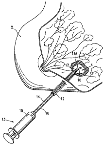
Figure 1 is a perspective view of a human breast partially cut
away having a lesion from which a biopsy specimen has been removed,
and showing a marker applicator syringe and introduction cannula
operatively positioned for introduction of a biopsy site marker
embodying features of the present invention into the cavity created by
removal of the biopsy specimen;
Figures 2A, 2B, 2C, 2D and 2E show exemplary conformations and
shapes of sintered or porous metal site marker embodiments of the
14
CA 02659518 2000-03-27
WO 02/41786 PCT/US01/43403
invention, Fig. 2A showing a sintered body having irregular pores, Fig.
2B showing a bubble-filled marker body, Fig. 2C showing a cylindrical-
shaped marker Figure 2D showing a polyhedral shaped marker and
Fig. 2E showing a cruciform-shaped marker;
Figure 3 shows an example of the alternative coil-shaped
embodiment of the marker of the invention;
Figure 4 shows an example of the alternative spheriod
embodiment of the marker of the invention;
Figure 5 Is a schematic view (scale exaggerated for clarity) of an
exemplary biopsy tissue site, in this case a human breast, showing a
biopsy cavity of the type obtained by a known type of vacuum assisted
large core biopsy sampler device, Into which a biopsy marker or
markers embodying features of the invention are deposited by a marker
applicator device inserted through the outer cannula of the large core
15, biopsy sampler.
Figure 6 shows schematically an embodiment of the invention
including one or more haptic elements and/or an adhesive component,
for resisting migration of the marker within the tissue.
Figure 7 shows schematically an embodiment of the invention
including an encapsulating element and optional adhesive component,
for resisting migration of the marker within the tissue.
CA 02659518 2000-03-27
FIG. 8 shows schematically an embodiment of the invention including an
encapsulating element and optional adhesive component, for resisting migration
of the marker within the tissue.
FIG. 9A is a schematic view of a biopsy sampler device at a tissue site with
an
alternative marker delivery system.
FIG. 9B is a perspective view of the petalled distal end of the delivery
device
shown in FIG. 9A.
FIG. 9C is a perspective view of the distal end of the delivery device shown
in
FIG. 9A with a marker exiting the petalled distal end.
FIG. 10 is a perspective view of an alternative marker having a gel body with
a
radiopaque collar disposed about the gel body.
15A
CA 02659518 2000-03-27
WO 02/41786 PCT/US01/43403
DETAILED DESCRIPTION OF THE INVENTION
The following detailed description, and the accompanying
drawings to which it refers are provided for purposes of exemplifying
and iilustrating representative examples and embodiments of the
invention only, and are not intended to limit the scope of the invention
in any way, and do not exhaustively illustrate and describe all possible
embodiments and configurations in which one or more features of the
present invention may take physical form.
Figure 1 shows the use and insertion into a biopsy site of any
one of the biopsy site marker embodim.. ents of the invention described
15% herein. Fig. 1 is a perspective view of a human breast 2 having a
lesion 3 from which a biopsy specimen has been removed, thereby
forming a biopsy cavity 4 within the lesion 3, into which a biopsy site
marker 10 of the of the present Invention Is implanted. The figure
shows an outer cannula 12 with the distal end thereof operatively
positioned within the biopsy site 4. The outer cannula 12 has been
Inserted percutaneously into the lesion 3 and a biopsy needle (not
shown) has been passed through the outer cannula 12 and used to
remove a biopsy specimen from the center of the lesion.
16
CA 02659518 2000-03-27
WO 02/41786 PCT/USO1/43403
Syringe-like marker application device 13 includes a marker
introduction tube or inner cannula 14. After removal of the biopsy
needle (not shown), the marker introduction cannula 14 has been
passed through the outer cannula 12 such that inner cannula distal
end 14d is located within the biopsy cavity 4, the marker 10 being
housed within cannula 14. Piston 15 of marker applicator 13 has an
extension 16 which passes through the interior of inner cannula 14.
Upon depressing piston 15, extenuation 16 pushes marker 10 outward
through an opening 17 in the tip 14d of inner cannula 14 into the cavity
4.
The outer cannula 12 may be an outer tube element of a
conventional vacuum assisted large core biopsy device, which has been
left in place to assist in site marker application following biopsy sample
recovery. One example of a applicator syringe device 13 is described
in further detail below with respect to Fig. 5.
Figures 2A, 2B, 2C and 2D show exemplary internal
conformations and shapes of the sintered or porous site marker
embodiments of the invention 20a-20e respectively.
Figures 2A and 2B show schematic cross sections of a
alternative porous or sintered marker body embodiments. Fig. 2A is a
cross section of a sintered site marker embodiment 20a. The matrix or
base material 21 encloses a plurality of irregular shaped pores 22
distributed within the body 20a, preferably throughout the body
17
CA 02659518 2000-03-27
WO 02/41786 PCT/US01/43403
volume. The term "sintered" will be used to describe the porous body
conformation, it being noted that conventional methods of production
other than sintering may be employed to produce a material containing
internal voids, pores, discontinuities, inclusions, bubbles and the like.
The pores 22 may be open celled, in which the pores 22
generally intersect or communicate with one another and the marker
body exterior, which may give the body surface 23 a pitted texture on
the scale of the pore size. Alternatively, the pores may be closed
celled., in which the pores 22 generally do not intersect one another or
the exterior. In the event that the pores 22
communicate with the marker exterior 23, the matrix material 21
is preferably hydrophobic (or treated to have hydrophobic surfaces) to
resist displacement of air entrained in pores 22.
The base or matrix composition 21 has may be of high acoustic
impedance relative to the surrounding tissue (not shown). Sintered
metal material may be shaped and sintered from commercially available
metallic powders comprising a metal or mixtures of metals, using
conventional sintering and forming techniques to produce body of
selected shaped, and selected pore size and surface texture, so as to
enhance acoustic reflectivity. The porosity of the sintered metal
provides an irregular surface texture as well as internal voids. A
suitable bio-compatible material is sintered 316L stainless steel, and
suitable sintered stainless steel stock is commercially available in
18
CA 02659518 2000-03-27
WO 02/41786 PCTIUSOI/43403
various forms, for example from the Mott Corporation. The sintered
stock may be economically cut and shaped by conventional methods.
Sintered stainless steel stock is commercially available with controlled
pore size, selectable over a range of pore sizes. The pores 22 of the
sintered body 20a may vary over a range of pore sizes, and is typically
from about 1 m to 100 m and preferably from about 5 m to 40 m.
In addition to sintered metal, alternative bio-compatible,
impedance materials may be included or substituted, such as ceramics,
metal oxides, polymers or composites/mixtures of these materials,
which may be configured to have a generally distributed internal
porosity and porous surface texture. Thus, the marker body 20a may
comprise a matrix or base composition 21 which has an acoustic
impedance close to that of the tissue at the marked body site, since the
air or other gas within the pores or internal spaces 22 provides a
dramatic contrast to the matrix material 21. Suitable bio-compatible
materials include polyethylene, polytetrafluoroethylene, PEBAX (made
by Autochem Corp.), and the like. Such porous materials may be
formed by conventional methods, such as heat bonding of polymer
powders, extrusion and the like.
Fig. 2B is a schematic cross section of an alternative site marker
embodiment 20b. The matrix or base material 24 encloses a plurality of
inclusions, suspended particles or bubbles 25 distributed within the
body 20b, preferably throughout the body volume. The inclusions 25
19
CA 02659518 2000-03-27
WO 02/41786 PCT/USO1/43403
may be low-density or gas-filled particles, such as foamed-in-place
bubbles; micro-beads, expanded beads, and the like, which have an
acoustic impedance substantially lower than the matrix material 24.
The matrix material 24 may as in the example of Fig. 2A.
Figures 2C and 2D show exemplary shapes of the sintered or
porous site marker embodiments of the invention 20c and 20d
respectively. Figure 2C shows schematically a cylindrical sintered
marker 20c. The marker 20c comprises a generally cylindrical body
having a diameter d and length I. The body may have diameter d of
from 0.5 to 5 mm, and preferably about 1.5 mm. The length I may be
from about 1 diameters to about 10 diameters, and preferably from
about 5 to 7 diameters. This biopsy site marker produces a distinct,
recognizable, marker image of artificial appearance when implanted at a
depth of about 2 to 4 cm in human breast tissue, and visualized by a
15, commercially available Accuson 128 US imaging system with an L7
transducer.
Fig. 2D illustrates a marker body having a polyhedral form of
multiple intersecting flat surfaces 26, 27 and 28.
Fig. 2E shows a cruciform
shaped marker 20e. The marker shown comprises a body 20e of
cruciform cross-section having four longitudinal fin-like portions 29,
CA 02659518 2000-03-27
WO 02/41786 PCT/US01/43403
which may be aligned at right angles to one another and joined at the
longitudinal central axis 30 providing a selectable number of side facets
(e.g., hexagonal cross-section). Optionally, medial web portions 31
may span laterally and join between adjacent fins 29, the webs 31
preferably being aligned perpendicularly to the fins 29. In the example
shown, there are four such web portions 31 positioned at about mid-
length of the body 20e, so that each fin 29 is joined by a pair of webs
31 , one on each side, to each adjacent fin. Thus, the planes of the
intersecting fins 29 and webs 31 form a pattern of eight mutually-
perpendicular "corner reflectors" 32 . The length I and characteristic
cross-section dimension d may be as described with respect to the
embodiments of Fig. 2C and 2D.
Fig. 3 illustrates yet another alternative where the marker body is
shaped to have the form, under ultrasound or radiological visualization,
preferably both, of a familiar symbol or letter, to by easily recognizable
as an artificial shape which is the lower-case Greek letter Gamma (y),
which when visualized in a biopsy site bears a resemblance to a familiar
breast-cancer-awareness symbol.
Figure 4 shows schematically an alternative coil marker 30 of the
invention. The marker 30 comprises a generally helical coil-like body
formed from one or more lengths of fine wire and/or fiber 31. The coil
has a generally cylindrical overall form. As with the other biopsy
site marker embodiments of the invention, the optimum dimensions of
21
CA 02659518 2000-03-27
WO 02/41786 PCT/US01/43403
the coil shaped marker embodiment will depend on such factors as the
type of visualization system used, its imaging resolution, and the
physical nature of the biopsy tissue region. The coil length I and
diameter d may be of a range of sizes, selected so as to be large
enough to provide a distinct, recognizable ultrasound marker image
within the tissue biopsy site, and small enough to avoid masking or
obscuring diagnostically important tissue features. For example, the
coil diameter d may be from 0.5 to 5 mm, and preferably about 1.5
mm. The coil length I is typically from about 1 coil diameters to about
10 coil diameters, and preferably from about 5 to 7 coil diameters.
The helical turns of the coil provide a body surface contour
including a outer helical g~pove 32 and inner helical groove 33 on the
coil surfaces (more than one such groove for a multiple helix). The
grooved coil body surface includes a plurality of lobes and crevices on
the exterior of the coil which enhance acoustic reflectivity. In addition
the similarly lobed internal surfaces of the coil provide additional
reflectivity. Optionally, the coil may be given a "frosted" or textured
surface, such as by particle blasting in the manner of the spheroid
marker described above. A uniform coil embodiment has a shape
which is markedly artificial in appearance under conventional
visualization methods, and is not easily confused tissue features of
biological origin.
22
CA 02659518 2000-03-27
WO 02/41786 PCT/US01/43403
The coil may comprise a fine wire 31 of a material of high
acoustic impedance relative to the tissue of the site, and may optionally
be radio-opaque. Suitable materials are biologically compatible metals,
such as stainless steel, titanium, platinum, palladium, alloys thereof and
the like. The coil may alternatively comprise a composite of different
materials, such as a composite of metal and polymeric materials. The
coil may be wound about a central core of the same or different
composition. Coil stock of suitable material, helical form and diameter
is available commercially, and may be cut to a selected length by
conventional means. A suitable material is 316 L stainless steel
surgical embolization coil currently used in arterial embolism repair
procedures, e.g., Cook 4 mm diameter embolization coil MWCE-25-2.5-
4 of 316L stainless steel and Dacron. Other suitable embolization coil
stock is available in a range of coil diameters. This biopsy site marker
produces a distinct, recognizable marker image as implanted at a depth
of about 2 to 4 cm in human breast tissue, when visualized by a
commercially available Accuson 128 US imaging system with an L7
transducer.
Figure 5 shows schematically the alternative spheroid marker 40
of the invention having a generally spherical body 40. Note that the
porous or sintered marker embodiments of ' Figs. 2A-2D may be
spherical also. However, the embodiment of Fig. 5 is a non-porous
example, and the biopsy site marker 40 comprises a high acoustic
23
CA 02659518 2000-03-27
WO 02/41786 PCT/US01/43403
impedance, biologically compatible material, such as 316 L stainless
steel and titanium, or radiopaque metals such as platinum, palladium, or
the like. Non-spherical shaped bodies may be used, however, metallic
spheres of suitable materials are readily commercially available, and
have a shape which is markedly artificial in appearance under
conventional visualization methods, i.e., not easily confused tissue
features of biological origin.
The generally spherical body may have a diameter d selected so
as to be large enough to provide a distinct, recognizable ultrasound
marker image within the tissue biopsy site, and small enough to avoid
obscuring tissue features. As with the other biopsy site marker
embodiments of the invention, the optimum size of the sphere will
depend on such factors as the type of visualization system used, its
imaging resolution, and the physical nature of the biopsy tissue region.
For example, the sphere diameter d is typically be from about 1 mm to
about 4 mm, and preferably from about 1.5 mm.
The spherical body 40 may include a pitted, matte, peened or
frosted surface texture 41, which may be produced by conventional
particle blasting or peening techniques. For example, the sphere may
be blasted with glass beads of about 100 m diameter to produce a
frosted surface. In another example, the sphere may be blasted with
aluminum oxide abrasive particles of about 25 m diameter to produce a
frosted surface. The frosted surface 41 thus produced provides
24
CA 02659518 2000-03-27
WO 02/41786 PCT/US01/43403
enhanced acoustic reflectivity in comparison to the untreated, smooth
sphere. Other conventional texturing, pitting or faceting methods may
alternatively be used to produce a frosted or irregular surface texture.
This biopsy site marker produces a distinct, recognizable marker
image of artificial appearance when implanted at a depth of about 2 to
4 cm in human breast tissue, and visualized by a commercially available
Acuson 128 US imaging system with an L7 transducer.
Figure 6 shows schematically in cut-away section an exemplary
marker applicator device 50 configured to be operated in association
with a conventional vacuum assisted large core biopsy device 6. The
dimensional size of the applicator device (particularly the inside
diameter) may be adjusted to correspond to the selected diameter or
characteristic dimension of the biopsy site marker to be deposited. In
this connection it should be understood that the biopsy markers of the
invention can be used without this applicator, and can be deposited in
accordance with the various methods and techniques utilized in the
state of the art.
The applicator 50 comprises an elongated cylindrical body 52
which has an outer diameter selected so that it fits, and may be
inserted through, the outer cannula 7 of vacuum assisted large core
biopsy device 6. As shown in Fig. 6, the outer cannula 7 is inserted
through the biopsy incision into the biopsy cavity 4 previously formed
CA 02659518 2000-03-27
WO 02/41786 PCT/USOI/43403
in the patient's tissue site 8, e.g., a human breast in the case of a
breast biopsy.
The cylindrical body 52 has an interior cavity and a piston 54
that fits and slides back and forth in the elongated cylindrical body 52.
The proximal end of the outer cannula 7 may be provided with
rectangularly shaped handle 56 the orientation of which indicates to the
operator the orientation of the opening 9 provided in the distal end of
the cannula 7. The cylindrical body 52 may have an enlarged finger
disk or handle 57 at its outer (exterior to the patient) end which permits
a user (not shown) to operate or move the piston 54 within the cylinder
52 of applicator 50. the orientation of the elongated finger disk 57
indicates the orientation of the opening 58 of body 53 adjacent its
other, closed end 59 (internal within biopsy cavity). The opening 58 is
configured to form a ramp in the side of the tube 52.
In this connection it should be understood that the selected
dimensions of the tube 52 are coordinated with the dimensions of the
piston 54 and with the cannula 7 of the vacuum assisted large core
biopsy device 6, thus permitting the tube 52 to both fit within cannula
7 and to contain one or more markers of the invention 10 within the
inside diameter of cylinder 52. The cylinder or tube 52 and the piston
54 may be made from any appropriate medical grade plastic material,
such as high density polyethylene or PEBAX, made by the Autochem
Corporation.
26
CA 02659518 2000-03-27
WO 02/41786 PCT/ITS01/43403
In one method of Implanting the biopsy markers 10 of the
present invention, the tube 52 is loaded with one or more of markers
10. The markers 10 may be any of the embodiments of the invention
described above, and Is shown schematically as a cyiindrica( object.
Optionally, in addition to the markers 10, pellets 57 composed of
various other materials may be inserted along with one of the
embodiments of the biopsy markers of the present invention described
herein=. For example, gelatin pellets of the type d.isciosed in our above
referenced US Patent No. 6,347,2411 may be
inserted in conjunction with the biopsy markers 10 of the present
invention.
With the markers 10 in the tube 52 and the tube 52 and cann.ula
7 inserted into the biopsy site 4, the opening 58 in the cylinder 52 is
moved into alignment with the opening or port 9 of the in the Internal
end of cannula 7 of biopsy sampler 6. The piston 54 is pressed Inward
by the operator so that the marker or markers 10 are expelled from the
tube 52 through the ramp shaped opening 58 as the piston 54 is
pushed into the cylinder or tube 52. The markers 10 are thereby
extruded through opening 59 and port 9 into the biopsy cavity 4. The
applicator 50 and biopsy device 6 are subsequently withdrawn.
Figure 7 shows schematically an alternative marker 60 of the
invention Including one or more optional tissue-engaging or haptic
elements 62 for resisting migration of the marker from the biopsy site.
27
CA 02659518 2000-03-27
WO 02/41786 PCTlUS01/43403
An exemplary cylindrical marker body 10 is shown, although each
embodiment of the biopsy site marker of the invention described above
may optionally comprises one or more such tissue engaging structures.
The haptic elements 62 may comprise an wire-like material fixed to the
marker body 10 at the proximal haptic end 64 and extending outward
from the marker body 10. The haptic 62 may be looped back at its
hook-like terminal end 66.
The haptic 62 assists in resisting migration of the marker from
the biopsy cavity, during initial placement, i.e., it engages the adjacent
tissue to resist being sucked back towards the applicator when the
applicator is withdrawn. The haptic also resists migration during later
movement, flexure or manipulation of the tissue surrounding the biopsy
site, such as when a patient's breast is decompressed upon removal
from a mammography device. Optionally, the marker body 10 may
include an adhesive component 68 coated onto its surface to cause the
marker body to adhere to adjacent tissue within the biopsy, site.
Figure 8 shows schematically the alternative marker 70 of the
invention including an encapsulating element 72 and optional adhesive
layer or component 74, for resisting migration of the marker within the
tissue. An exemplary cylindrical marker body 10 is shown, although
each of the biopsy site marker of the invention described above may
optionally comprise a pellet-shaped encapsulating element.
28
CA 02659518 2000-03-27
WO 02/41786 PCT/US01/43403
The pellet-shaped encapsulating element 72 is disposed
surrounding the marker body 10 and may fully or partially enclose the
marker body. The encapsulating element 72 may be of lower
impedance than the metallic marker body 10. Suitable materials are
gelatin or reconstituted coliagen material, polymers, or mixtures or
composites thereof. An optional adhesive component 74 is shown
coating the external surface of the encapsulating element, but may be
included within the composition the encapsulating element 72.
Fig. 9A illustrates an alternative device 80 for delivering markers
to a biopsy site which includes an elongated tube 81, a handle 82 on
the tubes proximal end and a closed distal end having a plurality of
leafs or petals 83. as shown in more detail in Fig. 9B. As shown in Fig.
9C, the petals 83 open up to allow a marker 84 to be discharged into
the biopsy site 85 as shown in Fig. 9C. The device 80 has an
elongated plunger or piston 86 slidably disposed within the tube 81 for
pushing one or more markers 84 through the petalled distal end by
pressing on the knob 87 on the proximal end of the shaft 86. The
orientation of the body 88 on the shaft 86 gives the operator an
indication of the orientation of the shaped distal end 89.
Figure 10 illustrates an alternative marker 90 which has an
elongated cylindrically shaped body of gel 91 surrounded with a
metallic band 92. The band 92 may conpletely or only partially
surround the body of gel 91.
29
CA 02659518 2000-03-27
WO 02/41786 PCT/US01/43403
In any of the above-described embodiments of the invention, the
marker body (and/or the optional ehcapsulating element) may include an
adhesive component to cause the marker body (or encapsulating
element) to adhere to adjacent tissue within the biopsy site. The
adhesive component may comprise a biocompatible adhesive, such as a
polyurethane, polyacrylic compound, polyhydroxymethacrylate, fibrin
glue (e.g., TissealTM"), collagen adhesive, or mixtures thereof.
While particular forms of the invention have been illustrated and
described, it will be apparent that various modifications can be made
without departing from the spirit and scope of the invention.
Accordingly, it is not intended that the invention be limited to the
specific embodiments illustrated. It is therefore intended that this
invention to be defined by the scope of the appended claims as broadly
as the prior art will permit, and in view of the specification if need be.