Note: Descriptions are shown in the official language in which they were submitted.
CA 02661804 2009-04-08
r 61211-1650D
- 1 -
STENT VALVES AND USES OF SAME
This application is a divisional of Canadian Patent
Application No. 2,398,640 filed on January 31, 2001.
Background
Technical field of the invention:
The invention includes a medical device and more
specifically to a valve found generally within a frame.
In preferred devices the frame is comprised of a
radially expandable stent which can be delivered
through a delivery device such as a catheter.
Background of the invention:
Lower extremity venous hypertension in addition to
venous insufficiency is a major cause of morbidity in
the United States. Symptoms of venous disease include
lower extremity edema, varicosities, skin pigmentation
changes, skin ulceration, and general poor circulation.
One solution to this problem is to replace the
defective valve or the vein with a valve assembly.
Current valves include a pressure responsive,
pressure directed ball movement valve assemblies. The
problem with mechanical ball valves is that mechanical =
valves are susceptible to clot formation. Additionally,
there are problems associated with long-term wear and
tear on the device.
Artificial valves such as biological valves are
also known. Biological valves include homografts,
allografts, and xenografts. Problems associated with
some biological valves include the supply of the
CA 02661804 2009-04-08
-
WO ,54625 PCMJS01/03095
- 2 -
valves, immunity response, or problems associated with
=
-
matching the size with the donor.
Finally other problems associated with valve
: !
repair include placement problems in which the device
cannot be repositioned once it is ejected from the
placement catheter, leakage that occurs around the
valve, and emboli formation.
In light of this background, there remains a need
for alternative and improved devices and methods for
= 10 providing valvular function within vessels of the body.
The present invention is addressed to these needs.
CA 02661804 2013-03-25
53672-11D
-3-
Summary of the Invention
Disclosed is a medical device comprising a frame that
has a valve generally located within. In preferred forms of
the invention, the frame is comprised of a radially-expandable
stent (including especially a self-expanding stent), which can
be delivered through a delivery device such as a catheter, and
then deployed and expanded at a target site in a body lumen
such as an artery or vein. For example, in one preferred use,
such a stent and method are used to treat incompetent veins in
the legs or feet.
The invention also relates to a percutaneously-
implantable vascular valve device, comprising: a percutaneously
implantable frame having a proximal frame end and a distal
frame end; a plurality of attached structures separately
fabricated from but attached to the frame, each attached
structure comprising a hole defined completely through the
attached structure; at least two valve leaflets comprising
flexible biomaterial and located at least partially within the
frame when the frame is in an expanded configuration, the at
least two flexible valve leaflets forming a valve opening
occurring between upper portions of the leaflets, and wherein
the leaflets are supported on the frame at least in part via
the attached structures; and a mechanism inward of the frame
and holding together the upper portions of the flexible
biomaterial of adjacent leaflets defining the valve opening.
The invention further relates to a percutaneously-
implantable vascular valve device, comprising: a percutaneously
implantable frame having a proximal frame end and a distal
frame end; a plurality of attached structures separately
fabricated from but attached to the frame, each attached
CA 02661804 2013-03-25
.53672-11D
-3a-
structure comprising a hole defined completely through the
attached structure; at least two valve leaflets comprising
pericardium and located at least partially within the frame
when the frame is in an expanded configuration, the at least
two flexible valve leaflets forming a valve opening occurring
between upper portions of the leaflets, wherein the leaflets
are supported on the frame at least in part via the attached
structures; a mechanism inward of the frame and holding
together the upper portions of the adjacent leaflets defining
the valve opening; and a sheath comprised of synthetic material
partially covering an outer surface of the percutaneously
implantable frame.
CA 02661804 2009-04-08
'WO 61/54625 PCT/US01/03095 =
=
- 4 -
Brief Description of the Drawings
.
.
FIGs. lA to 3 demonstrate one embodiment of the
invention comprising a stent.
FIGs. 4 to 8 demonstrate other embodiments of the
present invention comprising the valve.
FIGs. 9 to 11 demonstrate embodiments that
illustrate exemplary ways of attaching a plurality of
stents.
FIGs. 12 to 15 demonstrate exemplary embodiments
of the valve configuration in a variety of stent
embodiments.
FIG. 16 demonstrates one aspect of the invention
in situ.
FIGs. 17 to 19 demonstrate other alternative
embodiments.
FIG. 20 depicts a medical assembly of the
invention including a stent valve and a delivery device
for the stent valve.
CA 02661804 2012-05-23
53672-11D
- 5 -
Detailed description of the invention
With reference to FIG. 15, shown is one embodiment
of the present invention. The invention includes a
frame such as a wire stent that has a lumen extending
therethrough. Near one end of the stent is the valve
assembly comprising some leaflets or cusps. A valve
opening is generally located between the leaflets
through which fluid flows. Although shown as a two
leaflet valve, equally the invention can comprise, in
any embodiment described herein, at least one leaflet
such as two, three or four leaflets.
With respect to FIGs. 1A, 1B, and 1C, a frame is
partially shown. The frame can comprise a stent 20.
Choices of stent include a self expanding stent or a
non-self expanding stent. In one embodiment of the
present invention stent 20 is a self expanding stent
such as the Gianturco stent available from Cook Inc. of
Bloomington, IN as described in U.S. patent 4,580,568.
Such stent can be any
length, but in one embodiment, the stent is about 15 mm
long. Stent 20 includes a plurality of bends 22,
which generally form the area in which the stent struts
24 reverses direction. Bends 22 are generally rounded
to provide an atraumatic condition. Since the stent 20
is generally located in a vessel or body lumen of some
type, the stent 20 can be cylindrical and therefore has
a stent diameter 21 (shown in FIG. 3). In another
embodiment, the stent 20 can also have a plurality of
connectors 26 that connect adjacent struts 24. One
way to provide a connector 26 is to dispose a solder
CA 02661804 2009-04-08
(
4S:8/0 (r1/54625 PCT/US01/03095
- 6 -
bead between the adjacent struts. However connector 26
;
-
can also be a suture, weld, adhesive, rod, clamp, or
other well-known ways to connect adjacent struts 24.
Connector 26 provides several non-critical advantages.'
Connectors 26 can attach adjacent struts 24 to
minimize or prevent flaring of the ends of the stent
20. Furthermore, connector 26, if placed near the bend
22, can create a hole 28 wherein the boundaries of the
hole are the wires of the stent operating in general
conjunction with the connector 26. This creates a hole
28 through which a thread or suture can run. However,
as shown in FIG. 1C, a separate prefabricated hole can
be created by separately attaching a hole assembly,
such as a cap /9 over the bend 22. In any case, one
benefit of the connector 26 or cap 29 is that they
increase the radiographic visualization of the
invention. Particularly, if the connector 26 is a
solder bead, it has increased radiopacity.
With respect to FIGs. 2A and 2B, shown is part of
the stent in which connector 26 attaches adjacent
struts 24. As mentioned above, a thread or suture can
be threaded through the hole 28. A proximal suture 30
can be sewn through the stent proximal bends 22 or
stent proximal ends 31 of the stent. Similarly, a
distal suture 32 can be sewn through the stent distal
end 33 or the stent distal bends 22 of the stent. One
way to thread the suture is shown in FIG. 2B wherein
the suture 35 (generically any suture) runs over the
strut 24 to enter the hole 28, through hole 28 to come
behind the same strut 24, over the strut 24 and across
to the adjacent strut 24 running over the adjacent
strut 24, behind the adjacent strut 24 to come from
CA 02661804 2009-04-08
. ,
k
WO 01/54625
PCT/US01/0µ45
- 7 -
behind and through hole 28, and then run subsequently
over adjacent strut 24. Once the struts are connected
via the suture, the suture can be pulled to a
predetermined tightness to control the overall stent
size. Accordingly, the stent can be so constructed to
have a predetermined stent perimeter 34. To this end,
the stent lumen 36 will also have an appropriate size.
The stent can be constructed so as to have a different
perimeter length at the proximal or distal ends.
With regard to FIG. 3, shown is a cylindrical
stent 20 that has the proximal and distal sutures
running through the bends 22 or holes 28 of the
proximal and distal ends of the stent. By altering the
tautness of the sutures, the size of the stent lumen
36, the stent diameter 86, and the stent perimeters 34,
can be adjusted. As can be seen, distal perimeter
suture 32 runs along the stent distal end 33, whereas
proximal perimeter suture 30 runs along the stent
proximal end 31. The respective sutures run through
hole 28 of each bend 22.
With respect to FIGs. 4 and 5, the valve material
38 is shown, in this exemplary embodiment, as a sheet.
In so constructing the valve 41, the valve material 38
is draped across the stent lumen 36 opening (such as
shown on the proximal portion of the stent) and then
pushed down into the stent lumen 36 itself. Excess
material can be kept outside the stent, which will
later become a potential fold-over 42. However, the
excess material can also be trimmed off. The valve
material 38 is connected to the stent, using for
example, distal valve-stent suture 40. However, any
well known ways to connect the valve to the stent is
CA 02661804 2009-04-08
*001,A625 PCT/US01/03095
- 8 -
contemplated, such as but not limited to, sutures,
-
adhesives, folds, or the like. In one embodiment shown
in FIG. 5, the valve-stent suture 40 can share the hole
28 with distal suture 32 near the stent perimeter 34.
The valve material 38 can be any biocompatible
material such as polyethylene terephalate(PET),
polypropylene (PP), polytetrafluorethylene(PTFE), or
any polymer or derivative thereof, and also includes
commercially known materials such as GORE-TEX, DACRON,
or any other synthetic material. The preferred
material 38 will be advantageously compliant and
employed so as to permit effective value function as
described herein and in the case of
collapsible/expandable state devices will retain
integrity and function when cycled between tehse
states.
It is preferred to use a biomaterial that serves
as a biocompatible scaffold with the ability to remodel
host tissue. Accordingly, a naturally occurring
biomaterial is highly desirable. One such biomaterial
is collagen and more particularly, a collagen based
biomaterial called extracellular matrix (ECM).
Examples of ECM's include pericardium, stomach
submucosa, liver basement membrane, urinary bladder
submucosa, tissue mucosa, dura Mater, and small
intestine submucosa One such biomaterial is the ECM,
such as submucosa, and more particularly is small
intestine submucosa (SIS). SIS can be made in the
fashion described in Badylak et al., US Patent
4,902,508; Intestinal Collagen Layer described in US
Patent 5,733,337 to Carr and in 17 Nature Biotechnology
1083 (Nov. 1999); Cook et al., WIPO Publication WO
CA 02661804 2012-05-23
53672-11D
- 9 -
=
98/22158, dated 28 May 1998, which is the published
-application of PCT/US97/14855; Gastric Submucosa as .
described in WO 98/26291 (PCT/US97/22729), claiming
priority to US Provisional application 60/032,686;
Liver tissue as described in WO 98/25637
(PCT/US97/22727), claiming priority to 60/032,68.0;
Stomach Submucosa as described in WO 98/25636
(PCT/US97/23010), claiming priority to 60/032,683; and
Urinary Bladder Submucosa as described in US Patent
5,554,389.
Irrespective of
the origin of the valve material (synthetic versus
naturally occurring), the valve material can be made
thicker by making multilaminate constructs, for example
SIS constructs as described in US Patents 5,968,096;
5,955,110; 5,885,619; and 5,711,969.
With respect to FIGs. 6A and 6B, shown is the
connection of the valve to the stent frame. As
described above, the valve can be sutured at the distal
portion of the stent using distal .valve-stent suture
40. Similarly, the proximal portion of the valve can
be sutured to proximal portion of the stent, and more
particularly to proximal perimeter suture 30. Shown is
the valve connected to the proximal .portion of the
stent at proximal valve-stent suture 44. Suture 44 can
be through a bend 22 or can attach to the proximal
perimeter suture 30. In a traditional Gianturco Z-
stent, it is either an 8 (bend) point or 10 (bend)
point stent, so one leaflet of the valve can be sutured
to the four points of an 8 point stent thereby
CA 02661804 2009-04-08
= '
WO vi154625 PCT/US01/03095 =
=
- 10 -
=
comprising one half of the stent. To provide further
integrity, the valve can be sutured at the proximal and
distal end to the perimeter sutures themselves, without
actually being sutured to any or all of the stent bends
22.
With respect to FIG. 6B, shown is the valve with
the stent frame removed. Once the sutures are
generally in place, the valve sheet 38 will form a
valve pocket 46, extending inside the stent lumen in
which the fluid will fill. Proximal valve perimeter 48
will have the sutures connecting the valve to the stent
(not shown). Once the distal sutures are in place, the
general shape will likely resemble a pocket with the
pocket having a valve apex 50. There is a part of the
valve that will form central valve portion 49 that is
not directly sutured to the stent. This valve portion
49 will form the valve opening 52 through which fluid
will pass. Thus, upon filling of the valve pocket 46,
the fluid pressure will exert outwards causing valve
portion 49 to extend outward. When it does, it will
contact the other leaflets or cusps and form a seal to
stop or impede fluid flow.
FIG. 7 shows a top view of the stent valve. In
this particular non-limiting view, shown is the valve
opening 52 in a slightly open configuration. Valve
pockets 46 are shown in a slightly distended
configuration. The valve is connected, for example, by
sutures to the stent perimeter 34 and also forms a
valve perimeter 48. Because of the opening and closing
of the valve, there may be increased wear and tear at
the valve-stent-opening connection. At this point, one
embodiment of the present invention provides a
CD, 02661804 2009-04-08
.=,
PCT/US01/03095
W001/54625
- 11 -
reinforcement at this point. For example, this
reinforcement can be a plurality of reinforcement
sutures 54, adhesive, another material, or any other
mechanism that permits increased structural integrity.
FIG. 8 demonstrates a view of the stent valve once
the distal portion of the valve is sewn to a distal
bend 22 and also shows the proximal portion of the
valve being connected to the proximal portion of the
frame with one suture in the foreground, one suture in
the background. In addition, the reinforcement suture
54 is found in the foreground. Although only two
sutures 44 are seen at the proximal portion, it is of
course well-understood Lhat some or each of the
proximal bend of the frame can be connected to the
proximal portion of the valve. Similarly, although
only one distal suture 40 is shown, there may be as
many distal sutures necessary to connect the valve apex
50 or the distal portion of the valve to the frame. It
is well understood that this may be just one distal
suture or many distal sutures. Varying the number of
distal sutures will vary the shape, tightness, and
overall configuration of the valve, valve pocket 46,
and the valve apex 50.
The valve opening 52 although already described
above, is actually created in the final step of
preparation of the preferred device manufacture. The
construction mentioned above would be repeated on the
other side of the valve to create the valve pocket 46,
valve apex 50, and the like on the other side. At this
point, though, there is no valve opening 52. The valve
opening 52 is created by creating a slit in the sheet
to create the opening. The slit can be sized according
CA 02661804 2009-04-08
W6S...2&1625 PCT/US01/03095
'
- 12 -
to the intended flow rate of the passing fluid.
: =
Accordingly, a large slit would create a large valve
opening or orifice and permits a large volume of fluid
to pass therethrough. The slit can be created by poking
a scalpel through it and running it to the desired
length. However, due to potential fatigue at the
orifice, another set of reinforcements may be added to
the orifice perimeter. Therefore, as shown in FIGs. 7
and 8, an orifice reinforcement 53 may be created by
any known conventional ways, such as sutures
(resorbable or non-resorheble), adhesive, string,
staples, rings, or the. like.
Therefore, the stent valve as constructed can be
using one stent with the valve material enclosed
therein. Of course in the single stent configuration,
the overall length can be adjusted by elongating the
length of the struts 24. However, devices of the
invention can be built using a plurality of stents to
elongate the overall size of the stent, if desired. In
this regard, it will be preferred that the length of
the device 20 is sufficient to provide an aspect ratio
(length to expanded diameter) sufficiently high to
facilitate proper alignment of the device 20 within the
vessel, with the axis of the device lumen generally
aligned with the axis of the vessel. For example,
devices having a ratio of length:expanded diameter of
1:1 or greater, or about 2:1 or greater, will be
preferred. It will be understood that while such
dimensions will advantageously facilitate placement of
the inventive devices, they are not necessary to the
broader aspects of the invention
With reference to FIG. 15, shown is a double stent
CA 02661804 2009-04-08
- =
'
WO 01/54625
PCT/US01/03ti:
- 13 -
structure with the valve. Returning now to FIG. 9,
shown is a first stent 58 and a second stent 60. For
the purposes of discussion only, first stent 58 is
shown to be atop of the second stent 60. Ultimately as
shown herein by way of example only, the valve will
reside in the first stent 58. It should be noted
however that the valve can reside in the second stent
60 also as shown in FIG. 17. Furthermore, the overall
length can be increased by joining several stent valves
together as shown in FIGs. 18 and 19, thereby having a
plurality of stents, such as a first stent 58, second
stent 60, and a third stent 61. The valve 41 can be
placed in any or all stents, in any combination, for
example, as shown by the dotted lines. In this regard,
it is suggested and intended that many stents can be
joined and that each or any stent may house a valve or
plurality of valves. One benefit of having a plurality
of stents is that upon ejection of the placement
device, the invention will provide a self-aligning
feature in the vessel. This is because the plurality
of stents is generally longer with respect to the stent
diameter, or the plurality of valve device(s), as
discussed above.
Manufacture of the multi-stent or multi-valve
device will generally follow the same construction as
described above. The same considerations in making a
single valve single stent device applies equally to the
elongated device.
Returning now to FIGs. 9 and 10, shown are methods
of connecting the first stent 58 and second stent 60.
Equally, the construction shown from now on also
includes construction of at least two stents or at
CA 02661804 2009-04-08
.
PCT/US01/03095
WO t. i,44625
=
- 14 -
least two valves. First stent 58 and second stent 60
has bends 22 that are adjacent each other. Shown in
FIG. 9 is where the first stent 58 has its bends beside
- =
the bends of the second stent 60 such that they are not
touching each other (although they may touch). They
are connected together in the manner described above,
and for example by stent-stent suture 56. Stent-stent
suture 56 can be resorbable or non-resorbable. This
suture travels through the distal bends of the first
stent 56 and the proximal bends of the second stent 60.
The suturing pattern can be that described in FIG. 2B
and the accompanying discussion. As shown in FIG. 10,
the bends can be juxtaposed over each other to provide
an overlap suth that the stent-stent suture 56 will go
through the bends at the same time. Therefore, the
construction contemplates that the stent bends may
touch, overlap, or not at all.
= FIG. 11 shows one embodiment of the present
invention in which the valve apex 50 is sutured to at
least three bends; two bends of the first stent 56 and
one bend of the second stent 60. In this regard, the
valve also operates to keep the first stent 56
partially connected to the second stent 60. From the
bends, a plurality of valve apex sutures 66 are seen.
These sutures can emanate from the bends and each bend
can have many valve apex sutures 66 that travel in many
directions. Using multiple valve apex sutures 66 that
emanate in many directions and using a plurality of
bends (from either stent), generally functions to
minimize any parachuting or inversion of the valve
pocket 46.
FIG. 12 demonstrates a top view of the multi-stent
CA 02661804 2009-04-08
;
WO 01/54625
PCT/US01/036-:..
- 15 -
device in which the valve opening 52 is seen (in a
closed position) and the valve pocket 46 and valve apex
50 is connected to three bends. Again it should be
understood that many sutures may emanate from many
bends from any stent.
As described earlier, the excess material can
either be trimmed off or folded over the outer surface
of the device. Shown in FIGS. 13A and 13B, is the
excess material being folded over the device and
attached at the distal end of the first stent 58.
Shown in dotted lines is the first stent 58. FIG. 13B
shows that the fold-over 42 provides a second material
outer sheath so that the suture passes through the
inside and outside material to increase structural
integrity. Also, by folding over the excess material,
a smoother surface is presented rather than the naked
frame of the tip of the bend.
In all embodiments of the invention, the external
surface of the frame can be covered with a sheath that
is not necessarily the same material as the valve 41.
For example, while the valve can be a naturally
occurring material, the outer sheath can be synthetic
material such as described herein. The sheath,
therefore, can be the fold-over of the valve material,
another type of naturally occurring material, or a
synthetic material. Accordingly, the sheath may
partially or totally cover the frame.
FIG. 14 shows an embodiment in which both the
first stent 58 and second stent 60 are covered by the
fold over 42. Here, the fold-over 42 is connected to
the distal portion of the second stent 60. In this
manner, the entire device may be covered with an outer
CA 02661804 2009-04-08
W04:144625
PCT/US01/03095
- 16 -
sheath of biomaterial. The benefit of doing so,
especially if using SIS or other similar ECMs, is that
the regrowth and endothelialization of the device
embeds and encapsulates the frame. Accordingly, there
is a reduced risk of device migration. Furthermore,
due to the remarkable remodeling properties of SIS, the
outer SIS sheath acts as a conduit for host tissue to
infiltrate the device and remodel the valve itself.
Over the course of months, the valves are replaced by
host tissue and the SIS disappears.
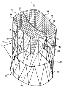
FIG. 15 shows yet another embodiment of the
present invention. In this demonstration, the valve is
located in the first stent 58, sutured at the proximal
end at the stent perimeter. The valve apex 50 is sewn
somewhat proximal of the stent-stent suture 56. The
valve apex 50 is sewn at the valve apex sutures 66 to
an intermediate portion of the frame. To minimize
parachuting or inversion, a valve intermediate portion
75 may be sutured using valve intermediate suture 76 to
connect the valve to the frame. In addition, the valve
may be so constructed to extend the valve's length to
create an elongated valve pocket 90 (shown by the
dotted lines). While the extended pocket 90 can be
connected to the distal perimeter of the second stent
distal suture 62:it can also be connected to an
intermediate portion of the second stent.
With further reference to FIG. 15, it is seen that
the valve opening 52 is a slit that extends across the
first stent diameter 21 but terminates several
millimeters before reaching the edge. In some
embodiments, this distance could be 1-5 mm from the
edge. Of course, it is understood that the invention
CA 02661804 2009-04-08
1, =
4 =
'1
WO 01/54625
PCT/US01/03b95
- 17 -
contemplates any distance that varies the length of the
slit. Also, shown in FIG. 15, but equally applies to
any device described herein, is an anchor 92, which can
be anchor barbs 92. These barbs 92 can dig into the
adjacent vessel wall to relatively affix the device at
its location. Anchor 92, although shown as barbs, may
include hooks, adhesives, knobs, a textured surface, or
any other treated surface that facilitates relative
affixation of the device in its location. Similarly,
the outer surface of the fold-over or sheath can be so
configured to provide anchoring.
FIG. 16 demonstrates the device upon implantation
into the patient. Upon implantation the device
generally resides in a vessel 80. For example, the
vessel could be vein, artery or the heart or wherever a
valve is necessary. In one preferred use, the vessel
is an incompetent vein in the leg or foot of a patient.
The device 20 reduces or prevents retrograde blood
flow, while normal blood flow is permitted to travel
through device 20. Illustrative veins in which the
device 20 may be used include, for example, saphenous
veins, femoral veins, popliteal veins, tibial veins,
and the inferior vena cava.
The vessel 80 has an inner lumenal surface 82 in
which the fluid flows. The fluid flow path is shown as
fluid path 70. Vessel 80 also has a vessel diameter
84. The medical device, upon implantation, will also
have a device outer stent diameter 86. The outer
diameter 86 will be chosen to permit contact with the
inner lumenal surface 82. The optimized fit will
decrease the leakage around the device by contacting
the inner lumenal surface 82. A tight fit can be
CA 02661804 2009-04-08
,
W0t1/54625 PCT/US01/03095
- 18 -
accomplished by sizing the stent diameter to be greater
.
.
than the vessel diameter. For example, a stent diameter
that is about 110 percent greater than (i.e. 1.1 times)
the vessel diameter provides a good fit. Expanded *
stent diameters of about 10 mm to about 30 mm will be
typical in many applications of the present invention.
Again, while it is shown in this FIG. 16 that the
valve is located in the first stent 58 and only the
first stent 58 is covered by the fold-over 42 or
sheath, it should be remembered that the valve could be
located in the second stent 60. Similarly, the fold-
over 42 or sheath could extend onto the second stent
60.
The standard method of deploying the medical
device 20 in a vessel 80 involves the use of a medical
assembly (see FIG. 20) including the device 20 and a
delivery device such as a percutaneous delivery device,
e.g. a catheter 100. The frame is configured to a
contracted state, e.g. by resiliently forming the frame
into a contracted configuration, to load into the
delivery device (catheter). The catheter can be
introduced into the patient via a suitable approach,
for example through the jugular or femoral vein. To
advance and deploy the device from the distal end of
the delivery catheter, a pusher 101 is placed into the
catheter lumen. When the device 20 is fully deployed,
it assumes the second, expanded configuration within
the vessel 80 as depicted in FIG. 16. The stent frame,
being made of resilient material, conforms to the shape
of the vessel wall such that when viewed on end, the
device 20 has a circular appearance when deployed in a
round vessel.
CA 02661804 2009-04-08
, .
WO 01/54625
PCT/US01/0045
- 19 -
FIGs. 17, 18, and 19 show other described
embodiments. FIG. 17 demonstrates the valve 41 in the
second stent 60. In this embodiment, the valve apex 50
is connected to the second stent's distal perimeter.
FIG. 18 demonstrates at least two stent frames
connected together. In this particular embodiment, the
valve is located in the first stent 58, with the valve
apex 50 being connected at the first stent 58-second
stent 60 junction. In dotted lines, however, there may
be many stents, such as first stent 58, second stent
60, and third stent 61. The valve 41 may be found in
any of the stents or in all. Similarly, in the three
stent configuration, the valve may begin at the first
stent and have the valve apex 50 be generally located
in the third stent 61. FIG. 19 shows another
embodiment of the present invention in which the valve
41 begins in the second stent 60 and extends into the
third stent 61 thereby having the first stent 58 being
empty.
Finally, since the device is located in an in vivo
environment, the device may be treated with therapeutic
agents to facilitate healing. For example, the frame
may be treated with therapeutic agents such as anti-
cancer drugs, plaque busters, anti-coagulants, or the
like. Similarly, the valve material can be treated with
therapeutics agents such as anti-cancer drugs, plaque
busters, anti-coagulants, proteins, growth factors,
proteoglycans, and the like. Furthermore, radiopaque
agents may be added, such as tantalum, barium, bismuth,
or the like to increase radiopacity. These ingredients
can be bonded to the frame or the valve material such
as rubbing the agent in, bonding it, adhering it, or
CA 02661804 2009-04-08
,
WO b.4625 PCT/US01/03095
- 20 -
the like.
While the invention has been illustrated and
=
described in detail in the drawings and the foregoing
text, it is understood that these are only some
embodiments and that the scope of the invention is not
solely defined by the description herein but also by
the appended claims. All modifications and changes
that come within the spirit of the invention are hereby
protected.