Note: Descriptions are shown in the official language in which they were submitted.
CA 02663220 2009-03-11
WO 2008/034013 PCT/US2007/078417
MEDICAL DEVICES AND METHODS OF MAKING THE SAME
CROSS-REFERENCE TO RELATED APPLICATIONS
This application claims priority under 35 USC 119(e) to U.S. Provisional
Patent
Application Serial No. 60/845,046, filed on September 15, 2006, the entire
contents of
which are hereby incorporated by reference herein.
TECHNICAL FIELD
The invention relates to medical devices, such as, for example,
endoprostheses,
and to related methods.
BACKGROUND
The body includes various passageways such as arteries, other blood vessels,
and
other body lumens. These passageways sometimes become occluded or weakened.
For
example, the passageways can be occluded by a tumor, restricted by plaque, or
weakened
by an aneurysm. When this occurs, a passageway can be reopened or reinforced,
or even
replaced, with a medical endoprosthesis. An endoprosthesis is typically a
tubular
member that is placed in a lumen in the body. Examples of endoprostheses
include
stents, stent-grafts, and covered stents.
An endoprosthesis can be delivered inside the body by a catheter that supports
the
endoprosthesis in a compacted or reduced-size form as the endoprosthesis is
transported
to a desired site. Upon reaching the site, the endoprosthesis is expanded, for
example, so
that it can contact the walls of the lumen.
The expansion mechanism may include forcing the endoprosthesis to expand
radially. For example, the expansion mechanism can include the catheter
carrying a
balloon, which carries a balloon-expandable endoprosthesis. The balloon can be
inflated
to deform and to fix the expanded endoprosthesis at a predetermined position
in contact
with the lumen wall. The balloon can then be deflated, and the catheter
withdrawn.
In another delivery technique, the endoprosthesis is formed of an elastic
material
that can be reversibly compacted and expanded (e.g., elastically or through a
material
phase transition). During introduction into the body, the endoprosthesis is
restrained in a
1
CA 02663220 2009-03-11
WO 2008/034013 PCT/US2007/078417
compacted condition. Upon reaching the desired implantation site, the
restraint is
removed, for example, by retracting a restraining device such as an outer
sheath, enabling
the endoprosthesis to self-expand by its own internal elastic restoring force.
To support a passageway and keep the passageway open, endoprostheses are
sometimes made of relatively strong materials, such as stainless steel or
Nitinol (a nickel-
titanium alloy), formed into struts or wires.
SUMMARY
In one aspect, the invention features an endoprosthesis including a generally
tubular member having a lumen and including at least one component selected
from
struts, bands, and combinations thereof. The component includes a bioerodible
material
selected from bioerodible metals, bioerodible metal alloys, and combinations
thereof.
The component has a first region including pores and a second region including
pores,
and the average maximum dimension (e.g., diameter) of the pores in the second
region is
greater than the average maximum dimension (e.g., diameter) of the pores in
the first
region.
In another aspect, the invention features an endoprosthesis including a
generally
tubular member having a lumen and including at least one component selected
from
struts, bands, and combinations thereof. The component includes a bioerodible
material
selected from bioerodible metals, bioerodible metal alloys, and combinations
thereof.
The component has a first region and a second region having a higher pore
density than
the first region.
In an additional aspect, the invention features an endoprosthesis including a
generally tubular member having a lumen. The generally tubular member includes
at
least one component selected from struts, bands, and combinations thereof. The
component includes a bioerodible material selected from bioerodible metals,
bioerodible
metal alloys, and combinations thereof. The component has at least one pore,
and the
endoprosthesis includes a polymer that is disposed within the pore.
In a further aspect, the invention features an endoprosthesis including a
generally
tubular member having a first region including pores and a second region
including
pores. The first region defines an interior surface of the generally tubular
member, and
2
CA 02663220 2009-03-11
WO 2008/034013 PCT/US2007/078417
the second region defines an exterior surface of the generally tubular member.
The
average maximum dimension of the pores in the second region is greater than
the average
maximum dimension of the pores in the first region. The generally tubular
member
includes a bioerodible material selected from bioerodible metals, bioerodible
metal
alloys, and combinations thereof.
In another aspect, the invention features an endoprosthesis including a
generally
tubular member. The generally tubular member has a first region defining an
interior
surface of the generally tubular member and a second region defining an
exterior surface
of the generally tubular member. The second region has a higher pore density
than the
first region. The generally tubular member includes a bioerodible material
selected from
bioerodible metals, bioerodible metal alloys, and combinations thereof.
In an additional aspect, the invention features an endoprosthesis including a
generally tubular member and a polymer. The generally tubular member has at
least one
pore, and the polymer is disposed within the pore. The generally tubular
member
includes a bioerodible material selected from bioerodible metals, bioerodible
metal
alloys, and combinations thereof.
In a further aspect, the invention features a method including delivering an
endoprosthesis into a lumen of a subject. The endoprosthesis includes a
generally tubular
member including a therapeutic agent and a bioerodible material selected from
bioerodible metals, bioerodible metal alloys, and combinations thereof. The
generally
tubular member erodes at an erosion rate and the therapeutic agent elutes into
the lumen
of the subject at an elution rate. The elution rate is slower than the erosion
rate.
In another aspect, the invention features an endoprosthesis including a
generally
tubular member having a lumen. The generally tubular member includes at least
one
component selected from struts, bands, and combinations thereof. The component
includes a reservoir surrounded by a matrix including a bioerodible material
and having
at least one pore. The bioerodible material is selected from bioerodible
metals,
bioerodible metal alloys, and combinations thereof.
Embodiments can include one or more of the following features.
The first and/or second region of the component and/or the generally tubular
member can include at least one pore (e.g., multiple pores). The average
maximum
3
CA 02663220 2009-03-11
WO 2008/034013 PCT/US2007/078417
dimension of the pores in the second region can be different from (e.g.,
greater than) the
average maximum dimension of the pores in the first region. In some
embodiments, the
average maximum dimension of the pores in the second region can be at least
about 1.5
times greater (e.g., at least about two times greater, at least about five
times greater, at
least about 10 times greater) than the average maximum dimension of the pores
in the
first region.
The endoprosthesis can include a therapeutic agent. The reservoir can contain
a
therapeutic agent. The endoprosthesis can include a polymer (e.g., a
bioerodible
polymer). The polymer can be supported by the component and/or the generally
tubular
member. The polymer can be disposed within pores of the component and/or the
generally tubular member. In some embodiments, the polymer can be disposed
within at
least one pore (e.g., multiple pores) in the first region and/or the second
region of the
component and/or the generally tubular member. In certain embodiments, the
endoprosthesis can include a composite including a therapeutic agent and a
polymer.
The generally tubular member can have an exterior surface and an interior
surface
that defines the lumen of the generally tubular member. In some embodiments,
the first
region of the component can define at least a portion of the interior surface
of the
generally tubular member. In certain embodiments, the second region of the
component
can define at least a portion of the exterior surface of the generally tubular
member.
The pore density of the second region of the component and/or the generally
tubular member can be different from (e.g., higher than) the pore density of
the first
region of the component and/or the generally tubular member. In some
embodiments, the
pore density of the second region can be at least about 1.5 times higher
(e.g., at least
about two times higher, at least about five times higher, at least about 10
times higher)
than the pore density of the first region.
In certain embodiments, the first and/or second regions of the component
and/or
the generally tubular member may not include any pores.
Embodiments can include one or more of the following advantages.
In some embodiments, a medical device (e.g., an endoprosthesis) including a
bioerodible material can be used to temporarily treat a subject without
permanently
remaining in the body of the subject. For example, the medical device can be
used for a
4
CA 02663220 2009-03-11
WO 2008/034013 PCT/US2007/078417
certain period of time (e.g., to support a lumen of a subject), and then can
erode after that
period of time is over.
In certain embodiments, a medical device (e.g., an endoprosthesis) including a
bioerodible metal and/or a bioerodible metal alloy can be relatively strong
and/or can
have relatively high structural integrity, while also having the ability to
erode after being
used at a target site.
In some embodiments, a medical device (e.g., an endoprosthesis) including a
bioerodible material and having regions with different pore densities and/or
with pores
having different average maximum dimensions can erode at different rates in
the different
regions. In certain embodiments, a medical device can be designed to erode at
a faster
rate in some regions than in other regions. For example, an endoprosthesis can
be
designed so that its end regions erode at a faster rate than its center
region. The result can
be that the endoprosthesis erodes as one piece, starting at its end regions
and progressing
toward its center region.
In some embodiments, a medical device (e.g., an endoprosthesis) that includes
a
bioerodible material can also include at least one other material that is
either bioerodible
or non-bioerodible. The other material can, for example, enhance the strength
and/or
structural integrity of the medical device.
In certain embodiments, a medical device (e.g., an endoprosthesis) can provide
a
controlled release of one or more therapeutic agents into the body of a
subject. For
example, in some embodiments in which a medical device includes a bioerodible
material
and a therapeutic agent, the erosion of the bioerodible material can result in
the release of
the therapeutic agent over a period of time.
In some embodiments, a medical device (e.g., an endoprosthesis) having regions
with different pore densities and/or with pores having different average
maximum
dimensions can deliver therapeutic agents at different rates and/or in
different amounts
from the different regions. For example, a region of an endoprosthesis having
a relatively
high pore density and/or having pores with a relatively high average maximum
dimension
may deliver therapeutic agent at a faster rate, and/or may deliver a greater
total volume of
therapeutic agent, than another region of the endoprosthesis having a
relatively low pore
density and/or having pores with a relatively low average maximum dimension.
In
CA 02663220 2009-03-11
WO 2008/034013 PCT/US2007/078417
certain embodiments, one region of a medical device can be designed to deliver
more
therapeutic agent, and/or to deliver therapeutic agent at a faster rate, than
another region
of the medical device. For example, a region of an endoprosthesis that is
located along
an outer diameter of the endoprosthesis can be designed to deliver a greater
volume of
therapeutic agent, and/or to deliver therapeutic agent at a faster rate, than
a region of the
endoprosthesis that is located along an inner diameter of the endoprosthesis.
In certain embodiments, a medical device (e.g., an endoprosthesis) having
regions
with different pore densities and/or with pores having different average
maximum
dimensions can deliver different therapeutic agents from the different
regions. As an
example, in some embodiments, a region of an endoprosthesis having a
relatively high
pore density and including pores having a relatively high average maximum
dimension
can deliver a therapeutic agent at a relatively fast rate, while another
region of the
endoprosthesis having a relatively low pore density and including pores having
a
relatively low average maximum dimension can be used to deliver a different
therapeutic
agent at a relatively slow rate.
In some embodiments in which a medical device (e.g., an endoprosthesis)
includes both a bioerodible material (e.g., a bioerodible metal) and a
therapeutic agent,
the erosion rate of the bioerodible material can be independent of the elution
rate of the
therapeutic agent. As an example, in certain embodiments, a medical device can
be
formed of a porous bioerodible metal, and can include a composite including a
bioerodible polymer combined with a therapeutic agent that is disposed within
the pores
of the bioerodible metal. As the polymer erodes, it can release the
therapeutic agent at a
rate that is different from the erosion rate of the bioerodible metal. In
certain
embodiments, the bioerodible metal can erode before all of the therapeutic
agent has been
released from the polymer. The remaining polymer can continue to elute the
therapeutic
agent. The therapeutic agent can be selected, for example, to help alleviate
the effects, if
any, of the erosion of the bioerodible metal on the body of the subject.
In some embodiments, a medical device (e.g., an endoprosthesis) including one
or
more metals (e.g., bioerodible metals) can be relatively radiopaque. This
radiopacity can
give the medical device enhanced visibility under X-ray fluoroscopy. Thus, the
position
6
CA 02663220 2009-03-11
WO 2008/034013 PCT/US2007/078417
of the medical device within the body of a subject may be able to be
determined
relatively easily.
An erodible or bioerodible endoprosthesis, e.g., a stent, refers to a device,
or a
portion thereof, that exhibits substantial mass or density reduction or
chemical
transformation, after it is introduced into a patient, e.g., a human patient.
Mass reduction
can occur by, e.g., dissolution of the material that forms the device and/or
fragmenting of
the device. Chemical transformation can include oxidation/reduction,
hydrolysis,
substitution, and/or addition reactions, or other chemical reactions of the
material from
which the device, or a portion thereof, is made. The erosion can be the result
of a
chemical and/or biological interaction of the device with the body
environment, e.g., the
body itself or body fluids, into which it is implanted and/or erosion can be
triggered by
applying a triggering influence, such as a chemical reactant or energy to the
device, e.g.,
to increase a reaction rate. For example, a device, or a portion thereof, can
be formed
from an active metal, e.g., Mg or Ca or an alloy thereof, and which can erode
by reaction
with water, producing the corresponding metal oxide and hydrogen gas (a redox
reaction). For example, a device, or a portion thereof, can be formed from an
erodible or
bioerodible polymer, or an alloy or blend erodible or bioerodible polymers
which can
erode by hydrolysis with water. The erosion occurs to a desirable extent in a
time frame
that can provide a therapeutic benefit. For example, in embodiments, the
device exhibits
substantial mass reduction after a period of time which a function of the
device, such as
support of the lumen wall or drug delivery is no longer needed or desirable.
In particular
embodiments, the device exhibits a mass reduction of about 10 percent or more,
e.g.
about 50 percent or more, after a period of implantation of one day or more,
e.g. about 60
days or more, about 180 days or more, about 600 days or more, or 1000 days or
less. In
embodiments, the device exhibits fragmentation by erosion processes. The
fragmentation
occurs as, e.g., some regions of the device erode more rapidly than other
regions. The
faster eroding regions become weakened by more quickly eroding through the
body of
the endoprosthesis and fragment from the slower eroding regions. The faster
eroding and
slower eroding regions may be random or predefined. For example, faster
eroding
regions may be predefined by treating the regions to enhance chemical
reactivity of the
regions. Alternatively, regions may be treated to reduce erosion rates, e.g.,
by using
7
CA 02663220 2009-03-11
WO 2008/034013 PCT/US2007/078417
coatings. In embodiments, only portions of the device exhibits erodibilty. For
example,
an exterior layer or coating may be erodible, while an interior layer or body
is non-
erodible. In embodiments, the endoprosthesis is formed from an erodible
material
dispersed within a non-erodible material such that after erosion, the device
has increased
porosity by erosion of the erodible material.
Erosion rates can be measured with a test device suspended in a stream of
Ringer's solution flowing at a rate of 0.2 m/second. During testing, all
surfaces of the
test device can be exposed to the stream. For the purposes of this disclosure,
Ringer's
solution is a solution of recently boiled distilled water containing 8.6 gram
sodium
chloride, 0.3 gram potassium chloride, and 0.33 gram calcium chloride per
liter.
Other aspects, features, and advantages are in the description, drawings, and
claims.
DESCRIPTION OF DRAWINGS
FIG. lA is a perspective view of an embodiment of a stent in a compressed
condition.
FIG. lB is a perspective view of the stent of FIG lA, in an expanded
condition.
FIG. 1C is a cross-sectional view of the stent of FIG. lA, taken along line 1C-
1C.
FIG. 1 D is an enlarged view of region 1 D of the stent of FIG. 1 C.
FIG. 2A is a cross-sectional view of an embodiment of a stent.
FIG. 2B is an enlarged view of region 2B of the stent of FIG. 2A.
FIG. 3 is a cross-sectional view of an embodiment of a stent.
FIG 4A is a perspective view of an embodiment of a stent.
FIG. 4B is a cross-sectional view of the stent of FIG. 4A, taken along line 4B-
4B.
FIG. 5 is a cross-sectional view of an embodiment of a stent.
FIG 6A is a perspective view of an embodiment of a stent.
FIG. 6B is a cross-sectional view of the stent of FIG. 6A, taken along line 6B-
6B.
FIG. 7 is a cross-sectional view of an embodiment of a stent.
FIG 8A is a perspective view of an embodiment of a stent.
FIG. 8B is an enlarged view of region 8B of the stent of FIG. 8A.
8
CA 02663220 2009-03-11
WO 2008/034013 PCT/US2007/078417
FIG. 8C is a cross-sectional view of region 8B of FIG. 8B, taken along line 8C-
8C.
FIG. 9 is a cross-sectional view of an embodiment of a component of a stent.
FIG. l0A is a perspective view of an embodiment of a stent.
FIG. l OB is a cross-sectional view of the stent of FIG. 10A, taken along line
l0B-
l OB.
FIG. 1 lA is a perspective view of an embodiment of a stent.
FIG. 1lB is a cross-sectional view of the stent of FIG. 11A, taken along line
11B-
11B.
FIG. 12A is a perspective view of an embodiment of a stent.
FIG. 12B is a cross-sectional view of the stent of FIG. 12A, taken along line
12B-
12B.
FIG. 13A is a perspective view of an embodiment of a stent.
FIG. 13B is an enlarged view of region 13B of the stent of FIG. 13A.
FIG. 13C is a cross-sectional view of region 13B of FIG. 13B, taken along line
13C-13C.
FIG. 14A is a perspective view of an embodiment of a stent.
FIG. 14B is a cross-sectional view of the stent of FIG. 14A, taken along line
14B-
14B.
FIG. 15A is a perspective view of an embodiment of a stent.
FIG. 15B is a cross-sectional view of the stent of FIG. 15A, taken along line
15B-
15B.
DETAILED DESCRIPTION
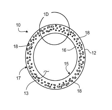
FIG. lA shows a stent 10 including a generally tubular member 12 capable of
supporting a body lumen and having a longitudinal axis A-A and a lumen 13.
Generally
tubular member 12 includes apertures 14 that are provided in a pattern to
facilitate stent
functions (e.g., radial expansion) and lateral flexibility. FIG. lA shows
stent 10 in a
compressed condition, such that stent 10 has a relatively small diameter D,
suitable for
delivery into a lumen of a subject. As shown in FIG. 1B, once stent 10 has
been
delivered into a lumen of a subject, stent 10 is expanded to a larger
diameter, Dexp. This
9
CA 02663220 2009-03-11
WO 2008/034013 PCT/US2007/078417
larger diameter can allow stent 10 to contact the walls of the lumen. In some
embodiments, a stent such as stent 10 can be expanded by a mechanical expander
(e.g.,
an inflatable balloon).
FIG. 1 C shows a cross-sectional view of stent 10. As shown in FIG. 1 C,
generally tubular member 12 has in interior surface 15 and an exterior surface
17, and is
formed of a metal matrix 16 including pores 18. Pores 18 can form an open pore
system
(in which different pores 18 are interconnected) or a closed pore system (in
which
different pores 18 are not interconnected). In certain embodiments, some pores
18 can be
interconnected, and other pores 18 may not be interconnected. While pores 18
are shown
as having an irregular cross-sectional shape, in some embodiments, the pores
in a metal
matrix can have one or more other cross-sectional shapes. For example, a pore
in a metal
matrix can be circular, oval (e.g., elliptical), and/or polygonal (e.g.,
triangular, square) in
cross-section.
Metal matrix 16 includes (e.g., is formed of) one or more bioerodible metals
and/or bioerodible metal alloys. In some embodiments (e.g., some embodiments
in
which metal matrix 16 is formed entirely of bioerodible metals and/or
bioerodible metal
alloys), generally tubular member 12 is bioerodible. In certain embodiments,
generally
tubular member 12 can erode after stent 10 has been used at a target site.
As shown in FIGS. 1C and 1D, different regions of generally tubular member 12
have different pore densities and/or include pores having different average
maximum
dimensions. As used herein, the pore density of a region is equal to the
number of pores
per square centimeter in that region. As an example, FIG. 1D shows a portion
of
generally tubular member 12 that has been divided by a line Ll into a region
Rl and a
region R2. Region Rl has a lower pore density than region R2, and also has
pores with a
lower average maximum dimension than the pores in region R2.
The variation in pore density and in the average maximum dimension of pores in
different regions of generally tubular member 12 can be designed, for example,
to result
in a particular pattern and/or rate of erosion by generally tubular member 12.
Typically,
as the pore density and/or average maximum dimension of the pores in a region
of
generally tubular member 12 increases, the erosion rate of that region can
also increase.
Without wishing to be bound by theory, it is believed that as the pore density
and/or
CA 02663220 2009-03-11
WO 2008/034013 PCT/US2007/078417
average pore volume of a region of generally tubular member 12 increases, the
surface
area of bioerodible material in that region that is exposed to blood and/or
other body
fluids (e.g., at a target site) can also increase. As a result, region R2 of
generally tubular
member 12, with its relatively high pore density and with its pores having a
relatively
high average maximum dimension, may erode at a faster rate than region Rl of
generally
tubular member 12, with its relatively low pore density and with its pores
having a
relatively low average maximum dimension.
In some embodiments, a medical device (e.g., stent 10) or a component of a
medical device (e.g., generally tubular member 12) that is formed of one or
more
bioerodible materials can substantially erode (can exhibit a mass reduction of
about 95
percent or more) over a period of at least about five days (e.g., at least
about seven days,
at least about 14 days, at least about 21 days, at least about 28 days, at
least about 30
days, at least about six weeks, at least about eight weeks, at least about 12
weeks, at least
about 16 weeks, at least about 20 weeks, at least about six months, at least
about 12
months). In some embodiments in which a medical device includes one or more
radiopaque materials, the erosion of the medical device within the body of a
subject can
be monitored using X-ray fluoroscopy. In certain embodiments, the erosion of a
medical
device within the body of a subject can be monitored using intravascular
ultrasound.
In some embodiments, region Rl can have a pore density of at least about 100
pores per square centimeter (e.g., at least about 500 pores per square
centimeter, at least
about 1000 pores per square centimeter, at least about 104 pores per square
centimeter, at
least about 105 pores per square centimeter, at least about 106 pores per
square
centimeter, at least about 10' pores per square centimeter, at least about 10
8 pores per
square centimeter) and/or at most about 109 pores per square centimeter (e.g.,
at most
about 108 pores per square centimeter, at most about 10' pores per square
centimeter, at
most about 106 pores per square centimeter, at most about 105 pores per square
centimeter, at most about 104 pores per square centimeter, at most about 1000
pores per
square centimeter, at most about 500 pores per square centimeter). In certain
embodiments, region R2 can have a pore density of at least about 100 pores per
square
centimeter (e.g., at least about 500 pores per square centimeter, at least
about 1000 pores
per square centimeter, at least about 104 pores per square centimeter, at
least about 105
11
CA 02663220 2009-03-11
WO 2008/034013 PCT/US2007/078417
pores per square centimeter, at least about 106 pores per square centimeter,
at least about
107 pores per square centimeter, at least about 108 pores per square
centimeter) and/or at
most about 109 pores per square centimeter (e.g., at most about 108 pores per
square
centimeter, at most about 10' pores per square centimeter, at most about 106
pores per
square centimeter, at most about 105 pores per square centimeter, at most
about 104 pores
per square centimeter, at most about 1000 pores per square centimeter, at most
about 500
pores per square centimeter). In some embodiments, the pore density of region
R2 can be
at least about 1.5 times greater (e.g., at least about two times greater, at
least about five
times greater, at least about 10 times greater, at least about 25 times
greater, at least about
50 times greater, at least about 75 times greater), and/or at most about 100
times greater
(e.g., at most about 75 times greater, at most about 50 times greater, at most
about 25
times greater, at most about 10 times greater, at most about five times
greater, at most
about two times greater), than the pore density of region Rl. While FIG. 1D
shows both
region Rl and region R2 as including pores 18, in certain embodiments, a
generally
tubular member such as generally tubular member 12 can have one or more
regions that
do not include any pores.
In some embodiments, the average maximum dimension (e.g., diameter, length,
width) of the pores in region Rl can be at least 0.01 micron (e.g., at least
0.05 micron, at
least about 0.1 micron, at least about 0.5 micron, at least about one micron,
at least about
five microns) and/or at most about 10 microns (e.g., at most about five
microns, at most
about one micron, at most about 0.5 micron, at most about 0.1 micron, at most
0.05
micron). In certain embodiments, the average maximum dimension (e.g.,
diameter,
length, width) of the pores in region R2 can be at least 0.01 micron (e.g., at
least 0.05
micron, at least about 0.1 micron, at least about 0.5 micron, at least about
one micron, at
least about five microns) and/or at most about 10 microns (e.g., at most about
five
microns, at most about one micron, at most about 0.5 micron, at most about 0.1
micron,
at most 0.05 micron). In some embodiments, the average maximum dimension of
the
pores in region R2 can be at least about 1.5 times greater (e.g., at least
about five times
greater, at least about 10 times greater, at least about 25 times greater, at
least about 50
times greater, at least about 75 times greater), and/or at most about 100
times greater
(e.g., at most about 75 times greater, at most about 50 times greater, at most
about 25
12
CA 02663220 2009-03-11
WO 2008/034013 PCT/US2007/078417
times greater, at most about 10 times greater, at most about five times
greater), than the
average maximum dimension of the pores in region Rl.
The bioerodible materials that are included in a medical device can include
one or
more metals and/or one or more metal alloys. Examples of bioerodible metals
include
alkali metals, alkaline earth metals (e.g., magnesium), iron, zinc, and
aluminum. As used
herein, a metal alloy refers to a substance that is composed of two or more
metals or of a
metal and a nonmetal intimately united, for example, by being fused together
and
dissolving in each other when molten. Examples of bioerodible metal alloys
include
alkali metal alloys, alkaline earth metal alloys (e.g., magnesium alloys),
iron alloys (e.g.,
alloys including iron and up to seven percent carbon), zinc alloys, and
aluminum alloys.
Metal matrix 16 of generally tubular member 12 can include one metal or metal
alloy, or
can include more than one (e.g., two, three, four, five) metal or metal alloy.
In some
embodiments, metal matrix 16 can include one or more metals and one or more
metal
alloys. Bioerodible materials are described, for example, in Weber, U.S.
Patent
Application Publication No. US 2005/0261760 Al, published on November 24,
2005,
and entitled "Medical Devices and Methods of Making the Same"; Colen et al.,
U.S.
Patent Application Publication No. US 2005/0192657 Al, published on September
1,
2005, and entitled "Medical Devices"; Weber, U.S. Patent Application Serial
No.
11/327,149, filed on January 5, 2006, and entitled "Bioerodible Endoprostheses
and
Methods of Making the Same"; Bolz, U.S. Patent No. 6,287,332; Heublein, U.S.
Patent
Application Publication No. US 2002/0004060 Al, published on January 10, 2002,
and
entitled "Metallic Implant Which is Degradable In Vivo"; and Park, Science and
Technology ofAdvanced Materials, 2, 73-78 (2001).
In some embodiments, stent 10 can include one or more therapeutic agents. As
an
example, stent 10 can include one or more therapeutic agents that are disposed
within
pores 18 of generally tubular member 12. During delivery and/or use in a body
of a
subject, stent 10 can elute the therapeutic agents. For example, as generally
tubular
member 12 erodes, the therapeutic agents within pores 18 can be released into
the body.
The erosion of generally tubular member 12 can result in a relatively
consistent release of
therapeutic agent, as pores 18 continue to become exposed.
13
CA 02663220 2009-03-11
WO 2008/034013 PCT/US2007/078417
The variation in pore density and in the average maximum dimension of the
pores
in different regions of generally tubular member 12 can be designed, for
example, to
result in a particular pattern and/or rate of therapeutic agent elution from
generally
tubular member 12. Typically, a region of generally tubular member 12 having a
relatively high pore density and/or including pores with a relatively high
average
maximum dimension can elute therapeutic agent at a faster rate than a region
of generally
tubular member 12 having a relatively low pore density and/or including pores
with a
relatively low average maximum dimension. For example, region R2 of generally
tubular
member 12 may elute therapeutic agent at a faster rate, and/or may elute a
higher total
volume of therapeutic agent, than region Rl.
Examples of therapeutic agents include non-genetic therapeutic agents, genetic
therapeutic agents, vectors for delivery of genetic therapeutic agents, cells,
and
therapeutic agents identified as candidates for vascular treatment regimens,
for example,
as agents targeting restenosis. In some embodiments, one or more therapeutic
agents that
are used in a medical device such as a stent can be dried (e.g., lyophilized)
prior to use,
and can become reconstituted once the medical device has been delivered into
the body
of a subject. A dry therapeutic agent may be relatively unlikely to come out
of a medical
device (e.g., a stent) prematurely, such as when the medical device is in
storage.
Therapeutic agents are described, for example, in Weber, U.S. Patent
Application
Publication No. US 2005/0261760 Al, published on November 24, 2005, and
entitled
"Medical Devices and Methods of Making the Same", and in Colen et al., U.S.
Patent
Application Publication No. US 2005/0192657 Al, published on September 1,
2005, and
entitled "Medical Devices".
Generally tubular member 12 of stent 10 can be formed by any of a number of
different methods. In some embodiments, generally tubular member 12 can be
formed by
molding a mixture of a bioerodible metal and a second bioerodible material
into a
generally tubular shape, and exposing the generally tubular shape to a solvent
that
solvates the second bioerodible material (without also solvating the
bioerodible metal),
and/or to a temperature that causes the second bioerodible material to melt
(without also
causing the bioerodible metal to melt). When the second bioerodible material
is solvated
14
CA 02663220 2009-03-11
WO 2008/034013 PCT/US2007/078417
and/or when it melts, it can result in the formation of pores in the metal,
thereby
producing metal matrix 16.
While a stent including regions having different pore densities and having
pores
with different average maximum dimensions has been described, in some
embodiments, a
stent can alternatively or additionally include regions having the same pore
density and/or
having pores with the same average maximum dimension. For example, FIG. 2A
shows
a cross-sectional view of a stent 100 including a generally tubular member
112.
Generally tubular member 112 has an interior surface 113, an exterior surface
114, and a
lumen 115, and is formed out of a metal matrix 116 formed of one or more
bioerodible
metals and/or bioerodible metal alloys. Metal matrix 116 includes pores 118.
FIG. 2B shows a portion of generally tubular member 112 that has been divided
by a line L2 into regions R3 and R4. As shown in FIG. 2B, regions R3 and R4
have the
same pore density, and also include pores 118 having the same average maximum
dimension.
While stents including generally tubular members formed out of a metal matrix
and/or including a therapeutic agent have been described, in some embodiments,
a stent
can include one or more other materials. The other materials can be used, for
example, to
enhance the strength and/or structural support of the stent. Examples of other
materials
that can be used in conjunction with a metal matrix in a stent include metals
(e.g., gold,
platinum, niobium, tantalum), metal alloys, and/or polymers (e.g., styrene-
isobutylene
styrene (SIBS), poly(n-butyl methacrylate) (PBMA)). Examples of metal alloys
include
cobalt-chromium alloys (e.g., L605), Elgiloy (a cobalt-chromium-nickel-
molybdenum-
iron alloy), and niobium-1 Zr alloy. In some embodiments, a stent can include
a
generally tubular member formed out of a porous magnesium matrix, and the
pores in the
magnesium matrix can be filled with iron compounded with a therapeutic agent.
In certain embodiments, a stent can include both a bioerodible metal matrix
and
one or more additional bioerodible materials that are different from the
bioerodible
materials in the bioerodible metal matrix. For example, in some embodiments, a
stent
can include both a bioerodible metal matrix and one or more non-metallic
bioerodible
materials (e.g., starches, sugars). In certain embodiments, a stent can
include a
bioerodible metal matrix and one or more additional bioerodible materials that
erode at a
CA 02663220 2009-03-11
WO 2008/034013 PCT/US2007/078417
different rate from the bioerodible metal matrix. The additional bioerodible
materials can
be added to the bioerodible metal matrix to, for example, tailor the erosion
rate of the
stent. For example, in some embodiments, a stent can include a generally
tubular
member that is formed of a porous bioerodible metal matrix, and a bioerodible
polymer
can be disposed within some or all of the pores of the bioerodible metal
matrix. For
example, FIG. 3 shows a cross-sectional view of a stent 200 including a
generally tubular
member 202. Generally tubular member 202 has an exterior surface 204, an
interior
surface 206, and a lumen 208, and is formed of a metal matrix 210 that is
formed of one
or more bioerodible metals and/or bioerodible metal alloys. Metal matrix 210
includes
pores 212 that are filled with a bioerodible polymer 214. Examples of
bioerodible
polymers include polyiminocarbonates, polycarbonates, polyarylates,
polylactides, and
polyglycolic esters. A stent including a metal matrix and a bioerodible
polymer disposed
within the pores of the metal matrix can be made, for example, by forming a
generally
tubular member out of a metal matrix (e.g., as described above), immersing the
generally
tubular member in a solution of the polymer, and allowing the solution to dry,
so that the
solvent in the solution evaporates, and the polymer is left behind on the
stent.
In some embodiments, a stent can include both a bioerodible metal matrix and
one
or more materials that carry a therapeutic agent. For example, a stent can
include a
generally tubular member that is formed of a porous bioerodible metal matrix,
and a
polymer containing a therapeutic agent can be disposed within the pores of the
metal
matrix. The polymer can be non-bioerodible, or can be bioerodible. In some
embodiments in which the polymer is bioerodible, the polymer can erode at a
different
rate from the metal matrix. As an example, in some embodiments, the polymer
can erode
at a faster rate than the metal matrix, causing all of the therapeutic agent
to be released
into the body before the generally tubular member has completely eroded. As
another
example, in certain embodiments, the polymer can erode at a slower rate than
the metal
matrix. The result can be that after the matrix has completely eroded, at
least some of the
therapeutic-agent containing polymer can remain in the body (e.g., in the form
of
polymeric particles). In some embodiments in which the stent has been
delivered into a
lumen of a subject, the polymer can be at least partially embedded in a wall
of the lumen.
As the polymer continues to erode, it can release the therapeutic agent into
the body.
16
CA 02663220 2009-03-11
WO 2008/034013 PCT/US2007/078417
Thus, the body can continue to be treated with the therapeutic agent, even
after the
generally tubular member has eroded. The therapeutic agent can be selected,
for
example, to alleviate the effects, if any, of the erosion of the stent on the
body. By
including a material (such as a polymer) containing a therapeutic agent, the
stent can
have a therapeutic agent elution rate that is independent of the erosion rate
of its
generally tubular member.
In certain embodiments, a stent can include one or more coatings on one or
more
surfaces of the stent. For example, FIGS. 4A and 4B show a stent 300 including
a
generally tubular member 302 defining a lumen 304. Generally tubular member
302 is
formed of a metal matrix 306 that is formed of one or more bioerodible metals
and/or
bioerodible metal alloys, and that includes pores 308. Stent 300 further
includes a
coating 310 disposed on the exterior surface 312 of generally tubular member
302.
Coating 310 can be used, for example, to regulate therapeutic agent release
from
generally tubular member 302. For example, pores 308 can contain one or more
therapeutic agents, and coating 310 (e.g., which can be bioerodible) can be
used to
control the release of the therapeutic agent(s) from pores 308 (e.g., by
delaying the
release of the therapeutic agent(s) until stent 300 has reached a target
site).
In certain embodiments, a stent can include a coating that contains a
therapeutic
agent or that is formed of a therapeutic agent. For example, a stent can
include a coating
that is formed of a polymer and a therapeutic agent. The coating can be
applied to a
generally tubular member of the stent by, for example, dip-coating the
generally tubular
member in a solution including the polymer and the therapeutic agent. Methods
that can
be used to apply a coating to a generally tubular member of a stent are
described, for
example, in U.S. Provisional Patent Application Serial No. [Attorney Docket
No. 10527-709P01 ], filed concurrently herewith and entitled "Medical
Devices".
While a stent with one coating has been shown, in some embodiments, a stent
can
include more than one (e.g., two, three, four, five) coating. For example,
FIG. 5 shows a
cross-sectional view of a stent 350 having a lumen 352. Stent 350 includes a
generally
tubular member 353 formed of metal matrix 354 that is formed of one or more
bioerodible metals and/or bioerodible metal alloys, and that includes pores
355. Stent
350 also includes a coating 356 on the exterior surface 358 of generally
tubular member
17
CA 02663220 2009-03-11
WO 2008/034013 PCT/US2007/078417
353, and a coating 360 on the interior surface 362 of generally tubular member
353.
Coatings 356 and 360 can include one or more of the same materials, or can be
formed of
different materials.
Examples of coating materials that can be used on a stent include metals
(e.g.,
tantalum, gold, platinum), metal oxides (e.g., iridium oxide, titanium oxide,
tin oxide),
and/or polymers (e.g., SIBS, PBMA). Coatings can be applied to a stent using,
for
example, dip-coating and/or spraying processes.
While stents including generally tubular members formed of a porous metal
matrix have been described, in certain embodiments, a stent can alternatively
or
additionally include a coating that is formed of a porous metal matrix. For
example,
FIGS. 6A and 6B show a stent 400 having a lumen 402. Stent 400 includes a
generally
tubular member 404 that is not formed of a porous metal matrix. Generally
tubular
member 404 can be formed of, for example, one or more metals (e.g., gold,
platinum,
niobium, tantalum), metal alloys, and/or polymers (e.g., SIBS, PBMA). Examples
of
metal alloys include cobalt-chromium alloys (e.g., L605), Elgiloy (a cobalt-
chromium-
nickel-molybdenum-iron alloy), and niobium-1 Zr alloy. Stent 400 further
includes a
coating 406 that is disposed on the exterior surface 408 of generally tubular
member 404.
Coating 406 is formed of a metal matrix 410 that is formed of one or more
bioerodible
metals and/or bioerodible metal alloys, and that includes pores 412. Metal
matrix 410
can be used, for example, as a reservoir for one or more therapeutic agents.
For example,
one or more therapeutic agents can be disposed within pores 412 of metal
matrix 410.
During and/or after delivery of stent 400 to a target site in a body of a
subject, metal
matrix 410 can erode, thereby eluting therapeutic agent into the body of the
subject.
A coating such as coating 406 can be formed using, for example, one or more
sintering and/or vapor deposition processes.
While coating 406 is shown as having a relatively uniform pore density and as
including pores having a relatively uniform average maximum dimension, in some
embodiments, a porous coating on a stent can have a non-uniform pore density
and/or can
include pores having a non-uniform average maximum dimension. For example,
FIG. 7
shows a cross-sectional view of a stent 450 including a generally tubular
member 452
that is not formed of a porous metal matrix. Generally tubular member 452 can
be
18
CA 02663220 2009-03-11
WO 2008/034013 PCT/US2007/078417
formed of, for example, one or more metals (e.g., gold, platinum, niobium,
tantalum),
metal alloys, and/or polymers (e.g., SIBS, PBMA). Examples of metal alloys
include
cobalt-chromium alloys (e.g., L605), Elgiloy (a cobalt-chromium-nickel-
molybdenum-
iron alloy), and niobium-1 Zr alloy. Stent 400 further includes a coating 456
that is
disposed on the exterior surface 458 of generally tubular member 452. Coating
456 is
formed of a metal matrix 460 that is formed of one or more bioerodible metals
and/or
bioerodible metal alloys, and that includes pores 462. Metal matrix 460 can be
used, for
example, as a reservoir for one or more therapeutic agents. As shown in FIG.
7, coating
456 has an interior surface 464 and an exterior surface 466. The pore density
of metal
matrix 460 is higher, and the average maximum dimension of pores 462 in metal
matrix
460 is greater, in the regions of generally tubular member 452 that are closer
to exterior
surface 466 than in the regions of generally tubular member 42 that are closer
to interior
surface 464.
While stents having certain configurations have been described, in some
embodiments, a stent including one or more bioerodible metals and/or
bioerodible metal
alloys can have a different configuration. For example, FIG. 8A shows a stent
520 that is
in the form of a generally tubular member 521 formed of one or more
bioerodible metals
and/or bioerodible metal alloys. Generally tubular member 521 is defined by a
plurality
of bands 522 and a plurality of connectors 524 that extend between and connect
adjacent
bands. Generally tubular member 521 has a lumen 523.
FIG. 8B shows an enlarged view of a connector 524 of stent 520, and FIG. 8C
shows a cross-sectional view of the connector of FIG. 8B. As shown in FIG. 8C,
connector 524 is formed of a metal matrix 530 including pores 534. Metal
matrix 530 is
formed of one or more bioerodible metals and/or bioerodible metal alloys. A
line L3
divides connector 524 into regions R5 and R6. As shown in FIG. 8C, region R5
has a
higher pore density than region R6, and the pores in region R5 have a higher
average
maximum dimension than the pores in region R6.
During delivery and/or use of stent 520, bands 522 and/or connectors 524 can
erode. The presence of pores 534 in connectors 524 can help to accelerate
and/or control
the erosion of connectors 524. In some embodiments, the presence of pores 534
in
connectors 524 can result in connectors 524 eroding at a faster rate than
bands 522. In
19
CA 02663220 2009-03-11
WO 2008/034013 PCT/US2007/078417
certain embodiments, it may be desirable for connectors 524 to completely
erode before
bands 522, allowing stent 520 to move and flex within a target site (e.g.,
within a lumen
in a body of a subject). By the time connectors 524 have completely eroded,
tissue may
have grown over the remaining parts of stent 520 (e.g., bands 522), thereby
helping to
hold bands 522 (and, therefore, stent 520) in place.
While a stent including connectors having regions with different pore
densities
and with pores having different average maximum dimensions has been described,
in
some embodiments, a stent can include one or more components having regions
with
relatively uniform pore densities and/or with pores having relatively uniform
average
maximum dimensions. For example, FIG. 9 shows a cross-sectional view of a
connector
550 of a stent. As shown in FIG. 9, connector 550 is formed of a metal matrix
554
including pores 558. Metal matrix 554 is formed of one or more bioerodible
metals
and/or bioerodible metal alloys. A line L4 divides connector 550 into regions
R7 and R8.
As shown in FIG. 9, regions R7 and R8 have the same pore density and the pores
in
regions R7 and R8 have the same average maximum dimension.
While stents including connectors including pores have been described, in some
embodiments, a stent can alternatively or additionally include one or more
other
components (e.g., bands) having pores.
While certain embodiments have been described, other embodiments are possible.
As an example, in some embodiments, a stent including a generally tubular
member formed of a bioerodible metal can be manufactured using powder
metallurgy
methods. For example, a stent can be formed by sintering and compacting
bioerodible
metal particles and/or metal alloy particles into the shape of a generally
tubular member.
A metal particle or metal alloy particle can have a dimension (e.g., a width,
a length, a
diameter) of, for example, at least about 0.1 micron (e.g., at least about 0.5
micron, at
least about one micron, at least about five microns) and/or at most about 10
microns (e.g.,
at most about five microns, at most about one micron, at most about 0.5
micron).
Sintering the metal particles and/or the metal alloy particles can include
exposing the
metal particles and/or the metal alloy particles to heat and pressure to cause
some
coalescence of the particles. A generally tubular member that is formed by a
sintering
process can be porous or non-porous, or can include both porous regions and
non-porous
CA 02663220 2009-03-11
WO 2008/034013 PCT/US2007/078417
regions. In some embodiments in which the generally tubular member includes
pores,
the sizes of the pores can be controlled by the length of the sintering and
compacting
period, and/or by the temperature and/or pressure of the sintering process.
Typically, as
the temperature and/or pressure of a sintering process increases, the pore
density of the
resulting generally tubular member, and the average maximum dimension of the
pores in
the generally tubular member, can decrease. In certain embodiments, a
generally tubular
member can be formed by sintering metal particles and/or metal alloy particles
having
different sizes.
In certain embodiments in which a generally tubular member includes different
regions having different pore densities and/or having pores with different
average
maximum dimensions, the generally tubular member can be formed using a
sintering
process employing thermal gradients. The sintering process can include
exposing certain
regions of the generally tubular member, as it is being formed, to higher
temperatures
than other regions of the generally tubular member. The regions that are
exposed to
higher temperatures ultimately can have relatively low pore densities and/or
pores with
relatively small average maximum dimensions, while the regions that are
exposed to
lower temperatures can have relatively high pore densities and/or pores with
relatively
large average maximum dimensions. Without wishing to be bound by theory, it is
believed that this variation in pore density and in the average maximum
dimension of the
pores can occur because as the temperature of the sintering process decreases,
the extent
by which the metal particles and/or the metal alloy particles come together
can decrease
as well. In some embodiments, a sintering process that is used to form a stent
can include
forming a generally tubular member around a mandrel that is selectively heated
so that
certain regions of the mandrel are hotter than other regions of the mandrel.
The result
can be that the generally tubular member has different regions having
different average
pore volumes and/or having pores with different average maximum dimensions.
In some embodiments, a stent that is formed by sintering metal particles
and/or
metal alloy particles can erode after being used at a target site in a body of
a subject, and
the erosion of the stent can result in the formation of metal particles and/or
metal alloy
particles having the same size as the particles that were originally sintered
together to
form the stent. Thus, the size of the particles formed from the erosion of a
stent can be
21
CA 02663220 2009-03-11
WO 2008/034013 PCT/US2007/078417
selected, for example, by sintering metal particles and/or metal alloy
particles of the
desired size to form the stent.
As another example, while stents with certain porosity patterns have been
described, in some embodiments, a stent can have a different porosity pattern.
For
example, FIGS. l0A and l OB show a stent 570 including a generally tubular
member 574
having an interior surface 578, an exterior surface 582, and a lumen 586.
Generally
tubular member 574 is formed of a metal matrix 590 that is formed of one or
more
bioerodible metals and/or bioerodible metal alloys, and that includes pores
594. As
shown in FIG. l OB, the pores in generally tubular member 574 that are
relatively far from
both interior surface 578 and exterior surface 582 are relatively large, while
the pores that
are relatively close to interior surface 578 or exterior surface 582 are
relatively small.
Stent 570 can be used, for example, to store a relatively large volume of
therapeutic agent
in the relatively large pores, and to provide a slow and/or controlled release
of the
therapeutic agent into the target site through the relatively small pores.
As an additional example, in some embodiments, a stent can include a porous
generally tubular member that includes more than one therapeutic agent in its
pores. For
example, FIGS. 1 lA and 1 lB show a stent 600 including a generally tubular
member 604
having an interior surface 605, an exterior surface 606, and a lumen 607.
Generally
tubular member 604 is formed of a metal matrix 608 that is formed of one or
more
bioerodible metals and/or bioerodible metal alloys. Metal matrix 608 includes
pores 610.
As shown in FIG. 11B, pores 610 are aligned in an inner circle 614 close to
interior
surface 605, and in an outer circle 618 close to exterior surface 606. In some
embodiments, the pores that form inner circle 614 can be filled with one type
of
therapeutic agent (e.g., an anticoagulant, such as heparin), while the pores
that form outer
circle 618 can be filled with a different type of therapeutic agent (e.g., an
anti-
proliferative, such as paclitaxel).
As a further example, in some embodiments, a stent can include a porous
bioerodible metal matrix surrounding a therapeutic agent-containing layer. For
example,
FIGS. 12A and 12B show a stent 650 including a generally tubular member 652
formed
of three layers 654, 656, and 658. Layer 654 is formed of a metal matrix 660
that is
formed of one or more bioerodible metals and/or bioerodible metal alloys, and
that
22
CA 02663220 2009-03-11
WO 2008/034013 PCT/US2007/078417
includes pores 662. Similarly, layer 658 is formed of a metal matrix 664 that
is formed
of one or more bioerodible metals and/or bioerodible metal alloys, and that
includes pores
668. Layer 656, which is located between layer 654 and layer 658, includes one
or more
therapeutic agents. For example, layer 656 can be formed entirely of one or
more
therapeutic agents, or can be formed of one or more materials (e.g., a
bioerodible
polymer) that are combined with one or more therapeutic agents. Layers 654 and
658 can
regulate the release of the therapeutic agent(s) from layer 656 into a target
site.
As another example, in certain embodiments, a stent can include one or more
components (e.g., bands and/or connectors) including a hollow reservoir that
can be filled
with, for example, one or more therapeutic agents. For example, FIG. 13A shows
a stent
720 that is in the form of a generally tubular member 721 formed of one or
more
bioerodible metals and/or bioerodible metal alloys. Generally tubular member
721 is
defined by a plurality of bands 722 and a plurality of connectors 724 that
extend between
and connect adjacent bands. Generally tubular member 721 has a lumen 723.
FIG. 13B shows an enlarged view of a connector 724 of stent 720, and FIG. 13C
shows a cross-sectional view of the connector of FIG. 13B. As shown in FIG.
13C,
connector 724 is formed of a metal matrix 730 surrounding a reservoir 732 and
including
pores 734. Metal matrix 730 is formed of one or more bioerodible metals and/or
bioerodible metal alloys. Reservoir 732 is filled with a therapeutic agent 750
that can, for
example, elute through pores 734 during and/or after delivery of stent 720 to
a target site.
As an additional example, in some embodiments, a stent can include a generally
tubular member having different regions along its length that have different
pore densities
and/or that include pores having different average maximum dimensions.
For example, FIGS. 14A and 14B show a stent 800 including a generally tubular
member 802 having a lumen 804. Generally tubular member 802 is formed of a
metal
matrix 806 including pores 808. Metal matrix 806 is formed of one or more
bioerodible
metals and/or bioerodible metal alloys. As shown in FIG. 14B, different
regions R9,
R10, and Rl 1 of generally tubular member 802 along the length L5 of generally
tubular
member 802 have different pore densities and include pores having different
average
maximum dimensions. More specifically, region R9 has a higher pore density
than
region R10, and includes pores with a higher average maximum dimension than
the pores
23
CA 02663220 2009-03-11
WO 2008/034013 PCT/US2007/078417
in region R10. Region R10, in turn, has a higher pore density than region Rl
l, and
includes pores with a higher average maximum dimension than the pores in
region R11.
These differences in the pore densities and average maximum dimensions of the
pores in
regions R9, R10, and Rl 1 can, for example, result in region R9 eroding at a
faster rate
than both regions R10 and Rl l, and region R10 eroding at a faster rate than
region Rl 1.
FIGS. 15A and 15B show a stent including a generally tubular member having
different regions along its length that include pores having different average
maximum
dimensions. As shown in FIGS. 15A and 15B, a stent 850 includes a generally
tubular
member 852 having a lumen 854. Generally tubular member 852 is formed of a
metal
matrix 856 including pores 858. Metal matrix 856 is formed of one or more
bioerodible
metals and/or bioerodible metal alloys. As shown in FIG. 15B, different
regions R12,
R13, and Rl4 of generally tubular member 852 along the length L6 of generally
tubular
member 852 include pores having different average maximum dimensions. More
specifically, the pores in end regions R12 and R14 have higher average maximum
dimensions than the pores in middle region R13. In some embodiments, one or
more of
the pores in generally tubular member 852 can contain one or more therapeutic
agents
that can treat thrombosis. The relatively large pores in end regions R12 and
R14 can
contain a higher volume of the therapeutic agent(s) than the relatively small
pores in
middle region R13.
As another example, in some embodiments, a stent including a metal matrix
including pores can be a self-expanding stent. For example, in certain
embodiments, a
self-expanding stent can include a generally tubular member that is formed of
Nitinol,
and can further include a porous bioerodible metal supported by the generally
tubular
member (e.g., the porous bioerodible metal can be in the form of a coating on
the
generally tubular member).
As a further example, while stents have been described, in some embodiments,
other medical devices can include pores, bioerodible metals, and/or
bioerodible metal
alloys. For example, other types of endoprostheses, such as grafts and/or
stent-grafts, can
include one or more of the features of the stents described above. Additional
examples of
medical devices that can have one or more of these features include bone
screws.
24
CA 02663220 2009-03-11
WO 2008/034013 PCT/US2007/078417
As another example, in some embodiments, a medical device can include regions
that are formed of a porous metal and/or a porous metal alloy (e.g., a
bioerodible porous
metal and/or a bioerodible porous metal alloy), and regions that are not
formed of a
porous metal or metal alloy. For example, a stent may include regions that are
formed of
a bioerodible porous metal, and regions that are formed of a metal that is
neither
bioerodible nor porous.
As an additional example, in certain embodiments, a medical device (e.g., a
stent)
including a coating formed of a porous metal and/or a porous metal alloy can
be further
coated with one or more other coatings. The other coatings can be formed of
porous
metals and/or porous metal alloys, or may not be formed of porous metals or
porous
metal alloys.
As a further example, in some embodiments, a coating can be applied to certain
regions of a medical device, while not being applied to other regions of the
medical
device.
As another example, in certain embodiments, a medical device (e.g., a stent)
can
include one or more metal foams, such as one or more bioerodible metal foams.
Medical
devices including metal foams are described, for example, in U.S. Provisional
Patent
Application Serial No. 60/844,967, which is incorporated by reference, filed
September
15, 2006 and entitled "Medical Devices".
All publications, applications, references, and patents referred to in this
application are herein incorporated by reference in their entirety.
Other embodiments are within the claims.