Note: Descriptions are shown in the official language in which they were submitted.
CA 02664662 2009-03-26
WO 2008/040014 PCT/US2007/079978
DELIVERY TOOL FOR PERCUTANEOUS DELIVERY OF A PROSTHESIS
RELATED APPLICATIONS
[0001] This application claims priority to U.S. Provisional Application Serial
No.
60/827,373 filed September 28, 2006 entitled Delivery Tool For Percutaneous
Delivery
Of A Prosthesis which is hereby incorporated by reference.
BACKGROUND OF THE INVENTION
[0002] There has been a significant movement toward developing and performing
cardiovascular surgeries using a percutaneous approach. Through the use of one
or
more catheters that are introduced through, for example, the femoral artery,
tools and
devices can be delivered to a desired area in the cardiovascular system to
perform any
number of complicated procedures that normally otherwise require an invasive
surgical
procedure. Such approaches greatly reduce the trauma endured by the patient
and
can significantly reduce recovery periods. The percutaneous approach is
particularly
attractive as an alternative to performing open-heart surgery.
[0003] Valve replacement surgery provides one example of an area where
percutaneous solutions are being developed. A number of diseases result in a
thickening, and subsequent immobility or reduced mobility, of heart valve
leaflets. Such
immobility also may lead to a narrowing, or stenosis, of the passageway
through the
valve. The increased resistance to blood flow that a stenosed valve presents
can
eventually lead to heart failure and ultimately death.
[0004] Treating valve stenosis or regurgitation has heretofore involved
complete
removal of the existing native valve through an open-heart procedure followed
by the
implantation of a prosthetic valve. Naturally, this is a heavily invasive
procedure and
inflicts great trauma on the body leading usually to great discomfort and
considerable
recovery time. It is also a sophisticated procedure that requires great
expertise and
talent to perform.
-1-
CA 02664662 2009-03-26
WO 2008/040014 PCT/US2007/079978
[0005] Historically, such valve replacement surgery has been performed using
traditional open-heart surgery where the chest is opened, the heart stopped,
the patient
placed on cardiopulmonary bypass, the native valve excised and the replacement
valve
attached. On the other hand, a proposed percutaneous valve replacement
alternative
method is disclosed in U.S. Pat. No. 6,168,614, which is herein incorporated
by
reference in its entirety. In this patent, the prosthetic valve is mounted
within a stent that
is collapsed to a size that fits within a catheter. The catheter is then
inserted into the
patient's vasculature and moved so as to position the collapsed stent at the
location of
the native valve. A deployment mechanism is activated that expands the stent
containing the replacement valve against the valve cusps. The expanded
structure
includes a stent configured to have a valve shape with valve leaflet supports
that
together take on the function of the native valve. As a result, a full valve
replacement
has been achieved but at a significantly reduced physical impact to the
patient.
[0006] More recent techniques have further improved over the drawbacks
inherent in
U.S. Pat. No. 6,168,614. For example, one approach employs a stentless support
structure as seen in U.S. Patent Application Serial Number 1 1 /44381 4,
entitled
Stentless Support Structure, filed May 26, 2006, the contents of which are
herein
incorporated by reference. The stentless support structure provides a tubular
mesh
framework that supports a new artificial or biological valve within a
patient's vessel. The
framework typically exhibits shape memory properties which encourage the
length of the
framework to fold back on itself at least once and possibly multiple times
during delivery.
In this respect, the framework can be percutaneously delivered to a target
area with a
relatively small diameter, yet can expand and fold within a vessel to take on
a
substantially thicker diameter with increased strength.
[0007] Typically, the stentless support structure is delivered at the location
of a
diseased or poorly functioning valve within a patient. The structure expands
against the
leaflets of the native valve, pushing them against the side of the vessel.
With the native
valve permanently opened, the new valve begins functioning in place of the
native valve.
Optimally placing the stentless support structure involves percutaneously
passing the
structure through the diseased valve, deploying a distal end of the structure
until the
distal end flares outwardly, then pulling the structure back through the
diseased valve
until the user can feel the flared distal end of the structure contact a
distal side of the
-2-
CA 02664662 2009-03-26
WO 2008/040014 PCT/US2007/079978
diseased valve. Once confident that the flared distal end of the structure is
abutting a
distal side of the diseased valve, the remaining portion of the structure is
deployed
within the diseased valve.
[0008] In any of the above mentioned percutaneous valve device implant
procedures, a significant challenge to device function is accurate placement
of the
implant. If the structure is deployed below or above the optimal device
position, the
native valve leaflets may not be captured by the prosthetic support structure
and can
further interfere with the operation of the implant. Further, misplacement of
the support
structure may result in interference between the prosthetic device and nearby
structures
of the heart, or can result in leakage of blood around the structure,
circumventing the
replacement valve.
[0009] Accurate placement of these devices within the native valve requires
significant technical skill and training, and successful outcomes can be
technique-
dependent. What is needed is a delivery tool for more reliably locating a
target
deployment area, for positioning a percutaneous aortic valve replacement
device or
other prosthetic device in which the device location during implantation is
critical (e.g.,
an occluder for vascular atrial septal defects, ventricular septal defects,
patent foramen
ovale or perforations of the heart or vasculature), and for the subsequent
deployment of
such a device to provide more reliable implant outcomes.
SUMMARY OF THE INVENTION
[0010] In one embodiment, the present invention provides an expandable
delivery
tool for deploying a prosthesis device within a patient. The delivery tool has
a generally
elongated shape with an expandable distal end region that flares outward in
diameter.
[0011] In one aspect, the delivery tool provides a tactile indication of a
desired target
area, such as a valve. For example, once expanded within a patient's vessel,
the
delivery device can be pulled proximally towards the user until it contacts a
desired
target valve. This contact is transmitted and thereby felt by the user on a
proximal end
of the device outside the patient, providing an indication that a desired
target location
has been located.
-3-
CA 02664662 2009-03-26
WO 2008/040014 PCT/US2007/079978
[0012] In another aspect, the delivery tool provides a stationary backstop
against
which a prosthesis can be deployed, further ensuring the prosthesis is
delivered at a
desired target location within the patient. For example, the expanded backstop
of the
delivery tool is positioned at a location just distal to a native valve within
a patient. The
prosthesis is deployed within the native valve and against the expanded
backstop,
ensuring the prosthesis maintains its intended target position within the
native valve.
[0013] In yet another aspect, the delivery tool is used to further expand the
prosthesis after deployment. For example, the expandable backstop is reduced
in size
to a desired expansion diameter (i.e., the diameter the user wishes to expand
the
prosthesis to), then pulled through the deployed prosthesis, causing the
diameter of the
prosthesis to expand. This expansion further anchors the prosthesis against
the vessel,
ensuring its position is maintained and minimal leakage occurs past the
periphery of the
prosthesis. Alternately, the distal end of the delivery tool can be expanded
within the
prosthesis to further expand the prosthesis within the patient's vessel.
BRIEF DESCRIPTION OF THE DRAWINGS
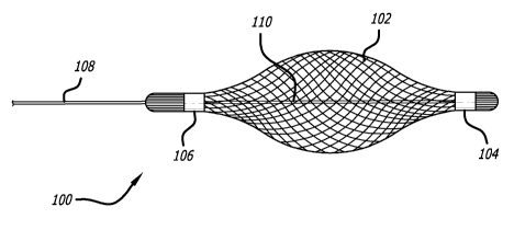
[0014] Figure 1 illustrates a side view of a delivery tool according a
preferred
embodiment of the present invention;
[0015] Figure 2 illustrates a side view of the delivery tool of Figure 1;
[0016] Figure 3 illustrates a perspective view of the delivery tool of Figure
1;
[0017] Figure 4 illustrates a side view of a valve prosthesis according to a
preferred
embodiment of the present invention;
[0018] Figure 5 illustrates a side view of a locking-pin mechanism connected
to a
support structure according to a preferred embodiment of the present
invention;
[0019] Figure 6 illustrates a magnified side view of the locking-pin mechanism
of
Figure 5;
[0020] Figure 7 illustrates a side perspective view of the locking-pin
mechanism of
Figure 5;
-4-
CA 02664662 2009-03-26
WO 2008/040014 PCT/US2007/079978
[0021] Figure 8 illustrates a bottom perspective view of the locking-pin
mechanism of
Figure 5;
[0022] Figure 9 illustrates a side view of the delivery tool of Figure 1;
[0023] Figure 10 illustrates a side view of the delivery tool of Figure 1;
[0024] Figure 11 illustrates a side view of the delivery tool of Figure 1,
with a valve
prosthesis in the initial stage of deployment;
[0025] Figure 12 illustrates a side view of the delivery tool of Figure 1,
with the initial
portion of the prosthesis further deployed;
[0026] Figure 13 illustrates a side view of the delivery tool of Figure 1,
with the initial
portion of the prosthesis further deployed;
[0027] Figure 14 illustrates a side view of the delivery tool of Figure 1 and
the
prosthesis retracted into a simulated valve site;
[0028] Figure 15 illustrates a side view of the delivery tool of Figure 1 with
the
prosthesis having been deployed into a simulated valve site;
[0029] Figure 16 illustrates a side view of the delivery tool of Figure 1
having been
relaxed from its expanded configuration;
[0030] Figure 17 illustrates a perspective view of the delivery tool of Figure
1 with the
prosthesis having been fully deployed;
[0031] Figure 18 illustrates a perspective view of the delivery tool of Figure
1 being
drawn within the prosthetic valve;
[0032] Figure 19 illustrates a perspective view of the delivery tool of Figure
1 drawn
into the prosthetic valve and expanded to provide a means for fully seating
the device
within the native valve;
[0033] Figure 20 illustrates a perspective view of a prosthesis and the
delivery tool of
Figure 1;
-5-
CA 02664662 2009-03-26
WO 2008/040014 PCT/US2007/079978
[0034] Figure 21 illustrates a side view of a prosthesis and the delivery tool
of Figure
1 with the tool having been fully withdrawn from the prosthetic valve;
[0035] Figure 22 illustrates a side view of a preferred embodiment of a
delivery tool
with mesh formed into an expanded shape constituting an inverted cone;
[0036] Figure 23 illustrates a side view of a preferred embodiment of a
delivery tool
with mesh formed into a conical cup shape without inversion of the mesh
layers;
[0037] Figure 24 illustrates a side view of a preferred embodiment of the
delivery tool
constructed with a series of superelastic wire loops for location and
placement; and
[0038] Figure 25 illustrates a side view of a preferred embodiment of the
delivery tool
constructed with a series of balloons for location and placement.
DETAILED DESCRIPTION OF THE INVENTION
[0039] Figure 1 illustrates an embodiment of an expandable delivery tool 100
according to the present invention. Generally, the expandable delivery tool
100 is
removably positioned within the vessel of a patient to assist in the delivery
and
positioning of a prosthesis at a target area. In this respect, a user can more
precisely
deploy a prosthesis while minimizing unwanted deployment complications.
[0040] The expandable delivery tool 100 includes a deformable mesh region 102
that
expands from a reduced diameter configuration seen in Figure 1 to a flared
expanded
diameter configuration seen in Figures 2 and 3. The diameter of the mesh
region 102 is
adjusted by increasing or decreasing the distance between a proximal and
distal end of
the mesh region 102. More specifically, a distal anchor 104 secures the distal
end of
the mesh region 102 to a control wire 110 that extends through the mesh region
102
and proximally towards the user. An outer sheath 108 slides over the control
wire 110
and is secured to the proximal anchor point 106. Thus, the outer sheath 108
can be
moved distally relative to the control wire 110 by the user to increase the
diameter of the
mesh region 102 and moved proximally relative to the control wire 110 to
reduce the
diameter of the mesh region 102.
-6-
CA 02664662 2009-03-26
WO 2008/040014 PCT/US2007/079978
[0041] The mesh of the mesh region 102 may be created by braiding together a
plurality of elongated filaments to form a generally tubular shape. These
elongated
filaments may be made from a shape memory material such as Nitinol, however
non
shape memory materials such as stainless steel or polymeric compounds can also
be
used. It should be noted that strength and shape of the mesh region 102 can be
modified by changing the characteristics of the filaments. For example, the
material,
thickness, number of filaments used, and braiding pattern can be changed to
adjust the
flexibility of the mesh region 102.
[0042] In a more specific example, the mesh region 102 of each filament has a
diameter of 0.008" and is made from Nitinol wire, braided at 8 to 10 picks per
inch. This
may result in an included braid angle between crossed wires of approximately
75
degrees.
[0043] While mesh is shown for the mesh region 102, other materials or
arrangements are possible which allow for selective expansion of this region
while
allowing profusion of blood past the delivery device 100.
[0044] The maximum diameter of the expanded configuration of the mesh region
102
may be increased by increasing the length of the mesh region 102 and therefore
allowing the ends of the mesh region 102 to be pulled together from a greater
distance
apart, or by decreasing the braid angle of the braided Nitinol tube.
Similarly, the
maximum diameter may be decreased by shortening the length of the mesh region
102
or by increasing the braid angle of the braided Nitinol tube. In other words,
the length of
the mesh region 102 and the braid angle used will generally determine the
maximum
expanded diameter that the mesh region 102 may achieve. Thus, the maximum
diameter of the mesh region 102 can be selected for a procedure based on the
diameter
of the target vessel.
[0045] In the embodiments shown, the proximal anchor 106 and the distal anchor
104 are metal bands that clamp the mesh region 102 to the outer sheath 108 and
control wire 110, respectively. However, other anchoring methods can be used,
such as
an adhesive, welding, or a locking mechanical arrangement.
-7-
CA 02664662 2009-03-26
WO 2008/040014 PCT/US2007/079978
[0046] The proximal and distal ends of the mesh region 102 may include
radiopaque
marker bands (not shown) to provide visualization under fluoroscopy during a
procedure. For example, these radiopaque bands may be incorporated into the
mesh
region 102 or may be included with the proximal and distal anchors 106 and
104. In this
respect, the user can better observe the position of the mesh region 102 and
its state of
expansion within the patient.
[0047] Figure 4 illustrates an example of a prosthesis that can be delivered
and
positioned with the delivery device 100. Specifically, the prosthesis is a
stentless
support structure 120 as seen in U.S. Patent Application Serial Number
11/443,814,
entitled Stentless Support Structure, filed May 26, 2006, the contents of
which are
herein incorporated by reference.
[0048] As described in the previously incorporated U.S. Patent Application
Serial
Number 11/443,814, the support structure 120 is typically inverted or folded
inward to
create a multilayer support structure during the delivery. To assist the user
in achieving
a desired conformation of the support structure 120, the delivery catheter
typically
includes connection members or arms that removable couple to the eyelets 132
of the
support structure 120. In this respect, the user can manipulate the support
structure
120, disconnect the connection members and finally, remove the delivery
catheter from
the patient.
[0049] Figures 5-8 illustrate a preferred embodiment of a removable coupling
mechanism between a connection member 124 of a delivery catheter and the
support
structure 120. Specifically, a locking-pin mechanism 130, best seen in Figures
7 and 8,
includes a first jaw member 136 having a locking pin 134 and a second jaw
member 138
having an aperture 140 to capture the locking pin 134 when the locking pin
mechanism
130 is closed. The jaw members 136 and 138 can be moved between open and
closed
positions (i.e., unlocked and locked positions) by adjusting control wires (or
alternately
rods) slideably contained within the connection member 124. The distal ends of
the
control wires are connected to the jaw members 136 and 138, causing the jaw
members
136 and 138 to move near or away from each other.
-8-
CA 02664662 2009-03-26
WO 2008/040014 PCT/US2007/079978
[0050] As best seen in Figures 5 and 6, the locking-pin mechanism 130 passes
through the eyelet 132 of the support structure 120. When the locking-pin
mechanism
130 is in the closed position, the eyelet 132 is locked around the connection
member
124. When the user wishes to release the support structure 120, the jaw
members 136
and 138 are opened allowing the eyelet 132 to slide off of the locking pin
134. In this
respect, the user can selectively release the support structure 120 by moving
the control
wires from a proximal location outside the body.
[0051] Preferably, the locking pin 134 has a longitudinal axis that is
perpendicular to
the longitudinal axis of the connection member 124. Because the locking pin
134 is
supported by both jaws 136 and 138 when the mechanism 130 is in the closed
position,
and because the resulting force placed on the locking pin 134 is normal to the
longitudinal axis of the locking pin 134, the locking-pin mechanism 130 is not
urged
toward the open position when under load. Accordingly, the locking-pin
mechanism 130
provides a strong and unbreakable connection with the eyelet 132 until the
user
disengages the locking-pin mechanism 130 from the eyelet 132 by opening the
jaws
136, 138.
[0052] One advantage of the configuration of the connection member 130 and the
location of the eyelets 132 is that even when all three connection members 130
are
attached to the eyelets 132 (see, e.g., Figure 21), there is no interference
between the
connection members 130 and the operation of the valve leaflets 125.
Additionally, blood
may flow around the delivery mechanism and through the prosthesis. Hence, the
operation and location of the prosthesis may be verified prior to release. If
the position
of the prosthesis is undesirable, or if the valve leaflets 125 are not
operating, the
prosthesis may be retracted into the delivery mechanism.
[0053] Alternately, other coupling mechanisms can be used to retain and
release the
support structure 120. For example, the connection member 124 may have hooks
or
breakable filaments at their distal end which allow the user to selectively
release the
support structure 120.
[0054] Operation of the device is now described in detail. Referring to
Figures 9-21,
the delivery tool 100 is illustrated delivering a prosthesis to a piece of
clear tubing that
-9-
CA 02664662 2009-03-26
WO 2008/040014 PCT/US2007/079978
represents a native valve 114 (e.g., aortic valve) within a patient. In this
example, the
prosthesis is the previously described stentless support structure 120.
However, it
should be understood that the present invention can be used for the delivery
of a variety
of prosthesis devices including stent devices as seen in the previously
discussed
Andersen '614 patent, as well as other devices used for occlusion of apertures
or
perforations of the heart or vasculature.
[0055] A distal end of a guidewire and introducer (not shown in the Figures)
are
typically advanced to the desired target area in the patient's vessel. In this
case the
target area is a native valve 114. Next, a delivery sheath 112 is slid over
the guide
catheter until its distal end is at the approximate location of the delivery
sheath 112, and
the guidewire and introducer are removed.
[0056] Referring now to Figure 9, the delivery tool 100 is advanced through
the
delivery sheath 112 until the mesh region 102 exits from the distal end of the
delivery
sheath 112 and passes to a location distal to the target area (i.e., past the
target
location which in this example is the native valve 114).
[0057] Turning now to Figure 10, the user moves the delivery tool 100 into its
expanded configuration by pulling on the proximal end of the control wire 110
relative to
the outer sheath 108. This moves the distal end of the control wire 108
towards the end
of the outer sheath 108, compressing the length of the mesh region 102 while
increasing
or flaring its diameter.
[0058] As seen in Figure 11, a stentless support structure 120 (for anchoring
a
replacement valve) is advanced out of the distal end of the delivery sheath
112 until it
contacts the mesh region 102 of the delivery tool 100. As it continues to
advance from
the delivery sheath 112, the support structure 120 expands in diameter as seen
in
Figures 12 and 13. In this respect, the support structure 120 becomes at least
partially
or even fully deployed distally to the native valve 114.
[0059] Next, the stentless support structure 120 is advanced from the delivery
sheath
112 by multiple connection members 124, seen best in Figures 18, 20 and 21.
Each of
the connection members 124 are removably connected to the stentless support
structure 120 at their distal ends and are longitudinally slidable within the
delivery
-10-
CA 02664662 2009-03-26
WO 2008/040014 PCT/US2007/079978
sheath 112. In this respect, the user can manipulate a proximal exposed end of
the
connection members 124 to advance and further position the stentless support
structure
120, even after the structure 120 has been partially deployed. Once the
stentless
support structure 120 has achieved a desired position, and the operation of
the
prosthesis has been verified, the connection members 124 can be uncoupled from
the
structure 120 and removed from the patient.
[0060] Turning to Figure 14, both the delivery tool 100 and the stentless
support
structure 120 are retracted in a proximal direction towards the native valve
114. As the
delivery tool 100 retracts, the expanded diameter of the mesh region 102
contacts the
native valve 114 to provide the user with a tactile indication. Thus, the user
is alerted
when the support structure 120 achieves the desired target location within the
native
valve 114.
[0061] As previously described in this application, the stentless support
structure 120
is folded inwards on itself to create a dual layer (or even a multiple layer)
support
structure. This folding configuration allows the stentless support structure
120 to
achieve a relatively small delivery profile within the delivery sheath 112
while deploying
to have increased wall thickness. While this folding may generally occur by
itself due to
the preconfigured characteristics of the shape memory material of the support
structure
120, additional force in a distal direction may be required to assist the
support structure
120 in achieving its final configuration. Typically, this extra force may be
generated by
advancing the delivery sheath 112 relative to the support structure 120 (i.e.,
pushing the
delivery sheath 112 or by advancing the connection members 124). However, this
extra
movement by the delivery sheath can dislodge the support structure 120 from
the native
valve 114, particularly in a distal direction.
[0062] To prevent the aforementioned movement of the support structure 120,
the
expanded mesh region 102 is held in place against the edge of the native valve
114,
preventing the support structure 120 from dislodging. In other words, the mesh
region
102 of the delivery device 100 acts as a stationary backstop, preventing
distal
movement of the support structure out of the native valve 114 and therefore
allowing the
user to more precisely determine the deployed location of the support
structure 120
within the patient.
-11-
CA 02664662 2009-03-26
WO 2008/040014 PCT/US2007/079978
[0063] In some circumstances, a user may simply wish to adjust the mesh region
102
to its contracted configuration and remove the delivery device from the
patient. In other
circumstances, the user may wish to further expand the support structure 120
to provide
additional anchoring force against the native valve and to ensure that the
leaflets of the
native valve remain captured under the support structure 120.
[0064] The further expansion of the support structure 120 can be achieved with
the
mesh region 102 of the delivery tool 100, similar to a balloon catheter. More
specifically,
the delivery tool 100 is advanced in a distal direction away from the native
valve 114, as
seen in Figure 15. As seen in Figures 16 and 17, the diameter of the mesh
region 102
is reduced to a desired target diameter of the support structure 120 (i.e.,
the diameter
the user wishes to expand the support structure 120 to).
[0065] Referring to Figures 18 and 19, once the desired diameter of the mesh
region
102 has been achieved, the user retracts the delivery device 100 in a proximal
direction
through the support structure 120 which causes the support structure 120 to
further
expand against the native valve 114. The resulting expansion of the support
structure
120 can be better demonstrated by comparing the perspective view of Figure 17
to the
view shown in Figure 20.
[0066] Once the delivery device has been pulled all the way through the
support
structure 120 and the native valve 114, as seen in Figure 21, the mesh region
102 can
be further reduced in diameter and removed from the patient. Finally, the
connection
members 124 can be disconnected from the support structure 120 and removed
with the
delivery sheath 112.
[0067] Alternately, this same expansion of the support structure 120 can be
achieved
by initially decreasing the diameter of the mesh region 102, positioning the
mesh region
102 within the support structure 120, then expanding the mesh region 102 to a
desired
diameter. Once a desired expansion of the support structure 120 has been
achieved,
the mesh region 102 can be decreased in diameter and pulled out of the
patient.
[0068] Other embodiments of the present invention may include a configuration
of
the mesh region that forms a variety of shapes in the expanded profile and can
be used
for other applications (e.g., implantable prosthetic devices having similar or
different
-12-
CA 02664662 2009-03-26
WO 2008/040014 PCT/US2007/079978
shapes or structures than the support structure 120). For example, Figure 22
illustrates
a delivery device 200 generally similar to the previously described delivery
device and
further includes an inverted cone shape mesh region 202 connected to an outer
sheath
204. In this respect, the mesh region 202 may be selectively expanded to a
cone shape
for delivery of a support structure.
[0069] Additionally, a pig tail 206 can be included on the end of the outer
sheath 204
or distal end of the delivery device 200 to act as a bumper, thereby
minimizing potential
damage that may otherwise be caused by the distal end of the device 200 during
delivery. The pigtail may be composed of a short tube composed of a flexible
polymer
and has a generally curved or circular shape.
[0070] In another example, Figure 23 illustrates a delivery device 300
including a
conical cup shaped mesh region 302 which is generally similar to the
previously
described preferred embodiments 100 and 200. Similarly, the device 300
includes an
outer sheath 304 and a pig tail 306 on the distal end of the device 300 to
prevent
damage to the patient. However unlike the relatively flat distal end of the
delivery device
200, the delivery device 300 inverts to form a cup shape having an open,
distal end.
[0071] As seen in Figure 24, a distal end of a delivery device 400 may be
constructed with individual arms 401 built from flexible or superelastic wire
402. These
arms 401 can be expanded and contracted similar to the previously described
embodiments and may also include a pigtail 406 disposed at a distal end of the
outer
sheath 404 or delivery device 400.
[0072] Referring to Figure 25, a distal end of a delivery device 500 may
alternately
include a series of expandable balloons 502 linked together to a catheter 504
to provide
delivery and positioning functions similar to the previously described
embodiment while
allowing blood flow through the balloon interstices. The balloons 502 may be
inflatable
and may be further expandable relative to each other by a mechanism similar to
the
previously described embodiments. Further, a pigtail may be included on the
distal end
of the delivery device 500.
[0073] While a stentless support structure 120 has been described with regards
to
the Figures, other prosthesis devices may similarly be delivered with the
present
-13-
CA 02664662 2009-03-26
WO 2008/040014 PCT/US2007/079978
invention. For example, the delivery tool 100 may be used to deploy a stent
with an
attached replacement valve at a poorly functioning target valve. Additionally,
this device
may be used independently as a tool to perform balloon aortic valvuloplasty or
other
balloon techniques in which, for example, device porosity and blood flow-
through are
desired during the procedure.
[0074] Although the invention has been described in terms of particular
embodiments
and applications, one of ordinary skill in the art, in light of this teaching,
can generate
additional embodiments and modifications without departing from the spirit of
or
exceeding the scope of the claimed invention. Accordingly, it is to be
understood that
the drawings and descriptions herein are proffered by way of example to
facilitate
comprehension of the invention and should not be construed to limit the scope
thereof.
-14-