Note: Descriptions are shown in the official language in which they were submitted.
WO 2008/058205 CA 02668692 2009-05-05
PCT/US2007/083965
MEDICAL IMPLANTS
BACKGROUND OF THE INVENTION
[0001] This invention relates generally to medical implants, and more
particularly
to medical implants having wear resistant geometries and wear resistant thin
films
thereon.
[0002] Medical implants, such as knee, hip, and spine orthopedic
replacement
joints and other joints and implants have previously consisted primarily of a
hard
metal motion element that engages a polymer contact pad. This has usually been
a
high density high wear resistant polymer, for example Ultra-High Molecular
Weight
Polyethylene (UHMWPE), or other resilient material. The problem with this type
of
configuration is the polymer eventually begins to degrade due to the caustic
nature of
blood, the high impact load, and high load cycle. As the resilient member
degrades,
pieces of polymer may be liberated into the joint area, often causing
accelerated wear,
implant damage, and tissue inflammation and harm.
[0003] It is desirable to employ a design using a hard member on a hard
member
e.g. metals or ceramics), thus eliminating the polymer. Such a design is
expected to
have a longer service life. Extended implant life is important as it is now
often
required to revise or replace implants. Implant replacement is undesirable
from a cost,
inconvenience, patient health, and resource consumption standpoint.
[0004] Implant using two hard elements of conventional design will be,
however,
subject to rapid wear. First, a joint having one hard, rigid element on
another will not
be perfectly shaped to a nominal geometry. Such imperfections will result in
points of
high stress, thus causing localized wear. Furthermore, two hard elements would
lack
the resilient nature of a natural joint. Cartilage has a definite resilient
property,
absorbing shock and distributing periodic elevated loads. This in turn extends
the life
of a natural joint and reduces stress on neighboring support bone and tissue.
If two
rigid members are used, this ability to absorb the shock of an active
lifestyle could be
diminished. The rigid members would transmit the excessive shock to the
implant to
- 1 -
WO 2008/058205 CA 02668692 2009-05-05
PCT/US2007/083965
bone interface. Some cyclical load in these areas stimulates bone growth and
strength;
however, excessive loads or shock stress or impulse loading the bone-to-
implant
interface will result in localized bone mass loss, inflammation, and reduced
support.
BRIEF SUMMARY OF THE INVENTION
[0005] These and other shortcomings of the prior art are addressed by the
present
invention, which according to one aspect provides a medical implant,
including: a first
member adapted to be implanted to bone and having a substantially rigid first
contact
surface; and a second member adapted to be implanted to bone and having a
substantially rigid second contact surface which bears against the first
contact surface
so as to transfer load from one member to the other while allowing relative
motion
between the two members. At least one of the first and second contact surfaces
is
adapted to have resilient properties when placed under load.
[0006] According to another aspect of the invention, a medical implant
includes: a
first member adapted to be implanted to bone and having a substantially rigid,
convex-curved first contact surface; and a second member adapted to be
implanted to
bone and having a substantially rigid, concave-curved second contact surface
riding
against the first contact surface. The second contact is adapted to bend
elastically in at
least one plane when placed under a preselected operating load.
BRIEF DESCRIPTION OF THE DRAWINGS
[0007] The invention may be best understood by reference to the following
description taken in conjunction with the accompanying drawing figures in
which:
[0008] Figure 1 is a side view of a lower portion of a hip implant
constructed in
accordance with the present invention;
[0009] Figure 2 is a schematic side view of a thin film treatment
apparatus for use
with the present invention;
[0010] Figure 3 is an enlarged view of a trabecular metal structure;
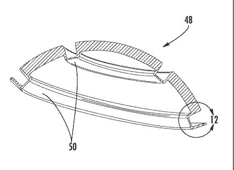
[0011] Figure 4 is a perspective view of a hip implant;
- 2 -
WO 2008/058205 CA 02668692 2009-05-05 PCT/US2007/083965
[0012] Figure 5 is a perspective view of a portion of a knee implant;
[0013] Figure 6 is a cross-sectional view of a portion of a resilient contact
member constructed in accordance with the present invention;
[0014] Figure 7 is an enlarged view of the contact member of Figure 7 in
contact
with a mating joint member;
[0015] Figure 8 is a side view of a resilient contact member in contact with
a
mating joint member;
[0016] Figures 9A and 9B are side and perspective views, respectively, of a
joint
with mating members;
[0017] Figures 10A and 10B are overall and detailed cross-sectional views of
the
joint of Figures 9A and 9B;
[0018] Figure 11 is a cross-sectional view of a cup for an implant according
to an
alternate embodiment of the invention;
[0019] Figure 12 is an enlarged view of a portion of the cup of Figure 11;
[0020] Figure 13 is a perspective view of a segmented implant constructed
according to the present invention;
[0021] Figure 14 is an enlarged view of a portion of Figure 13;
[0022] Figures 15A through 15C are various views of the implant of Figure 13;
[0023] Figures 16A through 16F are various views of another segmented implant
constructed according to the present invention;
[0024] Figure 17 is a cross-sectional view of an implant joint including a
flexible
seal;
[0025] Figure 18 is an enlarged view of a portion of Figure 17;
- 3 -
WO 2008/058205 CA 02668692 2009-05-05
PCT/US2007/083965
[0026] Figure 19 is a perspective view of a finite element model of a
joint
member; and
[0027] Figure 20 is a perspective view of a finite element model of
another joint
member.
DETAILED DESCRIPTION OF THE INVENTION
[0028] Referring to the drawings wherein identical reference numerals
denote the
same elements throughout the various views, Figure 1 depicts an exemplary
lower
member 10 of a hip implant constructed in accordance with the present
invention. The
lower member 10 is generally metallic and includes an elongated body 12 and a
ball
end 14. Although a hip implant is used as an example, the present invention is
equally
applicable to other types of implants
[0029] The surface of the ball end 14, or portions thereof, has a thin
film 16 of a
carbon-based material deposited thereon, referred to as a diamond-like carbon
(DLC)
material. This thin film material is essentially pure carbon, has a
noncrystalline
microstructure, and exhibits a flexural capability of approximately 8% or
better. The
carbon structure and bond layer enable the thin film 16 to endure significant
vibration
and deformation without cracking or detaching from the substrate or
delaminating.
Such thin films may be obtained from BioMedFlex LLC, Huntersville, NC, 28078.
[0030] The thin film 16 is applied in multiple layers, for example about 3
to about
30 layers may be used. The use of multilayers prevents residual stress build
up in the
individual layers and in the total film thickness This is in contrast to
typical prior art
thin films which have residual stress present and are brittle, limiting their
ability to
bear a localized load. The total thickness of the thin film 16 may be in the
range of
about 0.5 to about 6 gm. No post coating annealing or mechanical polishing is
required, and the thin film 16 has a high adhesion strength, for example about
8500
lb/in2 or greater.
[0031] Figure 2 illustrates a thin film apparatus 18 for applying the thin
film 16 to
the lower member 10. The thin film apparatus 18 is a chemical vapor deposition
- 4 -
WO 2008/058205 CA 02668692 2009-05-05 PCT/US2007/083965
(CVD) apparatus of a known type. It includes a processing chamber 20 which
receives the workpiece, a hydrocarbon gas source 22, an RF field generator 24
of a
known type, and a vacuum pump 26.
[0032] The thin film process proceeds as follows. First, the untreated lower
member 10 is plasma cleaned in a known manner to eliminate any foreign
material or
contaminants from the surface thereof The thin film 16 is then deposited over
the
exterior of the ball end 14 using a plasma assisted chemical vapor deposition
(CVD)
process. Since the thin film process is CVD, it does not require a direct line-
of-sight
to achieve a satisfactory thin film. Once the thin film cycle is complete, the
lower
member 10 is removed from the chamber 20.
[0033] It is also possible to construct the thin film 16 by alternating
layers of
metal doped DLC with layers of amorphous hydrogenated diamond like carbon.
Examples of suitable materials for the multilayers include: amorphous
hydrogenated
carbon, silicon doped amorphous hydrogenated carbon, boron doped amorphous
hydrogenated carbon, nitrogen doped amorphous hydrogenated carbon, boron
nitride
doped amorphous hydrogenated carbon, or other metal doped amorphous
hydrogenated carbon.
[0034] The thin film 16 does not require an intermediate film or coating
layer
(such as TiN). It has a high electrical resistivity and high thermal
conductivity. The
thin film 16 may be doped with one or more metallic, semi-metallic or other
elements
to produce a balance of high hardness without sacrificing typical DLC
coefficients of
reduced friction, adhesion layer strength, and overall bond strength.
[0035] The thin film 16 has several beneficial effects to the surface on
which it is
applied. The thin film is conformal and more uniform than physical vapor
deposition
methods. It creates a non-porous, chemically inert, protective boundary layer.
It
improves the ability to withstand a localized (Hertzian) load while still
providing
exceptional wear resistance and high adhesion. It provides unique flexural
property
that allows the thin film 16 (and underlying substrate) to flex under load.
This
combination of flexural nature and high wear resistance makes the thin film 16
a
- 5 -
WO 2008/058205 CA 02668692 2009-05-05 PCT/US2007/083965
solution for a variety of applications such as: gears (spiral bevel, hypoid,
helical, spur,
worm, etc.); medical implants; knees, hips, finger joint, spine, etc.; medical
instruments; cams (and cam shafts) lifters (e.g. flat tappet); valves
(automotive and
industrial); curvic couplings; hurth couplings; bearings (e.g. gothic arch and
planar
and roller surface); shafts (especially shaft faces or shoulders); and other
similar
applications.
[0036] The thin film 16 has the ability to withstand scuffing and galling. It
has a
high hardness, low friction, and resists chemical wear. The thin film 16
enhances
(fortifies) and protects the substrate surface to better preserve the exterior
(exposed
area) of the substrate to reduce the effects of micro surface damage (cracks
and
spalling); an initial wear indicator and mechanism. The high Hertzian contact
stress
tolerance makes it possible to actually maintain a hard carbon thin film 16
were prior
art DLCs would fail (due to cracking and adhesion layer failure)
[0037] Superfinishing of the thin film 16 is possible. This would produce an
even
better surface finish on a processed surface than originally existed on the
bare
substrate; even if the original substrate was finished to a sub micron (<1
micro-inch
Ra) surface finish.
[0038] The resilient hard carbon thin film 16 described above may be used on
implants having osseointegration surfaces, which are surfaces designed to be
infiltrated by bone growth to improve the connection between the implant and
the
bone. Osseointegration surfaces may be made from materials such as trabecular
metal,
textured metal, or sintered or extruded implant integration textures.
Trabecular metal
is an open metal structure with a high porosity (e.g. about 80%). An example
of a
trabecular metal structure is shown in Figure 3.
[0039] The thin film 16 may be applied to any osseointegration surface.
Figures
4-6 illustrate various examples of implants having osseointegration surfaces
"S",
including a hip joint shank 28, a hip joint cup 30, and a knee joint 32. The
thin film 16
may also be applied to other implants, such as plates and fasteners used for
reconstructive procedures The thin film 16 may be doped to facilitate
- 6 -
WO 2008/058205 CA 02668692 2009-05-05 PCT/US2007/083965
osseointegration, for example with titanium or fluorine. The thin film 16 may
be a
single layer of DLC material or a multilayer material as described above. If
desired, a
non-doped thin film may be used to create a wear resistant surface while
discouraging
bone integration. For example, in the hip joint lower member 10 of Figure 2,
the ball
end 14 may be coated with a non-doped thin film 16 as described above.
[0040] In order to utilize the superior characteristics of the thin films
described
above, a specialized implant contact interface (implant geometry) may be used.
In this
geometry, an implanted joint would include two typically hard (i.e. metal or
ceramic)
members; however, at least one of the members is formed such that it has the
characteristics of a resilient member, such as: the ability to absorb an
impact load; the
ability to absorb high cycle loading (high endurance limit); the ability to be
self
cleaning; and the ability to function as a hydrodynamic and/or hydrostatic
bearing.
One or both of these contact interface members may have thin film applied. If
thin
film is applied to two mating surfaces, it may be desirable to use two
different
compositions to improve the wear resistance and component compatibility. It
may
also be desired to apply thin film to one surface and a different surface
treatment or
coating to the mating surface.
[0041] Generally, the contact resilient member is flexible enough to allow
elastic
deformation and avoid localized load increases, but not so flexible as to risk
plastic
deformation, cracking and failure. In particular, the resilient member is
designed such
that the stress levels therein will be below the high-cycle fatigue endurance
limit. As
an example, the resilient member might be only about 10% to about 20% as stiff
as a
comparable solid member. It is also possible to construct the resilient member
geometry with a variable stiffness, i.e. having a low effective spring rate
for small
deflections and a higher rate as the deflections increase, to avoid failure
under sudden
heavy loads.
[0042] Figure 6 illustrates an exemplary contact member 34 including a basic
resilient interface geometry. The contact member 34 is representative of a
portion of a
medical implant and is made of one or more metals or ceramics (for example,
partially stabilized Zirconia). It is coated with a thin film (not shown) as
described
- 7 -
WO 2008/058205 CA 02668692 2009-05-05 PCT/US2007/083965
above. The geometry includes a lead in shape, Z1 and Z2, a contact shape, Z3
and Z4,
a lead out shape, Z5 and Z6, and a relieved shape, Z7. It may be desired to
vary the
cross-sectional thickness to achieve a desired mechanical stifthess to
substrate
resilience characteristic. The presence of the relieved region Z7 introduces
flexibility
into the contact member 34, reduces the potential for concentrated point
contact with
a mating curved member, and provides a reservoir for a working fluid.
[0043] The Z7 region may be local to the contact member 34 or may be one of
several. In any case, it may contain a means of providing fluid pressure to
the internal
contact cavity to produce a hydrostatic interface. A passive (powered by the
regular
motion of the patient) or active (powered by micro components and a dedicated
subsystem) pumping means and optional filtration may be employed to provide
the
desired fluid interaction.
[0044] A hydrodynamic interface is desirable as, by definition, it means the
contact member 34 is not actually touching the mating joint member. The lead-
in and
lead-out shapes Z1, Z2, Z5, Z6 are configured to generate a shear stress in
the
working fluid so as to create the fluid "wedge" of a hydrodynamic support.
However,
in this type of arrangement, there is a point where the two bearing surfaces
are resting
on each other in the absence of fluid shear between the two members of the
joint or
implant. This is what causes what is called stick-slip (the transition from
static to
dynamic friction then to hydrodynamic motion). The resilient nature of the
thin film
16, allows a design which has reduced wear even when the contact member 34
flexes
or is in a static friction regime.
[0045] Figure 7 shows a closer view of the contact member 34. It may be
desirable to make the contact radius (Z3 and Z4) larger or smaller, depending
on the
application requirement and flexural requirement. For example, Figure 8
illustrates
the contact member 34 in contact with a mating joint member 38 having a
substantially larger radius than the contact member 34. The radius ratio
between the
two joint members is not particularly critical, so long as one of the members
exhibits
the resilient properties described herein.
- 8 -
WO 2008/058205 CA 02668692 2009-05-05 PCT/US2007/083965
[0046] Another way to achieve a resilient member is to employ a design that
uses
contacting surfaces with similar geometric relationships but sandwiches a
resilient
media between two semi-rigid elements. For example, Figures 9A-9B and 10A-10B
illustrate a joint assembly with a cup 40 and a mating ball 42, both of
generally rigid
metals or ceramics. One or more ring-like rigid (i.e. metal or ceramic)
contact pads 44
are attached to the cup 40, with a resilient material (e.g. polymer) 46
sandwiched
between the two. In this case a polymer may be desirable as it is subjected to
a
distributed load versus the opportunity for localized wearing and degradation.
The cup
surface, including the contact pads 44, are coated with a thin film as
described above.
[0047] Figures 11 and 12 illustrate a coated cup 48 of metal or ceramic with
two
integrally-formed contact rings 50. More contact rings may be added if needed.
As
shown in Figure 12, the volume behind the contact rings 50 may be relieved.
This
relieved area 52 may be shaped so as to produce a desired balance between
resilience
and stiffness. A varying cross-section geometry defined by varying inner and
outer
spline shapes may be desired. In other words, a constant thickness is not
required. A
material such as a gel or non-Newtonian fluid (not shown) may be disposed in
the
relieved area 52 to modify the stiffness and damping characteristics of the
contact
rings 50 as needed for a particular application. The cup 48 could be used as a
stand-
alone portion of a joint, or it could be positioned as a liner within a
conventional liner.
The contact ring 50 is shown under load in Figure 19, which depicts contour
lines of
highest compressive stress at "Cl". This is the portion of the contact ring 50
that
would be expected to undergo bending first. The bearing interface portion of
the
resilient contact member could be constructed as a bridge cross-section
supported on
both sides as shown or as a cantilevered cross-section depending on the
desired static
and dynamic characteristics.
[0048] Figures 13-16 show a joint member 54 having a segmented shape. The
generally rectangular shape (in plan view) is illustrative and could be
modified to suit
a specific requirement. Contours Cl and C2 and C3 and C4 can be shaped as
needed
to yield the desired contact area and profile and contour coverage. Contact
profile Pb
can be modified to suit the load and resilience characteristic desired for the
specific
application. The joint member 54 may be solid in the center zone or open. The
contact
- 9 -
CA 02668692 2011-10-04
surface can have shaped grooves (for example in the profile PI) positioned to
allow particles to
move off the load bearing contact surface and eventually move back into the
joint for absorption
back into the body. The joint member 54 is shown under load in Figure 20,
which depicts an area
of highest compressive stress at "C2". This is the portion of the joint member
54 that would be
expected to undergo bending first.
[0049] Figures 17 and 18 illustrate an implant 56 of rigid material which
includes a wiper
seal 58. The wiper seal 58 keeps particles out of the contact area (seal void)
60 of the implant 58,
and working fluid (natural or synthetic) in. The seal geometry is intended to
be representative
and a variety of seal characteristics may be employed; such as a single lip
seal, a double or
multiple lip seal, a pad or wiper seal made from a variety of material
options. Different seal
mounting options may be used; lobe in shaped groove as shown in Figures 17 and
18, a retaining
ring or clamp, adhesion substance. The seal may also be incorporated into the
contact face of the
interface zone.
[0050] It may be desirable to create a return passage 62 from the seal void
region 60 back
into the internal zone 64 in order to stabilize the pressure between the two
and to allow for
retention of the Internal zone fluid if desired. This is especially relevant
when the hydrostatic
configuration is considered.
[0051] It is noted that it may be desirable to surface treat either or both
interfaces of any of
the above-described joints with a laser, shot peen, burnishing, or water shock
process, to reduce
wear. The benefit could be as much from surface annealing and microstructure
and microfracture
elimination as smoothing itself.
[0052] The foregoing has described medical implants with wear-resistant
geometries and
coatings. While specific embodiments of the present invention have been
described, it will be
apparent to those skilled in the art that various modifications thereto can be
made without
departing from the scope of the invention.
-10-