Note: Descriptions are shown in the official language in which they were submitted.
CA 02669516 2009-05-13
WO 2008/059483 PCT/IL2007/001375
1
A DIAGNOSTIC METHOD AND APPARATUS
The present invention relates to a diagnostic method correlating urine output
and urine flow for early prognosis of a disease affiliated with abnormal body
fluid
status. The present invention also provides an apparatus and system for
management of the hemodynamic state and kidney function of the body.
The field of the invention relates to management of a. patient's fluid, more
specifically providing an indication of "urine flow" such as an indication of
renal
perfusion, an indication of Glomerular Filtration Rate (GFR), changes in
extracellular
fluid, kidney function and urine irrigation problems, etc.
One of the most troublesome of all problems in critically ill patients is
maintenance of adequate body fluid and proper balance between fluid input and
fluid
output. To date, most patients that are hospitalized in the Intensive Care
Unit (ICU)
are monitored by continuous measurement of several hemodynamic parameters,
such as heart rate, invasive blood pressure measurement, central venous
pressure
(CVP) and occasionally, wedge pressure.
It is well known that one of the most important parameters that reflect proper
organ perfusion is the hourly urine output. However, currently the tools and
systems
that are used are not precise enough. One outcome of this is the high
occurrence of
acute renal failure (ARF) in ICU's. This complication occurs in a significant
percentage of critically ill patients. The most common underlying etiology is
acute
tubular necrosis, usually precipitated by hypoperfusion and/or nephrotoxic
agents.
On the other hand, overzealous use of fluid may result in fluid overload,
pulmonary
edema and ARDS.
Since appropriate management of the fluid balance and kidney function in the
critically ill patient is essential it is an object of the present invention
to provide a new
diagnostic method that continuously monitors and measures urine output and
urine
flow and correlates the same to provide real time warning with regard to
abnormal
fluctuations and perfusion to all the organs of the body and especially the
kidneys.
Thus according to the present invention there is now provided a diagnostic
method comprising continuously monitoring and transmitting urine output and
urine
flow rates of a catheterized patient to means which correlate the same with at
least
CA 02669516 2009-05-13
WO 2008/059483 PCT/IL2007/001375
2
one of renal perfusion, renal function, fluid status, polyuria, oleguria,
hypoperfusion,
hemorrhage shock and GFR.
In preferred embodiments of the present invention, said method utilizes a low
flow metering device.
In especially preferred embodiments of the present invention said low flow
metering device incorporates a drop generator and a droplet counter.
In a most preferred embodiment of the present invention, the present
invention utilizes a modified version of the low flow metering device
described and
claimed in U.S. Patent No:6,640,649, the relevant teachings of which are
incorporated herein by reference.
Preferably said method further comprises continuously monitoring and
graphically representing, in real minute unit time, fluctuations in renal flow
and renal
output.
The method of the present invention is especially useful for early prognosis
of
a disease affiliated with abnormal body fluid status and kidney stress in
medical
procedures such as surgery as well as being useful for providing an indicator
of
active nephron mass and kidney function.
As will be realized the method of the present invention is useful for
detecting a
disease affiliated with hypoperfusion.
The present invention is also useful for detecting a disease affiliated with
hyperperfusion.
In especially preferred embodiments of the present invention said method
further comprises providing alarm means.
Another aspect of the present invention relates to the use of a low flow
metering device for the manufacture of a diagnostic apparatus for continuous
monitoring and measuring of urine.output and urine flow of a catheterized
patient
further comprising linking the output of said device with means which
correlate the
same with at least one of renal perfusion, renal function, fluid status,
polyuria,
oleguria, hypoperfusion, hemorrhage shock and GFR.
In especially preferred embodiments of the present invention, there is
provided a diagnostic method comprising monitoring and transmitting urine flow
rates per minute units of a catheterized patient to means which correlate the
same
CA 02669516 2009-05-13
WO 2008/059483 PCT/IL2007/001375
3
with at least one of renal perfusion, renal function, fluid status, polyuria,
oleguria,
hypoperfusion, hemorrhage shock and GFR.
The term "urine flow rates per minute units" as used herein, is intended to
denote that in the apparatus and system of the present invention the volume of
urine
flow per a predetermined average of time intervals of minute units, such as
every
three minutes, is plotted on a graph.
In contradistinction to prior art systems, the present invention provides real
time information in terms of minute units and thus provides real time
information in
less than 30 minutes, preferably less than 20 minutes, and most preferred, in
some
of its aspects and utilizations, provides useful and critical information in
less than 10
minutes.
In a most preferred embodiment of the present invention there is provided a
diagnostic method for determining the hemodynamic state of a patient
comprising
administering a diuretic to a catheterized patient and monitoring and
displaying the
slope of urine output per minute units after administration thereof.
In another preferred embodiment of the present invention there is provided a
diagnostic method for determining the hemodynamic state of a patient
comprising
administering a bolus of fluid to the patient and monitoring and displaying
urine flow
reaction to said bolus to determine the state of hydration and hemorrhagic
shock.
The diagnostic method of the present invention allows for both the continuous
monitoring and transmission of urine output and flow rate information
regarding a
catheterized patient to means which correlate and display the same in real
time, and
will be integrated into an apparatus and system supplied to hospitals and
other
patient care facilities capable of showing an online and visual trend of urine
output
as well as a new clinical parameter, namely "urine flow". This parameter is
generated by online and continuous monitoring of urine production by the
kidneys.
Another aspect of the present invention, is directed to the use of a low flow
metering device for the manufacture of a diagnostic system for continuous
monitoring and measuring of urine output and urine flow of a catheterized
patient
further comprising linking the output of said device with means which
correlate the
same with at least one of renal perfusion, renal function, fluid status,
polyuria,
oleguria, hypoperfusion, hemorrhage shock and GFR.
CA 02669516 2009-05-13
WO 2008/059483 PCT/IL2007/001375
4
In another aspect of the present invention, there is provided a system for
management of the hemodynamic state and kidney function of the body comprising
a low flow metering device which continuously monitors and measures urine
output
and urine flow of a catheterized patient wherein the output of said device is
linked to
monitoring means and displaying means which display the slope of urine output
and
urine flow rates per minute units.
The present invention also provides an apparatus for management of the
hemodynamic state and kidney function of the body comprising a low flow
metering
device which continuous monitors and measures urine output and urine flow of a
catheterized patient wherein the output of said device is linked to means for
monitoring and displaying the slope of urine output and urine flow rates per
minute
units.
As is known, often the treating physicians are faced with a patient or body in
a
state of unconsciousness, semi-consciousness, or lack of control, as a result
of a
disease or trauma or induced by the medical staff, said patient being in the
operating
room, the ICU, the CCU, or in another critical care situation. When the body
is in a
steady state, and the kidneys are properly functioning with no blockages, and
when
the fluid flow into the body is constant, such as as a result of IV or IV
pumps, then
the amount of urine produced is constant and there is a continuous urine flow
which
is also constant.
Once it has been ascertained that the kidney is capable of producing urine at
a specific flow rate, then the urine flow rate can be maintained by
maintaining the
fluid flow rate into the body as a constant.
A rule of thumb usually accepted by most doctors establishes a urine
production rate of 1 ml / kg / hr.
A specific urine flow rate can then be calculated for a patient and fluid
input
can be adjusted and urine flow rate measured in order to establish this
specific urine
flow rate. As long as this fluid input rate is maintained and the kidneys
continue to
properly function, the urine flow rate will remain constant and the hydration
of a
patient can be managed accordingly.
By providing the apparatus of the present invention wherein the urine flow
rate is graphically represented in real minute unit times, it is possible to
immediately
CA 02669516 2009-05-13
WO 2008/059483 PCT/IL2007/001375
detect 'and deal with kidney stress and kidney malfunction, which have now
been
found to be accurate and early indicators of body dysfunction.
In preferred embodiments of the present invention said apparatus further
comprises means which correlate the same with at least one of renal perfusion,
renal
function, fluid status, polyuria, oleguria, hypoperfusion, hemorrhage shock
and GFR.
Preferably, said apparatus further comprises means for continuously
monitoring and graphically representing in real minute unit time fluctuations
in renal
flow and renal output.
In some preferred embodiments of the present invention said apparatus
comprises means for monitoring and measuring of urine output and urine flow of
a
catheterized patient after the administration of a diuretic.
In other preferred embodiments of the present invention said apparatus
comprises means which monitors and measures urine output and urine flow of a
catheterized patient after the administration of a bolus of fluid to a patient
in a stable
steady state with a constant fluid input and output.
In especially preferred embodiments of the present invention there is provided
an apparatus for management of the hemodynamic state and kidney function of
the
body comprising a low flow metering device for continuous monitoring and
measuring of urine output and urine flow of a catheterized patient wherein the
output
of said device is linked to means for monitoring and displaying the slope of
urine
output and urine flow rates per minute units during surgery.
In other preferred embodiments of the present invention there is provided an
apparatus for management of the hemodynamic state and kidney function of the
body comprising a low flow metering device which continuously monitors and
measures urine output and urine flow of a catheterized patient, wherein the
output of
said device is linked to means which monitor and display the slope of urine
output
and urine flow rates per minute units after administration of a nephrotoxic
drug.
In yet another preferred embodiments of the present invention there is
provided an apparatus for management of the hemodynamic state and kidney
function of the body comprising a low flow metering device for which
continuously
monitors and measures urine output and urine flow of a catheterized patient
wherein
the output of said device is linked to means which monitor and display the
slope of
CA 02669516 2009-05-13
WO 2008/059483 PCT/IL2007/001375
6
urine output and urine flow rates per minute units during administration of a
nephrotoxic drug
As is known, in most catheterized patients measurement of urine output is
performed by an hourly assessment of the urine volume in a canister or by
electronically measuring the volume in a canister. In contradistinction, by
providing
reliable, high resolution and continuous trends of the patient's "urine flow",
the
present invention enables continuous online provision of an indication of
renal
perfusion, renal function, fluid status, polyuria, oliguria and GFR.
The goal of the present invention is to continuously monitor and display in
real
time the urine flow in order to optimize fluid management thereby enabling
early
prognosis of disease affiliated with Hypoperfusion such as ARF caused by renal
Hypoperfusion, Intrinsic ARF and Postrenal azotemia etc. and Hyperperfusion
such
Edema, the use of diuretics etc.
It is to be noted that with the tools available in a standard emergency room
and ICU, there is no way to immediately check for hemorrhage shock or
hypoperfusion of an admitted patient.
As is known, blood pressure does not reflect blood loss, since in the case of
blood loss vasoconstriction cuts off the arterioles in less vital organs as
perceived by
the brain, i.e., the legs, the arms, the stomach and even the kidney, in order
to
maintain blood flow and blood pressure to the brain. It is for this reason
that up to
35% of patients in an ICU unit suffer from acute kidney injury since the
monitoring
teams have no way of knowing that blood has been cut off from the kidney when
the
patient is in a hypoperfusion state, or is suffering from kidney damage as a
result of
drugs which act as nephrotoxins.
Thus, many drugs, and especially anti-cancer drugs, function as
nephrotoxins.
It has now been found, according to the present invention, that by measuring
urine flow, one can tell when the kidney is in stress and based thereon,
treatment
with said drug can be slowed whereby the drug is administered in a regulated
way in
order to limit kidney damage such as by administering the drug over a 4 hour
period
instead of in a single immediate dose.
Today, creatinine is used as a measure of kidney state, however, creatinine in
the blood occurs when the kidney cannot remove some or all of the creatinine
from
CA 02669516 2009-05-13
WO 2008/059483 PCT/IL2007/001375
7
the body, and this occurs only when there is already between about 50%-70%
kidney damage. Thus the creatinine test is ineffective for showing kidney
damage of
up to and even greater than 50%.
As is known, the rise of creatinine in the blood as a result of kidney damage
takes hours and even days to occur and therefore the creatinine test gives its
results
much too late to reverse kidney damage.
It has now been found that with early detection of kidney damage, i.e., within
the first half hour or so, the damage can be reversed by corrective action, or
at a
later stage, e.g., within 1- 1.5 hours, can be reversed with certain drugs and
therefore there is a need for a marker providing early detection of renal
failure, also
known as AKI (acute kidney injury).
According to the present invention, it has now been discovered that
administration of a loop diuretic such as Fusid (furosamide) to a patient,
results in a
rise in the flow rate of urine per minute units which can be plotted on a
graph and the
slope of which is a linear slope. Thus it has now been discovered that this
slope is
proportional to the peak flow rate of urine which is proportional to the total
volume of
urine produced as a result of the administration of a diuretic which in turn
is
proportional to the state of the kidney in terms of active nephron mass and
which
slopes therefore represent the percent of damage to the kidney. Therefore
displaying
and noting the linear slope in a graph generated in a relatively short period
of time
e.g., in the first 5 minutes after administration or a similarly chosen minute
time unit,
is sufficient to establish a clear picture of kidney function.
Similarly it is possible to effect a fluid challenge to the body, e.g., by
administering a predetermined amount of fluid to the system, such as 200-300
ml,
and then monitoring and displaying the slope of urine output per minute unit
after
administration thereof, wherein either the slope, the peak or the total time
for the flow
of urine to return to steady state flow, serves to determine the state of the
kidney and
the existence or absence of hemorrhagic shock in a patient.
Thus in a preferred embodiment of the present invention, there is provided a
diagnostic method for determining the hemodynamic state of a patient
comprising
administering a bolus of fluid to the patient and monitoring and displaying
urine flow
reaction to said bolus to determine the state of hydration and hemorrhagic
shock.
CA 02669516 2009-05-13
WO 2008/059483 PCT/IL2007/001375
8
Thus the present invention provides a novel and greatly needed tool for early
detection of kidney damage and the degree thereof, thereby enabling the timely
treatment for reversing the same
As a side benefit of the present invention, it has been discovered that it is
not
necessary to administer large doses such as 500 mg of a diuretic such as
Fusid,
since diuretic drugs are known to be nephrotoxins and it is sufficient to
administer a
smaller dose of 40-50 mg in order to obtain the same effect.
According to the present invention, it is now possible to monitor and display
kidney function during surgery and to detect kidney stress in real time, in
minute
units during surgery, whereby the surgeon then has a much earlier indicator,
than
presently available, that corrective action is immediately required.
The following is a partial list of diseases that are associated with and
indicated by abnormal urine flow: kidney perfusion, renal failure, organ
perfusion,
pre-operative/post-operative complications, surgical success, undetected
internal
trauma, dehydration, response to medication (antibiotics, diuretics etc),
jaundice,
shock, preeclampsia, bladder infection, cystitis, prostatitis, urinary tract
infection,
kidney stones, low blood pressure, anuria (lack of urine), hypovolemia,
hypervolemia, pulmonary edema, and hyponatremia,
The following are explanations of terms and diseases referred to herein.
ARF (Acute Renal Failure)
Acute Renal Failure (ARF) is a syndrome characterized by rapid decline in
glomerular filtration rate (hours to days), retention of nitrogenous waste
products,
and perturbation of extracellular fluid volume and electrolyte and acid-base
homeostasis. ARF complicates approximately 5% of hospital admissions and up to
30% of admissions to intensive care units. Oliguria (urine output < 400 mUd)
is a
frequent but not invariable clinical feature (50%). ARF is usually
asymptomatic and
diagnosed when biochemical monitoring of hospitalized patients elevates. a
recent
increase in blood urea and creatinine concentrations. It may complicate a wide
range
of diseases, which for purposes of diagnosis and management are conveniently
divided into three categories:
(1) Diseases that cause renal hypoperfusion without compromising the
integrity of renal parenchyma (prerenal ARF, prerenal azotemia)
(55%),
CA 02669516 2009-05-13
WO 2008/059483 PCT/IL2007/001375
9
(2) Diseases that directly involve renal parenchyma (intrinsic renal ARF,
renal azotemia) (40%);
(3) Diseases associated with urinary tract obstruction (postrenal ARF,
postrenal azotemia) (5%).
Most ARF is reversible, the kidney being relatively unique among major organs
in
its ability to recover from almost complete loss of function. Nevertheless,
ARF is
associated with major in-hospital morbidity and mortality, in large part due
to the
serious nature of the illnesses that precipitate the ARF. Severe cases may
show
clinical or pathologic evidence of ATN. Contrast nephropathy classically
presents as
an acute (onset within 24 to 48 h) but reversible.
GFR
The GFR was originally determined by injecting inulin into the plasma. Since
inulin is not reabsorbed by the kidney after glomerular filtration, its rate
of excretion
is directly proportional to the rate of filtration of water and solutes across
the
glomerular filter. In clinical practice however, creatinine clearance is used
to
measure GFR. Creatinine is an endogenous molecule, synthesized in the body,
which is freely filtered by the glomerulus (but also secreted by the renal
tubules in
very small amounts). Creatinine clearance is therefore a close approximation
of the
GFR. The GFR is typically recorded in milliliters per minute (mI/min).
Example: A person has a plasma creatinine concentration of 0.01 mg/mI and in
1 hour he excretes 75 mg of creatinine in the urine. The GFR is calculated as
M/P
(where M is the mass of creatinine excreted per unit time and P is the plasma
concentration of creatinine).
75 Lnb
GFR = 60 Miuls = 125 inl/min
0.01 ing/iml
= Chronic Renal Failure (CRF) develops slowly and gives few
symptoms initially. It can be the complication of a large number of kidney
diseases, such as IgA nephritis, glomerulonephritis, chronic pyelonephritis
and urinary retention. End-stage renal failure (ESRF) is the ultimate
consequence, in which case dialysis is generally required until a donor for a
renal transplant is found.
CA 02669516 2009-05-13
WO 2008/059483 PCT/IL2007/001375
~ Acute Renal failure (ARF) is, as the name implies, a rapidly
progressive loss of renal function, generally characterised by oliguria
(decreased urine production, quantified as less than 400 mL per day in
adults,113 less than 0.5 mUkg/h in children or less than 1 mL/kg/h in
infants),
body water and body fluids disturbances and electrolyte derangement. An
underlying cause must be identified to arrest the progress, and dialysis may
be necessary to bridge the time gap required for treating these undertying
causes
Acute renal failure can be present on top of chronic renal failure. This is
called
acute-on-chronic renal failure (AoCRF). The acute part of AoCRF may be
reversible
and the aim of treatment, like in ARF, is to return the patient to their
baseline renal
function, which is typically measured by serum creatinine. AoCRF, like ARF,
can be
difficult to distinguish from chronic renal failure, if the patient has not
been followed
by a physician and no baseline (i.e., past) blood work is available for
comparison.
Before the advancement of modern medicine renal failure might be referred to
as
uremic poisoning. Uremia was the term used to describe the contamination of
the
blood with urine. Starting around 1847 this term was used to describe reduced
urine
output, now known as oliguria that was thought to be caused by the urine
mixing with
the blood instead of being voided through the urethra.
Prerenal azotemia is relatively common, especially in hospitalized patients.
The kidneys normally filter the blood. When the volume or pressure of blood
flow
through the kidney drops, blood filtration also drops drastically, and may not
occur at
all. Waste products remain in the bloodstream and little or no urine is
formed, even
though the internal structures of the kidney are intact and functional.
Lab tests show that nitrogen-type wastes, such as creatinine and urea, are
accumulating in the body (azotemia). These waste products act as poisons when
they accumulate, damaging tissues and reducing the ability of organs to
function.
The build-up of nitrogen waste products and accumulation of excess fluid in
the body
are responsible for most of the symptoms of prerenal azotemia and acute renal
failure.
Prerenal azotemia is the most common form of kidney failure seen in
hospitalized patients. Any condition that reduces blood flow to the kidney may
cause
CA 02669516 2009-05-13
WO 2008/059483 PCT/IL2007/001375
11
it, including loss of blood volume, which may occur with dehydration,
prolonged
vomiting or diarrhea, bleeding, burns, and other conditions that allow fluid
to escape
from circulation.
Conditions in which the volume is not lost, but in which the heart cannot pump
enough blood, or the blood is pumped at low volume, also increase risk for
prerenal
azotemia. These conditions include shock (such as septic shock), heart
failure, and
conditions where the blood flow to the kidney is interrupted, such as trauma
to the
kidney, surgery of various types, renal artery embolism, and other types of
renal
artery occlusion.
Thus it will be realized that the method of the present invention provides the
ICU and other medical facilities and departments with a valuable new
diagnostic tool
heretofore not available.
While the invention will now be described in connection with certain preferred
embodiments in the following examples and with reference to the attached
figures so
that aspects thereof may be more fully understood and appreciated, it is not
intended
to limit the invention to these particular embodiments. On the contrary, it is
intended
to cover all alternatives, modifications and equivalents as may be included
within the
scope of the invention as defined by the appended claims. Thus, the following
examples which include preferred embodiments will serve to illustrate the
practice of
this invention, it being understood that the particulars shown are by way of
exampie
and for purposes of illustrative discussion of preferred embodiments of the
present
invention only and are presented in the cause of providing what is believed to
be the
most useful and readily understood description of formulation procedures as
well as
of the principies and conceptual aspects of the invention.
In the figures,
Figure 1 is a graphical representation of urine volume measured by prior art
methods during open heart surgery of a patient;
Figure 2 is a graphical representation of urine flow measured according to the
method of the present invention during open heart surgery of a patient;
Figure 3 is a graphical representation of standard blood pressure
measurements over time as well as that of urine flow rate according to the
present
invention taken during open heart surgery.
CA 02669516 2009-05-13
WO 2008/059483 PCT/IL2007/001375
12
Figure 4 is a graphical representation of mean flow rate of urine of a patient
during bypass open heart surgery;
Figure 5 is a graphical representation of standard blood pressure
measurement over time of said patient during said bypass surgery;
Figure 6 is a graphical representation of online per minute urine flow rate
output as an indication for hemorrhagic shock, as opposed to blood pressure
which
remains within the range of normal pressure;
Figure 7 is a graphical representation of flow rate versus time, as well as
blood pressure versus time when a bolus of liquid was administered during
induction
of hemorrhagic shock;
Figure 8 is a graphical representation of urine flow versus time during bypass
surgery;
Figure 9 is a graphical representation of urine flow versus time during bypass
surgery in a patient with kidney under stress;
Figure 10 is a graphical representation of online per minute urine flow rate
output after administration of a diuretic medication to different patients
with different
kidney status;
Figure 11 is a graphical representation of urine flow versus time after
administration of a diuretic from which it can be noted that the flow slope
and the
flow peak are proportional.
Figure 12 is a graphical representation of urine flow output as related to
dose
of diuretic drug administered; and
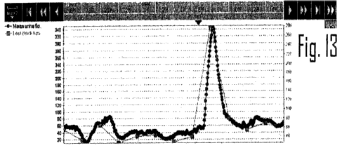
Figure 13 is a graphical representation of urine flow as a function of time
when a body is in a stable state and when a bolus of fluid is administered.
Referring now to Figure 1, there is seen a graphical representation of urine
volume over time of a patient during open heart surgery wherein according to
the
graph, there is a constant increase in volume and therefore no probiems are
detected.
Referring now to Figures 2 and 3, Figure 2 is a graphical representation of
urine flow rate measured according to the method of the present invention and
Figure 3 is a graphical representation of said flow rate on a graph also
showing
standard blood pressure measurements of the same patient during the same
period
of time. As will be noted, the method according to the present invention
detected
CA 02669516 2009-05-13
WO 2008/059483 PCT/IL2007/001375
13
and displayed a severe drop in flow rate, more than an hour before a drop was
noted
by the standard blood pressure measurements.
Thus from Figures 1, 2 and 3, it will be noted that the standard methods and
tools available indicated that the urine volume continued to increase
throughout the
procedure and the reduction in flow was detected by the present method more
than
an hour before the reduction of blood pressure was noted, wherein measurement
of
blood pressure is used today as the standard for determining fluid status of a
patient.
Referring now to Figures 4 and 5, Figure 4 records the flow rate of urine as a
function of time of a patient undergoing bypass open heart surgery, while
Figure 5
records the`standard blood pressure measurements taken of the same patient
during
the same period of time. As will be noted, Figure 5 does not show any problem
in
the blood pressure of the patient, while Figure 4 which recorded flow rate of
urine
according to the present invention indicated significant fluctuations in flow,
indicating
that the patient was not receiving sufficient blood to the kidneys which could
have
been corrected based on the information provided by the flow rate graph of the
present method by increasing cardiac output during bypass.
Referring to Figure 6, there is graphically represented the monitoring of a
trial
of hemorrhagic shock to pigs wherein adult pigs weighing between 50 - 70 kg
were
anesthetized and monitored with the first hour serving as a reference, the
urinary
bladder was pierced and directly catheterized using a foley catheter and then
the
pigs were bled with a break between bleeding, each bleeding being of 10% of
the
blood volume of the pig for four repeated bleedings and the final bleeding
being of
5%.. A diuretic was administered prior to the test and the flow rate against
time in
minutes was monitored.
As will be noted, the urine flow drops drastically after the first few minutes
of
bleeding and after 180 minutes, the urine flow goes to 0. At this point,
vasoconstriction occurs, cutting off blood to the kidney. The nephrons will be
damaged shortly thereafter. As noted however, the blood pressure remains
within
the normal range after 45% of the blood has been removed from the pigs.
Referring to Figure 7, the procedure used in Figure 6 was repeated, however
a bolus of 500 ml of water was administered shortly after bleeding was
induced. As
can be seen the kidney reacted within minutes resulting in increased urine
flow
during the period of the first bleeding. A second bolus of 500 ml was
administered at
CA 02669516 2009-05-13
WO 2008/059483 PCT/IL2007/001375
14
the outset of bleeding of the second 10%. Once again, the kidney reacted
accordingly resulting in increased urine flow. A third bolus of 500 ml was
administered at the outset of bleeding of the third 10% however the kidney did
not
react thereto, indicating that the kidney was no longer functioning at this
point.
Referring to Figure 8, there is seen a graphical representation of urine flow
rate as a function of time as well as mean blood pressure during bypass
surgery
wherein bypass began at minute 100 and ended at around minute 200.
Before commencement of the bypass surgery either a large volume of fluid is
administered to the patient or a diuretic is administered or both, in order to
maintain
kidney activity. As will be noted, urine flow increased in a typical bell
shape as seen
in the figure. At around minute 200, the operation was completed and the
bypass
was disconnected. In this case the kidneys were not affected.
Referring now to Figure 9, there is seen a further graph of urine flow and
mean blood pressure for a different patient undergoing bypass surgery. As will
be
noted, the urine flow was not smooth and instead was very erratic. This flow
pattern
which was immediately observable indicated that the kidney was under stress
and
was damaged and that the patient had acute kidney injury (AKI).
As will be realized, by observing urine flow during surgery, the flow pattern
will
show the kidney state and indicate when the kidney gets into stress enabling
the
surgeon to effect early intervention and immediately attempt corrective
action.
Referring to Figure 10, there are seen the patterns of urine flow as a
function
of time of different patients to whom a diuretic medication was administered.
It will
be noted that the degree of kidney injury will give different flow peaks. Thus
a
normal kidney will give a very high peak whiie a damaged kidney will give a
very low
peak.
Referring to Figure 11, there is seen a graphical representation of urine flow
per minute after administration of a diuretic from which it can be seen that
the siope
is substantially linear and proportional to the peak and therefore its path
can be
extrapolated within several minutes after administration of the diuretic
thereby
providing a very valuable early assessment tool for kidney function.
Referring to Figure 12, there is seen a graph of a urine flow slope as a
function of a diuretic dose of fusid. It will be noted that after 40 mg of
diuretic the
CA 02669516 2009-05-13
WO 2008/059483 PCT/IL2007/001375
drug has no further effect thus indicating that it is not necessary to
administer high
drug doses and that a drug dose of less than 50 mg is sufficient.
Since diuretic drugs such as fusid are nephrotoxic, this finding enables the
determination of optimum effective doses of similar drugs, and obviates the
administration of excess drugs which are harmful to the body.
Thus, by observing urine flow parameters, optimal amount of drugs can be
determined and administered.
Referring to Figure 13, there is seen a graphical representation of urine flow
versus time.
It will be noted that when a patient or body is in a stable state and the
fluids
administered to the body are constant, a healthy kidney is in a condition that
it can
produce urine flow at a constant rate. When a small bolus of fluid is
administered to
the body, the kidney reacts within minutes to remove the fluid and return the
body to
the original steady state.
Thus, the apparatus of the present invention provides an invaluable tool for
early detection of abnormal conditions not provided by the standard measuring
tools
available today, and has multiple uses in body hydration and kidney
management.
It will be evident to those skilled in the art that the invention is not
limited to
the details of the foregoing illustrative examples and that the present
invention may
be embodied in other specific forms without departing from the essentiai
attributes
thereof, and it is therefore desired that the present embodiments and examples
be
considered in all respects as illustrative and not restrictive, reference
being made to
the appended claims, rather than to the foregoing description, and all changes
which
come within the meaning and range of equivalency of the claims are therefore
intended to be embraced therein.