Note: Descriptions are shown in the official language in which they were submitted.
CA 02676225 2009-08-28
VOLAR FIXATION SYSTEM
BACKGROUND OF THE INVENTION
This application is a divisional application of Canadian application serial
number
2,396,850.
1. Field of the Invention
This invention relates broadly to surgical devices. More particularly, this
invention
relates to a bone fixation system, and particularly to a fixation system
adapted to fixate a
Colles' (or distal radial) fracture.
2. State of the Art
Referring to Fig. 1, a Colles' fracture is a fracture resulting from
compressive forces
being placed on the distal radius 10, and which causes backward displacement
of the distal
fragment 12 and radial deviation of the hand at the wrist 14. Often, a Colles'
fracture will
result in multiple bone fragments 16, 18, 20 which are movable and out of
alignment relative
to each other. If not properly treated, such fractures result in permanent
wrist deformity. It is
therefore important to align the fracture and fixate the bones relative to
each other so that
proper healing may occur.
Alignment and fixation are typically performed by one of several methods:
casting,
external fixation, interosseous wiring, and plating. Casting is non-invasive,
but may not be
able to maintain alignment of the fracture where many bone fragments exist.
Therefore, as an
alternative, external fixators may be used. External fixators utilize a method
known as
ligamentotaxis, which provides distraction forces across the joint and pen-
nits the fracture to
be aligned based upon the tension placed on the surrounding ligaments.
However, while
external fixators can maintain the position of the wrist bones, it may
nevertheless be difficult
in certain fractures to first provide the bones in proper alignment. In
addition, external
fixators are often not suitable for fractures resulting in multiple bone
fragments. Interosseous
wiring is an invasive procedure whereby screws are positioned into the various
fragments
CA 02676225 2009-08-28
2
and the screws are then wired together as bracing. This is a difficult and
time consuming
procedure. Moreover, unless the bracing is quite complex, the fracture may not
be properly
stabilized. Plating utilizes a stabilizing metal plate typically against the
dorsal side of the
bones, and a set of parallel pins extending from the plate into the holes
drilled in the bone
fragments to provide stabilized fixation of the fragments. However, the
currently available
plate systems fail to provide desirable alignment and stabilization.
SUMMARY OF THE INVENTION
It is therefore an object of the invention to provide an improved fixation and
alignment system for a Colles' fracture.
It is another object of the invention to provide a volar fixation system which
desirably
aligns and stabilizes multiple bone fragments in a distal radial fracture to
permit proper
healing.
It is also an object of the invention to provide a volar fixation system which
is highly
adjustable to provide a customizable framework for bone fragment
stabilization.
In accordance with these objects, which will be discussed in detail below, a
volar
fixation system is provided which generally includes a T-shaped plate intended
to be
positioned against the volar side of the radial bone, a plurality of bone
screws for securing
the plate along a non-fractured portion of the radial bone, and a plurality of
bone pegs which
extend from the plate and into bone fragments of a Colles' fracture.
Parent application CA 2,396,850 claims a volar fixation plate comprising a
substantially rigid plate including a distal head portion and a proximal body
portion angled
relative to said head portion, said head portion defining a plurality of
threaded peg holes
adapted to individually receive fixation pegs therein, said peg holes defining
a plurality of
axes at least two of which are oblique relative to each other, and said body
portion including
at least one screw hole.
CA 02676225 2009-08-28
3
This application relates to a volar fixation plate, comprising: a
substantially rigid
plate including a distal head portion and a proximal body portion angled
relative to said head
portion, said head portion defining a plurality of threaded peg holes adapted
to individually
receive fixation pegs therethrough, said peg holes linearly arranged in a
generally medial to
lateral direction wherein successive lateral peg holes are situated distally
relative to adjacent
peg holes, and said body portion including at least one screw hole.
In another aspect, there is disclosed the use of a volar fixation plate as
described
above for aligning and stabilizing a Colles' fracture of the radius bone at
the wrist, the radius
bone having a volar side and a dorsal side, and the fracture including at
least one bone
fragment.
The plate is generally a T-shaped plate defining an elongate body, a head
portion
angled relative to the body, a first side which is intended to contact the
bone, and a second
side opposite the first side. The body portion includes a plurality of
countersunk screw holes
for the extension of the bone screws therethrough. The head portion includes a
plurality of
threaded peg holes for receiving the pegs therethrough. According to a first
embodiment, the
peg holes are preferably non-linearly arranged. According to a second
embodiment, the peg
holes are preferably linearly arranged. In either embodiment, the peg holes
are positioned
increasingly distal in a medial to lateral direction along the second side.
According to a third
embodiment, which preferably uses a volar plate with peg holes arranged
according to either
of the first and second embodiments, the pegs are adjustable relative to the
peg holes and can
be independently fixed in selectable orientations.
In use, the volar plate is positioned with its first side against the volar
side of the
radius and bone screws are inserted through the bone screw holes into the
radius to secure the
volar plate to the radius. The bone fragments are then aligned and the guide
plate is
positioned on the second side of the volar plate. A drill drills holes into
the bone fragments.
CA 02676225 2009-08-28
4
The pegs are then inserted through the peg holes and into the holes in the
bone. In
some embodiments, the heads of the pegs are threadably engaged in the volar
plate. In other
embodiments, the pegs are inserted into the peg holes and into the drilled
holes at an angle
chosen by the surgeon, and a set screw is inserted over each peg to lock the
peg in the volar
plate at the chosen orientation. The volar fixation system thereby stabilizes
and secures the
bone fragments in their proper orientation.
Additional objects and advantages of the invention will become apparent to
those
skilled in the art upon reference to the detailed description taken in
conjunction with the
provided figures.
BRIEF DESCRIPTION OF THE DRAWINGS
Fig. 1 is an illustration of an extremity subject to a Colles' fracture;
Fig. 2 is a top volar view of a right hand volar fixation system according to
a first
embodiment of the invention;
Fig. 3 is a side view of a bone peg according to the first embodiment of the
volar
fixation system of the invention;
Fig. 4 is a side view of a bone screw of the volar fixation system of the
invention;
Fig. 5 is a side view of the right hand volar plate of the volar fixation
system
according to the first embodiment of the invention;
Fig. 6 is a front end view of the right hand volar plate of the volar fixation
system
according to the first embodiment of the invention;
Fig. 7 is an exploded side view of the right hand volar plate and guide plate
according
to the first embodiment of the fixation system of the invention;
CA 02676225 2009-08-28
Fig. 8 is a side view of the guide plate positioned on the right hand volar
plate to
provide drill guide paths in accord with the invention;
Fig. 9 is an illustration of the first embodiment of the volar fixation system
provided
in situ aligning and stabilizing a Colles' fracture;
Fig. 10 is a top volar view of a left hand volar fixation system according to
the second
embodiment of the invention;
Fig. 11 is a lateral side view of the left hand volar fixation system
according to the
second embodiment of the invention;
Fig. 12 is a bottom view of the left hand volar fixation system according to
the second
embodiment of the invention;
Fig. 13 is an enlarged side elevation of a bone peg according to the second
embodiment of the volar fixation system of the invention;
Fig. 14 is a proximal end view of the bone peg of Fig. 13;
Fig. 15 is first partial top view of the head portion of the left hand volar
plate
according to the second embodiment of the volar fixation system of the
invention;
Figs. 16-19 are section views across line 16-16, 17-17, 18-18, and 19-19,
respectively
in Fig. 15;
Fig. 20 is second partial top view of the head portion of the left hand volar
plate
according to the second embodiment of the volar fixation system of the
invention;
CA 02676225 2009-08-28
6
Figs. 21-24 are section views across line 21-21, 22-22, 23-23, and 24-24,
respectively
in Fig. 20;
Fig. 25 is a broken partial longitudinal section view across a distal end of a
third
embodiment of the volar fixation system of the invention;
Fig. 26 is a proximal perspective view of a bone peg according to the third
embodiment of the invention; and
Figs. 27 and 28 are proximal and distal perspective views, respectively, of a
set screw
according to the third embodiment of the invention.
DETAILED DESCRIPTION OF THE PREFERRED EMBODIMENTS
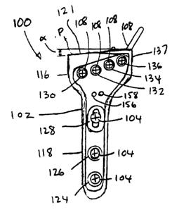
Turning now to Figs. 2 through 4, a first embodiment of a volar fixation
system 100
for aligning and stabilizing multiple bone fragments in a Colles' fracture
generally includes a
substantially rigid T-shaped plate 102 intended to be positioned against the
volar side of the
radial bone, a plurality of preferably self-tapping bone screws 104 for
securing the plate 102
along a non- fractured portion of the radial bone, and a plurality of bone
pegs 108 which
extend from the plate 102 and into bone fragments of a Colles' fracture.
Referring to Figs. 2, 5 and 6, more particularly, the T-shaped plate 102
defines a head
portion 116, an elongate body portion 118 angled relative to the head portion,
a first side 120
which is intended to contact the bone, and a second side 122 opposite the
first side. The first
side 120 at the head portion is preferably planar, as is the first side at the
body portion. As the
head portion and body portion are angled relative to each other, the first
side preferably
defines two planar portions. The angle 0 between the head portion 116 and the
body portion
118 is preferably approximately 18 and bent at a radius of approximately 1.00
inch (Fig. 5).
The distal edge 121 of the head portion 116 is preferably angled proximally
toward the
medial side at an angle a, e.g., 5 , relative to a line P, which is
perpendicular to the body
portion. The head portion 116 preferably has a width of 0.913 inch and a
greatest proximal-
CA 02676225 2009-08-28
7
distal dimension (i.e., from the comer of angle a to the body portion) of
approximately 0.69
inch, and the body portion preferably has a width of 0.375 inch and a length
of 1.40 inches.
The plate 102 preferably has a thickness of approximately 0.098 inch. The
plate 102 is
preferably made from a titanium alloy, such as Ti-6A-4V.
The body portion 118 includes three preferably countersunk screw holes 124,
126,
128 for the extension of the bone screws 104 therethrough. The first screw
hole 124 has a
center preferably 0.235 inch from the end of the body portion, the second
screw hole 126 has
a center preferably 0.630 inch from the end of the body portion, and the third
screw hole 128
is preferably generally elliptical (or oval) and defines foci-like locations
at 1.020 inches and
1.050 inches from the end of the body portion. The head portion 116 includes
four threaded
peg holes 130, 132, 134, 136 for individually receiving the pegs 108
therethrough. According
to a first preferred aspect of the first embodiment of the invention, the peg
holes 130, 132,
134, 136, preferably 0.100 inch in diameter, are preferably non-linearly
arranged along the
head portion 116, and are provided such that the adjacent peg holes are
provided further
distally in a medial to lateral direction along the second side. More
particularly, according to
a preferred aspect of the first embodiment of the invention, the peg holes are
preferably
arranged along a parabolic curve, with the center of peg hole 130 located
approximately
0.321 inch proximal line P and approximately 0.719 inch medial of the lateral
edge 137 of
the head portion, the center of peg hole 132 located approximately .296 inch
proximal line P
and approximately 0.544 inch medial of the lateral edge 137, the center of peg
hole 134
located approximately 0.250 inch proximal line P and approximately 0.369 inch
medial of
the lateral edge 137, and the center of peg hole 136 located approximately
0.191 inch
proximal line P and approximately 0. 194 inch medial of the lateral edge 137.
In addition, according to a second preferred aspect of the first embodiment of
the
invention, the peg holes define axes Al, A2, A3, A4 which are oblique (not
parallel) relative to
each other, and more preferably are angled in two dimensions (medial/lateral
and
proximal/distal) relative to each other; i. e., the pegs once inserted into
the peg holes are also
angled in two dimensions relative to each other. More particularly, the first
axis Al of the
first peg hole 130 (that is, the most proximal and medial peg hole) is
preferably directed
CA 02676225 2009-08-28
8
normal to the first side 120 of the head portion 116. The axis A2 of the
adjacent peg hole 132,
i.e., the second axis, is preferably angled approximately 1-7 distal and
lateral relative to the
first axis A1, and more preferably approximately 2.5 distal and lateral
relative to the first
axis Al. The axis A3 of the peg hole 134 laterally adjacent the second peg
hole 132, i.e., the
third axis, is preferably angled approximately 7-13 distal and lateral
relative to the first axis
Al, and more preferably approximately 10 distal and lateral relative to the
first axis Al. The
axis A4 of the peg hole 134 laterally adjacent the third peg hole 132, i.e.,
the fourth axis, is
preferably angled approximately 10-30 distal and lateral relative to the
first axis Al, and
more preferably approximately 20 distal and lateral relative to the first
axis & The second
side of the head portion 116, distal of the peg holes 130, 132, 134, 136 is
preferably bevelled.
Referring back to Fig. 3, the pegs 108, preferably approximately 0.872 inch in
length,
each have a threaded head 138 adapted to threadably engage the threads about
the peg holes
130, 132, 134, 136, and have a relatively smooth non-threaded cylindrical
shaft 140. The
shafts 140 are preferably approximately 0.0675 inch in diameter and 0.765 inch
in length.
Such dimensions permit the pegs to adequately support the bone fragments such
that the bone
is able to heal correctly. The pegs 108 are also preferably made from titanium
alloy, and may
be coated in a ceramic, e.g., titanium nitride, to provide a bone interface
which will not
adversely affect bone healing.
Turning now to Figs. 7 and 8, the system 100 preferably also includes a guide
plate
146 which temporarily sits on the second side 122 of the volar plate 102 and
includes guide
holes 148, 150, 152, 154 (illustrated in overlapping section in Fig. 8)
oriented according to
the axes Al, A2, A3, A4 of the peg holes for guiding a drill into the bone
fragments at the
required orientation. That is, the guide holes together with the peg holes
define a drill guide
path along the axes with sufficient depth to accurately guide a drill (not
shown) to drill holes
at the desired pin orientations. The volar plate 102 and guide plate 146 are
also preferably
provided with mating elements, such as a plurality of holes 156, 158 on the
second side of
the volar plate (Fig. 2), and a plurality of protuberances 160 on the mating
side of the guide
plate (Fig. 7), to temporarily stabilize the guide plate on the volar plate
during the hole
drilling process.
CA 02676225 2009-08-28
9
Referring to Figs. 2 through 9, in use, the volar plate 102 is positioned with
its first
side 120 against the volar side of the radius. Bone screws 104 (either self-
tapping or inserted
with the aid of pre-drilled pilot holes) are inserted through the bone screw
holes 124, 126,
128 into the radius bone 10 to secure the volar plate 102 to the radius. The
bone fragments
16, 18, 20 are then aligned with the radius 10. Next, the guide plate 146 is
positioned on the
second side of the volar plate. A drill, guided by a guide path formed by the
peg holes and
the guide holes, drills holes into and between the bone fragments 16, 18, 20
(and possibly
also a portion of the integral radius, depending upon the particular location
and extent of the
fracture), and the guide plate is then removed. The pegs 108 are then inserted
through the peg
holes 130, 132, 134, 136 and into the holes drilled into the fragments, and
the heads of the
pegs are threadably engaged in the volar plate. The pegs 108, extending
through the oblique-
axis peg holes 130, 132, 134, 136, are positioned immediately below the
subcondylar bone of
the radius and support the bone fragments for proper healing. The volar
fixation system
thereby secures the bone fragments in their proper orientation.
Referring to Figs. 10-12, a second embodiment of a volar plate 210,
substantially
similar to the first embodiment (with like parts having numbers incremented by
100) and
used in substantially the same manner as the first embodiment is shown. The
plate 210
preferably has a length of approximately 2.35 inches, which is approximately
0.35 inch
greater than in the first embodiment. This additional length accommodates an
extra bone
screw hole 229 in the body of the volar plate such that the volar plate
preferably includes
four bone screw holes 224, 226, 228, 229. The additional bone screw in screw
hole 229
increases plate stability over the three holes of the first embodiment. The
plate 210 preferably
tapers in thickness from the body portion 218 to the head portion 216. A
preferred taper
provides a proximal body portion 218 thickness of approximately .098 inch and
head portion
216 thickness of approximately .078 inch. The taper decreases the thickness of
the head
portion 216 relative to the body such that the weight of the volar plate is
reduced and an
improved tendon clearance is provided. The distal edge of the head portion 216
has an
increased taper (preferably approximately 60 relative to a line normal to the
head) to a distal
CA 02676225 2009-08-28
edge 221. The edge 221 is broken (i.e., made blunt) to prevent irritation or
disturbance to the
surrounding anatomy.
The head portion 216 includes four threaded peg holes 230, 232, 234, 236 for
individually receiving pegs 208 therethrough (Figs. 13 and 14), and a guide
hole 256 for
alignment of a guide plate. According to a preferred aspect of the second
embodiment of the
invention, the peg holes 230, 232, 234, 236, preferably 0.100 inch in
diameter, are preferably
linearly arranged along the head portion 216, and are provided such that the
adjacent peg
holes are provided further distally in a medial to lateral direction along the
first and second
sides. Referring to Fig. 15, more particularly, according to a preferred
dimensions of the
second embodiment of the invention, the center of peg hole 230 is located
approximately
0.321 inch proximal line P and approximately 0.750 inch medial of the lateral
edge 237 of
the head portion, the center of peg hole 232 is located approximately 0.306
inch proximal
line P and 0.557 inch medial of the lateral edge 237, the center of peg hole
234 is located
approximately 0.289 inch proximal line P and approximately 0.364 inch medial
of the lateral
edge 237, and the center of peg hole 236 is located approximately 0.272 inch
proximal line P
and approximately 0.171 inch medial of the lateral edge 237. As such, the
distance from each
of the peg holes to the distal edge 221 of the volar plate is relatively
greater than in the first
embodiment, and provides a preferred alignment with respect to the tapered
distal edge 221.
Referring to Figs. 15-24, in addition, as in the first embodiment, the peg
holes define
axes Al, A2, A3, A4 which are oblique relative to each other, and more
preferably are angled
in two dimensions (medial/lateral and proximal/distal) relative to each other;
i. e., the pegs
208 once inserted into the peg holes are also angled in two dimensions
relative to each other.
More particularly, as in the first embodiment, the first axis Al of the first
peg hole 230 is
preferably directed normal (Figs. 16 and 21) to the first side 220 of the head
portion 216. The
axis A2 of peg hole 232 is preferably angled approximately 1-7 distal (Fig.
17) and
approximately 1-7 lateral (Fig. 22) relative to the axis Al, and more
preferably
approximately 2.5 both distal. and lateral relative to axis Al. The axis A3
of peg hole 234 is
preferably angled approximately 7-130 distal (Fig. 18) and approximately 7-13
lateral (Fig.
23) relative to axis Al, and more preferably approximately 10 both distal.
and lateral relative
CA 02676225 2009-08-28
11
to axis Al . Axis A4 of the peg hole 234 is preferably angled approximately 10-
30 distal.
(Fig. 19) and approximately 10-30 lateral (Fig. 24) relative to axis Al, and
more preferably
approximately 20 both distal and lateral relative to axis Al.
Referring to Figs. 13 and 16-19, each of the peg holes has a countersunk
portion 270,
272, 274, 276, respectively, for receiving the head 238 of peg 208.
Countersunk portions 270,
272 are each preferably approximately .030 inch deep and threaded according to
the head of
the pegs, as described below. Countersunk portion 274 is preferably
approximately .042 inch
deep and likewise threaded. Countersunk portion 276 is preferably
approximately 0.056 inch
deep and also threaded. The respective depths of the countersunk portions are
adapted to
better accommodate the heads 238 of the pegs 208 relative to the respective
axes of the peg
holes.
Referring to Figs. 13 and 14, the pegs 208, preferably approximately 0.872
inch in
length, each have a threaded head 238 adapted to threadably engage threads
about the peg
holes 230, 232, 234, 236, and have a relatively smooth non-threaded
cylindrical shaft 240.
The heads 238 preferably include a no. 5 thread 280 at a count of 44 per inch.
In addition, the
heads 238 are rounded and include a hex socket 282 to facilitate stabilized
threading into the
peg holes. This design accommodates the reduced thickness of the volar plate
at the head
portion 216. The shafts 240 are preferably approximately 0.0792 inch (2 mm) in
diameter
and 0.765 inch in length. Such dimensions permit the pegs to adequately
support the bone
fragments such that the bone is able to heal correctly. The pegs 208 are also
preferably made
from titanium alloy, and are preferably 'tiodized' to provide a strong finish
which does not
adversely affect bone healing.
Turning now to Fig. 25, a volar fixation system 300 according to a third
embodiment
is shown in which each peg can be articulated through a range of angles within
a respective
peg hole and fixed at a desired angle within the range. The system includes a
volar plate 302,
four pegs 308, and four set screws 3 10, as well as bone screws, not shown but
described
above, for mounting the volar plate to the radius.
CA 02676225 2009-08-28
12
The volar plate 310 is substantially similar to the first or second
embodiments, with
the exception of the shape of the peg holes described below, and is used in
substantially the
same manner as the first embodiment. Each peg hole 3 12 in the volar plate
includes a
cylindrical upper bore 314 provided with threads 316 and a lower portion 318
having a radius
of curvature. The surface 320 of the lower portion and/or the surface 330 of
the head of the
peg is preferably roughened, e.g., by electrical, mechanical, or chemical
abrasion, or by the
application of a coating or material having a high coefficient of friction.
The lower opening
322 of each peg hole includes a circumferential bevel 324.
Referring to Figs. 25 and 26, each peg 308 includes a head 330 and a
cylindrical shaft
332. The proximal portion 334 of the head 330 includes a cup 336 having an
outer radius Ro
substantially corresponding to the radius of the lower portion 318 of the peg
holes 312, and a
relatively smaller inner radius R; of curvature. The head 330 defines
preferably
approximately 160 of a sphere. The shaft 332 includes a slight taper 336 at
the intersection
with the head 330, and a rounded distal end 338. According to a preferred
manufacture of the
pegs 308, the cylindrical shaft 332 is first provided with a sphere (not
shown) or a hemispher
(not shown) at a proximal end. If a sphere is provided, it is cut to a
hemisphere. The
hemisphere is then hollowed and further reduced to the 160 shape. Finally,
the taper 336 is
provided at the intersection.
Turning now to Figs. 25, 27 and 28, each set screw 310 includes a proximal hex
socket 340, circumferential threads 342 adapted to engage the threads 316 of
the upper bore
314 of the peg hole, and distal hemispherical portion 344 having substantially
the same
radius of curvature as the inner radius of curvature of the cup 336, and
preferably
substantially smaller than a radius of the peg holes 312.
In accord with the third embodiment, the volar plate is positioned on the
radius, a
hole is drilled through the elliptical screw hole on the volar plate and into
the radius. A bone
screw is inserted through the plate and into the bone. The fractured bones are
then adjusted
under the plate into their desired stabilized positions, and the bone screw is
tightened. Then,
through the peg holes, the surgeon drills holes into the fracture location for
the stabilization
CA 02676225 2009-08-28
13
pegs. Unlike the previous embodiments, the holes may be drilled at any angle
within a
predefined range, and preferably at any angle within a range of 20 relative
to an axis normal
AN to the lower surface of the head of the volar plate. Each hole may be
drilled at the same
angle or at relatively different angles. After each hole is drilled, a peg 308
is inserted therein.
The bevel 324 at the lower end 322 of the peg hole 312 and the taper 336 on
the shaft
cooperate to pen-nit the peg to be oriented with greater angularity relative
to the axis AN, if
required, as interference between the peg hole and peg shaft is thereby
reduced. Once the peg
308 has been appropriately positioned within the peg hole, one of the set
screws 310 is
threaded into the upper bore 314 of the peg hole 312. The hemispherical
portion 344 contacts
the head 330 of the peg, seating in the concavity of the cup 336. As the set
screw 310 is
tightened, the head of the peg, which may be roughened, is sandwiched between
the set
screw and the roughened inner surface of the lower portion of the peg hole,
thereby securing
the peg in the selected orientation. The other pegs are similarly positioned
and angularly
fixed.
There have been described and illustrated herein embodiments of a volar
fixation
system and a method of aligning and stabilizing a Colles' fracture. While
particular
embodiments of the invention have been described, it is not intended that the
invention be
limited thereto, as it is intended that the invention be as broad in scope as
the art will allow
and that the specification be read likewise. Thus, while particular materials
for the elements
of the system have been disclosed, it will be appreciated that other materials
may be used as
well. In addition, while a particular number of screw holes in the volar
plates and bone
screws have been described, it will be understood another number of screw
holes and screws
may be provided. Further, fewer screws than the number of screw holes may be
used to
secure to the volar plate to the radius. Also, fewer or more peg holes and
bone pegs may be
used, preferably such that at least two pegs angled in two dimensions relative
to each other
are provided. Moreover, while in the first embodiment it is prefer-red that
the peg holes lie
along a parabolic curve, it will be appreciated that they can lie along
another curve. In
addition, while a particular preferred angle between the head portion and body
portion has
been disclosed, other angles can also be used. Furthermore, while particular
distances are
disclosed between the peg holes and line P, it will be appreciated that the
peg holes may be
CA 02676225 2009-08-28
14
provided at other distances relative thereto. Moreover, while particular
preferred
medial/lateral and proximal/distal angles for the peg hole axes has been
disclosed, it will be
appreciated that yet other angles may be used in accord with the invention.
Also, while a
right-handed volar plate is described with respect to the first embodiment,
and a left-handed
volar plate is described with respect to the second embodiment, it will be
appreciated that
each embodiment may be formed in either a right- or left-handed model, with
such alternate
models being mirror images of the models described. In addition, while a range
of 20 in
which the pins may articulate is disclosed, the peg holes and pegs may be
modified to permit
a greater or smaller range of articulation. Furthermore, while a hex socket is
disclosed on the
set screws for applying rotational force thereto, it will be appreciated that
other rotational
engagement means, e. g., a Phillips, slotted, star, rectangular, or other
configuration may be
used. In addition, aspects from each of the embodiments may be combined. It
will therefore
be appreciated by those skilled in the art that yet other modifications could
be made to the
provided invention without deviating from its spirit and scope as claimed.